
Submitted:
22 November 2024
Posted:
26 November 2024
You are already at the latest version
Abstract
In the present paper we discuss the results of the study of magnetic properties and micromagnetic structure of multilayer [Co(4×t) Å/Pt(5×t) Å]10 thin films with varied bilayer thickness (t). The structures were fabricated by alternating electron beam evaporation of Co and Pt targets. The measurements of the element distribution profile, magnetic field dependence of magnetization and magnetic force microscopy pictures have shown that varying the t coefficient allows manipulating the degree Co and Pt layers intermixing, which, in turn, changes the magnetic properties of the films over a wide range. In particular, in structures representing a CoxPt1-x solid solution with a variable composition, magnetic skyrmions were revealed upon magnetization of the films. Varying the Co vs Pt composition heterogeneity makes it possible to control the skyrmion density. The results are believed to be promising for fabrication of skyrmion-based memory elements.
Keywords:
magnetic anisotropy
; thin films
; ferromagnetism
; skyrmions
1. Introduction
The study of thin ferromagnetic/heavy metal multilayer films is believed to be an urgent task of nanospintronics. The interest in such films fabrication and investigation is due to a number of structural and magnetic features that provide an unique set of magnetic properties. The first to mention is a pronounced anisotropy of magnetization with an easy axis directed towards normal the growth plane even in the case of very small film thicknesses [1]. The atomic ordering of the films provides specific magnetic ordering within Dzyaloshinskii-Moriya interphase exchange interaction mechanism [2]. The attributive elements of such interaction are the set of nano-size vortex magnetic domains (skyrmions) which are highly interesting objects for magnetic memory applications [3,4]. The magnetic properties of the films are retained at temperatures significantly exceeding room temperature. Finally, since noble metals (usually Pt) are used as heavy metal part of the multilayer structure such films possess improved corrosion resistance.
One prospective representative of ferromagnet/heavy metal system is a Co/Pt multilayer film [5,6,7,8]. These films with varied Co vs Pt thicknesses are widely investigated which is due to the highest technological and structural flexibility of the layers: minor changes in the deposition technology lead to significant modifications of the phase composition of the films. A wide variation in the composition, in turn, allows a wide variation in the magnetic properties and micromagnetic structure [6,7,8]. In this paper, we have studied Co/Pt films fabricated by electron-beam alternating evaporation of Co and Pt targets in a high vacuum. The films consisted of 10 layers of cobalt and 10 layers of platinum sequentially deposited on the surface of a Si substrate. The selection of evaporation modes ensures the formation of either non-homogeneous layers of a CoxPt1-x solid solution with a periodic composition modulation (varying x), or a multilayer structure of the Co/Pt type with blurred interfaces. We report on investigating the effect of such a phase composition modification on the magnetic properties and micromagnetic structure of the films. The conditions that ensure the formation of isolated micromagnetic domains (skyrmions) in a pre-magnetized film are discussed.
2. Experiment
The Co/Pt ferromagnetic films were formed in a vacuum chamber on Si substrates by electron beam evaporation at a temperature of 200ºC. Prior to the Co/Pt growth, a 2 nm thick Al2O3 layer was deposited on the surface of the samples to prevent Co diffusion into the wafer [9]. The Co/Pt growth was performed by alternately evaporating separate Co and Pt targets with an electron beam in a high vacuum of 3·10-6 Torr. The layer thicknesses in the Co/Pt multilayer structure were controlled by a piezoelectric weighing sensor. In the present experiment the base thickness values were set to 4 Å for Co and 5 Å for Pt with a total number of repetitions of 10. The exact thickness value was adjusted using a tuning factor (tooling), which varied from t = 0.85 to t = 1.5. The value of t is a dimensionless tuning coefficient, which is the one of operating parameters in the technological equipment used. The tooling is responsible for the simultaneous change in the thickness of the Co and Pt layers. Since the tooling remained unchanged throughout the growth experiment, a structure of the type [Co(4×t)/Pt(5×t)]10 was formed, and the layer thicknesses of Co and Pt in each of 10 layers were the same: 4×t Å for Co and 5×t Å for Pt. The entire composition of Co and Pt thus remained constant (although the total film thickness varied). The impact of the t coefficient at fixed compositions was considered in [6]. In this work, three values of the coefficient were considered: t = 0.85 – bilayer thickness is 7.5 Å (structure A), t = 1 – bilayer thickness is 9 Å (structure B) and t = 1.5 – bilayer thickness is 13.5 Å (structure C).
LIBRA 200MC transmission electron microscope (TEM, Carl Zeiss NTS GmbH, Germany) which operated at 200 kV was used to study the structural properties of the samples. Cross section samples were prepared using Fischione XTEM toolkit. At final stage of preparation samples were polished by 1 kV Ar+ ions at small angles according to the samples plane. Thus, we got extended wedge in cross-sections. This allowed us to find area of the same thickness along electron beam for every sample to be sure experimental conditions for amplitude and phase contrast were the same in every case.
The element distribution profiles within the deposited films were measured using X-ray photoelectron spectroscopy (XPS) method. Before measuring the CoPt films, the surface was cleaned by ion beam. During the cleaning procedure, the film surface was bombarded with Ar+ ions with energy of 1 keV and ion current of density 0.7 μA/cm2 for 3 minutes. The ion beam axis was directed at an angle of 45° to the sample normal. The ion beam diameter was 20 mm. The necessity for the procedure was dictated by the presence of various adsorbates on the surface. The profiles were measured by recording the angular dependence of the Co 2p3/2 and Pt 4f photoelectron lines with a step of 1°. At the next stage, the concentrations of chemical elements were estimated for each angle using the relative sensitivity coefficient method [10]. Rotating the sample changes the penetration depth of X-rays, so information is collected from a different depth of the sample at each angle. Thus, the angular dependence of the concentration can be recalculated as a function of depth using the mathematical apparatus of the angle-resolved X-ray photoelectron spectroscopy method [10,11,12]. The maximum penetration depth for the studied samples can be estimated as ~ 4 nm, which corresponds to at least 3-5 nominal periods of the multilayer structure.
The magnetic properties were estimated based on the results the anomalous Hall effect measurements at room temperature. The magnetic field was directed normal to a film plane. The Hall resistance RH(H) is a nonlinear function of the external magnetic field and has both a component proportional to the magnetic field strength (B0) and a component proportional to the structure magnetization Rs(M) [13,14]. For the studied structures, R0 << Rs in the magnetization saturation region. Therefore, R0 was considered to be zero, and the RH(H) dependence is thus similar to the magnetic field dependence of magnetization. Comparison of the RH(H) curves for different structures allows drawing conclusions about the modulation of magnetization when varying the process parameters. The full similarity between RH(H) and M(H) dependences was confirmed in our earlier work [6].
The micromagnetic structure of the deposited Co/Pt films was studied by magnetic force microscopy (MFM) using a Smart SPM microscope (AIST-NT) according to the tow-pass technique involving a probe with a low magnetic moment [15]. Changes in the domain structure were made by applying a magnetic field of a PPP-LM-MFMR probe (Nanosensors) during MFM scanning. The magnetic moment of Nanosensors probe is sufficient to partly magnetize the scanned area of the film and to form skyrmions using the method described in [16,17]. To manipulate the magnetic domain structure, the samples were “magnetized”, which consisted in bringing the MFM probe to a minimum distance from the surface and moving it over a certain area (in a mode similar to MFM scanning). As a result of the impact of the probe’s magnetic field on the surface, a magnetization reversal of some region of the films with dimensions from 3x3 to 5x5 µm2 was performed.
2. Results and Discussion
Figure 1 shows the bright-field TEM micrographs of samples A and C with the lowest and highest tooling correspondingly.
The layers of Co/Pt (dark contrast), Al2O3 (bright contrast) and Si can be visualized at the images. We note that the technologically set Co/Pt layer thickness is in a good agreement with the values obtained by TEM. The multilayer structure of the Co/Pt can be visualized in case of high bilayer thickness, however due to a polycrystalline structure of the film the multilayer contrast is quite nonuniform. In Sample A with the lowest t value the multilayer contrast was not visualized indicating that Co and Pt in sample A are highly intermixed.
Further investigation of multilayer structure was carried out via the element distribution profiles measurements performed by XPS technique. Figure 2 shows the profiles for samples A, B, and C, which differ in bilayer thickness. Since the upper layer of the sample was subjected to ion cleaning and its structure was changed by the ion beam, the data obtained for a distance of ≤ 1 nm from the surface were excluded from consideration. The TEM signal intensity profiles were plotted at the same graph for samples A and C. The results indicate that the Co and Pt layers in the studied structures are strongly mixed with one another. At the same time, nearly uniform mixing (with respect to the experimental error) is typical only for sample A with the smallest bilayer thickness (t = 0.85). No difference between Co and Pt content with respect to experimental error was revealed which is in the good agreement with vague bright-field contrast from obtained from TEM microgrpaph (Figure 2a). For two other samples, at an analysis depth above 1 nm, periodic oscillations of the Co×Pt composition are visualized. The oscillation period is approximately equal to the period of the multilayer structure (9 Å for sample B and 13.5 Å for sample C). We note that the oscillation period for sample C is in the very good agreement with the period of bright-field contrast oscillation revealed from the TEM micrograph (Figure 2c), evidencing that indeed the Co vs Pt composition is an oscillating function in this case.
We believe that strong mixing between the pure Co and Pt, which could have taken place during the film deposition, has led to formation of CoxPt1-x solid solution with a periodically modulated composition. In such case the modulation period is equal to the bilayer period of the multilayer structure. In addition to the period, the modulation depth (the average difference between the maximum of one component and the minimum of the other) also changes. For sample C with the greatest bilayer thickness (13.5 Å for t = 1.5), this parameter was 10 %, for sample B (t = 1), the composition modulation depth does not exceed ≈ 7 %, for sample A the modulation depth is below the experimental error of 2.5 %.
Figure 3 shows the magnetic field dependences of the Hall resistance, reflecting changes in magnetization. For structures A and B, the RH(H) dependence is a closed hysteresis loop. Note that for sample B, the RH(H) dependence exhibits a “wasp waist” feature (visual “narrowing” of the loop in the region of zero magnetization). A similar feature was reported for two-phase magnetic films [6]. The RH(H) dependence for structure C differs significantly from the previous ones: a hysteresis loop is not observed, and the magnetic field of magnetization saturation HS increases significantly compared to structures A and B (from ~ 1000 to ~ 4000 Oe). Figure 4 shows the MFM images of the studied structures. In the initial unmagnetized state, a labyrinthine domain structure with close domain sizes is formed in films A and B. For structure C with the greatest bilayer thickness, it differs significantly from the data for structures A and B: a scan of a smaller area allows one to reveal a domain structure, but the domain size is significantly smaller than that of structures A and B. The shape of the domains does not correspond to the labyrinthine domain structure. When part of the sample is magnetized by the magnetic field of the magnetic force microscope tip, dark areas with ~ 0.1 µm diameter appear on the MFM image (Figure 4d). These features were previously [6] interpreted as skyrmions based on Lorentz transmission electron microscopy data. The skyrmion density (NSk), defined as the number of skyrmions per unit area, is ~ 6 μm-2 for structure B and 3 μm-2 for structure A. For structure C, it is impossible to detect magnetic skyrmions. The research results are systematized in Table 1.
Let us proceed to a discussion of the obtained results. First of all, we note the similarity of the magnetic properties and micromagnetic structure for samples A and B (Figure 3, curves A and B, Figure 4a,b respectively). At the same time, the layer structure revealed for these two samples differs significantly: in the first case, a close to uniformly mixed CoxPt1-x solid solution is revealed, whereas structure B corresponds to a solid solution with a periodically modulated composition (Figure 1a,b). In the case of structure C with the greatest bilayer thickness (t = 1.5), the magnetic loop parameters and micromagnetic structure differ fundamentally from the results obtained for the samples A and B (Figure 3 and Figure 4), although the layer structure for samples B and C is similar (Figure 2b,c respectively). In other words, in the batch under study, samples A and B are close to one another in magnetic properties, whereas samples B and C are close in the structure of layers. We associate this discrepancy with the degree of mixing between the Co and Pt layers. Presumably, the resolution of the angle-scanning XPS method is approximately equal to the period of structure A (0.85 nm). For this reason, the modulation of the composition in Figure 1a is not resolved. In the TEM micrographs (Figure 1a) the modulation of the composition was not resolved neither which is believed to be due to low contrast resolution in polycrystalline films.
The calculated value of the modulation depth for structure C (10 %) is affected by the resolution capabilities of the methods used. The significant difference in the properties of structure C indicates that the modulation depth in this structure is significantly higher, and the film structure is closer to a multilayer [Co/Pt]10 with highly blurred heterointerfaces. In the case of a decreased layer thickness for structures A and B (t=1 and t=0.85 respectively), the mixing between Co and Pt increases, and film structure is closer to a CoxPt1-x solid solution with a periodically modulated composition. The proximity of magnetic properties of samples A and B is determined by the proximity of their structure. In particular, in both cases the presence of skyrmions in magnetized Co-Pt films is observed, which is caused by the Dzyaloshinsii-Moriya interaction (DMI), leading to a special type of magnetic ordering [18,19,20]. A change in the degree of blurring of heterointerfaces significantly changes the value of the DMI constant, as well as the magnetic anisotropy constant [21]. This explains the change in the skyrmion density from structure to structure.
4. Conclusions
Thus, in the present paper, multilayer magnetic [Co(4×t)/Pt(5×t)]10 films were grown and investigated. The adjusted growth parameter was the thickness of the bilayers (t) whereas the Co vs Pt composition remained unchanged. It was found that during the growth process, the Co and Pt layers were significantly intermixed, and the value of t makes it possible to control intermixing degree and the phase composition of the layers. Selecting relatively small values of t, a CoxPt1-x solid solution can be formed, whereas larger t values lead to a growth of a multilayer structure with blurred heterointerfaces. The variation of a structure type changes drastically the magnetic properties. In particular, skyrmions were observed in the films representing solid solutions. The density of skyrmions depends on the composition modulation depth, which was revealed earlier [6]. The bilayer thickness coefficient (t) has proven to be a very sensitive adjusting parameter, providing control over the magnetic properties and micromagnetic structure of CoPt layers.
Author Contributions
Conceptualization, Mikhail Dorokhin and Anton Zdoroveyshchev; Data curation, Mikhail Dorokhin, Polina Demina, Alexey Kudrin and Irina Kalentyeva; Formal analysis, Mikhail Dorokhin, Polina Demina, Irina Kalentyeva and Mikhail Ved'; Investigation, Yurii Kuznetsov, Daniil Zdoroveishchev, Marina Temiryazeva, Alexei Temiryazev, Ruslan Kryukov, Sergey Zubkov and Dmitry Tatarskiy; Methodology, Anton Zdoroveyshchev, Yurii Kuznetsov, Daniil Zdoroveishchev, Alexey Kudrin, Marina Temiryazeva, Alexei Temiryazev, Ruslan Kryukov, Sergey Zubkov and Dmitry Tatarskiy; Supervision, Mikhail Dorokhin; Visualization, Polina Demina, Irina Kalentyeva and Mikhail Ved'; Writing – original draft, Mikhail Dorokhin, Anton Zdoroveyshchev and Ruslan Kryukov; Writing – review & editing, Mikhail Dorokhin, Polina Demina, Irina Kalentyeva and Mikhail Ved'.
Funding
This work was supported by Russian Science Foundation (Project No. 21-79-20186).
Institutional Review Board Statement
Not applicable.
Data Availability Statement
The original contributions presented in this study are included in the article. Further inquiries can be directed to the corresponding authors.
Conflicts of Interest
The authors declare no conflict of interest.
References
- Zhao, R.H.; Ren, Z.Y.; Cao, J.P. .; Yuan Y.S.; Zhao G.L.; Xu X.G.; Meng K.K.; Miao J.; Jiang Y. Influence of heavy-metal capping layers on perpendicular magnetic anisotropy and spin-orbit torques of Pt/Co/HM stacks structures. Solid State Communications 2021, 332, 114340. [Google Scholar] [CrossRef]
- Zhang, X.; Yang, X.; Li, P.; Ouyang, J.; Mingzhong, W.; Voyles, P.M.; Wang, W. Effects of Growth Order on Perpendicular Magnetic Anisotropy of Heavy-Metal/Ferromagnet/MgO Trilayered Structures. IEEE Magnetics Letters 2019, 10, 4503504. [Google Scholar] [CrossRef]
- Bi, C.; Sun, C.; Xu, M.; Newhouse-Illige, T.; Voyles, P.M.; Wang, W. Electrical Control of Metallic Heavy-Metal–Ferromagnet Interfacial States. Phys. Rev. Applied 2017, 8, 034003. [Google Scholar] [CrossRef]
- Song, C.; Zhang, R.; Liao, L.; Zhou, Y.; Zhou, X.; Chen, R.; You, Y.; Chen, X.; Pan, F. Spin-orbit torques: Materials, mechanisms, performances, and potential applications. Progress in Materials Science 2021, 118, 100761. [Google Scholar] [CrossRef]
- Tejo, F.; Velozo, F.; Elias, R.G. .; Escrig J. Oscillations of skyrmion clusters in Co/Pt multilayer nanodots. Scientific Reports 2020, 10, 16517. [Google Scholar] [CrossRef] [PubMed]
- Dorokhin, M.V.; Zdoroveyshchev, A.V.; Temiryazeva, M.P.; Temiryazev, A.G.; Demina, P.B.; Vikhrova, O.V.; Kudrin, A.V.; Kalentyeva, I.L.; Ved, M.V.; Orlova, A.N.; Trushin, V.N.; Sadovnikov, A.V.; Tatarskiy, D.A. Manipulation of micromagnetic structure of thin Co/Pt multilayer films by precise variation of Co and Pt thicknesses. Journal of Alloys and Compounds 2022, 926, 166956. [Google Scholar] [CrossRef]
- Cheng, S.; Bagues, N.; Selcu, C.M. .; Freyermuth J.B.; Li Z.; Wang B.; Das S.; Hammel P.C.; Randeria M.; McComb D.W.; Kawakami R.K. Room-temperature magnetic skyrmions in Pt/Co/Cu multilayers. Phys. Rev. B 2023, 108, 174433. [Google Scholar] [CrossRef]
- Dorokhin, M.V.; Demina, P.B.; Zdoroveyshchev, A.V.; Zdoroveyshchev, D.A.; Temiryazev, A.G.; Temiryazeva, M.P.; Kalentyeva, I.L.; Trushin, V.N. Manipulating the Micromagnetic Structure of Multiphase CoPt Thin Films by Varying Layer Thicknesses. Phys. Solid State 2023, 65, 947–952. [Google Scholar] [CrossRef]
- Zdoroveyshchev, A.V.; Dorokhin, M.V.; Demina, P.B.; Kudrin, A.V.; Vikhrova, O.V.; Ved’, M.V.; Danilov, Yu.A.; Erofeeva, I.V.; Krjukov, R.N.; Nikolichev, D.E. CoPt ferromagnetic injector in light-emitting Schottky diodes based on InGaAs/GaAs nanostructures. Semiconductors 2015, 49, 1601–1604. [Google Scholar] [CrossRef]
- Hofmann, S. Auger- and X-ray photoelectron spectroscopy in materials science; Springer: Heidelberg, Berlin; 2013; p. 528. [Google Scholar]
- Cumpson, P.J. The Thickogram: a method for easy film thickness measurement in XPS. Surface and Interface Analysis 2000, 29, 403–406. [Google Scholar] [CrossRef]
- Seah, M.P.; Dench, W.A. Quantitative electron spectroscopy of surfaces: A standard data base for electron inelastic mean free paths in solids. Surface and Interface Analysis 1979, 1, 2–11. [Google Scholar] [CrossRef]
- Woolley, J.C.; Phillips, J.H.; Clark, J.A. Ordering in CoPt CrPt and CoPt MnPt alloys. J. of Less-Com. Metals 1964, 6, 461–471. [Google Scholar] [CrossRef]
- Nagaosa, N.; Sinova, J.; Onoda, S.; MacDonald, A.H.; Ong, N.P. Anomalous Hall effect. Rev. Mod. Phys 2010, 82, 1539–1592. [Google Scholar] [CrossRef]
- Zdoroveyshchev, A.V.; Vikhrova, O.V.; Demina, P.B.; Dorokhin, M.V.; Kudrin, A.V.; Temiryazev, A.G.; Temiryazeva, M.P. Magneto-optical and micromagnetic properties of ferromagnet/heavy metal thin films structures. Int. J. Nanosci 2019, 18, 1940019. [Google Scholar] [CrossRef]
- Temiryazev, A.G.; Temiryazeva, M.P.; Zdoroveyshchev, A.V.; Vikhrova, O.V.; Dorokhin, M.V.; Demina, P.B.; Kudrin, A.V. Formation of a Domain Structure in Multilayer CoPt Films by Magnetic Probe of an Atomic Force Microscope. Phys. Solid State 2018, 60, 2200–2206. [Google Scholar] [CrossRef]
- Temiryazev, A.G.; Zdoroveyshchev, A.V.; Temiryazeva, M.P. Formation of a Domain Structure in Multilayer CoPt Films by Magnetic Probe of an Atomic Force Microscope. Bull. Russ. Acad. Sci. 2023, 87, 318–321. [Google Scholar] [CrossRef]
- Wang, L.; Liu, C.; Mehmood, N.; Han, J.; Wang, Y.; Xu, X.; Feng, C.; Hou, Z.; Peng, Y.; Gao, X.; Yu, G. Construction of a Room-Temperature Pt/Co/Ta Multilayer Film with Ultrahigh-Density Skyrmions for Memory Application. ACS Appl. Mater. Interfaces 2019, 11, 12098–12104. [Google Scholar] [CrossRef] [PubMed]
- Yu, G.; Upadhyaya, P.; Shao, Q.; Wu, H.; Yin, G.; Li, X.; He, C.; Jiang, W.; Han, X.; Amiri, P.K.; Wang, K.L. Room-Temperature Skyrmion Shift Device for Memory Application. Nano Lett. 2017, 17, 261–268. [Google Scholar] [CrossRef] [PubMed]
- Tejo, F.; Toneto, D.; Oyarzún, S.; Hermosilla, J.; Danna, C.S.; Palma, J.L.; da Silva, R.B.; Dorneles, L.S.; Denardin, J.C. Stabilization of Magnetic Skyrmions on Arrays of Self-Assembled Hexagonal Nanodomes for Magnetic Recording Applications. ACS Appl. Mater. Interfaces 2020, 12, 53454–53461. [Google Scholar] [CrossRef] [PubMed]
- Ariake, J.; Chiba, T.; Watanabe, S.; Honda, N.; Ouchi, K. Magnetic and structural properties of Co–Pt perpendicular recording media with large magnetic anisotropy. J. Magn. Magn. Mater 2005, 287, 229–233. [Google Scholar] [CrossRef]
Figure 1.
Bright-field TEM micrographs of Co/Pt/Al2O3/Si cross-section for investigated samples: (a) – structure A; (b) –structure C.
Figure 1.
Bright-field TEM micrographs of Co/Pt/Al2O3/Si cross-section for investigated samples: (a) – structure A; (b) –structure C.
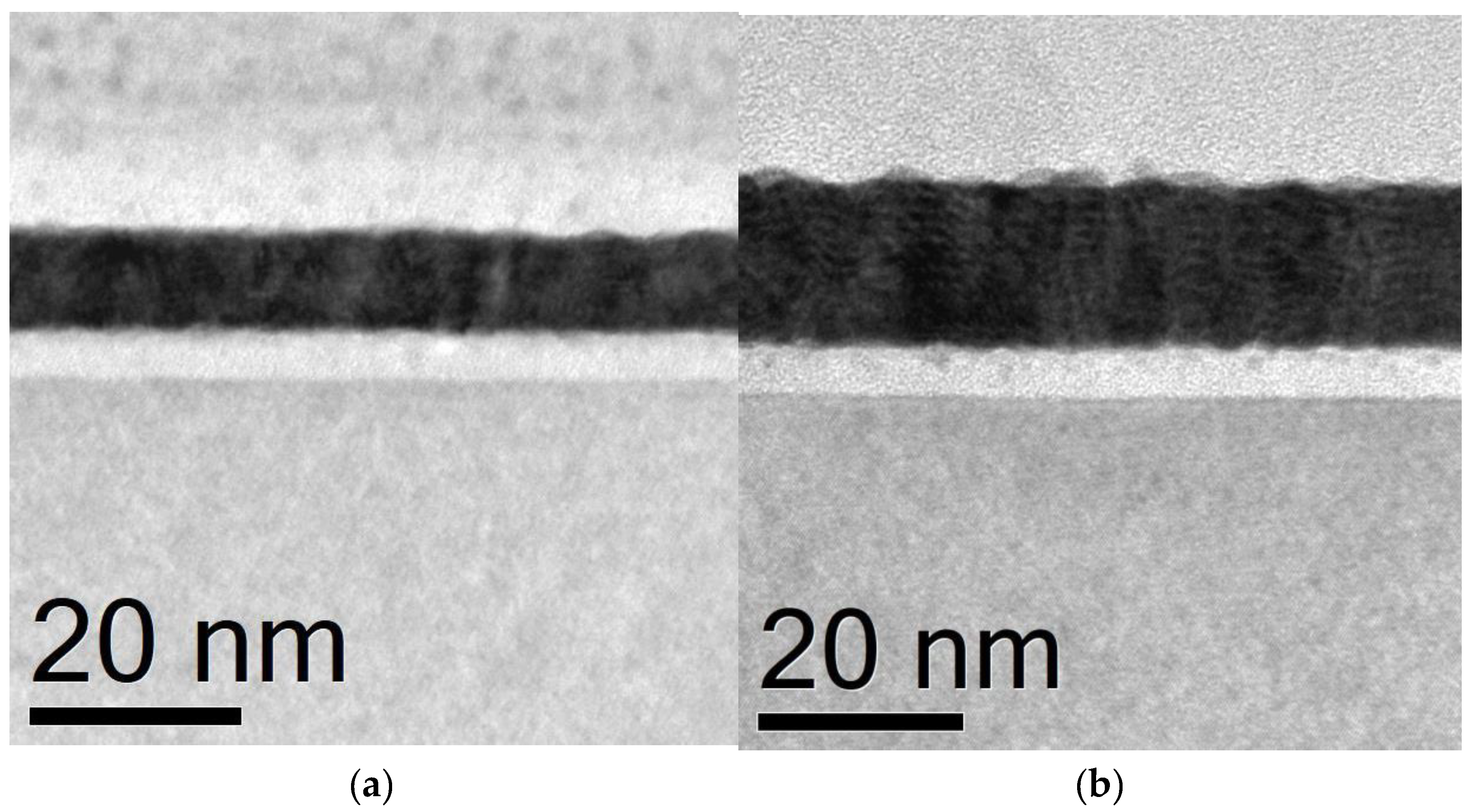
Figure 2.
Distribution profiles of Co and Pt in thin Co/Pt films measured by XPS with sample tilting; (a) – structure A; (b) – structure B; (c) – structure C. Blue lines correspond to bright-field contrast profiles obtained from TEM micrograph where 0 corresponds to the top of the film.
Figure 2.
Distribution profiles of Co and Pt in thin Co/Pt films measured by XPS with sample tilting; (a) – structure A; (b) – structure B; (c) – structure C. Blue lines correspond to bright-field contrast profiles obtained from TEM micrograph where 0 corresponds to the top of the film.
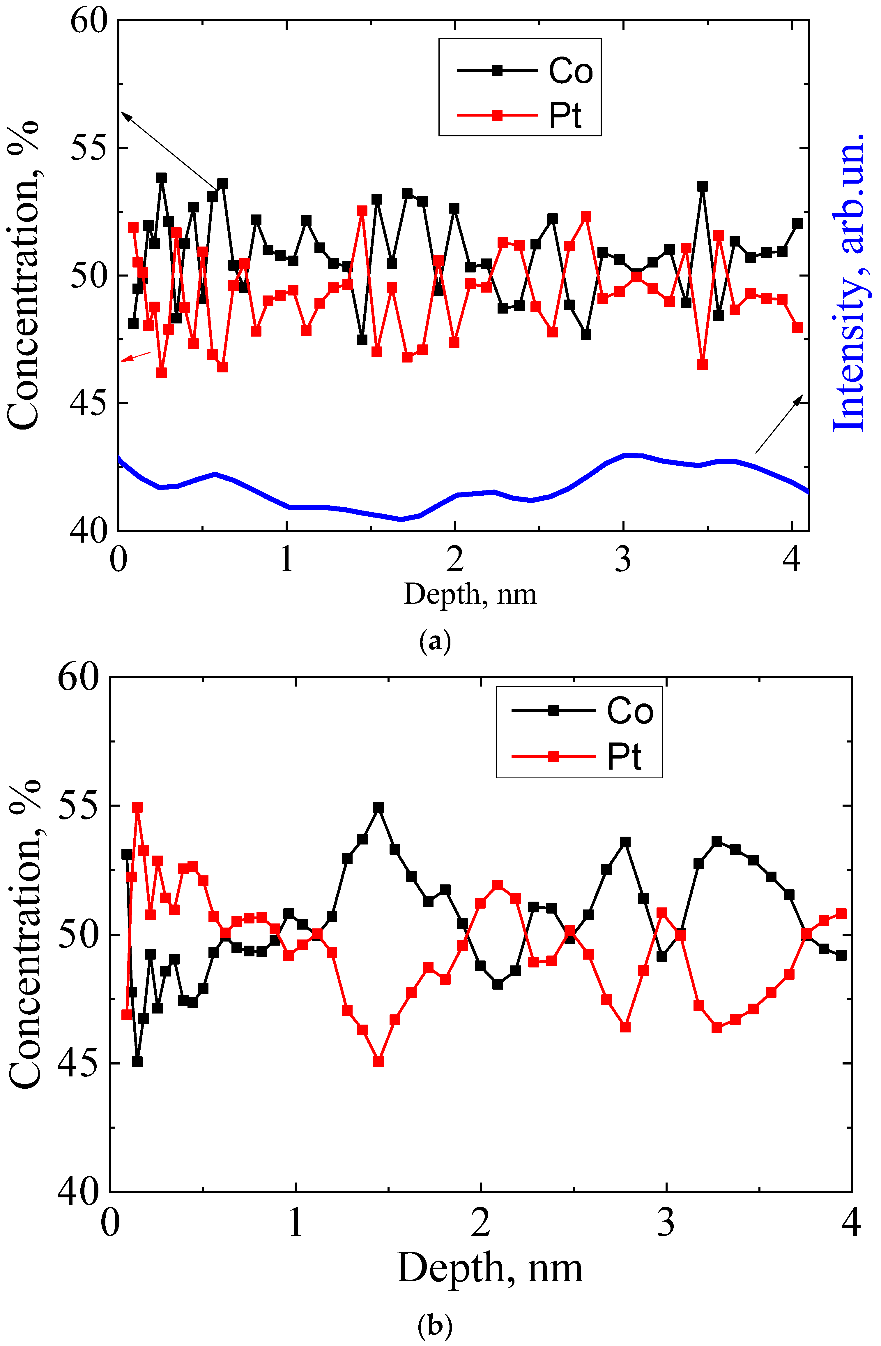
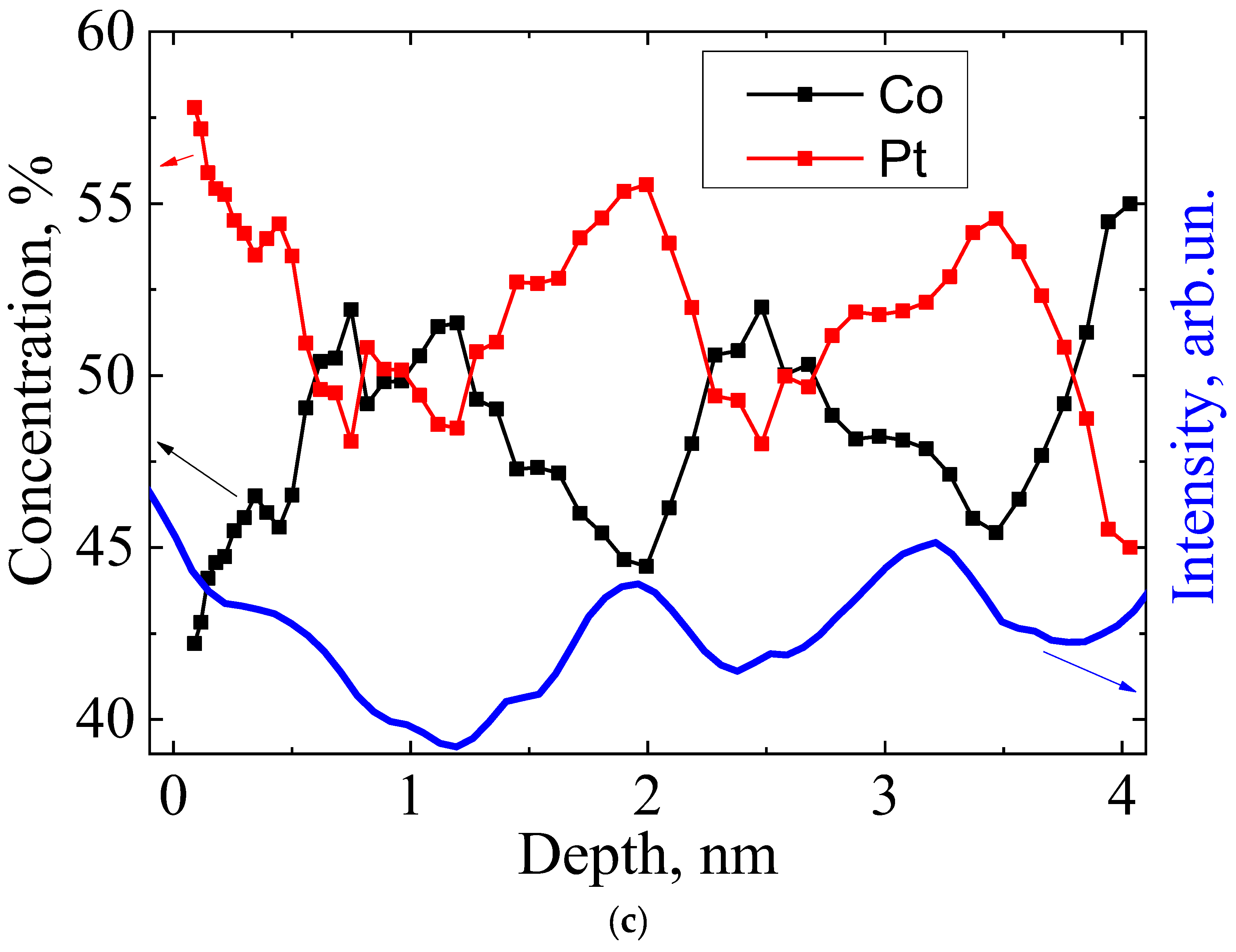
Figure 3.
magnetic field dependence of the Hall constant RH, measured for structures A, B and C. The inset shows the dependence RH(H), measured for sample C in a wider range of magnetic fields.
Figure 3.
magnetic field dependence of the Hall constant RH, measured for structures A, B and C. The inset shows the dependence RH(H), measured for sample C in a wider range of magnetic fields.
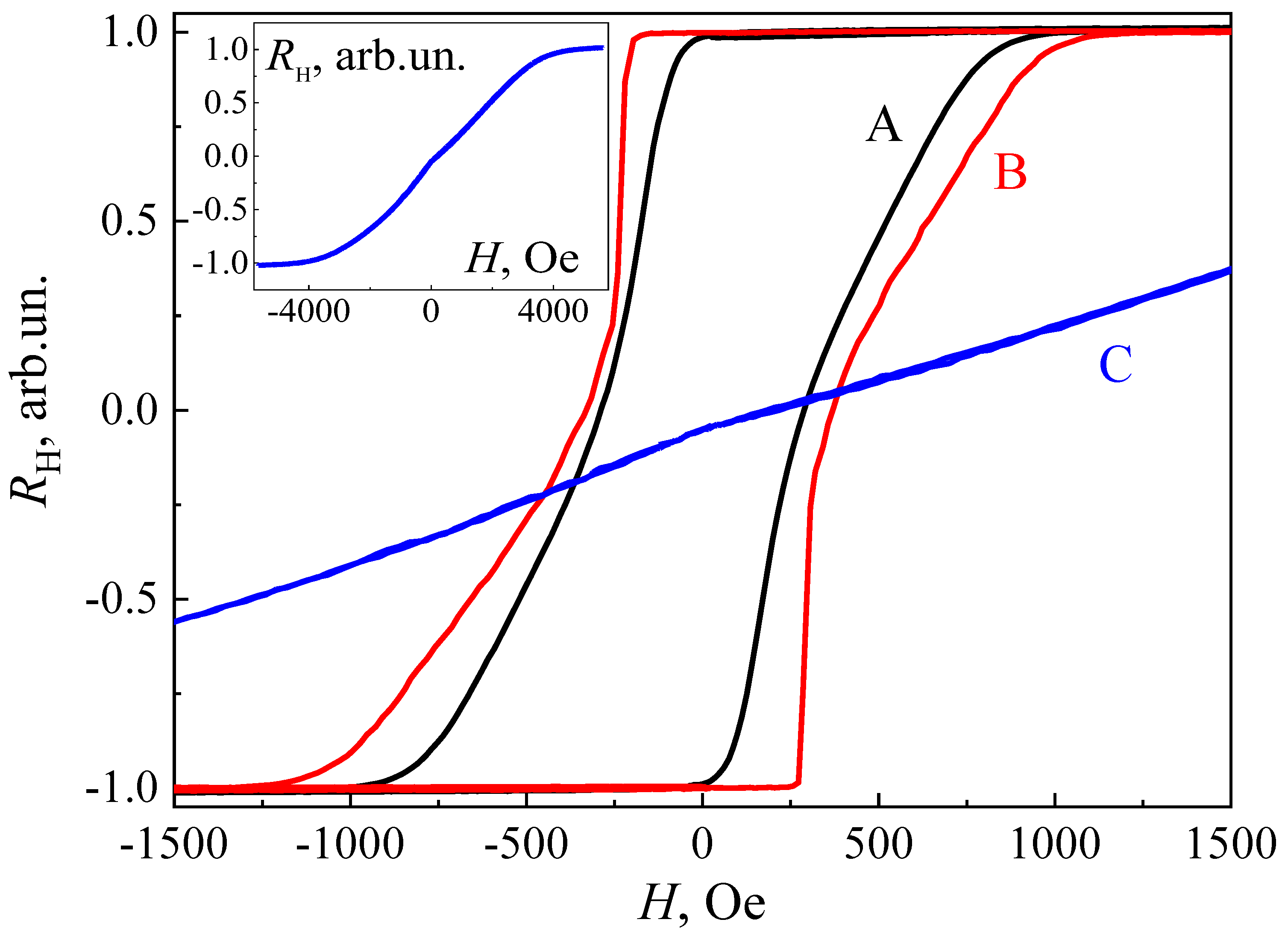
Figure 4.
MFM images of unmagnetized samples A (a), B (b) and C (c), as well as sample A, with a 3x3 μm2 region magnetized by the probe of the magnetic force microscope(d).
Figure 4.
MFM images of unmagnetized samples A (a), B (b) and C (c), as well as sample A, with a 3x3 μm2 region magnetized by the probe of the magnetic force microscope(d).
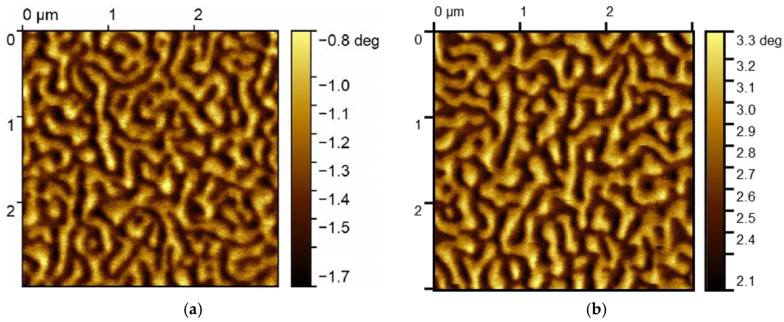
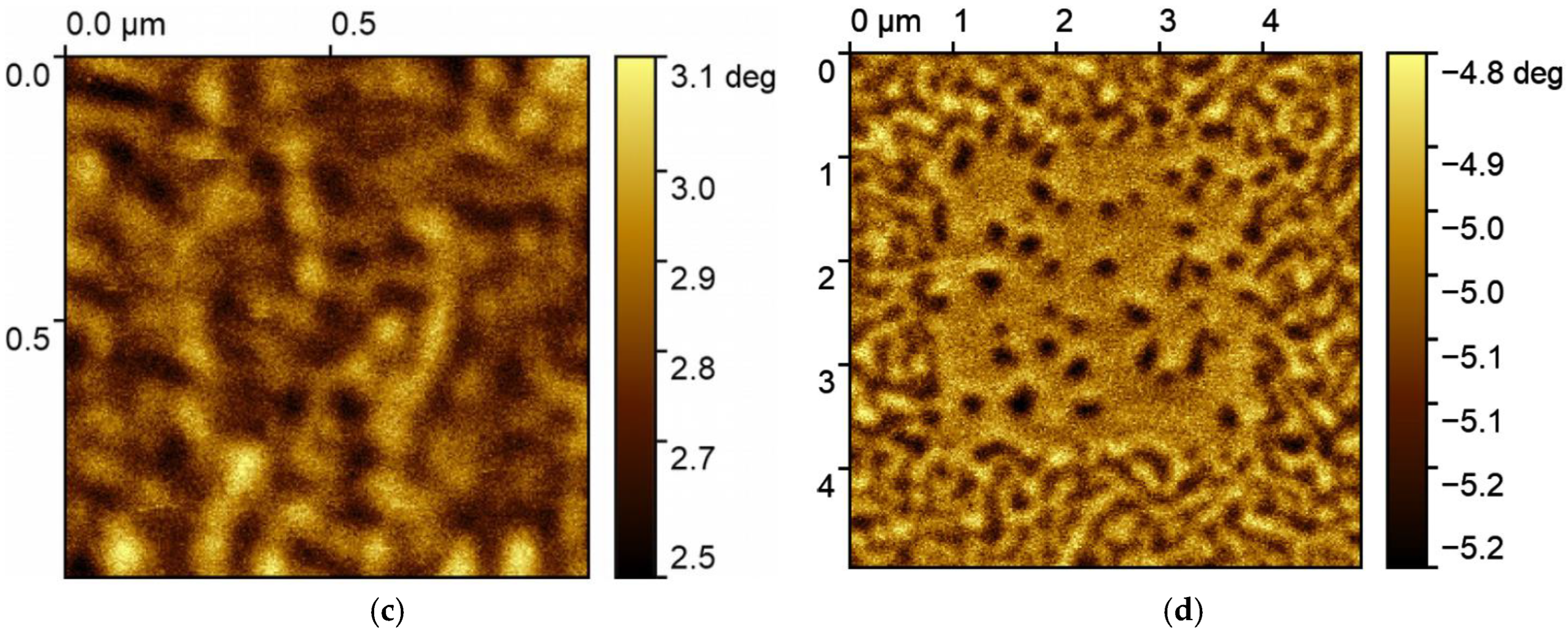

Table 1.
Magnetic properties and parameters of the micromagnetic structure of the studied samples.
| Sample (t) | Period, nm | Coercive field, Ое | Hs (Ое) | NSk, μm-2 |
|---|---|---|---|---|
| A (0.85) | ----- | 282 | 1000 | 3 |
| B (1) | 1±0.2 | 364 | 1100 | 6 |
| C (1.5) | 1.5±0.2 | 0 | 4000 | 0 |
Disclaimer/Publisher’s Note: The statements, opinions and data contained in all publications are solely those of the individual author(s) and contributor(s) and not of MDPI and/or the editor(s). MDPI and/or the editor(s) disclaim responsibility for any injury to people or property resulting from any ideas, methods, instructions or products referred to in the content. |
© 2024 by the authors. Licensee MDPI, Basel, Switzerland. This article is an open access article distributed under the terms and conditions of the Creative Commons Attribution (CC BY) license (http://creativecommons.org/licenses/by/4.0/).
Copyright: This open access article is published under a Creative Commons CC BY 4.0 license, which permit the free download, distribution, and reuse, provided that the author and preprint are cited in any reuse.


