Preprint
Article

This version is not peer-reviewed.

Polyamide Microparticles with Immobilized Enological Pectinase as Efficient Biocatalysts for Wine Clarification: The Role of the Polymer Support

A peer-reviewed article of this preprint also exists.

Submitted:

21 November 2024

Posted:

25 November 2024

You are already at the latest version

Abstract

Free pectinase is commonly employed as a biocatalyst in wine clarification; however, its removal, recovery, and reuse are not feasible. To address these limitations, this study focuses on the immobilization of a commercial pectinolytic preparation (Pec) onto highly porous polymer microparticles (MP). Seven microparticulate polyamide (PA) supports, namely PA4, PA6, PA12 (with and without magnetic properties), and the copolymeric PA612 MP, were synthesized through activated anionic ring-opening polymerization of various lactams. Pectinase was noncovalently immobilized on these supports by adsorption, forming Pec@PA conjugates. Comparative activity and kinetic studies revealed that the Pec@PA12 conjugate exhibited more than twice the catalytic efficiency of the free enzyme, followed by Pec@PA6-Fe and Pec@PA4-Fe. All Pec@PA complexes were tested in the clarification of industrial rosé must, demonstrating similar or better performance compared to the free enzyme. Some immobilized biocatalysts supported up to seven consecutive reuse cycles, maintaining up to 50% of their initial activity and achieving complete clarification within 3-30 hours across three consecutive cycles of application. These findings highlight the potential for industrial application of noncovalently immobilized pectinase on various polyamide microparticles, with possibilities for customization of the conjugates' properties.

Keywords: 
;  ;  ;  ;  ;  ;  

1. Introduction

Wine is one of the oldest and most widely consumed alcoholic beverages, produced through the enzymatic alcoholic fermentation of grape juice or must. Most of the enzymes required for this process are derived from the grapes themselves, the grape’s natural microflora, and/or microorganisms involved in fermentation [1,2]. However, in modern winemaking, particularly at large scales, the endogenous enzymes from grapes, yeasts, and naturally occurring microorganisms often prove insufficient to catalyze the numerous chemical reactions involved in the process of fermentation. Consequently, the use of commercial enzyme supplements at various stages of winemaking has become common practice in large-scale production [3,4].
Clarification is a crucial step in vinification, aimed at eliminating turbidity from the final product. This turbidity is primarily caused by high-molecular-weight pectic substances originating from grape cell walls, which are transferred into the must. In rosé wine production, the addition of exogenous pectinolytic enzymes is essential not only to enhance the aesthetic and sensory qualities of the wine but also to improve vinification efficiency [5].
The most commonly used enzymes for clarification are pectinases, a heterogeneous group of enzymes that catalyze the controlled degradation of pectin, the key substance responsible for turbidity. Beyond clarifying the must, enzymatic breakdown of pectin offers several additional benefits: (i) it reduces must viscosity, facilitating downstream processes, e.g., minimizing filtration system clogging [6,7]; (ii) it increases juice yield by up to 15% [8]; and (iii) it can release volatile aroma compounds, particularly terpenes and esters trapped within the pectin structure, thereby enhancing the wine’s aromatic profile and sensory complexity [9].
Pectinases used in winemaking industry are typically applied as preparations, i.e., enzyme mixtures derived from fungal sources, particularly species of Aspergillus and Penicillium. The primary enzymes in these preparations are polygalacturonase (E.C. 3.2.1.15), pectin lyase (E.C. 4.2.2.10), and pectin methyl esterase (E.C. 3.1.1.11), working synergistically during the clarification step to degrade pectin into simpler carbohydrates [10].
Currently, industrial enological pectinases are mainly used as free enzymes, i.e., they are soluble in the aqueous reaction medium. However, in this form, they often exhibit insufficient short-term operational stability and are difficult to recover and reuse [11], complicating the automation and continuous operation of enzymatic processes [12]. To overcome these limitations, the immobilization of enzymes has been widely studied, as highlighted by recent reviews disclosing various immobilization strategies [2,13,14]. In summary, immobilization involves attaching enzymes to various carriers, frequently selected from the group of natural or synthetic polymers. This can be achieved through physical methods (adsorption, entrapment) or chemical reactions (covalent binding, crosslinking). These approaches improve enzyme stability, facilitate the removal of biocatalysts from the final product, and enable enzyme reuse, thus enhancing process efficiency.
In the food industry in general and particularly in winemaking, supports for enzyme immobilization must be nontoxic and biocompatible, ensuring that they do not alter the sensory or chemical properties of the final product, such as color, taste, or texture [15]. The enzyme/support complex must be robust enough to withstand external factors encountered during processing, such as filtration, mixing, and exposure to temperatures or pH levels that are unusual for the free enzyme. A large surface area is crucial for maximizing enzyme loading and promoting efficient mass transfer during catalysis. Finally, the supports should enable cost-effective catalytic complexes with high activity while being easy to recover and reuse across multiple operation cycles [16].
For the particular case of pectinases’ immobilization, a number of polymer-based supports have been reported, e.g., alginate [11], polyacrylamide [17] and polyethyleneimine gels [18], as well as cationic polystyrene resin beds [19], among others. Special attention has been given to polyamides (PA), the closest synthetic analogues of the protein apoenzyme matrix. Polyamides are readily obtainable in various forms via ring-opening polymerization or polycondensation. Thus, Omelková et al. [20] and Rexová-Benková et al. [21] used PA6 powders as supports for polygalacturonase covalently immobilized via glutaraldehyde binding. The activity of the immobilized enzyme decreased by half, most likely due to steric hindrance near its active site [21]. Shukla et al. [22] immobilized polygalacturonase on PA6 beads with a similar method, achieving 70 μg/g enzyme loading, and retaining almost 50% activity after four clarification cycles. Ben-Othman and Rinken [23] immobilized pectinolytic enzymes on PA66 pellets or threads, proving a 40-fold higher immobilization yield in the latter case. The immobilized pectinase retained 40% activity after five cycles and over 20% after twenty cycles. Other authors [24,25] also employed PA6 and PA66 pellets as supports for pectinases, with covalent binding via glutaraldehyde or dimethyl sulfate. Notably, these two reagents are far too toxic and are not approved in food processing.
In summary, synthetic polyamides could serve as useful supports for pectinase immobilization, as long as the reduced catalytic activity of the resulting conjugates is addressed, eliminating also the use of toxic compounds for covalent binding. These challenges can be overcome by employing noncovalent immobilization of pectinases onto polyamide microparticles (MP), enabling the formation of multiple hydrogen bonds between the enzyme and the highly porous, scaffold-like polyamide MP. As demonstrated in our previous work [26], such porous PA6 microparticles, which may also possess magnetic susceptibility, can be synthesized through activated anionic ring-opening polymerization (AAROP) of ε-caprolactam. The effectiveness of PA6 as a carrier for the noncovalent immobilization of pectinase, particularly for enological applications, was recently confirmed [27]. Compared to the free enzyme, PA6-immobilized pectinase (Pec@PA6) exhibited more than double the specific activity toward pectin, showed slightly increased substrate affinity, but acted as faster catalysts with greater resistance to substrate inhibition. Additionally, the Pec@PA6 biocatalysts could be reused in up to three consecutive cycles, achieving complete rosé must clarification within 3–36 hours, which is considered acceptable for industrial applications.
This study extends our previous approach by synthesizing other types of polyamide MP (i.e., PA4, PA12, and the copolymeric PA612) with and without magnetic susceptibility, by means of AAROP. These microparticles are used to noncovalently immobilize pectinase, resulting in Pec@PA catalytic conjugates. After structural and morphological characterization, their enzymatic activity and the kinetics of pectin breakdown in each case are evaluated for rosé must clarification. The results are compared with Pec@PA6 systems and free, non-immobilized pectinase. Based on these findings, the influence of the chemical structure and morphology of different polyamide MP supports on the pectinolytic performance of the immobilized pectinase is determined. The best solutions for potential industrial application are also identified.

2. Results

2.1. Synthesis of Different Polyamide MP

Seven different MP supports were synthesized and designated as shown in Table 1 and explained in Section 3.3. The MP without Fe were white powders, while those with Fe were gray and exhibited ferromagnetic properties. Main characteristics of these samples are also presented in the same table. The structures of the monomers and catalysts used in each synthesis and the resulting polymers are shown schematically in Figure S1 of the Supporting Materials.
Table 1 shows that AAROP in suspension resulted in relatively good yields of purified polyamide MPs, with the highest yield for the copolymeric PA612 sample and the lowest for PA12 MP. In the case of PA12, the process was more difficult to control due to rapid spontaneous MP growth leаding to the formation of large PA12 lumps instead of microparticles. To avoid this, the polymerization was stopped earlier, which explains the lower yield. In contrast, the high yield of the copolymeric system can be attributed to the stable growth of PA612 MP, allowing the process to be carried out at maximum temperature and stirring rates. Yields for the other MP were around 60%. The viscosity-average molecular weights (Mη) were around 30 kDa or slightly higher, sufficient for use as enzymatic supports. PA4 exhibited the lowest Mη, likely due to the low polymerization temperature of 40°C. Raising the temperature above 60°C led to a marginal increase in molecular weight and very low yields of PA4, most probably related to spontaneous depolymerization due to unfavorable thermodynamics of AAROP in the case of five-membered polymerizable rings [28].
The Fe percentages of the PA-Fe supports were based on the amounts introduced into the reaction mixture before polymerization. As shown in previous thermogravimetric measurements [29], the actual Fe content in the final polyamide MPs (Table 1, values in brackets) depends on the degree of monomer conversion. At a conversion rate of 60%, the Fe content is typically twice that of the initial monomer feed.

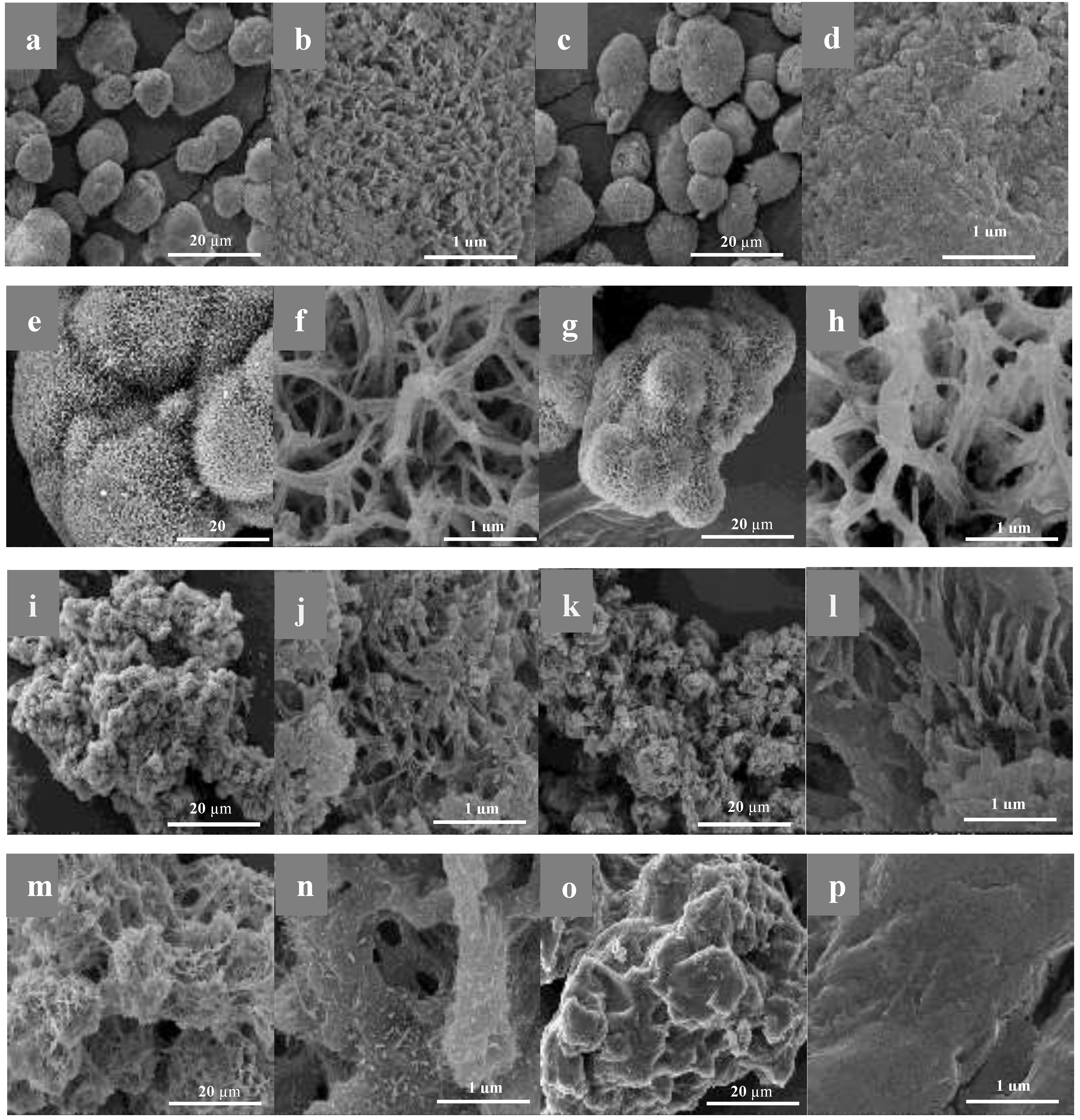
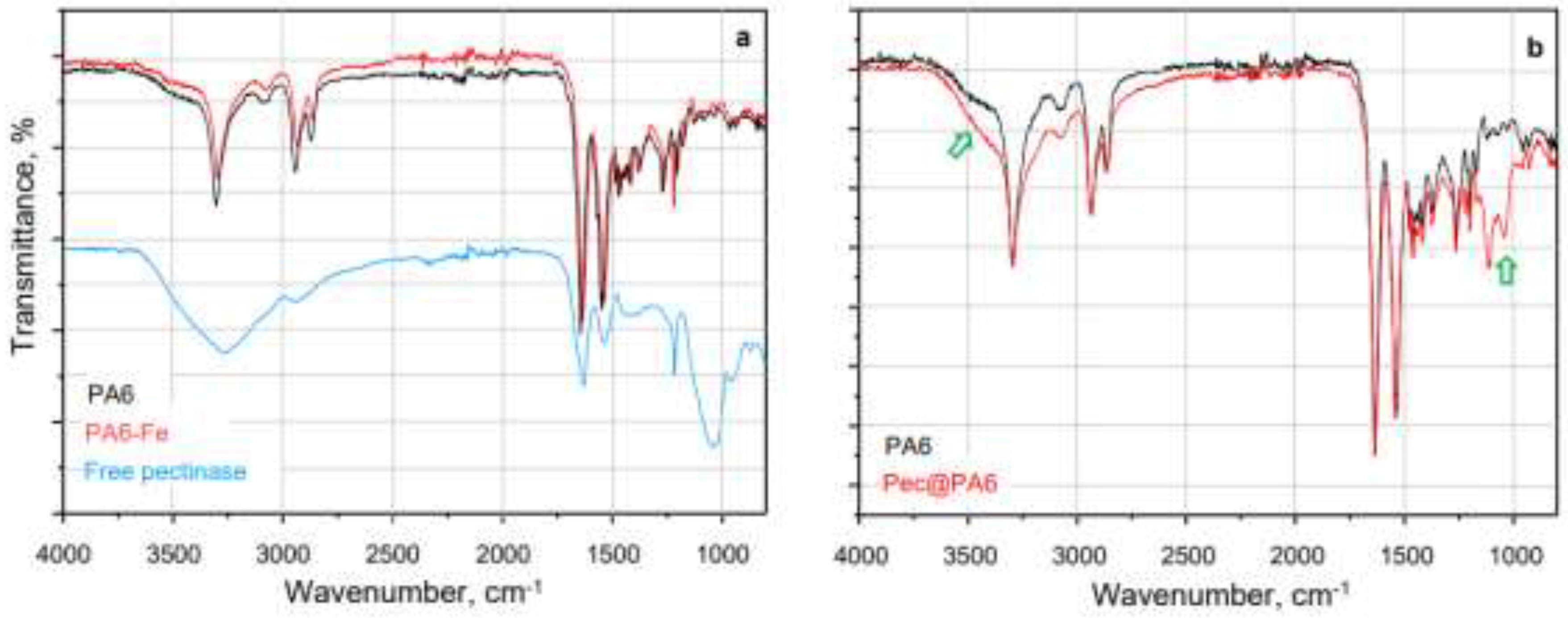
2.2. Morphological Studies by SEM

The morphology of the polyamide MP supports can be studied by SEM (Figure 1). For easier comparison, the micrographs of the neat support particles and the respective Pec@PA immobilized complexes are presented side by side at the same magnification.
Several important conclusions can be drawn about the morphology of the neat supports and the corresponding pectinase-immobilized systems. The PA4 and PA4-Fe MP exhibit predominantly near-spherical morphologies with diameters in the 10-40 µm range (Table 1, Figure 1a and Figure S2a). PA4-Fe shows significantly larger pores as seen from Figure S2, images a and b). No iron particles are observed on the surface of the PA4-Fe MP, but they are found deeper inside the particles. As shown in Figure 2S, image c, the inset, the two Fe peaks in EDX for Kα and Kβ are clearly observable at 6.4 keV and 7.2 keV directing the electron beam directly into a surface crack. After pectinase immobilization, the visual appearance of the Pec@PA4 complexes at lower magnification, both with and without Fe, remains largely unchanged (Figure 1, images a and c). At the micropores´ level, however, the enzyme fills in the pores, significantly altering the surface topology of Pec@PA4-Fe (see images 1b and 1d).
The neat PA6 support consists of aggregates of irregular shape, up to 150 µm in size, formed by smaller fused spheres (Figure 1e, 1f). Introducing Fe results in the formation of a second MP fraction with higher aspect ratios, exhibiting pea-pod-like morphologies up to 300-500 µm in size (Figure S2, e), likely caused by magnetic attraction of Fe microparticles followed by coating with the PA4 formed during AAROP. Before immobilization, the PA6 MP at higher magnification displays a scaffold-like structure composed of a 3D network of interlaced PA6 filaments (like balls of yarn) with large meshes of 300-500 µm (Figure 1, image f). Enzyme immobilization does not alter significantly the form or size of the PA6 MP, nor the mesh filling (image 1g, 1h), with the enzyme appearing to adhere to the filaments without obstructing significantly the 3D network.
In contrast to PA4 and PA6, the neat PA12 MP lack spherical symmetry (Figure 2i). At lower magnifications (Figure S2, f), most particles represent massive formations with irregular form, with sizes approaching 700 µm, and a finer fraction measuring 50-100 µm. The surface topography is sponge-like, with large, deep channels rather than pores (Table 1, Figure 1j). In PA12-Fe, these channels are even larger, but the size of the microparticles significantly decreases (Table 1, Figure S2, g). The presence of Fe in the AAROP reaction mixture most probably promotes heterogeneous nucleation of the forming PA12, facilitating its cold crystallization that results in a better control over MP size. After immobilization, Pec@PA12 and Pec@PA12-Fe behave similarly, retaining the morphology of the neat support (Figure S2, h, Figure 1, images i-l), with smaller surface pores filled, while the large channels remain unobstructed. In the Fe-containing PA12 MP small Fe particles may become visible on or slightly beneath the MP surface (Figure 2S, i, the bright circular spot with clear EDX signals for Fe).
The copolymeric PA612 support exhibits a morphology more similar to PA12 than to PA6 MP, also with a bimodal size distribution. The predominant morphology consists of irregularly shaped particles up to 150 µm, with a sponge-like surface topography (Figure 1, images m, n). Pectinase immobilization covers all surface channels and pores (Figure 1, images o, and p).

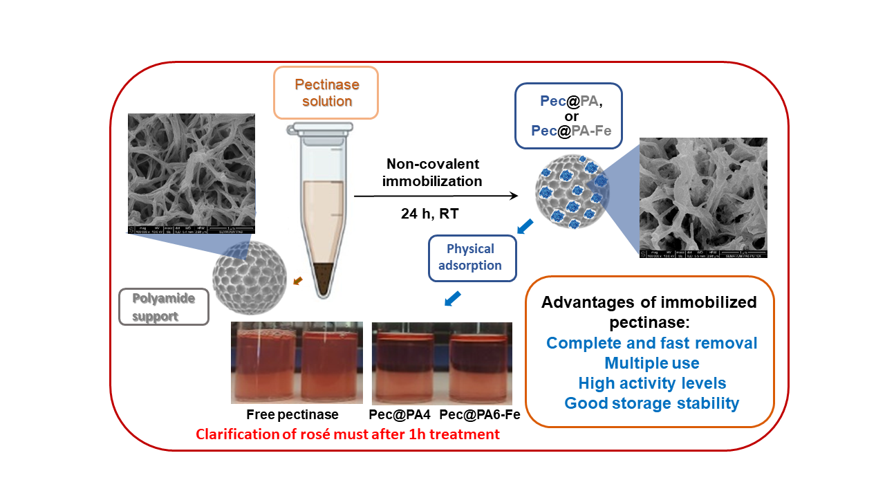
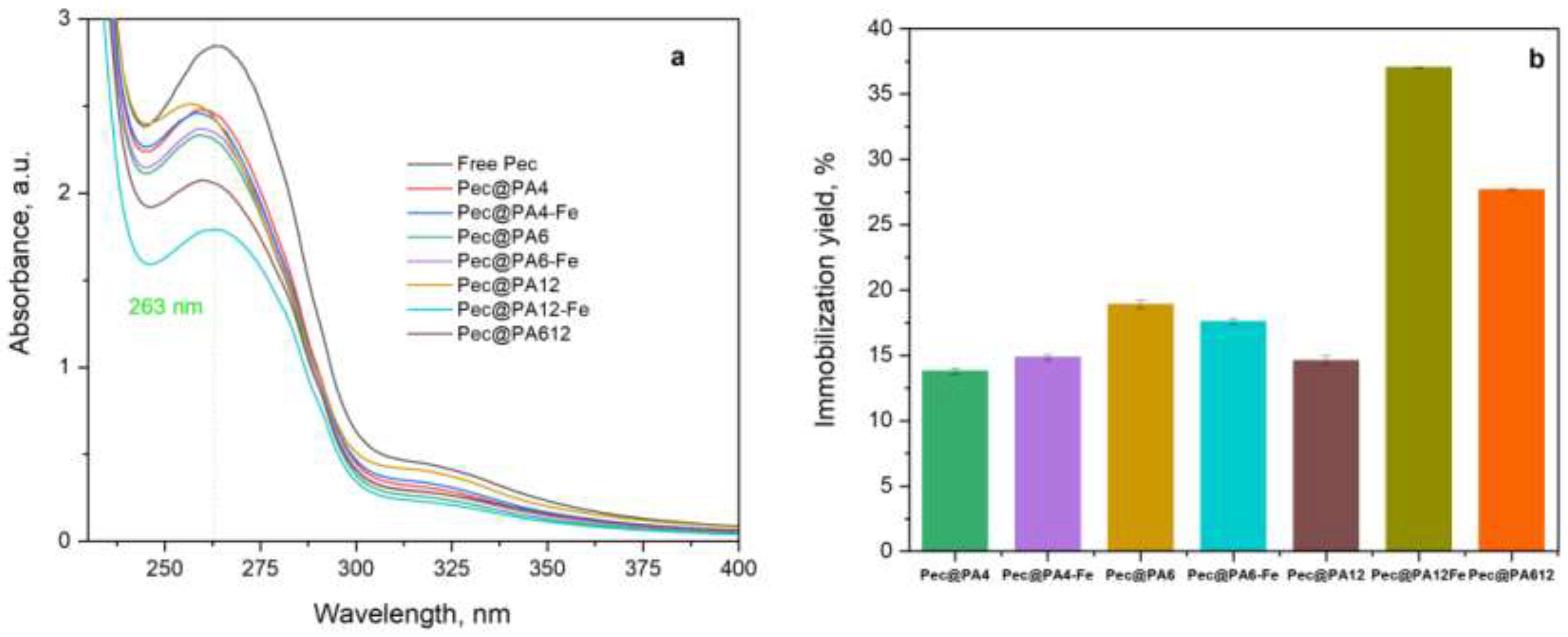
2.3. Noncovalent Immobilization of PEC on PA MP

The amount of noncovalently immobilized pectinolytic enzymes on both PA6 MP supports was calculated using Equation 3, Section 3.5. Figure 2a compares the UV/VIS spectra of the Viazyme stock solution used for immobilization (Free Pec curve) and the remaining seven supernatants after removal of the respective immobilized Pec@PA complexes. The absorption peak at λ = 263 nm corresponds to protein moieties containing substituted benzene rings, likely from tryptophan, phenylalanine, or tyrosine in the pectinase apoenzymes. As shown in previous studies [27], this direct spectroscopic method provides reliable data on protein adsorption immobilization, with a standard deviation of 3–5%, whereas the indirect protein determination by the Bradford and bicinchoninic acid assays can show data dispersion of up to 15%.
Figure 2b illustrates the immobilization yield of Pec@PA6 and Pec@PA6-Fe complexes, expressed as the percentage of pectinolytic enzymes immobilized relative to the initial concentration. Standard deviations were calculated from three consecutive trials for each sample.
Immobilization yield was between 13-18% for the PA4 and PA6-containing complexes and reached 38% for the PA12-Fe-supported pectinase, highlighting the significant influence of the polyamide support on the noncovalent immobilization of the enzyme. Two main factors most probably influence the amount of immobilized enzyme: (i) the density of hydrogen bonds between the proteic matrix of the pectinase apoenzyme and the polyamide support, and (ii) the surface topology of the respective MP carrier. Based on the data in Figure 2b, it seems that the second factor—the large channels in the PA12 MP—plays a more important role than the H-bond density at the enzyme-support interface. This is evidenced by the fact that the PA4-based complexes, which should have the highest H-bond density, exhibit the lowest immobilization yield.
The formation of hydrogen bonds between the apoenzyme and the amide groups of the supports was investigated by means of FTIR. Figure 3 below and Figure S3 in the Supporting Materials provide representative spectra comparing selected polyamide MP before and after pectinase immobilization.
The two spectra of PA6 and PA6-Fe supports in Figure 3a are identical and both exhibit bands at around 3300 cm−1, corresponding to the stretching vibrations in linearly associated through H-bonds secondary NH groups. Additionally, these spectra feature well-defined peaks for Amide I band at 1631 cm−1 and Amide II at 1535 cm−1 with similar intensities, indicating a fixed trans-conformation of the NH–CO group, typical of high molecular weight polyamides. The free Pec spectrum (Figure 3a) proves the enzyme’s proteic matrix as a structural analogue of synthetic polyamides, with similar Amide I and Amide II peaks, showing also a broad peak centered at 3250 cm−1, representative for associated secondary and primary amino groups. An intensive band at 1050 cm−1 typical only for Pec is also present. In the Pec@PA6 spectrum (Figure 3b), this band appears as a weak doublet at 1050-1120 cm−1, confirming the presence the Pec on the support. The increased intensity of the shoulder for associated –NH– and– NH2 groups suggests hydrogen bond formation between the enzyme's amide groups and those of the PA6 support.

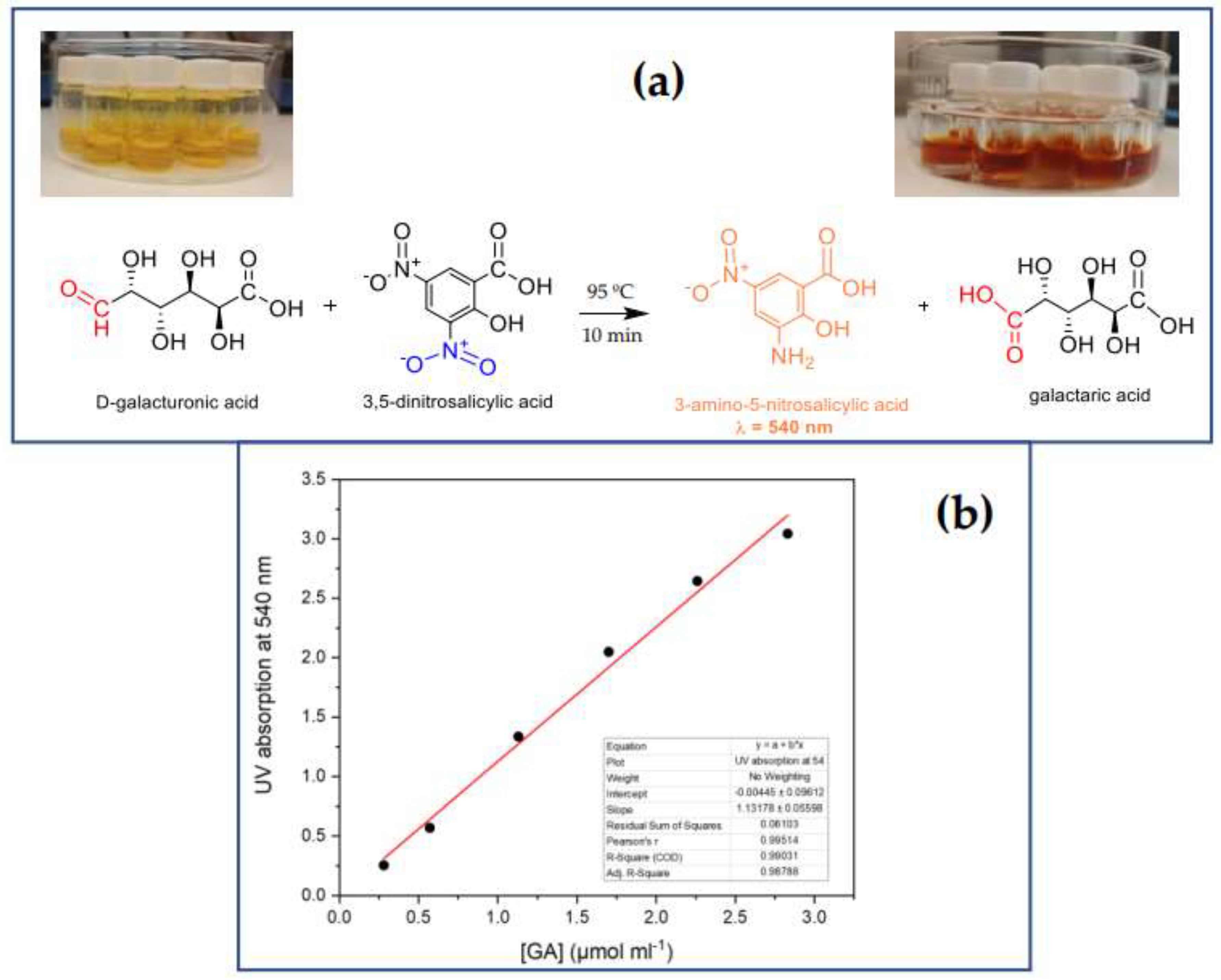
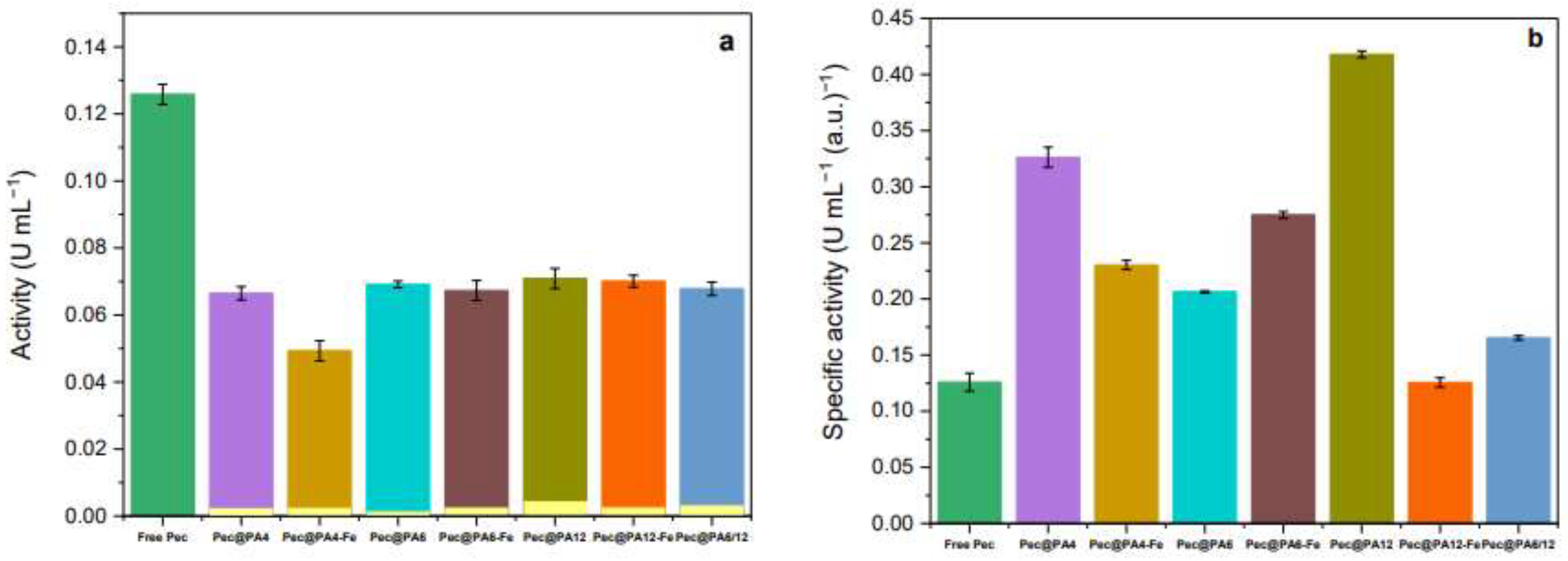
2.4. Activity by the DNS methods

At temperatures near 100 ºC, reducing sugars like galacturonic acid (GA), which contain an aldehyde group, donate a hydrogen atom to 3,5-DNS reagent (2-hydroxy-3,5-dinitrobenzoic acid), converting it into 3-amino-5-nitrosalicylic acid (3,5-ANS) while oxidizing GA to galactaric acid. This redox reaction causes a color change as shown in Figure 4a that serves as the basis for UV-VIS estimation of pectinase activity [30].
In the present study, both free and immobilized pectinase are expected to depolymerize pectic substrates found in the rosé must, producing GA through the hydrolysis of the pectin main chain. The amount of GA generated will be proportional to the concentration of 3,5-ANS, which can be quantified via UV/VIS spectroscopy by the intensity of the band at λ = 540 nm. Thus, the pectinolytic activity is determined by the concentration of 3,5-ANS released per unit time using the calibration plot in Figure 4b.
It is important to underline here that the activity assay should be made on the basis of the absorbance at 540 nm. As seen from Figure S4 of the Supplementary materials, the nominal 3,5-ANS peak becomes clearly resolved exactly at 540 nm only at higher contents of the reducing GA.
Figure 5 presents the results from the activity tests for free and immobilized pectinolytic preparations obtained at room temperature. All pectin-immobilized polyamide MP supports, both with and without Fe, are shown for comparison. As depicted in Figure 5a, most all conjugates—except for Pec@PA4-Fe—demonstrate statistically equal absolute activity levels, approximately half that of the free enzyme. The small yellow bars in Figure 5a represent the measured “activity” of the polymer supports without any pectinase, showing values within the margin of the experimental error. This confirms that, as expected, the plain microparticulate supports do not possess pectinolytic activity.
After normalizing activity values by the protein content in each Pec@PA conjugate or in the free enzyme (Figure 5b), it becomes clear that the specific activities of the immobilized systems are either equivalent to (Pec@PA12-Fe) or 2.5–3.5 times higher than the free enzyme (Pec@PA4 and Pec@PA12, respectively). This result contrasts with the reduced activity often seen in covalently immobilized pectinase (e.g., in [21]), underscoring an important advantage of the noncovalent immobilization.
The observed significant increase in the recovered activity of adsorption-immobilized pectinolytic preparations on polyamide MP may be due to a more favorable active site configuration and/or to reduced inactivation. To further investigate and validate the activity data, comparative kinetic studies were conducted on all biocatalysts in this study.

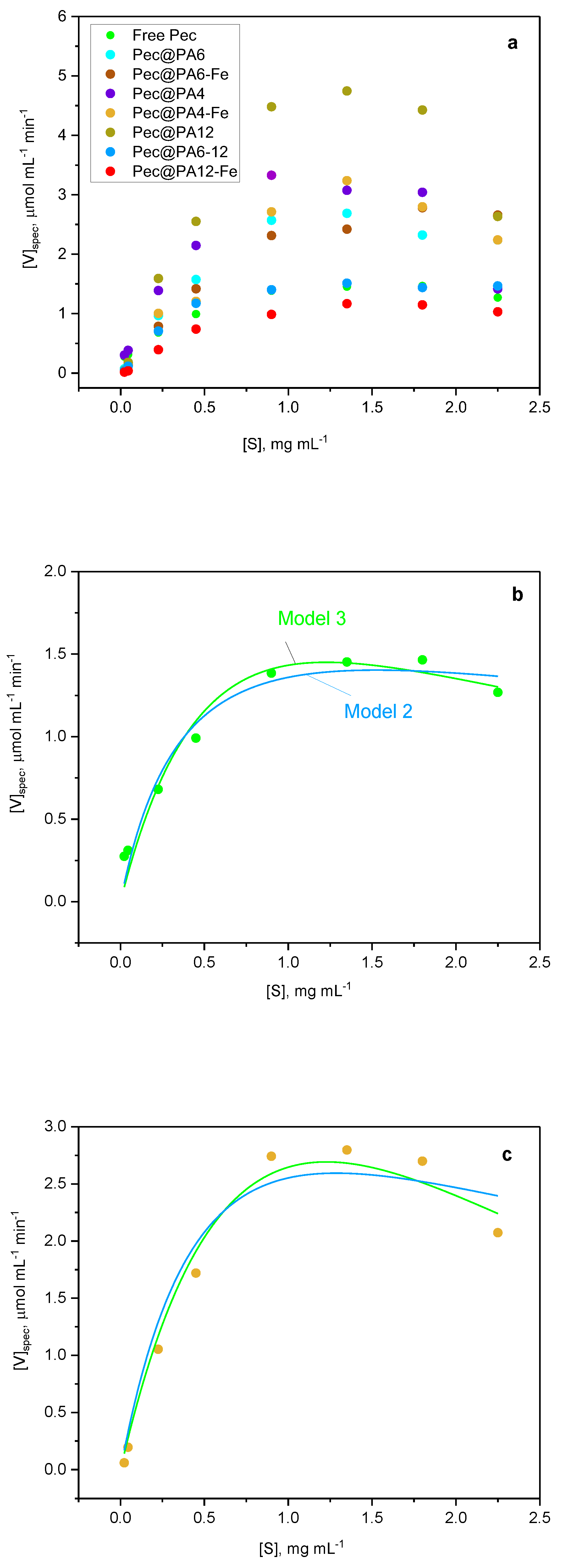
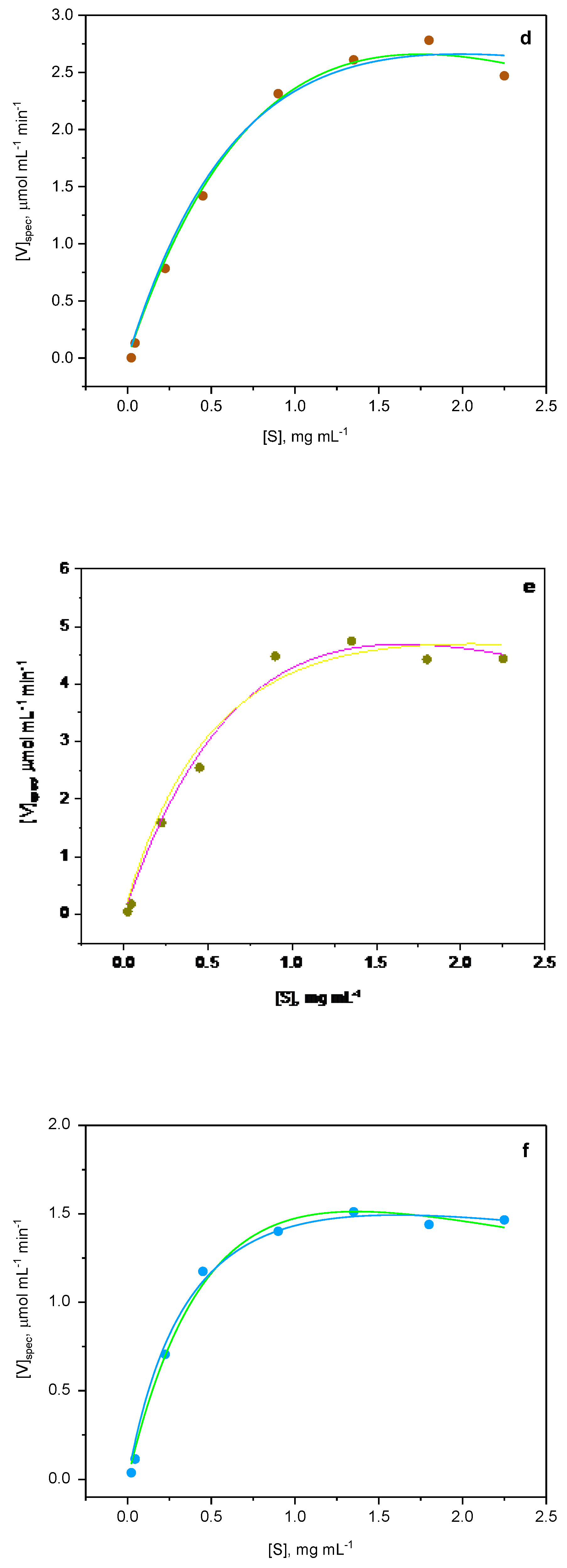
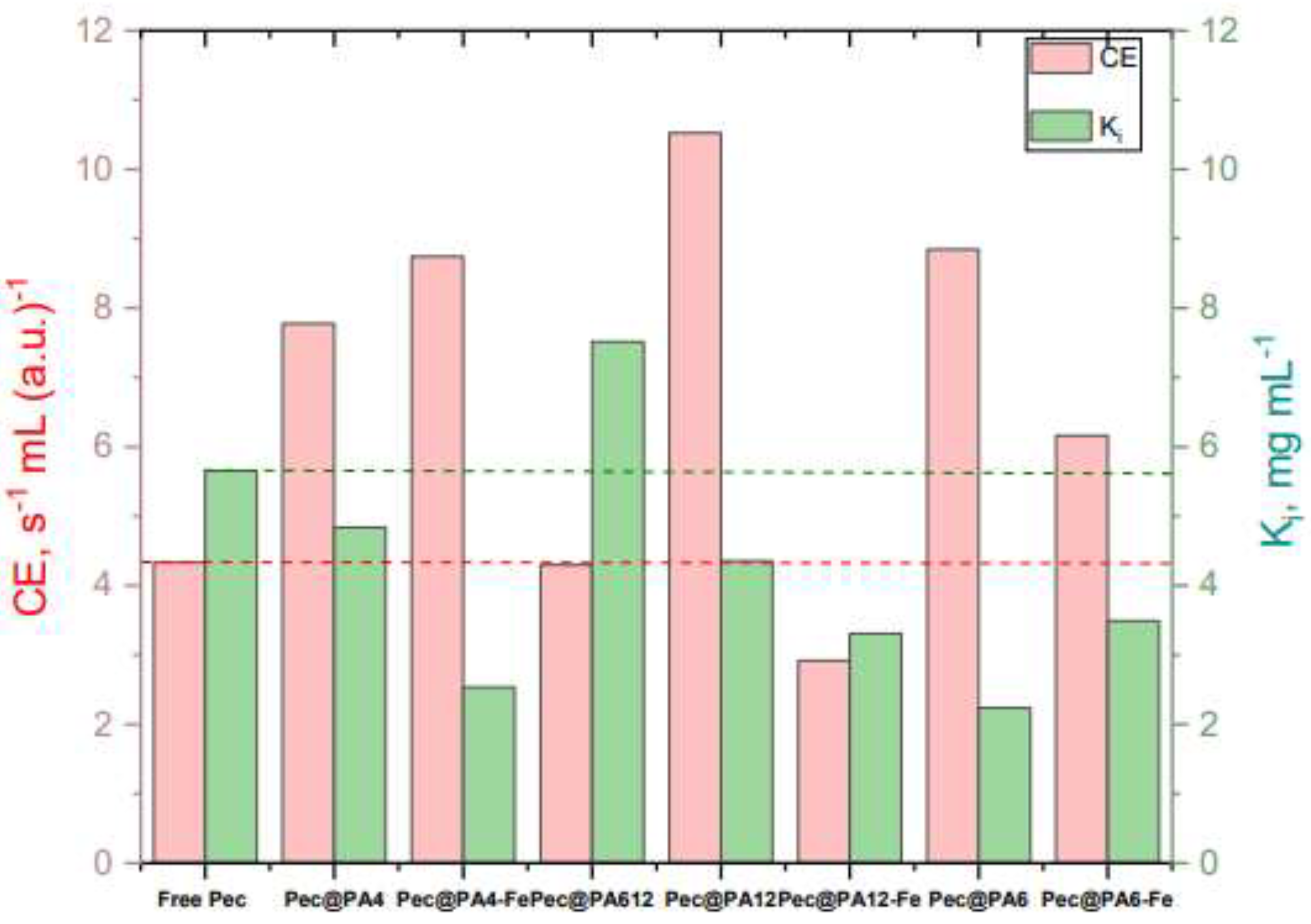
2.5. Kinetics Studies

The kinetic parameters of pectin hydrolysis catalyzed by free Pec and its seven noncovalently immobilized complexes were analyzed by plotting the reaction velocity per unit protein (Vspec) against pectin substrate concentration (S). As demonstrated in Figure 6a, neither the free enzyme nor any of its immobilized complexes showed saturation of Vspec at higher pectin concentrations, which deviates from traditional Michaelis-Menten kinetics (Model 1, Equation 4 in Section 3.7). Instead, a well-expressed inhibition effect occurred, with Vspec decreasing above 1.0–1.2 mg/mL of pectin. This substrate inhibition was previously observed for free pectinase-mediated pectin degradation by Gummadi and Panda [31] who demonstrated that the process can be most adequately described by two models: i) conventional substrate-inhibition model (Model 2, Equation 5,) derived by Haldane [32] who supposed the formation of multiple inactive enzyme-substrate complexes, and ii) the model proposed by Edwards [33], which combines diffusion-controlled substrate supply with substrate inhibition effects (Model 3, Equation 6).
Figure 6b-f depicts the fits of Models 2 and 3 to the experimental kinetic curves for the free enzyme and the most representative Pec@PA pectinolytic conjugates.
To ensure that fitting the experimental curves to Equations 5 and 6 will render Vmax, Km and Ki values with meaningful physical interpretation and reliable statistics, the procedure of Cleland for a single inhibitory substrate was followed [34]. First, kinetic data were plotted in double-reciprocal form (1/Vspec vs. 1/S), and the linear portions of the plots were used to estimate the apparent Vmax and Km values, as in standard Michaelis-Menten kinetics (Equation 4). The missing Ki constant was initially estimated using the following approximate relationship [34]:
K i = S m a x 2 / K m
where Smax is the substrate concentration at maximum Vspec values. These starting Vmax, Km and Ki values were then input into a nonlinear fitting program based on Equations 5 and 6. The best fits for the most representative biocatalysts are presented in Figure 6b-6f, with the corresponding Vmax, Km and Ki for all systems listed in Table 2.
To decide which substrate inhibition method—conventional substrate inhibition (Model 2, Section 3.7) or the double exponential model (Model 3)—best describes the kinetics with the systems in Table 2 and Figure 6, we first examined the coefficient of determination R2. It can be seen that Model 3 better captures the trend of the data points for the free enzyme, Pec@PA4, and Pec@PA6, suggesting that these biocatalysts exhibit a combination of diffusion-controlled substrate supply and substrate inhibition effects. In contrast, Model 2 not accounting for diffusion provides a more suitable fit for Pec@PA6-Fe, while both Model 2 and 3 are equally adequate for the remaining four complexes, yielding similar Vmax, Km and Ki values. This finding is likely due to the distinct morphology of the Pec@PA6 conjugates, as detailed in Section 2.2.
In enzymatic biocatalysis, the constant Ki​ is a significant indicator of the substrate inhibition phenomenon, reflecting the affinity between the enzyme and the inhibitory form of the substrate. A lower Ki​ value indicates a higher tendency for inhibitory binding, meaning that inhibition occurs even at lower substrate concentrations. Conversely, a higher Ki​ suggests that significant inhibition only occurs at high substrate concentrations. As shown in Table 2, the Pec@PA612 conjugate, with Ki​ = 7.5, is notably more resistant to inhibition compared to the free Pec reference, which has Ki​ ​=5.7. On the other end of the range, the Pec@PA4-Fe and Pec@PA6 conjugates are more easily inhibited, with Ki​ ​values ranging from 2.2 to 2.6. The Ki​ values for the remaining samples fluctuate between 3.5 and 4.8, which is close to but still below the value for the free enzyme.
Key parameters characterizing an enzymatic process include the specific maximum catalytic activity (Vmax) and the Michaelis-Menten constant (Km). Vmax represents the maximum reaction rate of the enzyme system, while Km indicates the enzyme's substrate affinity, with a lower Km signifying higher affinity. Additionally, catalytic efficiency (CE) can be derived from these parameters. Since, in this study, Vmax is already normalized to the immobilized protein amount in each system, the following relationship can be expressed:
C E = V m a x K m
The CE value reflects the enzyme's performance, with high velocity and a low Km indicating high catalytic efficiency. This means the catalyst is both fast and effective at binding to the substrate.
With these concepts in mind, Figure 7 was constructed presenting the consolidated data for CE and Ki​ for all biocatalysts in this study.
Analyzing the CE and Ki​ values for all catalytic systems in Figure 7, the most suitable candidates for further use in larger-scale applications are catalysts with both high CE and Ki​, i.e., combining efficient catalysis with resistance to inhibition at high substrate levels. The ideal choice, compared to the free Pec reference, is the Pec@PA12 system, which achieves the highest CE with a sufficiently high Ki​. Another promising option is the Pec@PA612 conjugate, featuring the same CE as free Pec but with significantly higher Ki. The next best options are the PA4-based complexes, Pec@PA4 and Pec@PA4-Fe, followed by the PA6-supported systems, with and without Fe filler. The only system with notably low CE and Ki​ as compared to the free Pec is Pec@PA12-Fe, where the addition of Fe appears to have compromised both parameters.
In summary, the morphology and chemical structure of the polyamide MP support significantly influence the activity and robustness of noncovalently immobilized Pec against the pectin substrate. This insight could guide the design of customized biocatalytic systems for applications in the food industry, particularly for controlling pectin levels.

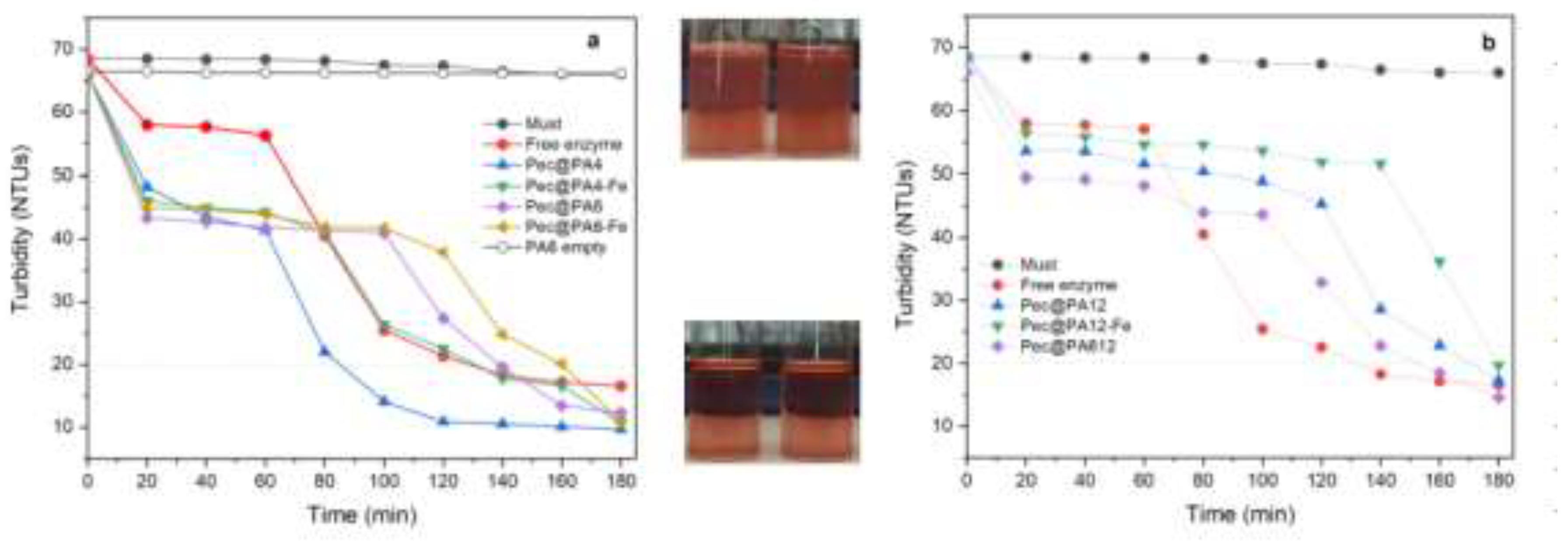
2.6. Rosé Must Clarification

The next step in this study was to use all biocatalysts for clarification of real rosé must samples containing pectin concentrations of 2.0 mg mL-1, i.e., significantly above the threshold of 1.2 mg mL-1, above which the inhibition effect was registered in the kinetics studies. To ensure comparability of the results, the amounts of the pectinolytic preparation immobilized on all seven supports were kept identical to those of the free enzyme, as expressed in arbitrary units, by appropriate selection of the mass of the Pec@PA conjugate. The results of all clarification studies performed at 23ºC are presented in Figure 8.
As shown in Figure 8a, without enzyme treatment, including the presence of a neat PA6 MP carrier, the initial turbidity remained constant at approximately 70 NTU over time. During the first hour of clarification, all immobilized complexes in Figure 8a demonstrated faster clarification than the free Pec, which reached the target turbidity of 20 NTU within 2 hours. The Pec@PA4-Fe complex followed a similar trend, while Pec@PA6 and Pec@PA6-Fe achieved 20 NTU within 160 minutes. Notably, Pec@PA4 exhibited the best clarification performance, reaching the target turbidity level in just 80 minutes.
A comparable turbidity reduction pattern was observed for the PA12-based complexes in Figure 8b. All of them reduced turbidity from 70 to 45 NTU within 60 minutes, outpacing the free Pec. This initial effect was the strongest with the Pec@PA612 complex. Thereafter, the clarification rate of the immobilized systems in Figure 8b slowed slightly, achieving the desired clarity between 160 and 180 minutes.
The results in Figure 8 align with the kinetic data, indicating that Pec@PA4 and Pec@PA12 serve as faster catalysts than the free enzyme. However, under real clarification conditions, factors beyond kinetics appear to influence performance. The pulverulent catalytic complexes—depending on the polyamide’s nature, density, and molecular weight—float on the must surface, disperse within it, or rapidly precipitate to the bottom of the container, all of which certainly impacts additionally the clarification rate.

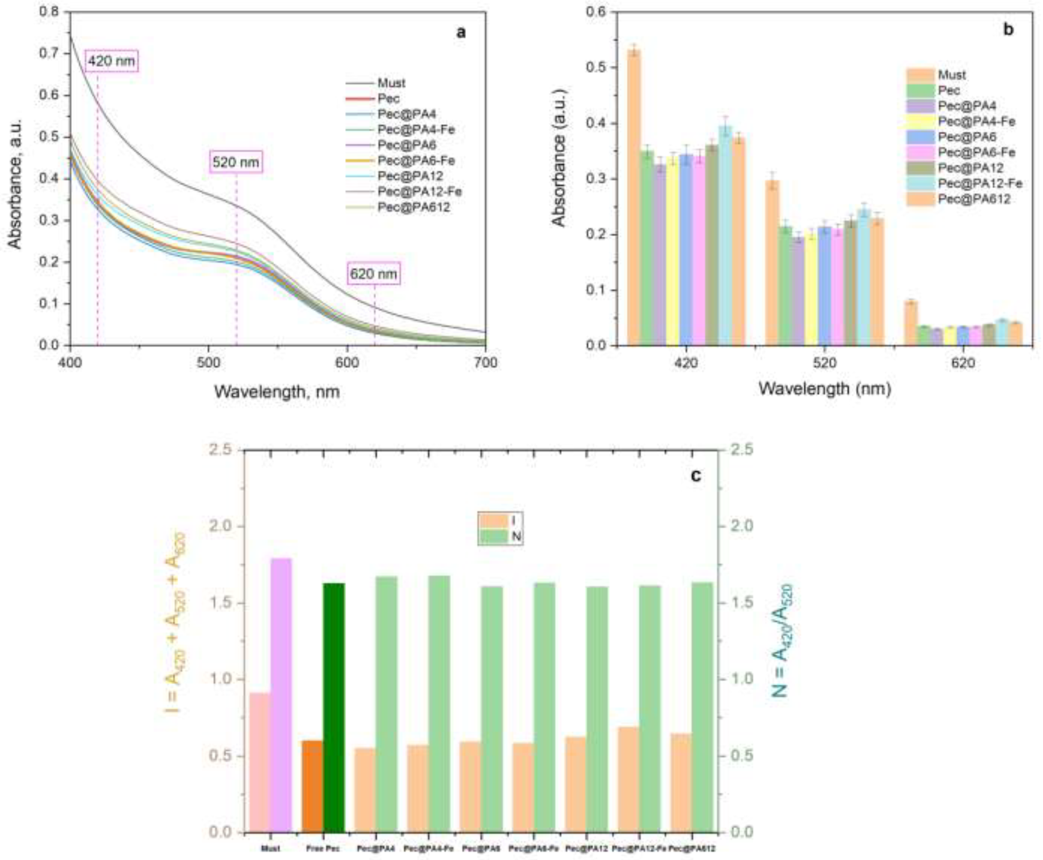
2.7.Color Retention After Clarification

Alongside transparency, color is a critical characteristic of rosé and white wines, subject to rigorous control. Color is typically analyzed across three bands in the UV/VIS spectrum: 420 nm (yellow), 520 nm (red), and 620 nm (blue) [35,36]. Figure 9 presents comparative results for rosé musts treated with the free enzyme and the seven immobilized conjugates.
Theoretically, immobilized enzymes could introduce undesirable color changes due to possible selective adsorption of wine color components onto the microparticulate PA supports, an effect not present with the free enzyme preparation. However, the UV/VIS curves in Figure 9a and the bar graphs in Figure 9b show that the absorbances at 420 nm, 520 nm, and 620 nm for immobilized conjugates are statistically comparable to, or even better than those of the commercial free enzyme. Figure 9c illustrates the color intensity I and shade N as functions of the Pec@PA type, calculated as per Equations 7 and 8, indicating once again that color changes caused by the free Pec are equivalent to those by the Pec@PA conjugates.
In summary, the microparticulate polyamide supports in all noncovalently immobilized conjugates do not induce any undesirable color changes in the rosé must, despite potentially adsorbing some pectin degradation products, as suggested by observations on the multiple uses of the seven conjugates.

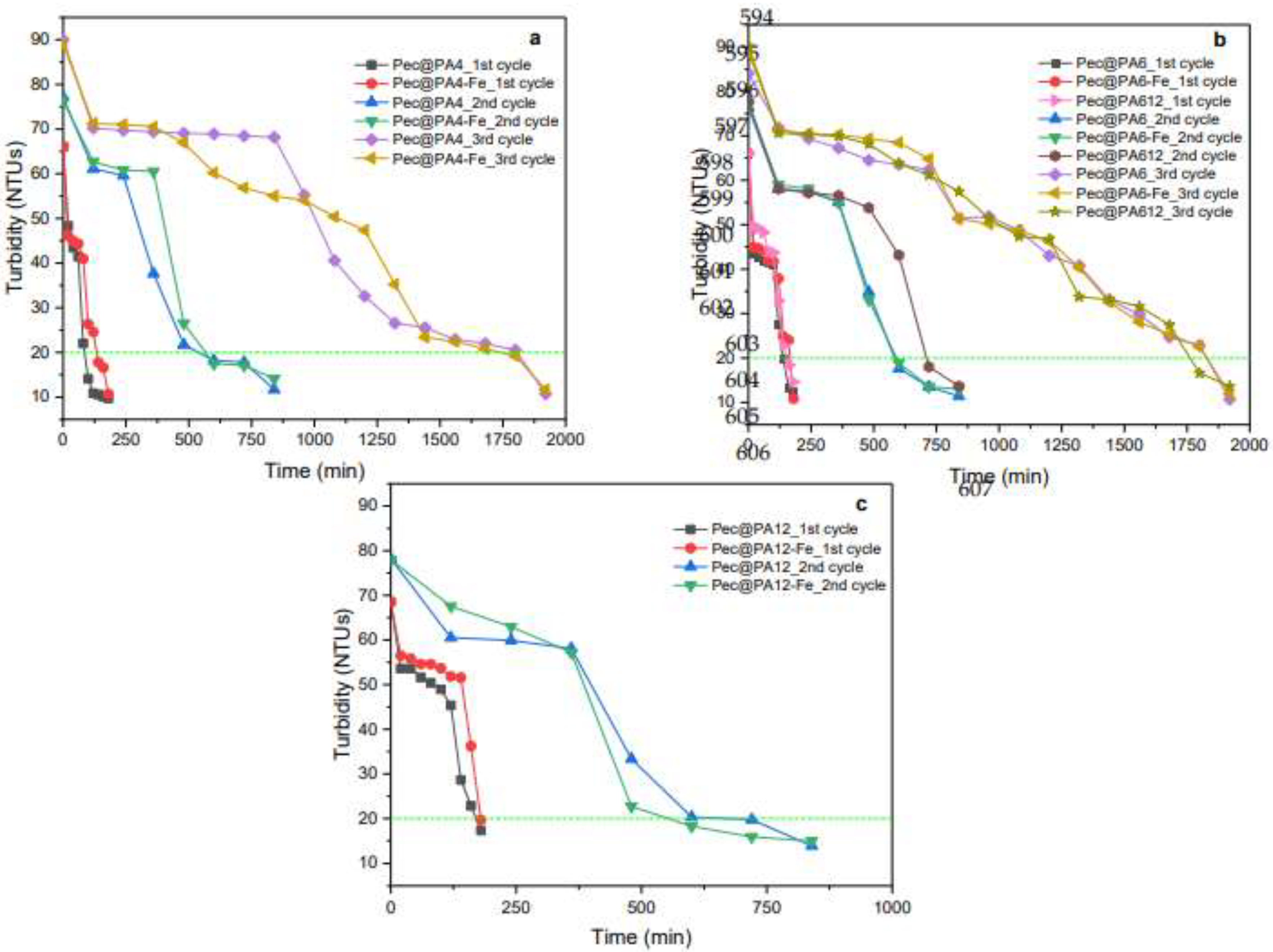
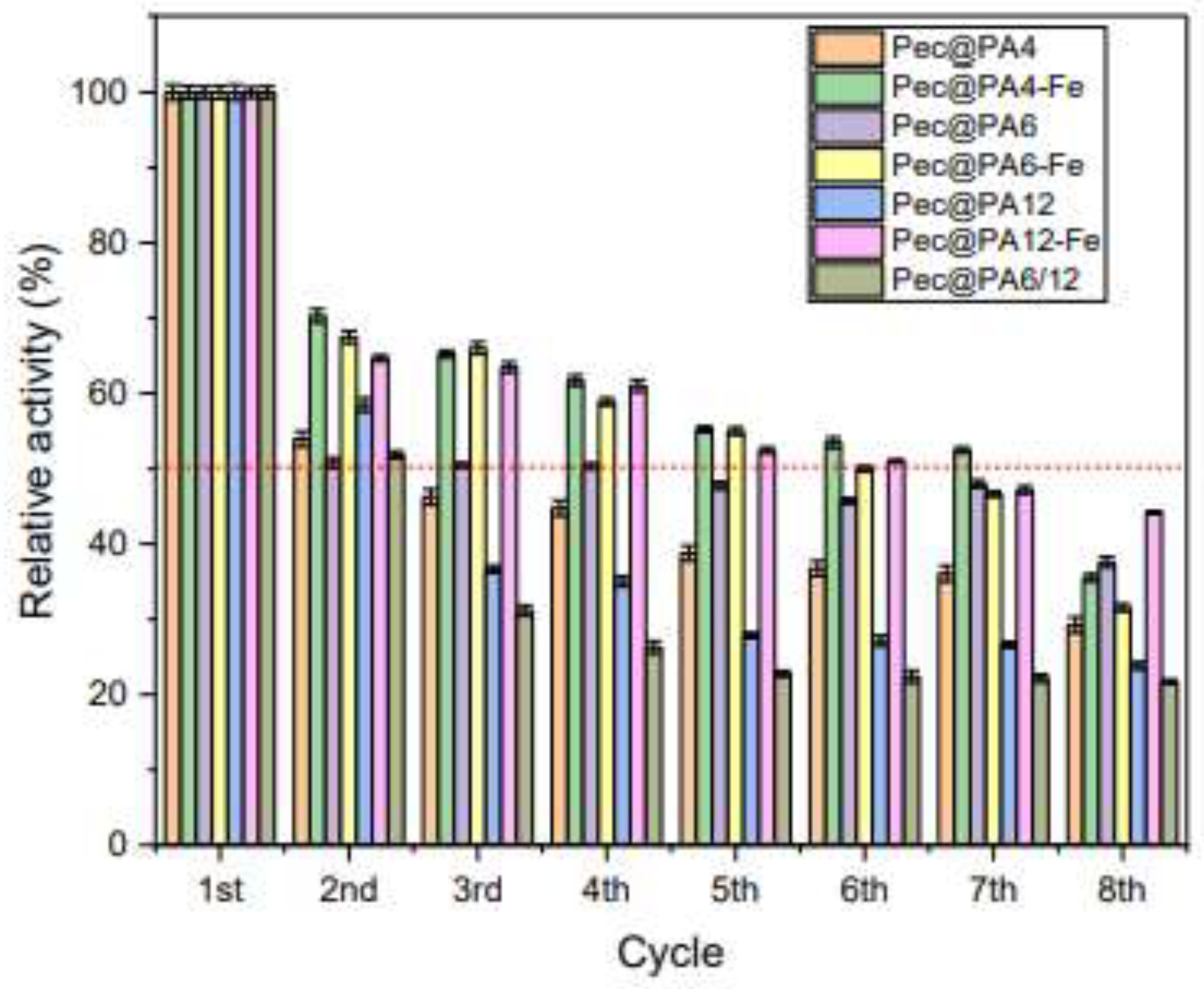
2.8. Multiple Use of Pec@PA Conjugates

The primary advantage of the MP-supported pectinolytic Pec@PA catalysts over the free Pec resides in the ability to completely remove and reuse the immobilized biocatalysts. The reusability of the Pec@PA systems was evaluated by repeatedly using defined amounts of each biocatalyst for rosé must clarification, followed by enzymatic activity tests, as described in Section 3.10.
Figure 10 illustrates the results of rosé must clarification using immobilized conjugates over three consecutive applications. During the first use (red points and black squares in Figure 10a and c; pink triangles in Figure 10b), all seven complexes reduced turbidity to below 20 NTUs within approximately 3 hours. However, during the second use (blue and green triangles in Figure 10a and c; brown circles in Figure 10b), achieving the same turbidity level required about 14 hours.
The third application was performed using Pec@PA4 (yellow triangles and purple diamonds, Figure 10a) and Pec@PA6 (yellow triangles and purple diamonds, Figure 10b) complexes, which took 32 hours to reduce turbidity to 20 NTUs. It was not possible to conduct a third cycle with Pec@PA12 complexes, as their morphology and low density, as well as the lack of magnetic properties, prevented recovery after the second cycle.
The reduced relative activity during the second and third cycles most probably results from byproduct adsorption on the catalytic complex surface, which hinders substrate transport to the enzyme's active center, prolonging the clarification process. At the industrial scale, clarification times of 14 or 32 hours are still considered acceptable.
The reusability of the immobilized enzyme was also analyzed by performing consecutive cycles of enzymatic activity, as described in Section 3.10. The assays were carried out at 50°C and pH 3.8 the results of residual activity after each cycle are shown in Figure 11. As shown in Figure 11, after the first application cycle, the three Fe-containing systems (Pec@PA4-Fe, Pec@PA6-Fe, and Pec@PA12-Fe) retain 66–70% of their initial activity, while the non-Fe systems stabilize around 50–55%. This higher activity retention probably results from the easier and more complete removal of the Fe-containing systems using a constant magnet, which minimizes catalyst loss and indirectly supports higher relative activity. The three magnetically susceptible systems maintain activity above or around 50% through the 7th cycle, indicating good potential for large-scale reuse. Among the non-Fe systems, Pec@PA6 shows the best performance. In all Pec@PA conjugates, possible causes of activity decline may include also enzyme leaching and/or surface contamination of the support particles by pectin degradation products. After the final reusability cycle, the used Pec@PA particles can be boiled in distilled water to remove all accumulated contaminants, dried, and reused for a new immobilization.

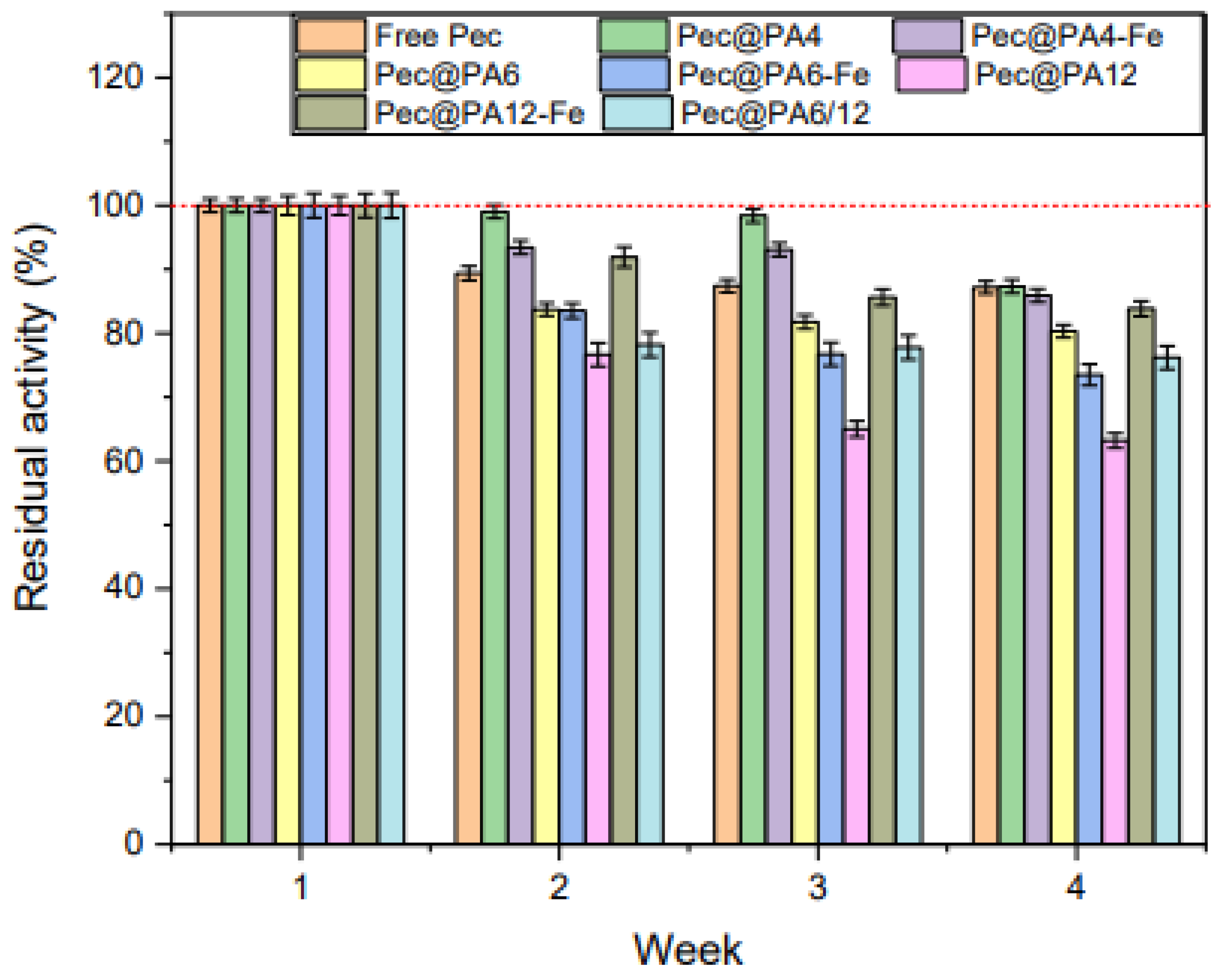
2.9. Storage Stability of Pec@PA Conjugates

Figure 11 illustrates the storage stability of all biocatalysts in this study at 4ºC for one month, determined as detailed in Section 3.11. By the end of the test, all immobilized catalysts, except for Pec@PA612 and Pec@PA12, show stability comparable to the free enzyme. Notably, Pec@PA4 and Pec@PA4-Fe exhibit higher residual activity than the free enzyme up to the 3rd week of storage, suggesting that the support may offer some enzyme protection.
Based on the results in Figure 11, it is recommended to use the Pec@PA pectinolytic systems within 2–3 weeks after immobilization to preserve their enhanced activity as compared to free Pec.

3. Materials and Methods

3.1. Reagents

The monomers 2-pyrrolidone (2PD, purum >98%) and laurolactam (LL, purum >99%) were purchased from Sigma Aldrich–Merck (Lisbon, Portugal) and used as received. The ε-caprolactam (ECL) monomer for anionic polymerization (AP-NylonR), and the Brüggolen C20® (C20) polymerization activator were both obtained from Bruggemann Chemical (Heilbronn, Germany). According to the manufacturer, C20 contains 80 wt.% of aliphatic diisocyanate blocked in ECL. The initiator, sodium dicaprolactamato-bis-(2-methoxyethoxo)-aluminate (80 wt.% in toluene) (Dilactamate®, DL), was purchased from Katchem (Prague, Czech Republic) and used without further purification.
The soft, non-insulated iron particles (Fe content >99.8%), with average diameters of 3–5 μm, were generously donated by BASF (Ludwigshafen, Germany). Viazym Clarif Extrem®, an enological pectinase preparation derived from Aspergillus Niger, was sourced from Martin Vialatte (Magenta, France). This preparation is typically used for the industrial clarification of white wine and rosé musts, and, like the sample quantities of industrial rosé must, was kindly donated by Sogrape Vinhos SA (Avintes, Portugal).
All other chemical reagents, such as pectin from citrus peel, and solvents used in this study were of analytical grade and supplied by Sigma Aldrich–Merck (Lisbon, Portugal).

3.2. Characterization Methods

The scanning electron microscopy (SEM) studies were carried out with a NanoSEM-200 apparatus from FEI Nova (Hillsboro, OR, USA) using mixed secondary electron/backscattered electron in-lens detection. All the samples were observed after sputter coating with Au/Pd alloy with a 208 HR instrument from Cressington Scientific Instruments (Watford, UK) with high-resolution thickness control. The UV-VIS spectral measurements were performed with a Shimadzu model 1900i double-beam spectrophotometer (Tokyo, Japan) working in photometric or spectral modes. Fourier-transform infra-red spectroscopy with attenuated total reflection (FTIR-ATR): the spectra were collected in a Perkin-Elmer Spectrum 100 apparatus (Waltham, MA, USA) using a horizontal ATR attachment with ZnSe crystal. Spectra were acquired between 4000 and 600 cm−1 accumulating up to 16 spectra with a resolution of 2 cm−1. The samples were studied in the form of fine powders. The turbidity measurements were made with a METRIA M10 portable turbidimeter (Labbox Labware, Barcelona, Spain) performing up to 5 parallel measurements with every sample, according to ISO 7027.

3.3. Synthesis of PA6 Microparticles

The PA6, PA12, and the copolymeric PA612 MPs were prepared by AAROP in suspension, following the general method described by Dencheva et al. [26]. Polymerizations were conducted in a 250 mL flask equipped with a thermometer, magnetic stirrer, Dean-Stark attachment for azeotropic distillation with a reflux condenser, and a nitrogen inlet. The monomers (0.3 mol of ECL for PA6 MP, 0.2 mol of LL for PA12 MP, and a mixture of 0.1 mol CL and 0.1 mol LL for PA612 MP) were added to 100 mL of a mixed hydrocarbon solvent (toluene/xylene, 1:1 by volume) under nitrogen atmosphere, and the mixture was refluxed for 10–15 minutes. For Fe-containing MPs, 1–3 wt.% of iron microparticles (relative to monomer) were introduced at this point. Then, 3.0 mol% of DL and 1.5 mol% of C20 were added simultaneously. The reaction proceeded for 2 hours after the catalyst addition, maintaining the temperature between 125–135°C for PA6 synthesis or around 120°C for PA12 and PA612, with constant stirring at 800 rpm. The fine powders produced were isolated by dissolving the reaction mixture in acetone to remove unpolymerized monomer, followed by vacuum filtration and washing with methanol to eliminate low molecular weight oligomers. The resulting microparticles were dried for 2 hours in a vacuum oven at 80°C. All MP powders were subjected to Soxhlet extraction with methanol for 4 hours to remove traces of oligomers or impurities, then dried and stored in a desiccator.
For the PA4-Fe MPs, low-temperature solventless AAROP was employed [37]. In this case, 0.2 mol of 2PD was stirred with 1 wt.% Fe at 25°C for 30 minutes. C20 activator and DL initiator (1.5 mol % and 3.0 mol. % respectively) were added under stirring in an inert atmosphere at 20ºC. The temperature was then raised to 40°C, and the pressure reduced to 50 mbar for 6 hours. After completion, the reaction mixture was dispersed in acetone, filtered, and washed twice with methanol to remove unreacted monomer. Oligomers were removed by Soxhlet extraction with methanol for 4 hours, followed by drying in vacuum and storage in a desiccator. The procedure for neat PA4 MP followed the same steps, without the addition of magnetic fillers in the monomer feed.

3.4 Immobilization of Pectinase by Physical Absorption

The commercial enological pectinase preparation, Viazym Clarif Extrem®, was immobilized by adsorption on the seven different microparticulate supports in the following way. The PA microparticles (100 mg) were incubated with 5 mL of the pectinase preparation diluted 10-fold in double-distilled water, under gentle agitation, at room temperature. It was assumed that the commercial pectinase preparation contained 10 a.u. of protein per mL After 24 h, all samples were centrifuged, and the supernatant was decanted and stored at 4°C for further analyses. The final Pec@PA complexes were washed twice with double-distilled water to remove any non-immobilized enzymes and stored at 4°C for subsequent use.

3.5 Determination of the Total Amount of Protein

After immobilization, the supernatants were subjected to UV analysis to determine the residual protein content and calculate the total protein (TP) incorporated into the PA microparticles (MPs), expressed as:
T P   =   C     C s , ( a , u )
where C₀ is the initial protein content before immobilization, and Cₛ is the protein content in the supernatant after the immobilization process. TP was determined by direct quantification of the UV absorption peak at λ ≈ 263 nm. The absorbance was measured, and Cs was determined using a standard calibration curve. In this study TP is considered as immobilization yield.

3.6. Pectinase Activity Assay

Pectinase activity was determined by measuring the amount of reducing sugars released from a pectin solution using the DNS method proposed by Miller [30]. The DNS reagent consists of 2-hydroxy-3,5-dinitrobenzoic acid (1% w/v), potassium sodium tartrate (30% w/v), and sodium hydroxide (1% w/v). Citric pectin (0.25% w/v) in citrate buffer (50 mM, pH 3.8) was used as the substrate. Typically, free pectinase preparation (0.1 mL, 0.1 a.u. total protein) or immobilized enzyme (0.020 g) was mixed with the substrate solution (0.90 mL) and incubated at 50°C for 15 minutes. The reaction mixture (1 mL) was then combined with DNS solution (1 mL), and the samples were kept in boiling water for 10 minutes before being cooled on ice to stop the reaction. Absorbance was measured at 540 nm against the blank DNS reagent. The amount of released reducing sugar was quantified using D-galacturonic acid standard calibration plot in the concentration range of 0.28–2.83 µmol/mL. One unit of pectinase activity is defined as the amount of enzyme required to release 1 μmol of galacturonic acid per minute under standard assay conditions.

3.7. Kinetics of Pectin Degradation

The kinetic parameters of pectin degradation by free pectinase and the immobilized Pec@PA complexes were determined by measuring the pectin degradation to D-galacturonic acid at various substrate concentrations (0.05–5 mg/mL), with an incubation time of 1 minute under the conditions previously described in Section 3.6. The total amount of enzyme (0.15 a.u.) added to the reaction mixture was the same in all cases. Three kinetic models, based on previous studies, were selected to calculate the kinetic parameters, namely the Michaelis–Menten model and two models accounting for substrate inhibition. The equations for these models are presented below: Equations (4)–(6) [31]:
V = V m a x S K m + S   ( Model   1 ,   Michaelis-Menten   equation )
V = V m a x S ( K m + S ) ( 1 + S K i )   ( Model   2 ,   conventional   substrate   inhibition   model )
V = V m a x exp S K i exp S K m   ( Model   3 ,   double   exponential   model )
The kinetic parameters, including the maximal reaction rate ( V m a x ), Michaelis–Menten constant ( K m ) and the constant characterizing the formation of an inactive enzyme–substrate complex ( K i )) were computed using nonlinear multiple regression analysis with the commercial software package OriginPro, version 9.8.0.200 by OriginLab corporation (Northampton, MA, USA).

3.8. Application of Immobilized Pectinase for Rosé Must Clarification

Clarification of the rosé must was carried out at room temperature using both free and immobilized pectinase preparation. The manufacturer’s recommended concentration of free enzymatic preparation and complexes containing the same amount of immobilized enzyme were used. The enzyme was mixed with 14 mL of rosé must for three hours, and turbidity was measured in nephelometric turbidity units (NTUs) every 20 minutes. A pectin test was then performed to detect the presence of residual pectin by adding acidified alcohol (5% v/v HCl, 5 mL) to 2.5 mL of the must sample under gentle agitation. After 10 minutes, the must was visually inspected. In the presence of residual pectin, insoluble flakes form; if the liquid is clear, the sample contains no residual pectin.

3.9. Color Analyses

The color of the must was analyzed by UV/VIS spectroscopy (method OIV-MA-AS2-07B [35] to evaluate any color changes resulting from enzymatic clarification. Spectra were obtained from all treated samples both before and after clarification. The color of the must was measured at three wavelengths: 420, 520, and 620 nm.
The color intensity (I) was calculated using Equation (7). The darkness or lightness of the color, known as the shade (N), was determined using Equation (8).
I = A 420 + A 520 + A 620
N = A 420 A 520
where A 420 ,   A 520 and A 620 represent the absorbance values at 420 nm, 520 nm, and 620 nm, respectively.

3.10. Reusability Studies

The Pec@PA conjugates, each containing 0.15 a.u. of enzyme, were used for must clarification over three consecutive cycles. Turbidity was measured until levels of around 20 NTUs were reached, as described in Section 4.8. After the first clarification cycle, the complexes were recovered from the must by centrifugation, washed twice with 1 mL of deionized water, and tested for their clarification capacity with fresh rosé must following the same procedure.
For enzymatic activity tests in this study, Pec@PA complexes (0.020 g) were mixed with 0.90 mL of citric pectin substrate, prepared as previously described in Section 3.6 and incubated at 50ºC. After a 15-minute reaction, the conjugates´ microparticles were washed twice with deionized water to remove any residual substrate, and their activity was tested with fresh substrate. This process was repeated for 8 consecutive cycles. Activity after each cycle was measured in terms of residual activity, with the first cycle set at 100%.

3.11. Storage Stability Studies

Free pectinase and immobilized enzyme complexes Pec@PA were stored at 4°C for one month, the latter in double-distilled water. Every week enzyme samples were taken, and their residual enzymatic activity was measured by DNS method as previously described. The activity of the first week was considered 100%.

4. Conclusions

This study presents systematic investigation of noncovalently immobilized commercial pectinase formulation on seven distinct microparticulate polyamide supports. These are previously synthesized by AAROP, each varying in chemical structure, composition, shape, size, and surface topology. The immobilization method involves a simple, one-pot process: incubating the supports in buffered aqueous enzyme solutions without the need for hazardous chemicals, making it compatible with the food industry requirements.
The results demonstrate a significant effect of polyamide type (PA4, PA6, PA12, with and without Fe carrier, and copolymeric PA612) on the enzyme immobilization yield and the activity of the resulting Pec@PA catalytic conjugates. Comparative kinetic studies revealed that, except for Pec@PA12-Fe, all conjugates exhibited higher catalytic efficiency than free pectinase. This finding should be ascribed to the different structure and morphology of the polyamide supports.
The polyamide-immobilized pectinase biocatalysts in this study achieved effective rosé must clarification at the laboratory scale within an industrially relevant time frame of 80–180 minutes during the first clarification round. Most immobilized complexes were reusable for second and third consecutive clarification cycles, with clarification times extending to approximately 15 and 30 hours, respectively, which is still within industrially acceptable limits. Additionally, each conjugate supported up to seven consecutive clarification rounds, with most systems retaining about 50% of their initial activity.
Notably, the polyamide supports in the Pec@PA conjugates did not alter the must color, as confirmed by UV/VIS measurements across three key wavelength ranges. Furthermore, all immobilized systems, except for Pec@PA12, retained up to 75% of their initial activity after four weeks of storage. These properties are comparable to those of the free pectinase preparation.
Currently, enological assessments are underway to evaluate the sensorial properties of the clarified musts using the best-performing polyamide-immobilized pectinase conjugates. These studies will provide a foundation for practical applications of these novel biocatalysts.

Supplementary Materials

The following supporting information can be downloaded at the website of this paper posted on Preprints.org: Figure S1: Chemical structures of the reagents participating in AAROP and of the microparticulate polyamides prepared; Figure S2: Selected SEM micrographs complementary to those in Figure 1 of the main text; Figure S3: Selected FTIR spectra of PA12 and PA4 supports before and after Pec immobilization; Figure S4: Evolution of the UV/VIS absorption maxima of various concentrations of galacturonic acid treated with the DNS reagent.

Author Contributions

Conceptualization, N.V.D., Z.Z.D.; methodology, N.V.D. and S.C.O..; validation, S.C.O., N.V.D.; formal analysis, N.V.D. and Z.Z.D.; investigation, S.C.O., S.M.A.; resources, Z.Z.D. and N.V.D.; data curation, S.C.O., S.M.A. and N.V.D.; writing—original draft preparation, S.C.O. and N.V.D.; writing—review and editing, Z.Z.D. and N.V.D; visualization, S.C.O, S.M.A.; supervision, N.V.D., Z.Z.D.; project administration, Z.Z.D..; funding acquisition, N.V.D. and Z.Z.D. All authors have read and agreed to the published version of the manuscript.

Funding

N. Dencheva is also grateful for the financial support by the Fundação para a Ciência e Tecnologia, Portugal (FCT) for the personal program contract CTTI-51/18-IPC. S. C. Oliveira acknowledges the financial support of the FCT through the 2021.07246.BD Ph.D. grant.

Data Availability Statement

All the data generated during this research are presented in the manuscript and in the Supplementary Materials.

Acknowledgments

The authors gratefully acknowledge the support of Sogrape Vinhos S.A., Avintes, Portugal, for generously providing the necessary quantities of unclarified rosé must and the Viazym Clarif Extrem commercial pectinolytic preparation used in this study.

Conflicts of Interest

The authors declare no conflict of interest.

References

  1. Rodicio, R.; Heinisch, J. Carbohydrate Metabolism in Wine Yeasts. In Biology of Microorganisms on Grapes, in Must and in Wine, 2nd ed.; Konig, H., Unden, G., Frohlich, J., Eds.; Springer: Cham, Switzerland, 2017; pp. 189–214. [CrossRef]
  2. Ottone, C.; Romero, O.; Aburto, C.; Illanes, A.; Wilson, L. Biocatalysis in the winemaking industry: Challenges and opportunities for immobilized enzymes. Compr. Rev. Food Sci. Food Saf. 2020, 19, 595–621. [CrossRef]
  3. Mojsoc, K. Use of enzymes in wine making: A review. Int. J. Mark. Technol. 2013, 3, 112–127.
  4. Pretorius, I.S. Tailoring wine yeast for the new millennium: Novel approaches to the ancient art of winemaking. Yeast. 2000, 16, 675–729. [CrossRef]
  5. Ribéreau-Gayon, P.; Dubourdieu, D.; Donèche, B.; Lonvaud, A. White winemaking, chapter 13. In Handbook of Enology: The Microbiology of Wine and Vinifications, 2nd ed.; John Wiley & Sons, Ltd, 2006; Volume 1, pp. 421-429; 445-448.
  6. Espejo, F. Role of commercial enzymes in wine production: a critical review of recent research. J. Food Sci. Technol. 2021, 58, 9–21. [CrossRef]
  7. Kassara, S.; Li, S.; Smith, P.; Blando, F.; Bindon, K. Pectolytic enzyme reduces the concentration of colloidal particles in wine due to changes in polysaccharide structure and aggregation properties, Int. J. Biol. Macromol. 2019, 140, 546-555. [CrossRef]
  8. Jiang, X.; Lu, Y.; Liu, S.Q. Effects of pectinase treatment on the physicochemical and oenological properties of red dragon fruit wine fermented with Torulaspora delbrueckii, LWT 2020, 132, 109929. [CrossRef]
  9. Rocha, S.M.; Coutinho, P.; Delgadillo I.; Cardoso, A.D.; Coimbra, M.A. Effect of enzymatic aroma release on the volatile compounds of white wines presenting different aroma potentials, J. Sci. Food Agric. 2005, 85, 199–205. [CrossRef]
  10. Jayani, R.S.; Saxena, S.; Gupta, R. Microbial pectinolytic enzymes: A review. Process Biochem. 2005, 40, 2931–2944. [CrossRef]
  11. Martín, M.C.; López, O.V.; Ciolino, A.E.; Morata, V.I.; Villar, M.A.; Ninago, M.D. Immobilization of enological pectinase in calcium alginate hydrogels: A potential biocatalyst for winemaking. Biocatal. Agric. Biotechnol. 2019, 18, 101091. [CrossRef]
  12. Benucci, I.; Lombardelli, C.; Cacciotti, I.; Liburdi, K.; Nanni, F.; Esti, M. Immobilized native plant cysteine proteases: Packed-bed reactor for white wine protein stabilization. J. Food Sci. Technol. 2016, 53, 1130–1139. [CrossRef]
  13. Barbosa, O.; Ortiz, C.; Berenguer-Murcia, Á.; Torres, R.; Rodrigues, R.C.; Fernandez-Lafuente, R. Strategies for the one-step immobilization–purification of enzymes as industrial biocatalysts. Biotechnol. Adv. 2015, 33, 435–456. [CrossRef]
  14. Yushkova, E.D.; Nazarova, E.A.; Matyuhina, A.V.; Noskova, A.O.; Shavronskaya, D.O.; Vinogradov, V.V.; Skvortsova, N.N.; Krivoshapkina, E.F. Application of Immobilized Enzymes in Food Industry. J. Agric. Food Chem. 2019, 67, 11553–11567. [CrossRef]
  15. Hosseini, S.S.; Khodaiyan, F.; E. Mousavi, S.M.; Kennedy, J.F.; Azimi, S.Z. A Health-Friendly Strategy for Covalent-Bonded Immobilization of Pectinase on the Functionalized Glass Beads. Food Bioprod. Process. 2021, 14, 177–186. [CrossRef]
  16. Behram, T.; Pervez, S.; Nawaz, M.A.; Ahmad, S.; Jan, A.U.; Rehman, H.U.; Ahmad, S.; Khan, N.M.; Khan, F.A. Development of Pectinase Based Nanocatalyst by Immobilization of Pectinase on Magnetic Iron Oxide Nanoparticles Using Glutaraldehyde as Crosslinking. Agent. Molecules 2023, 28, 404. [CrossRef]
  17. ​Rehman, H.U.; Nawaz, M.A.; Pervez, S.; Jamal, M.; Attaullah, M.; Aman, A.; Qader, A.U. Encapsulation of pectinase within polyacrylamide gel: characterization of its catalytic properties for continuous industrial uses. Heliyon 2020, 6, e04578. [CrossRef]
  18. Rajdeo, K.; Harini, T.; Lavanya, K.; Fadnavis, N. W. Immobilization of pectinase on reusable polymer support for clarification of apple juice. Food Bioprod. Process. 2016, 99, 12-19. [CrossRef]
  19. Miao, Q.; Zhang, C.; Zhou, S.; Meng, L.; Huang, L., Ni, Y.; Chen, L. Immobilization and Characterization of Pectinase onto the Cationic Polystyrene Resin. ACS Omega. 2021, 6, 31683-31688. [CrossRef]
  20. Omelková, J.; Rexová-Benková, Ł.; Kubánek, V.; Veruovič, B. Endopolygalacturonase immobilized on a porous poly(6-caprolactame). Biotechnol Lett. 1985, 7, 99–104. [CrossRef]
  21. Rexová-Benková, L.; Omelková, J.; Veruovič, B.; Kubánek, V. Mode of action of endopolygalacturonase immobilized by adsorption and by covalent binding via amino groups. Biotechnol. Bioeng. 1989, 34, 79–85. [CrossRef]
  22. Shukla, S.K.; Saxena, S.; Thakur, J.; Gupta, R. Immobilization of polygalacturonase from Aspergillus Niger onto glutaraldehyde activated Nylon-6 and its application in apple juice clarification. Acta Aliment. 2010, 39, 277–292. [CrossRef]
  23. Ben-Othman, S.; Rinken, T. Immobilization of Pectinolytic Enzymes on Nylon 6/6 Carriers. Appl. Sci. 2021, 11, 4591. [CrossRef]
  24. Lozano, P.; Manjón, A.; Iborra, J.L.; Gálvez, D.M. Characteristics of the immobilized pectin lyase activity from a commercial pectolytic enzyme preparation. Acta Biotechnol. 1990, 10, 531–539. [CrossRef]
  25. Diano, N.; Grimaldi, T.; Bianco, M.; Rossi, S.; Gabrovska, K.; Yordanova, G.; Godjevargova, T.; Grano, V.; Nicolucci, C.; Mita, L.; et al. Apple Juice Clarification by Immobilized Pectolytic Enzymes in Packed or Fluidized Bed Reactors. J. Agric. Food Chem. 2008, 56, 11471–11477. [CrossRef]
  26. Dencheva, N.; Denchev, Z.; Lanceros-Méndez, S.; Ezquerra, T.E. One-step in-situ synthesis of polyamide microcapsules with inorganic payload and their transformation into responsive thermoplastic composite materials. Macromol. Mater. Eng. 2016, 301, 119–124. [CrossRef]
  27. Oliveira, S.C.; Dencheva, N.V.; Denchev, Z.Z. Immobilization of Enological Pectinase on Magnetic Sensitive Polyamide Microparticles for Wine Clarification. Foods 2024, 13, 420. [CrossRef]
  28. Hong, M.; Chen, Y.-X. Completely Recyclable Biopolymers with Linear and Cyclic Topologies via Ring-Opening Polymerization of γ-Butyrolactone. Nature Chem, 2016, 8, 42-49. [CrossRef]
  29. Brêda, C.; Dencheva, N.; Lanceros-Méndez, S.; Denchev, Z. Preparation and properties of metal-containing polyamide hybrid composites via reactive microencapsulation. J. Mater. Sci. 2016, 51, 10534–10554. [CrossRef]
  30. Miller, G.L. Use of Dinitrosalicylic Acid Reagent for Determination of Reducing Sugar. Anal. Chem. 1959, 31, 426–428. [CrossRef]
  31. Gummadi, S.N.; Panda, T. Analysis of Kinetic Data of Pectinases with Substrate Inhibition. J. Microbiol. Biotechnol. 2003, 13, 332–337.
  32. Haldane, J.B.S. Enzymes, Featured ed.; MIT Press: Cambridge, USA, 1965; pp. 154-196.
  33. Edwards, V.H. Influence of high substrate concentration on microbial kinetics. Biotechnol. Bioeng. 1970, 27, 679-712. [CrossRef]
  34. Cleland W.W. Substrate Inhibition, chapter 20. In Enzyme Kinetics and Mechanism – Part A: Initial Rate and Inhibition Methods. Academic Press: Cambridge, MA, USA, 1976, volume 63, pp. 500-513.
  35. IOV (International Organization of Vine and Wine). Compendium of international methods of wine and must analysis - Chromatic Characteristics (Type-IV). OIV-MA-AS2-07B. 2022. Available online: https://www.oiv.int/standards/compendium-of-international-methods-of-wine-and-must-analysis (accessed on October 16, 2024).
  36. Faragó, P.; Gălătuș, R.; Hintea, S.; Boșca, A.B.; Feurdean, C.N.; Ilea, A. An Intra-Oral Optical Sensor for the Real-Time Identification and Assessment of Wine Intake. Sensors 2019, 19, 4719. [CrossRef]
  37. Dencheva, N.; Braz, J.; Nunes, T.G.; Oliveira, F.D.; Denchev, Z. One-pot low temperature synthesis and characterization of hybrid poly(2-pyrrolidone) microparticles suitable for protein immobilization. Polymer 2018, 145, 402–415. [CrossRef]
Figure 1. Selected SEM micrographs of empty supports and pectine-immobilized conjugates: a, b – PA4; c, d – Pec@PA4; e, f – PA6; g, h – Pec@PA6; i, j – PA12; k, l – Pec@PA12-Fe; m, n – PA612; o, p - Pec@PA612 complexes.
Preprints 140472 g001
Figure 2. Quantification of the pectinase immobilization: (a) UV/VIS spectra of the Viazym solutions before and after adsorption immobilization on 7 different MP supports; (b) Immobilization yield for all Pec@PA and Pec@PA-Fe pectinolytic complexes. For more data, see the text.
Preprints 140472 g002
Figure 3. Selected FTIR spectra of PA6 supports, before and after noncovalent Pec immobilization. The two arrows in Figure 3b point at the Pec bands present in the Pec@PA6 spectra. .
Preprints 140472 g003
Figure 4. Activity assay of pectinolytic biocatalysts by the DNS method. (a) Reaction scheme of the interaction between 3,5-DNS and D-galacturonic acid (GA); (b) The standard calibration curve constructed with various concentrations of GA.
Preprints 140472 g004
Figure 5. Activity of Pec@PA immobilized conjugates in comparison to free Pec: a – absolute activity. The small yellow bars in Fig. 5a show the “activity” values of the respective empty PA supports.; b – specific activity per unit enzyme (free or immobilized).
Preprints 140472 g005
Figure 6. Selected experimental kinetic curves and their best fits with Model 2 and Model 3 for: a – comparison of all experimental curves; b – Free pectinase; c – Pec@PA4-Fe; d – Pec@PA6-Fe; e – Pec@PA12; – Pec@PA612; .
Preprints 140472 g006aPreprints 140472 g006b
Figure 7. Consolidated results from the kinetics studies. CE = catalyst efficiency, Ki = inhibition constant. The dashed lines are guides for the eye highlighting the data of the free enzyme reference.
Preprints 140472 g007
Figure 8. Clarification studies with free and immobilized pectinase preparation: a – free Pec compared to PA4 and PA6-based Pec@PA complexes; b – free Pec compared to PA12-based Pec@PA complexes. All immobilized catalysts contain 0.15 a.u. of pectinolytic enzyme; the same is the amount of the free Pec catalyst. The dashed lines indicate the level of 20 NTUs, below which the must is considered completely clarified, i.e., not containing pectin. The visual aspects of the must before and after clarification is presented in the upper and bottom photographs, respectively. .
Preprints 140472 g008
Figure 9. Color changes in treated rosé must samples: a – numerical data comparison considering the three spectral intervals at 420, 520 and 620 nm; b – the source UV/VIS spectra; c – Comparison of the I and N color parameters for rosé must before and after treatment with various pectinllytic catalysts. For more details see Section 3.9. .
Preprints 140472 g009
Figure 10. Clarification studies of rosé must with Pec@PA systems in several different clarification cycles: a – with Pec@PA4 and Pec@PA4-Fe; b – with Pec@PA6, Pec@PA6-Fe and Pec@PA612; c – with Pec@PA12 and Pec@PA12-Fe. All immobilized catalysts contain 0.15 a.u. of the pectinolytic enzyme. The dashed lines indicate the level of 20 NTUs, below which the must is completely clarified.
Preprints 140472 g010
Figure 11. Relative activity of immobilized Pec@PA pectinolytic catalysts over eight application cycles. The red dashed line indicates 50% relative activity. Initial activity (Figure 5b) is set to 100% for each complex. See text for details.
Preprints 140472 g011
Figure 12. Storage stability of immobilized Pec@PA pectinolytic catalysts over four weeks at 4ºC. Initial activity (Figure 5b) is normalized to 100% for each complex.
Preprints 140472 g012
Table 1. Some basic characteristics of the synthesized microparticulate polyamide supports
Sample Yield of
poly-
mer, %
Oligo-
mers1,
wt.%
Mη
kDa2
Average particle size, µm Average pore size,
nm
Fe
content4,
wt.%
PA4 59.4 4.4 26.4 10-25 50-120 -
PA4-Fe 61.2 5.9 - 3 10-40 120-250 1.0 [1.6]
PA6 56.0 2.0 33.7 50-150 300-500 -
PA6-Fe 58.8 2.4 - 50-200 200-300 3.0 [5.6]
PA12 42.0 3.2 33.8 50-700 250-900 -
PA12-Fe 58.5 3.4 - 50-80 750-1500 2.0 [4.2]
PA612 77.0 2.1 28.9 100-150 250-2000 -
1 After Soxhlet extraction; 2 Viscosity-average molecular weight Mη determined in H2SO4.3 Formation of precipitate, intrinsic viscosity η impossible to determine; 4 In relation to monomer before polymerization and in the final polymer (in brackets).
Table 2. Kinetic parameters estimated for Pec and the seven Pec@PA catalytic conjugates
Biocatalyst Parameters a) Model 1 Model 2 Model 3
Free Pec Vmax
Km
Ki
1.956 ± 0.024 2.182 ± 0.071 1.933 ± 0.001
0.423 ± 0.009 0.423 ± 0.043 0.446 ± 0.041
≈ 3.0 5.520 ± 0.850 5.656 ± 0.687
Adj. R2 0.995 0.958 0.997
Pec@PA4 Vmax 5.672 ± 0.020 9.364 ± 0.014 5.592 ± 0.101
Km 0.752 ± 0.010 1.444 ± 0.035 0.720 ± 0.020
Ki 2.5 - 3.0 1.920 ± 0.060 4.838 ± 0.304
Adj. R2 0.989 0.965 0.994
Pec@PA4-Fe Vmax 4.193 ± 0.027 5.387 ± 0.015 5.981 ± 0.164
Km 0.662 ± 0.012 0.695 ± 0.012 0.684 ± 0.027
Ki 2.0 - 3.0 2.390 ± 0.025 2.536 ± 0.100
Adj. R2 0.977 0.981 0.985
Pec@PA6 Vmax 4.669 ± 0.020 5.568 ± 0.005 7.679 ± 0.170
Km 0.863 ± 0.009 0.887 ± 0.002 0.868 ± 0.050
Ki ≈ 2.5 2.359 ± 0.021 2.236 ± 0.131
Adj. R2 0.995 0.988 0.995
Pec@PA6-Fe Vmax 5.100 ± 0.020 6.136 ± 0.003 6.133 ± 0.644
Km 1.222 ± 0.008 1.297 ± 0.002 0.995 ± 0.020
Ki ≈ 2.5 – 3.0 3.035 ± 0.060 3.491 ± 0.554
Adj. R2 0.995 0.998 0.984
Pec@PA12 Vmax 8.666 ± 0.019 8.335 ± 0.016 8.408 ± 0.196
Km 1.010 ± 0.007 0.869 ± 0.032 0.799± 0.030
Ki ≈ 2.5 5.323 ± 0.066 4.354 ± 0.226
Adj. R2 0.988 0.990 0.990
Pec@PA12-Fe Vmax 1.872 ± 0.070 2.217 ± 0.040 2.395 ± 0.040
Km 0.820 ± 0.020 0.855 ± 0.030 0.821 ± 0.019
Ki ≈ 2.5 3.737 ± 0.021 3.308 ± 0.167
Adj. R2 0.976 0.993 0.994
Pec@PA612 Vmax 1.931 ± 0.043 2.254 ± 0.020 1.924 ± 0.020
Km 0.372 ± 0.022 0.454 ± 0.010 0.448 ± 0.020
Ki ≈ 5.0 5.708 ± 0.083 7.516 ± 0.539
Adj. R2 0.966 0.990 0.993
a) Estimations for Vmax (µmol mL-1 min-1) and Km (mg mL-1) from the linear part of the double reciprocal Lineweaver-Burk plot (Model 1, Equation 4, Section 4.7). Ki, (mg mL-1), estimation from Equation 1. The final Vmax, Km and Ki are determined after fitting of the estimates to Models 2 and 3.
Disclaimer/Publisher’s Note: The statements, opinions and data contained in all publications are solely those of the individual author(s) and contributor(s) and not of MDPI and/or the editor(s). MDPI and/or the editor(s) disclaim responsibility for any injury to people or property resulting from any ideas, methods, instructions or products referred to in the content.
Copyright: This open access article is published under a Creative Commons CC BY 4.0 license, which permit the free download, distribution, and reuse, provided that the author and preprint are cited in any reuse.
Prerpints.org logo

Preprints.org is a free preprint server supported by MDPI in Basel, Switzerland.

Subscribe

Disclaimer

Terms of Use

Privacy Policy

Privacy Settings

© 2025 MDPI (Basel, Switzerland) unless otherwise stated