
Submitted:
22 November 2024
Posted:
22 November 2024
You are already at the latest version
Abstract
This study documents Euphorbiaceae plants in Southern Africa, focusing on their traditional medicinal uses, pharmacological properties, toxicity, and active secondary metabolites. Literature from scientific journals, books, dis-sertations, and conference papers from 1962 to 2023 was reviewed for 15 Euphorbia species. Recent findings indicate that certain compounds in Eu-phorbia plants have significant biological and pharmacological properties. However, the white sticky latex sap they contain is highly toxic yet can also have medicinal applications. Phytochemical analysis has shown these plants exhibit beneficial effects, including antibacterial, antioxidant, antiprolifera-tive, anticancer, anti-inflammatory, antiviral, antifungal, and anti-HIV activ-ities. Key phytochemicals such as euphol, cycloartenol, tirucallol, and triterpenoids contribute to their therapeutic efficacy, along with various proteins like lectin and lysozyme. Despite some Euphorbiaceae species being screened for medicinal compounds, many have not been thoroughly exam-ined, highlighting a critical gap. Given their historical usage, further research is essential to evaluate the medicinal significance of Euphorbia species through detailed studies of isolated compounds and their pharmacokinetics and pharmacodynamics. This research will serve as a valuable resource for future inquiries into the benefits of lesser-studied Euphorbia species.
Keywords:
Euphorbia species
; toxicity
; secondary metabolites
; pharmacological properties
; medicinal plants
; cancer
1. Introduction
The Euphorbiaceae is a highly varied group of flowering plants, having over 300 genera and 8 000 species [1]. Within this family, the Euphorbia genus stands out as one of the largest, with more than 2 000 species [2]. These species can take the form of herbs, shrubs and trees, sometimes resembling succulents or cacti. They are mainly found in tropical and subtropical regions of Africa and America, as noted by Adedapo et al. [3]. Many species in this family produce a toxic white sap, but this sap can also have medicinal properties [4]. The genus contains economically significant species, making it a vital genus with high research potential.
South Africa has a rich collection of Euphorbia species, with at least 188 indigenous to the country (SANBI). The Euphorbia genus has attracted the attention of many researchers due to its diverse chemical compositions, which include euphol, triterpenoids, diterpene ester and tirucallol. These compositions have been found to have remarkable therapeutic properties such as antimicrobial, antidiabetic, antiviral and anticancer properties, as noted by Betancur-Galvis et al. [5] and also Mwine and Damme [6]. However, there is a lack of scientific documentation on the anticancer properties of many Euphorbia species in Southern Africa. This is due to a lack of research on isolated compounds, as well as a lack of testing and discovery of new compounds. Furthermore, there is limited research on unexplored Euphorbia species that may have potential anticancer properties. Additionally, the majority of published articles on Euphorbia focus on already explored species, with little to no information on the unexplored ones [7,8]. It is worth noting that there is currently only one comprehensive review available on the Euphorbia species found in Southern Africa. This review highlights the limited scientific documentation available on these plants and emphasizes the need for further research to unlock their full potential. The review, however, did not focus on the compounds found in the selected plants and anticancer properties [9]. Therefore, the current study focuses on gathering more data on the use of isolated compounds as anticancer lead agents in traditional medicine to advance research in the field of discovery and development of anticancer agents.
For this reason, the purpose of this review is to provide a comprehensive overview of the pharmacological activities of the isolated compounds from various Euphorbia species found in Southern Africa. This review also aims to highlight the diversity of the Euphorbiaceae family and its potential for further research. The study focuses on several plants, including Euphorbia trigona (E. trigona), Euphorbia tirucalli (E. tirucalli), Euphorbia ammak (E. ammak), Euphorbia bupleurifolia (E. bupleurifolia), Euphorbia enopla (E. enopla), Euphorbia polygona (E. polygona), Euphorbia cooperi (E. cooperi), Euphorbia stellata (E. stellata), Euphorbia ferox (E. ferox), Euphorbia clavariodes (E. clavarioides), Euphorbia gorgonis (E. gorgonis), Euphorbia coerulescens (E. coerulescens), Euphorbia horrida (E. horrida), Euphorbia arabica (E. arabica), and Euphorbia ledienii (E. ledienii). These plants were selected for their pharmacological activities and toxicological evaluation on breast cancer cells as part of the authors’ recent research project. This review serves as a valuable resource for other researchers to explore the remaining Euphorbia species for their potentially active compounds and anticancer therapeutic properties.
2. Data Collection
To conduct this review, original articles from journals indexed in PubMed Central, Springer Link, Scopus, Science Direct, Hindawi, and Google scholars on Euphorbia species, their medicinal uses, toxicity and pharmacological properties, were analysed. For this review, only texts published in English between 1962 and 2023 were considered. The years of publication were selected due to the limited data available on Euphorbia plants. Throughout the search process, 881 reports were found across various databases. After reviewing the titles and abstracts, the search was refined and narrowed down to 293 articles, which were fully examined and are now being discussed in this study.
The research criteria for inclusion was based on the guidelines set by Lu et al. [10] with a few modifications. The criteria included: (1) studies on the isolated compounds of Euphorbia species conducted in vitro and in vivo; (2) evaluation of their biological activities; (3) studies published in peer-reviewed journals written in English; and (4) studies that provided full-text papers. There were no limitations on the study’s location. The study excluded (1) human studies, (2) a combination of isolated compounds and (3) non-original articles.
The retrieved data was carefully examined to identify possible medical applications, biological activities, isolated compounds, toxicity and pharmacological properties. Additional confirmation of the correct plant names was verified from the plantlist.org website. After analyzing all the data collected, conclusive results were obtained.
The cytotoxic tendency of selected compounds on various cancer cell lines and human tumor types was computed using the Cell Line Cytotoxicity Predictor (CLCPred) webserver (https://www.way2drug.com/Cell-line/) a free online service for in silico evaluation of human cell line cytotoxicity tendency of bioactive compounds [11]. This prediction was based on Prediction of Activity Spectra for Substances (PASS) technology (https://www.way2drug.com/PASSonline) where the training set was generated based on cytotoxicity data derived from ChEMBLdb (version 23) (https://www.ebi.ac.uk/chembldb/). Compounds with robust anticancer potential were selected and subjected to further analyses to extract physicochemical and pharmacokinetics parameters using the SwissADME (http://www.swisadme.ch/index.php) [12] and pKCSM (http://biosig.unimelb.edu.au/pkcsm/) [13] webservers as demonstrated earlier [14,15].
3. Results
The current study reports on 15 Euphorbia species, their traditional and pharmacological properties, and also isolated compounds. Euphorbia species have been used in folk medicine for various ailments, which is why scientists have taken an interest in investigating this genus and fully documenting the secondary metabolites responsible for these properties, see Table 1.1.
Out of the 15 plant species examined, 8 have been traditionally used as medicine for treating 7 distinct diseases. The most commonly used plants are for treating cancer (7), followed by warts and wounds (5), as well as other ailments, see Table 1.1.
An investigation into the chemical composition of various Euphorbia species unveiled distinctive profiles of isolated compounds. E. tirucalli exhibited the highest diversity with a total of 30 isolated compounds, spanning triterpenoids (Lupane, Oleanane, Tirucallane, Phorbol-type), phenolic compounds (Gallic Acid Derivatives, Ellagic Acid Derivatives, Flavonoids), phytosterols (Sterols), glycosides (Triterpene Glycosides, Other Glycosides), and diterpene esters [9,37,100]. Following closely, E. cooperi presented 18 compounds, including triterpenoids (Lupane, Oleanane, Phorbol-type), phytosterols (Sterols), and glycosides (Triterpene Glycosides) [95]. E. trigona showcased 16 compounds, including triterpenoids (Lupane, Oleanane, Phorbol-type), phenolic compounds (Gallic Acid Derivatives, Ellagic Acid Derivatives, Flavonoids), phytosterols (Sterols), and glycosides (Other Glycosides) [17,70,99]. E. coerulescens and E. ledienii exhibited 9 and 8 compounds, respectively, encompassing triterpenoids, phenolic compounds and phytosterols [66,71], see Table 1.2 and Table 1.3. Notably, certain plants demonstrated fewer or no isolated compounds.
The breakdown of these compounds into subclasses revealed diverse chemical categories, including Euphane-type Triterpenoids (Euphol, Euphorbol), Cycloartane-type Triterpenoids (Cycloartenol, Cycloartanol, 24-ethylene cycloartanol), Lupane-type Triterpenoids (Lupeol), Oleanane-type Triterpenoids (α-amyrin, β-amyrin), Pentacyclic Triterpenes (Betulinic acid, Glut-5-en-3-β-ol), Taraxarane-type Triterpenoids (Taraxerol, Taraxerol acetate), Phorbol-type Diterpenoids (12-Deoxyphorbol-13-isobutyrate-20-acetate, Phorbol, 12-Deoxy phorbol, 12-Deoxy-16-hydroxy phorbol, 12-Deoxyphorbol esters, 12, 20-Dideoxyphorbol-13-isobutyrate), Tirucallane-type Triterpenoids (Tirucalicine, Tirucallol, Tirucallin A, Tirucallin B), Steroidal Triterpenoids (Terpenic alcohol), Ingenane-type Triterpenoids (Ingol-7,8,12-acetate, ditiglate, Ingenol, Ingenol triacetate, Angelate acetate isobutyrate), Flavonoids (Tri-methyl ellagic acid, 17-Hydroxyingenol-17-benzoate-20-angelate, Kampferol-3-O-ß-D-rutinoside), Sterols (β-sitosterol, Stigmasterol, Taraxasterol), Ellagic Acid Derivatives (Ellagic acid, 3,3′-Di-O-methylellagic acid), Medium-chain Fatty Acids (Laurate) and no specific subclass records (Rhoiptlenone, 2-Methylbutyric acid, Euphorbin A, Euphorbin B, Euphorbol hexacosonate, 12-Deoxy-4β-hydroxyphorbol-13-phenyl acetate-20-acetate, 20-Acetoxy-16-angeloyloxy13α-isobutanoyloxy-4β,9α,20-tetrahydroxytiglia-1,5-diene-3-one, Resin, Arachiside A, 3, 4, 4′ Trimethoxyellagic acid, 12-Deoxyphorbol-13-(2-methylbutyrate)-20-acetate, 2-Methylbutyric acid) (see Table 1.4).
E. tirucalli as well as E. ammak are among the Euphorbia species that received substantial research attention. These findings are in agreement with studies conducted by Mavundza et al. [9], which stated that E. tirucalli was among the most studied species.
The investigation into the chemical composition of various Euphorbia species are presented in Table 1.4, and has revealed a diverse array of isolated compounds, each showcasing unique pharmacological and biological activities. Euphol, classified within the Euphane subclass, demonstrates a diverse profile, including anticancer, cytotoxicity, anti-nociceptive, anti-inflammatory and HIV-1 reverse transcriptase inhibitor activities [76,102,103]. The Cycloartenol and Cycloartanol compounds, falling under the Cycloartane subclass, exhibit a wide range of effects such as anti-inflammatory, antitumor, antioxidant, antibiosis, anti-Alzheimer’s disease, apoptotic, analgesic and antifungal activities [104,105,106,107,108,109,110]. Lupeol, belonging to the Lupane subclass, displays diverse properties like anticancer, anti-inflammatory, antimicrobial, antiprotozoal, antiproliferative, antiangiogenic and cholesterol-lowering effects [40,111,112,113,114,115,116,117]. Oleanane-type triterpenoids, represented by α-amyrin and β-amyrin, demonstrate activities such as cytotoxicity, antifungal, anti-inflammatory, nitric oxide inhibition, reactive oxygen species activation and anticancer effects [118,119,120,121,122,123,124,125]. Pentacyclic triterpenes, including Betulinic acid, exhibit antitumor, antidiabetic, anti-inflammatory, HIV-1 reverse transcriptase inhibition, antiviral and hepatoprotective activities [126,127,128,129,130,131,132,133,134,135]. The Taraxarane subclass, represented by compounds like Taraxerol and Taraxerol acetate, showcases properties like anticancer, anti-inflammatory, apoptotic, antioxidative, antimicrobial, antifungal and antidiabetic effects [136,137,138,139,140,141]. Sterols, represented by β-sitosterol, display a spectrum of activities, including anti-inflammatory, anticancer, antiproliferative, analgesic and antimicrobial effects [125,142,143,144,145]. Tirucallane-type triterpenoids, such as Tirucalicine and Tirucallol, currently have no specific records of biological activity [146,147]. Terpenic alcohol, a steroidal triterpenoid, demonstrates antibacterial and irritant effects [148,149]. Ellagic Acid Derivatives, including Tri-methyl ellagic acid, exhibit anticancer properties [148,150,151]. Ingenane-type Triterpenoids, represented by Ingenol and its derivatives, showcase cytotoxicity and HIV-1 reverse transcriptase inhibition [102,152]. Various subclasses such as Taraxarane, Euphane-type, Sterol, Tirucallane and Phorbol-type exhibit diverse pharmacological activities, including anti-inflammatory, antifungal, antibacterial, hepatoprotective, antioxidant, antiproliferative and anti-HIV effects, see Table 1.5.
E. trigona has the most isolated anticancer compounds (14), followed by E. tirucalli (13), E. cooperi (8), and the others have fewer or none. The Euphorbia species found in Southern Africa are rich sources of various types of bioactive compounds, including triterpenoids, phorbol esters, alkaloids, flavonoids, phytosterols, glycosides and saponins [9,60,65]. Furthermore, minor classes that were isolated from Euphorbia species from Southern Africa are: anthraquinone, polyphenols and tannins [9,60,65].
It has further been reported that most spurges contain an acidic and burning vesicant juice, as well as cyanoglycosides, which can be toxic [207]. Reports have shown that ingestion of a large quantity of the latex may cause gastro-intestinal haemorrhage and even result in death [41]. Although incidents of poisoning in children and animals are rare, it is important to handle these plants with great caution [208]. The latex from these plants can also cause blisters on the skin and temporary blindness [34]. Furthermore, they have been used as fish poison and bird-lime; see Table 1.6.
Taxonomic classification of all Euphorbia plants discussed in this review:
Domain: Eukaryote; Kingdom: Plantae; Order: Malpighiales; Family: Euphorbiaceae; Genus: Euphorbia (https://www.mindat.org/taxon-4691.html)
4. Discussion
4.1. Ethnopharmacological Use, Phytochemistry and Toxicity
4.1.1. Euphorbia trigona
E. trigona, a plant native to Central Africa, tropical Africa and India has been used in traditional Indian Ayurvedic medicine to treat respiratory and urinary tract infections and gonorrhea; Figure 1.1 [18]. Studies by Nashikkar et al. [17] have also shown that E. trigona is effective in treating various ailments, such as tumors, warts, intestinal parasites, rheumatoid arthritis, hepatitis and inflammation. A combination of the roots and ginger can be consumed in the morning to alleviate piles [19]. Additionally, some people take latex drops in palm wine for severe constipation or during an epileptic attack [20].
Phytochemistry
The plant E. trigona was analyzed for its phytochemical composition and was found to contain saponins, alkaloids, flavonoids, glycosides, sterols and triterpenoids [99]. Additional studies by Nashikkar et al. [17] also identified the presence of sterols, alkaloids, flavonoids and saponins, as well as tannins that were not detected by [99]. The absence of tannins may be due to environmental factors. Nielsen et al. [71] found that the latex of E. trigona contains a high level of sterols, with the main components being euphol and cycloartenol. Anjaneyulu and Rao [70] isolated several triterpenoids from the latex, including euphol, cycloartanol, cycloartenol, lupeol, α-amyrin and β-amyrin. They also identified five diterpenes ester skin-irritants. E. trigona is also a good source of lectin, which has been shown to have a potency in human erythrocyte agglutination [21].
Toxicity
A study, conducted by EL-Hawary et al. [98], tested the cytotoxic effects of a methanolic extract of E. trigona against HEPG2, MCF-7 and CACO2 cell lines. The findings revealed a pronounced cytotoxic effect on MCF-7 and Caco-2 cell lines, with IC50 values of 16.1 and 15.6 µg/mL, respectively. In 2022, Anju and Rameshkumar evaluated a methanol extract’s cytotoxic effect on HeLa and H9C2 cell lines but found no notable cytotoxic effect on either cell line [156]. Another study evaluated the cytotoxicity effect of Hex, DCM, MeOH and EtoAc extract against Vero cell lines [65]. The results indicated that none of the four extratcs from E. trigona exhibited cytotocity, as they did not hinder 50% of the cell growth at concentrations of 10 µg/mL and below.
4.1.2. Euphorbia ledienii
E. ledienii is a plant species that is indigenous to the Western Cape, South Africa (see Figure 1.2). The plant does not have any traditional or pharmacological data.
Phytochemistry
Despite a lack of traditional or pharmacological data, several compounds have been identified from the plant, including 12-Deoxyphorbol-13-isobutyrate-20-acetate and 12-Deoxyphorbol-13-(2-methylbutyrate)-20-acetate [72]. Evans and Kinghorn [74] discovered three variations of taglines which are phorbol, 12-Deoxyphorbol, and 12-Deoxy-16-hydroxy phorbol. E. ledienii has been reported to contain Ingol-7,8,12-acetate and ditiglate [160]. Redei et al. [73] identified Isobutyric and 2-methylbutyric acid. Other researchers discovered several hydrolytic proteins in E. ledienii, including N-acetyl-b-glucosamidase, chitobiosidase, endochitinase, and lysozyme activity [212]. According to Domsalla et al. [213], the E. ledienii plant has been found to have high proteolytic activity.
Toxicity
It is crucial to highlight that the plant is extremely toxic to humans, leading to potential skin irritation [69]. It is important to note that none of the isolated compounds have undergone toxicity assessments to date.
4.1.3. Euphorbia horrida
E. horrida is a plant species that can be found in Wittepoort/Karoo, South Africa (see Figure 1.3). There is currently no known traditional or pharmacological information about this plant.
Phytochemistry
In a study by El-Hawary et al. [98], 17-Hydroxyingenol-17-benzoate-20-angelate from the plant was isolated. The plant also contains diterpene esters that can cause skin irritation. In addition, Mampa et al. [65] found various other classes of compounds in the plant, including phytosterols, pentose, tannins, glycosides, triterpenoids, anthraquinones, saponins, flavonoids and alkaloids.
Toxicity
The cytotoxic effect of dichloromethane (DCM) extract derived from E. horrida var. was evaluated on a Vero cell line. The results demonstrated notable cytotoxicity, as evidenced by its achievement of an IC50 value at a concentration of 10 µg/mL, highlighting its potential cytotoxic effects [65]. A study conducted by El-Hawary et al. [98] evaluated the cytotoxicity effect of methanol extract of E. horrida against HeLa and H9C2 cell lines and no significant effect was observed.
4.1.4. Euphorbia enopla
E. enopla, a plant that is native to the Eastern Cape and semi-arid Karoo, South Africa, has no documented traditional or pharmacological data (see Figure 1.4).
Phytochemistry
The plant contains euphol and tirucallol, which were isolated by Ponsinet and Ourisson [93]. Furthermore, Mampa et al. [65]) extracted phytosterols, glycosides, triterpenoids, flavonoids, alkaloids, tanins and anthraquinone. Sytwala et al. [212] isolated N-acetyl-b-glucosamidase, chitobiosidase, endochitinase and lysozyme hydrolytic proteins.
Toxicity
A study conducted by Mampa et al. [65] assessed the toxicity of the hexane extract of E. enopla on the Vero cell line. The results indicated that the extract had a substantial inhibitory effect on cell growth, particularly at a concentration of 10 µg/mL. Notably, the highly non-polar hexane fraction exhibited the most potent effects [65].
4.1.5. Euphorbia coerulescens
E. coerulescens which is native to the Cape province, South Africa, has no documented traditional or pharmacological data (see Figure 1.5). However, it has a number of compounds isolated from it.
Phytochemistry
Studies by Evans [66] resulted in the isolation of angelate acetate isobutyrate, acetate a-methyl butyrate, acetate laurate, α-Methyl butyrate, heptanoate and laurate. In other studies, euphol, tirucallol and euphorbol were also isolated [89]. In addition, Sytwala et al. [212] isolated several hydrolytic proteins, including N-acetyl-b-glucosamidase, chitobiosidase, endochitinase and lysozyme activity. Lynn and Clevette-Radford [214] also isolated homogeneous lectins.
Toxicity
During an irritancy test conducted by Evans [66], it was observed that the latex of E. coerulescences induced ear inflammation in mice. Furthermore, this latex has the potential to cause skin irritation and, if ingested, may result in a burning sensation in the throat. Direct contact with the eyes can lead to severe consequences, including the risk of blindness. It is worth noting that none of the extracted compounds have been subjected to toxicity evaluations at this time.
4.1.6. Euphorbia cooperi
E. cooperi is a plant that grows naturally in KwaZulu Natal and Limpopo province, South Africa (see Figure 1.6). Research has shown that it may have potential in treating various health conditions. It has been reported that the liquid from the soaked roots and stem has been used as an enema for sore stomachs and bloatedness [25]. In South Africa, the Venda tribe uses this plant to cure paralysis and apply it to infected wounds [25]. Farmers have historically used this plant to treat various bacterial infections in their livestock [22,215]. Furthermore, the latex is utlilized for poisoning fish in South Africa’s Limpopo province [38,216].
Phytochemistry
Phytochemical analysis of E. cooperi revealed that the latex of E. cooperi comprises of numerous diesters and triesters [97]. The chloroform fraction of the plant’s latex was found to contain three compounds that had never been isolated before. These were euphol, obtusifoliol and 12-Deoxyphorbol-13-isobutyrate-16-angelate-20-acetate, which belong to the triterpene, steroid and diterpenoid families, respectively [24]. A study by El-Toumy et al. [96] isolated 7-galloyl catechin, kaempferol 3-O-β-(6”- O-galloyl)-glucopyranoside and triesters-16-hydroxy-12-desoxy-phorbol from the flower of E. cooperi.
A study of the aerial part of E. cooperi has uncovered some interesting findings. Hlengwa [95] has discovered a unique norsesquiterpenoid called euphorbilactone, as well as its glycoside, arachiside A. The researcher further identified a triterpenoid, glutinol, a known phorbol ester (16-Angeloyloxy-13α-isobutanoyloxy-4β,9α,20-trihydroxytiglia-1,5-diene-3,7-dione) and a new phorbol esters (20-Acetoxy-16-angeloyloxy-13α-isobutanoyloxy-4β,9α,20-tetrahydroxytiglia-1,5-diene-3-one). Upon a thorough review of existing literature, it has come to light that certain compounds such as Euphol, Obtusifoliol, 12-Deoxyphorbol-13-isobutyrate-16-angelate-20-acetate, Euphorbilactone, Norsesquiterpenoid, Arachiside A, Glutinol, 16-Angeloyloxy-13α-isobutanoyloxy-4β,9α,20- trihydroxytiglia-1,5-diene-3,7-dione, 20-Acetoxy-16-angeloyloxy13α-isobutanoyloxy-4β,9α,20-tetrahydroxytiglia-1,5-diene-3-one, Bervifolin, carboxylic acid, Kampferol-3-O-ß-D-rutinoside, 1-O-Galloyl-3,6-hexahydroxydiphenyl-β-D glucopyranoside, 3, 3’ Dimethoxy ellagic acid and 3, 4, 4’ Trimethoxyellagic acid have not been previously discussed.
Toxicity
A study conducted by El-Sherei et al. [24] evaluated the cytotoxic effect of the chloroform extract of E. Cooperi. Their findings revealed that the chloroform extract demonstrated significant cytotoxic effects against MCF-7, HepG2 and HeLa cell lines. The IC50 values for these cell lines were 4.23, 10.80 and 26.6, respectively. The results align with Mavundza et al.‘s [9] findings, as no further research has been conducted to date.
4.1.7. Euphorbia tirucalli
E. tirucalli is a plant that is indigenous to Eastern tropical Africa, South Africa and the Indian Ocean Island (see Figure 1.7). It has been proven to be effective in treating various illnesses. Hargreaves [38] stated that E. tirucalli is used to induce vomiting for treating snakebites. Other authors have reported the use of its latex for various purposes such as treating sexual impotence, skin disorders, swollen glands, edema, haemorrhoids, rheumatoid arthritis, epilepsy, tooth and ear pain, and tumor [39,40,217]. In addition, the latex has pharmacological properties that include antibacterial, molluscicidal, antiherpetic and antimutagenic effects [5,40,44,45,46,47,48,49,177]. Studies have demonstrated that extracts from E. tirucalli have myelomodulating activity and suppress the formation of colonies [50]. Additionally, the latex of the plant contains compounds that have antitumor effects on various cell lines [51,52]. In fact, E. tirucalli has been patented as a possible medicinal treatment for prostate cancer [6].
Phytochemistry
Extensive studies have analyzed the chemical makeup of E. tirucalli and diterpenes have been identified as the primary isolated compound in all parts of the plant [9]. The latex of E. tirucalli contains several phytoconstituents, including triterpenes euphol, diterpene esters of phorbol, 12-Deoxyphorbol esters and ingenol, β-sitosterol, euphorbol hexacosonate, 12-Deoxy-4β-hydroxyphorbol-13-phenyl acetate-20-acetate, 12, 20-Dideoxyphorbol-13-isobutyrate, glut-5-en-3-β- and euphol, 12-O-2Z-4E-octadienoy1-4-deoxyphorbo1-13-acetate, cycloart-23-ene-3-β-, 25-diol,Euphorcinol, 4-Deoxyphorbol di-ester, cycloeuphordenol, cyclotirucanenol, diterpene ester, serine proteases, euphol, steroids, tirucalicine, tri-methyl ellagic acid, terpenic alcohol, isoeuphorol, taraxasterol, tirucallol (fresh latex), ketone euphorone and resin [8,21,75,76,78,80,81,82,83,84,86,87,88,89,90,91]. Researchers have identified several compounds in the stem of E. tirucalli, including ellagic acid, taraxerol, 3,3′-Di-O-methylellagic acid, β -sitosterol, euphorbin A (a type of polyphenol), euphorbin F (dimers), tirucallin A (a type of tannin) and tirucallin B. [79,80,92]. Euphorbiane which is a triterpenoid was isolated from the stem [77]. Rasool et al. [85] isolated euphorginol from the stem bark of E. tirucalli. The bark, on the other hand, was found to contain phorbol, β-sitosterol, cycloartenol, 24-Methylene cycloartenol and ingenol triacetate [35]. Other studies have revealed that β-amyrin is present in the leaves of this plant, as reported by Kajikawa et al. [36]. Additionally, Shivkumar [37] found various compounds such as phenols, flavonoids, tannins, alkaloids, saponins, glycosides, triterpenes and steroids. However, Kgosiemang et al. [100] isolated only tannins, glycosides, triterpenoids and saponins from the same plant. Upon review, it was noted that several compounds, such as diterpene esters, 12-Deoxyphorbol esters, ingenol, hexacosonate, 12-Deoxy-4β hydroxyphorbol-13- phenyl acetate -20- acetate, 12, 20 Dideoxyphorbol-13 isobutyrate, tirucalicine, tri-methyl ellagic acid, terpenic alcohol, isoeuphorol, taraxasterol, tirucallol, ketone euphorone, resin, ellagic acid, 3,3′-Di-O-methylellagic acid, euphorbin A, euphorbin B, tirucallin A, tirucallin B, cycloartenol, 24-Methylenecycloartenol, ingenol triacetate, rhoiptlenone, 3β-friedelinol, epi-friedelinyl acetate, 24-ethylene cycloartanol, friedelan 3 α - and 3 βα –ols, taraxerol acetate, betulinic acid, α amyrin, lupeol, Cycloartanol and β-amyrone were not discussed in other reviews compared to the current review [9].
Toxicity
Silva et al. [218] assessed the antitumor effect of euphol from E. tirucalli against a wide range of human cancer cell lines. The study demonstrated that euphol exhibits cytotoxic properties against various cancer cell lines, exhibiting IC50 values ranging from 1.41 to 38.89 µM. The highest impact was observed in esophageal squamous cell lines (11.08 µM) and pancreatic carcinoma cells (6.84 µM), with notable effects also observed in prostate, melanoma and colon cancer cells. Letícia et al. [219] evaluated the antiproliferative efficacy of E. tirucalli extracts against leukaemia (HL-60), lymphoma (Daudi) and melanoma (B16F10) cell lines using methyl thiazol tetrazolium assay (MTT) at concentrations of 62, 125, 250 and 500 μg/mL. There was a notable regional variation in the cytotoxicity of the extracts, displaying a dose-dependent pattern. The extracts exhibited comparable effectiveness against the leukaemia cell line HL-60, resulting in a reduction of cell viability to approximately 60–70%. In a separate study, other researchers assessed anti-proliferation effects of highly diluted latex and E. tirucalli homeopathic remedies on melanoma cells In vitro. The researchers created solutions of 0.5% and 5% concentration in 70ºGL EtOH, which were then prepared for use. The findings indicated that the 0.5% latex solution at 30cH reduced melanoma cell growth by 19.7%, while the 0.5% E. tirucalli solution at 30cH showed a 32.1% reduction [220]. In another study, Waczuk et al. [217] conducted an evaluation of the cytotoxic effect of an aqueous extract obtained from E. tirucalli on human leukocytes. The results revealed that exposure to high concentrations of the extract induced a notable reduction in cell viability. In a study by Abdel-Aty et al. [221], the cytotoxicity of phenol content from E. tirucalli was evaluated against various cancer cell lines (HepG2, MCF7, A549, HL-60, HCT116) and human normal melanocyte, HFB4. The results showed that low concentrations of the phenolic content exhibited significant potent cytotoxicity against HL-60, with an IC50 value of 22.76 ± 2.85 μg/ml. Moreover, the extract demonstrated moderate cytotoxicity against MCF-7 and A549 cells, with IC50 values of 31.65 ± 3.67 and 35.36 ± 3.82 μg/ml, respectively. Upon thorough evaluation of the anti-proliferative potential of the methanolic extract on MiaPaCa-2 cancer cell line, it was unequivocally observed that the extract exhibited a remarkable ability to significantly impede the growth of MiaPaCa-2 cancer cells [222]. The presented toxicity is in line with what was presented by Mavundza et al. [9].
4.1.8. Euphorbia ammak
Phytochemistry
Abdel-Sattar et al. [94] screened the leaves of E. ammak and found three compounds: euphol, α-glutinous and stigmasterol. Furthermore, Ponsinet and Ourisson [93] isolated euphol and euphorbol. Alkaloids, saponins and glycosides were detected by Al-Hajj et al. [31]. According to Al-Hajj et al. [31], it has been found to have good anti-leishmanial activity against Cutaneous leishmaniasis.
Toxicity
Studies have also shown that the methanolic extract of E. ammak could potentially reduce the H1N1 influenza virus and exhibits considerable cytotoxic activity against MDCK cells [32]. Additionally, Abdel-Satter et al. [33] found that the methanolic extract has anti-parasitic properties and that the euphol compound, which was also isolated, demonstrated significant cytotoxic effect against various human cancer cell lines in vitro. According to research conducted by Mampa et al. [65], the effect of four distinct E. ammak extracts on cell proliferation was evaluated. The extracts were obtained using various solvents such as Hexane, DCM, MeOH and EtOAc. The findings of the study indicated that the DCM extract exhibited the most significant inhibition of cell growth at concentrations as low as 1 µg/mL. Almehdar et al. [30] evaluated the cytotoxic effect of E. ammak latex on MCF-7 breast cancer cells. The study found that the latex showed significant toxicity against MCF-7 cells, with an IC50 value of 14.3.
4.1.9. Euphorbia clavarioides
E. clavarioides is a plant that originates from South Africa and Lesotho (see Figure 1.9). It has been traditionally used to treat various skin conditions, such as skin rashes in children, acne, sores, bruises, burns, eczema, ulcers, cracked heels and wounds [53,54,223]. According to a study by Mbhele [224], E. clavarioides is effective in wound healing, confirming its traditional use. Additionally, in Lesotho, E. clavarioides is used for bathing swollen feet and, when mixed with Berkheya onopordifolia, it is used as a remedy for leprosy [224]. Other reports suggest that it can be used to treat herpes, HIV-related infections, high blood pressure, and diabetes [55,56,57].
Phytochemistry
In 2019, Moteetee et al. conducted a phytochemical analysis of E. clavarioides and found that it contains alkaloids, flavonoids, saponins, terpenoids and tannins [225]. In 2020, Mampa et al. conducted a study on the same plant and isolated phytosterols, glycosides, triterpenoids, anthraquinone, flavonoids and alkaloids. Despite these findings, there is still a lack of research on this plant [65].
Toxicity
In a study conducted by Mampa et al. [65], the effect of Hex, DCM, MeOH and EtOAc derived from E. clavariodes extracts on cell proliferation was evaluated against the Vero cell line. The Hex and DCM extracts exhibited the highest cell growth inhibition of Vero cells.
4.1.10. Euphorbia gorgonis
E. gorgonis is a plant species that can only be found in the Eastern Cape of South Africa (see Figure 1.10). Studies have shown that it has medicinal properties that can be used to treat cancer, wounds, swelling and skin problems [58,59]. The plant has also been found to have high antibacterial and antimicrobial activity [60].
Phytochemistry
Toxicity
When tested on human intestinal cancer cells, the aqueous extracts of E. gorgonis extract showed cell reduction, while the acetone and ethanol extracts showed cytotoxic and reduced cell viability patterns, respectively. In another study, the acetone extract was found to reduce cell viability in rat hepatoma cells, while the aqueous extract showed a high percentage of cell viability [60]. Mampa et al. [65], evaluated the cytotoxicity effect of Hex, DCM, MeOH and EtOAc extracts on a Vero cell line. It was observed that none of the extracts displayed toxicity against the cell line.
4.1.11. Euphorbia bupleurifolia
E. bupleurifolia is a plant native to South Africa and is commonly found in the Eastern Cape Province and Natal (see Figure 1.11). The milky latex of this plant has been historically used to treat various ailments including cancerous sores, painful cracked feet, eczema, pimples, rashes and wounds [61]. Additionally, the twigs of E. bupleurifolia have been utilized as a teeth-cleaning agent [61]. It has also been used to treat swelling in the lower limbs and cancer [62,63]. Some have even used it to help with retained placenta [64].
Phytochemistry
Toxicity
An investigation conducted by Mampa et al. [65] examined the potential cytotoxicity of E. bupleurifolia extracts on the Vero cell line. The findings indicated that both the Hex and DCM extracts had an antiproliferative effect on the Vero cell line. It is worth noting that the Hex and DCM extracts showed significant efficacy at concentrations of 1 and 10 µg/mL respectively.
4.1.12. Euphorbia polygona
E. polygona is a plant species that originates in the Eastern Cape, South Africa (see Figure 1.12). There is no recorded information on its traditional and pharmacological use.
Phytochemistry
A study conducted by Mampa et al. [65] discovered the presence of various compounds in the plant, including phytosterols, tannins, glycosides, triterpenoids, flavonoids and alkaloids.
Toxicity
The study conducted by Mampa et al. [65] evaluated the antiproliferative effects of the Hex, DCM, MeOH and EtOAc extracts of E. polygona against the Vero cell line. The results of the study indicated that the Hex and DCM extracts exhibited the highest antiproliferative effect compared to the MeOH and EtOAc extracts.
4.1.13. Euphorbia arabica
E. arabica can be found in various regions, including Botswana, southern Mozambique, Zimbabwe and South Africa (see Figure 1.13). This plant has been utilized in the past as an antibacterial agent, according to El-Shanwani [68]. It has also been used to treat ailments such as warts and stomachaches, while the juice has been used to treat skin infections [67,68].
Phytochemistry
A recent study by Mampa et al. [65] has revealed the presence of phytosterols, tannins, glycosides, triterpenoids, anthraquinones and flavonoids.
Toxicity
When the cytotoxic effect of Hex, DCM, MeOH and EtOAc extracts of E. arabica were evaluated against a Vero cell line. The findings of the study indicate that the hexane extract of E. arabica inhibited cell growth of the Vero cell line, achieving IC50 at all concentrations tested. It is worth noting that the DCM extract showed IC50 at a concentration of 10 µg/mL.
4.1.14. Euphorbia ferox
E. ferox is a plant species that is indigenous to the Western Cape region of South Africa (see Figure 1.14). Currently, there is no available information on the traditional and pharmacological activities and isolated compounds of E. ferox.
4.1.15. Euphorbia stellata
E. stellata is a plant species that can be found in the Eastern Cape region of South Africa (see Figure 1.15). There is currently no recorded information on the traditional and pharmacological uses or the isolated compounds of E. stellata.
4.2. Pharmacological Activities
The use of medicinal plants has gained attention due to their effectiveness in treating various ailments, and some of these claims have been supported by scientific evidence. Natural products are significant because of their diverse biological activities and drug-like properties, making them useful in the development of new lead compounds, natural drugs, pharmacological tools and herbal remedies [226]. The therapeutic properties of these plants are attributed to their unique bioactive chemicals.
Many species of Euphorbia are known for their medicinal properties, with over 5% of them used to treat various ailments such as warts, wounds, skin complications, tumor, respiratory disorder, sexually transmitted diseases, urinary tract infections and intestinal parasites throughout the world [17,24,25,28]. This is likely due to the presence of unique secondary metabolites and isolated compounds [227]. However, it is worth noting that the latex found in most Euphorbia species is toxic and can cause serious skin irritation and blindness. Despite the toxicity, latex also contains crucial biological active compounds such as terpenes, diterpenoids and triterpenes [228].
Several species of Euphorbia have demonstrated medicinal properties, as their extracts have been patented as prescription drugs. For example, E. lathyrism extract (US 5707631) is used to treat arthritis, high cholesterol, Alzheimer’s and blood pressure. Extracts from E. peplus, E. hirta and E. drummondii (US 6844013) have shown selective cytotoxicity against various cancer cell lines and their compounds are used to treat malignant melanomas and squamous cell carcinomas. E. aaron-rossii, E. tirucalli, E. tomentella and E. tomentose (US 2003/0171334 A1) are used in treating prostate cancer. Additionally, E. tirucalli latex (US 2009/0142421 A1) has been found to be potent in treating diseases related to cell proliferation or angiogenesis [229]. Extract from E. obesa (US 6923993) has been shown to stimulate apoptosis and inhibit the growth of cancer cells. E. hirta (US 2007/0248694 A1) is used to reduce inflammation and E. antiquorum (US 2003/0165579 A1) has been found to inhibit tumor growth [6].
This study has analyzed 15 different species of Euphorbia and found a diverse range of secondary metabolites, which are important in biomedical sciences. The analysis identified several phytochemicals, including diterpenoids, triterpenes, sesquiterpenoids, phloracetophenones, cerebrosides, glycerols, flavonoids, and steroids, as well as isolated compounds (listed in Table 1.5).
4.2.1. Flavonoids
a type of polyphenolic compound, were found in 9 species of Euphorbia, including E. horrida, E. trigona, E. clavarioides, E. enopla, E. gorgonis, E. bupleurifolia, E. polygona, E. arabica and E. tirucalli. Additionally, these have been observed in other species of the Euphorbiaceae family, including E. microsciadia, E. heterophylla, E. hirta, E. neriifolia, E. paralysis, E. lunulata and E. larica, indicating their prevalence within the Euphorbiacaece family. Studies have shown that flavonoids have medicinal properties that make them useful for treating different health conditions. These properties include antiinflammatory, enzyme inhibition, antimicrobial, estrogenic activity, antiallergic and antioxidant activity [230,231,232]. According to Mai et al. [233], E. helioscopia contains a significant amount of flavonoids and has shown to have a cytotoxic effect on triple-negative breast cancer cells. In addition, according to research by Yang et al. [234] found that there was a decrease in the differentiation of 3T3-L1 preadipocytes, a reduction in triglyceride accumulation in mature adipocytes and a decrease in nitric oxide production in RAW 264.7 cells. According to a report by Galleggiante et al. [235], flavonoids can prevent the degradation of cAMP by phosphodiesterases and extend cAMP signaling by the enzyme. This leads to the development of antiinflammatory properties. According to El-Hawary et al. [98], the methanol extracts from E. trigona were found to cause cell death in MCF-7 and Caco-2 cells with IC50 values of 16.1 and 15.6 µg/mL, respectively. Previous research has shown that the butanol extract derived from E. tirucalli demonstrated effective cytotoxicity against MCF-7 cells and MDA-MB231, with IC50 values of 15 and 30 µg/mL [236]. Hence, researchers have been drawn towards using this secondary metabolite for pharmaceutical purposes.
4.2.2. Alkaloids
are a type of organic compound that occur naturally and have at least one nitrogen atom [237]. It was found in 9 plant species, including E. horrida, E. trigona, E. clavarioides, E. gorgonis, E. enopla, E. bupleurifolia, E. ammak, E. polygona and E. tirucalli. The fact that alkaloids were found in nearly all of the chosen plants should not come as a surprise since previous studies have documented the presence of alkaloids in species from the Euphorbiaceae family [238,239,240]. Prescription medicines derived from plants with alkaloids have been used for many years. Their potent effects are attributed to the presence of alkaloids. Morphine, currently used as an analgesic, was the first alkaloid from Opium poppy [241]. Furthermore, other alkaloids such as vinblastine, quinine, morphine, atropine, nicotine, caffeine, ephedrine and strychnine are also used in medicine production. Alkaloids have several physiological effects on humans, such as antibacterial, antimitotic, antiinflammatory, local anesthetic, hypnotic, antitumor activity and several others [242].
4.2.3. Saponins
which are glycosides, were present in 7 plants, namely; E. horrida, E. trigona, E. clavarioides, E. gorgonis, E. bupleurifolia, E. polygona and E. ammak. According to a report by Bigoniya and Rana [243], saponins have been found in E. neriifolia, E. paralias and E. terracina [244]. Saponins possess pharmacological and medicinal properties such as cell membrane permeability, hemolytic activity, antiviral, antifungal, antiinflammatory and antiallergic [245,246,247]. In addition, they have shown potential for cancer treatment by reducing cell invasiveness, induce cell cycle arrest and apoptosis, and also for suppressing angiogenesis [248]. Along with other antitumor medicines, saponins have been added to enhance their cytotoxicity in tumor treatment [249,250]. A study conducted by Xiao et al. [251] discovered that triterpene saponin found in the root bark of Aralia dasyphylla Miq. had the ability to inhibit cancer cells in two types of cell lines: KB and HeLa-S3. Another study discovered that eight steroidal saponins found in Allium porrum L. were able to inhibit WEHI 164 and J774 cells [252]. In 2001, Tran et al. conducted a study on Dracaena angustifolia Roxb. roots and rhizomes, testing spirostanol and furostanol-type saponins for their antiproliferative activity against murine colon 26-L5 carcinoma, human HT-1080 fibrosarcoma and B-16 BL6 melanoma cells [253]. The study found that three of the tested compounds were highly effective in inhibiting the growth of HT-1080 fibrosarcoma cells. According to a study by Yokosuka et al. [254], two types of saponins (ruscogenin glycoside and 26-glycosyloxyfurostanol saponin) demonstrated cytostatic activity against HL-60 human leukaemia cells. Researchers have discovered two new triterpenoid saponins, namely glycoside A and B, in the aerial parts of Glinus oppositifolius L. These saponins were found to be effective against Plasmodium falciparum, a type of protozoan [255]. In a study conducted by Iorizzi et al. [256], three new furostanol saponins and seven previously known saponins were extracted from the seeds of Capsicum annuum L. var. acuminatum Fingerh. However, the analysis showed that these saponins had little to no effect on the growth of Gram-positive and Gram-negative bacteria.
4.2.4. Tannins
which are polyphenols, were found in 9 plants. These plants are E. horrida, E. trigona, E. clavarioides, E. gorgonis, E. bupleurifolia, E. enopla, E. polygona, E. arabica and E. tirucalli. These findings are in agreement with a study by Aleksandrov et al. [257], which also observed high levels of tannins in E. hirta. According to Fraga-Corral et al. [258], applying tannin topically can help remove irritants from the skin, reduce inflammation and be beneficial in treating burns and wounds due to its antihaemorrhagic and antiseptic properties. Tannins have been used in various studies for treating different diseases due to their high antioxidant content, ability to scavenge free radicals and their antimicrobial, antiviral properties, and they have also been found to be effective in cancer chemotherapy [259,260,261,262,263]. Tannins were also reported to inhibit various coronavirus strains [264].
4.2.5. Glycosides
which are acetal derivatives of monosaccharides, were present in 10 Euphorbia spp including E. horrida, E. trigona, E. enopla, E. clavarioides, E. gorgonis, E. bupleurifolia, E. polygona, E. arabica, E. tirucalli and E. ammak. Studies by Mshvildadze et al. [265] discovered that glycosides from the bark of Betula papyrifera and they had a significant cytotoxic effect against lung carcinoma, colorectal adenocarcinoma and normal skin fibroblast. Liu et al. [266] further reported the cytotoxic activity of glycosides from Antiaris toxicaria against human NIHH460 lung cancer cells.
4.2.6. Anthraquinones
which are phenolics, were present in 4 Euphorbia spp. namely; E. horrida, E. enopla, E. clavarioides and E. arabica. Anthraquinones are potent compounds found in various plant-based medicines that possess numerous health benefits, including acting as laxatives, diuretics, estrogenic agents and immunomodulators. Additionally, they are also utilized in cancer treatment. Moreover, they exhibit antibacterial, antiparasitic, insecticidal, fungicidal and antiviral properties as well [267].
According to studies by Hanson [268] and Berdy [269], several plants including E. bupleurifolia, E. gorgonis, E. horrida, E. polygona and E. coerulescens contain alkaloids, saponins and terpenoids. These compounds are believed to have pharmacological effects such as anticancer and antibacterial properties [270]. In addition, the report found that a single plant can treat multiple illnesses and many recorded plants have similar secondary metabolites. According to the data, seven different Euphorbia plants were used to treat cancer, these include E. trigona, E. tirucalli, E. clavarioides, E. gorgonis, E. bupleurifolia and E. cooperi. Five plants were used for wound healing and warts, which included E. trigona, E. tirucalli, E. clavarioides, E. arabica and E. bupleurifolia. Three plants, E. trigona, E. bupleurifolia and E. gorgonis, were used for inflammation, while E. arabica and E. cooperi were used for stomach aches, skin infections and cracked heels. The seven plants with cancer-fighting properties all contained common secondary metabolites, including phytosterols, tannins, glycosides, triterpenoids, saponins, upholds, flavonoids and alkaloids. It has been reported that the latex from Euphorbia species can be highly toxic if ingested and can cause severe skin irritation [34,41].
4.2.7. Terpenoids
4.2.7.1. Triterpenoids
Tritepenoids which are triterpenes, are present in most Euphorbia spp. In this study, it was discovered in 10 plants, including E. horrida, E. enopla, E. trigona, E. clavarioides, E. gorgonis, E. bupleurifolia, E. polygona, E. cooperi, E. arabica and E. tirucalli. It has been found that these secondary metabolites have medicinal properties, including being anticarcinogenic, antimalarial, antiulcer, antiinflammatory, cytotoxic, antiHIV, antiangiogenic, hepaticidal, antimicrobial and antiviral [271,272,273]. According to Munro et al. [222], the tetracyclic triterpene euphol found in E. tirucalli latex has been proven to have anticancer properties. According to Yasukawa et al. [103], E. kansui contains lanostane-type triterpenes that can inhibit inflammation caused by TPA. The main triterpene in this plant, euphol, was found to effectively prevent tumor promotion induced by TPA.
Euphol, a tetracyclic triterpene, is commonly found in the Euphorbiaceae family, including E. trigona, E. enopla, E. tirucalli, E. coerulescens and E. ammak. Popplewell et al. [72] discovered that this compound showed moderate activity against the HepG2 cells. Ahmed et al. [181] also reported remarkable cytotoxic effect of euphol from E. bothae against the MCF-7 cells. Silva et al. [102] evaluated the antitumor effect of euphol on a number of human cancer cell lines and discovered that euphol from E. tirucalli was cytotoxic to several cancer cell lines, including esophageal squamous and pancreatic carcinoma cells. Meanwhile, Abdel-Sattar et al. [94] found that euphol from E. ammak exhibited notable cytotoxic effect against HeLa cells. In other studies, euphol from E. umbellata was found to exhibit noteworthy cytotoxic effect against HL-60, K-562 and B16F10 cells, while Yasukawa et al. [103] discovered that topical application of euphol from E. kansui greatly reduced the cancer-promoting impact of TPA in mouse skin, lowering it by 90%. Akihisa et al. [147] reported that euphol was a successful inhibitor of HIV-1 reverse transcriptase. Additionally, euphol has shown potential in reducing inflammation and pain by blocking PGE2 and protein C kinase epsilon mediators [168]. However, data has yet to be documented on the euphol of E. trigona, E. enopla, E. tirucalli, E. coerulescens and E. ammak.
Cycloartanol is a type of sterol lipid that has been found in various plants including E. trigona, E. glareosa Pall. Ex M. Bieb., E. amygdaloides L. and E. palustris L. [105]. According to a recent report by Salome-Abarca et al. [105], cycloartenol extracted from these plants was noted to have antifungal properties against B. cinerea. Barla et al. [110] also discovered cycloartanol in E. helioscopia, but observed no vasodepressor activity. Meanwhile, Heliawati et al. [104] conducted a study on the cytotoxicity of cycloartanol extracted from the bark of Cory-pha utan Lamk on leukemia cells. The research indicated that cycloartanol was able to restrict the growth of these cells. Nevertheless, there is currently no information available on the effects of cycloartanol from E. trigona.
Lupeol, a triterpenoid, is found in many plants including E. trigona, Tamarindus indica, Celastrus paniculatus, Zanthoxylum riedelianum, Allanblackia monticola, Himatanthus sucuuba, Leptadenia hastata, Crataeva nurvala, Bombax ceiba, Sebastiania adenophora, Aegle marmelos and Emblica officinalis [114,274,275,276,277]. You et al. [116] studied the effects of Lupeol from Bombax ceiba on various cells such as HUVEC, SK-MEL-2, A549, and B16-F10 melanoma cells. Their findings showed that lupeol effectively inhibited the formation of HUVEC tubes by over 80% at a concentration of 50 µg/mL. However, there was no notable cytotoxic effect observed in the three cancer cell lines. Nguemfo et al. [278] conducted a study on the antiinflammatory properties of lupeol found in Allanblackia monticola Staner. The findings demonstrated that within just 30 minutes, lupeol effectively reduced paw edema by approximately 57.14%. Zhang et al. [111] conducted a study on lupeol’s ability to inhibit the growth of liver cancer cells called HCCLM3. It was found that lupeol effectively stopped liver cancer cell growth by suppressing the secretion of Brain-Derived Neurotrophic Factor, phosphatidyl inositol 3-kinase and Wnt pathways. Borgati et al. [113] conducted a study on the effectiveness of lupeol from Parahancornia fasciculata against chloroquine-resistant W2 clones in treating malaria. The study found that the compound had limited activity. Other studies have explored the antidiabetic properties of lupeol found in Solanum xanthocarpum [279]. The findings showed that lupeol can effectively hinder the advancement of diabetes by decreasing glucose levels, nitric oxide, further increases serum insulin and antioxidant levels. Sudhakar et al. [280] conducted a study on the impact of lupeol from the Crataeva nurvala plant on cholesterol levels. They discovered that this compound can reduce oxidative stress and inflammatory cytokines, which in turn leads to a decrease in nitric oxide production and ultimately results in lower cholesterol levels. However, data has yet to be documented on the lupeol of E. trigona.
α-amyrin, a type of triterpenoid, can be found in several plants including E. trigona, E. tirucalli, E. aphylla Brouss, E. schimperi C. Presl and E. hirta [123,125,281]. Abdel-Monem et al. [123] also discovered that α-amyrin obtained from E. schimperi C. Presl displayed moderate cytotoxicity, resulting in a 40-50% reduction in u251 and MCF-7 cell lines at a concentration of 10 µg/mL. Other studies reported insignificant cytotoxicity against HCT 116 cell line [281]. Jabeen et al. [120] conducted an analysis of the antifungal properties of α-amyrin from Melia azedarach L. in vitro. The results indicated that the compound was effective against Ascochyta rabiei, with a MIC of 0.0156 mg mL−1. However, there is currently no available data on the effects of α-amyrin from E. trigona and E. tirucalli.
β-amyrin, a triterpenoid, was found in E. trigona and E. hirta and has demonstrated anticancer properties against Hep-G2 cancer [118]. The same compound also exhibited weak cytotoxic effect against NTUB1, A549 and HL-60 [119]. According to Ragasa and Cornelio [281] insignificant cytotoxicity against HCT 116 cell line was observed. According to Vazquez et al. [125], a study showed that the antiinflammatory activity in mice ears was effective. Another study by Shih et al. [121] found that β-amyrin from E. hirta could treat arthritis inflammation by blocking the function of the nitric oxide pathways. According to Lin et al. [119], the use of β-amyrin together with cisplatin caused reactive oxygen species to trigger cell cycle arrest and apoptosis in NTUB1 cells. However, there is currently no documented information on the β-amyrin found in E. trigona.
Betulinic acid, a triterpenoid, has been discovered in various plants such as E. trigona, Tetracarpidium conophorum seeds, Uapaca paludosa, Manniophyton fulvum (Euphorbiaceae) and Agathosma betulina [282,283,284,285]. A study conducted by Zhang et al. [111] showed that betulinic acid has the potential to inhibit the growth of more than 20 different cancer cells. According to studies conducted by Damle et al. [128], betulinic acid was found to have cytotoxic effects on MCF-7 cells with an IC50 value of 13.5 mg/mL. Additionally, the compound was found to reduce the level of specificity of protein transcription factors, which are known to be overproduced in cancer cells compared to normal cells [130]. A study conducted by Mbeunkeu et al. [283] found that betulinic acid from Manniophyton fulyum had a substantial impact on HeLa cells, resulting in a cell viability rate of only 4%. A study conducted by Foo et al. [127] discovered that the DCM fraction of Dillenia suffruticosa can cause cell cycle arrest and apoptosis of MCF-7 cells through the p35/p21 pathway. The compound was found to inhibit the growth and ability of all human melanoma cell lines to form colonies [135]. According to Oriakhi et al. [282], there was a 54% inhibition of HepG2 cells as well as hepatoprotective activity, whereby a binding energy of −11.2 kcal/mol to the Hepatitis B virus DNA was observed. Other studies reported antidiabetic activity, where the pancreatic α-amylase is inhibited [131,286]. According to Bernard et al. [134], betulinic acid was found to have an antiinflammatory effect by inhibiting phospholipase A2. Additionally, Mukherjee et al. [287] reported that betulinic acid reduced rat paw edema caused by carrageenan and serotonin. According to additional research, Hypericum hircinum L. contains betulinic acid which can prevent HIV-1 from replicating by stopping reverse transcriptase-associated DNA polymerase. This is achieved by either hindering the fusion of HIV or disrupting a certain stage of its maturation process [129,133]. However, data has yet to be documented on the betulinic acid of E. trigona.
Taraxerol, a triterpenoid, has previously been isolated from E. trigona, E. neriifolia Linn, Artemisia roxburghiana, Taraxacum japonicum and Fruits of Dregea volubilis [136,139,141,288]. Taraxerol isolated from Vepris punctate showed limited activity against the A2780 cells [289]. Studies by Cao et al. [290] also showed minimal inhibition of cell growth of A2780 cells at the highest tested concentration. Taraxerol from Conyza canadensis was found to have a maximum antiproliferative effect against A431. However, taraxerol displayed no activity against HeLa, MCF-7 and MRC-5 [291]. In addition, it was found that taraxerol effectively hindered the growth of AGS cells by causing G(2)/M arrest and stimulating cell apoptosis [138]. According to Takasaki et al. [141], taraxerol has been found to be highly effective in preventing tumors in mice during two-stage carcinogenesis tests. In addition, Singh et al. [140] reported that taraxerol has antiinflammatory properties, reducing paw edema by 48.61%. Furthermore, it was also observed that taraxerol exhibited moderate antimicrobial activity against certain gram negative and gram positive bacteria [140]. According to a study conducted by Min et al. [292], taraxerol found in Styrax japonica showed low effectiveness in scavenging free radicals based on the DPPH assay. Sangeetha et al. [293] investigated the use of taraxerol from Mangifera indica as an antidiabetic agent. According to the findings, the compound has dual activity as a glucose activator and stimulator of glycogen, making it a potential treatment for type 2 diabetes. However, there is currently no documented information available on the taraxerol of E. trigona.
Taraxerol acetate, a triterpenoid, was previously isolated from E. trigona, E. pubescens and Artemisia roxburghiana [294]. Studies have shown that taraxerol acetate from A. roxburghiana has the ability to significantly reduce edema in mice caused by carrageen [136]. In addition, Rehman et al. [137] discovered that taraxerol acetate can inhibit Cycloxygenases enzymes 1 and 2. However, there is currently no documented data on the taraxerol acetate found in E. trigona.
Friedelin, which is a type of triterpenoid, has been found in plants such as E. trigona, E. tortilis Rottler, Mangifera indica and Lentinus edodes [153,295,296]. Previous studies have shown that when investigated for its antibacterial properties against certain gram positive and gram negative bacteria, friedelin had weak activity with a minimum inhibitory concentration (MIC) value of over 250 μg/mL [153]. However, it was discovered that friedelin from Mangifera indica had anti-colorectal cancer activity [154]. Although studies have yet to be conducted on friedelin from E. trigona, it is an area of interest for future research.
Friedelan-3-β–ol, a triterpenoid, was also isolated from E. trigona and Mangifera indica. The compound exhibited significant inhibition of thymidylate synthase, thereby displaying its anti-colorectal potential [154]. However, data has yet to be documented on the friedelan-3-β-ol of E. trigona.
3β-Friedelinol, a triterpenoid, has been found in E. trigona, E. vajravelui, E. kamerunica and Maytenus robusta [157,158]. Sousa et al. [158] discovered that 3β-friedelinol from Maytenus robusta exhibited cytotoxicity against 4T1 cells. Meanwhile, Ogunnusi et al. [157] investigated the antibacterial activity of 3β-Friedelinol from E. kamerunica against certain bacterias and found that the compound had inhibitory activity. However, there is currently no available data on the 3β-Friedelinol of E. trigona. The latex of E. trigona has ingenol esters that are highly irritating to the skin. Studies have shown that it can cause dermatitis in open patch tests and blisters in closed patch tests [16]. Two compounds isolated from E. trigona, Epi-Friedelinyl acetate and Rhoiptlenone, have no known biological effects.
4.2.8. Phytosterols
Phytosterols which are sterols, were present in 9 plants, including E. horrida, E. trigona, E. clavarioides, E. gorgonis, E. enopla, E. bupleurifolia, E. cooperi, E. polygona and E. arabica. Research suggests that phytosterols possess bioactive properties that offer multiple health benefits, such as reducing inflammation, preventing oxidative stress, fighting against cancer and lowering cholesterol levels [297,298]. A mixture of phytosterols inhibited tumor development in various cancer forms, including cholangiocarcinoma and breast cancer, at physiological doses [299,300].
Cycloartenol is a phytosterol compound found in certain plants such as E. trigona, E. nicaeensis, E. broteri, E. macrosteigia, and E. boetica (NHI). It serves as a precursor to various sterol compounds and has several pharmacological benefits such as antiinflammatory, antitumor, antioxidant, antibacterial and antiAlzheimer’s properties [108,109]. Niu et al. [107] conducted a study on the potential anticancer properties of cycloartenol in relation to glioma U87 cells. The findings indicated that cycloartenol was able to hinder the growth and ability of the glioma U87 cells to form colonies. This was due to the induction of Sub-G1 cell cycle arrest and apoptosis, both of which contributed to the antiproliferative effects observed. According to Zare et al. [109], cycloartenol has displayed anticancer, analgesic and bactericidal properties. Meanwhile, Sawale et al. [106] discovered that cycloartenol from E. neriifolia, at concentrations of 10, 20, 40, 60, 80 and 100μg /mL, exhibited antioxidant activity ranging from 34.56% to 72.87%. However, data has yet to be documented on the Cycloartenol of E. trigona.
β-sitosterol, a sterol, was previously isolated from E. trigona, E. abyssinica, Pinellia ternate and Nyctanthes arbortristis [142,143,145]. β-sitosterol was investigated for its effect on the growth of Caski and HeLa cells; investigating its potential antiproliferation and anticancer effects. The results revealed that the compound was found to reduce the expression of antigen responsible for cell proliferation in both cervical carcinoma cells [142]. In their 2015 study, Cheng et al. discovered that elevated levels of P53 and decreased levels of HPVE6 viral oncogenes resulted in anticancer activity in Caski and HeLa cells [142,153]. Previous research has indicated that β-sitosterol derived from Nyctanthes arbortristis leaves had a considerable antiinflammatory impact on the paw edema of rats [143]. β-sitosterol derived from E. abyssinica exhibits notable antimicrobial properties against Candida albicans [145]. According to reports, β-sitosterol found in E. hirta was able to prevent inflammation caused by TPA [125]. However, there is currently no documented data on the β-sitosterol of E. trigona.
24-Methylenecycloartanol, a type of sterol, was previously isolated from E. hirta, E. heteradena, E. trigona, E. broteri, E. palustris, Moroccan propolis and E. aleppica [155,301,302,303]. According to Krstić et al. [155], it was found that 24-Methylenecycloartanol from E. palustris has a stronger antifungal effect against Fusarium sporotrichioides and Alternaria alternata. Previous research has shown that 24-Methylenecycloartanol found in E. hirta can reduce inflammation caused by TPA [125]. However, there is currently no available data on the effects of 24-Methylenecycloartanol of E. trigona.
Tirucallol, a tetracyclic triterpene, was found in E. enopla and E. lacteal latex. This compound has antiinflammatory effects. Other studies have shown that mice models experienced a decrease in ear edema and that nitrate production in stimulated macrophages was inhibited [146,304]. Furthermore, it was discovered that it inhibits HIV-1 reverse transcriptase [147]. However, there is currently no available data on the tirucallol of E. enopla.
Obtusifoliol is a type of sterol that is present in various plants, including E. cooperi, E. bothae, E. chamaesyce and E. sogdiana [72,305]. A study by Aghaei et al. [198] found that obtusifoliol from E. Sogdiana has reduced the cytotoxic effect against MCF-7 and MDA-231, with an IC50 value of 29.33 ± 1.52 and 41.81 ± 2.42 μM; respectively. Similarly, Ahmed [197] reported weak cytotoxicity of obtusifoliol from E. cooperi against MCF-7.
Glutinol, a triterpenoid, was isolated from E. cooperi, E. ammak, E. chamaesyce and Scoparia dulcis [94,306]. This compound exhibited a notable cytotoxic effect against HeLa cells [94]. A study conducted by Ding et al. [194] investigated the potential toxicity of glutinol from Acer mandshuricum on different types of cells; including leukaemia, ovary, lung and human colon. The findings showed that the cell lines were significantly inhibited, with GI50 values ranging from 11.6 to 16.0. Another research by Chen and Li [307] also reported the antiproliferation of ovarian cells. However, there is currently no available data on the effects of glutinous from E. cooperi and E. ammak.
Triterpene euphol, a steroidal alcohol, is found in plants such as E. tirucalli, E. umbellata and Synadenium grantii [308,309]. Studies have shown that triterpene euphol, the primary compound present in E. umbellata and E. tirucalli, shows potential as a supplementary cancer treatment [52,308]. According to Lin et al. [310], triterpene euphol exhibited moderate cytotoxic activity against gastric adenocarcinoma cells. Additionally, Silva et al. [102] reported that triterpene euphol had a cytotoxic effect on glioblastoma cells. de Oliveira et al. [309] conducted a study on the antitumoral effect of triterpene euphol from Synadenium grantii. The study found that the compound did not exhibit any antitumoral effect on B16F10 melanoma cells. A study conducted by Dutra et al. [168] focused on the potential of triterpene euphol found in E. tirucalli in preventing and treating inflammation in the colons of mice. Their findings showed that this compound was highly effective in reducing pro-inflammatory mediators in vitro.
24-Methylene cycloartenol, a sterol, was previously extracted from the E. tirucalli and E. neriifolia. The plant was tested for its pharmacological activities, rather than the compound itself. Additionally, 24-Methylene which was isolated from E. hirta, has been shown to have significant antiinflammatory effects when used to treat ear inflammation caused by acetate anti-inflammatory [189]. There is currently no information available regarding the anticancer properties of 24-Methylene cycloartenol from E. tirucalli.
Ingenol triacetate, a diterpene, has shown to have an antimicrobial effect against different pathogens that cause infectious diseases [190]. Ingenol triacetate was also found to be a non-tumor promotor [190]. However, a separate study by Tilabi and Upadhyay [191] discovered that when ingenol triacetate was applied topically to female NMRI mice, it resulted in a high occurance of lung adenoma. There is currently no information available regarding the anticancer properties of Ingenol triacetate from E. tirucalli.
Terpenic alcohol which is a terpene, from E. tirucalli, has demonstrated antibacterial properties against S. aureus. This is due to its ability to damage cell membrane [148,311]. However, this study was not directly done on terpenic alcohol [149]. More studies need to be performed on this compound, especially that which is isolated from E. tirucalli.
Taraxasterol, which is a phytosterol was extracted from E. tirucalli, Carthamus tinctorius, Chrysanthemum morifolium and Helianthus annuus [178,312]. Taraxasterol was effective in reducing ear inflammation caused by TPA in mice and also in preventing tumor growth on mouse skin. When taraxasterol was tested on the skin of mice, it demonstrated strong antitumor activity during two-stage carcinogenesis tests. Futhermore, the research showed that taraxasterol has the ability to suppress spontaneous mammary tumors in C3H/OuJ mice. The experiment was conducted with taraxasterol, which was not extracted from Euphorbia species [178]. Further pharmacological testing is required on Euphorbia species, especially on E. tirucalli, as suggested by Ovesnâ et al. [178].
Stigmasterol which falls under the sterol class, was extracted from E. ammak and Butea monosperma [195]. In a separate study by Abdel-Sattar et al. [94], the cytotoxicity activity of stigmasterol from E. ammak was examined. The study showed that the compound was effective against HeLa cells. In 2009, Panda and colleagues conducted a study on the effects of stigmasterol from Butea monosperma [195]. Their findings showed that stigmasterol had properties that could inhibit the thyroid and lower blood glucose levels. The study further showed that stigmasterol has the potential to reduce liver damage caused by oxidative stress, as it decreased the level of harmful lipid peroxidation and increased the activity of protective enzymes. Studies have shown that this compound exhibits cytotoxicity against MCF-7 breast cancer cells [24,28]. Although some Euphorbia diterpenoids (phorbol ester) are toxic, recent research indicates that certain compounds in this plant have noteworthy bioactivity [26,27].
4.2.9. Other terpenoids
12-Deoxyphorbol-13-isobutyrate-20-acetate is a type of phorbol ester that can be found in various plants such as E. ledienii, E. coerulescens, E. tirucalli, E. triangularis, E. resinifera and E. bothae. Studies by Ourhzif et al. [161] reported cytotoxicity of 12-Deoxyphorbol-13-isobutyrate-20-acetate from E. resinifera latex which inhibited the growth of A. carbonarius. However, there is currently no available data on the 12-Deoxyphorbol-13-isobutyrate-20-acetate found in E. ledienii, E. coerulescens and E. tirucalli.
Phorbol, a diterpenoid, was extracted from E. ledienii and Croton tiglium, a plant belonging to the Euphorbiaceae family. It was also discovered in E. tirucalli by Fürstenberger and Hecker [7]. While some research suggests that phorbol can irritate and promote tumor growth, other studies have found that it may induce apoptosis in tumor cells by activating protein kinase C [162]. However, there is currently no available information on the phorbol found in E. ledienii and E. tirucalli.
12-Deoxyphorbol ester, a diterpenoid, was isolated from E. ledienii and E. grandicornis. According to a study by Zayed et al. [163], this compound was found to activate protein kinase C instead of promoting tumor growth. Previous research has also shown that this activation can have antiproliferative effects on certain types of cancer cells. According to research conducted by Shen et al. [164], 12-Deoxyphorbol ester was found to prevent the growth of myeloid leukemia cells. Additionally, other studies showed that 12-Deoxyphorbol ester increased cell death in breast cancer cells compared to normal breast epithelial cells [165]. According to a study by Tsai et al. [166], 12-Deoxyphorbol ester has the capacity to stop the growth and cause cell death in human lung cancer cells by activating the PKC-δ/PKD/ERK Signaling Pathway. However, there is currently no information available on the effects of this compound from E. ledienii.
12-Deoxy-16-hydroxy-phorbol, is a component of DHPB which has been found in the latex of several plants including E. ledienii, E. poisonii Pax, E. cooperi, Jatropha curcas and Jatropha gossypiifolia [313,314]. It was found to stimulate ornithine decarboxylase in mouse skin, restrict the binding of [3H]-12-O-tetradecanoylphorbol-13-acetate to phorbol ester receptors and activate protein kinase C in vitro [167]. However, data has yet to be documented on the 12-Deoxy-16-hydroxy-phorbol of E. ledienii and E. cooperi.
16-Hydroxy-12-desoxyphorbol, a diterpenoid, was isolated from E. cooperi, E. bothae, E. triangularis, E. ingens and Croton rhanmmifolius, and E. rowlandii [196,315]. Dinala et al. [196] documented that this compound showed activity against HCC70 and MCF-7 with EC50 values of 0.592 and 1.003µM; respectively. Contrary to this, Gschwendt and Hecker [97] reported the compound as a tumor promoter. However, data has yet to be documented on the 16-Hydroxy-12-desoxyphorbol of E. cooperi.
12-Deoxyphorbol-13-isobutyrate-16-angelate-20-acetate, which belongs to the diterpene class, was discovered in E. cooperi and E. bothae [72]. Ahmed [197] reported that 12-Deoxyphorbol-13-isobutyrate-16-angelate-20-acetate from E. cooperi demonstrated noteworthy cytotoxicity against MCF-7 cells.
12-Deoxy phorbol esters, which fall under the phorbol ester class, were previously isolated from E. tirucalli, E. triangular, E. resinifera and Excoecaria bicolor ([97,163,316]. It was reported that 12-Deoxy phorbol esters are carcinogenic and a tumor promotor [97,175]. Studies by Driedger and Blumberg [174] reported inflammation in mice ears after exposure to 12-Deoxy phorbol esters.
Ingenol, a diterpenoid, was discovered in E. tirucalli and E. sikkimensis. In 2019, Silva and colleagues conducted research on the impact of ingenol, derived from E. tirucalli, on multiple human cancer cell lines [102]. The findings showed that the compound had varying degrees of effectiveness, ranging from weak to potent, against the different cell lines. In other studies, ingenol effectively prevented the replication of HIV-1 subtype B and C in both MT-4 cells and human PBMCs [152].
Glut-5-en-3-β-ol, a triterpenoid, was discovered in E. tirucalli, E. pseudocactus Berger [176]. Abdel-Monem and Abdelrahman [176] conducted a study on Glut-5-en-3-β-ol’s ability to fight against various microorganisms. However, the results showed that Glut-5-en-3-β-ol did not exhibit any activity against any of the tested bacteria.
4.2.10. Phenolic Compounds
Gallic acid, a phenolic acid from E. cooperi, is effective in protecting the liver and preventing liver damage caused by paracetamol. It also has antioxidant properties that can improve the levels of serum alanine aminotransferase, aspartate aminotransferase, alkaline phosphatase and bilirubin antioxidant [181].
Bervifolin carboxylic acid, a tannin from E. cooperi, has a strong hepatoprotective effect. It also acts as an antioxidant, reducing the levels of serum alanine aminotransferase, aspartate aminotransferase, alkaline phosphatase and bilirubin in rats with paracetamol-induced hepatotoxicity [181].
3, 3’-Dimethoxy ellagic acid a tannin, from E. cooperi displayed significant hepatoprotective effect as well as antioxidant effect. In particular, it was able to lower levels of serum alanine aminotransferase, aspartate aminotransferase, alkaline phosphatase and bilirubin in rats with paracetamol-induced hepatotoxicity. In addition, the compound showed moderate activity against HepG2 cells [181].
Ellagic acid, a tannin from E. cooperi, has been shown to have many benefits for liver health in rats. In the same study, it has been found to protect the liver, prevent the growth of cells and act as an antioxidant [181,197]. It also reduces the levels of certain enzymes and bilirubin in the blood, which are commonly elevated in cases of liver damage caused by paracetamol antioxidant [181,197].
Kaempferol and Kampferol-3-O-ß-D-rutinoside, which are a class of flavonoids from E. cooperi. They were found to be effective in protecting the liver, preventing excessive cell growth and reducing the levels of certain enzymes and bilirubin in the blood, all of which are associated with liver damage caused by paracetamol antioxidant [181,197].
1-O-Galloyl-3,6-hexahydroxydiphenyl-β-D-glucopyranoside/corilagin a tannin, was extracted from various plants including E. cooperi, E. prostrata, Phyllanthus amarus L, Phyllanthus niruri L and Euphoria longana Lam (Euphorbiaceace) [200,203,206,317,318]. In 2013, Ming and colleagues conducted a study on the potential antitumor effects of corilagin from Phyllanthus niruri L. on three types of cells - Chang-liver, SMMC7721 and Bel7402 [201]. The findings showed that the compound had a moderate level of effectiveness against SMMC7721 and Bel7402 cells. However, it only exhibited weak activity against Chang-liver cells. In a study by Bai et al. [202], corilagin extracted from Dimocarpus longan Lour was found to have a cytotoxic effect on A549 cells. Another research by Tong et al. [199] discovered that corilagin from Phyllanthus urinaria induced apoptosis through reactive oxygen species and autophagy. During autophagy mediation, the Akt/mTOR/p70S6K pathway was suppressed and there was an increase in the formation of autophagic vacuoles and conversion of LC3-I to LC3-II. According to a report by Ahmed [197], corilagin showed notable toxicity against MCF-7 cells. Kolodziej et al. [203] studied the effects of corilagin from the Phyllanthus amarus L. plant on various genes related to inflammation including iNOS. Their findings showed that corilagin increased the mRNA levels of iNOS and cytokines in parasitized cells. In 2013, Jin and colleagues conducted a study on animals to explore the effects of corilagin on liver protection and inflammation reduction [205]. The findings indicated that corilagin was able to inhibit the NF-κB pathway, leading to an increase in the level of superoxide dismutase, a key defense mechanism against superoxide radical toxicity and nitric oxide, which plays an important role in liver metabolism. Previous research has shown that corilagin, derived from E. longana Lam, can decrease blood pressure by reducing the release of plasma noradrenaline and inducing direct vasorelaxation [206]. Yang et al. [319] studied the impact of corilagin from Terminalia bellerica Roxb on HepG2 cellss; specifically, its potential to treat diabetes. Their findings showed that corilagin increased PPARγ signaling, which led to an increase in adipogenesis. According to studies by Latté and Kolodziej [320], from the Zeitschrift für Naturforschung, corilagin has a strong antifungal effect against Candida glabrata. Notka et al. [321] reported HIV-1 replication inhibition from corilagan of Phyllanthus amarus.
Tri-methyl ellagic acid is a tannin which was previously isolated from E. tirucalli and E. sorori. In 2008, Zhang et al. conducted a study to assess the antibacterial effect of tri-methyl ellagic acid on various types of bacteria [151]. The results indicated that the compound had a moderate effect on B. subtilis and S. aureus, but did not show any inhibitory activity against E. coli. There is currently no information available regarding the anticancer properties of tri-methyl ellagic acid from E. tirucalli.
3′-Di-O-methylellagic acid, which is a phenol, has been isolated from E. tirucalli, E. lunulata and E. schimperiana [322]. Researchers have tested the cytotoxicity of this compound from E. schimperiana extract on four human cancer cell lines. The results showed that 3,3′-Di-O-methylellagic acid had promising cytotoxicity against PC3 cells [184,323], with an IC50 of 5.5 μg/mL. This depicts the likely anticancer activity of the compound [184]. Research has found that 3′-di-O-methylellagic acid, which was extracted from E. schimperiana, has exhibited promising antibacterial properties. This is reported by the same author. Aljubiri et al. [184] confirmed the antiinflammatory activity of 3′-Di-O-methylellagic acid from the E. lunulate isolate. Guo et al. [185] discovered that E. hylonoma contains 3’-Di-O-methylellagic acid, which has a strong antioxidant effect. A great efficacy of a potential antimicrobial agent by the compound was observed from the isolate of E. thymifolia.
4.2.11. Fatty Acids
Laurate, a fatty acid, has been found in E. coerulescens and has limited antimicrobial activity against beneficial lactic acid bacteria but strong antimicrobial activity against harmful Bacteroides and Clostridium bacteria [192,324]. However, there is currently no available data on the effect of laurate of E. coerulescens.
4.2.12. Miscellaneous
Diterpene esters, a terpene, were extracted from the latex of E. horrida, E. periplus, E. tirucalli and E. cauducifolia [7,325]. It is a skin irritant and tumor promoter [7,325]. Contrary, studies by Kedei et al. [326] reported that diterpene ester is an anticancer agent as it activates the protein kinase C, which has previously been proven to induce an anticancer effect on cells. According to research conducted by Ogbourne et al. [327], applying high doses of diterpene ester topically cured skin cancer in mice through protein kinase C. Another study by Hampson et al. [328] found that applying a much lower dose of diterpene ester resulted in cell death through a protein kinase C mechanism in leukemia cells. Nothias-Scaglia et al. [329] reported that diterpene ester could also inhibit HIV replication at the nanomolar level. However, there is currently no available data on the effects of diterpene esters from E. horrida and E. tirucalli.
Out of the 15 Euphorbia plants that were studied, some of the compounds were left unexplored. These include Ingol-7,8,12-acetate and ditiglate from E. ledienii, as well as 12-Deoxyphorbol-13-(2-methyl butyrate)-20-acetate. One compound from E. horrida was identified as 17-Hydroxyingenol-17-benzoate-20-angelate. Additionally, five compounds were identified from E. coerulescens: Angelate acetate isobutyrate, Fatty acids, Acetate laurate, α-Methyl butyrate and Heptanoate. E. cooperi had five compounds identified: Euphorbilactone, Norsesquiterpenoid, Arachiside A, 16-Angeloyloxy-13α-isobutanoyloxy-4β,9α,20-trihydroxytiglia-1,5-diene-3,7-dione and 20-Acetoxy-16-angeloyloxy13α-isobutanoyloxy-4β,9α,20-tetrahydroxytiglia-1,5-diene-3-one. Lastly, E. tirucalli had ten compounds identified: Euphorbol hexacosonate, 12-Deoxy-4β-hydroxyphorbol-13-phenyl acetate-20-acetate, 12, 20-Dideoxyphorbol-13-isobutyrate, tirucalicine, isoeuphorol, ketone euphorone, euphorbin A, euphorbin B, tirucallin A and tirucallin B.
4.3. In Silico Evaluation of Selected Phytochemicals
Euphorbia species are well reported for various biological activities and pharmacological potential indicating their potential as anticancer agents. Some extracts and isolates of the plants are well reported to show cytotoxic effects, as reviewed in this study. However, the phytochemical constituents that may account for the cytotoxic effects and other biological activities are largely unexplored. In silico techniques, which are valuable for resource maximization and search space minimization, may help to provide insightful information on the biological activities, pharmacokinetics, pharmacodynamics and pharmacotherapeutics of phytochemicals reported from Euphorbia species; and thereby serve as a fundamental resource for researchers to design more targeted experiments and manage future studies into the medicinal benefits of the unexplored and underutilized Euphorbia species.
4.3.1. Cell Line Cytotoxicity Tendency of Selected Compounds
Promising Euphorbia-derived compounds as shown in Table 1.7 were evaluated in silico for cytotoxicity and anticancer tendency using the Cell Line Cytotoxicity Predictor (CLCPred) webserver [11]. Among 74 compounds subjected to prediction, 40 compounds were prioritized based on the availability of information and high cytotoxic activity. The compounds predicted to have high cytotoxic activity exhibited high anticancer potential activity when the probability cut-off was set as both P(a)ctive > P(i)nactive and Pa > 0.50. These compounds exhibit sufficient cytotoxic tendencies against multiple cancer cell lines and tumor types in human. Among these are the several Euphorbia derived top compounds (EDCs) viz: Euphol, cycloartenol, lupeol, α-amyrin, betulinic acid, 24-Methylene-cycloartanol, 12 Deoxyphorbol 13 tiglate 20 acetate, Ingol 7,8,12 acetate, diterpene glycoside, Tirucallol, Isobutyl angelate, Kaempferol-3-O-rutinoside exhibit high cytotoxic probability against 3 or more cell lines (Table 1.7). Considering the huge potential of plant-based natural products in drug discovery programs and the contribution of terpenoids in the production of anti-cancer compounds, terpenoids from Euphorbia species can be exploited for more reliable economical and environmentally safe bioactive molecules. Collectively, the terpenoids feature cytoxicity tendencies against the various cell lines of stomach carcinoma, kidney carcinoma, skin melanoma, liver hepatoblastoma, lung carcinoma, brain glioma, colon adenocarcinoma and thyroid carcinoma (see Table 1.7).
4.3.2. Physicochemical and Drug-Like Properties
The physicochemical parameters and drug-likeness of the EDCs were computed based on their chemical structures using the SwissADME platform as presented in Table 1.8 (physicochemical properties; drug-like properties). Table 1.7 and Table 1.8 show that most of the compounds show good and desirable physicochemical properties. All the EDCs except C8, C9 and C12 have molecular weight less than 500. Other desirable physicochemical properties exhibited by several compounds are lipophilicity (LogP), solubility, Polar Surface Area (PSA), Hydrogen Bond Donors and Acceptors. Moderate molecular weight is preferred for drug-likeness. Extremely large or small molecules might have issues with absorption, distribution, metabolism and excretion (ADME) properties. Balanced lipophilicity is desirable. Too hydrophilic or too hydrophobic molecules might face issues with absorption or distribution in the body. Good solubility in physiological fluids is essential for a drug candidate to be effective. Poorly soluble compounds might have bioavailability issues. Smaller polar surface area often correlates with better oral absorption. Fewer hydrogen bond donors and acceptors often indicate better drug-like properties.
Drug-likeness refers to the set of characteristics that make a molecule more likely to become an effective and safe drug candidate. Understanding physicochemical properties helps in predicting a molecule’s behavior in biological systems and its potential as a drug. Many of these properties are essential for drug development and violation of any of these properties may result in delay or failure of the drug candidate to reach market operation. All the compounds except C9 and C12 exhibit desirable Lipinski and Veber features. They also feature Synthetic accessibility. The Lipinski’s Rule-of-Five (RO5) is a guideline used to assess drug-likeness based on physicochemical properties. It suggests that for a compound to have good oral bioavailability, it should have: Molecular weight <500 Daltons, LogP <5, Hydrogen bond donors <5, Hydrogen bond acceptors <10 [330,331]. While adherence to the RO5 is not a strict requirement for a compound to become a drug, it serves as a useful guideline in early-stage drug discovery to prioritize compounds likely to have favorable properties.
4.3.3. ADMET Properties of Top Compounds
The fate and behavior of a drug or chemical compound within the human body are significantly influenced by Absorption, Distribution, Metabolism, Excretion and Toxicity (ADMET) factors. Understanding these properties is crucial in drug development to ensure both efficacy and safety. Computational tools play a pivotal role in predicting these properties and in this study we utilized the pKCSM webserver [13] to predict the ADMET properties of the top compounds, as detailed in Table 1.8.
The predictions regarding the intestinal absorption of Euphorbia-derived terpenoids in humans, based on computed molecular descriptors, indicate that all the compounds exhibit good human intestinal absorption (%Absorbed). Additionally, most of these compounds are predicted as P-glycoprotein inhibitors (PGIs). PGIs interfere with glycoprotein activity, preventing the pumping of drug compounds out of cells. This inhibition can lead to the maintenance of high intracellular drug concentration, potentially improving therapeutic efficacy. Notably, several FDA-approved drugs and natural compounds are recognized as PGIs [332].
Human volume of distribution (VDss) is a pivotal parameter for determining the human dose. While some Euphorbia-derived compounds feature low VDss, the majority exhibit a low plasma binding tendency. A drug with strong plasma protein binding may struggle to dissociate from plasma proteins and bind to target receptors, even if present in the bloodstream. Thus, a candidate with very high plasma levels due to very low VDss may appear promising, but it is essential to examine the magnitude of the pharmacological effect to avoid potential misinterpretations [333].
The computed interactions of the compounds with liver phase I drug metabolism, using various cytochrome P450 descriptors, reveal that most of the top compounds do not exhibit inhibitory tendencies against various cytochrome P450s. This suggests that these compounds may not adversely affect phase I drug metabolism in the liver.
Organic cation transporter 2 (OCT2) mediates the initial step in the renal secretion of organic cations. While several compounds exhibit a high clearance rate tendency, all are predicted to be non-substrates of OCT2.
Computed molecular descriptors assessing the toxicity tendency of the compounds indicate that the majority may not be toxic, as indicated by AMES toxicity and maximum dose (human) predictions. hERG I, which blocks the hERG channel, may cause cardiotoxicity and oral rat acute toxicity. However, none of the six phytocompounds in this study exhibit the potential to be hERG channel blockers, suggesting that they may not cause hERG channel-related cardiotoxicity [330,334].
4.3.4. Biosynthesis of Tepernoid Class
The Euphorbia species contains a diverse array of compounds, including terpenoids, steroids, fatty acids and phenolic compounds. Each compound has its own specific biosynthetic pathway. However, the current study will only focus on the biosynthesis pathway of terpenoid, as it is the main class of compounds isolated from Euphorbia species. A schematic diagram of the biosynthesis is presented in Figure 1.16.
5. Conclusions and Future Perspective
This review focuses on the medicinal properties of 15 species of Euphorbia, particularly their potential as alternative treatments for cancer. These plants contain unique compounds such as triterpenoids, tannins, diterpene esters and sterols. Several studies have reported that the Euphorbiacea family contains phytochemicals that lead to the isolation of various classes of triterpenoids. Some of these triterpenoids include euphol (found in E. trigona, E. tirucalli, E. enopla, E. coerulescens, E. ammak, E. cooperi), cycloartenol, lupeol, amyrins, taraxerols, friedelin and tirucallol (found in E. trigona). Tannins classes include ellagic acid, 1-O-Galloyl-3,6-hexahydroxydiphenyl-β-D-, tri methyl ellagic acid and unknown structures Tirucallin A and Tirucallin B. Sterols classes such as β-sitosterol, Stigmasterol and Obtusifoliol are also present. Diterpernoid classes such as diterpene esters, 12-Deoxy phorbol, phorbol, ingenol, ingenol triacetate, 16-Hydroxy-12-desoxyphorbol, 12-Deoxyphorbol-13-isobutyrate-16-angelate-20-acetate are also found. These compounds have shown cytotoxic effects through various mechanisms of action, including cell proliferation and differentiation, and also apoptosis. Out of all the Euphorbia species studied, E. trigona had the most anti-cancer isolated compounds with a total of 14. E. tirucalli followed closely behind with 13, while E. cooperi had 8 and the others had fewer or none at all. While some studies have reported promising results, others have found no significant inhibitory activity against certain cell lines. The insilico results further revealed that several Euphorbia-derived top compounds show good cytotoxic potential against multiple cancer cell lines indicating anticancer tendency against stomach carcinoma, kidney carcinoma, skin melanoma, liver hepatoblastoma, lung carcinoma, brain glioma, colon adenocarcinoma and thyroid carcinoma, etc. Most of these compounds are drug-like as indicated by Lipinski screening and Veber parameters which were derived from good physicochemical properties. Most of the EDCs were predicted to possess good pharmacokinetic tendencies as indited by the ADMET properties.
Therefore, building upon the diverse compounds identified, such as triterpenoids, tannins, diterpene esters and sterols, requires further indepth pharmacological studies to unravel the precise mechanisms of action and potential synergies among these compounds. Subsequent clinical trials would be pivotal in assessing the safety and efficacy of these compounds in human subjects. Additionally, exploring combination therapies with existing cancer treatments, investigating the bioavailability of isolated compounds and identifying new, yet-to-be-evaluated compounds like Euphorbin A, Euphorbin B and Tirucallin A are crucial areas of focus. Development of optimized formulations, understanding the factors contributing to variability in study results and fostering collaboration between diverse research fields are imperative for advancing the potential of Euphorbia species in cancer therapeutics. Furthermore, public awareness initiatives and the integration of ethnobotanical knowledge can contribute to a holistic understanding of the historical and contemporary significance of these plants in medicinal contexts. This comprehensive and multidisciplinary approach underscores the need for ongoing research to harness the full therapeutic potential of Euphorbia species for cancer treatment. Overall, this report aims to provide scientific credibility to the traditional use of Euphorbia species for medicinal purposes.
Author Contributions
IKR, conceptualized, read, reviewed, wrote the draft and corrected the manuscript. AM, OO, R, MP, and MM wrote sections of the manuscript, read, reviewed and made corrections. All authors made significant contributions to the analysis, arrangement, and manuscript preparation. The published version of the manuscript has been read and approved by all authors.
Funding
This study was financially supported by the NRF [grant numbers 105946 and 108456], and the Innovation fund from the Central University of Technology.
Data Availability Statement
Data is contained within the article .
Acknowledgments
The authors declare non.
Conflicts of Interest
The authors declare no conflict of interest.
References
- Bijekar, S. R.; Gayatri, M. C. Ethanomedicinal Properties of Euphorbiaceae Family—A Comprehensive Review. Int. J. Phytomedicine 2014, 6, 144. [Google Scholar]
- Aleksandrov, M.; Maksimova, V.; Koleva Gudeva, L. Review of the anticancer and cytotoxic activity of some species from genus Euphorbia. Agric. Conspec. Sci. 2019, 84, 1–5. [Google Scholar] [CrossRef]
- Adedapo, A. A.; Saba, A. B.; Dina, O. A.; Oladejo, G. M. A. Effects of dexamethasone on the infectivity of Trypanosoma vivax Y486 and the haematological changes in Nigerian domestic chickens (Gallus gallus domesticus). Vet. Arhiv 2004, 74, 371–381. [Google Scholar]
- Villanueva, J.; Quirós, L. M.; Castañón, S. Purification and Partial Characterization of a Ribosome-Inactivating Protein from the Latex of Euphorbia Trigona Miller with Cytotoxic Activity toward Human Cancer Cell Lines. Phytomedicine 2015, 22, 689–695. [Google Scholar] [CrossRef] [PubMed]
- Betancur-Galvis, L. A.; Morales, G. E.; Forero, J. E.; Roldan, J. Cytotoxic and antiviral activities of Colombian medicinal plant extracts of the Euphorbia genus. Mem. Inst. Oswaldo Cruz 2002, 97, 541–546. [Google Scholar] [CrossRef]
- Mwine, T. J.; Damme, V. P. Why Do Euphorbiaceae Tick as Medicinal Plants? A Review of Euphorbiaceae Family and Its Medicinal Features. 2011. [Google Scholar]
- Fürstenberger, G.; Hecker, E. On the Active Principles of the Spurge Family (Euphorbiaceae) XI. The Skin Irritant and Tumor Promoting Diterpene Esters of Euphorbia tirucalli L. Originating from South Africa. Z. Naturforsch. C 1985, 40, 631–646. [Google Scholar] [CrossRef]
- Cataluna, P.; Rates, S. M. K. The traditional use of the latex from Euphorbia tirucalli Linnaeus (Euphorbiaceae) in the treatment of cancer in South Brazil. In II WOCMAP congress medicinal and aromatic plants, part 2: pharmacognosy, pharmacology, phytomedicine, toxicology 1997, 501, 289–296. [Google Scholar] [CrossRef]
- Mavundza, E. J.; Street, R.; Baijnath, H. A Review of the Ethnomedicinal, Pharmacology, Cytotoxicity, and Phytochemistry of the Genus Euphorbia in Southern Africa. S. Afr. J. Bot. 2022, 144, 403–418. [Google Scholar] [CrossRef]
- Lu, L.; Zhang, J.; Xie, Y.; Gao, F.; Xu, S.; Wu, X.; et al. Wearable health devices in health care: narrative systematic review. J. Med. Internet Res. 2020, 8, e18907. [Google Scholar] [CrossRef]
- Lagunin, A. A.; Dubovskaja, V. I.; Rudik, A. V.; Pogodin, P. V.; Druzhilovskiy, D. S.; Gloriozova, T. A.; Poroikov, V. V. CLC-Pred: A Freely Available Web-Service for In Silico Prediction of Human Cell Line Cytotoxicity for Drug-Like Compounds. PLOS ONE 2018, 13, e0191838. [Google Scholar] [CrossRef] [PubMed]
- Daina, A.; Michielin, O.; Zoete, V. SwissADME: A Free Web Tool to Evaluate Pharmacokinetics, Drug-Likeness, and Medicinal Chemistry Friendliness of Small Molecules. Sci. Rep. 2017, 7, 42717. [Google Scholar] [CrossRef] [PubMed]
- Pires, D. E. V.; Blundell, T. L.; Ascher, D. B. pkCSM: Predicting Small-Molecule Pharmacokinetic and Toxicity Properties Using Graph-Based Signatures. J. Med. Chem. 2015, 58, 4066–4072. [Google Scholar] [CrossRef] [PubMed]
- Adetunji, J. A.; Ogunyemi, O. M.; Gyebi, G. A.; Adewumi, A. E.; Olaiya, C. O. Atomistic Simulations Suggest Dietary Flavonoids from Beta Vulgaris (Beet) as Promising Inhibitors of Human Angiotensin-Converting Enzyme and 2-Alpha-Adrenergic Receptors in Hypertension. Bioinform. Adv. 2023, 3. [Google Scholar] [CrossRef] [PubMed]
- Ogunyemi, O. M.; Gyebi, G. A.; Ibrahim, I. M.; Esan, A. M.; Olaiya, C. O.; Soliman, M. M.; Batiha, G. E.-S. Identification of Promising Multi-Targeting Inhibitors of Obesity from Vernonia Amygdalina through Computational Analysis. Mol. Divers. 2022. [CrossRef]
- Tada, M.; Seki, H. Toxic Diterpenes from Euphorbia trigona (Saiunkaku: An Indoor Foliage Plant in Japan). Agric. Biol. Chem. 1989, 53, 425–430. [Google Scholar] [CrossRef]
- Nashikkar, N.; Begde, D.; Bundale, S.; Mashitha, P.; Rudra, J.; Upadhyay, A. Evaluation of the Immunomodulatory Properties of Euphorbia trigona—An In Vitro Study. Int. J. Inst. Pharm. Life Sci. 2012, 2, 88–105. [Google Scholar]
- Marathe, K.; Nashikkar, N.; Bundale, S.; Upadhyay, A. Analysis of Quorum Quenching Potential of Euphorbia trigona Mill. Int. J. Pharm. Sci. Res. 2019, 10, 1372–1386. [Google Scholar] [CrossRef]
- Siddique, N. A.; da Silva, J. A. T.; Bari, M. A. Preservation of Indigenous Knowledge Regarding Important and Endangered Medicinal Plants in Rajshahi District of Bangladesh. J. Plant Sci. 2010, 5, 201–215. [Google Scholar] [CrossRef]
- Bouquet, A. J. Natural products as an alternative remedy. 24th ed. Royal Botanic Garden Kew 1969, 166 -179. https://www.researchgate.net/publication/242213547_Acute_effect_of_administration_of_ethanol_extracts_of_Ficus_exasperata_vahl_on_kidney_function_in_albino_rats.
- Lynn, K. R.; Clevette-Radford, N. A. Four serine proteases from the latex of Euphorbia tirucalli. Can. J. Biochem. Cell Biol. 1985, 63, 1093–1096. [Google Scholar] [CrossRef]
- Luseba, D.; Van der Merwe, D. Ethnoveterinary medicine practices among Tsonga speaking people of South Africa. Onderstepoort J. Vet. Res. 2006, 73, 115–122. [Google Scholar] [CrossRef] [PubMed]
- Gildenhuys, S. The three most abundant tree Euphorbia species of the Transvaal (South Africa). Euphorbia World 2006, 2, 9–14. [Google Scholar]
- El-Sherei, M. M.; Islam, W. T.; El-Dine, R. S.; El-Toumy, S. A.; Ahmed, S. R. Phytochemical investigation of the cytotoxic latex of Euphorbia cooperi NE Br. Aust. J. Basic Appl. Sci. 2015, 9, 488–493. [Google Scholar]
- Hedberg, I.; Staugård, F. Traditional Medicinal Plants; Ipelegeng Publishers: 1989; Vol. 3.https://www.sciencedirect.com/science/article/pii/S0378874108004637.
- Gundidza, M.; Sorg, B.; Hecker, E. A skin irritant phorbol ester from Euphorbia cooperi NE Br. Cent. Afr. J. Med. 1992, 38, 444–447. [Google Scholar]
- Engi, H.; Vasas, A.; Redei, D.; Molnár, J.; Hohmann, J. New MDR Modulators and Apoptosis Inducers from Euphorbia Species. Anticancer Res. 2007, 27, 3451–3458. [Google Scholar]
- Morsi, S. R. A. Phytochemical and Biological Study of Certain Euphorbia Species Cultivated in Egypt. Doctoral Dissertation, Cairo University, 2015. http://erepository.cu.edu.eg/index.php/cutheses/article/view/5355.
- Carter, S. A preliminary classification of Euphorbia subgenus Euphorbia. Ann. Mo. Bot. Gard. 1994, 368–379. [Google Scholar] [CrossRef]
- Almehdar, H.; Abdallah, H. M.; Osman, A. M. M.; Abdel-Sattar, E. A. In vitro cytotoxic screening of selected Saudi medicinal plants. J. Nat. Med. 2012, 66, 406–412. [Google Scholar] [CrossRef]
- Al-Hajj, M. M. A.; Al-Shamahy, H. A.; Alkhatib, B. Y.; Moharram, B. A. In vitro anti-leishmanial activity against cutaneous Leishmania parasites and preliminary phytochemical analysis of four Yemeni medicinal plants. Univ. J. Pharm. Res. 2018, 3, 48–54. [Google Scholar] [CrossRef]
- Kiyohara, H.; Ichino, C.; Kawamura, Y.; Nagai, T.; Sato, N.; Yamada, H.; et al. In vitro anti-influenza virus activity of a cardiotonic glycoside from Adenium obesum (Forssk. ). Phytomedicine 2012, 19, 111–114. [Google Scholar] [CrossRef]
- Abdel-Sattar, E.; Maes, L.; Salama, M. M. In Vitro Activities of Plant Extracts from Saudi Arabia Against Malaria, Leishmaniasis, Sleeping Sickness, and Chagas Disease. Phytother. Res. 2010, 24, 1322–1328. [Google Scholar] [CrossRef]
- Watt, J. M.; Breyer-Brandwijk, M. G. The Medicinal and Poisonous Plants of Southern and Eastern Africa Being an Account of Their Medicinal and Other Uses, Chemical Composition, Pharmacological Effects and Toxicology in Man and Animal; Edn. 2, 1962. https://www.cabdirect.org/cabdirect/abstract/19622704780.
- Baslas, R. K.; Gupta, N. C. Chemical constituents of the bark of Euphorbia tirucalli. Indian J. Pharm. Sci. 1982. Available online: https://scholar.google.com/scholar_lookup?title=Chemical%20constituents%20of%20the%20bark%20of%20<italic>Euphorbia</italic>%20tirucalli&publication_year=1982&author=R.K.%20Baslas&author=N.C.%20Gupta.
- Kajikawa, M.; Yamato, K. T.; Fukuzawa, H.; Sakai, Y.; Uchida, H.; Ohyama, K. Cloning and characterization of a cDNA encoding β-amyrin synthase from petroleum plant Euphorbia tirucalli L. Phytochemistry 2005, 66, 1759–1766. [Google Scholar] [CrossRef] [PubMed]
- Shivkumar, S. P. Ethnopharmacological Validation of Medicinal Plants Treating Skin Diseases in Hyderabad Karnataka Region. http://hdl.handle.net/10603/37479, 2015.
- Hargreaves, B. J. The spurges of Botswana. Botswana Notes Rec. 1991, 23, 115–130. [Google Scholar]
- Van Damme, P. Studie van Euphorbia Tirucalli L.: Morfologie, Fysiologie, Teeltvoorwaarden; Doctoral Dissertation, Ghent University, 1989. http://hdl.handle.net/1854/LU-8551967. 1854. [Google Scholar]
- Gupta, N.; Vishnoi, G.; Wal, A.; Wal, P. Medicinal value of Euphorbia tirucalli. Syst. Rev. Pharm. 2013, 4, 40. [Google Scholar] [CrossRef]
- Voigt, W. E. Euphorbia Tirucalli L. (Euphorbiaceae). [Online] Available: http://pza.sanbi.org/Euphorbia-tirucalli. Accessed March 9, 2023.
- MacNeil, A.; Sumba, O. P.; Lutzke, M. L.; Moormann, A.; Rochford, R. Activation of the Epstein–Barr virus lytic cycle by the latex of the plant Euphorbia tirucalli. Br. J. Cancer 2003, 88, 1566–1569. [Google Scholar] [CrossRef] [PubMed]
- Van den Bosch, C.; Griffin, B. E.; Kazembe, P.; Dziweni, C.; Kadzamira, L. Are Plant Factors a Missing Link in the Evolution of Endemic Burkitt’s Lymphoma? Br. J. Cancer 1993, 68, 1232–1235. [Google Scholar] [CrossRef]
- Kajikawa, M.; Yamato, K. T.; Fukuzawa, H.; Sakai, Y.; Uchida, H.; Ohyama, K. Cloning and characterization of a cDNA encoding β-amyrin synthase from petroleum plant Euphorbia tirucalli L. Phytochemistry 2005, 66, 1759–1766. [Google Scholar] [CrossRef]
- Lirio, L. G.; Hermano, M. L.; Fontanilla, M. Q. Antibacterial activity of medicinal plants from the Philippines. Pharm. Biol. 1998, 36, 357–359. [Google Scholar] [CrossRef]
- Tiwari, S.; Singh, P.; Singh, A. Toxicity of Euphorbia Tirucalli Plant against Freshwater Target and Non-Target Organisms. Pak. J. Biol. Sci. 2003. [CrossRef]
- Rezende, E. L.; Bozinovic, F.; Garland, T., Jr. Climatic Adaptation and the Evolution of Basal and Maximum Rates of Metabolism in Rodents. Evolution 2004, 58, 1361–1374. [Google Scholar] [CrossRef]
- Parekh, J.; Chanda, S. In Vitro Antimicrobial Activity and Phytochemical Analysis of Some Indian Medicinal Plants. Turk. J. Biol. 2007, 31, 53–58. [Google Scholar]
- Altamimi, M.; Jaradat, N.; Alham, S.; Al-Masri, M.; Bsharat, A.; Alsaleh, R.; Sabobeh, R. Antioxidant, anti-enzymatic, antimicrobial and cytotoxic properties of Euphorbia tirucalli L. bioRxiv 2019, 2019–12. [Google Scholar] [CrossRef]
- Valadares, M. C.; Carrucha, S. G.; Accorsi, W.; Queiroz, M. L. Euphorbia Tirucalli L. Modulates Myelopoiesis and Enhances the Resistance of Tumor-Bearing Mice. Int. Immunopharmacol. 2006, 6, 294–299. [Google Scholar] [CrossRef] [PubMed]
- Silva, V. A.; Rosa, M. N.; Miranda-Gonçalves, V.; Costa, A. M.; Tansini, A.; Evangelista, A. F. , et al. Euphol, a Tetracyclic Triterpene, from Euphorbia tirucalli Induces Autophagy and Sensitizes Temozolomide Cytotoxicity on Glioblastoma Cells. Invest. New Drugs 2019, 37, 223–237. [Google Scholar] [CrossRef] [PubMed]
- De Souza, L. S.; Puziol, L. C.; Tosta, C. L.; Bittencourt, M. L.; Santa Ardisson, J.; et al. Analytical methods to access the chemical composition of an Euphorbia tirucalli anticancer latex from traditional Brazilian medicine. J. Ethnopharmacol. 2019, 237, 255–265. [Google Scholar] [CrossRef] [PubMed]
- Moteetee, A.; Moffett, R. O.; Seleteng-Kose, L. A Review of the Ethnobotany of the Basotho of Lesotho and the Free State Province of South Africa (South Sotho). S. Afr. J. Bot. 2019, 122, 21–56. [Google Scholar] [CrossRef]
- Shale, T. L.; Stirk, W. A.; van Staden, J. Screening of Medicinal Plants Used in Lesotho for Antibacterial and Anti-inflammatory Activity. J. Ethnopharmacol. 1999, 67, 347–354. [Google Scholar] [CrossRef]
- Maliehe, E. B. Medicinal plants and herbs of Lesotho. Mafeteng Development Project 1997. [CrossRef]
- Kose, L. S.; Moteetee, A.; Van Vuuren, S. Ethnobotanical survey of medicinal plants used in the Maseru district of Lesotho. J. Ethnopharmacol. 2015, 170, 184–200. [Google Scholar] [CrossRef]
- Cock, I. E.; Ndlovu, N.; Van Vuuren, S. F. The use of South African botanical species for the control of blood sugar. J. Ethnopharmacol. 2021, 264, 113234. [Google Scholar] [CrossRef]
- Johnson, T. CRC ethnobotany desk reference. CRC Press 2019. https://books.google.co.za/books?hl=en&lr=&id=uTSoDwAAQBAJ&oi=fnd&pg=PA2&dq=Johnson.
- Komoreng, L.; Thekisoe, O.; Lehasa, S.; Tiwani, T.; Mzizi, N.; Mokoena, N.; et al. An ethnobotanical survey of traditional medicinal plants used against lymphatic filariasis in South Africa. S. Afr. J. Bot. 2017, 111, 12–16. [Google Scholar] [CrossRef]
- Tiwani, T. Phytochemical Screening, Cytotoxicity, Antimicrobial and Anthelmintic Activity of Medical Plants Used in the Treatment of Lymphatic Filariasis in the Eastern Cape, South Africa; Doctoral Dissertation, University of the Free State, 2017.http://hdl.handle.net/11660/10002.
- Afolayan, A. J.; Grierson, D. S.; Mbeng, W. O. Ethnobotanical survey of medicinal plants used in the management of skin disorders among the Xhosa communities of the Amathole District, Eastern Cape, South Africa. J. Ethnopharmacol. 2014, 153, 220–232. [Google Scholar] [CrossRef] [PubMed]
- Van Wyk, B. E.; Oudtshoorn, B. V.; Gericke, N. Medicinal Plants of South Africa; Briza, 1997.https://www.cabdirect.org/cabdirect/abstract/20006782435.
- Iwalewa, E. O.; McGaw, L. J.; Naidoo, V.; Eloff, J. N. Inflammation: The Foundation of Diseases and Disorders. A Review of Phytomedicines of South African Origin Used to Treat Pain and Inflammatory Conditions. Afr. J. Biotechnol. 2007, 6, 2868–2885. [Google Scholar] [CrossRef]
- Assefa, A.; Bahiru, A. Ethnoveterinary botanical survey of medicinal plants in Abergelle, Sekota and Lalibela districts of Amhara region, Northern Ethiopia. J. Ethnopharmacol. 2018, 213, 340–349. [Google Scholar] [CrossRef] [PubMed]
- Mampa, S. T. M.; Mashele, S. S.; Sekhoacha, M. P. Applications of Chromatographic Techniques for Fingerprinting of Toxic and Non-toxic Euphorbia Species. Pak. J. Biol. Sci. 2020, 23, 552–560. [Google Scholar] [CrossRef]
- Evans, F. J. The irritant toxins of Blue Euphorbia (Euphorbia coerulescens Haw.). Toxicon 1978, 16, 51–57. [Google Scholar] [CrossRef]
- Al-Harbi, N. A. Diversity of medicinal plants used in the treatment of skin diseases in Tabuk region, Saudi Arabia. J. Med. Plants Res. 2017, 11, 549–555. [Google Scholar] [CrossRef]
- El-Shanwani, M. A. A. Al-nibatat al-mustakhdima fi al-tibb al-sha’abi al-Saudi [Plants used in Saudi folk medicine]. General Directorate of Research Grants Program, KACST, Riyadh 1996. scholar.google.com/scholar_lookup?title=Plants+Used+in+Saudi+Folk+Medicine&author=Al-Shanwani,+M.&.
- Abdel-Fattah, M. R. The Chemical Constituents and Economic Plants of the Euphorbiaceae. Bot. J. Linn. Soc. 1987, 94, 293–326. [Google Scholar]
- Anjaneyulu, V.; Rao, G. S.; Connolly, J. D. Occurrence of 24-epimers of cycloart-25-ene-3β, 24-diols in the stems of Euphorbia trigona. Phytochemistry 1985, 24, 1610–1612. [Google Scholar] [CrossRef]
- Nielsen, P. E.; Nishimura, H.; Otvos, J. W.; Calvin, M. Plant Crops as a Source of Fuel and Hydrocarbon-Like Materials. Science 1977, 198, 942–944. [Google Scholar] [CrossRef]
- Popplewell, W. L.; Marais, E. A.; Brand, L.; Harvey, B. H.; Davies-Coleman, M. T. Euphorbias of South Africa: Two New Phorbol Esters from Euphorbia bothae. S. Afr. J. Chem. 2010, 63, 175–179. [Google Scholar]
- Rédei, D.; Forgo, P.; Hohmann, J. New Tigliane Diterpenes from Euphorbia grandicornis. Planta Med. 2010, 76, 1256. [Google Scholar] [CrossRef]
- Evans, F. J.; Kinghorn, A. D. A comparative phytochemical study of the diterpenes of some species of the genera Euphorbia and Elaeophorbia (Euphorbiaceae). Bot. J. Linn. Soc. 1977, 74, 23–35. [Google Scholar] [CrossRef]
- Martins, C. G.; Appel, M. H.; Coutinho, D. S.; Soares, I. P.; Fischer, S.; de Oliveira, B. C.; de Souza, L. M. Consumption of Latex from Euphorbia tirucalli L. Promotes a Reduction of Tumor Growth and Cachexia, and Immunomodulation in Walker 256 Tumor-Bearing Rats. J. Ethnopharmacol. 2020, 255, 112722. [Google Scholar] [CrossRef] [PubMed]
- Lin, M. W.; Lin, A. S.; Wu, D. C.; Wang, S. S.; Chang, F. R.; Wu, Y. C.; Huang, Y. B. Euphol from Euphorbia tirucalli Selectively Inhibits Human Gastric Cancer Cell Growth through the Induction of ERK1/2-Mediated Apoptosis. Food Chem. Toxicol. 2012, 50, 4333–4339. [Google Scholar] [CrossRef]
- Yu, H.-C.; Shen, C.; Yi, H.-M.; Chen, T.-H.; Hsueh, M.-L.; Lin, C.; Don, M. Euphorbiane: A Novel Triterpenoid with an Unprecedented Skeleton from Euphorbia tirucalli. J. Chin. Chem. Soc. 2012, 60, 191–194. [Google Scholar] [CrossRef]
- Dutra, R. C.; da Silva, K. A. B. S.; Bento, A. F.; Marcon, R.; Paszcuk, A. F.; Meotti, F. C.; et al. Euphol, a tetracyclic triterpene produces antinociceptive effects in inflammatory and neuropathic pain: The involvement of cannabinoid system. Neuropharmacology 2012, 63, 593–605. [Google Scholar] [CrossRef]
- Yoshida, T.; Yokoyama, K. I.; Namba, O.; Okuda, T. Tannins and Related Polyphenols of Euphorbiaceous Plants. VII. Tirucallins A, B and Euphorbin F, Monomeric and Dimeric Ellagitannins from Euphorbia Tirucalli L. Chem. Pharm. Bull. 1991, 39, 1137–1143. [Google Scholar] [CrossRef]
- Khan, A. Q.; Malik, A. A new macrocyclic diterpene ester from the latex of Euphorbia tirucalli. J. Nat. Prod. 1990, 53, 728–731. [Google Scholar] [CrossRef]
- Khan, A. Q.; Ahmed, Z.; Kazmi, N. U. H.; Malik, A.; Afza, N. The structure and absolute configuration of cyclotirucanenol, a new triterpene from Euphorbia tirucalli Linn. Z. Naturforsch. B 1988, 43, 1059–1062. [Google Scholar] [CrossRef]
- Khan, A. Q.; Rasheed, T.; Kazmi, S. N. U. H.; Ahmed, Z.; Malik, A. Cycloeuphordenol, a new triterpene from Euphorbia tirucalli. Phytochemistry 1988, 27, 2279–2281. [Google Scholar] [CrossRef]
- Khan, A.; Ahmed, Z.; Najam-ul-Hussain Kazmi; Malik, A. Further triterpenes from the stem bark of Euphorbia tirucalli. Planta Med. 1987, 53, 577–577. [Google Scholar] [CrossRef] [PubMed]
- Khan, A.; Kazmi, S.; Ahmed, Z.; Malik, A. Euphorcinol: A new pentacyclic triterpene from Euphorbia tirucalli. Planta Med. 1989, 55, 290–291. [Google Scholar] [CrossRef] [PubMed]
- Rasool, N.; Khan, A. Q.; Malik, A. A Taraxerane Type Triterpene from Euphorbia tirucalli. Phytochemistry 1989, 28, 1193–1195. [Google Scholar] [CrossRef]
- Baslas, R. K.; Gupta, N. C. Constituents with potential effective agents from the latex of some Euphorbia species. Herba Hung. 1984. https://scholar.google.com/scholar_lookup?title=Constituents%20with%20potential%20effective%20agents%20from%20the%20latex%20of%20some%20Euphorbia%20species&publication_year=1984&author=R.K.%20Baslas&author=N.C.%20Gupta.
- Yamamoto, Y.; Mizuguchi, R.; Yamada, Y. Chemical Constituents of Cultured Cells of Euphorbia Tirucalli and E. Millii. Plant Cell Rep. 1981, 1, 29–30. [Google Scholar] [CrossRef] [PubMed]
- Biesboer, D. D.; Mahlberg, P. G. The effect of medium modification and selected precursors on sterol production by short-term callus cultures of Euphorbia tirucalli. J. Nat. Prod. 1979, 42, 648–657. [Google Scholar] [CrossRef]
- Nielsen, P. E.; Nishimura, H.; Liang, Y.; Calvin, M. Steroids from Euphorbia and Other Latex-Bearing Plants. Phytochemistry 1979, 18, 103–104. [Google Scholar] [CrossRef]
- Kinghorn, A. D. Characterization of an irritant 4-deoxyphorbol diester from Euphorbia tirucalli. J. Nat. Prod. 1979, 42, 112–115. [Google Scholar] [CrossRef]
- Fürstenberger, G.; Hecker, E. New highly irritant Euphorbia factors from latex of Euphorbia tirucalli L. Experientia 1977, 33, 986–988. [Google Scholar] [CrossRef]
- Gupta, R. K.; Mahadevan, V. Chemical examination of the stems of Euphorbia tirucalli. Indian J. Pharm. 1967, 29, 152–154. [Google Scholar]
- Ponsinet, G.; Ourisson, G. Chemotaxonomic Studies in the Family Euphorbiaceae III: Distribution of Triterpenes in the Latexes of Euphorbia. Phytochemistry 1968, 7, 89–98. [Google Scholar] [CrossRef]
- Abdel-Sattar, E.; Abou-Hussein, D.; Petereit, F. Chemical constituents from the leaves of Euphorbia ammak growing in Saudi Arabia. Pharmacogn. Res. 2015, 7, 14. [Google Scholar] [CrossRef]
- Hlengwa, S. S. Isolation and characterisation of bioactive compounds from Antidesma venosum E. Mey. ex Tul. and Euphorbia cooperi NE Br. ex A. Berger. [Doctoral Dissertation] 2018, University of KwaZulu-Natal. https://researchspace.ukzn.ac. 1041. [Google Scholar]
- El-Toumy, S. A.; Salib, J. Y.; El-Kashak, W. A.; Marty, C.; Bedoux, G.; Bourgougnon, N. Antiviral effect of polyphenol rich plant extracts on herpes simplex virus type 1. Food Sci. Hum. Wellness 2018, 7, 91–101. [Google Scholar] [CrossRef]
- Gschwendt, M.; Becker, E. Tumor promoting compounds from Euphorbia triangularis: mono-and diesters of 12-desoxy-phorbol. Tetrahedron Lett. 1970, 10, 3509–3512. [Google Scholar] [CrossRef] [PubMed]
- El-Hawary, S. S.; Mohammed, R.; Tawfike, A. F.; Lithy, N. M.; AbouZid, S. F.; Amin, M. N.; et al. Cytotoxic activity and metabolic profiling of fifteen Euphorbia Species. Metabolites 2020, 11, 15. [Google Scholar] [CrossRef]
- Boshara, O. A. A. Phytochemical Screening for Leaves, Cortex and Pith of the Cactus Euphorbia trigona L. [Doctoral Dissertation] 2014, University of Gezira.
- Kgosiemang, I. K.; Lefojane, R.; Direko, P.; Madlanga, Z.; Mashele, S.; Sekhoacha, M. Green synthesis of magnesium and cobalt oxide nanoparticles using Euphorbia tirucalli: Characterization and potential application for breast cancer inhibition. Inorg. Nano-Met. Chem. 2020, 50, 1070–1080. [Google Scholar] [CrossRef]
- Kose, L. E. S. Evaluation of commonly used medicinal plants of Maseru District in Lesotho for their ethnobotanical uses, antimicrobial properties and phytochemical compositions. [Doctoral Dissertation] 2017, University of Johannesburg (South Africa). https://hdl.handle.net/10210/243109.
- Silva, V. A.; Rosa, M. N.; Miranda-Gonçalves, V.; Costa, A. M.; Tansini, A.; Evangelista, A. F. , et al. Euphol, a Tetracyclic Triterpene, from Euphorbia tirucalli Induces Autophagy and Sensitizes Temozolomide Cytotoxicity on Glioblastoma Cells. Invest. New Drugs 2019, 37, 223–237. [Google Scholar] [CrossRef]
- Yasukawa, K.; Akihisa, T.; Yoshida, Z. Y.; Takido, M. Inhibitory Effect of Euphol, a Triterpene Alcohol from the Roots of Euphorbia Kansui, on Tumor Promotion by 12-O-Tetradecanoylphorbol-13-Acetate in Two-Stage Carcinogenesis in Mouse Skin. J. Pharm. Pharmacol. 2000, 52, 119–124. [Google Scholar] [CrossRef]
- Heliawati, L.; Kurnia, D.; Apriyanti, E.; Adriansyah, P. N. A.; Ndruru, S. T. C. L. Natural Cycloartane Triterpenoids from Corypha utan Lamk. with Anticancer Activity towards P388 Cell Lines and their Predicted Interaction with FLT3. Comb. Chem. High Throughput Screen. 2023, 26, 001–011. [Google Scholar] [CrossRef]
- Salomé-Abarca, L. F.; Gođevac, D.; Kim, M. S.; Hwang, G. S.; Park, S. C.; Jang, Y. P.; et al. The Instantaneous Multi-Pronged Defense System of Latex against General Plant Enemies. bioRxiv 2020, 2020–06. [Google Scholar] [CrossRef]
- Sawale, J. A.; Patel, J. R.; Kori, M. L. Antioxidant Properties of Cycloartenol Isolated from Euphorbia neriifolia Leaves. Indian J. Nat. Prod. 2019, 33. [Google Scholar] [CrossRef]
- Niu, H.; Li, X.; Yang, A.; Jin, Z.; Wang, X.; Wang, Q.; et al. Cycloartenol Exerts Antiproliferative Effects on Glioma U87 Cells via Induction of Cell Cycle Arrest and p38 MAPK-Mediated Apoptosis. J. BUON 2018, 23, 1840–1845. [Google Scholar] [PubMed]
- Zhang, Z. L.; Luo, Z. L.; Shi, H. W.; Zhang, L. X.; Ma, X. J. Research Advance of Functional Plant Pharmaceutical Cycloartenol about Pharmacological and Physiological Activity. Zhongguo Zhong Yao Za Zhi 2017, 42, 433–437. [Google Scholar] [CrossRef] [PubMed]
- Zare, S.; Ghaedi, M.; Heiling, S.; Asadollahi, M.; Baldwin, I. T.; Jassbi, A. R. Phytochemical Investigation on Euphorbia Macrostegia (Persian Wood Spurge). Iran. J. Pharm. Res. 2015, 14, 243. [Google Scholar] [PubMed]
- Barla, A.; Birman, H.; Kültür, Ş.; Öksüz, S. Secondary metabolites from Euphorbia helioscopia and their vasodepressor activity. Turk. J. Chem. 2006, 30, 325–332. [Google Scholar]
- Zhang, D. M.; Xu, H. G.; Wang, L.; Li, Y. J.; Sun, P. H.; Wu, X. M. , et al. Betulinic Acid and Its Derivatives as Potential Antitumor Agents. Med. Res. Rev. 2015, 35, 1127–1155. [Google Scholar] [CrossRef]
- Kumari, A.; Kakkar, P. Lupeol prevents acetaminophen-induced in vivo hepatotoxicity by altering the Bax/Bcl-2 and oxidative stress-mediated mitochondrial signaling cascade. Life Sci. 2012, 90, 561–570. [Google Scholar] [CrossRef]
- Borgati, T. F.; Pereira, G. R.; Brandão, G. C.; Oliveira, A. B. D. Synthesis of triazol derivatives of lupeol with potential antimalarial activity. Orbital: Electron. J. Chem. 2012, 4, 21–22. [Google Scholar]
- Siddique, H. R.; Saleem, M. Beneficial Health Effects of Lupeol Triterpene: A Review of Preclinical Studies. Life Sci. 2011, 88, 285–293. [Google Scholar] [CrossRef]
- Gallo, M. B.; Sarachine, M. J. Biological Activities of Lupeol. Int. J. Pharm. Biomed. Sci. 2009, 3, 46–66. [Google Scholar]
- You, Y. J.; Nam, N. H.; Kim, Y.; Bae, K. H.; Ahn, B. Z. Antiangiogenic Activity of Lupeol from Bombax Ceiba. Phytother. Res. 2003, 17, 341–344. [Google Scholar] [CrossRef]
- Sudhahar, V.; Kumar, S. A.; Mythili, Y.; Varalakshmi, P. Remedial Effect of Lupeol and Its Ester Derivative on Hypercholesterolemia-Induced Oxidative and Inflammatory Stresses. Nutr. Res. 2007, 27, 778–787. [Google Scholar] [CrossRef]
- Wen, S.; Gu, D.; Zeng, H. Antitumor Effects of Beta-Amyrin in Hep-G2 Liver Carcinoma Cells Are Mediated via Apoptosis Induction, Cell Cycle Disruption and Activation of JNK and P38 Signaling Pathways. J. BUON 2018, 23, 965–970. [Google Scholar] [PubMed]
- Lin, K. W.; Huang, A. M.; Tu, H. Y.; Lee, L. Y.; Wu, C. C.; Hour, T. C.; et al. Xanthine oxidase inhibitory triterpenoid and phloroglucinol from guttiferaceous plants inhibit growth and induced apoptosis in human NTUB1 cells through a ROS-dependent mechanism. J. Agric. Food Chem. 2011, 59, 407–414. [Google Scholar] [CrossRef] [PubMed]
- Jabeen, K.; Javaid, A.; Ahmad, E.; Athar, M. Antifungal compounds from Melia azedarach leaves for management of Ascochyta rabiei, the cause of chickpea blight. Nat. Prod. Res. 2011, 25, 264–276. [Google Scholar] [CrossRef] [PubMed]
- Shih, M. F.; Cheng, Y. D.; Shen, C. R.; Cherng, J. Y. A Molecular Pharmacology Study into the Anti-inflammatory Actions of Euphorbia hirta L. on the LPS-Induced RAW 264.7 Cells through Selective iNOS Protein Inhibition. J. Nat. Med. 2010, 64, 330–335. [Google Scholar] [CrossRef]
- Singh, A. B.; Yadav, D. K.; Maurya, R.; Srivastava, A. K. Antihyperglycaemic Activity of α-Amyrin Acetate in Rats and db/db Mice. Nat. Prod. Res. 2009, 23, 876–882. [Google Scholar] [CrossRef]
- Abdel-Monem, A. R.; Abdel-Sattar, E.; Harraz, F. M.; Petereit, F. Chemical Investigation of Euphorbia schimperi C. Presl. Rec. Nat. Prod. 2008. Available online: https://acgpubs.org/RNP/2008/Volume%202/Issue%201/RNP_0807_34.pdf.
- Johann, S.; Soldi, C.; Lyon, J. P.; Pizzolatti, M. G.; Resende, M. A. Antifungal activity of the amyrin derivatives and in vitro inhibition of Candida albicans adhesion to human epithelial cells. Lett. Appl. Microbiol. 2007, 45, 148–153. [Google Scholar] [CrossRef]
- Vazquez, M. M.; Apan, T. O. R.; Lazcano, M. E.; Bye, R. Anti-Inflammatory Active Compounds from the n-Hexane Extract of Euphorbia Hirta. J. Mex. Chem. Soc. /: (3–4), 103–105. https, 4754. [Google Scholar]
- Silva, F. S.; Oliveira, P. J.; Duarte, M. F. Oleanolic, Ursolic, and Betulinic Acids as Food Supplements or Pharmaceutical Agents for Type 2 Diabetes: Promise or Illusion? J. Agric. Food Chem. 2016, 64, 2991–3008. [Google Scholar] [CrossRef]
- Foo, J. B.; Yazan, L. S.; Tor, Y. S.; Wibowo, A.; Ismail, N.; How, C. W.; et al. Induction of cell cycle arrest and apoptosis by betulinic acid-rich fraction from Dillenia suffruticosa root in MCF-7 cells involved p53/p21 and mitochondrial signalling pathway. J. Ethnopharmacol. 2015, 166, 270–278. [Google Scholar] [CrossRef]
- Damle, A. A.; Pawar, Y. P.; Narkar, A. A. Anticancer Activity of Betulinic Acid on MCF-7 Tumors in Nude Mice. 2013.
- Esposito, F.; Sanna, C.; Del Vecchio, C.; Cannas, V.; Venditti, A.; Corona, A.; et al. Hypericum hircinum L. components as new single-molecule inhibitors of both HIV-1 reverse transcriptase-associated DNA polymerase and ribonuclease H activities. Pathog. Dis. 2013, 68, 116–124. [Google Scholar] [CrossRef]
- Mertens-Talcott, S. U.; Noratto, G. D.; Li, X.; Angel-Morales, G.; Bertoldi, M. C.; Safe, S. Betulinic Acid Decreases ER-Negative Breast Cancer Cell Growth In Vitro and In Vivo: Role of Sp Transcription Factors and microRNA-27a: ZBTB10. Mol. Carcinog. 2013, 52, 591–602. [Google Scholar] [CrossRef]
- Kumar, S.; Kumar, V.; Prakash, O. Enzymes inhibition and antidiabetic effect of isolated constituents from Dillenia indica. Biomed Res. Int. 2013. [CrossRef]
- Alakurtti, S.; Mäkelä, T.; Koskimies, S.; Yli-Kauhaluoma, J. Pharmacological properties of the ubiquitous natural product betulin. Eur. J. Pharm. Sci. 2006, 29, 1–13. [Google Scholar] [CrossRef] [PubMed]
- Aiken, C.; Chen, C. H. Betulinic acid derivatives as HIV-1 antivirals. Trends Mol. Med. 2005, 11, 31–36. [Google Scholar]
- Bernard, P.; Scior, T.; Didier, B.; Hibert, M.; Berthon, J. Y. Ethnopharmacology and bioinformatic combination for leads discovery: application to phospholipase A2 inhibitors. Phytochemistry 2001, 58, 865–874. [Google Scholar] [CrossRef]
- Selzer, E.; Pimentel, E.; Wacheck, V.; Schlegel, W.; Pehamberger, H.; Jansen, B.; Kodym, R. Effects of Betulinic Acid Alone and in Combination with Irradiation in Human Melanoma Cells. J. Invest. Dermatol. 2000, 114, 935–940. [Google Scholar] [CrossRef]
- ur Rahman, U.; Ali, S.; Khan, I.; Khan, M. A.; Arif, S. Anti-Inflammatory Activity of Taraxerol Acetate. J. Med. Sci. 2016, 24, 216–219. [Google Scholar]
- Rehman, U. U.; Shah, J.; Khan, M. A.; Shah, M. R.; Khan, I. Molecular Docking of Taraxerol Acetate as a New COX Inhibitor. Bangladesh J. Pharmacol. 2013, 8, 194–197. [Google Scholar] [CrossRef]
- Tan, B.; Shi, H. L.; Ji, G.; Xie, J. Q. Effects of Taraxerol and Taraxerol Acetate on Cell Cycle and Apoptosis of Human Gastric Epithelial Cell Line AGS. J. Chin. Integr. Med. 2011, 9, 638–642. [Google Scholar] [CrossRef]
- Biswas, M.; Biswas, K.; Ghosh, A.; Haldar, P. A pentacyclic triterpenoid possessing anti-inflammatory activity from the fruits of Dregea volubilis. Pharmacogn. Mag. 2009, 5, 64. [Google Scholar] [CrossRef]
- Singh, B.; Sahu, P. M.; Sharma, M. K. Anti-inflammatory and Antimicrobial Activities of Triterpenoids from Strobilanthes callosus Nees. Phytomedicine 2002, 9, 355–359. [Google Scholar] [CrossRef] [PubMed]
- Takasaki, M.; Konoshima, T.; Tokuda, K.; Masuda, K.; Arai, Y.; Shiojima, K.; Ageta, H. Anti-Carcinogenic Activity of Taraxacum Plant. II. Biol. Pharm. Bull. 1999, 22, 606–610. [Google Scholar] [CrossRef] [PubMed]
- Cheng, D.; Guo, Z.; Zhang, S. Effect of β-sitosterol on the expression of HPV E6 and p53 in cervical carcinoma cells. Contemp. Oncol. (Pozn) 2015, 19, 36–42. [Google Scholar] [CrossRef] [PubMed]
- Nirmal, S. A.; Pal, S. C.; Mandal, S. C.; Patil, A. N. Analgesic and Anti-Inflammatory Activity of β-Sitosterol Isolated from Nyctanthes arbortristis Leaves. Inflammopharmacology 2012, 20, 219–224. [Google Scholar] [CrossRef] [PubMed]
- Loizou, S.; Lekakis, I.; Chrousos, G. P.; Moutsatsou, P. β-Sitosterol exhibits anti-inflammatory activity in human aortic endothelial cells. Mol. Nutr. Food Res. 2010, 54, 551–558. [Google Scholar] [CrossRef]
- El-Fiky, F.; Asres, K.; Gibbons, S.; Hammoda, H.; Badr, J.; Umer, S. Phytochemical and antimicrobial investigation of latex from Euphorbia abyssinica Gmel. Nat. Prod. Commun. 2008, 3. [Google Scholar] [CrossRef]
- Fernandez-Arche, A.; Saenz, M. T.; Arroyo, M.; De la Puerta, R.; Garcia, M. D. Topical anti-inflammatory effect of tirucallol, a triterpene isolated from Euphorbia lactea latex. Phytomedicine 2010, 17, 146–148. [Google Scholar] [CrossRef]
- Akihisa, T.; Ogihara, J.; Kato, J.; Yasukawa, K.; Ukiya, M.; Yamanouchi, S.; Oishi, K. Inhibitory effects of triterpenoids and sterols on human immunodeficiency virus-1 reverse transcriptase. Lipids 2001, 36, 507–512. [Google Scholar] [CrossRef]
- Mali, P. Y.; Panchal, S. S. Euphorbia tirucalli L.: Review on morphology, medicinal uses, phytochemistry and pharmacological activities. Asian Pac. J. Trop. Biomed. 2017, 7, 603–613. [Google Scholar] [CrossRef]
- Inoue, Y.; Shiraishi, A.; Hada, T.; Hirose, K.; Hamashima, H.; Shimada, J. The antibacterial effects of terpene alcohols on Staphylococcus aureus and their mode of action. FEMS Microbiol. Lett. 2004, 237, 325–331. [Google Scholar] [CrossRef]
- Wardana, A. P.; Abdjan, M. I.; Aminah, N. S.; Fahmi, M. Z.; Siswanto, I.; Kristanti, A. N.; Takaya, Y. 3,4,3′-Tri-O-Methylellagic Acid as an Anticancer Agent: In Vitro and In Silico Studies. RSC Adv. 2022, 12, 29884–29891. [Google Scholar] [CrossRef] [PubMed]
- Zhang, W. K.; Xu, J. K.; Zhang, X. Q.; Yao, X. S.; Ye, W. C. Chemical Constituents with Antibacterial Activity from Euphorbia Sororia. Nat. Prod. Res. 2008, 22, 353–359. [Google Scholar] [CrossRef] [PubMed]
- Abreu, C. M.; Price, S. L.; Shirk, E. N.; Cunha, R. D.; Pianowski, L. F.; Clements, J. E.; Tanuri, A.; Gama, L. Dual role of novel ingenol derivatives from Euphorbia tirucalli in HIV replication: inhibition of de novo infection and activation of viral LTR. PLoS One 2014, 9, e97257. [Google Scholar] [CrossRef] [PubMed]
- Chen, J.; Wei, S. L.; Gao, K. Chemical constituents and antibacterial activities of compounds from Lentinus edodes. Chem. Nat. Compd. 2015, 51, 592–594. [Google Scholar] [CrossRef]
- Abdul-Hammed, M.; Bello, I. A.; Olajide, M.; Adedotun, I. O.; Afolabi, T. I.; Ibironke, A. A.; et al. Exploration of bioactive compounds from Mangifera indica (Mango) as probable inhibitors of thymidylate synthase and nuclear factor kappa-B (NF-Κb) in colorectal cancer management. Phys. Sci. Rev. 2023. [Google Scholar]
- Krstić, G.; Anđelković, B.; Choi, Y. H.; Vajs, V.; Stević, T.; Tešević, V.; Gođevac, D. Metabolic changes in Euphorbia palustris latex after fungal infection. Phytochemistry 2016, 131, 17–25. [Google Scholar] [CrossRef]
- Anju, V.; Rameshkumar, K. B. Phytochemical investigation of Euphorbia trigona. J. Indian Chem. Soc. 2022, 99, 100253. [Google Scholar] [CrossRef]
- Ogunnusi, T. A.; Oso, B. A.; Dosumu, O. O. Isolation and Antibacterial Activity of Triterpenes from Euphorbia kamerunica Pax. Int. J. Biol. Chem. Sci. 2010, 4. [Google Scholar] [CrossRef]
- Sousa, G. F. D.; Soares, D. C. F.; Mussel, W. D. N.; Pompeu, N. F. E.; Silva, G. D. D. F.; Vieira Filho, S. A.; Duarte, L. P. Pentacyclic Triterpenes from Branches of Maytenus robusta and In Vitro Cytotoxic Property against 4T1 Cancer Cells. J. Braz. Chem. Soc. 2014, 25, 1338–1345. [Google Scholar] [CrossRef]
- Yang, J.; Fa, J.; Li, B. Apoptosis Induction of Epifriedelinol on Human Cervical Cancer Cell Line. Afr. J. Tradit. Complement. Altern. Med. 2017, 14, 80–86. [Google Scholar] [CrossRef]
- Sosath, S.; Ott, H. H.; Hecker, E. Irritant Principles of the Spurge Family (Euphorbiaceae) XIII. Oligocyclic and Macrocyclic Diterpene Esters from Latices of Some Euphorbia Species Utilized as Source Plants of Honey. J. Nat. Prod. 1988, 51, 1062–1074. [Google Scholar] [CrossRef] [PubMed]
- Ourhzif, E. M.; Ricelli, A.; Stagni, V.; Cirigliano, A.; Rinaldi, T.; Bouissane, L. , et al. Antifungal and Cytotoxic Activity of Diterpenes and Bisnorsesquiterpenoides from the Latex of Euphorbia resinifera Berg. Molecules 2022, 27, 5234. [Google Scholar] [CrossRef] [PubMed]
- Brodie, C.; Blumberg, P. M. Regulation of cell apoptosis by protein kinase C δ. Apoptosis 2003, 8, 19–27. [Google Scholar] [CrossRef] [PubMed]
- Zayed, S.; Sorg, B.; Hecker, E. Structure Activity Relations of Polyfunctional Diterpenes of the Tigliane Type, VI1. Planta Med. 1984, 50, 65–69. [Google Scholar] [CrossRef] [PubMed]
- Shen, X.; Xiong, G. L.; Jing, Y.; Xiao, H.; Cui, Y.; Zhang, Y. F. , et al. The Protein Kinase C Agonist Prostratin Induces Differentiation of Human Myeloid Leukemia Cells and Enhances Cellular Differentiation by Chemotherapeutic Agents. Cancer Lett. 2015, 356, 686–696. [Google Scholar] [CrossRef]
- Alotaibi, D.; Amara, S.; Johnson, T. L.; Tiriveedhi, V. Potential anticancer effect of prostratin through SIK3 inhibition. Oncol. Lett. 2018, 15, 3252–3258. [Google Scholar] [CrossRef]
- Tsai, J. Y.; Rédei, D.; Hohmann, J.; Wu, C. C. 12-Deoxyphorbol Esters Induce Growth Arrest and Apoptosis in Human Lung Cancer A549 Cells via Activation of PKC-δ/PKD/ERK Signaling Pathway. Int. J. Mol. Sci. 2020, 21, 7579. [Google Scholar] [CrossRef]
- Hirota, M.; Suttajit, M.; Suguri, H.; Endo, Y.; Shudo, K.; Wongchai; et al. A new tumor promoter from the seed oil of Jatropha curcas L., an intramolecular diester of 12-deoxy-16-hydroxyphorbol. Cancer Res. 1988, 48, 5800–5804. [Google Scholar]
- Dutra, R. C.; Claudino, R. F.; Bento, A. F.; Marcon, R.; Schmidt, E. C.; Bouzon, Z. L.; et al. Preventive and therapeutic euphol treatment attenuates experimental colitis in mice. PLoS One 2011, 6, e27122. [Google Scholar] [CrossRef]
- Wu, T. S.; Lin, Y. M.; Haruna, M.; Pan, D. J.; Shingu, T.; Chen, Y. P.; et al. Antitumor Agents, 119. Kansuiphorins A and B, Two Novel Antileukemic Diterpene Esters from Euphorbia kansui. J. Nat. Prod. 1991, 54, 823–829. [Google Scholar] [CrossRef]
- Fatope, M. O.; Zeng, L.; Ohayaga, J. E.; Shi, G.; McLaughlin, J. L. Selectively cytotoxic diterpenes from Euphorbia poisonii. J. Med. Chem. 1996, 39, 1005–1008. [Google Scholar] [CrossRef] [PubMed]
- Rizk, A. M.; Hammouda, F. M.; El-Missiry, M. M.; Radwan, H. M.; Evans, F. J. Biologically Active Diterpene Esters from Euphorbia peplus. Phytochemistry 1985, 24, 1605–1606. [Google Scholar] [CrossRef]
- Kinghorn, A. D.; Kinghorn, A. Cocarcinogenic irritant Euphorbiaceae. Toxic Plants. Columbia University Press, New York 1979, 137-160.
- Driedger, P. E.; Blumberg, P. M. Structure–Activity Relationships in Chick Embryo Fibroblasts for Phorbol-Related Diterpene Esters Showing Anomalous Activities In Vivo. Cancer Res. 1980, 40, 339–346. [Google Scholar] [PubMed]
- Priya, C. L.; Rao, K. V. B. A Review of Phytochemical and Pharmacological Profile of Euphorbia tirucalli. Pharmacologyonline 2011, 2, 384–390. [Google Scholar]
- Adolf, W.; Hecker, E. On the Active Principles of the Spurge Family, X. Skin Irritants, Cocarcinogens, and Cryptic Cocarcinogens from the Latex of the Manchineel Tree. J. Nat. Prod. 1984, 47, 482–496. [Google Scholar] [CrossRef]
- Abdel-Monem, A. R.; Abdelrahman, E. H. New abietane diterpenes from Euphorbia pseudocactus berger (Euphorbiaceae) and their antimicrobial activity. Pharmacogn. Mag. 2016, 12 (Suppl 3), S346. [Google Scholar] [CrossRef]
- Wal, A.; Wal, P.; Gupta, N.; Vishnoi, G.; Srivastava, R. S. Medicinal Value of Euphorbia tirucalli. Int. J. Pharm. Biol. 2013, 31–40. [Google Scholar]
- Ovesná, Z.; Vachálková, A.; Horváthová, K. Taraxasterol and β-Sitosterol: New Natural Compounds with Chemoprotective/Chemopreventive Effects. Neoplasma 2004, 51, 407–414. [Google Scholar]
- Upadhyay, B.; Singh, K. P.; Kumar, A. Ethno-Medicinal, Phytochemical, and Antimicrobial Studies of Euphorbia tirucalli L. J. Phytol. 2010, 2. [Google Scholar]
- Upadhyaya, C.; Sathish, S. A Review on Euphorbia neriifolia Plant. Int. J. Pharm. Chem. Res. 2017, 3, 149–154. [Google Scholar] [CrossRef]
- Ahmed, S. R.; Elsherei, M. M.; Salah El Dine, R.; Eltomy, S. Phytoconstituents, Hepatoprotective, and Antioxidant Activities of Euphorbia cooperi NE Br. Egypt. J. Chem. 2019, 62 Pt 2, 831–840. [Google Scholar] [CrossRef]
- Herawati, N.; Firdaus, F. 3,3′-Di-O-Methylellagic Acid, an Antioxidant Phenolic Compound from Sonneratia alba Bark. 2013, 63–67. [CrossRef]
- Vigbedor, B. Y.; Akoto, C. O.; Neglo, D. Isolation and Characterization of 3,3′-Di-O-Methyl Ellagic Acid from the Root Bark of Afzelia africana and Its Antimicrobial and Antioxidant Activities. Sci. Afr. 2022, 17, e01332. [Google Scholar] [CrossRef]
- Aljubiri, S. M.; Mahmoud, K.; Mahgoub, S. A.; Almansour, A. I.; Shaker, K. H. Bioactive Compounds from Euphorbia schimperiana with Cytotoxic and Antibacterial Activities. S. Afr. J. Bot. 2021, 141, 357–366. [Google Scholar] [CrossRef]
- Guo, Z.; Xu, Y.; Han, L.; Bo, X.; Huang, C.; Ni, L. Antioxidant and cytotoxic activity of the acetone extracts of root of Euphorbia hylonoma and its ellagic acid derivatives. J. Med. Plants Res. 2011, 5, 5584–5589. [Google Scholar]
- Yoshida, T.; Yokoyama, K. I.; Namba, O.; Okuda, T. Tannins and Related Polyphenols of Euphorbiaceous Plants. VII. Tirucallins A, B, and Euphorbin F, Monomeric and Dimeric Ellagitannins from Euphorbia tirucalli L. Chem. Pharm. Bull. 1991, 39, 1137–1143. [Google Scholar] [CrossRef]
- Mazoir, N.; Benharref, A.; Bailén, M.; Reina, M.; González-Coloma, A.; Martínez-Díaz, R. A. Antileishmanial and Antitrypanosomal Activity of Triterpene Derivatives from Latex of Two Euphorbia Species. Z. Naturforsch. C. [CrossRef]
- Ekpo, O. E.; Pretorius, E. Asthma, Euphorbia hirta and its anti-inflammatory properties: News and views. S. Afr. J. Sci. 2007, 103, 201–203. [Google Scholar]
- Martínez-Vázquez, M.; Apan, T. O. R.; Lazcano, M. E.; Bye, R. Antiinflammatory Active Compounds from the n-Hexane Extract of Euphorbia hirta. J. Mex. Chem. Soc. 1999, 43, 103–105. [Google Scholar]
- Sultana, A.; Hossain, M. J.; Kuddus, M. R.; Rashid, M. A.; Zahan, M. S.; Mitra, S. . Naina Mohamed, I. Ethnobotanical Uses, Phytochemistry, Toxicology, and Pharmacological Properties of Euphorbia neriifolia Linn. Against Infectious Diseases: A Comprehensive Review. Molecules 2022, 27, 4374. [Google Scholar] [CrossRef]
- Tilabi, J.; Upadhyay, R. R. Adenoma Formation by Ingenol 3, 5, 20-Triacetate. Cancer Lett. 1983, 18, 317–320. [Google Scholar] [CrossRef]
- Matsue, M.; Mori, Y.; Nagase, S.; Sugiyama, Y.; Hirano, R.; Ogai, K.; et al. Measuring the Antimicrobial Activity of Lauric Acid against Various Bacteria in Human Gut Microbiota Using a New Method. Cell Transplant. 2019, 28, 1528–1541. [Google Scholar] [CrossRef]
- Chen, Y.; Li, J. Glutinol inhibits the proliferation of human ovarian cancer cells via PI3K/AKT signaling pathway. Trop. J. Pharm. Res. 2021, 20, 1331–1335. [Google Scholar] [CrossRef]
- Ding, Y.; Liang, C.; Kim, J. H.; Lee, Y. M.; Hyun, J. H.; Kang, H. K.; et al. Triterpene Compounds Isolated from Acer mandshuricum and Their Anti-Inflammatory Activity. Bioorg. Med. Chem. Lett. 2010, 20, 1528–1531. [Google Scholar] [CrossRef] [PubMed]
- Panda, S.; Jafri, M.; Kar, A.; Meheta, B. K. Thyroid Inhibitory, Antiperoxidative and Hypoglycemic Effects of Stigmasterol Isolated from Butea monosperma. Fitoterapia 2009, 80, 123–126. [Google Scholar] [CrossRef] [PubMed]
- Dinala, M. M.; Siwe-Noundoub, X.; Augustyna, W.; Tembu, V. J. Metabolomics and phytochemicals of Euphorbia rowlandii with anticancer properties. The 2nd African Traditional and Natural Product Medicine Conference. Poster 6. 2022, University of Limpopo.
- Ahmed, S. R. Phytochemical and Biological Study of Certain Euphorbia Species Cultivated in Egypt. [Doctoral Theses] 2015. [CrossRef]
- Aghaei, M.; Yazdiniapour, Z.; Ghanadian, M.; Zolfaghari, B.; Lanzotti, V.; Mirsafaee, V. Obtusifoliol related steroids from Euphorbia sogdiana with cell growth inhibitory activity and apoptotic effects on breast cancer cells (MCF-7 and MDA-MB231). Steroids 2016, 115, 90–97. [Google Scholar] [CrossRef] [PubMed]
- Tong, Y.; Zhang, G.; Li, Y.; Xu, J.; Yuan, J.; Zhang, B. , et al. Corilagin Inhibits Breast Cancer Growth via Reactive Oxygen Species-Dependent Apoptosis and Autophagy. J. Cell. Mol. Med. 2018, 22, 3795–3807. [Google Scholar] [CrossRef]
- Zheng, Z. Z.; Chen, L. H.; Liu, S. S.; Deng, Y.; Zheng, G. H.; Gu, Y.; Ming, Y. L. Bioguided Fraction and Isolation of the Antitumor Components from Phyllanthus niruri L. Biomed Res. Int. 2016, 2016, 9729275. [Google Scholar] [CrossRef]
- Ming, Y.; Zheng, Z.; Chen, L.; Zheng, G.; Liu, S.; Yu, Y.; Tong, Q. Corilagin Inhibits Hepatocellular Carcinoma Cell Proliferation by Inducing G2/M Phase Arrest. Cell Biol. Int. 2013, 37, 1046–1054. [Google Scholar] [CrossRef]
- Bai, X.; Pan, R.; Li, M.; Li, X.; Zhang, H. HPLC profile of longan (cv. Shixia) pericarp-sourced phenolics and their antioxidant and cytotoxic effects. Molecules 2019, 24, 619. [Google Scholar] [CrossRef]
- Kolodziej, H.; Burmeister, A.; Trun, W.; Radtke, O. A.; Kiderlen, A. F.; Ito, H.; et al. Tannins and related compounds induce nitric oxide synthase and cytokines gene expressions in Leishmania major-infected macrophage-like RAW 264.7 cells. Bioorg. Med. Chem. 2005, 13, 6470–6476. [Google Scholar] [CrossRef]
- Tang, Y. Y.; He, X. M.; Sun, J.; Li, C. B.; Li, L.; Sheng, J. F. , et al. Polyphenols and Alkaloids in Byproducts of Longan Fruits (Dimocarpus Longan Lour.) and Their Bioactivities. Molecules 2019, 24, 1186. [Google Scholar] [CrossRef]
- Jin, F.; Cheng, D.; Tao, J. Y.; Zhang, S. L.; Pang, R.; Guo, et al. Anti-inflammatory and anti-oxidative effects of corilagin in a rat model of acute cholestasis. BMC Gastroenterol. 2013, 13, 1–10. [Google Scholar] [CrossRef] [PubMed]
- Cheng, J. T.; Lin, T. C.; Hsu, F. L. Antihypertensive effect of corilagin in the rat. Can. J. Physiol. Pharmacol. 1995, 73, 1425–1429. [Google Scholar] [CrossRef] [PubMed]
- Al-Sultan, S. I.; Hussein, Y. A. Acute toxicity of Euphorbia heliscopia in rats. Pak. J. Nutr. 2006, 5, 135–140. [Google Scholar] [CrossRef]
- Rizk, A. F. M. The Chemical Constituents and Economic Plants of the Euphorbiaceae. Bot. J. Linn. Soc. [CrossRef]
- Van Damme, P. L. Euphorbia Tirucalli for High Biomass Production. Combating Desertification with Plants 2001, 169–187. [Google Scholar] [CrossRef]
- Vogg, G.; Mattes, E.; Rothenburger, J.; Hertkorn, N.; Achatz, S.; Sandermann Jr, H. Tumor Promoting Diterpenes from Euphorbia Leuconeura L. Phytochemistry 1999, 51, 289–295. [Google Scholar] [CrossRef]
- Shlamovitz, G. Z.; Gupta, M.; Diaz, J. A. A Case of Acute Keratoconjunctivitis from Exposure to Latex of Euphorbia tirucalli (Pencil Cactus). J. Emerg. Med. 2009, 36, 239–241. [Google Scholar] [CrossRef]
- Sytwala, S.; Günther, F.; Melzig, M. F. Lysozyme- and Chitinase Activity in Latex Bearing Plants of Genus Euphorbia–A Contribution to Plant Defense Mechanism. Plant Physiol. Biochem. 2015, 95, 35–40. [Google Scholar] [CrossRef]
- Domsalla, A.; Görick, C.; Melzig, M. F. Proteolytic Activity in Latex of the Genus Euphorbia – A Chemotaxonomic Marker? Pharmazie 2010, 65, 227–230. [Google Scholar] [CrossRef]
- Lynn, K. R.; Clevette-Radford, N. A. Lectins from latices of Euphorbia and Elaeophorbia species. Phytochemistry 1986, 25, 1553–1557. [Google Scholar] [CrossRef]
- Arnold, H. J.; Gulumian, M. Pharmacopoeia of traditional medicine in Venda. J. Ethnopharmacol. 1984, 12, 35–74. [Google Scholar] [CrossRef]
- Watt, J. M.; Breyer-Brandwijk, M. G. The Medicinal and Poisonous Plants of Southern and Eastern Africa Being an Account of Their Medicinal and Other Uses, Chemical Composition, Pharmacological Effects and Toxicology in Man and Animal; Edn. 2, 1962.
- Waczuk, E. P.; Kamdem, J. P.; Abolaji, A. O.; Meinerz, D. F.; Caeran Bueno, D.; do Nascimento Gonzaga, T. K. S. , et al. Euphorbia Tirucalli Aqueous Extract Induces Cytotoxicity, Genotoxicity and Changes in Antioxidant Gene Expression in Human Leukocytes. Toxicol. Res. 2015, 4, 739–748. [Google Scholar] [CrossRef]
- Silva, V. A. O.; Rosa, M. N.; Tansini, A.; Oliveira, R. J.; Martinho, O.; Lima, J. P. . Reis, R. M. In Vitro Screening of Cytotoxic Activity of Euphol from Euphorbia tirucalli on a Large Panel of Human Cancer-Derived Cell Lines. Exp. Ther. Med. 2018, 16, 557–566. [Google Scholar] [CrossRef] [PubMed]
- Letícia, M.; Victório, C. P.; Costa, H. B.; Romão, W.; Kuster, R. M.; Gattass, C. R. Antiproliferative activity of extracts of Euphorbia tirucalli L (Euphorbiaceae) from three regions of Brazil. Trop. J. Pharm. Res. 2017, 16, 1013. [Google Scholar] [CrossRef]
- Macedo, R.; Furtado Teixeira, L. D.; Cristina, T. Analysis of in vitro activity of high dilutions of Euphorbia tirucalli L. in human melanoma cells. Int. J. High Dilution Res. 2021, 10, 183–193. [Google Scholar] [CrossRef]
- Abdel-Aty, A. M.; Hamed, M. B.; Salama, W. H.; Ali, M. M.; Fahmy, A. S.; Mohamed, S. A. Ficus carica, Ficus sycomorus and Euphorbia tirucalli latex extracts: Phytochemical screening, antioxidant and cytotoxic properties. Biocatal. Agric. Biotechnol. 2019, 20, 101199. [Google Scholar] [CrossRef]
- Munro, B.; Vuong, Q. V.; Chalmers, A. C.; Goldsmith, C. D.; Bowyer, M. C.; Scarlett, C. J. Phytochemical, Antioxidant and Anticancer Properties of Euphorbia tirucalli Methanolic and Aqueous Extracts. Antioxidants 2015, 4, 647–661. [Google Scholar] [CrossRef]
- Guillarmod, A. J. Flora of Lesotho (Basutoland); Cramer: Lehre, Germany, 1971. [Google Scholar]
- Mbhele, N. Ethnobotanical Survey, Pharmacological Evaluation and Chemical Characterization of Selected Medicinal Plants Used in South Africa in the Management of Wounds. Doctoral Dissertation, University of Johannesburg (South Africa), 2021.
- Moteetee, A.; Moffett, R. O.; Seleteng-Kose, L. A Review of the Ethnobotany of the Basotho of Lesotho and the Free State Province of South Africa (South Sotho). S. Afr. J. Bot. 2019, 122, 21–56. [Google Scholar] [CrossRef]
- Shi, Q.; Li, L.; Huo, C.; Zhang, M.; Wang, Y. Study on Natural Medicinal Chemistry and New Drug Development. Zhongcaoyao 2010, 41, 1583–1589, https://www.cabdirect.org/cabdirect/abstract/20113119759. [Google Scholar]
- Shi, Q. W.; Su, X. H.; Kiyota, H. Chemical and Pharmacological Research of the Plants in Genus Euphorbia. Chem. Rev. 2008, 108, 4295–4327. [Google Scholar] [CrossRef]
- Hua, J.; Liu, Y.; Xiao, C. J.; Jing, S. X.; Luo, S. H.; Li, S. H. Chemical profile and defensive function of the latex of Euphorbia peplus. Phytochemistry 2017, 136, 56–64. [Google Scholar] [CrossRef]
- Goutam, M.; Sadhan, K. R.; Jnanojjal, C. Euphorbia tirucalli L.: a review on its potential pharmacological use in chronic diseases. Int. J. Sci. Res. 2017, 6, 241–245. [Google Scholar] [CrossRef]
- Cowan, M. M. Plant products as antimicrobial agents. Clin. Microbiol. Rev. 1999, 12, 564–582. [Google Scholar] [CrossRef] [PubMed]
- Tapas, A. R.; Sakarkar, D. M.; Kakde, R. B. Flavonoids as Nutraceuticals: A Review. Trop. J. Pharm. Res. 2008, 7, 1089–1099. [Google Scholar] [CrossRef]
- Abdel-Hameed, E. S. S.; Bazaid, S. A.; Salman, M. S. Characterization of the phytochemical constituents of Taif rose and its antioxidant and anticancer activities. Biomed Res. Int. 2013, 2013, 13. [Google Scholar] [CrossRef] [PubMed]
- Mai, Z. P.; Ni, G.; Liu, Y. F.; Li, L.; Shi, G. R.; Wang, X.; et al. Heliosterpenoids A and B, Two Novel Jatrophane-Derived Diterpenoids with a 5/6/4/6 Ring System from Euphorbia helioscopia. Sci. Rep. 2017, 7, 4922. [Google Scholar] [CrossRef]
- Yang, Z. G.; Jia, L. N.; Shen, Y.; Ohmura, A.; Kitanaka, S. Inhibitory Effects of Constituents from Euphorbia Lunulata on Differentiation of 3T3-L1 Cells and Nitric Oxide Production in RAW264.7 Cells. Molecules 2011, 16, 8305–8318. [Google Scholar] [CrossRef]
- Galleggiante, V.; De Santis, S.; Cavalcanti, E.; Scarano, A.; De Benedictis, M.; Serino, G.; et al. Dendritic cells modulate iron homeostasis and inflammatory abilities following quercetin exposure. Curr. Pharm. Des. 2017, 23, 2139–2146. [Google Scholar] [CrossRef]
- Choene, M.; Motadi, L. Validation of the antiproliferative effects of Euphorbia tirucalli extracts in breast cancer cell lines. Mol. Biol. 2016, 50, 98–110. [Google Scholar] [CrossRef]
- Seigler, D. S. Alkaloids Derived from Anthranilic Acid. Plant Second. Metab. 1998, 568–577. [Google Scholar] [CrossRef]
- Kapingu, M. C.; Mbwambo, Z. H.; Moshi, M. J.; Magadula, J. J. Brine shrimp lethality of a glutarimide alkaloid from Croton sylvaticus Hochst. East Cent. Afr. J. Pharm. Sci. 2005, 8, 3–5. [Google Scholar] [CrossRef]
- Boudiar, T.; Hichem, L.; Khalfallah, A.; Kabouche, A.; Kabouche, Z.; Brouard, I.; Bruneau, C. A new alkaloid and flavonoids from the aerial parts of Euphorbia guyoniana. Nat. Prod. Commun. 2010, 5, 1934578X1000500109. [Google Scholar] [CrossRef]
- Ndam, L. M.; Mih, A. M.; Tening, A. S.; Fongod, A. G. N.; Temenu, N. A.; Fujii, Y. Phytochemical Analysis, Antimicrobial and Antioxidant Activities of Euphorbia golondrina L.C. Wheeler (Euphorbiaceae Juss.): An Unexplored Medicinal Herb Reported from Cameroon. SpringerPlus 2016, 5, 1–15. [Google Scholar] [CrossRef] [PubMed]
- Demirkapu, M. J.; Yananli, H. R. Opium Alkaloids. Bioactive compounds in nutraceutical and functional food for good human health. 2020.
- Molyneux, R. J.; Nash, R. J.; Asano, N. Alkaloids: Chemical and Biological Perspectives; Vol. 11, Pelletier, S. W., Ed.; 1996. [CrossRef]
- Bigoniya, P.; Rana, A. C. Radioprotective and in-vitro cytotoxic sapogenin from Euphorbia neriifolia (Euphorbiaceae) leaf. Trop. J. Pharm. Res. 2009, 86. [Google Scholar] [CrossRef]
- Jannet, S. B.; Hymery, N.; Bourgou, S.; Jdey, A.; Lachaal, M.; Magné, C.; Ksouri, R. Antioxidant and selective anticancer activities of two Euphorbia species in human acute myeloid leukemia. Biomed. Pharmacother. 2017, 90, 375–385. [Google Scholar] [CrossRef] [PubMed]
- Glauert, A. M.; Dingle, J. T.; Lucy, J. A. Action of saponin on biological cell membranes. Nature 1962, 196, 953–955. [Google Scholar] [CrossRef]
- El Izzi, A.; Benie, T.; Thieulant, M. L.; Le Men-Olivier, L.; Duval, J. Stimulation of LH release from cultured pituitary cells by saponins of Petersianthus macrocarpus: A permeabilizing effect. Planta Med. 1992, 58, 229–233. [Google Scholar] [CrossRef]
- Francis, G.; Kerem, Z.; Makkar, H. P.; Becker, K. The biological action of saponins in animal systems: a review. Br. J. Nutr. 2002, 88, 587–605. [Google Scholar] [CrossRef]
- Fuchs, H.; Bachran, D.; Panjideh, H.; Schellmann, N.; Weng, A.; Melzig, M. F.; et al. Saponins as tool for improved targeted tumor therapies. Curr. Drug Targets 2009, 10, 140–151. [Google Scholar] [CrossRef]
- Gaidi, G.; Correia, M.; Chauffert, B.; Beltramo, J. L.; Wagner, H.; Lacaille-Dubois, M. A. Saponins-mediated potentiation of cisplatin accumulation and cytotoxicity in human colon cancer cells. Planta Med. 2002, 68, 70–72. [Google Scholar] [CrossRef]
- Jia, W. W. G.; Bu, X.; Philips, D.; Yan, H.; Liu, G.; Chen, X.; et al. Rh2, a compound extracted from ginseng, hypersensitizes multidrug-resistant tumor cells to chemotherapy. Can. J. Physiol. Pharmacol. 2004, 82, 431–437. [Google Scholar] [CrossRef] [PubMed]
- Xiao, K.; Yi, Y. H.; Wang, Z. Z.; Tang, H. F.; Li, Y. Q.; Lin, H. W. A Cytotoxic Triterpene Saponin from the Root Bark of Aralia Dasyphylla. J. Nat. Prod. 1999, 62, 1030–1032. [Google Scholar] [CrossRef] [PubMed]
- Fattorusso, E.; Lanzotti, V.; Taglialatela-Scafati, O.; Di Rosa, M.; Ianaro, A. Cytotoxic saponins from bulbs of Allium porrum L. J. Agric. Food Chem. 2000, 48, 3455–3462. [Google Scholar] [CrossRef] [PubMed]
- Tran, Q. L.; Tezuka, Y.; Banskota, A. H.; Tran, Q. K.; Saiki, I.; Kadota, S. New Spirostanol Steroids and Steroidal Saponins from Roots and Rhizomes of Dracaena Angustifolia and Their Antiproliferative Activity. J. Nat. Prod. 2001, 64, 1127–1132. [Google Scholar] [CrossRef] [PubMed]
- Yokosuka, A.; Mimaki, Y.; Sashida, Y. Spirostanol Saponins from the Rhizomes of Tacca Chantrieri and Their Cytotoxic Activity. Phytochemistry 2002, 61, 73–78. [Google Scholar] [CrossRef] [PubMed]
- Traore, F.; Faure, R.; Ollivier, E.; Gasquet, M.; Azas, N.; Debrauwer, L. , et al. Structure and Antiprotozoal Activity of Triterpenoid Saponins from Glinus Oppositifolius. Planta Med. 2000, 66, 368–371. [Google Scholar] [CrossRef]
- Iorizzi, M.; Lanzotti, V.; Ranalli, G.; De Marino, S.; Zollo, F. Antimicrobial furostanol saponins from the seeds of Capsicum annuum L. var. acuminatum. J. Agric. Food Chem. 2002, 50, 4310–4316. [Google Scholar] [CrossRef]
- Aleksandrov, M.; Maksimova, V.; Koleva Gudeva, L. Review of the anticancer and cytotoxic activity of some species from genus Euphorbia. Agric. Conspec. Sci. 2019, 84, 1–5. [Google Scholar]
- Fraga-Corral, M.; Otero, P.; Cassani, L.; Echave, J.; Garcia-Oliveira, P.; Carpena, M.; et al. Traditional applications of tannin rich extracts supported by scientific data: Chemical composition, bioavailability and bioaccessibility. Foods 2021, 10, 251. [Google Scholar] [CrossRef]
- Chung, K. T.; Wong, T. Y.; Wei, C. I.; Huang, Y. W.; Lin, Y. Tannins and human health: a review. Crit. Rev. Food Sci. Nutr. 1998, 38, 421–464. [Google Scholar] [CrossRef]
- Amarowicz, R.; Troszynska, A.; Barylko-Pikielna, N.; Shahidi, F. Polyphenolics extracts from legume seeds: correlations between total antioxidant activity, total phenolics content, tannins content and astringency. J. Food Lipids 2004, 11, 278–286. [Google Scholar] [CrossRef]
- Ho, P. L.; Yung, R. W. H.; Tsang, D. N. C.; Que, T. L.; Ho, M.; Seto; et al. Increasing resistance of Streptococcus pneumoniae to fluoroquinolones: results of a Hong Kong multicentre study in 2000. J. Antimicrob. Chemother. 2001, 48, 659–665. [Google Scholar] [CrossRef] [PubMed]
- Buzzini, P.; Arapitsas, P.; Goretti, M.; Branda, E.; Turchetti, B.; Pinelli, P.; et al. Antimicrobial and antiviral activity of hydrolysable tannins. Mini-Rev. Med. Chem. 2008, 8, 1179. [Google Scholar] [CrossRef] [PubMed]
- Koleckar, V.; Kubikova, K.; Rehakova, Z.; Kuca, K.; Jun, D.; Jahodar, L.; Opletal, L. Condensed and hydrolysable tannins as antioxidants influencing the health. Mini-Rev. Med. Chem. 2008, 8, 436–447. [Google Scholar] [CrossRef] [PubMed]
- Chinsembu, K. C. Coronaviruses and nature’s pharmacy for the relief of coronavirus disease 2019. Rev. Bras. Farmacogn. 2020, 30, 603–621. [Google Scholar] [CrossRef] [PubMed]
- Mshvildadze, V.; Legault, J.; Lavoie, S.; Gauthier, C.; Pichette, A. Anticancer Diarylheptanoid Glycosides from the Inner Bark of Betula papyrifera. Phytochemistry 2007, 68, 2531–2536. [Google Scholar] [CrossRef]
- Liu, Q.; Tang, J. S.; Hu, M. J.; Liu, J.; Chen, H. F.; Gao, H.; et al. Antiproliferative cardiac glycosides from the latex of Antiaris toxicaria. J. Nat. Prod. 2013, 76, 1771–1780. [Google Scholar] [CrossRef]
- Yadav, A. N.; Kour, D.; Rana, K. L.; Yadav, N.; Singh, B.; Chauhan, V. S. , et al. Metabolic Engineering to Synthetic Biology of Secondary Metabolites Production. New Future Dev. 2019. [Google Scholar] [CrossRef]
- Hanson, J. R. Natural Products: The Secondary Metabolites; Royal Society of Chemistry: 2003, 17. [CrossRef]
- Berdy, J. Bioactive microbial metabolites. J. Antibiot. 2005, 58, 1–26. [Google Scholar] [CrossRef]
- Kemboi, D.; Peter, X.; Langat, M.; Tembu, J. A review of the ethnomedicinal uses, biological activities, and triterpenoids of Euphorbia species. Molecules 2020, 25, 4019. [Google Scholar] [CrossRef]
- Langenheim, J. H. Higher plant terpenoids: a phytocentric overview of their ecological roles. J. Chem. Ecol. 1994, 20, 1223–1280. [Google Scholar] [CrossRef]
- Dudareva, N.; Pichersky, E.; Gershenzon, J. Biochemistry of Plant Volatiles. Plant Physiol. 2004, 135, 1893–1902. [Google Scholar] [CrossRef] [PubMed]
- Si, L.; Meng, K.; Tian, Z.; Sun, J.; Li, H.; Zhang, Z. , et al. Triterpenoids Manipulate a Broad Range of Virus-Host Fusion via Wrapping the HR2 Domain Prevalent in Viral Envelopes. Sci. Adv. 2018, 4, eaau8408. [Google Scholar] [CrossRef] [PubMed]
- Saleem, M. Lupeol, a Novel Anti-inflammatory and Anti-cancer Dietary Triterpene. Cancer Lett. 2009, 285, 109–115. [Google Scholar] [CrossRef] [PubMed]
- Erazo, S.; Rocco, G.; Zaldivar, M.; Delporte, C.; Backhouse, N.; Castro, C.; Belmonte, E.; Monache, F. D.; García, R. Active Metabolites from Dunalia spinosa Resinous Exudates. Zeitschrift für Naturforschung C 2008, 63, 492–496. [Google Scholar] [CrossRef]
- Imam, S.; Iqbal Azhar, M. Two triterpenes lupanone and lupeol, isolated and identified from Tamarindus indica. Linn. Pak. J. Pharm. Sci. 2007, 20, 125–127. [Google Scholar]
- de Miranda, A. L. P.; Silva, J. R.; Rezende, C. M.; Neves, J. S.; Parrini, S. C.; Pinheiro, M. L.; et al. Antiinflammatory and analgesic activities of the latex containing triterpenes from Himatanthus sucuuba. Planta Med. 2000, 66, 284–286. [Google Scholar] [CrossRef]
- Nguemfo, E. L.; Dimo, T.; Dongmo, A. B.; Azebaze, A. G. B.; Alaoui, K.; Asongalem, A. E.; et al. Anti-Oxidative and Anti-Inflammatory Activities of Some Isolated Constituents from the Stem Bark of Allanblackia monticola Staner L. C. (Guttiferae). Inflammopharmacology 2009, 17, 37–41. [Google Scholar] [CrossRef]
- Gupta, R.; Sharma, A. K.; Sharma, M. C.; Dobhal, M. P.; Gupta, R. S. Evaluation of antidiabetic and antioxidant potential of lupeol in experimental hyperglycaemia. Nat. Prod. Res. 2012, 26, 1125–1129. [Google Scholar] [CrossRef]
- Sudhahar, V.; Kumar, S. A.; Mythili, Y.; Varalakshmi, P. Remedial Effect of Lupeol and Its Ester Derivative on Hypercholesterolemia-Induced Oxidative and Inflammatory Stresses. Nutr. Res. 2007, 27, 778–787. [Google Scholar] [CrossRef]
- Ragasa, C. Y.; Cornelio, K. B. Triterpenes from Euphorbia hirta and Their Cytotoxicity. Chin. J. Nat. Med. 2013, 11, 528–533. [Google Scholar] [CrossRef]
- Oriakhi, K.; Uadia, P. O.; Shaheen, F.; Jahan, H.; Ibeji, C. U.; Iqbal, C. M. Isolation, Characterization, and Hepatoprotective Properties of Betulinic Acid and Ricinine from Tetracarpidium conophorum Seeds (Euphorbiaceae). J. Food Biochem. 2014, 38, 491–501. [Google Scholar] [CrossRef]
- Mbeunkeu, A. B. D.; Azebaze, A. G. B.; Tala, M. F.; Teinkela, J. E. M.; Noundou, X. S.; Krause, R. W. M.; et al. Three New Pentacyclic Triterpenoids from Twigs of Manniophyton fulvum (Euphorbiaceae). Phytochem. Lett. 2018, 27, 1–8. [Google Scholar] [CrossRef]
- Banzouzi, J. T.; Soh, P. N.; Ramos, S.; Toto, P.; Cavé, A.; Hemez, J.; Benoit-Vical, F. Samvisterin, a new natural antiplasmodial betulin derivative from Uapaca paludosa (Euphorbiaceae). J. Ethnopharmacol. 2015, 173, 100–104. [Google Scholar] [CrossRef] [PubMed]
- Sekhoacha, M.; Campbell, W.; Smith, P. In vitro and in vivo antimalarial activity of DCM extract of Agathosma betulina. Afr. J. Tradit. Complement. Altern. Med. 2009, 6, 323–324. [Google Scholar]
- Tamamura, H.; Kobayakawa, T.; Ohashi, N. Springer Briefs in Pharmaceutical Science and Drug Development. Springer: Singapore, 2018. [CrossRef]
- Mukherjee, P. K.; Saha, K.; Das, J.; Pal, M.; Saha, B. P. Studies on the Anti-Inflammatory Activity of Rhizomes of Nelumbo nucifera. Planta Med. 1997, 63, 367–369. [Google Scholar] [CrossRef]
- Bigoniya, P.; Rana, A. A comprehensive phyto-pharmacological review of Euphorbia neriifolia Linn. Pharmacogn. Rev. 2008, 2, 57. [Google Scholar]
- Chaturvedula, V. P.; Schilling, J. K.; Miller, J. S.; Andriantsiferana, R.; Rasamison, V. E.; Kingston, D. G. New cytotoxic terpenoids from the wood of Vepris punctata from the Madagascar Rainforest. J. Nat. Prod. 2004, 67, 895–898. [Google Scholar] [CrossRef]
- Cao, S.; Brodie, P.; Miller, J. S.; Birkinshaw, C.; Rakotondrafara, A.; Andriantsiferana, R.; et al. Antiproliferative compounds of Helmiopsis sphaerocarpa from the Madagascar rainforest. Nat. Prod. Res. 2009, 23, 638–643. [Google Scholar] [CrossRef]
- Csupor-Löffler, B.; Hajdú, Z.; Zupkó, I.; Molnár, J.; Forgo, P.; Vasas, A.; et al. Antiproliferative constituents of the roots of Conyza canadensis. Planta Med. 2011, 77, 1183–1188. [Google Scholar] [CrossRef]
- Min, B. S.; Na, M. K.; Oh, S. R.; Ahn, K. S.; Jeong, G. S.; Li, G.; et al. New Furofuran and Butyrolactone Lignans with Antioxidant Activity from the Stem Bark of Styrax japonica. J. Nat. Prod. 2004, 67, 1980–1984. [Google Scholar] [CrossRef]
- Sangeetha, K. N.; Sujatha, S.; Muthusamy, V. S.; Anand, S.; Nithya, N.; Velmurugan, D.; Balakrishnan, A.; Lakshmi, B. S. 3β-Taraxerol of Mangifera indica, a PI3K-Dependent Dual Activator of Glucose Transport and Glycogen Synthesis in 3T3-L1 Adipocytes. Biochim. Biophys. Acta, Gen. Subj. 2010, 1800, 359–366. [Google Scholar] [CrossRef] [PubMed]
- Carréu, J. P. M. Bioactive terpenoids from Euphorbia pubescens: isolation and derivatization. [Masters Dissertation] 2020, University of Lisbon.
- Anju, V.; Singh, A.; Shilpa, G.; Kumar, B.; Priya, S.; Sabulal, B.; Rameshkumar, K. B. Terpenes and biological activities of Euphorbia tortilis. Lett. Org. Chem. 2018, 15, 221–225. [Google Scholar] [CrossRef]
- Anjaneyulu, V.; Satyanarayana, P.; Viswanadham, K. N.; Jyothi, V. G.; Rao, K. N.; Radhika, P. Triterpenoids from Mangifera indica. Phytochemistry 1999, 50, 1229–1236. [Google Scholar] [CrossRef]
- Vilahur, G.; Ben-Aicha, S.; Diaz-Riera, E.; Badimon, L.; Padró, T. Phytosterols and Inflammation. Curr. Med. Chem. 2019, 26, 6724–6734. [Google Scholar] [CrossRef] [PubMed]
- Vezza, T.; Canet, F.; de Marañón, A. M.; Bañuls, C.; Rocha, M.; Víctor, V. M. Phytosterols: Nutritional Health Players in the Management of Obesity and Its Related Disorders. Antioxidants 2020, 9, 1266. [Google Scholar] [CrossRef] [PubMed]
- Kazłowska, K.; Lin, H. T. V.; Chang, S. H.; Tsai, G. J. In vitro and in vivo anticancer effects of sterol fraction from red algae Porphyra dentata. Evid.-Based Complement. Altern. Med. 2013. [Google Scholar] [CrossRef]
- Kangsamaksin, T.; Chaithongyot, S.; Wootthichairangsan, C.; Hanchaina, R.; Tangshewinsirikul, C.; Svasti, J. Lupeol and stigmasterol suppress tumor angiogenesis and inhibit cholangiocarcinoma growth in mice via downregulation of tumor necrosis factor-α. PLoS One 2017, 12, e0189628. [Google Scholar] [CrossRef]
- De, P. T.; Urones, J. G.; Marcos, I. S.; Basabe, P.; Cuadrado, M. S.; Moro, R. F. Triterpenes from Euphorbia broteri. Phytochemistry 1987, 26, 1767–1776. [Google Scholar] [CrossRef]
- Öksüz, S.; Gürek, F.; Lin, L. Z.; Gil, R. R.; Pezzuto, J. M.; Cordell, G. A. Aleppicatines A and B from Euphorbia aleppica. Phytochemistry 1996, 42, 473–478. [Google Scholar] [CrossRef]
- Öksüz, S.; Ulubelen, A.; Barla, A.; Voelter, W. Terpenoids and Aromatic Compounds from Euphorbia heteradena. Turk. J. Chem. 2002, 26, 457–464. [Google Scholar]
- Miranda, R. D. S.; de Jesus, B. D. S. M.; da Silva Luiz, S. R.; Viana, C. B.; Adao Malafaia, C. R.; Figueiredo, F. D. S.; Martins, R. C. C. Anti-Inflammatory Activity of Natural Triterpenes—An Overview from 2006 to 2021. Phytother. Res. 2022, 36, 1459–1506. [Google Scholar] [CrossRef] [PubMed]
- Tanaka, R.; Kasubuchi, K.; Kita, S.; Matsunaga, S. Obtusifoliol and Related Steroids from the Whole Herb of Euphorbia chamaesyce. Phytochemistry 1999, 51, 457–463. [Google Scholar] [CrossRef]
- National Center for Biotechnology Information. “PubChem Compound Summary for CID 9932254, Glutinol.” PubChem. https://pubchem.ncbi.nlm.nih.gov/compound/Glutinol (accessed April 5, 2023).
- Chen, Y.; Li, J. Glutinol inhibits the proliferation of human ovarian cancer cells via PI3K/AKT signaling pathway. Trop. J. Pharm. Res. 2021, 20, 1331–1335. [Google Scholar] [CrossRef]
- Latansio de Oliveira, T.; Reder Custodio de Souza, A.; Dias Fontana, P.; Carvalho Carneiro, M.; Beltrame, F. L.; de Messias Reason, I. J.; Bavia, L. Bioactive secondary plant metabolites from Euphorbia umbellata (PAX) Bruyns (Euphorbiaceae). Chem. Biodivers. 2022, 19, e202200568. [Google Scholar] [CrossRef] [PubMed]
- De Oliveira, T. L.; Munhoz, A. C. M.; Lemes, B. M.; Minozzo, B. R.; Nepel, A.; Barison, A.; et al. Antitumoral effect of Synadenium grantii Hook f. (Euphorbiaceae) latex. J. Ethnopharmacol. 2013, 150, 263–269. [Google Scholar] [CrossRef]
- Lin, M. W.; Lin, A. S.; Wu, D. C.; Wang, S. S.; Chang, F. R.; Wu, Y. C.; Huang, Y. B. Euphol from Euphorbia tirucalli selectively inhibits human gastric cancer cell growth through the induction of ERK1/2-mediated apoptosis. Food Chem. Toxicol. 2012, 50, 4333–4339. [Google Scholar] [CrossRef]
- Duong, T. H.; Beniddir, M. A.; Genta-Jouve, G.; Nguyen, H. H.; Nguyen, D. P.; Mac, D. H.; et al. Further terpenoids from Euphorbia tirucalli. Fitoterapia 2019, 135, 44–51. [Google Scholar] [CrossRef]
- Akihisa, T.; Yasukawa, K.; Oinuma, H.; Kasahara, Y.; Yamanouchi, S.; Takido, M.; Tamura, T. Triterpene alcohols from the flowers of compositae and their anti-inflammatory effects. Phytochemistry 1996, 43, 1255–1260. [Google Scholar] [CrossRef]
- Schmidt, R. J.; Evans, F. J. Candletoxins A and B, 2 new aromatic esters of 12-deoxy-16-hydroxy-phorbol, from the irritant latex of Euphorbia poisonii Pax. Experientia 1977, 33, 1197–1198. [Google Scholar] [CrossRef]
- Hecker, E. New toxic, irritant and cocarcinogenic diterpene esters from Euphorbiaceae and from Thymelaeaceae. Pure Appl. Chem. 1977, 49, 1423–1431. [Google Scholar] [CrossRef]
- Sakata, K.; Kawazu, K.; Mitsui, T. Studies on a Piscicidal Constituent of Hura crepitans: Part II. Chemical Structure of Huratoxin. Agric. Biol. Chem. 1971, 35, 2113–2126. [Google Scholar] [CrossRef]
- Karalai, C.; Wiriyachitra, P.; Sorg, B.; Hecker, E. Medicinal plants of Euphorbiaceae occurring and utilized in Thailand. V. Skin irritants of the daphnane and tigliane type in latex of Excoecaria bicolor and the uterotonic activity of the leaves of the tree. Phytother. Res. 1995, 9, 482–488. [Google Scholar] [CrossRef]
- Yang, M. H.; Baek, S. H.; Hwang, S. T.; Um, J. Y.; Ahn, K. S. Corilagin Exhibits Differential Anticancer Effects through the Modulation of STAT3/5 and MAPKs in Human Gastric Cancer Cells. Phytother. Res. 2022, 36, 2449–2462. [Google Scholar] [CrossRef] [PubMed]
- Chen, L.; Chen, R.; Wei, K. Constituents of tannins from Euphorbia prostrata Ait. Zhongguo Zhong Yao Za Zhi = Zhongguo Zhongyao Zazhi = China J. Chin. Mater. Med. 1992, 17, 225–226. [Google Scholar]
- Yang, M. H.; Vasquez, Y.; Ali, Z.; Khan, I. A.; Khan, S. I. Constituents from Terminalia Species Increase PPARα and PPARγ Levels and Stimulate Glucose Uptake without Enhancing Adipocyte Differentiation. J. Ethnopharmacol. 2013, 149, 490–498. [Google Scholar] [CrossRef]
- Latté, K. P.; Kolodziej, H. Antifungal effects of hydrolysable tannins and related compounds on dermatophytes, mould fungi and yeasts. Z. Naturforsch. C 2000, 55, 467–472. [Google Scholar] [CrossRef]
- Notka, F.; Meier, G. R.; Wagner, R. Inhibition of Wild-Type Human Immunodeficiency Virus and Reverse Transcriptase Inhibitor-Resistant Variants by Phyllanthus amarus. Antiviral Res. 2003, 58, 175–186. [Google Scholar] [CrossRef]
- Aljubiri, S. M.; Elsalam, E. A.; Abd El Hady, F. K.; Radwan, M. O.; Almansour, A. I.; Shaker, K. H. In Vitro Acetylcholinesterase, Tyrosinase Inhibitory Potentials of Secondary Metabolites from Euphorbia schimperiana and Euphorbia balsamifera. Zeitschrift für Naturforschung C 2023, 78, 209–216. [Google Scholar] [CrossRef]
- Lan, Y. H.; Yen, C. H.; Leu, Y. L. Chemical constituents from the aerial parts of Euphorbia formosana Hayata and their chemotaxonomic significance. Biochem. Syst. Ecol. 2020, 88, 103967. [Google Scholar] [CrossRef]
- Wu, Y.; Zhang, H.; Zhang, R.; Cao, G.; Li, Q.; Zhang, B. , et al. Serum Metabolome and Gut Microbiome Alterations in Broiler Chickens Supplemented with Lauric Acid. Poult. Sci. 2021, 100, 101315. [Google Scholar] [CrossRef]
- Baloch, I. B.; Baloch, M. K. Irritant and co-carcinogenic diterpene esters from the latex of Euphorbia cauducifolia L. J. Asian Nat. Prod. Res. 2010, 12, 600–613. [Google Scholar] [CrossRef] [PubMed]
- Kedei, N.; Lundberg, D. J.; Toth, A.; Welburn, P.; Garfield, S. H.; Blumberg, P. M. Characterization of the interaction of ingenol 3-angelate with protein kinase C. Cancer Res. 2004, 64, 3243–3255. [Google Scholar] [CrossRef] [PubMed]
- Ogbourne, S. M.; Suhrbier, A.; Jones, B.; Cozzi, S. J.; Boyle, G. M.; Morris, M.; et al. Antitumor Activity of 3-Ingenyl Angelate: Plasma Membrane and Mitochondrial Disruption and Necrotic Cell Death. Cancer Res. 2004, 64, 2833–2839. [Google Scholar] [CrossRef] [PubMed]
- Hampson, P.; Chahal, H.; Khanim, F.; Hayden, R.; Mulder, A.; Assi, L. K.; et al. PEP005, a selective small-molecule activator of protein kinase C, has potent antileukemic activity mediated via the delta isoform of PKC. Blood 2005, 106, 1362–1368. [Google Scholar] [CrossRef]
- Nothias-Scaglia, L. F.; Pannecouque, C.; Renucci, F.; Delang, L.; Neyts, J.; Roussi, F.; et al. Antiviral Activity of Diterpene Esters on Chikungunya Virus and HIV Replication. J. Nat. Prod. 2015, 78, 1277–1283. [Google Scholar] [CrossRef]
- Gyebi, G.; Ogunyemi, O.; Ibrahim, I.; Afolabi, S.; Ojo, R.; Ejike, U.; Adebayo, J. Inhibitory Potentials of Phytocompounds from Ocimum Gratissimum against Anti-Apoptotic BCL-2 Proteins Associated with Cancer: An Integrated Computational Study. Egypt. J. Basic Appl. Sci. 2022, 9, 588–608. [Google Scholar] [CrossRef]
- Lipinski, C. A. Lead- and Drug-Like Compounds: The Rule-of-Five Revolution. Drug Discov. Today Technol. 2004, 1, 337–341. [Google Scholar] [CrossRef]
- Amin, M. L. P-Glycoprotein Inhibition for Optimal Drug Delivery. Drug Target Insights 2013, 7, DTI.S12519. [Google Scholar] [CrossRef]
- White, R. E. Role of ADME/PK in Drug Discovery, Safety Assessment, and Clinical Development. In Comprehensive Medicinal Chemistry III; Chackalamannil, S., Rotella, D., Ward, S. E., Eds.; Elsevier, 2017; pp. 1–33. [CrossRef]
- Kratz, J. M.; Grienke, U.; Scheel, O.; Mann, S. A.; Rollinger, J. M. Natural Products Modulating the hERG Channel: Heartaches and Hope. Nat. Prod. Rep. 2017, 34, 957–980. [Google Scholar] [CrossRef]
Figure 1.1.
Euphorbia trigona tree and pot plant (source: http://tropical.theferns.info/image.php?id=Euphorbia+trigona; https://laidbackgardener.blog/2018/12/09/when-a-red-euphorbia-turns-green/).
Figure 1.1.
Euphorbia trigona tree and pot plant (source: http://tropical.theferns.info/image.php?id=Euphorbia+trigona; https://laidbackgardener.blog/2018/12/09/when-a-red-euphorbia-turns-green/).
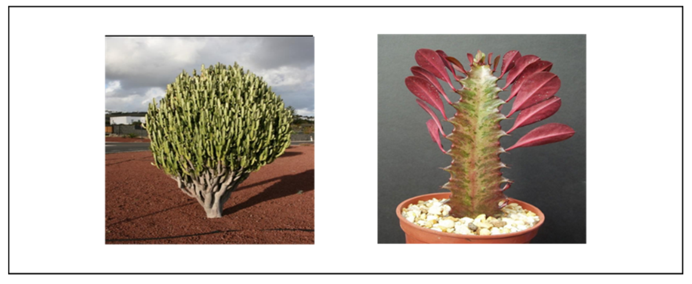
Figure 1.2.
Euphorbia ledienii plant with stem and flowers (source: https://www.agaveville.org/viewtopic.php?f=54andt=3571, http://www.llifle.com/Encyclopedia/SUCCULENTS/Family/Euphorbiaceae/14292/Euphorbia_ledienii ).
Figure 1.2.
Euphorbia ledienii plant with stem and flowers (source: https://www.agaveville.org/viewtopic.php?f=54andt=3571, http://www.llifle.com/Encyclopedia/SUCCULENTS/Family/Euphorbiaceae/14292/Euphorbia_ledienii ).
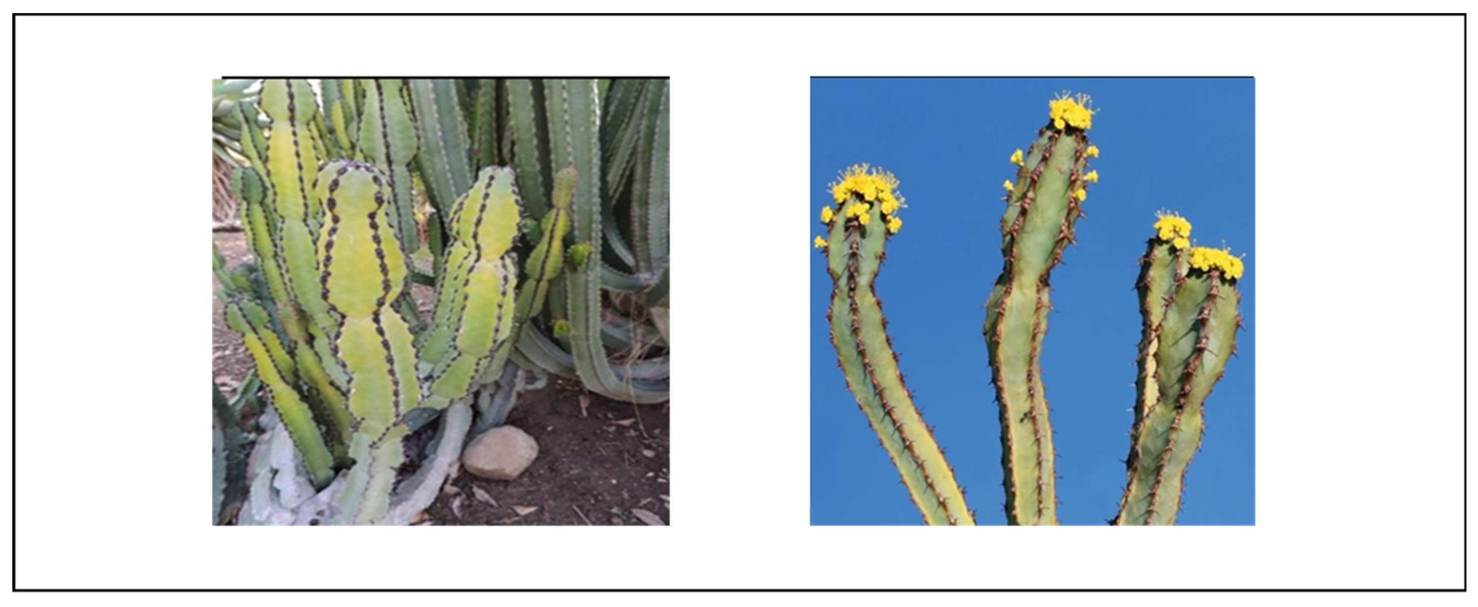
Figure 1.3.
Euphorbia horrida plant with stem and flowers (source: http://pza.sanbi.org/euphorbia-horrida).
Figure 1.3.
Euphorbia horrida plant with stem and flowers (source: http://pza.sanbi.org/euphorbia-horrida).
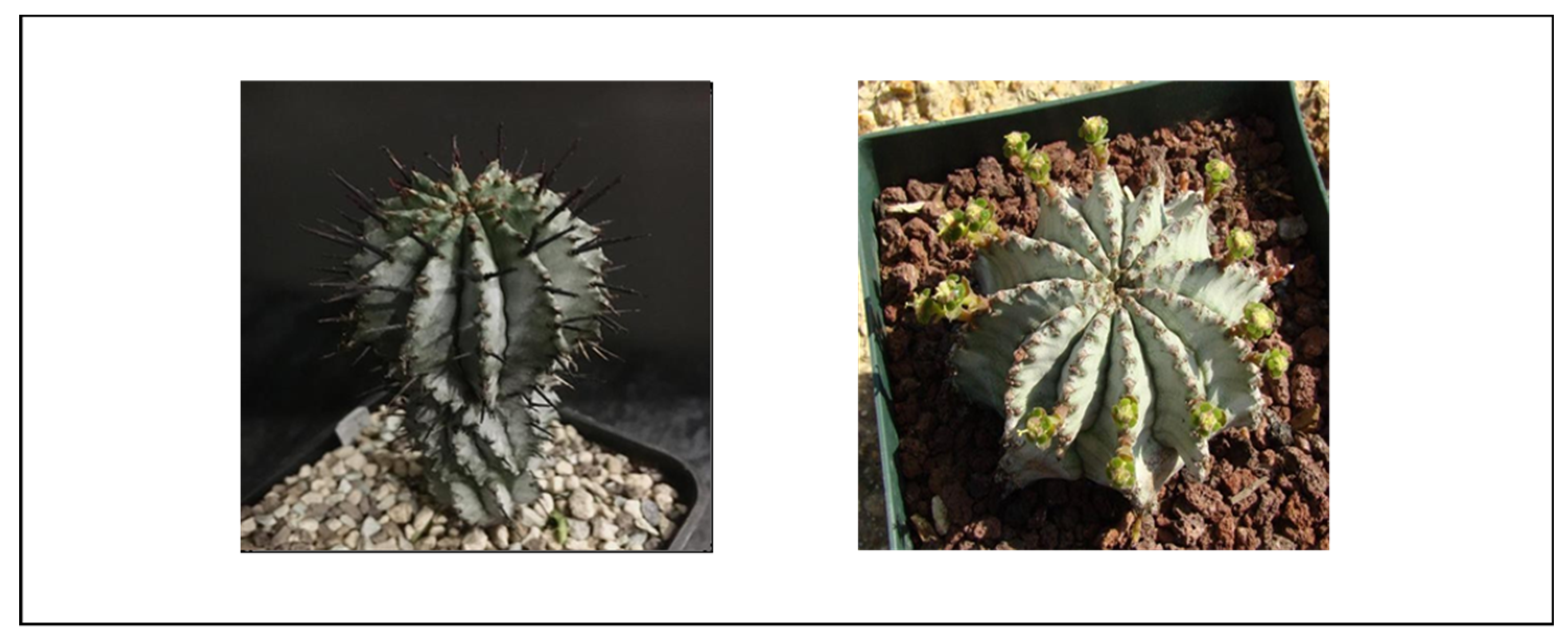
Figure 1.4.
Euphorbia enopla plant with stem and flowers (source: https://www.cactus-art.biz/schede/EUPHORBIA/Euphorbia_enopla/Euphorbia_enopla/Euphorbia_enopla.htm).
Figure 1.4.
Euphorbia enopla plant with stem and flowers (source: https://www.cactus-art.biz/schede/EUPHORBIA/Euphorbia_enopla/Euphorbia_enopla/Euphorbia_enopla.htm).
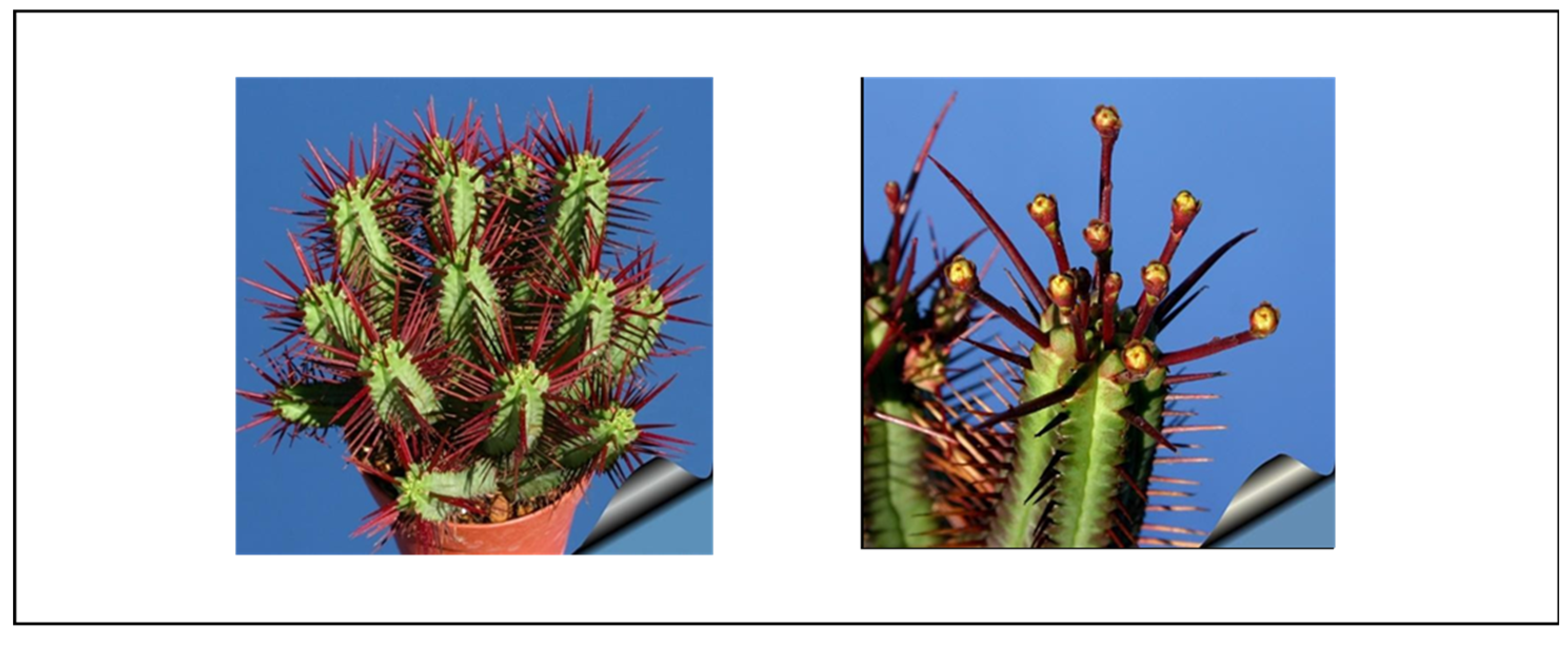
Figure 1.5.
Euphorbia coerulescences plant with stem and flowers (source: https://www.ebay.com.au/itm/euphorbia-coerulescens-POT-10 CM /233323039902?nma=true&si=DenzQanzHzMK8eHcDW6MHzA0s9s%253D&orig_cvip=true&nordt=true&rt=nc&_trksid=p2047675.l2557.
Figure 1.5.
Euphorbia coerulescences plant with stem and flowers (source: https://www.ebay.com.au/itm/euphorbia-coerulescens-POT-10 CM /233323039902?nma=true&si=DenzQanzHzMK8eHcDW6MHzA0s9s%253D&orig_cvip=true&nordt=true&rt=nc&_trksid=p2047675.l2557.
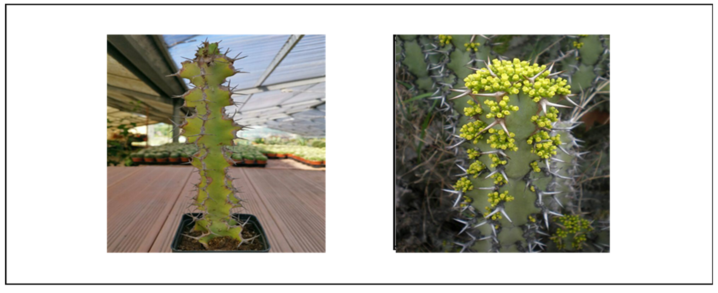
Figure 1.6.
Euphorbia cooperi tree and fruits (source: http://pza.sanbi.org/euphorbia-cooperi).
Figure 1.6.
Euphorbia cooperi tree and fruits (source: http://pza.sanbi.org/euphorbia-cooperi).
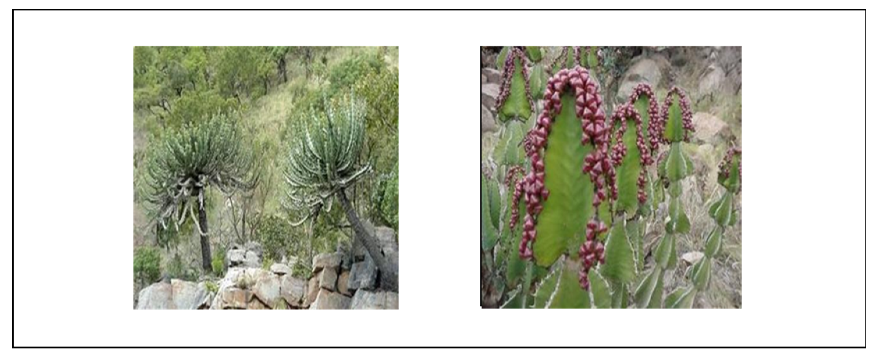
Figure 1.7.
Euphorbia tirucalli plant and flower (source: https://za.pinterest.com/pin/298785756510953587/; https://za.pinterest.com/pin/474426141998696380/).
Figure 1.7.
Euphorbia tirucalli plant and flower (source: https://za.pinterest.com/pin/298785756510953587/; https://za.pinterest.com/pin/474426141998696380/).
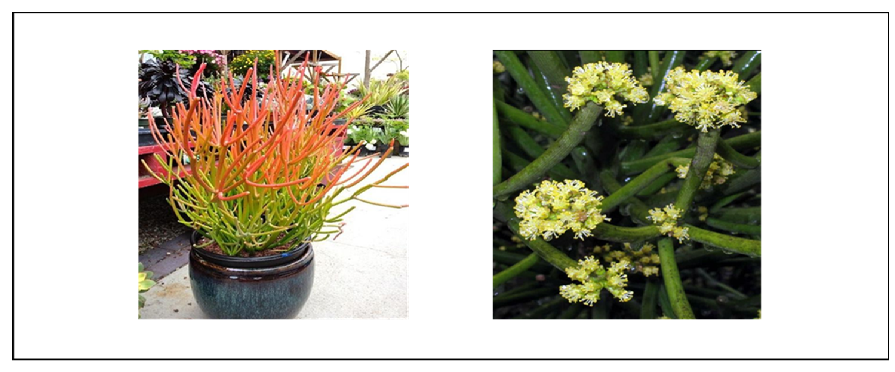
Figure 1.8.
Euphorbia ammak tree and flower (source: http://luirig.altervista.org/cpm/albums/bot-units07/euphorbia-ammak16454.jpg).
Figure 1.8.
Euphorbia ammak tree and flower (source: http://luirig.altervista.org/cpm/albums/bot-units07/euphorbia-ammak16454.jpg).
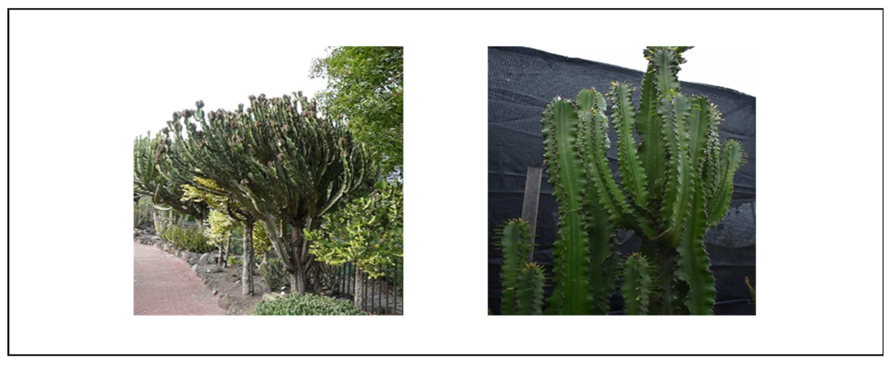
Figure 1.9.
Euphorbia clavarioides plant with flowers and stem (source: https://www.inaturalist.org/observations/38217609; http://www.llifle.com/Encyclopedia/SUCCULENTS/Family/Euphorbiaceae/32951/Euphorbia_clavarioides_var._truncata).
Figure 1.9.
Euphorbia clavarioides plant with flowers and stem (source: https://www.inaturalist.org/observations/38217609; http://www.llifle.com/Encyclopedia/SUCCULENTS/Family/Euphorbiaceae/32951/Euphorbia_clavarioides_var._truncata).
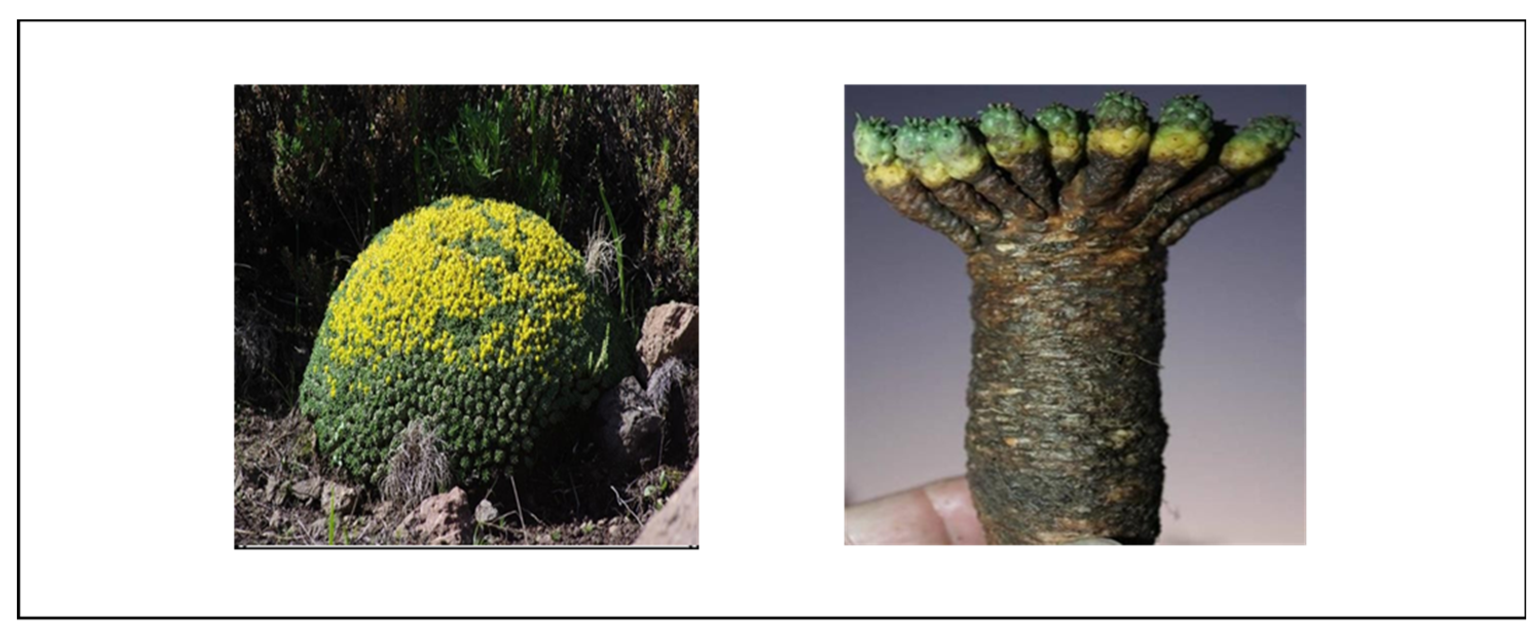
Figure 1.10.
Euphorbia gorgonis plant with flowers and stem (source: http://www.bihrmann.com/caudiciforms/SUBS/eup-gor-sub.asp).
Figure 1.10.
Euphorbia gorgonis plant with flowers and stem (source: http://www.bihrmann.com/caudiciforms/SUBS/eup-gor-sub.asp).
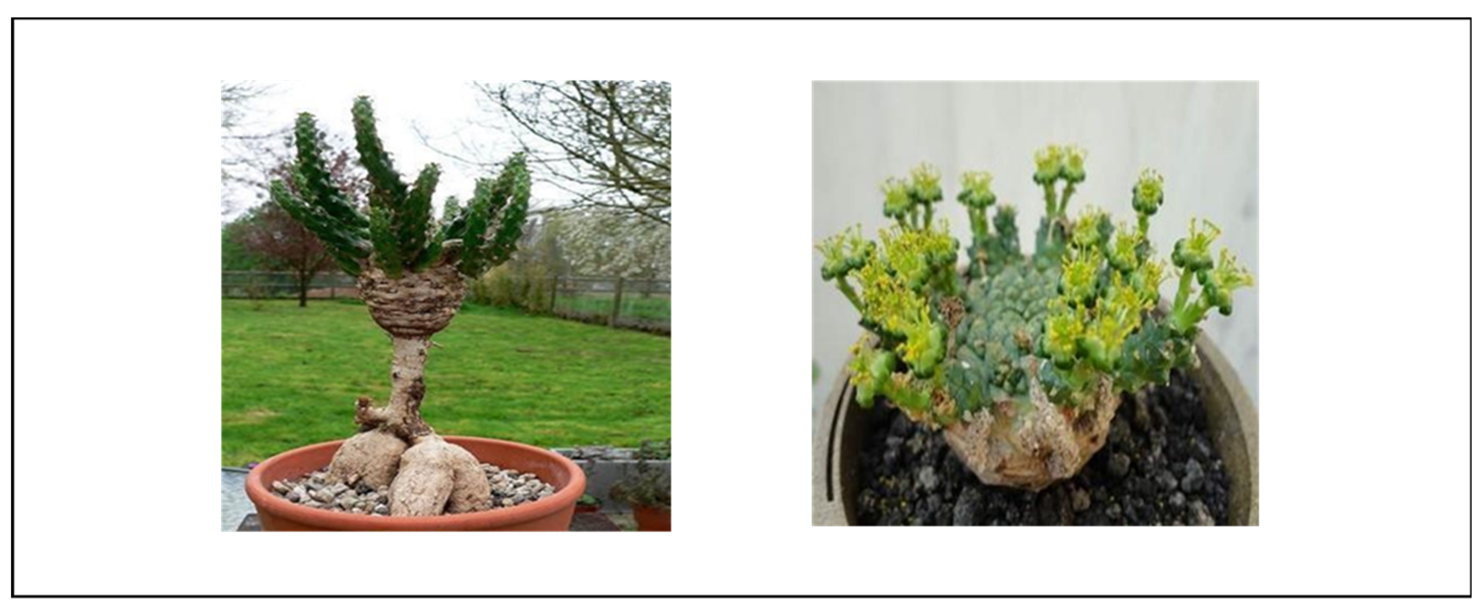
Figure 1.11.
Euphorbia bupleurifolia plant with stem and flowers (source:https://davesgarden.com/guides/pf/showimage/238493/#b; https://planetdesert.com/products/euphorbia-bupleurifolia-large).
Figure 1.11.
Euphorbia bupleurifolia plant with stem and flowers (source:https://davesgarden.com/guides/pf/showimage/238493/#b; https://planetdesert.com/products/euphorbia-bupleurifolia-large).
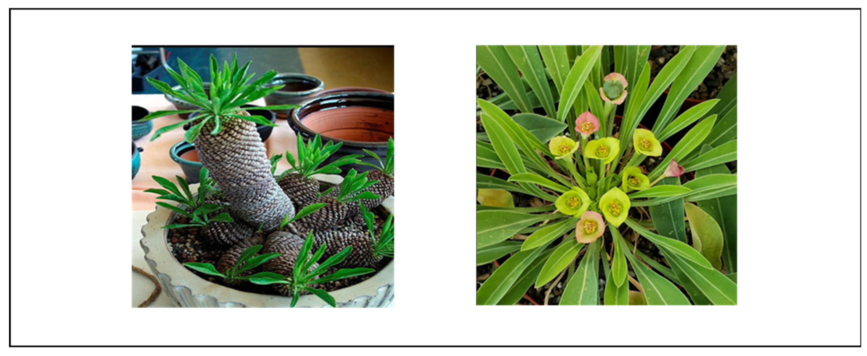
Figure 1.12.
Euphorbia polygona plant with stem and flowers (source: http://www.biodiversityexplorer.info/plants/euphorbiaceae/euphorbia_polygona.htm).
Figure 1.12.
Euphorbia polygona plant with stem and flowers (source: http://www.biodiversityexplorer.info/plants/euphorbiaceae/euphorbia_polygona.htm).
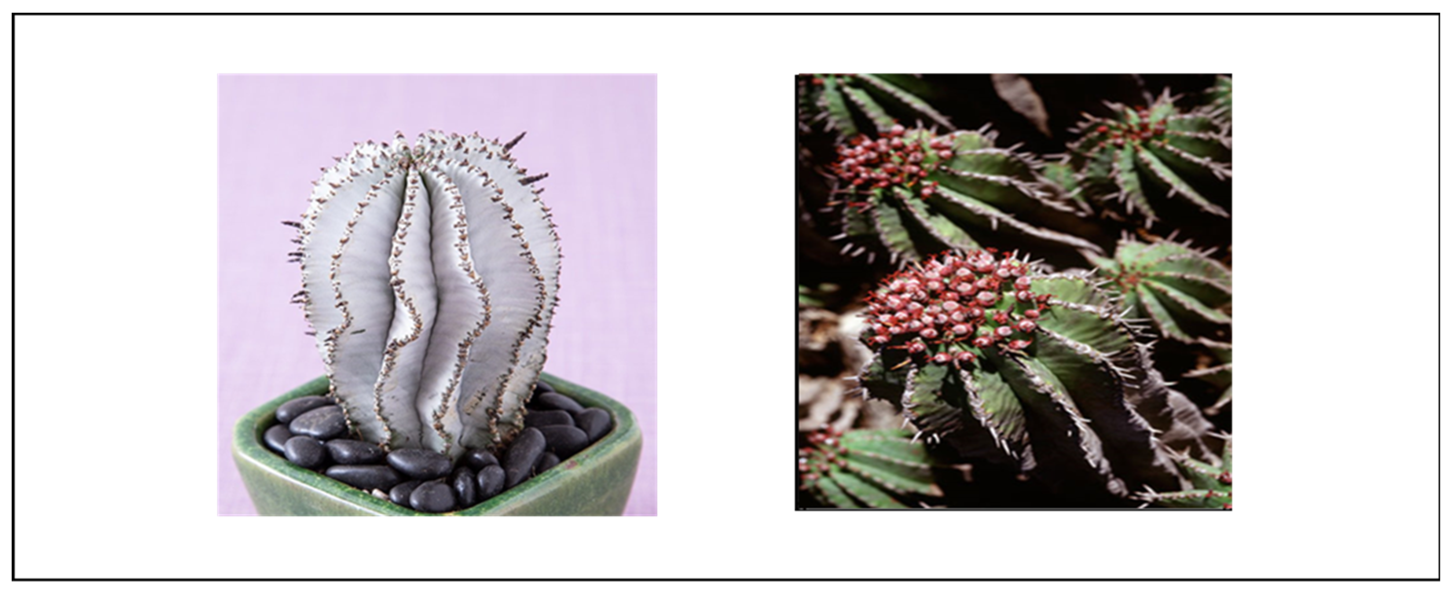
Figure 1.13.
Euphorbia arabica plant with stem and flowers (source: http://www.biodiversityexplorer.info/plants/euphorbiaceae/images/1362101_658w.jpg ,http://www.biodiversityexplorer.info/plants/euphorbiaceae/images/136210-2_658w.jpg).
Figure 1.13.
Euphorbia arabica plant with stem and flowers (source: http://www.biodiversityexplorer.info/plants/euphorbiaceae/images/1362101_658w.jpg ,http://www.biodiversityexplorer.info/plants/euphorbiaceae/images/136210-2_658w.jpg).
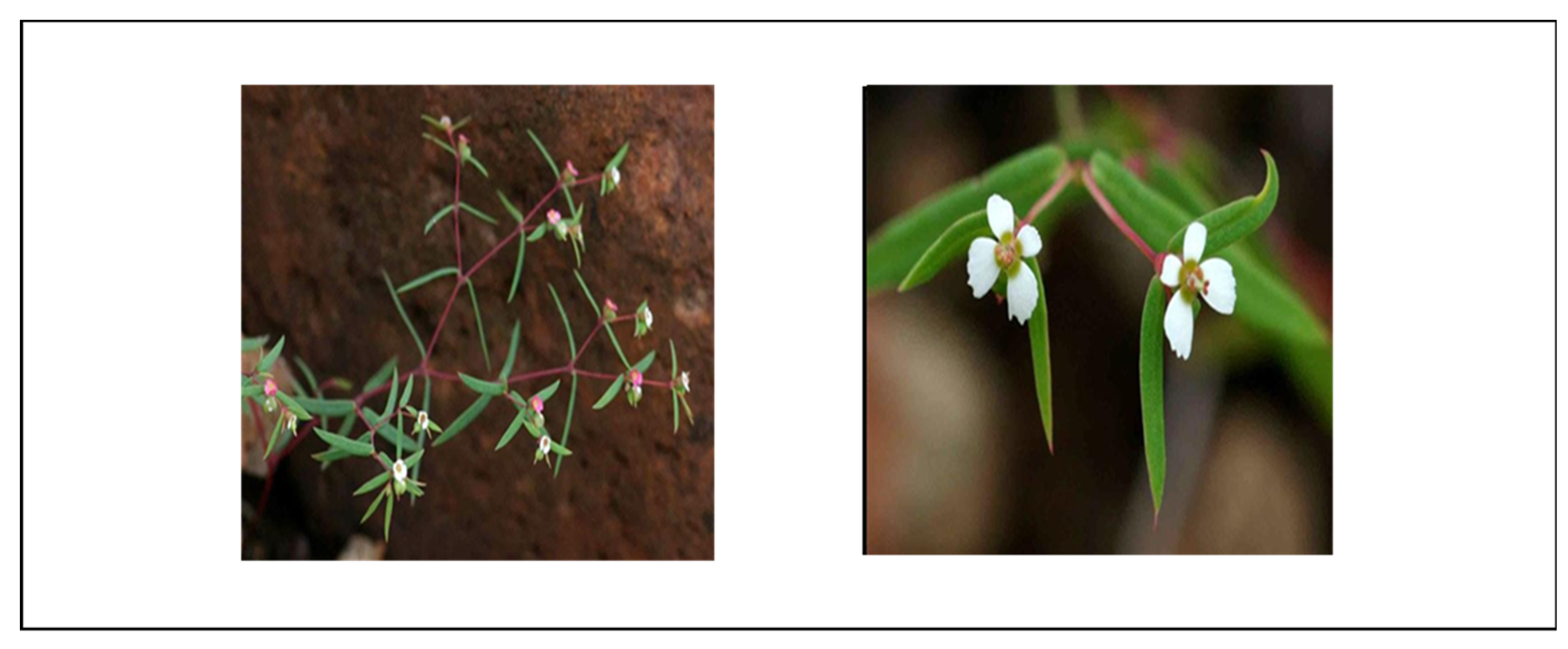
Figure 1.14.
Euphorbia ferox plant with stem and flowers (source:communities/southern-africa/view/observation/586073,http://gadi.agric.za/Grootfontein%20plants/Euphorbia%20ferox.html).
Figure 1.14.
Euphorbia ferox plant with stem and flowers (source:communities/southern-africa/view/observation/586073,http://gadi.agric.za/Grootfontein%20plants/Euphorbia%20ferox.html).
Figure 1.15.
Euphorbia stellata plant with stem and flowers (source:communities/southern-africa/view/observation/586073, http://gadi.agric.za/Grootfontein%20plants/Euphorbia%20ferox.html).
Figure 1.15.
Euphorbia stellata plant with stem and flowers (source:communities/southern-africa/view/observation/586073, http://gadi.agric.za/Grootfontein%20plants/Euphorbia%20ferox.html).
Figure 1.16.
Terpene biosynthesis in the cytosol through the mevalonate pathway. Starting with acetyl-CoA, this process produces terpene precursors (isopentenyl pyrophosphate and dimethylallyl pyrophosphate), serving as building blocks for various terpenes with diverse biological functions. The diagram was adopted from Qiao et al. (2018).
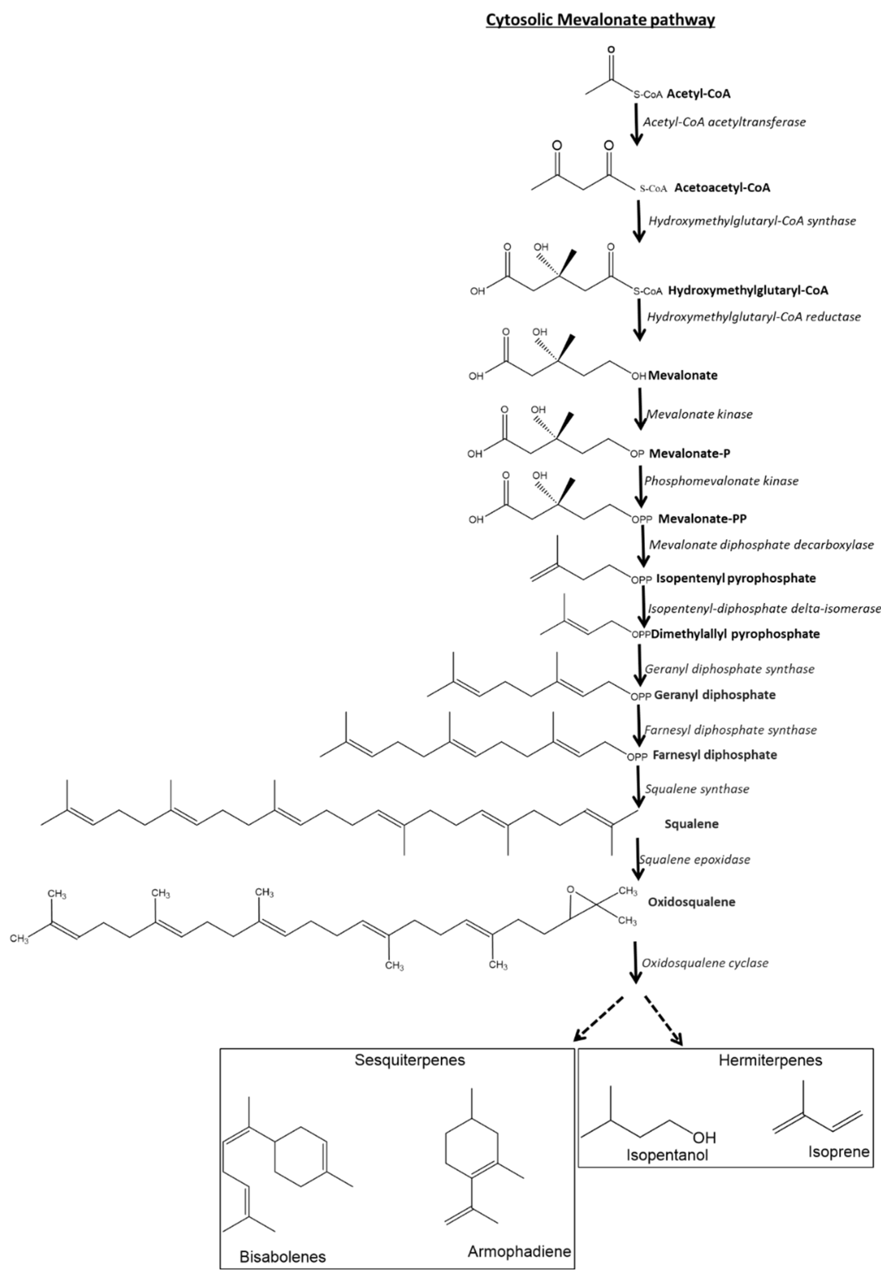

Table 1.1.
Euphorbia spp of Southern Africa and its neighboring countries, their toxicity and traditional use.
Table 1.1.
Euphorbia spp of Southern Africa and its neighboring countries, their toxicity and traditional use.
| Botanical name Synonym in bold |
Vernacular name (E)= English; (S)= Sotho (Z)= Zulu; (X)= Xhosa; (A)=Afrikaans (N)= Ndebele (T)= Tsonga (Ts)= Tswana (V)= Venda |
Location | Growing condition | Traditional use |
Bioactive property | Toxicity | Reference |
|---|---|---|---|---|---|---|---|
|
Euphorbia trigona Euphorbia hermentiana Lem |
African milk tree (E) | West Africa, tropical Asia, India | Dry tropical forests and semiarid environment. Direct sunlight | Respiratory infections, urinary tract infections, gonorrhoea, tumors and warts, intestinal parasites, rheumatoid arthritis, hepatitis, inflammation, piles, constipation, epileptic attack. | Anticancer | Skin irritation | [16,17,18,19,20,21] |
|
Euphorbia cooperi E. cooperi var. cooperi E. cooperi var calidicola |
Candelabra Euphorbia (E), Umhlonhlo (N), Tshikondengala (V), Mokhoto (T) Mohlohlo (S) | South Africa (KwaZulu-Natal, Swaziland, North-West Province, Mpumalanga and Limpopo) Mozambique, Zimbabwe, Botswana | Well-drained soil. Direct sunlight. | Sore stomach, bloatedness, paralysis, wound healing | Breast cancer inhibitor | Skin irritation, blindness, throat burning sensation | [22,23,24,25,26,27,28] |
| Euphorbia ammak | African candelabra /Desert candelabra (E) | Saudi Arabia, Yemen peninsulaα | No data reported | No data reported | Anticancer Antileishmanial activity, (H1N1) influenza virus, MDCK cells, antiparasitic activity |
Skin and eye irritation. Normal melanocyte (HFB4). |
[29,30,31,32,33] |
|
Euphorbia tirucalli Euphorbia laro Drake |
Rubber-hedge Euphorbia (E), umSululu (Z), umHlonthlo (X), motsetse/ setlharesetola (Ts), mahumbana (T), muṱungu (V) kraalmelkbos (A) | Eastern tropical Africa, South Africa, Indian Ocean Island | Rock garden, well-drained soils, mild to warm climates. | Snakebites, sexual impotence, warts, wounds, skin complaints, swollen glands, oedema, haemorrhoids, rheumatism, epilepsy toothache, earache and tumor. | Antibacterial, molluscicide, antiherpetic, antimutagenic antibacterial, myelomodulating activity, antitumor effect on different cell lines. Tumor promotor | Skin irritation, blindness, fish poisoning, gastro-intestinal haemorrhage, fatality, Burkitt’s lymphoma, Epstein-Barr virus | [5,6,34,35,36,37,38,39,40,41,42,43,44,45,46,47,48,49,50,51,52] |
|
Euphorbia clavarioides Euphorbia basutica |
lions Spoor (E), melkpol (A), iSantilele/ isihlekehleke (Z), sehlehle/sehloko (S) | South Africa (Eastern Cape, KwaZulu-Natal, Northern Cape, Western Cape), Lesotho | Direct sunlight, mineral soil , grassland gardens | Skin rash in children, acne, sores, bruises, burns, eczema, ulcer, cracked heels and wounds, cancerous sores, warts, swollen feet, leprosy remedy, herpes, HIV related infections, high blood pressure, diabetes | No data reported | Skin irritation, bird lime | [53,54,55,56,57] |
| Euphorbia gorgonis | Nkalimasane (Z) melkbol (A) | South Africa (Eastern Cape) | Well-drained soil, Direct sunlight | Wounds, swelling, skin problems. | Antibacterial, antimicrobial activity, cancer cell line (HuTu), hepatoma cells (H411E) | Skin irritation | [58,59,60] |
|
Euphorbia bupleurifolia Tithymalus bupleurifolius |
Pine cone plant (E), melkbol (A), intsele (X), insema (Z) |
South Africa (Eastern Cape Province, Transkei and Natal) | Warm and moist conditions,moderate sun light. | Cancerous sores, for painful cracked feet, eczema, pimples, rashes and wounds in topical formulation, clean teeth, swellings of the lower limbs, cancer, retained placenta. | No data reported | Skin irritation | [61,62,63,64] |
|
Euphorbia enopla Euphorbia enopla var. enopla |
Milk-barrel/Pincushion Euphorbia (E) | South Africa ( Eastern Cape in the Noorsveld, in arid to semi-arid Karoo) | Well-drained soil, direct sunlight | No data reported |
No data reported | Skin irritation, toxic to Vero cell line | [65] |
|
Euphorbia coerulescens Euphorbia virosa |
Sweetnoors (A) | South Africa (Cape province) | Sunny to half-shady, rocky and gritty-sandy soils | No data reported |
No data reported | Inflammation in mice, skin irritation, blindness, throat irritation if swollen | [66] |
| Euphorbia polygona | African Milk Barrel (E) | South Africa (Eastern Cape) | Well-drained soil, direct sunlight and high temperature environment. | No data reported | No data reported | Eye irritation, skin irritation, paralysis | [65] |
|
Euphorbia horrida Var Euphorbia horrida Boiss. |
African Milk Barrel (E) | South Africa ( Wittepoort/ Karoo) | Well-drained soil, direct sunlight and dry conditions. | No data reported | No data reported | Inhibition of Vero cell line | [65] |
|
Euphorbia arabica Euphorbia neopolycnemoides |
Klein Bont Euphorbia (E), umhlonhlo (Z) | Botswana, southern Mozambique, Zimbabwe, Limpopo, Mpumalanga and KwaZulu-Natal, South Africa | Stony grassland area. | Warts, stomach ache, skin infections. | Antibacterial | Inhibition of Vero cell line | [67,68] |
|
Euphorbia ledienii Euphorbia ledienii A. Berger var. ledienii |
Crested Euphorbia (E) | South Africa (Western Cape) | Well-drained soil, sunny conditions | No data reported |
No data reported | Skin irritation | [69] |
|
Euphorbia ferox Euphorbia capitosa |
Milkweed (E) | South Africa (Western Cape) | Drained sandy soil, rocky outcrops and in sunny environment | No data reported | No data reported | Poisonous latex | |
|
Euphorbia stellata Euphorbiasquarrosa Euphorbia radiata Euphorbia scolopendrea Euphorbia uncinata |
Spurge (E) | South Africa (Eastern Cape) | Well-drained soil and sunny environment | No data reported | No data reported | Skin and eye irritation. |
Table 1.2.
Comprehensive overview of Isolated Compounds from Individual Plants.
| Plant | Compounds | Reference |
|---|---|---|
| Euphorbia trigona | Euphol , Cycloartenol, Cycloartanol, Lupeol, α amyrin, β –amyrin, Betulinic acid, Taraxerol , β-sitosterol, Taraxerol acetate, Friedelin, Friedelan 3 α - and 3 βα –ols, 24-ethylene cycloartanol, Epi-friedelinyl acetate, 3β, friedelinol, Rhoiptlenone | [70,71] |
| Euphorbia ledienii | Isobutyric, 2-Methylbutyric acid, 12-Deoxyphorbol-13-isobutyrate-20-acetate, 12-Deoxyphorbol-13-(2-methylbutyrate)-20-acetate, Phorbol, 12-Deoxy phorbol, 12-Deoxy-16-hydroxy phorbol, Ingol-7,8,12-acetate,ditiglate | [72,73,74] |
| Euphorbia tirucalli |
Triterpenes, Euphol, Diterpene esters, Euphorbiane, 12-Deoxyphorbol esters, Ingenol ,β-Sitosterol, Euphorbol, hexacosonate, 12-Deoxy-4β-hydroxyphorbol-13-phenyl acetate-20-acetate, 12, 20-Dideoxyphorbol-13-isobutyrate, Glut-5-en-3-β-ol, Tirucalicine, Tri-methyl ellagic acid, Terpenic alcohol, Isoeuphorol, Taraxasterol, Tirucallol, Ketone euphorone, Resin, Ellagic acid, Taraxerol, 3,3′-Di-O-methylellagic acid, Euphorbin A, Euphorbin B, Tirucallin A, Tirucallin B, Euphorbol, Cycloartenol, 24-Methylenecycloartenol, Ingenol triacetate, β-amyrin, Rhoiptlenone, 3β-friedelinol, Epi-friedelinyl acetate, 24-ethylene cycloartanol, Friedelan 3 α - and 3 βα –ols, Friedelin, Taraxerol acetate, Betulinic acid, β –amyrin, α amyrin, Lupeol, Cycloartanol, β-amyrone, glutinone, taraxerone, Glut-5-en-3-β-ol and cycloart-23-ene-3-β, 25-diol, Euphorcinol, Euphorginol, 12, 13,20-tri- O-acetylphorbol, 3,5,20-tri-O-acetlingenol 20-acetate | [8,9,21,35,36,75,76,77,78,79,80,81,82,83,84,85,86,87,88,89,90,91,92] |
| Euphorbia enopla |
Euphol, Tirucallol | [93] |
| Euphorbia coerulescens | Angelate acetate isobutyrate, Fatty acids, Acetate laurate, α-Methyl butyrate, Heptanoate, Laurate, Euphol, Tirucallol, Euphorbol | [66,89] |
| Euphorbia ammak | α-glutinol, Stigmasterol, Euphol, Euphorbol | [93,94] |
| Euphorbia cooperi | 16-Hydroxy-12-desoxyphorbol, Euphol, Obtusifoliol, 12-Deoxyphorbol-13-isobutyrate-16-angelate-20-acetate, Euphorbilactone, Norsesquiterpenoid, Arachiside A, Glutinol, 16-Angeloyloxy-13α-isobutanoyloxy-4β,9α,20- trihydroxytiglia-1,5-diene-3,7-dione, 20-Acetoxy-16-angeloyloxy13α-isobutanoyloxy-4β,9α,20-tetrahydroxytiglia-1,5-diene-3-one, Gallic acid, Bervifolin, carboxylic acid, Kampferol-3-O-ß-D-rutinoside, 1-O-Galloyl-3,6-hexahydroxydiphenyl-β-D glucopyranoside, 3, 3′ Dimethoxy ellagic acid 3, 4, 4′ Trimethoxyellagic acid, Ellagic acid, Kaempferol, 7-galloyl catechin, kaempferol 3-O-β-(6”- O-galloyl)-glucopyranoside, triesters-16-hydroxy-12-desoxy-phorbol | [9,24,95,96,97] |
| Euphorbia horrida | 17-Hydroxyingenol-17-benzoate-20-angelate, Diterpene esters | [98] |
Table 1.3.
Phytochemicals extracted from the selected Euphorbia spp. This table outlines the diverse array of crude phytochemicals extracted from carefully chosen Euphorbia species, shedding light on the rich chemical composition inherent in these selected plants.
Table 1.3.
Phytochemicals extracted from the selected Euphorbia spp. This table outlines the diverse array of crude phytochemicals extracted from carefully chosen Euphorbia species, shedding light on the rich chemical composition inherent in these selected plants.
| Euphorbia trigona | Saponins, alkaloids, flavonoids, glycosides, sterols and triterpernoids, tannins | [17,71,99] |
| Euphorbia cooperi | Triterpernoid | [95] |
| Euphorbia ammak | alkaloids, saponins, glycosides | [31] |
| Euphorbia tirucalli | Triterpernoid, phenols, flavonoids, tannins, alkaloids, saponins, glycosides, and steroids. | [37,100] |
| Euphorbia clavarioides | Alkaloids, flavonoids, saponins, tannins, terpenoids, phytosterols, glycosides, triterpenoids, anthraquinone | [65,101] |
| Euphorbia gorgonis | Phytosterols, glycosides, triterpenoids, flavonoids, alkaloids, saponins | [60,65] |
| Euphorbia bupleurifolia | Phytosterols, tannins, glycosides, triterpernoids, saponins, flavonoids and alkaloids | [62,65] |
| Euphorbia enopla | Phytosterols, glycosides, triterpenoids, flavonoids, alkaloids, tanins, anthraquinone | [65] |
| Euphorbia polygona | Phytosterols, tannins, glycoside, triterpenoids, flavonoids, alkaloids | [65] |
| Euphorbia horrida Var | phytosterols, pentose, tannins, glycosides, triterpenoids, anthraquinones, saponins, flavonoids | [65] |
| Euphorbia arabica | Phytosterols, tannins, glycosides, triterpenoids, anthraquinones, flavonoids | [65] |
| Euphorbia ledienii | No data reported | |
| Euphorbia ferox | No data reported | |
| Euphorbia stellata | No data reported | |
| Euphorbia coerulescens | No data reported |
Table 1.4.
Classification of Crude and Isolated Compounds from 15 Euphorbia Specie. This table delineates the categorization of both crude extracts and individually isolated compounds obtained from 15 distinct Euphorbia species. The compounds are organized based on their respective classes, providing a comprehensive overview of the chemical constituents present in each species.
| Compounds | Class | subclass |
|---|---|---|
| Euphol (Tetracyclic Triterpene), Cycloartanol (Sterol Lipid), Lupeol, α-amyrin, β-amyrin, Betulinic acid, Taraxerol, Taraxerol acetate, Friedelin, Friedelan-3-β–ol, 3β-Friedelinol | Terpenoids | Triterpenoids |
| Cycloartenol, β-sitosterol, 24-Methylenecycloartanol, Tirucallol (Tetracyclic Triterpene), Obtusifoliol, Glutinol, Triterpene euphol (Steroidal Alcohol), 24-Methylene cycloartenol, Ingenol triacetate (Diterpene), Terpenic alcohol (Terpene), Taraxasterol (Phytosterol), Stigmasterol | Phytosterol | |
| 12-Deoxyphorbol-13-isobutyrate-20-acetate (Phorbol Ester), Phorbol (Diterpenoid), 12-Deoxyphorbol ester (Diterpenoid), 12-Deoxy-16-hydroxy-phorbol (Component of DHPB), 16-Hydroxy-12-desoxyphorbol (Diterpenoid), 12-Deoxyphorbol-13-isobutyrate-16-angelate-20-acetate (Diterpene), 12-Deoxy phorbol esters (Phorbol Ester), Ingenol (Diterpenoid), Glut-5-en-3-β-ol (Triterpenoid) | Other Terpenoids | |
| Gallic acid (Phenolic Acid), Bervifolin carboxylic acid (Tannin), 3, 3′-Dimethoxy ellagic acid (Tannin), Ellagic acid (Tannin), Kaempferol (Flavonoid), Kampferol-3-O-ß-D-rutinoside (Flavonoid), 1-O-Galloyl-3,6-hexahydroxydiphenyl-β-D-glucopyranoside/corilagin (Tannin), Tri-methyl ellagic acid (Tannin), 3′-Di-O-methylellagic acid (Phenol) | Phenolic | |
| Laurate | Fatty acids | |
| Diterpene esters | Miscellaneous | |
| Flavonoids | Polyphenolic compounds | |
| Alkaloids | Organic compounds | |
| Saponins | glycosides | |
| Tannins | Polyphenols | |
| Glycosides | Acetal Derivatives of Monosaccharides | |
| Anthraquinones | Phenolics |
Table 1.5.
Bioactive compounds isolated from 15 Euphorbia species alongside their biological activities and subclasses. This table provides an in-depth exploration of bioactive compounds isolated from 15 distinct Euphorbia species, highlighting their diverse biological activities.
Table 1.5.
Bioactive compounds isolated from 15 Euphorbia species alongside their biological activities and subclasses. This table provides an in-depth exploration of bioactive compounds isolated from 15 distinct Euphorbia species, highlighting their diverse biological activities.
| Compounds | Subclasses | Pharmacological/ biological activity | Reference |
|---|---|---|---|
| Euphol | Euphane | Anticancer Cytotoxicity Anti-nociceptive Antiinflammatory HIV-1 reverse transcriptase inhibitor |
[76,102,103] |
| Cycloartenol | Cycloartane | Antiinflammatory Antitumor Antioxidant Antibiosis Anti-alzheimer’s disease Apoptotic Analgesic Bactericidal |
[106,107,108,109] |
| Cycloartanol | Cycloartane | Antifungal Vasodepressor Antitumor |
[104,105,110] |
| Lupeol | Lupane | Anticancer Antiinflammatory Antimicrobial Antiprotozoal Antiproliferative Antiangiogenic Anti-invasive Cholesterol lowering |
[40,111,112,113,114,115,116,117] |
| α amyrin | Oleanane | Cytotoxicity Antifungal Antiinflammatory |
[120,122,123,124] |
| β -amyrin | Oleanane | Antiinflammatory Nitric oxide inhibitor Reactive oxygen species activator Anticancer |
[118,119,121,122,125] |
| Betulinic acid | Pentacyclic triterpenes | Antitumor Antidiabetic Antiinflammatory HIV-1 reverse transcriptase inhibitor Antiviral Hepatoprotective activity |
[126,127,128,129,130,131,132,133,134,135] |
| Taraxerol | Taraxarane | Anticancer Antiinflammatory Apoptotic Anti-oxidative activity Antimicrobial Antifungal Antidiabetic |
[138,139,140,141] |
| β-sitosterol | Sterols | Antiinflammatory Anticancer Antiproliferation Analgesic Antimicrobial |
[125,142,143,144,145] |
| Taraxerol acetate | Taraxarane | Antiinflammatory Cycloxygenases inhibitor |
[136,137] |
| Friedelin | Friedelane | Cytotoxicity Antibacterial |
[153,154] |
| Friedelan 3 α - and 3 βα -ols | Friedelane | Anticancer | [154] |
| 24-ethylene cycloartanol | Cycloartane | Antiinflammatory Antifungal |
[125,155] |
| Epi-friedelinyl acetate | Friedelane | [156] | |
| 3β-friedelinol | Friedelane | Antibacterial Cytotoxicity |
[156,157,158,159] |
| Rhoiptlenone | No data reported | No data reported | [156] |
| Isobutyric | Carboxylic acid | irritant | [73,160] |
| 2-Methylbutyric acid | No data reported | No data reported | [73] |
| 12-Deoxyphorbol-13-isobutyrate-20-acetate | Phorbol | Antifungal | [161] |
| 12-Deoxyphorbol-13-(2-methylbutyrate)-20-acetate | Phorbol | No data reported | [72] |
| Phorbol | Phorbol | Tumor promoter Apoptosis |
[7,162] |
| 12-Deoxy phorbol | Phorbol | Antitumor Apoptotic |
[163,164,165,166] |
| 12-Deoxy-16-hydroxy phorbol | Phorbol | Irritant Tumor promoter |
[167] |
| Ingol-7,8,12-acetate,ditiglate | Ingenane | No data reported | [160] |
| Triterpenes euphol | Glycosides | Anticancer Antiinflammatory |
[52,88,168] |
| Diterpene esters | Terpenoids | Anticancer Cytotoxicity Tumor promoter Irritant Pro-inflammatory |
[7,80,169,170,171] |
| 12 Deoxyphorbol esters | Phorbol | Irritant Pro-inflammatory Tumor promoter |
[172,173,174,175] |
| Ingenol | Ingenane | Cytotoxicity HIV- reverse transcriptase Inhibitor |
[102,152] |
| Euphorbol hexacosonate | No data reported | No data reported | [148] |
| 12-Deoxy-4β-hydroxyphorbol-13-phenyl acetate-20-acetate | No data reported | No data reported | [148] |
| 12, 20-Dideoxyphorbol-13-isobutyrate | Phorbol | No data reported | [148] |
| Glut-5-en-3-β-ol | Pentacyclic triterpene | Antibacterial | [176] |
| Tirucalicine | Tirucallane | No data reported | [81,148,177] |
| Tri-methyl ellagic acid | Ellagic Acid Derivatives |
Anticancer | [148,150,151] |
| Terpenic alcohol | Steroidal Triterpenoids |
Antibacterial Irritant |
[148,149] |
| Isoeuphorol | Ingenane-type Triterpenoids | No data reported | [148] |
| Taraxasterol | Sterol |
Inhibition of tumor promotion Antiproliferation |
[178] |
| Tirucallol | Tirucallane | Antiinflammatory HIV-Inhibition reverse transcriptase |
[146,147] |
| Ketone euphorone | Euphane-type Triterpenoids | No data reported | [179] |
| Resin | No data reported | Digestive enzyme Antioxidant Antispasmodic Hypotensive Hepatoprotective Antiviral Antifungal Anticancer Anxiolytics Anthelmintic |
[180] |
| Ellagic acid | Ellagic Acid Derivatives | Hepatoprotective activity Antiproliferative activity Antioxidant |
[181] |
| 3,3′-Di-O-methylellagic acid | Ellagic Acid Derivatives | Antioxidant Moderate antibacterial activity Antimicrobial activity Anticancer |
[182,183,184,185] |
| Euphorbin A | No data reported | No data reported | [186] |
| Euphorbin B | No data reported | No data reported | [186] |
| Tirucallin A | Tirucallane | No data reported | [79] |
| Tirucallin B | Tirucallane | No data reported | [79] |
| Euphorbol | Euphane-type Triterpenoids | Antibacterial Antiinflammatory |
[94,187] |
| 24-Methylenecycloartenol | Cycloartane | Anti-oxidant Antiinflammatory |
[188,189] |
| Ingenol triacetate | Ingenane-type Triterpenoids | Antimicrobial Antitumor |
[190,191] |
| Angelate acetate isobutyrate | Ingenane-type Triterpenoids | No data reported | [66] |
| Fatty acids | No data reported | No data reported | [66] |
| Acetate laurate | No data reported | No data reported | [66] |
| α-Methyl butyrate | No data reported | No data reported | [66] |
| Heptanoate | No data reported | No data reported | [66] |
| Laurate | Medium-chain fatty acids | Antibacterial | [66,192] |
| α-glutinol | Pentacyclic triterpenes | Ant-proliferation Cytotoxicity |
[94,193,194] |
| Stigmasterol | Phytosterol | Cytotoxicity Antioxidant Hypoglycemic Thyroid inhibitor |
[94,195] |
| 16-Hydroxy-12-desoxyphorbol | Phorbol | Antitumor Tumor promoter |
[97,196] |
| Obtusifoliol | Lanostane | Cytotoxicity | [24,95,197,198] |
| 12-Deoxyphorbol-13-isobutyrate-16-angelate-20-acetate | Phorbol-type diterpenoid | Cytotoxicity | [24,95,197] |
| Euphorbilactone | Ingenane-type Triterpenoids | No data reported | [95] |
| Norsesquiterpenoid | No data reported | No data reported | [95] |
| Arachiside A | Ingenane-type Triterpenoids | No data reported | [95] |
| Glutinol | Pentacyclic triterpenes | Antiproliferation | [193] |
| 16-Angeloyloxy-13α-isobutanoyloxy-4β,9α,20- trihydroxytiglia-1,5-diene-3,7-dione | No data reported | No data reported | [95] |
| 20-Acetoxy-16-angeloyloxy13α-isobutanoyloxy-4β,9α,20-tetrahydroxytiglia-1,5-diene-3-one | No data reported | No data reported | [95] |
| Gallic acid | Gallic Acid Derivatives |
Hepatoprotective activity Antioxidant |
[181] |
| Bervifolin carboxylic acid | Gallic Acid Derivatives |
Hepatoprotective activity Antioxidant |
[181] |
| Kampferol-3-O-ß-D-rutinoside | Flavonoids | Hepatoprotective activity Antioxidant |
[181] |
| 1-O-Galloyl-3,6-hexahydroxydiphenyl-β-D-glucopyranoside | Gallic Acid Derivatives |
Antitumor Antiinflammation Antioxidant Hepatoprotective activity Antimicrobial Antihypertensive Antidiabetic Anti-HIV Antifungal |
[197,199,200,201,202,203,204,205,206] |
| 3, 3′ Dimethoxy ellagic acid | Ellagic Acid Derivatives |
Hepatoprotective activity Antioxidant |
[181] |
| 3, 4, 4′ Trimethoxyellagic acid | Ellagic Acid Derivatives |
Hepatoprotective activity Antioxidant |
[181] |
| Kaempferol | Flavonoids | Hepatoprotective activity Antiproliferative activity Antioxidant |
[197] |
| 17-Hydroxyingenol-17-benzoate-20-angelate | Flavonoids | No data reported | [98] |
Table 1.6.
Type of toxicity caused by some Euphorbia species.
| Type of toxicity | Euphorbia species | References |
|---|---|---|
| Fish poisoning | Euphorbia scheffleri Pax, Euphorbia tirucalli L., and Euphorbia inaequilatera Sond | [208] |
| Human poisoning | Euphorbia ledienii A. Berger, Euphorbia heterophylla L., Euphorbia cooperi N.E.Br. ex A. Berger, Euphorbia candelabrum Kotschy, Euphorbia virosa Willd., Euphorbia poissonii Pax, Euphorbia unispina N.E.Br. and Euphorbia venenifica Tremaux ex Kotschy; | [208] |
| Domestic animals poisoning | Euphorbia caput-medusae L., Euphorbia silenifolia (Haworth) Sweet, Euphorbia ingens E. Mey. Ex Boiss; as well as irritating ones: E. tirucalli, Euphorbia poissonii, Euphorbia unispina and E. venenifica. I | [208] |
| Carcinogen/ promotor of cell division | E. tirucalli, Euphorbia leuconeura, J. curcas | [167,209,210] |
| Conjunctivitis | E. tirucalli and Euphorbia royleana | [39,211] |
Table 1.7.
Compounds with high cytotoxic probability against 3 or more cancer cell lines.
| Compounds | Pa | Pi | Cancer cell line | Cell line full name | Tissue | Tumor type |
|---|---|---|---|---|---|---|
| Euphol (C1) | 0.591 | 0.002 | MKN- 7 | Gastric carcinoma | Stomach | Carcinoma |
| 0.555 | 0.014 | UO-31 | Renal carcinoma | Kidney | Carcinoma | |
| 0.554 | 0.014 | SK-MEL-2 | Melanoma | Skin | Melanoma | |
| Cycloartenol (C2) | 0.783 | 0.001 | MKN-7 | Gastric carcinoma | Stomach | Carcinoma |
| 0.577 | 0.015 | HepG2 | Hepatoblastoma | Liver | Hepatoblastoma | |
| 0.523 | 0.029 | DMS-114 | Lung carcinoma | Lung | Carcinoma | |
| 0.501 | 0.017 | U-251 | Glioma | Brain | Glioma | |
| Lupeol (C3) | 0.785 | 0.003 | 8505C | Thyroid-gland-undifferentiated carcinoma | Thyroid | Carcinoma |
| 0.62 | 0.001 | FaDu | Hypopharyngeal squamous carcinoma | Upper aerodigestive tract | Carcinoma | |
| 0.563 | 0.013 | SK-MEL- 2 | Melanoma | Skin | Melanoma | |
| 0.539 | 0.004 | DLD-1 | Colon adenocarcinoma | Colon | Adenocarcinoma | |
| 0.526 | 0.005 | SW480 | Colon adenocarcinoma | Colon | Adenocarcinoma | |
| Alpha-amyrin (C4) | 0.52 | 0.005 | PANC-1 | Pancreatic carcinoma | Pancreas | Carcinoma |
| 0.632 | 0.004 | 8505C | Thyroid-gland-undifferentiated (anaplastic) carcinoma | Thyroid | Carcinoma | |
| 0.523 | 0.003 | MKN-7 | Gastric carcinoma | Stomach | Carcinoma | |
| Betulinic acid (C5) | 0.695 | 0.003 | 8505C | Thyroid-gland-undifferentiated (anaplastic) carcinoma | Thyroid | Carcinoma |
| 0.667 | 0.001 | FaDu | Hypopharyngeal-squamous carcinoma | Upper aerodigestive tract | Carcinoma | |
| 0.562 | 0.013 | SK-MEL-2 | Melanoma | Skin | Melanoma | |
| 24-Methylene-cycloartanol (C6) | 0.555 | 0.003 | MKN-7 | Gastric carcinoma | Stomach | Carcinoma |
| 0.539 | 0.023 | HL-60 | Promyeloblast leukemia | Haematopoietic and lymphoid tissue | Leukemia | |
| 0.52 | 0.031 | DMS-114 | Lung carcinoma | Lung | Carcinoma | |
| 0.505 | 0.026 | HL-60 | Promyeloblast leukemia | Haematopoietic and lymphoid tissue | Leukemia | |
| 12-Deoxyphorbol-13-tiglate-20-acetate (C7) | 0.638 | 0.033 | A549 | Lung carcinoma | Lung | Carcinoma |
| 0.568 | 0.024 | NCI-H838 | Non-small cell lung cancer. 3 stage | Lung | Carcinoma | |
| 0.556 | 0.021 | HL-60 | Promyeloblast leukemia | Haematopoietic and lymphoid tissue | Leukemia | |
| Ingol-7,8,12-acetate,ditiglate (C8) | 0.663 | 0.012 | NCI-H838 | Non-small cell lung cancer. 3 stage | Lung | Carcinoma |
| 0.592 | 0.005 | SK-MEL-1 | Metastatic melanoma | Skin | Melanoma | |
| 0.513 | 0.033 | DMS-114 | Lung carcinoma | Lung | Carcinoma | |
| Diterpene glycoside (C9) | 0.682 | 0.008 | SK-MEL-2 | Melanoma | Skin | Melanoma |
| 0.614 | 0.015 | HL-60 | Promyeloblast leukemia | Haematopoietic and lymphoid tissue | Leukemia | |
| 0.534 | 0.034 | NCI-H838 | Non-small cell lung cancer. 3 stage | Lung | Carcinoma | |
| Tirucallol (C10) | 0.591 | 0.002 | MKN-7 | Gastric carcinoma | Stomach | Carcinoma |
| 0.555 | 0.014 | UO-31 | Renal carcinoma | Kidney | Carcinoma | |
| 0.554 | 0.014 | SK-MEL-2 | Melanoma | Skin | Melanoma | |
| Isobutyl angelate (C11) | 0.616 | 0.012 | HepG2 | Hepatoblastoma | Liver | Hepatoblastoma |
| 0.545 | 0.03 | NCI-H838 | Non-small cell lung cancer. 3 stage | Lung | Carcinoma | |
| 0.544 | 0.052 | A549 | Lung carcinoma | Lung | Carcinoma | |
| Kaempferol-3-O-rutinoside (C12) | 0.546 | 0.004 | Caco-2 | Colon adenocarcinoma | Colon | Adenocarcinoma |
| 0.548 | 0.016 | SK-MEL-1 | Metastatic melanoma | Skin | Melanoma | |
| 0.55 | 0.022 | HL-60 | Promyeloblast leukemia | Haematopoietic and lymphoid tissue | Leukemia | |
| 0.542 | 0.031 | NCI-H838 | Non-small cell lung cancer. 3 stage | Lung | Carcinoma |
* Pa - Probability to be Active, Pi - Probability to be Inactive.
Table 1.8.
Physicochemical parameters and solubility tendency.
| Descriptors | Euphol | Cycloartenol | Lupeol | α-amyrin | Betulinic acid | 24-Methylene-cycloartanol | |
|---|---|---|---|---|---|---|---|
| Physiochemical characteristic | |||||||
| Molecular weight (g/mol) | 426.72 | 426.72 | 426.7 2 | 426.72 | 456.70 | 440.74 | |
| Num.rotatable bonds | 4 | 4 | 1 | 0 | 2 | 5 | |
| H-bond acceptors | 1 | 1 | 1 | 1 | 1 | 1 | |
| H-bond donors | 1 | 1 | 1 | 1 | 2 | 1 | |
| Molar Refractivity | 137.04 | 135.14 | 135.1 4 | 135.14 | 136.91 | 139.95 | |
| TPS | 20.23 | 20.23 | 20.23 | 20.23 | 57.53 | 20.23 | |
| Solubility (Log Po/w) | 5.09 | 5.17 | 4.68 | 4.77 | 3.79 | 5.20 | |
| Solubility class | Poorly soluble | Poorly soluble | Poorly soluble | Poorly soluble | Poorly soluble | Poorly soluble | |
| Drug-likeness | |||||||
| Lipinski | Yes | Yes | Yes | Yes | Yes | Yes | |
| Bioavailabilitv | 0.55 | 0.55 | 0.55 | 0.55 | 0.85 | 0.55 | |
| PAINS | 0 alert | 0 alert | 0 alert | 0 alert | 0 alert | 0 alert | |
| Brenk | 1 alert | 1 alert | 1 alert | 1 alert | 1 alert | 1 alert | |
| Synthetic accessibility | Yes | Yes | Yes | Yes | Yes | Yes | |
| Absorption | |||||||
| Caco2 permeability(log Papp in 10-6 cm/s) | 1.203 | 1.194 | 1.226 | 1.227-J | 1.175 | 1.221 | |
| Human intestinal absorption (% Absorbed) | 93.119 | 95.248 | 95.782 | 94.062 | 99.763 | 95.309 | |
| P-glycoprotein SUbstrate | Neg. | Neg. | Neg. | Neg. | Neg. | Neg. | |
| P-glycoprotein Iinhibitor | Pos. | Pos. | Pos. | Pos. | Neg. | Neg. | |
| P-glycoprotein II inhibitor | Pos. | Pos. | Pos. | Pos. | Neg. | Pos. | |
| Distribution | |||||||
| VDss (human) (log L/kg) | 0.661 | -0.075 | 0 | 0.266 | -1.18 | -0.072 | |
| BBB permeability (Log BB) | 0.683 | 0.794 | 0.726 | 0.674, | -0.322 | 0.845 | |
| CNS permeability (Log PS) | -2.254 | -1.714 | -1.714 | -1.773 | -1.343 | -1.462 | |
| Metabolism | |||||||
| CYP2D6 substrate | Neg. | Neg. | Neg. | Neg. | Neg. | Neg. | |
| CYP3A4 substrate | Pos. | Pos. | Pos. | Pos. | Pos. | Pos. | |
| CYP1A2 inhibition | Neg. | Neg. | Neg. | Neg. | Neg. | Neg. | |
| CYP2C19 inhibitior | Neg. | Neg. | Neg. | Neg. | Neg. | Neg. | |
| CYP2C9 inhibitior | Neg. | Neg. | Neg. | Neg. | Neg. | Neg. | |
| CYP2D6 inhibitior | Neg. | Neg. | Neg. | Neg. | Neg. | Neg. | |
| CYP3A4 inhibitior | Neg. | Neg. | Neg. | Neg. | Neg. | Neg. | |
| Excretion | |||||||
| Total Clearance (log ml/min/kg) | 0.403 | 0.262 | 0.153 | 0.119 | 0.116 | 0.255 | |
| Renal OCT2 substrate | Neg. | Neg. | Neg. | Neg. | Neg. | Neg. | |
| Toxicity | |||||||
| Max. tolerated dose (human) (log mg/kg/day) | -0.568 | -0.46 | 0.502 | -0.571 | 0.144 | 0.303 | |
| hERG Iinhibitor | Neg. | Neg. | Neg. | Neg. | Neg. | Neg. | |
| Oral Rat Acute _ Toxicity (LOSO)( mol/ka) | 1.906 | 2.627 | 2.563 | 2.4 | 2.256 | 2.542 | |
| Oral Rat Chronic Toxicity (LOAEL)( (log mg/ka bw/day) | 0.788 | 0.806 | 0.8 | 0.856 | 2.206 | 0.802 | |
| Hepatotoxicity | Neg. | Neg. | Neg. | Neg. | Pos. | Neg. | |
[Neg. – Negative; Pos. – Positive].
Disclaimer/Publisher’s Note: The statements, opinions and data contained in all publications are solely those of the individual author(s) and contributor(s) and not of MDPI and/or the editor(s). MDPI and/or the editor(s) disclaim responsibility for any injury to people or property resulting from any ideas, methods, instructions or products referred to in the content. |
© 2024 by the authors. Licensee MDPI, Basel, Switzerland. This article is an open access article distributed under the terms and conditions of the Creative Commons Attribution (CC BY) license (http://creativecommons.org/licenses/by/4.0/).
Copyright: This open access article is published under a Creative Commons CC BY 4.0 license, which permit the free download, distribution, and reuse, provided that the author and preprint are cited in any reuse.


