
Submitted:
21 December 2024
Posted:
23 December 2024
You are already at the latest version
Abstract
It is estimated that 70–90% of drug candidates at the development stage are hydrophobic, with poor water solubility. Examples of hydrophobic drug candidates include quercetin, cannabidiol (CBD), tetrahydrocannabinol (THC), retinoic acid, tocotrienols, and ivermectin. This physical property can limit the development of these molecules for new drug use, and may be associated with low bioavailability, reduced therapeutic effects, and increased dosage that could cause un-wanted adverse effects. Background: In previous work, we developed a novel Facilitated Self-assembling Technology (FAST for short) with several specific practical methods. The major advantage of FAST is that the hydrophobic compound is not engineered nor encapsulated, and there is no addition of lipid, surfactant, or metal component. The nanoparticles prepared using this technology are highly hydrophilic and stable. This nanotechnology would allow many drug candidates to be developed with increased solubility and bioavailability in their own nanoparticle form. Objectives: The purpose of the current study is to use three practical methods of FAST to prepare water nanosuspensions of several known poorly soluble drug candidates. Methods: ZetaView nanoparticle tracking analysis and electron microscopy imaging. Results: The results demonstrate that this novel nanotechnology is able to generate stable and water-soluble nano-particles of these compounds, which could be used in new drug development or reformulate and improve the solubility and bioavailability of currently used drugs.
Keywords:
Nanoparticles
; EGCG-palmitate
; CBD
; THC
; Quercetin
; Ivermectin
; Retinoic Acid
; Bioavailability
; Solubility
1. Introduction
Various attempts have been made to increase the solubility and bioavailability of drug candidates, including methods employing nanotechnology. The first FDA approved medication using nanotechnology was Rapamune (sirolimus), which was solubilized through a wet milling method [1]. Since then, various nanotechnologies have been developed, including solid lipid nanoparticles, nanostructured lipid carriers, which require solid or solid/liquid fats as carriers. Advanced nanotechnology such as supercritical antisolvent (SAS) methods can increase the solubility of certain molecules, but it is a complicated process with limited use. Other nanotechnology methods include nano emulsions, nanogels, with either oil and surfactant, or crosslinked polymers. In addition, engineered nanoparticles can be made through metal organic frameworks, carbon nanotubes, and mesoporous silica [2,3]. The recently emerged nanocrystal technology using methods such as wet milling and high-pressure homogenization allows drugs with poor solubility to form nanocrystal particles. An increased number of FDA-approved drugs are associated with nanocrystal technology [4]. However, these nanocrystals were not self-assembled crystals, therefore often require a stabilizer such as surfactants or polymers that coat the nanoparticles to maintain stability of the suspension formulations. In summary, current nanotechnology is often involved in time-consuming processes with additional ingredients/components and/or specific engineering methods. Specifically, nanoparticles generated by wet milling or high-pressure homogenization do not involve self-assembly of the molecules according to the amphipathic molecular structures.
In our previous research on the use of lipid-soluble green tea polyphenols, we invented technology to convert these lipid-soluble compounds to nanoparticles readily suspended in aqueous formulations. This technology is referred to as Facilitated Self-assembling Technology (FAST), which is simple and easy to perform, and has been used to generate nanoparticles comprised of epigallacatechin-3-gallate-palmitates (EC16) with consistent size range, and high stability [5,6]. The FAST was developed in a project using an EC16 nanoformulation for nasal application against human respiratory viruses. We were able to prepare nasal nanoformulations with high efficacy against human coronavirus without mucociliary toxicity [5,6]. The virucidal efficacy of EC16 nanoparticles is more than 100-fold than EGCG dissolved in DMSO [5,7,8]. These particles can be seen under electron microscopes as tightly packed self-assembled structures [9].
Methods derived from FAST to generate EC16 nanoparticles are easy, economical, and rapid, with self-assembled nanoparticles consist of amphipathic molecules with a hydrophilic (negatively charged) moiety facing the aqueous phase, therefore the surface charge is sufficient to maintain stability by expulsion force in aqueous suspensions. These nanoparticles are easy to suspend in water and other aqueous solutions and are stable in room temperature. The major advantage of FAST is that it is not engineered or associated with other components. The EC16 nanoparticles can be dried or in liquid form. Thus, EC16 nanoparticles can be used in various formulations, drugs, and consumer products for antiviral/virucidal, anti-biofilm, anti-inflammatory, anti-neurodegeneration, antiaging, and sporicidal purposes.
Based on the data generated from our laboratories, we hypothesize that FAST can be applied to many hydrophobic compounds with poor water solubility and/or bioavailability, to generate nanoparticles/nanocrystals in stable aqueous nanosuspension or dry form to improve effectiveness and/or delivery efficiency. Examples of medications for improvement include azole antifungal drugs and insoluble glucocorticoids.
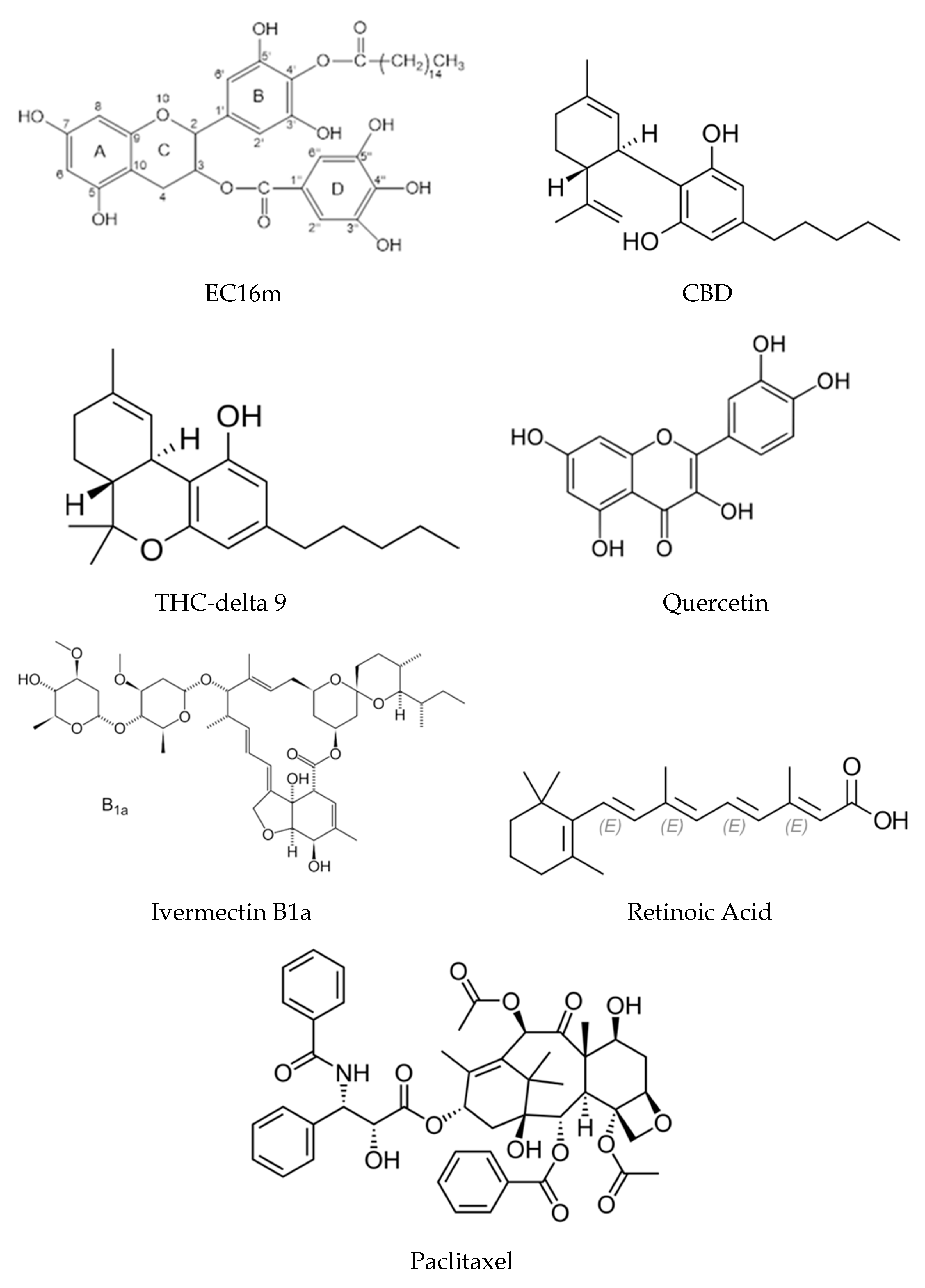
The current study is aimed at testing our hypothesis by generating nanoparticles of Cannabidiol (CBD), Delta-9-tetrahydrocannabinol (THC-9), quercetin, ivermectin, retinoic acid, and paclitaxel (see structures of these hydrophobic compounds below from Wikipedia, except for EC16m, which was provided by Camellix, LLC. USA). If successful, FAST could lead to a wide range of applications for hydrophobic compounds to be developed for effective delivery to specific targets by oral, topical, nasal, inhalational, injectable, and other applications.
2. Materials and Methods
2.1. Compounds and Oral Rinse
Epigallocatechin-3-Gallate-Palmitates (EC16) and Epigallocatechin-3-Gallate-Mono-Palmitate (EC16m) were obtained from Camellix, LLC (Evans, GA, USA). Delta-9-Tetrahydrocannabinol (THC-9, 06-722-453), Quercetin hydrate, 95% (AC174070100), Ivermectin (AAJ6277703), and Retinoic Acid, all trans isomer (MP021902695) were purchased from Thermo Fisher Scientific Inc. (Waltham, MA, USA). Cannabidiol (CBD) isolate powder was purchased from Crescent Canna (New Orleans, LA). Paclitaxel was purchased from MedKoo Biosciences (Durham, NC. USA). Both unflavored and peppermint flavored oral rinse were provided by International Nutrition, Inc. (Middle River, MD, USA).
2.2. Preparation of Nanoparticles
The nanoparticles of the compounds were prepared using FAST (proprietary, patent pending) with three specific practical methods (A, A1 and A2). Method A was the main method that was used for nanoparticle preparations from EC16m [7] and other compounds except EC16 in this study, from which were formed by simplified methods Method A1 and Method A2. All nanoparticle stocks at 1% were stored in pure glycerol for further dilution with sterilized double-stilled water. In addition, a food grade dispersing agent was used in the water nanosuspensions of some nanoparticles as described [7]. The oral rinse formulations were mixed directly with the EC16 nanoparticle stock to 0.01% w/v. EC16 nanoparticle powder was obtained by centrifugation of 0.1% EC16 water nanosuspension at 13,400 RPM for 45 minutes, followed by washing with purified water twice before final centrifugation. Subsequently, pellets were collected and dried. The EC16 nanoparticle powder was then reconstituted with water for analysis.
2.3. Evaluation of Particle Size Distribution and Zeta Potential
ZetaView nanoparticle tracking analysis was performed according to a method described previously [6,7]. The particle size distribution and concentration were measured using the ZetaView ×20 (Particle Metrix, Meerbusch, Germany) and corresponding software. The measuring range for particle diameter is 10–2000 nm. All samples were diluted by the same volume of 1× PBS and then loaded into the cell. Particle information was collected from the instrument at 11 different positions across the cell, with two cycles of readings. Standard operating procedure was set to a temperature of 23 °C, a sensitivity of 70, a frame rate of 30 frames per second, and a shutter speed of 100. The post-acquisition parameters were set to a minimum brightness of 20, a maximum area of 1000, a minimum area of 10, and a trace length of 15 [6,7].
2.4. Electron Microscopy Imaging of EC16 Nanoparticles
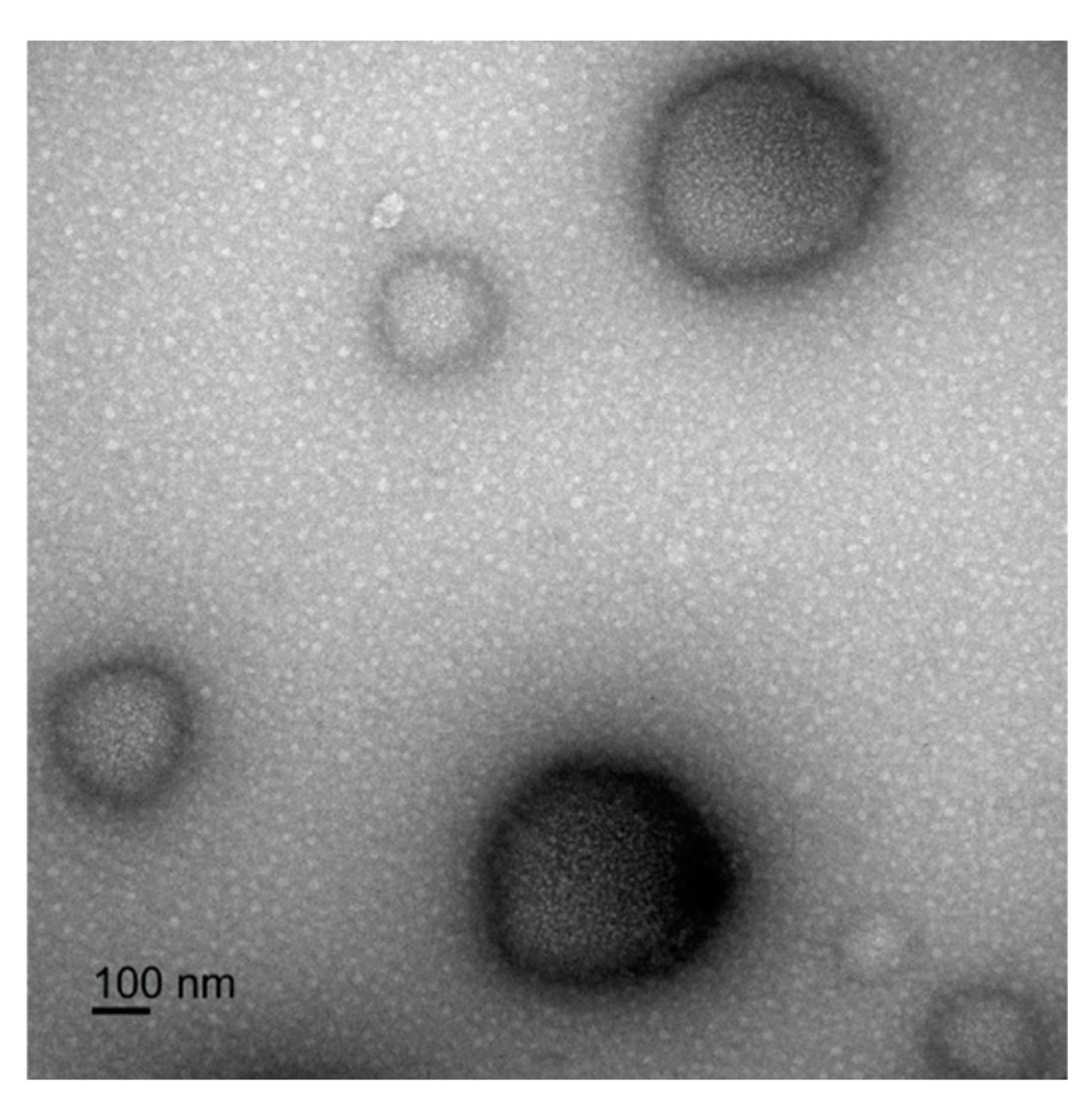
The 1% EC16 nanoparticle stock was diluted with PBS to 0.01% and fixed in 4% paraformaldehyde and 2% glutaraldehyde. After mixing, 5 µl of the sample was removed and transferred to a Formvar/Copper 200 mesh grid and allowed to dry for 15 minutes. Excess solution was then removed using filter paper particles and was negatively stained by addition of 5 µl of 2% aqueous uranyl acetate. Multiple images were captured from each sample in a JEM 1400 Flash Transmission Electron Microscope (JEOL, Peabody, MA) at 120kV, using a Gatan OneView Digital Camera (Gatan Inc., Pleasanton, CA).
3. Results
3.1. EC16 and EC16m Nanoparticles
Three different methods based on FAST were used for preparing the nanoparticles. Method A is the main method that was used for nanoparticle preparations, except EC16 nanoparticles, which were formed by simplified methods Method A1 and Method A2. In addition, a food grade dispersing agent was used in the water nanosuspensions of some nanoparticles as described [6,7]. Figure 1 shows the size distribution of EC16 nanosuspensions prepared using Method A1 and with a food grade dispersing agent. The median size of the EC16 nanoparticles was152.5 ± 78.8 (SD) nm, with a range of 95 to 218 nm. At 0.01% w/v EC16, the density of the nanoparticles was 3.2x10⁹ particles/ml, and the Zeta potential was -60.11 ± 0.59 (SD) mV (Figure 1A). When the dispersing agent was added to the water suspension, the median size of the nanoparticles became 163.8 ± 104.2 nm, ranging from 74 to 435 nm. At 0.01% w/v EC16, the density of the nanoparticles was 2.4x10⁹ particles/ml, and the Zeta potential was -58.08 ± 0.55 mV (Figure 1B).
When EC16 nanoparticles were prepared by Method A2, the median size of the nanoparticles was 128.1 ± 65.9 nm, ranging from 66 to 143.9 nm. At 0.01% EC16, the density of the nanoparticles is 1x10¹⁰ particles/ml and the Zeta potential was -56.65 ± 0.65 (SD) mV (Figure 2A). With the addition of the dispersing agent, the median size of the nanoparticles was 147.7 ± 63.8 nm, ranging from 57.8 to 162.1 nm. At 0.01% EC16, the density of the nanoparticles is 6.5x10⁹ particles/ml, and the Zeta potential was -55.22 ± 0.88 mV (Figure 2B).
Method A was used in all previously published data [5,6,7,9] and for EC16m preparation (Figure 3). The median size of EC16m nanoparticles was 115.9 ± 57.5 nm, ranging from 30 to 120.9 nm. At 0.02% EC16m, the density of the nanoparticles was 2.3x10¹⁰ particles/ml, with Zeta potential of -50.33 ± 0.98 mV (Figure 3A). With the addition of the dispersing agent, the median size of the nanoparticles was 154.9 ± 77.7 nm, ranging from 65 to 182.5 nm. At 0.03% EC16m, the density of nanoparticles was 4.8x10⁹ particles/ml, and the Zeta potential was -60.56 ± 0.73 mV (Figure 3B).
To test if the nanoparticle powder produced by a FAST method can be easily reconstituted in water, an EC16 water nanosuspension at 0.1% prepared by Method A2 was condensed and dried, producing a white powder. The dry EC16 nanoparticles were then reconstituted in double distilled water as a nanosuspension. At 0.01% EC16, the density of the particles reached 1x10¹⁰ particles/ml, with median size of 141.4 ± 105.4 nm and size range from 49 to 178.9 nm (Figure 5B). The size distribution was comparable to that of EC16 nanoparticles prepared by Method A2 (Figure 2), as was the Zeta potential.
3.2. CBD Nanoparticles
The median size of CBD nanoparticles is 206 ± 103.4 nm, ranging from 147 to 438 nm. At 0.06% CBD, the density of the nanoparticles was 4.7x10⁸ particles/ml and the Zeta potential was -51.64 ± 0.85 mV (Figure 4A). With inclusion of 1% w/v of the dispersing agent, the median size of nanoparticles was 222.7 ± 135.3 nm, ranging from 69 to 271.9 nm. At 0.06% CBD, the density of nanoparticles was 4.7x10⁸ particles/ml and the Zeta potential was -48.09 ± 0.14 mV (Figure 4B).
3.3. THC-9 and Reconstituted EC16 Nanoparticles
The median size of the THC-9 nanoparticles was 232.3 ± 151.3 nm, ranging from 149 to 605 nm. At 0.01% THC-9 the density of the nanoparticles is 2.2x10⁸ particles/ml, and the Zeta potential was -38 ± 0.51 mV (Figure 5A). The median size of the water-reconstituted EC16 nanoparticles was 141.4 ± 105.4 nm, ranging from 49 to 178.9 nm. At 0.01% EC16 the density of nanoparticles was 1010 particles/ml and Zeta potential was -56 ± 0.58 mV (Figure 5B).
3.4. Quercetin Nanoparticles
The median size of quercetin nanoparticles was 163.8 ± 102.2 nm, ranging from 40 to 269.8 nm. At 0.02% w/v quercetin the density of the nanoparticles was 3.2x10⁹ particles/ml, and the Zeta potential was -62 ± 1.0 mV (Figure 6A). With 1% w/v of the dispersing agent the median size of nanoparticles was 184.9 ± 119.6 nm, ranging from 69.6 to 256 nm. At 0.02% w/v quercetin the density of nanoparticles was 1.9x10⁹ particles/ml and the Zeta potential was -38.4 ± 0.78 mV (Figure 6B).
3.5. Ivermectin Nanoparticles
The median size of ivermectin nanoparticles was 176 ± 112.1 nm, ranging from 115 to 420.7 nm. At 0.02% w/v ivermectin the density of the nanoparticles is 3.5x10⁸ particles/ml with Zeta potential of -52.32 ± 0.69 mV (Figure 7A). With 1% w/v of the dispersing agent the median size of nanoparticles was 160.6 ± 90.4 nm, ranging from 98 to 344.5 nm. At 0.02% w/v ivermectin, the density of nanoparticles was 5x10⁸ particles/ml and the Zeta potential was -53.92 ± 1.49 mV (Figure 7B).
3.6. Retinoic Acid Nanoparticles
The median size of retinoic acid nanoparticles was 147 ± 155.4 nm, ranging from 75.5 to 341.5 nm. At 0.02% w/v retinoic acid, the density of nanoparticles was 3.7x10⁸ particles/ml, and the Zeta potential was -48.34 ± 1.07 mV (Figure 8A). With 1% w/v of the dispersing agent the median size of nanoparticles was 162.8 ± 107.8 nm, ranging from 120.3 to 407.3 nm. At 0.02% w/v retinoic acid the density of nanoparticles was 7.2x10⁸ particles/ml and the Zeta potential was -53.99 ± 1.50 mV (Figure 8B).
3.7. Paclitaxel Nanoparticles
The median size of paclitaxel nanoparticles was 119 ± 111.0 nm, ranging from 117.3 to 265.8 nm. At 0.01% w/v paclitaxel, the density of nanoparticles was 3.2x10⁸ particles/ml, and the Zeta potential was -49.5 ± 2.13 mV (Figure 9A). With 1% w/v of the dispersing agent the median size of nanoparticles was 130.1 ± 138.2 nm, ranging from 42.3 to 432.6 nm. At 0.01% w/v paclitaxel the density of nanoparticles was 3.9x10⁸ particles/ml and the Zeta potential was -42.27 ± 1.40 mV (Figure 9B).
3.8. EC16 Nanoparticles in Two Water-Based Oral Rinse Formulations
The median size of the nanoparticles in unflavored oral rinse formulation was 176.7 ± 184.4 nm, ranging from 81 to 268 nm. At 0.01% w/v EC16 the density of the nanoparticles is 1.6x10⁹ particles/ml and the Zeta potential was -39 ± 1.35 mV (Figure 10A). In the peppermint flavored oral rinse formulation, the median size of nanoparticles was 121.6 ± 63.6 nm, ranging from 103 to 126.9 nm. At 0.01% w/v EC16, the density of nanoparticles was 8.6x10⁸ particles/ml with Zeta potential of -51.54 ± 0.01 mV (Figure 10B).
3.9. Transmission Electron Microscopy Image of EC16 Nanoparticles
4. Discussion
FAST is a relatively simple tool to prepare nanoparticles/nanocrystals without the use of sophisticated technology of agents that could cause adverse effects. We previously reported the use of FAST Method A to generate EC16 and EC16m nanoparticle for nasal aqueous formulations [5,6,7]. To explore approaches to further simplify the method, specific proprietary Methods A1 and A2 were used for EC16 nanoparticle preparation. In addition, an FDA approved, commonly used food additive dispersing agent (proprietary, patent pending) was tested with the suspensions. Figure 1A shows the size distribution of EC16 nanoparticles in water suspension; Figure 1B is the size distribution of the water suspension with the dispersing agent at 1% w/v. There was no statistical difference in median size between the two formulations, but the dispersing agent altered the size range and resulted in smaller particles around 74 nm (10.1%) and 3.6% of larger particles around 435 nm; compared to the EC16 nanosuspension without the dispersing agent, which is ranging from 95 to 218 nm. Both nanosuspensions had excellent stability with Zeta potential at about -60 mV (a Zeta Potential value above +30 mV or below −30 mV is generally considered stable). Therefore, FAST Method A1 was a suitable method to prepare EC16 nanoparticles.
Method A2 is another simplified method derived from Method A. As shown in Figure 2, the EC16 nanoparticles have a median size of 128.1 ± 65.9 nm, with a range of 66 to 143.9 nm (Figure 2A), while the dispersing agent resulted in a slightly larger median size of 147.7 ± 63.8 nm, with similar size range 57.8 to 162.1 nm (Figure 2B). It appears that the dispersing agent had little effect on the nanoparticle size, and the charges measured by Zeta potentials were similar to each other (-56.65 ± 0.65 mV vs. -55.22 ± 0.88 mV), although slightly lower than those for particles produced by Method A1. Also, both Method A2 suspensions had a narrower size range than that of EC16 nanosuspensions made with Method A1 (Figure 1), suggesting Method A2 can be used for producing EC16 nanoparticles with smaller range in size.
In summary, both Methods A1 and A2 simplified versions of Method A were capable of producing EC16 nanoparticles with high stability in terms of high surface charges, consistent with previously published data using Method A. It is important to note that all three methods are simple, economical, require a short time (<30 min), and little equipment.
Method A was used to prepare EC16m nanoparticles, as well nanosuspensions for all other compounds. As shown in Figure 3, Method A was able to produce EC16m nanoparticles with a narrow range (30 to 120.9 nm) (Figure 3A), compared to the nanoparticles in dispersing agent suspension (65 to 182.5 nm) (Figure 3B). The median size of particles was smaller in the suspension without dispersing agent (115.9 ± 57.5 nm) in comparison to the suspension with the dispersing agent (154.9 ± 77.7 nm). Interestingly, the EC16 nanosuspension without dispersing agent has significantly more nanoparticles than the nanosuspension with the dispersing agent (2.3x10¹⁰ vs. 4.6x10⁹/ml). However, it appears that the EC16m nanosuspension with the dispersing agent was potentially more stable, a with Zeta potential exceeding -60 mV. On the other hand, both nanosuspensions were potentially very stable, with Zeta potential greater than -50 mV. We chose 0.02% EC16m and EC16 based on the previously tested nasal application formulations [5], and two ongoing animal studies, all of which have this concentration of EC16m or EC16, and which did not show mucociliary toxicity [5] of adverse effect in experimental mice (data not shown, study ongoing).
For CBD nanoparticles (Figure 4), the dispersing agent reduced the range of particle size (69 to 271 nm vs. 147 to 438 nm) but somewhat increased the median size, although without statistical difference (232.3 ± 135.3 vs. 206 ± 103.4 nm). Interestingly, the two CBD nanosuspensions with 0.06% CBD have identical particle density of 4.7x10⁸ particles/ml, and a potentially high stability with a Zeta potential around -50 mV (Figure 4). Compared to the EC16 nanoparticles, the CBD nanoparticles were larger in diameter, leading to a lower particle density. The stability of the two compounds in terms of surface charges in the nanosuspensions are similarly high.
A noticeable difference was found in the nanosuspension of THC-9 without the dispersing agent. As shown in Figure 5A, the median size of the particles (232.3 ± 151.3 nm) was similar to that of CBD. However, unlike the other compounds tested, the size distribution was discontinued, with four discrete subpopulations (Figure 5A). Although the nanosuspension was stable with a Zeta potential at -38 ± 0.51, this charge was significantly lower than that of other compounds. However, it is important to note that the THC-9 sample was in a methanol solution at a very low concentration, which was different from other compounds that were obtained in powdered form. It is known that there are different nanotechnology methods to produce nanoparticles of both CBD and THC [10]. The FAST-generated nanoparticles could provide an alternative approach.
The properties of the reconstituted suspension prepared from dried EC16 nanoparticles were similar to those of the original suspension. This result demonstrates that EC16 nanoparticles can be condensed to a powder form and reconstituted in aqueous suspensions or in dry delivery forms. This process could be used for other compounds if dry powder form is preferred.
A number of nanotechnologies have been applied to generate nanoparticles of quercetin, a flavonoid with poor solubility but the potential to benefit human health. Previous studies used lipid-based nanocarriers, polymer-based nanocarriers, micelles and hydrogels composed of natural or synthetic polymers to produce nanoparticles of quercetin [11]. Figure 6 demonstrates that quercetin is suitable for nanosuspension preparation using Method A. The results from the two nanosuspensions indicate that the dispersing agent caused the suspension to have a significantly reduced surface charge, potentially decreasing stability (Zeta potential of -38.4 mV vs. -62 mV), and associated with decreased particle density (1.9x10⁹ vs. 3.2x10⁹/ml at 0.02%) in comparison to the nanosuspension without the dispersing agent. These differences suggest that the dispersing agent may not be beneficial to every compound in terms of stability and particle size.
Ivermectin was initially used as a veterinary medicine for treating parasite infections, but consistently faced limitations due to its poor water solubility and low bioavailability. Various strategies have been applied to increase the solubility of this drug, including lipid-based, polymer-based, drug-loaded nanoparticles, and nanostructured carriers [12]. In the current study, the results demonstrate that both tested nanosuspensions of ivermectin had identical Zeta potential (Figure 7). The particle size distributions appeared different. In the nanosuspension with the dispersing agent only 14.2% of particles had diameters greater than 200 nm, while the counterpart has more than 35% particles with diameters greater than 200 nm. This effect of the dispersing agent resulted in significantly more particles (5x10⁸/ml) in the presence of the dispersing agent compared to the suspension without the agent (3.5x10⁸/ml) at 0.02% w/v ivermectin (Figure 7). Therefore, the addition of the dispersing agent is dependent on the specific compound for its effect.
Retinoids present considerable potential to treat multiple conditions, but one of the major challenges to their use is their low solubility. Attempts to produce nanoparticles of retinoic acid have been reported, including encapsulation in other nanoparticles, micelles, liposomes, films, or by attaching to a carrier [13]. Retinoic acid has a hydrocarbon chain and a 6-carbin ring, as an amphipathic compound similar to other compounds tested. Despite the carboxylic acid group is charged at pH 7, retinoic acid is hydrophobic with poor water solubility. The current study demonstrates that these chemical properties did not prevent retinoic acid from being self-assembled into nanoparticles using FAST Method A (Figure 8). The dispersing agent gave a similar Zeta potential (-54 mV) to that of nanosuspension without the dispersing agent (-48.34 mV). Another advantage of using the dispersing agent is that the particle density almost doubled (7.2x10⁸ vs. 3.7x10⁸/ml) at 0.02% concentration, by shifting the distribution from larger to smaller particles as seen in Figure 8.
Paclitaxel, a cancer drug with poor solubility, is able to self-assemble to form nanoparticles using Method A (Figure 9). In water suspension, the median size of paclitaxel nanoparticles was 119 nm, ranging from 117.3 to 265.8 nm. At 0.01% w/v paclitaxel, the density of nanoparticles was 3.2x10⁸ particles/ml, and the Zeta potential was -49.55 mV (Figure 9A). An interesting observation is that the size distribution of nanoparticles is narrow, with approximately 90% particles around the major peak of 117.3 nm (Figure 9A). The Zeta potential of the suspension indicates the surface of the surface of the particles are also negatively charged, and provides strong electrostatic repulsion among the particles, leading to higher stability of the suspension. The addition of the food-grade dispersing agent has a slightly lower Zeta potential (-42.27 mV) but higher particle density (3.9x10⁸). Currently used FDA approved Paclitaxel formulations include a formulation of 50:50 mix of ethanol and a polyoxyethylated castor oil (Taxol), a formulation involving human serum albumin (Abraxane), and a liposome formulation containing lecithin and cholesterol (Lipusu) [14]. Our result suggests that FAST technology could provide another option for paclitaxel (and docetaxel) formulations with self-assembled nanoparticles associated with strong surface charge.
To examine the feasibility of using EC16 nanoparticles in oral care products we initially tested 0.05% and 0.005% w/v of EC16 nanoparticles in an unflavored oral rinse product containing erythritol, provided by International Nutrition, Inc. Both concentrations were stable and compatible with the oral rinse with particle size range of 40.7 to 251.4 nm, and median size of 170.6 ± 97.3 nm (data not shown). To further investigate the feasibility of EC16 nanoparticles, the 1% EC16 stock was directly added to two oral rinse products containing xylitol. One oral rinse also contains natural peppermint flavor. As shown in Figure 10A, the unflavored oral rinse with EC16 nanoparticles has a similar size distribution to the previously tested unflavored product, with more than 60% particles under 200 nm, resulting in a high density of 1.6x10⁹ particles/ml at 0.01% EC16. In contrast, the peppermint oral rinse with EC16 nanoparticles has significantly higher surface charges, with Zeta potential of -51.54 mV vs. -39 mV of the unflavored oral rinse. In addition, the particle range is narrow, with most particles at around 100 to 130 nm range (Figure 10B). These results demonstrate that EC16 nanoparticles can be easily incorporated into aqueous products with high surface charges.
Transmission electron microscopy was performed to investigate the EC16 nanoparticle structure, shape, and size, after the self-assembling process. As shown in Figure 11, the rounded nanoparticles showed high polydispersity, with diameters ranging from approximately 100 nm to >300 nm, consistent with the ZetaView results. The characters of EC16 nanoparticles were described in a recent publication [9]. It is postulated that other compounds in the current study would have similar particle structures and characteristics, pending future studies. The limitations of FAST include 1: It is only for hydrophobic and poorly soluble molecules with chemical structures suitable to self-assembly into nanoparticles/nanocrystals; 2: it may not be suitable for large compounds that are soluble in aqueous solutions.
5. Conclusions
In summary, unlike the nanoparticles/nanocrystals generated by wet milling and high-pressure homogenizations, the Facilitated Self-assembling Technology (FAST) allows compounds with poor solubility and bioavailability to form molecularly organized nanoparticles/nanocrystals by themselves with strong surface charges to either suspend in an aqueous formulation or in a dried powder form for a variety of delivery methods such as oral, nasal, topical, injectable, etc. Methods based on FAST can be used in drug development and drug improvement, as well as for healthcare, disease control and prevention, cosmetic, and consumer products, pending future studies.
Author Contributions
Conceptualization, S.H., D.D.; methodology, S.H., N.F., Zeta View validation, Y.L., H.Y., J.C. and N.F.; data analysis, S.H., D.D., N.F.; investigation, N.F., D.D., and S.H.; re-sources, Y.L.; data curation, S.H.; writing—original draft preparation, S.H.; writing—review and editing, N.F.; D.D.; visualization, N.F.; supervision, S.H. and D.D.; project administration, S.H.; funding acquisition, S.H., D.D. All authors have read and agreed to the published version of the manuscript.
Funding
This work was funded by a grant from the National Institute on Deafness and Other Communication Disorders (NIDCD) (1R41DC020678-01). The content is solely the responsibility of the authors and does not necessarily represent the official views of the National Institutes of Health.
Acknowledgments
The authors want to thank Dr. Brenden Marshal for TEM work and support from Augusta University Research Institute and Office of Innovation Commercialization.
Conflicts of Interest
The authors declare no conflicts of interest.
References
- Ogden, J.; Parry-Billings, M. Nanotechnology approaches to solving the problems of poorly water-soluble drugs. Drug Discov 2005, 6:71–76.
- Kumari, L.; Choudhari, Y.; Patel, P.; Gupta, G.D.; Singh, D.; Rosenholm, J.M.; Bansal, K.K.; Kurmi, B.D. . Advancement in Solubilization Approaches: A Step towards Bioavailability Enhancement of Poorly Soluble Drugs. Life (Basel) 2023, 13:1099. [CrossRef]
- Bhalani, D.V.; Nutan, B.; Kumar, A.; Chandel, A.K.S. Bioavailability Enhancement Techniques for Poorly Aqueous Soluble Drugs and Therapeutics. Biomedicines 2022, 10:2055. [CrossRef]
- Liu, J.; Liangxing Tu, L.; Cheng, M.; Feng, J.; Jin, Y. Mechanisms for oral absorption enhancement of drugs by nanocrystals Journal of Drug Delivery Science and Technology 2020, 56(Part A).
- Frank, N.; Dickinson, D.; Garcia, W.; Liu, Y.; Yu, H.; Cai, J.; Patel, S.; Yao, B.; Jiang, X.; Hsu, S. Feasibility Study of Developing a Saline-Based Antiviral Nanoformulation Containing Lipid-Soluble EGCG: A Potential Nasal Drug to Treat Long COVID. Viruses 2024, 16:196. [CrossRef]
- Frank, N.; Dickinson, D.; Lovett, G.; Liu, Y.; Yu, H.; Cai, J.; Yao, B.; Jiang, X.; Hsu, S. Evaluation of Novel Nasal Mucoadhesive Nanoformulations Containing Lipid-Soluble EGCG for Long COVID Treatment. Pharmaceutics 2024, 16:791. [CrossRef]
- Frank, N.; Dickinson, D.; Garcia, W.; Xiao, L.; Xayaraj, A.; Lee, L.; Chu, T.; Kumar, M.; Stone, S.; Hsu, S. Evaluation of Aqueous Nanoformulations of Epigallocatechin-3-Gallate-Palmitate (EC16) Against Human Coronavirus as a Potential Intervention Drug. Biomed J Sci & Tech Res 2023, 50:2023.
- Hurst, B.L.; Dickinson, D.; Hsu, S. Epigallocatechin-3-Gallate (EGCG) Inhibits Sars-Cov-2 Infection in Primate Epithelial Cells. Microbiol Infect Dis 2021, 5: 1-6.
- Frank, N.; Dickinson, D.; Dudish, D.; James, W.; Lovett, G.; Liu, Y.; Yu, H.; Cai, J.; Yao, B.; Jiang, X.; Hsu, S. Potential Therapeutic Use of EGCG-Palmitate Nanoparticles for Norovirus Infection. Biomed J Sci & Tech Res 2024, 59:2024.
- Rebelatto, E.R.L.; Rauber, G.S.; Caon, T. An update of nano-based drug delivery systems for cannabinoids: Biopharmaceutical aspects & therapeutic applications. International Journal of Pharmaceutics 2023, 635:122727. [CrossRef]
- Tomou, E.M.; Papakyriakopoulou, P.; Elmina-Marina Saitani, E.M.; Georgia Valsami, G.; Pippa, N.; Skaltsa, H. Recent Advances in Nanoformulations for Quercetin Delivery. Pharmaceutics 2023, 15:1656. [CrossRef]
- Maiara Callegaro Velho, M.C.; Funk, N.L.; Deon, M.; Benvenutti, E.V.; Buchner, S.; Hinrichs, R.; Diogo André Pilger, D.A.; Ruy Carlos Ruver Beck, R.C.R. Ivermectin-Loaded Mesoporous Silica and Polymeric Nanocapsules: Impact on Drug Loading, In Vitro Solubility Enhancement, and Release Performance. Pharmaceutics 2024, 16:325. [CrossRef]
- Napoli, R.F.J. ; Tariq Enver, Liliana Bernardino, L.; Ferreira, L. Advances and challenges in retinoid delivery systems in regenerative and therapeutic medicine. Nature Communications 2020, 11: 4265. [CrossRef]
- Haddad, R.; Alrabadi, N.; Altaani, B.; Li, T. Paclitaxel Drug Delivery Systems: Focus on Nanocrystals' Surface Modifications. Polymers (Basel) 2022, 9;14:658. [CrossRef]
Figure 1.
Size and distribution of EC16 nanoparticles. A. Preparation Method A1. The median size of the nanoparticles was 152.5 ± 78.8 (SD) nm, with a range from 95 to 218 nm. At 0.01% EC16, the density of the nanoparticles was 3.2x 10⁹ particles/ml. The Zeta potential was -60.11 ± 0.59 (SD) mV. B. Preparation Method A1 with addition of a food-grade dispersing agent. The median size of the nanoparticles was 163.8 ± 104.2 nm, with a range from 74 to 435 nm. At 0.01% EC16, the density of the nanoparticles was 2.4x10⁹ particles/ml. The Zeta potential was -58.08 ± 0.55 mV.
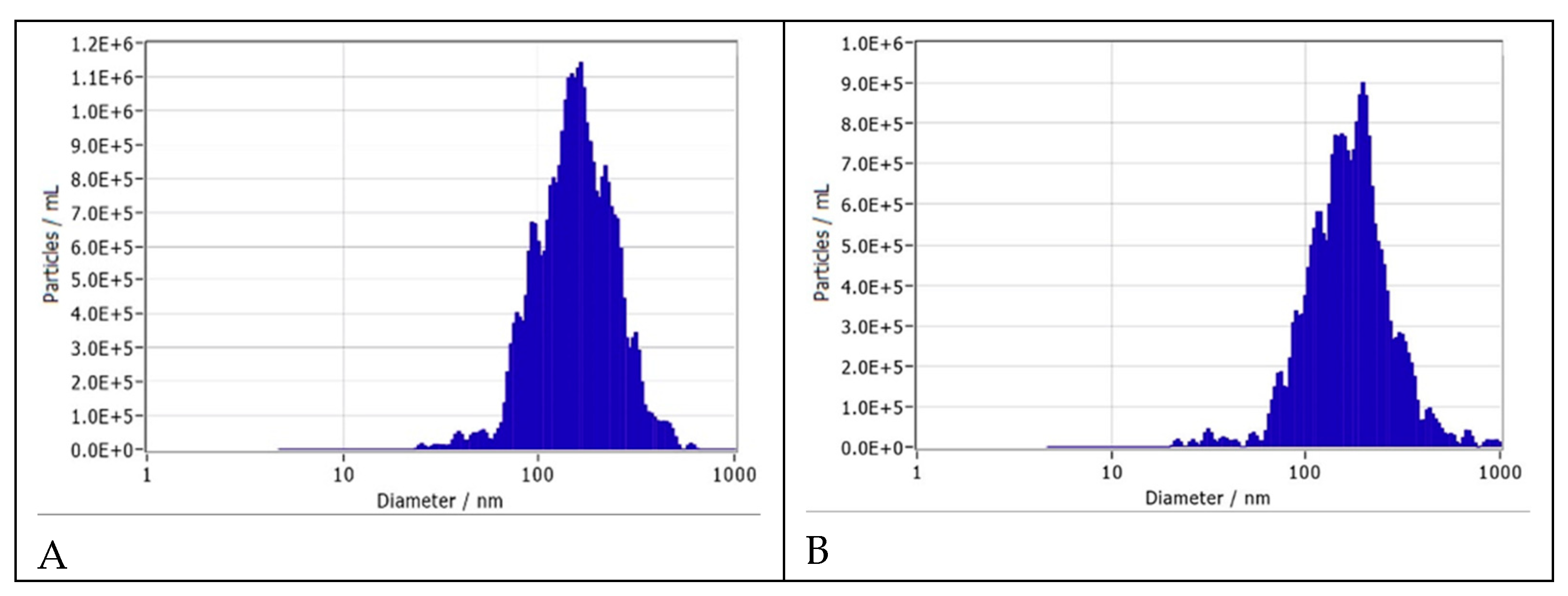
Figure 2.
Size and distribution of EC16 nanoparticles. A. Preparation Method A2. The median size of the nanoparticles was 128.1 ± 65.9 nm, with a range from 66 to 143.9 nm. At 0.01% EC16, the density of the nanoparticles was 1x10¹⁰ particles/ml. The Zeta potential was -56.65 ± 0.65 mV. B. Preparation Method A2 with addition of a food-grade dispersing agent. The median size of the nanoparticles was 147.7 ± 63.8 nm, with a range from 57.8 to 162.1 nm. At 0.01% EC16, the density of the nanoparticles was 6.5x10⁹ particles/ml. The Zeta potential was -55.22 ± 0.88 mV.
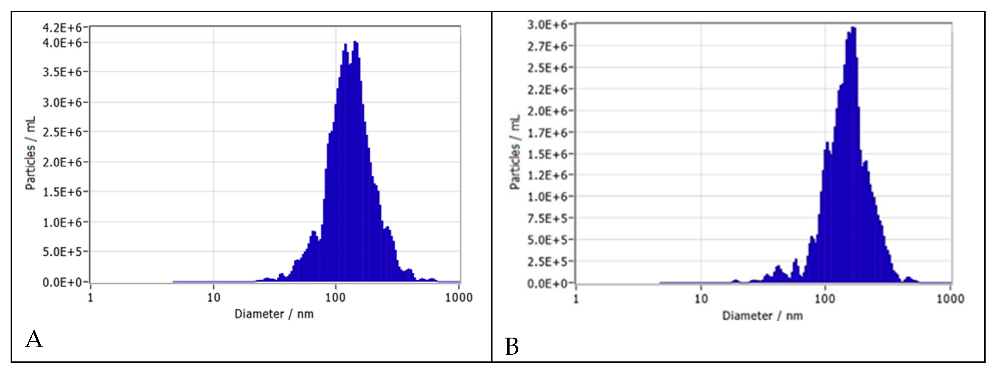
Figure 3.
Size and distribution of EC16m nanoparticles. A. Preparation Method A. The median size of the nanoparticles was 115.9 ± 57.5 nm, ranging from 30 to 120.9 nm. At 0.02% EC16m, the density of the nanoparticles was 2.3x10¹⁰ particles/ml. The Zeta potential was -50.33 ± 0.98 mV. B. Preparation Method A with addition of a food-grade dispersing agent. The median size of the nanoparticles was 154.9 ± 77.7 nm, ranging from 65 to 182.5 nm. At 0.03% EC16m, the density of nanoparticles was 4.8x10⁹ particles/ml. The Zeta potential was -60.56 ± 0.73 mV.
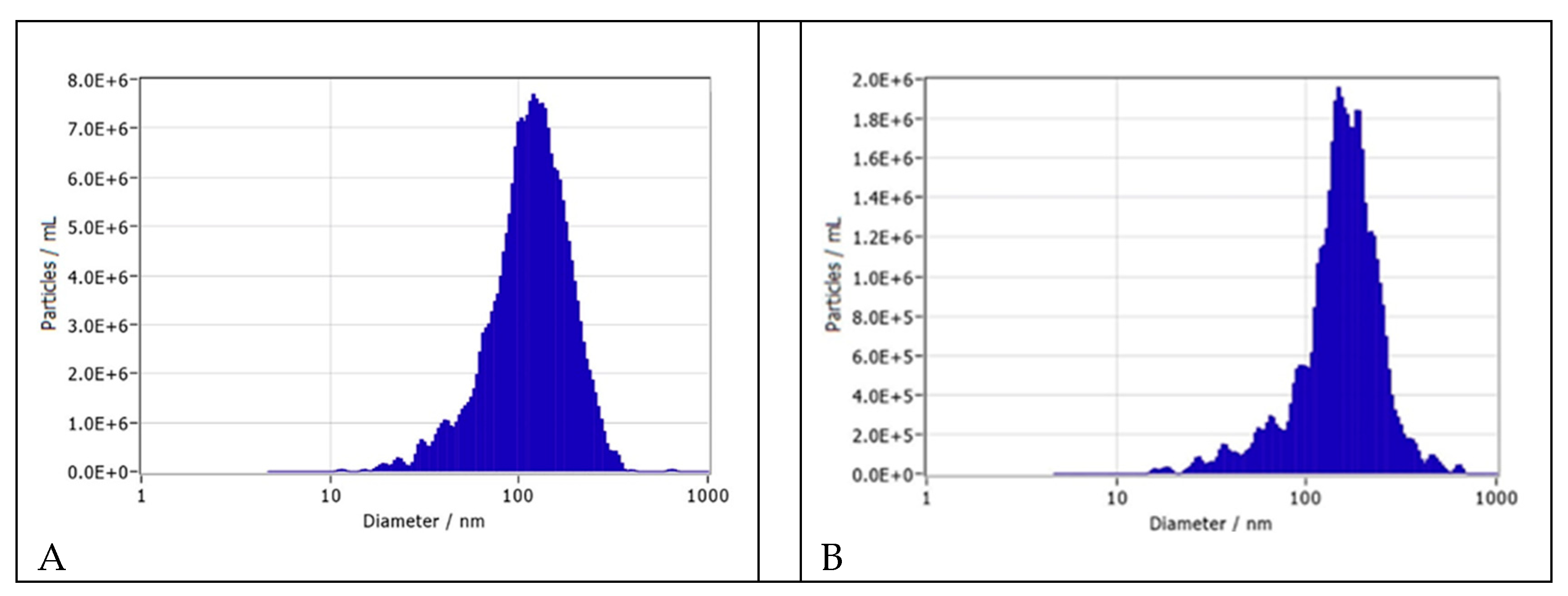
Figure 4.
Size and distribution of CBD nanoparticles. A. Preparation Method A. The median size of the nanoparticles was 206 ± 103.4 nm, ranging from 147 to 438 nm. At 0.06% CBD, the density of the nanoparticles was 4.7x10⁸ particles/ml. The Zeta potential was -51 ± 0.85 mV. B. Preparation Method A with addition of a food-grade dispersing agent. The median size of the nanoparticles was 222.7 ± 135.3 nm, ranging from 69 to 271.9 nm. At 0.06% CBD, the density of nanoparticles was 4.7x10⁸ particles/ml. The Zeta potential was -48.09 ± 0.14 mV.
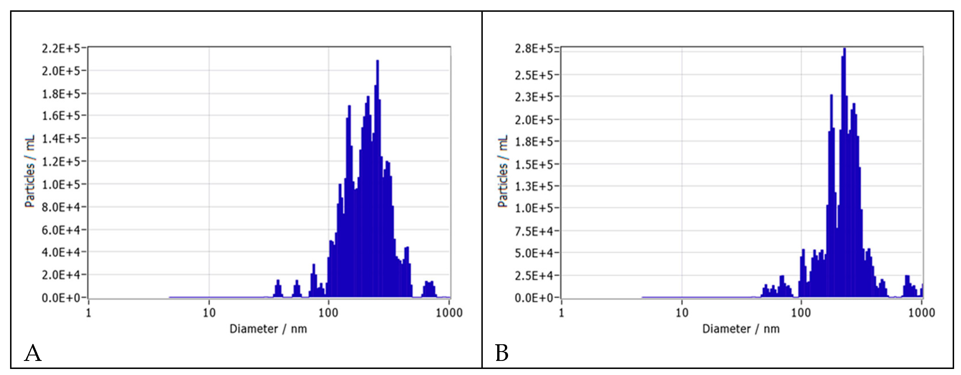
Figure 5.
Size and distribution of THC-9 nanoparticles and EC16 nanoparticles reconstituted from died powder. A. Preparation Method A. The median size of the THC-9 nanoparticles was 232.3 ± 151.3 nm, ranging from 149 to 605 nm. At 0.01% w/v THC-9, the density of the nanoparticles was 2.2x10⁸ particles/ml. The Zeta potential was -38 ± 0.51 mV. B. Preparation Method A. The median size of the water-reconstituted EC16 nanoparticles was 141.4 ± 105.4 nm, ranging from 49 to 178.9 nm. At 0.01% EC16, the density of nanoparticles was 1x10¹⁰ particles/ml. The Zeta potential was -56 ± 0.58 mV.
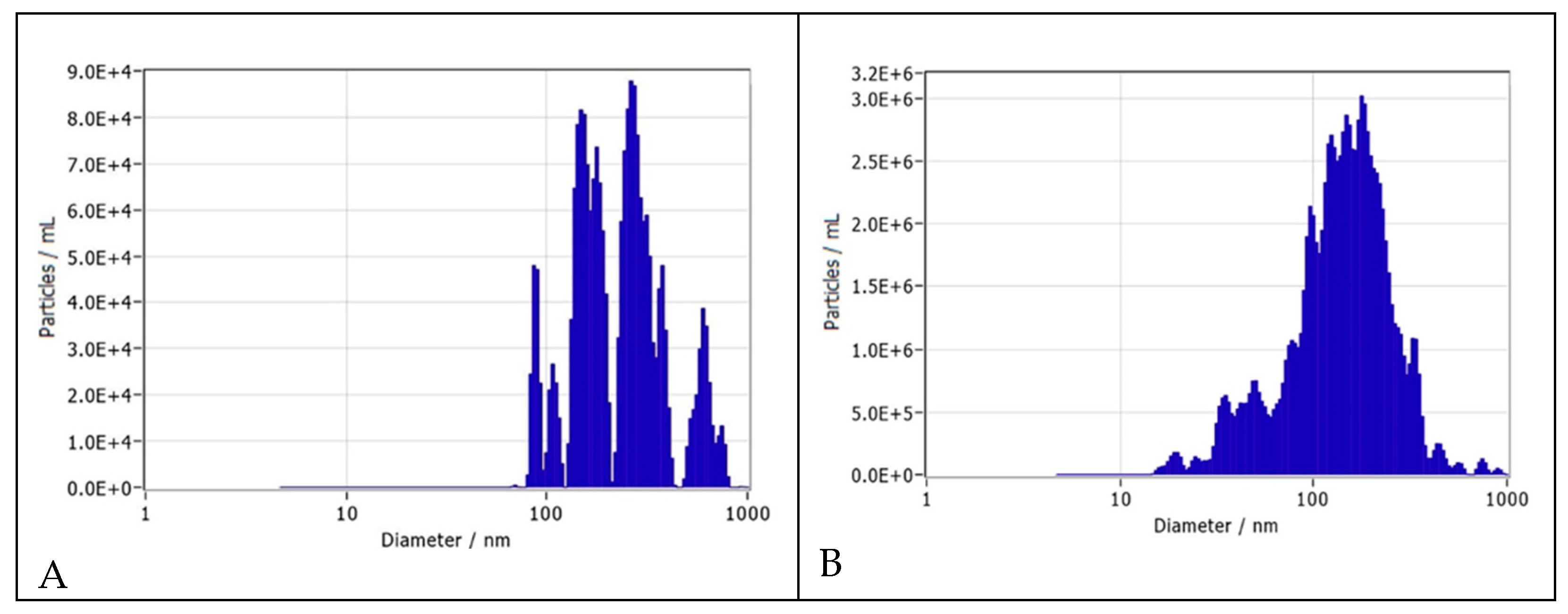
Figure 6.
Size and distribution of quercetin nanoparticles. A. Preparation Method A. The median size of the nanoparticles was 163.8 ± 102.2 nm, ranging from 40 to 269.8 nm. At 0.02% quercetin, the density of the nanoparticles was 3.2x10⁹ particles/ml. The Zeta potential was -62 ± 1.0 mV. B. Preparation Method A with addition of a food-grade dispersing agent. The median size of the nanoparticles was 184.9 ± 119.6 nm, ranging from 69.6 to 256 nm. At 0.02% quercetin, the density of nanoparticles was 1.9x10⁹ particles/ml. The Zeta potential was -38.4 ± 0.78 mV.
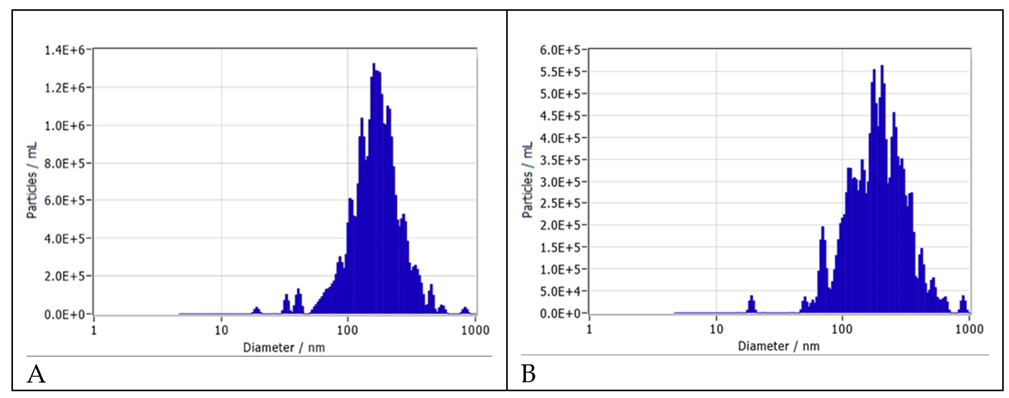
Figure 7.
Size and distribution of Ivermectin nanoparticles. A. Preparation Method A. The median size of the nanoparticles is 176 ± 112.1 nm, ranging from 115 to 420.7 nm. At 0.02% ivermectin, the density of the nanoparticles is 3.5x10⁸ particles/ml. Zeta potential is -52.32 ± 0.69 mV. B. Preparation Method A with addition of a food-grade dispersing agent. The me-dian size of nanoparticles is 160.6 ± 90.4 nm, ranging from 98 to 344.5 nm. At 0.02% ivermectin, the density of nanoparticles is 5x10⁸ particles/ml. Zeta potential is -53.92 ± 1.49 mV.
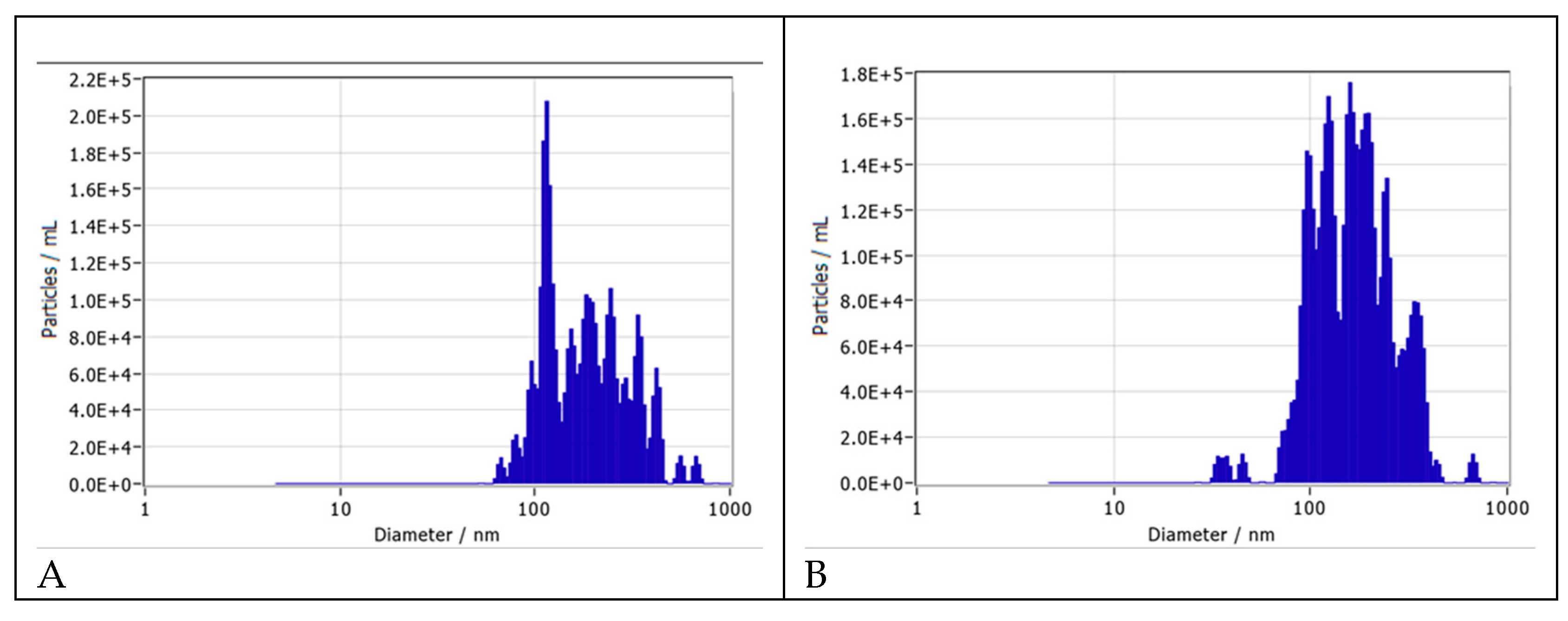
Figure 8.
Size and distribution of retinoic acid nanoparticles. A. Preparation Method A. The median size of the nanoparticles was 147 ± 155.4 nm, ranging from 75.5 to 341.5 nm. At 0.02% ivermectin, the density of the nanoparticles was 3.7x10⁸ particles/ml. The Zeta potential was -48.34 ± 1.07 mV. B. Preparation Method A with addition of a food-grade dispersing agent. The median size of the nanoparticles was 162.8 ± 107.8 nm, ranging from 120.3 to 407.3 nm. At 0.02% ivermectin, the density of the nanoparticles was 7.2x10⁸ particles/ml. The Zeta potential was -53.99 ± 1.50 mV.
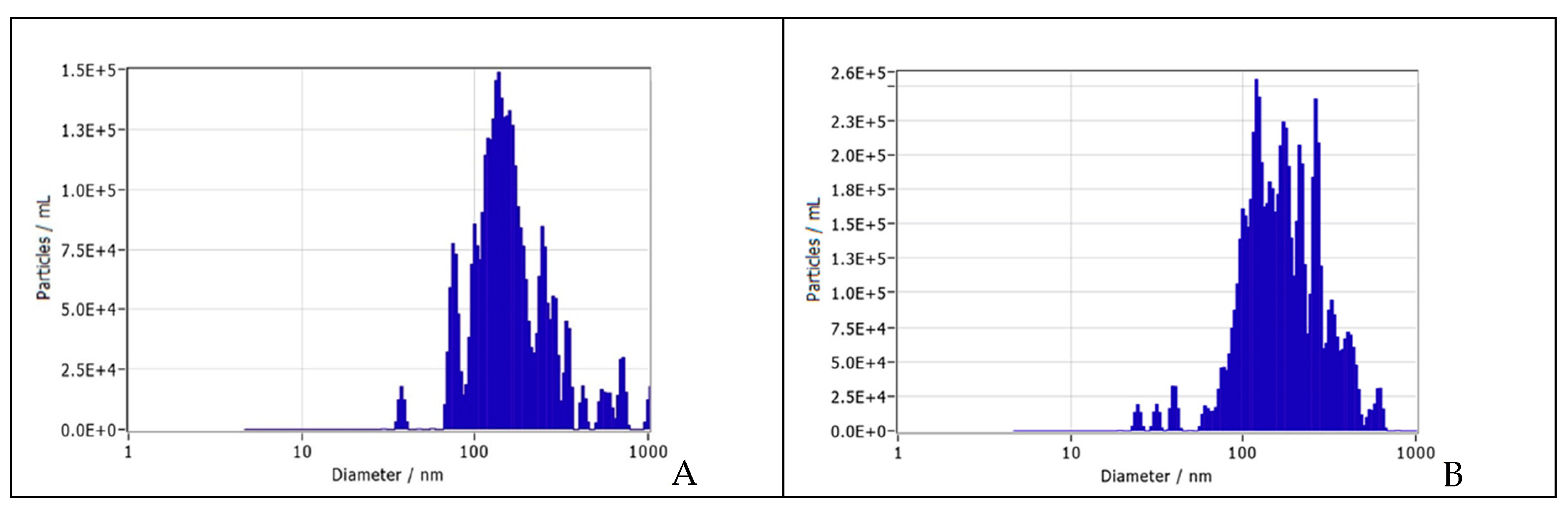
Figure 9.
Size and distribution of paclitaxel nanoparticles. A. Preparation Method A. The median size of paclitaxel nanoparticles was 119 ± 111.0 nm, ranging from 117.3 to 265.8 nm. At 0.01% w/v paclitaxel, the density of nanoparticles was 3.2x10⁸ particles/ml, and the Zeta potential was -49.55 ± 2.13 mV. B. Preparation Method A with addition of a food-grade dispersing agent. The median size of nanoparticles was 130.1 ± 138.2 nm, ranging from 42.3 to 432.6 nm. At 0.01% w/v paclitaxel the density of nanoparticles was 3.9x10⁸ particles/ml and the Zeta potential was -42.27 ± 1.40 mV.
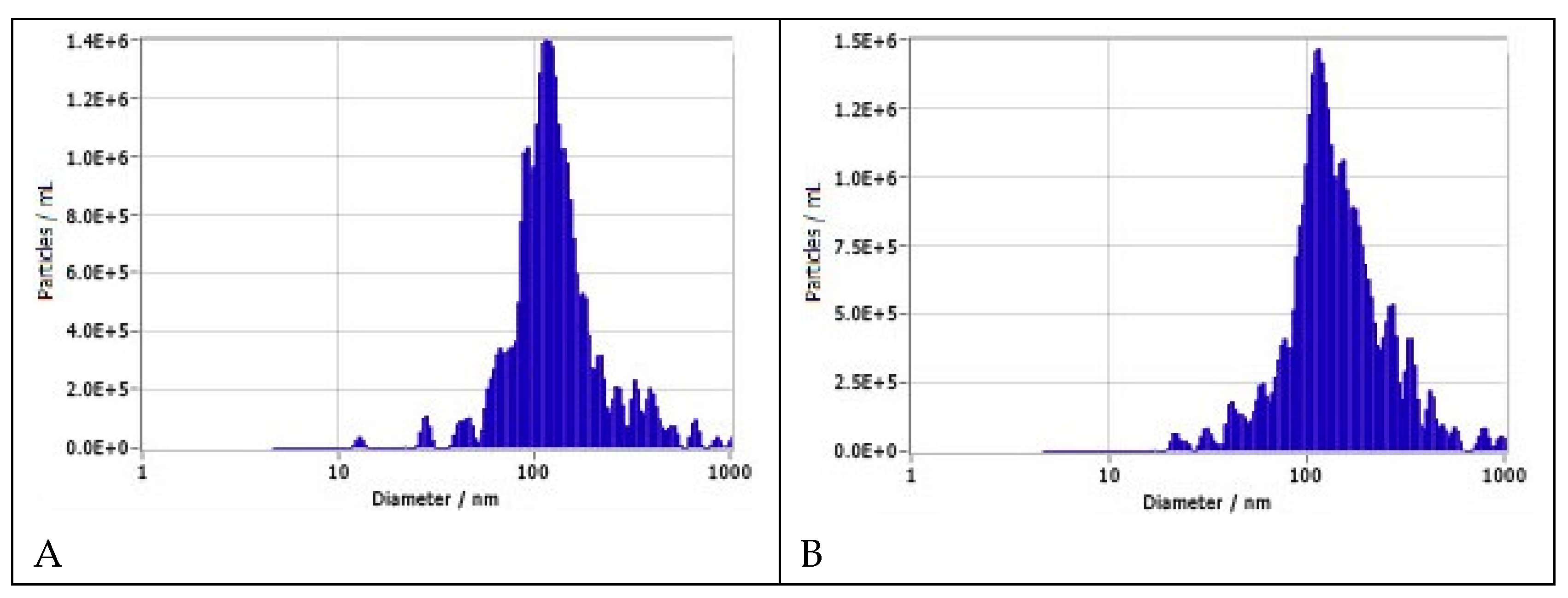
Figure 10.
Size and distribution of EC16 nanoparticles in two water-based oral rinse formulations. A. Unflavored oral rinse. The median size of the nanoparticles was 176.7 ± 184.4 nm, ranging from 81 to 268 nm. At 0.01% EC16 w/v, the density of the nanoparticles was 1.6x10⁹ particles/ml. The Zeta potential was -39 ± 1.35 mV. B. Peppermint oral rinse. The median size of nanoparticles was 121.6 ± 63.6 nm, ranging from 103 to 126.9 nm. At 0.01% EC16 nanoparticles, the density of nanoparticles was 8.6x10⁸ particles/ml. The Zeta potential was -51.54 ± 0.01 mV.
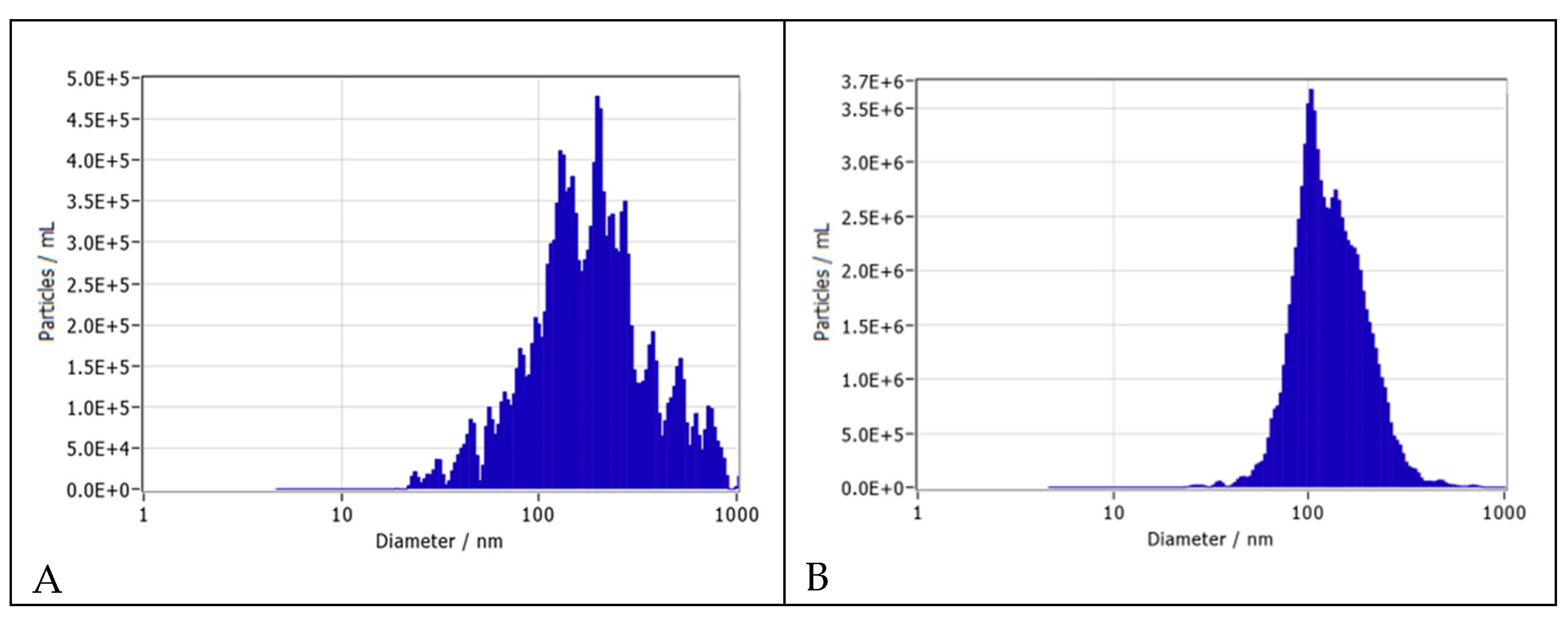
Figure 11.
Representative transmission electron microscopy image of EC16 nanoparticles.
Disclaimer/Publisher’s Note: The statements, opinions and data contained in all publications are solely those of the individual author(s) and contributor(s) and not of MDPI and/or the editor(s). MDPI and/or the editor(s) disclaim responsibility for any injury to people or property resulting from any ideas, methods, instructions or products referred to in the content. |
© 2024 by the authors. Licensee MDPI, Basel, Switzerland. This article is an open access article distributed under the terms and conditions of the Creative Commons Attribution (CC BY) license (http://creativecommons.org/licenses/by/4.0/).
Copyright: This open access article is published under a Creative Commons CC BY 4.0 license, which permit the free download, distribution, and reuse, provided that the author and preprint are cited in any reuse.


