Submitted:
13 November 2024
Posted:
14 November 2024
Read the latest preprint version here
Abstract
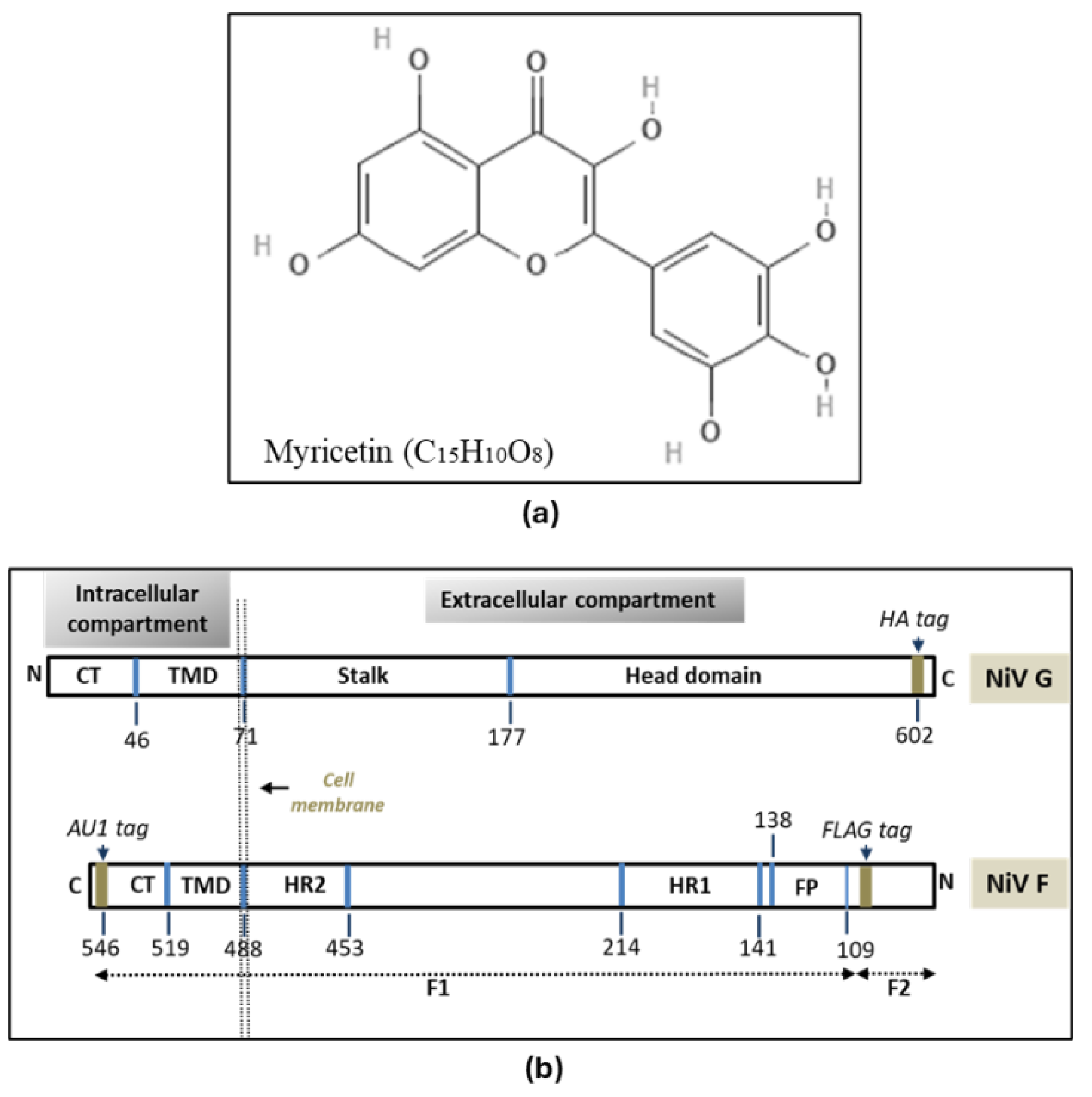
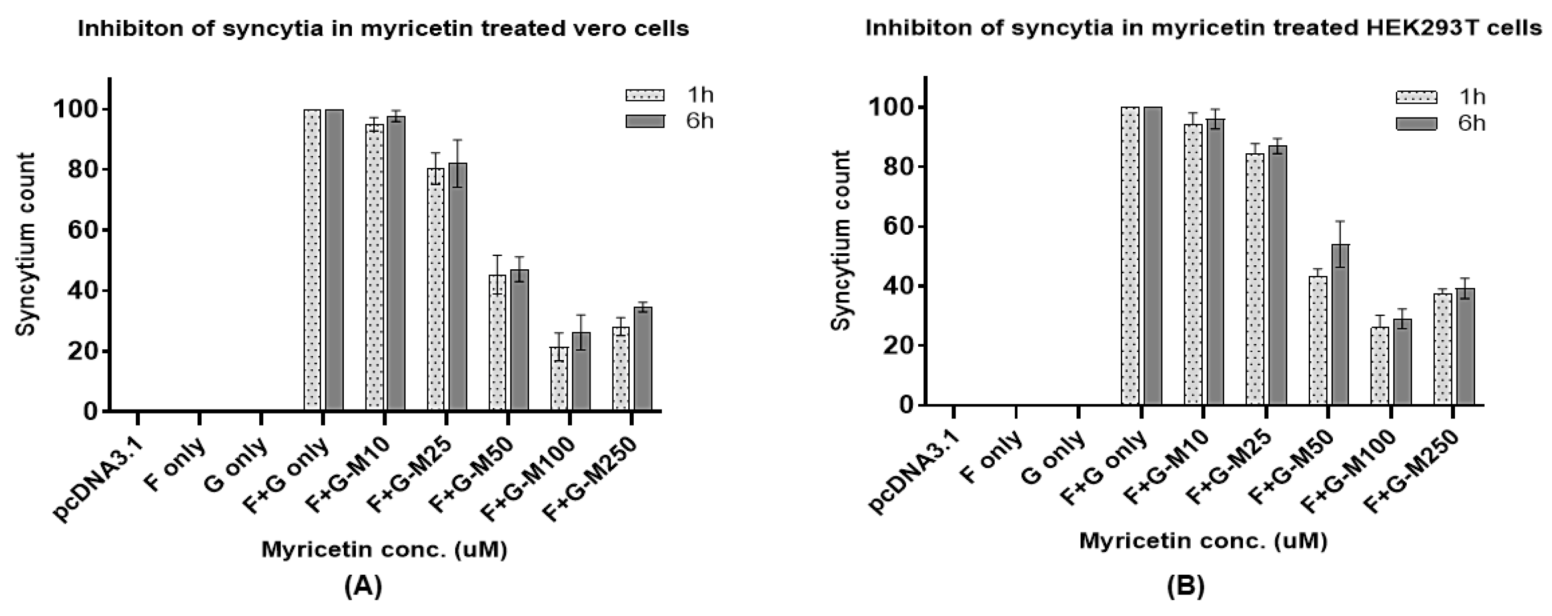
Myricetin (MYR), a flavonoid demonstrated antiviral activity in effectively inhibiting nipah virus envelope glycoprotein mediated syncytial development in-vitro. Time-of-addition assays were designed to evaluate the effective concentration of MYR using codon-optimized NiV wild type (WT) F and G plasmids followed by the addition of MYR in HEK and vero cells. From the seven different concentrations tested, MYR was effective at 100μM concentration with a ~80% decline in syncytial count in HEK293T and vero cells. Results from the time-of-addition were substantiated with Western blotting and flow cytometry assays indicated that MYR might play a critical role in the reduction of NiV F and G glycoprotein expression levels and(or) interfere with their receptor binding, F and G interactions rendering the glycoproteins inefficient in cell-to-cell fusion leading to a quantifiable reduction in the syncytial development. This study is the first to determine antiviral potential of MYR in mitigating the effects of NiV pathogenesis in-vitro.
Keywords:
2. Materials & Methods
2.1. Cell lines and Reagents
2.2. Plasmid Constructs
2.3. Confirmation of NiV F and G Plasmids
2.4. MTT (Cell Viability) Assay
2.4. Transfection with F and G Plasmids
2.5. Time-of-Addition (Time-Course) Assay of Antivirals
2.6. Quantification of Syncytia
2.7. Protein Expression, SDS-Polyacrylamide Gel Electrophoresis (PAGE) and Western Blotting
2.8. Flow Cytometry Quantification of NiV-F Cell Surface Expression (CSE)
3. Results
3.1. Confirmation of NiV-F and G Plasmids
3.2. MTT Assay
3.3. Transfection & Syncytia Development
3.4. Inhibition of Syncytia and Quantification
3.5. Western Blotting and Flow Cytometry
4. Discussion
5. Conclusion
Supplementary Materials
References
- Agrawal, P. K., Agrawal, C., & Blunden, G. Antiviral and Possible Prophylactic Significance of Myricetin for COVID-19. Nat. Prod. Comm. 18(4), 1934578X231166283, (2023). [CrossRef]
- Aguilar, H. C., Matreyek, K. A., Filone, C. M., Hashimi, S. T., Levroney, E. L., Negrete, O. A., ... & Lee, B. N-glycans on Nipah virus fusion protein protect against neutralization but reduce membrane fusion and viral entry. J. Virol. 80(10), 4878-4889, (2006). [CrossRef]
- Ang, B. S., Lim, T. C., & Wang, L. Nipah virus infection. J. Clin. microbial. 56(6), 10-1128, (2018).
- Bishop, K. A., Hickey, A. C., Khetawat, D., Patch, J. R., Bossart, K. N., Zhu, Z., Wang, L. F., Dimitrov, D. S., and Broder, C. C. J. Virol. 82, 11398–11409, (2008).
- Carrasco-Hernandez, R., Jácome, R., López Vidal, Y., & Ponce de León, S. Are RNA viruses candidate agents for the next global pandemic? A review. ILAR J, 58(3), 343-358, (2017). [CrossRef]
- Chakraborty, S., Deb, B., Barbhuiya, P. A., & Uddin, A. Analysis of codon usage patterns and influencing factors in Nipah virus. Virus Res. 263, 129-138, (2019). [CrossRef]
- Chen L, Sun M, Zhang H, Zhang X, Yao Y, Li M, Li K, Fan P, Zhang H, Qin Y, Zhang Z, Li E, Chen Z, Guan W, Li S, Yu C, Zhang K, Gong R, Chiu S. Potent human neutralizing antibodies against Nipah virus derived from two ancestral antibody heavy chains. Nat Commun. 2024 Apr 6;15(1):2987. PMID: 38582870; PMCID: PMC10998907. [CrossRef]
- Devnath, P., & Masud, H. M. A. A. Nipah virus: a potential pandemic agent in the context of the current severe acute respiratory syndrome coronavirus 2 pandemic. New Microb. New Inf., 41, 100873, (2021). [CrossRef]
- Epstein, J. H., Anthony, S. J., Islam, A., Kilpatrick, A. M., Ali Khan, S., Balkey, M. D., & Daszak, P. Nipah virus dynamics in bats and implications for spillover to humans. Proc. Natl. Acad. Sci. 117(46), 29190-29201, (2020). [CrossRef]
- Gao, Z., Li, T., Han, J., Feng, S., Li, L., Jiang, Y., ... & Li, C. Assessment of the immunogenicity and protection of a Nipah virus soluble G vaccine candidate in mice and pigs. Front. Microbiol. 13, 1031523, (2022). [CrossRef]
- Hans, N. Malik, A., & Naik, S. Antiviral activity of sulfated polysaccharides from marine algae and its application in combating COVID-19: Mini review. Bioresour. Technol. Rep. 13, 100623, (2021). [CrossRef]
- Hao, C., Yu, G., He, Y., Xu, C., Zhang, L., & Wang, W. Marine glycan–based antiviral agents in clinical or preclinical trials. Rev. Med. Virol. 29(3), e2043, (2019). [CrossRef]
- Harcourt, A. H., Parks, S. A., & Woodroffe, R. Human density as an influence on species/area relationships: double jeopardy for small African reserves?. Biodiv. Conserv. 10, 1011-1026, (2001). [CrossRef]
- Harit, A. K., Ichhpujani, R. L., Gupta, S., & Gill, K. S. (2006). Nipah/Hendra virus outbreak in Siliguri, West Bengal, India in 2001. Ind. J. Med. Res. 123(4), 553.
- Hauser, N., Gushiken, A. C., Narayanan, S., Kottilil, S., & Chua, J. V. Evolution of Nipah virus infection: past, present, and future considerations. Trop. Med. Infec. Dis. 6(1), 24, (2021). [CrossRef]
- Kalbhor, M. S., Bhowmick, S., Alanazi, A. M., Patil, P. C., & Islam, M. A. Multi-step molecular docking and dynamics simulation-based screening of large antiviral specific chemical libraries for identification of Nipah virus glycoprotein inhibitors. Biophys. chem. 270, 106537 (2021). [CrossRef]
- Levroney, E. L., Aguilar, H. C., Fulcher, J. A., Kohatsu, L., Pace, K. E., Pang, M., Gurney, K. B., Baum, L. G., & Lee, B. Novel innate immune functions for galectin-1: galectin-1 inhibits cell fusion by Nipah virus envelope glycoproteins and augments dendritic cell secretion of proinflammatory cytokines. J. Immunol. 175: 413–420, (2005). [CrossRef]
- Li, W., Xu, C., Hao, C., Zhang, Y., Wang, Z., Wang, S., Wang, W. Inhibition of herpes simplex virus by myricetin through targeting viral gD protein and cellular EGFR/PI3K/Akt pathway. Antiviral Res. 177: 104714, (2020). [CrossRef]
- Lipin, R., Dhanabalan, A. K., Gunasekaran, K., & Solomon, R. V. Piperazine-substituted derivatives of favipiravir for Nipah virus inhibition: What do in silico studies unravel?. SN Appl. Sci. 3(1), 110, (2021). [CrossRef]
- Liu, T. F., & Yu, H. Z. (2014). Research Progress on plant origins and extraction methods of Myricetin. J. Anhui Agric. Sci, 42, 4781-4783.
- Medina-Magües, E.S., Lopera-Madrid, J., Lo, M.K. et al. Immunogenicity of poxvirus-based vaccines against Nipah virus. Sci. Rep. 13, 11384, (2023). [CrossRef]
- Muñoz, A. L., Cuéllar, A. F., Arévalo, G., Santamaría, B. D., Rodríguez, A. K., Buendia-Atencio, C., & Losada-Barragán, M. Antiviral activity of myricetin glycosylated compounds isolated from Marcetia taxifolia against chikungunya virus. EXCLI J. 22, 716- 718, (2023).
- Nahar, N., Mondal, U.K., Hossain, M.J., Uddin Khan, M.S., Sultana, R., Gurley, E.S., Luby, S.P. Piloting the promotion of bamboo skirt barriers to prevent Nipah virus transmission through date palm sap in Bangladesh. Glob. Health Promot. Int. 28: 378 –386, (2013). http://doi.org/10.1093/heapro /das020. [CrossRef]
- Pan, H., He, J., Yang, Z., Yao, X., Zhang, H., Li, R., & Liu, L. Myricetin possesses the potency against SARS-CoV-2 infection through blocking viral-entry facilitators and suppressing inflammation in rats and mice. Phytomed., 116, 154858, (2023).
- Peng S, Fang C, He H, et al. Myricetin exerts its antiviral activity against infectious bronchitis virus by inhibiting the deubiquitinating activity of papain-like protease. Poultry Science. 2022 Mar;101(3):101626. PMCID: PMC8741506. [CrossRef] [PubMed]
- Perkin, A. G., & Hummel, J. J. LXXVI.—The colouring principle contained in the bark of Myrica nagi. Part I. J. Chem. Soc. Trans. 69, 1287-1294, (1896).
- Pillai, V., Krishna, G., & Valiya Veettil, M. Nipah virus: past outbreaks and future containment. Viruses, 12(4), 465, (2020). [CrossRef]
- Ramharack, P., Devnarain, N., Shunmugam, L., & Soliman, M. E. Navigating Research Toward the Re-emerging Nipah Virus-A New Piece to the Puzzle. Curr. Pharmaceut. Des. 25(12), 1392-1401, (2019). [CrossRef]
- Ren, R., Yin, S., Lai, B., Ma, L., Wen, J., Zhang, X., ... & Li, L. Myricetin antagonizes semen-derived enhancer of viral infection (SEVI) formation and influences its infection-enhancing activity. Retrovirology, 15(1), 1-24, (2018).
- Sambrook J. & Russell D. W. Molecular cloning: a laboratory manual (3rd ed.). Cold Spring Harbor Laboratory Press, (2001)..
- Sazzad, H. M., Hossain, M. J., Gurley, E. S., Ameen, K. M., Parveen, S., Islam, M. S., ... & Luby, S. P. Nipah virus infection outbreak with nosocomial and corpse-to-human transmission, Bangladesh. Emerg. Inf. Dis. 19(2), 210, (2013). [CrossRef]
- Shao, Q., Guo, Q., Xu, W. P., Li, Z., & Zhao, T. T. Specific inhibitory effect of κ-carrageenan polysaccharide on swine pandemic 2009 H1N1 influenza virus. PloS one, 10(5), e0126577 (2015). [CrossRef]
- Shariff, M. Nipah virus infection: A review. Epidem. Inf. 147, e95, (2019). [CrossRef]
- Sharma, P., Kumar, R., Sharma, A., Hajam, Y. A., & Kumar, N. Nipah Virus: An Active Causative Agent For Respiratory And Neuronal Ailments. Epidem. Transm. Inf. dis. 78, (2020).
- Singh, N., Brar, R. S., Chavan, S. B., & Singh, J. Scientometric analyses and visualization of scientific outcome on Nipah virus. Curr. Sci. 117(10), 1574-1584, (2019). [CrossRef]
- Stone JA, Vemulapati BM, Bradel-Tretheway B, Aguilar HC. Multiple Strategies Reveal a Bidentate Interaction between the Nipah Virus Attachment and Fusion Glycoproteins. J Virol. 2016 Nov 14;90(23):10762-10773. PMCID: PMC5110167. [CrossRef] [PubMed]
- Talarico, L. B., & Damonte, E. B. Interference in dengue virus adsorption and uncoating by carrageenans. Virology, 363(2), 473-485, (2007). [CrossRef]
- Tsurudome M, Ohtsuka J, Ito M, Nishio M, Nosaka T. The Hemagglutinin-Neuraminidase (HN) Head Domain and the Fusion (F) Protein Stalk Domain of the Parainfluenza Viruses Affect the Specificity of the HN-F Interaction. Front Microbiol. 2018 Mar 13;9:391. PMCID: PMC5859044. [CrossRef] [PubMed]
- Wong JJ, Chen Z, Chung JK, Groves JT, Jardetzky TS. EphrinB2 clustering by Nipah virus G is required to activate and trap F intermediates at supported lipid bilayer-cell interfaces. Sci Adv. 2021 Jan 27;7(5):eabe1235. PMCID: PMC7840137. [CrossRef] [PubMed]
- Wong KT, Shieh W-J, Kumar S, Norain K, Abdullah W, Guarner J, Goldsmith CS, Chua KB, Lam SK, Tan CT. Nipah virus infection: pathology and pathogenesis of an emerging paramyxoviral zoonosis. Am. J. Pathol. 161: 2153–2167, (2002). [CrossRef]
- Xiao, T., Cui, M., Zheng, C., Wang, M., Sun, R., Gao, D., & Zhou, H. Myricetin inhibits SARS-CoV-2 viral replication by targeting Mpro and ameliorates pulmonary inflammation. Fron. Pharm. 12, 669642, (2021).
- Yeo YY, Buchholz DW, Gamble A, Jager M, Aguilar HC. Headless Henipaviral Receptor Binding Glycoproteins Reveal Fusion Modulation by the Head/Stalk Interface and Post-receptor Binding Contributions of the Head Domain. J Virol. 2021 Sep 27;95(20):e0066621. . Epub 2021 Jul 21. PMCID: PMC8475510. [CrossRef] [PubMed]
Disclaimer/Publisher’s Note: The statements, opinions and data contained in all publications are solely those of the individual author(s) and contributor(s) and not of MDPI and/or the editor(s). MDPI and/or the editor(s) disclaim responsibility for any injury to people or property resulting from any ideas, methods, instructions or products referred to in the content. |
© 2024 by the authors. Licensee MDPI, Basel, Switzerland. This article is an open access article distributed under the terms and conditions of the Creative Commons Attribution (CC BY) license (http://creativecommons.org/licenses/by/4.0/).


