
Submitted:
23 September 2024
Posted:
23 September 2024
You are already at the latest version
Abstract
Species of the genus Kalanchoe have a long history of therapeutic use in ethnomedicine linked to their remarkable medical properties. One of them is Kalanchoe blossfeldiana succulents occurring in tropical regions. Despite the great interest in this plant, there are no reports about its therapeutic effects on the skin. In this study, the antioxidant properties and skin permeation of a topical hydrogel containing K. blossfeldiana ethanol extract was assessed. Additionally, the content of active compounds in the K. blossfeldiana extract was evaluated by UHPLC-MS and HPLC-UV. The extract was analysed with three antioxidant assays: ABTS, DPPH and FRAP. Furthermore, antielastase and antihialuronidase properties of the tested extract were assessed. The ex vivo penetration studies were performed using the Franz diffusion cells. The obtained results show that antioxidant properties of K. blossfeldiana extract were similar to ascorbic acid, while antielastase and antihialuronidase tests indicated strong antiaging and anti-inflammatory activity (IC50 was 26.8 ± 0.13 and 77.31 ± 2.44 µg/mL, respectively). Moreover, active ingredients contained in K. blossfeldiana extract penetrated through human skin and accumulate in it. In conclusion, the hydrogel containing K. blossfeldiana extract can be considered as an interesting and new alternative to dermatologic and cosmetic preparations.
Keywords:
Kalanchoe blossfeldiana
; hydrogels
; antioxidant activity
; skin penetration
; elastase
; hyaluronidase
1. Introduction
Kalanchoe blossfeldiana Poelln. belonging to the Crassulaceae family growsnaturally in tropical and subtropical regions and is commonly cultivated as household and garden plants. The interest in this plant in recent years is primarily due to its health benefits. The plant has long been used to treat different diseases. Many studies report its antioxidant, anticancer and antibacterial effects [1,2,3,4]. In addition, K. blossfeldiana contains various secondary metabolites, including flavonoids such as kaempferol and quercetin as well as gallic and benzoic acid derivatives [1,3,5]. These metabolites showed among others a high ability to scavenge free radicals and anti-inflammatory, antibacterial and anticancer properties which play a very important role in medicinal preparations, including those applied to the skin. This is important because in recent years, more and more attempts are made to use less popular plants and their extracts in preparations applied to the skin. There are no reports in the available literature on this type of K. blossfeldiana use. Also, the extracts from K. blossfeldiana have never been investigated in terms of the penetration of the plant metabolites through the skin and their accumulation there.
Assessment of penetration and accumulation in the skin after application of the preparation is very important because the vehicle used may significantly affects the release and penetration of active substances through the skin [6,7,8]. Moreover, in recent years, there has been a search for safe carriers that are easy to use and whose consistency allows for easy placement of plant extracts in them [9]. Such this vehicles include among others hydrogels, which are characterized by simple composition and ease to use. Many authors have used in their studies the hydrogels containing various plant extracts. For example, hydrogels films composed of agarose, κ-carrageenan and glycerol contained the aqueous extract of Cryphaea heteromalla with solid antioxidant activity [10]. Hydrogels with Epilobium angustifolium extract in an amount of 5% showed anti-inflammatory and antioxidant effects and also accelerated wound healing in vitro [6]. Moreover, some phenolic acids contained in hydrogels with the addition of Epilobium angustifolium extracts penetrated the skin or accumulated in it, exhibiting additional antioxidant effects. In other studies, the hydrogels containing 0.5% of Cannabis sativa extracts had a moisturizing effect on the skin and affected the restoration of the hydrolipid balance and rebuilding of the hydrolipid barrier of the skin damaged in the cleansing process by surfactants such as sodium laurosulfate [11].
The aim of our study was to estimate antioxidant, antielastase, and antihialuronidase activity, and the permeation of selected phenolic acids from a hydrogel with K. blossfeldiana ethanol extract through the human skin and their accumulation in it.
2. Results
2.1. Phytochemical profiles of K. blossfeldiana
The phytochemical analysis of the ethanol extract of K. blossfeldiana was done with high-resolution mass spectrometry LC-QTOF-MS. Data analysis revealed the presence of at least 143 main components. Ninety-three components were tentatively identified and classified into 23 arbitrarily established general compound categories (Table 1, Table S1, and Figure S1 in Supplementary Materials). The most frequently occurring in the investigated extract were phenylpropanoid derivatives, observed mainly as glycosides (12 compounds), gallic acid derivatives and acyclic alcohol glycosides (10 and 9 compounds each, respectively), benzoic acid derivatives and organic acids (9 and 8 compounds each, respectively), acyclic nitrile glycosides and flavanoles (7 and 6 compounds each, respectively). Among the benzoic acid derivatives group, it was possible to observe, among others, derivatives of vanillic and shikimic acid. The analyzed extract was rich in gallic acid derivatives, and these were compounds belonging to gallotannins. Subsequently, the following groups of compounds were identified in smaller quantities: megastigmane glycosides, flavonole glycosides, dimeric proanthocyanidins, phenol derivatives, and aminoacids (Table 1 and S1).
Moreover, the content of selected phenolic acids in a hydrogel with K. blossfeldiana (HKB) was estimated and is presented in Table 2 and Figure 1. The following phenolic acids were found: gallic acid, protocatechuic acid, p-hydroxybenzoic acid, m-hydroxybenzoic acid, vanillic acid, gentistic acid and hypogallic acid. The content of the analyzed phenolic acids ranges from 0.85 ± 0.11 µg·mL extract-1 for m-hydroxybenzoic acid to 284.74 ± 15.64 µg·mL extract-1 for gallic acid. The gallic acid and protocatechuic acid were the most abundant in the extract tested (Table 2).
2.2. Biological Activity of Ethanol Extract of K. Blossfeldiana
2.2.1. Antioxidant Activity
To estimate antioxidant activity of ethanol extract of K. blossfediana, three antioxidant tests were performed. Ascorbic acid was used as a standard. The obtained results show that the extract had strong antioxidant properties, and in DPPH and ABTS assays the IC50 values for the extract were lower than for ascorbic acid (Table 3). The highest activity of the extract we obtained in ABTS test. In FRAP test, the IC50 value of K. blossfeldiana extract was about two times higher than the standard compound. All the tests confirmed potent antiradical and reduction activity of the plant extract.
2.2.2. Enzyme Inhibition
In this study, the activity of the K. blossfeldiana ethanol extract on skin enzymes was estimated on elastase and hyaluronidase. The obtained results indicate that the tested extract inhibited the activity of both enzymes and this effect was dose-dependent. In the case of hyaluronidase, the extract caused a complete inhibition of the enzyme at a concentration of 400 µg/mL (Figure 2), while elastase was completely inhibited at the extract concentration of 50 µg/mL (Figure 3). The IC50 values calculated for the extract were only 1.5 times higher in comparison to the standard – oleanolic acid used in the experiments (Table 4).
2.3. Stability Test of HKB
The tested hydrogel with K. blossfeldiana ethanol extract showed appropriate physical properties. After performing the vortex test, no separation of the extract was observed. Similarly, we did not observe changes in colour and odour of HKB in comparison to HKB before the heating-cooling test.
2.4. Ex Vivo Study with HKB
2.4.1. Permeation Through the Skin
The results of permeation of phenolic acids from HKB during the 24-h study are shown in Table 5. The highest penetration was observed for gallic acid and protocatechuic acid, the cumulative mass of which collected after 24 hours of permeation were 249.73 ± 13.69 and 97.55 ± 5.31 µg·cm-2, respectively. It has been observed that these two acids penetrated human skin the fastest. Gallic acid penetrates within the first hour of applying the hydrogel to the skin, while protocatechuic acid was identified in the acceptor fluid collected after the second hour of the study. The remaining acids penetrated the skin much slower and were identified in the acceptor fluid only in the last hours of the experiment.
2.4.2. Accumulation in the Skin
The accumulation of phenolic acids in the skin after 24 hours from application of the hydrogel to the skin is presented in Figure 4. All analyzed phenolic acids accumulated in the skin except for hypogallic acid. It was observed that in the largest amount accumulated in the skin gallic acid (107.58 ± 15.27 µg·g skin-1) and then protocatechuic acid (72.28 ± 8.08 µg·g skin-1).
3. Discussion
The plants are an valuable source of bioactive compounds and are important for the pharmaceutical and cosmetic industries. In recent years, interest in the use of plant extracts for the production of dermatological and cosmetic preparations has also increased significantly. The main reason for this are valuable secondary metabolites found in large amounts in plants, which have primarily antioxidant, but also anti-inflammatory, antibacterial and antiaging effects [11,12]. In recent years, there has been a growing interest in lesser-known plants that could have high health potential. Kalanchoe blossfeldiana, still little known in this respect, has attracted increasing interest. There are not many reports in the available literature on the identification of individual phenolic acids in K. blossfeldiana leaves. Previous analysis of aqueous fraction from the extract of K. blossfeldiana using high-resolution mass spectrometry data showed the presence of benzoic acid derivatives as well as a high content of gallic acid derivatives [3]. In our study, the phytochemical analysis revealed that the most frequently detected group of compounds in the K. blossfeldiana ethanol extract were phenylpropanoid derivatives. In this group identified metabolites were coumaroyl, feruloyl and caffeoyl derivatives that were previously identified in K. blossfeldiana and K. pinnata [3,13,14]. Other important groups identified in the extract we analyzed were derivatives of benzoic acid and gallic acid, which were additionally confirmed by HPLC-UV analysis. It was: gallic acid, protocatechuic acid, p-hydroxybenzoic acid, m-hydroxybenzoic acid, vanillic acid, gentistic acid and hypogallic acid. In our study, gallic acid was detected in the highest amount. A similarly high content of this compound in the leaves of K. blossfeldiana was confirmed by Pryce [15]. Phenolic acids are important plant components that have an impact on skin health. The main properties of these compounds include high antioxidant activity. When applied to the skin, they protect it from excessive oxidative stress caused by reactive oxygen species (ROS). Excessive ROS production leads to many negative effects, including cell damage [16]. The natural antioxidants contained in plants play a key role here as they support the endogenous skin defense system. The K. blossfeldiana ethanol extract applied to the skin in our study was characterized by high antioxidant activity. High levels of ROS may hinder faster wound healing, especially chronic changes. Providing exogenous antioxidants stimulates cell migration and angiogenesis. Therefore, the added of natural antioxidants to cosmetics/dermatologic preparation may be very effective in this case. For example, the hydrogel with antioxidant activity, applied to the skin can reduce oxidative stress, improve the wound microenvironment, and ultimately achieve rapid skin repair [17]. Moreover, to keep the skin in good condition for as long as possible, it is necessary to use preparations with ingredients that inhibit the activity of skin enzymes - components of extracellular matrix (ECM). Kalanchoe blossfeldiana extract can inhibit elastase and hyaluronidase significantly and this effect was slightly weaker than the effect of oleanolic acid.
Antioxidants contained in plant extracts applied to the skin can accumulate in it or penetrate into its deeper layers. Therefore, when designing new cosmetic/dermatological preparations, the study of skin penetration plays an increasingly important role. Active substances are first released from the preparation and then penetrate the stratum corneum (SC). In addition, penetration can be limited by factors, such as lipophilicity or the chemical structure of the active substance itself [7,18,19]. The SC is a thin membrane consisting primarily of cornified epidermal cells, while the main components are lipids, namely cholesterol, ceramides and its esters, and fatty acids [20,21]. The analysis of the penetration of phenolic acids through the skin as well as the assessment of their accumulation therein is an important step in the selection of an appropriate carrier for the plant extract. Plant extracts contain a whole pool of secondary metabolites with different physicochemical properties. In our research, we assessed the penetration of selected phenolic acids from a hydrogel containing dry ethanol extract from leaves of K. blossfeldiana. The penetration test was performed in Franz diffusion cell using the human skin. There are no reports in the available literature about the application of K. blossfeldiana extracts to the skin and the subsequent analysis of the penetration and accumulation of phenolic acids. Therefore in the next stage of our research, the penetration of phenolic acids from the hydrogel containing the extract of K. blossfeldiana was assessed. The phenolic acids after applications of HKB on the skin penetrated the skin to varying degrees. Some phenolic acids, such as gallic acid and protocatechuic acid penetrated the skin already in the first and second hour after application. For these phenolic acids the highest permeation values were observed. Some of the phenolic acids analyzed in our study were identified in the acceptor fluid only after the 8th hour of the experiment and these were p-hydroxybenzoic acid, m-hydroxybenzoic acid and vanillic acid. However, gentistic acid penetrated the skin only after 24 hours, and in the case of hypogallic acid, the 24-hour test period was too short for this compound to cross the skin barrier. As we know, plant extracts contain various secondary metabolites which, depending on their concentration, may interact synergistically, which may also increase the permeation of some of them. The concentration of gallic acid and protocatechuic acid was the highest in the prepared plant extract, which was probably reflected in the highest penetration of these compounds. Previous authors also reported rapid skin penetration of gallic acid from microemulsions containing Glochidion wallichianum extract and E. angustifolium ethanolic extract [22,23]. However, some authors have shown very low or negligible penetration of phenolic acids from preparations containing plant extracts. For example, phenolic acids were released to a very low degree from a hydrogel containing 5% coffee seed extract [19]. Whereas, the rosmarinic acid contained in Plectranthus ecklonii did not permeate the human skin, neither from the water solution nor from the ethanol/PG solution prepared from this plant [24]. Of course, the vehicle used plays a key role in releasing active substances into the skin. In our study, we chose hydrogel as the vehicle for preparation. The simple composition of hydrogels as well as high water content and high biocompatibility with skin cells make them very popular vehicles. Hydrogels are playing an increasingly important role in pharmacy and cosmetology and they seem to be one of the most promising groups of biomaterials [9]. The hydrogels containing plant extracts with many active substances can have a wide range of effects, including antibacterial, anti-inflammatory or antioxidant [9,25,26]. For example, phenolic acids contained in E. angustifolium extract penetrated from the hydrogel to skin better than from the emulsion [7]. Whereas, Žilius et al. showed the highest penetration of phenolic acids from a hydrogel containing a propolis extract, from which fast released acids such as coumaric, caffeic, and ferulic. These authors suggested that the higher viscosity of the emulsions, containing oil content, hampers these substances diffusion [27].
Active substances have to penetrate the stratum corneum to reach the cells localized in the lower strata of the epidermis and the dermis. Many authors suggest that in the case of cosmetic preparations including plant extracts, greater accumulation in the skin is preferred, where they will show, among others, antioxidant or antiaging effects [6,19,28]. In our study, most of the analyzed phenolic acids accumulated in the skin, with gallic acid and protocatechuic acid being the most abundant. Taking into account the beneficial effects of a cosmetic or dermatological preparation on the skin, the accumulation of some of the analyzed compounds in the skin may be very important. For example, a gallic acid can exhibit variety of biological activities for skin including antioxidant [18,29,30], anticancer [31,32,33], anti-inflammatory [32], antibacterial [34,35], and antiaging [36].
4. Materials and Methods
4.1. Chemicals
Ethanol, methanol, acetic acid, acetone were from Chempur (Piekary Śląskie, Poland), propylene glycol and hydroxyethylcellulose were from Pol-Aura (Morąg, Poland), acetonitrile for HPLC was from J.T. Baker (Berlin, Germany). Hypogallic acid, protocatechuic acid, gentistic acid, and vanillic acid were obtained from Sigma Aldrich (Steinheim am Albuch, Germany), p-hydroxybenzoic acid, m-hydroxybenzoic acid, gallic acid, and phosphate-buffered saline (PBS; pH 7.00 ± 0.05) were from Merck (Darmstadt, Germany). Furthermore, the 31 reference standards (listed in [37]) were obtained from Merck (Darmstadt, Germany) and used for calibration of semi-quantitation method described previously [3]. In antioxidant assays and tests with hyaluronidase and elastase, DPPH (2,2-diphenyl-1-picrylhydrazyl), ABTS (diammonium 2,2'-azinobis[3-ethyl-2,3-dihydrobenzothiazole-6-sulphonate]), TPTZ (2,4,6-Tris(2-pyridyl)-s-triazine), potassium persulfate, neutrophil elastase, hyaluronidase (from bovine testes, 400–1000 U/mg), hyaluronic acid, bovine serum albumin (BSA), N-Sccinyl-Ala-Ala-Ala-p-nitroanilide (SANA), ascorbic acid, and oleanolic acid were obtained from Merck Millipore (Burlington, MA, USA). TRIS-HCl, HCl (77 mM), acetate buffer (0.3 M, pH 3.6), sodium acetate, sodium phosphate, phosphoric acid, and FeCl3 × 6 H2O were purchased from P.O.Ch. (Gliwice, Poland).
4.2. Plant Material and Extraction
The pink-flowered cultivar of K. blossfeldiana was obtained from a commercial garden (Garden Center Justyna, Gdansk, Poland) and one specimen was deposited in GDMA Herbarium (Herbarium of the Medical University of Gdansk, No. 21759). The plant leaves were divided into two parts and used for preparation of the extracts. From the first part, the leaves (100 g) were macerated and stirred with 95% ethanol (0.5 L) for 24 h at RT. The ethanol extract were filtered, concentrated under reduced pressure at 40 ◦C and lyophilized. The lyophilizates were dissolved in DMSO and submitted for antioxidant, antielastase, and antihialuronidase analyses.
The extract needed to produce the hydrogel was prepared from the second part of leaves. Namely, after drying at RT in a well-ventilated area to a constant weight, 2.5 g of dried leaves were extracted with 100 mL 70% (v/v) ethanol for 60 min in an ultrasonic bath (Polsonic Sonic 5, Poland) at a frequency of 40 kHz. After filtration, the extract was divided into two parts. The first part was transferred for HPLC analysis and the second part was evaporated under reduced pressure at 40 °C and used to prepare a hydrogel. All the samples were stored in the dark at 4 °C until further analysis and the hydrogel preparations.
4.3. Phytochemical Analysis of K. Blossfeldiana Ethanol Extract
Semi-quantitative data were acquired following the methodology outlined in a prior publication [3]. For UHPLC-MS analyses, samples were prepared utilizing a simplified protocol adapted from Salem et al. [38]. Specifically, 10 mg (± 0.1 mg) of dried extract underwent a 45-minute incubation at 4 °C with a mixture of methyl-tertbutyl ether and methanol (in a 3:1 v/v ratio). Subsequently, the samples were sonicated in an ice water bath for 15 minutes before adding a distilled water/methanol mixture (in a 3:1 v/v ratio). Following centrifugation, the water/methanol phases were collected, evaporated under nitrogen (at 30 °C), and reconstituted in methanol (1 mL of 40%). Three replicate samples were prepared, each containing 10 mg of the original extract per mL.
Prior to analysis, the reconstituted samples underwent filtration through 0.2 µm cellulose centrifugal filters. The UHPLC-MS analyses were conducted using a Thermo Ultimate 3000 RS coupled Bruker Impact II QTOF mass spectrometer. Separations were done on a Waters HSS T3 column with a constant flow rate of 0.4 mL/min using 0.1% (v/v) formic acid (mobile phase A) and acetonitrile with 0.1% (v/v) formic acid (mobile phase B). The elution profile began with 5% of phase B for 1 minute, followed by linear gradients from 5% to 14% over 7.5 minutes and 14 to 48% of phase B over 18.5 minutes. The concentration of the mobile phase was then increased over 4.5 minutes to 95% of phase B, maintained at that level for 7 minutes, and then equilibrated for 5 minutes with 5% of phase B. The column effluent was 3:1 proportioned with a fixed flow splitter between the charged aerosol detector (CAD, Thermo Corona Veo RS) and the ion source of the QTOF. Ions were measured in the negative electrospray mode, in the m/z range of 100–1200 at a 5 Hz scanning frequency. MS/MS spectra were obtained in DDA (data-depended analysis) mode, with the two most intense precursors in each scan fragmented by CID (collision-induced dissociation, Ar collision gas).
The concentration of each analyte could not be quantified in an absolute manner due to the lack of commercially available reference standards. Instead, observed analytes were semi-quantified using signals from charged aerosol detector. The influence of the mobile phase on the signal intensity was investigated with 31 reference standards differing in retention time. The concentration of each analyte was then calculated as a weighted average of the responses of the pair of adjacent calibration standards with RT lower and higher than the RT of the analyte. After each block of 10 regular sample injections, a pooled quality control sample was analyzed. Data processing and analysis were done using Bruker Data Analysis (ver. 4.4SR1). High-resolution m/z measurements were used for preliminary, tentative identification of metabolites. Chemical formulas were calculated based on these results. The obtained formulas were validated based on the acquired MS/MS spectra.
The high-performance liquid chromatography HPLC-UV method (Knauer, Germany) was used to determine selected phenolic acids in the tested plant extract. The mobile phase contained 1% acetic acid and MeOH in a ratio of 92:8, v/v. A 125 mm × 4 mm C18 column containing Eurospher 100 with a particle size of 5 μm was used at a flow rate of 1 mL/min. The amount of the tested extract injected into the column was 20 μL. Individual phenolic acids were identified based on patterns and retention times. Results are presented as mean ± standard deviation (± SD). All samples were analyzed in triplicate. The following standards were used: gallic acid (r=0.9999, y = 30106x – 1.2008, tR – 4.944 min); protocatechuic acid (r=0.9998, y=21474x - 1.3075, tR – 9.123 min); p-hydroxybenzoic acid (r=0.9929, y=17279x + 3.623, tR – 25.892 min); m-hydroxybenzoic acid (r=0.9997; y=65401x - 0.0413, tR – 18.776 min); vanillic acid (r=0.9998, y=17122x - 0.2184, tR – 18.776 min); gentistic acid (r=0.9995, y=14706x - 0.8531, tR – 16.394 min), and hypogallic acid (r=0.9999; y=51231x + 2.0769, tR – 23.583 min).
4.4. Antioxidant Tests
4.4.1. DPPH Assay
The DPPH method [3,39] was used for the assessment of antioxidant activity of the ethanol extract of K. blossfeldiana. Briefly, the ethanol extract (dissolved in DMSO) was mixed with DPPH (0.06 mM) and its absorbance was measured by a 96-well microplate reader at λ = 510 nm (Epoch, BioTek System, Winooski, VT, USA). The standard was ascorbic acid, the control was DPPH solution with DMSO.
DPPH inhibition was calculated as follows:
DPPH Inhibition (%) = [(Control − Sample)/Control] × 100%
The program GraphPad Prism v. 9.0.0 (GraphPad Software, Boston, MA, USA) was used for calculation of IC50 values for the extract and ascorbic acid.
4.4.2. ABTS Assay
The ABTS method was used for the estimation of the antioxidant activity of the ethanol extract of K. blossfeldiana [3,40]. Briefly, the ethanol extract (dissolved in DMSO) was mixed with ABTS solution (3.5 mM potassium persulfate, 2 mM ABTS diammonium salt). The absorbance of the solution was analyzed with a 96-well microplate reader at λ = 750 nm (Epoch, BioTek System, Winooski, VT, USA). The standard was ascorbic acid, the control was ABTS solution with DMSO.
ABTS inhibition was calculated as follows:
ABTS Inhibition (%) = [(Control − Sample)/Control] × 100%
The program GraphPad Prism v. 9.0.0 (GraphPad Software, Boston, MA, USA) was used for calculation of IC50 values for the extract and ascorbic acid.
4.4.3. FRAP Assay
The FRAP assay was used for the estimation of the reducing ability of the ethanol extract of K. blossfeldiana. Briefly, the extract was mixed with FRAP reaction mixture [41]. The absorbance was read with a 96-well microplate reader at λ = 593 nm (Epoch, BioTek System, Winooski, VT, USA). The calibration curve plotted for ascorbic acid as a standard (1–1000 µg/mL) was used for determination of the percentage of reduced iron ions.
The program GraphPad Prism v. 9.0.0 (GraphPad Software, Boston, MA, USA) was used for calculation of IC50 values for the extract and ascorbic acid.
4.5. Inhibition of Enzymes
4.5.1. Antihyaluronidase Assay
The inhibition of hyaluronidase activity by the ethanol extract of K. blossfeldiana was evaluated spectrophotometrically [42,43]. Sodium phosphate buffer hyaluronidase, and series of dilutions of the analyzed extract or standard were incubated (37 °C, 10 min). After mixed with hyaluronic acid (HA), the mixture was then incubated (37 °C, 45 min). Acid albumin solution was used for precipitation of the undigested HA. A 96-well microplate reader (Epoch, BioTek System, Winooski, VT, USA) was used for measuring the absorbance of the reaction mixture (λ = 600 nm). The standard was oleanolic acid.
The percentage of inhibition was calculated as:
Hyaluronidase inhibition = [(Aextract ˗ Acontrol)/Ahyaluronic acid ˗ Acontrol] x 100%
The program GraphPad Prism v. 9.0.0 (GraphPad Software, Boston, MA, USA) was used for calculation of IC50 values for the extract and oleanolic acid.
4.5.2. Antielastase Assay
The activity of the extract on enzyme elastase was done according to the previously described method [44,45]. Briefly, the ethanol extract was mixed with porcine pancreatic elastase and Tris-HCl buffer (pH 8.0). After incubation, the substrate—SANA was added and absorbance was measured with a plate reader (Epoch, BioTek System, Winooski, VT, USA) with respect to p-nitroaniline at λ = 410 nm every 20 s for 20 min. The standard was oleanolic acid.
The percentage of inhibition was calculated as:
Elastase inhibition = [(Acontrol-Aextract)/ Acontrol] x 100%
The program GraphPad Prism v. 9.0.0 (GraphPad Software, Boston, MA, USA) was used for calculation of IC50 values for the extract and oleanolic acid.
4.6. Prepared HKB Hydrogel
The hydrogel was prepared according to a modified procedure by Zagórska-Dziok et al. [11]. 0.25 g of the evaporated extract was dissolved in 0.5 g of propylene glycol and then suspended in 4.25 g of gel containing 1% hydroxyethylcellulose (HEC). HEC was added to distilled water and mixed on a magnetic stirrer (Chemland MS-H280-Pro, Poland) at 40 °C and 250 rpm. The final concentration of the dry extract in the hydrogel was 5% (HKB).
4.7. Stability of Hydrogel with K. Blossfeldiana Extract
The stability of the hydrogel was tested based on the previous method described by Muthachan and Tewtrakul [46]. The stability of the hydrogel was performed by heating–cooling test: incubation at 45 °C (drying oven, DHG-9075A) for 48 h, followed by incubation at 4 °C (refrigerator) for 48 h. The stability test was repeated six times. During the stability test, the preparation was also visually assessed. The phase separation test of the preparation was evaluated by centrifuge test. Samples of the preparation in the amount of 1 g were centrifuged (MPW-223e, Mechanika Precyzyjna, Warsaw, Poland) at 4000 rpm at 25 °C for 10 min.
4.8. Ex Vivo Skin Permeation Studies
4.8.1. Human Skin
The study used human skin obtained from the abdomen of living patients as a result of plastic surgery. The material was collected with the consent of the patients, and the study was approved by the Ethical Committee of the Pomeranian Medical University in Szczecin, No. KB0012/02/18. The obtained fresh skin was washed several times in PBS buffer (pH 7.4). Then the skin was cut into 0.5 mm thick pieces using a dermatome and divided into pieces measuring 2 cm × 2 cm. The skin samples were stored at −20 °C wrapped in aluminum foil. The material was stored for no longer than 3 months. Such storage time does not change the barrier properties of the skin [47].
4.8.2. Permeation Studies
The permeation study was performed using Franz diffusion cells (Phoenix DB-6, ABL&E-JASCO, Austria) with diffusion areas of 1 cm2. The diffusion cells were kept at a constant temperature of 37.0 ± 0.5 °C [48]. The volume of the acceptor chamber was 10 mL, and the volume of the donor chamber was 2 mL. The contents of the acceptor chamber were constantly mixed using a magnetic stirrer. After placing the skin between the donor and acceptor chambers, its integrity was tested using an LCR 4080 meter (Voltcraft LCR 4080, Conrad Electronic, Germany) [12,49]. The penetration test was conducted for 24 h. Acceptor fluid in the amount of 0.3 mL was collected after 1, 2, 3, 5, 8 and 24 h, after which the chamber was refilled with the same volume of PBS (pH 7.4). The analyzed hydrogel in the amount of 1 g was applied to the donor chamber. The collected acceptor fluid was sent for HPLC analysis for the content of selected phenolic acids, which were calculated as cumulative mass (µg).
4.9. Accumulation of the Phenolic Acids in the Skin
The assessment of phenolic acid accumulation in the skin after the completion of 24 h of permeation was determined as in our previous studies [22]. After the completion of the permeation study, the skin was collected and rinsed several times with 0.5% sodium lauryl sulfate solution. Then, the diffusion area was weighed and cut into small pieces, which were incubated in 2 mL of methanol for 24 h. After incubation, the skin samples were homogenized using a homogenizer (IKA ®T18 digital ULTRA TURRAX, Staufen, Germany). The supernatant was centrifuged and analyzed with HPLC for phenolic acid content. The accumulation of phenolic acids in the skin was expressed as the mass of phenolic acid per mass of skin (µg·g skin-1).
4.10. Statistical Analysis
Results are presented as the mean ± standard deviation (± SD). The Student’s t-test was used to compare the antioxidant or enzymes inhibition results with the standard compounds. The statistical significance was set at p < 0.05.
5. Conclusions
Kalanchoe blossfeldiana ethanol extract may be a valuable component of dermatological and cosmetic preparations due to its high antioxidant, antihyaluronidase and antielastase activity. The phenolic acids contained in the plant extract accumulate in the human skin and may play a crucial role in the protection of this barrier against damaging factors.
Supplementary Materials
The following supporting information can be downloaded at: www.mdpi.com/xxx/s1, Figure S1: Base peak chromatograms (ESI(-), ESI(+), PDA) and CAD chromatogram from the UHPLC-HR-MS analyses of Kalanchoe blossfeldiana extract. Peak numbers as in Table S1.; Table S1: The identified compounds in Kalanchoe blossfeldiana extract with UHPLC-HR-MS.
Author Contributions
Conceptualization, J.S-H.; A.N.; A.H.; methodology, J.S-H.; A.N.; A.H.; M.K.; software, J.S-H.; A.N.; M.K.; validation, J.S-H.; A.N.; M.K.; formal analysis, J.S-H.; A.N.; A.H.; M.K.; investigation, J.S-H.; A.N.; A.H.; Ł.K.; P.G.; M.K.; T.S.; A.M-Sz.; resources, J.S-H.; A.N.; M.K.; data curation, J.S-H.; A.N.; A.H.; Ł.K.; M.K.; A.M-Sz.; writing-original draft preparation, J.S-H.; A.N.; A.H.; M.K.; writing—review and editing, J.S-H.; A.N.; A.H.; M.K.; A.M-Sz.; visualization, J.S-H.; A.N.; A.H.; M.K.; supervision, J.S-H.; project administration, J.S-H.; funding acquisition, J.S-H. All authors read and approved the manuscript.
Funding
This research received no external funding.
Institutional Review Board Statement
The study was conducted in accordance with the Declaration of Helsinki, and approved by Ethical Committee of Pomeranian Medical University in Szczecin (protocol code KB-0012/02/18, date of approval, 05 February 2018).
Informed Consent Statement
Not applicable.
Data Availability Statement
The data presented in this study are available in this article.
Acknowledgments
The authors would like to thank the Medical University of Gdańsk, Gdansk, Poland, for financial support (statutory funds) for this work.
Conflicts of Interest
The authors declare no conflicts of interest.
References
- Sarkar, R.; Kumar, A.; Divya, L.K.; Samanta, S.; Adhikari, D.; Karmakar, S.; Sen, T. Antioxidant Properties of Kalanchoe Blossfeldiana – A Focus on Erythrocyte Membrane Stability and Cytoprotection. Curr. Tradit. Med. 2017, 3. [Google Scholar] [CrossRef]
- Stefanowicz-Hajduk, J.; Gucwa, M.; Hajduk, A.; Ochocka, Jr. Kalanchoe Blossfeldiana Extract Induces Cell Cycle Arrest and Necrosis in Human Cervical Cancer Cells. Pharmacogn. Mag. 2019, 15, 527. [Google Scholar] [CrossRef]
- Stefanowicz-Hajduk, J.; Hering, A.; Kowalczyk, M.; Hałasa, R.; Gucwa, M.; Ochocka, J.R. Kalanchoe Sp. Extracts—Phytochemistry, Cytotoxic, and Antimicrobial Activities. Plants 2023, 12, 2268. [Google Scholar] [CrossRef] [PubMed]
- Aldalbahi, A.; Alterary, S.; Ali Abdullrahman Almoghim, R.; Awad, M.A.; Aldosari, N.S.; Fahad Alghannam, S.; Nasser Alabdan, A.; Alharbi, S.; Ali Mohammed Alateeq, B.; Abdulrahman Al Mohsen, A.; et al. Greener Synthesis of Zinc Oxide Nanoparticles: Characterization and Multifaceted Applications. Molecules 2020, 25, 4198. [Google Scholar] [CrossRef] [PubMed]
- Assis de Andrade, E.; Machinski, I.; Terso Ventura, A.C.; Barr, S.A.; Pereira, A.V.; Beltrame, F.L.; Strangman, W.; Williamson, R.T. A Review of the Popular Uses, Anatomical, Chemical, and Biological Aspects of Kalanchoe (Crassulaceae): A Genus of Plants Known as “Miracle Leaf”. Molecules 2023, 28, 5574. [Google Scholar] [CrossRef] [PubMed]
- Nowak, A.; Zagórska-Dziok, M.; Perużyńska, M.; Cybulska, K.; Kucharska, E.; Ossowicz-Rupniewska, P.; Piotrowska, K.; Duchnik, W.; Kucharski, Ł.; Sulikowski, T.; et al. Assessment of the Anti-Inflammatory, Antibacterial and Anti-Aging Properties and Possible Use on the Skin of Hydrogels Containing Epilobium Angustifolium L. Extracts. Front. Pharmacol. 2022, 13, 896706. [Google Scholar] [CrossRef]
- Nowak, A.; Zagórska-Dziok, M.; Ossowicz-Rupniewska, P.; Makuch, E.; Duchnik, W.; Kucharski, Ł.; Adamiak-Giera, U.; Prowans, P.; Czapla, N.; Bargiel, P.; et al. Epilobium Angustifolium L. Extracts as Valuable Ingredients in Cosmetic and Dermatological Products. Molecules 2021, 26, 3456. [Google Scholar] [CrossRef]
- Ossowicz-Rupniewska, P.; Nowak, A.; Klebeko, J.; Janus, E.; Duchnik, W.; Adamiak-Giera, U.; Kucharski, Ł.; Prowans, P.; Petriczko, J.; Czapla, N.; et al. Assessment of the Effect of Structural Modification of Ibuprofen on the Penetration of Ibuprofen from Pentravan® (Semisolid) Formulation Using Human Skin and a Transdermal Diffusion Test Model. Materials 2021, 14, 6808. [Google Scholar] [CrossRef]
- Zagórska-Dziok, M.; Sobczak, M. Hydrogel-Based Active Substance Release Systems for Cosmetology and Dermatology Application: A Review. Pharmaceutics 2020, 12, 396. [Google Scholar] [CrossRef]
- Ditta, L.A.; Rao, E.; Provenzano, F.; Sánchez, J.L.; Santonocito, R.; Passantino, R.; Costa, M.A.; Sabatino, M.A.; Dispenza, C.; Giacomazza, D.; et al. Agarose/κ-Carrageenan-Based Hydrogel Film Enriched with Natural Plant Extracts for the Treatment of Cutaneous Wounds. Int. J. Biol. Macromol. 2020, 164, 2818–2830. [Google Scholar] [CrossRef]
- Zagórska-Dziok, M.; Bujak, T.; Ziemlewska, A.; Nizioł-Łukaszewska, Z. Positive Effect of Cannabis Sativa L. Herb Extracts on Skin Cells and Assessment of Cannabinoid-Based Hydrogels Properties. Molecules 2021, 26, 802. [Google Scholar] [CrossRef]
- Nowak, A.; Zagórska-Dziok, M.; Ossowicz-Rupniewska, P.; Makuch, E.; Duchnik, W.; Kucharski, Ł.; Adamiak-Giera, U.; Prowans, P.; Czapla, N.; Bargiel, P.; et al. Epilobium Angustifolium L. Extracts as Valuable Ingredients in Cosmetic and Dermatological Products. Molecules 2021, 26, 3456. [Google Scholar] [CrossRef] [PubMed]
- Nielsen, A.H.; Olsen, C.E.; Møller, B.L. Flavonoids in Flowers of 16 Kalanchoë Blossfeldiana Varieties. Phytochemistry 2005, 66, 2829–2835. [Google Scholar] [CrossRef]
- Akhmad Darmawan 3’,4’-Dimethoxy Quercetin, a Flavonol Compound Isolated from Kalanchoe Pinnata. J. Appl. Pharm. Sci. 2013. [CrossRef]
- Pryce, R.J. Gallic Acid as a Natural Inhibitor of Flowering in Kalanchoe Blossfeldiana. Phytochemistry 1972, 11, 1911–1918. [Google Scholar] [CrossRef]
- Briganti, S.; Picardo, M. Antioxidant Activity, Lipid Peroxidation and Skin Diseases. What’s New. J. Eur. Acad. Dermatol. Venereol. 2003, 17, 663–669. [Google Scholar] [CrossRef]
- Xu, Z.; Han, S.; Gu, Z.; Wu, J. Advances and Impact of Antioxidant Hydrogel in Chronic Wound Healing. Adv. Healthc. Mater. 2020, 9, 1901502. [Google Scholar] [CrossRef]
- Alonso, C.; Martí, M.; Barba, C.; Lis, M.; Rubio, L.; Coderch, L. Skin Penetration and Antioxidant Effect of Cosmeto-Textiles with Gallic Acid. J. Photochem. Photobiol. B 2016, 156, 50–55. [Google Scholar] [CrossRef]
- Bertges, F.S.; da Penha Henriques do Amaral, M.; Rodarte, M.P.; Vieira Fonseca, M.J.; Sousa, O.V.; Pinto Vilela, F.M.; Alves, M.S. Assessment of Chemical Changes and Skin Penetration of Green Arabica Coffee Beans Biotransformed by Aspergillus Oryzae. Biocatal. Agric. Biotechnol. 2020, 23, 101512. [Google Scholar] [CrossRef]
- Murphy, B.; Grimshaw, S.; Hoptroff, M.; Paterson, S.; Arnold, D.; Cawley, A.; Adams, S.E.; Falciani, F.; Dadd, T.; Eccles, R.; et al. Alteration of Barrier Properties, Stratum Corneum Ceramides and Microbiome Composition in Response to Lotion Application on Cosmetic Dry Skin. Sci. Rep. 2022, 12, 5223. [Google Scholar] [CrossRef]
- Zillich, O.V.; Schweiggert-Weisz, U.; Hasenkopf, K.; Eisner, P.; Kerscher, M. Release and in Vitro Skin Permeation of Polyphenols from Cosmetic Emulsions. Int. J. Cosmet. Sci. 2013, 35, 491–501. [Google Scholar] [CrossRef] [PubMed]
- Nowak, A.; Cybulska, K.; Makuch, E.; Kucharski, Ł.; Różewicka-Czabańska, M.; Prowans, P.; Czapla, N.; Bargiel, P.; Petriczko, J.; Klimowicz, A. In Vitro Human Skin Penetration, Antioxidant and Antimicrobial Activity of Ethanol-Water Extract of Fireweed (Epilobium angustifolium L.). Molecules 2021, 26, 329. [Google Scholar] [CrossRef] [PubMed]
- Sae Yoon, A.; Sakdiset, P. Development of Microemulsions Containing Glochidion Wallichianum Leaf Extract and Potential for Transdermal and Topical Skin Delivery of Gallic Acid. Sci. Pharm. 2020, 88, 53. [Google Scholar] [CrossRef]
- Nicolai, M.; Mota, J.; Fernandes, A.S.; Pereira, F.; Pereira, P.; P. Reis, C.; Robles Velasco, M.V.; Baby, A.R.; Rosado, C.; Rijo, P. Assessment of the Potential Skin Application of Plectranthus Ecklonii Benth. Pharmaceuticals 2020, 13, 120. [Google Scholar] [CrossRef]
- Gavan, A.; Colobatiu, L.; Hanganu, D.; Bogdan, C.; Olah, N.; Achim, M.; Mirel, S. Development and Evaluation of Hydrogel Wound Dressings Loaded with Herbal Extracts. Processes 2022, 10, 242. [Google Scholar] [CrossRef]
- Zagórska-Dziok, M.; Ziemlewska, A.; Mokrzyńska, A.; Nizioł-Łukaszewska, Z.; Wójciak, M.; Sowa, I. Evaluation of the Biological Activity of Hydrogel with Cornus Mas L. Extract and Its Potential Use in Dermatology and Cosmetology. Molecules 2023, 28, 7384. [Google Scholar] [CrossRef]
- Žilius, M.; Ramanauskienė, K.; Juškaitė, V.; Briedis, V. Formulation of Propolis Phenolic Acids Containing Microemulsions and Their Biopharmaceutical Characterization. Evid. Based Complement. Alternat. Med. 2016, 2016, 1–7. [Google Scholar] [CrossRef] [PubMed]
- Ossowicz-Rupniewska, P.; Bednarczyk, P.; Nowak, M.; Nowak, A.; Duchnik, W.; Kucharski, Ł.; Klebeko, J.; Świątek, E.; Bilska, K.; Rokicka, J.; et al. Evaluation of the Structural Modification of Ibuprofen on the Penetration Release of Ibuprofen from a Drug-in-Adhesive Matrix Type Transdermal Patch. Int. J. Mol. Sci. 2022, 23, 7752. [Google Scholar] [CrossRef] [PubMed]
- Alonso, C.; Lucas, R.; Barba, C.; Marti, M.; Rubio, L.; Comelles, F.; Morales, J.C.; Coderch, L.; Parra, J.L. Skin Delivery of Antioxidant Surfactants Based on Gallic Acid and Hydroxytyrosol. J. Pharm. Pharmacol. 2015, 67, 900–908. [Google Scholar] [CrossRef]
- Monteiro e Silva, S.; Calixto, G.; Cajado, J.; de Carvalho, P.; Rodero, C.; Chorilli, M.; Leonardi, G. Gallic Acid-Loaded Gel Formulation Combats Skin Oxidative Stress: Development, Characterization and Ex Vivo Biological Assays. Polymers 2017, 9, 391. [Google Scholar] [CrossRef]
- Jiang, Y.; Pei, J.; Zheng, Y.; Miao, Y.; Duan, B.; Huang, L. Gallic Acid: A Potential Anti-Cancer Agent. Chin. J. Integr. Med. 2022, 28, 661–671. [Google Scholar] [CrossRef] [PubMed]
- Sguizzato, M.; Valacchi, G.; Pecorelli, A.; Boldrini, P.; Simelière, F.; Huang, N.; Cortesi, R.; Esposito, E. Gallic Acid Loaded Poloxamer Gel as New Adjuvant Strategy for Melanoma: A Preliminary Study. Colloids Surf. B Biointerfaces 2020, 185, 110613. [Google Scholar] [CrossRef] [PubMed]
- Subramanian, A.P.; John, A.A.; Vellayappan, M.V.; Balaji, A.; Jaganathan, S.K.; Supriyanto, E.; Yusof, M. Gallic Acid: Prospects and Molecular Mechanisms of Its Anticancer Activity. RSC Adv. 2015, 5, 35608–35621. [Google Scholar] [CrossRef]
- Borges, A.; Ferreira, C.; Saavedra, M.J.; Simões, M. Antibacterial Activity and Mode of Action of Ferulic and Gallic Acids Against Pathogenic Bacteria. Microb. Drug Resist. 2013, 19, 256–265. [Google Scholar] [CrossRef] [PubMed]
- Keyvani-Ghamsari, S.; Rahimi, M.; Khorsandi, K. An Update on the Potential Mechanism of Gallic Acid as an Antibacterial and Anticancer Agent. Food Sci. Nutr. 2023, 11, 5856–5872. [Google Scholar] [CrossRef]
- Manosroi, A.; Jantrawut, P.; Akihisa, T.; Manosroi, W.; Manosroi, J. In Vitro and in Vivo Skin Anti-Aging Evaluation of Gel Containing Niosomes Loaded with a Semi-Purified Fraction Containing Gallic Acid from Terminalia Chebula Galls. Pharm. Biol. 2011, 49, 1190–1203. [Google Scholar] [CrossRef]
- Liudvytska, O.; Ponczek, M.B.; Ciesielski, O.; Krzyżanowska-Kowalczyk, J.; Kowalczyk, M.; Balcerczyk, A.; Kolodziejczyk-Czepas, J. Rheum Rhaponticum and Rheum Rhabarbarum Extracts as Modulators of Endothelial Cell Inflammatory Response. Nutrients 2023, 15, 949. [Google Scholar] [CrossRef] [PubMed]
- Salem, M.A.; Jüppner, J.; Bajdzienko, K.; Giavalisco, P. Protocol: A Fast, Comprehensive and Reproducible One-Step Extraction Method for the Rapid Preparation of Polar and Semi-Polar Metabolites, Lipids, Proteins, Starch and Cell Wall Polymers from a Single Sample. Plant Methods 2016, 12, 45. [Google Scholar] [CrossRef]
- Sakdawattanakul, R.; Panapisal, P.; Tansirikongkol, A.A. Comparative in Vitro Anti-Aging Activities of Phyllanthus Emblica L. Extract, Manilkara Sapota L. Extract and Its Combination. Thai J. Pharm. Sci. 2016, 40, 108–111. [Google Scholar]
- Geeta, *!!! REPLACE !!!*; Widodo, W.S.; Widowati, W.; Ginting, C.N.; Lister, I.N.E.; Armansyah, A.; Girsang, E. Comparison of Antioxidant and Anti-Collagenase Activity of Genistein and Epicatechin. Pharm. Sci. Res. 2019, 6. [Google Scholar] [CrossRef]
- Bahorun, T.; Luximon-Ramma, A.; Crozier, A.; Aruoma, O.I. Total Phenol, Flavonoid, Proanthocyanidin and Vitamin C Levels and Antioxidant Activities of Mauritian Vegetables. J. Sci. Food Agric. 2004, 84, 1553–1561. [Google Scholar] [CrossRef]
- Kaessler, A.; Nourrisson, M.-R.; Duflos, M.; Jose, J. Indole Carboxamides Inhibit Bovine Testes Hyaluronidase at pH 7.0 and Indole Acetamides Activate the Enzyme at pH 3.5 by Different Mechanisms. J. Enzyme Inhib. Med. Chem. 2008, 23, 719–727. [Google Scholar] [CrossRef] [PubMed]
- Hering, A.; Stefanowicz-Hajduk, J.; Hałasa, R.; Olech, M.; Nowak, R.; Kosiński, P.; Ochocka, J.R. Polyphenolic Characterization, Antioxidant, Antihyaluronidase and Antimicrobial Activity of Young Leaves and Stem Extracts from Rubus caesius L. Molecules 2022, 27, 6181. [Google Scholar] [CrossRef] [PubMed]
- Thring, T.S.; Hili, P.; Naughton, D.P. Anti-Collagenase, Anti-Elastase and Anti-Oxidant Activities of Extracts from 21 Plants. BMC Complement. Altern. Med. 2009, 9, 27. [Google Scholar] [CrossRef]
- Ochocka, R.; Hering, A.; Stefanowicz–Hajduk, J.; Cal, K.; Barańska, H. The Effect of Mangiferin on Skin: Penetration, Permeation and Inhibition of ECM Enzymes. PLOS ONE 2017, 12, e0181542. [Google Scholar] [CrossRef]
- Muthachan, T.; Tewtrakul, S. Anti-Inflammatory and Wound Healing Effects of Gel Containing Kaempferia Marginata Extract. J. Ethnopharmacol. 2019, 240, 111964. [Google Scholar] [CrossRef]
- Badran, M.M.; Kuntsche, J.; Fahr, A. Skin Penetration Enhancement by a Microneedle Device (Dermaroller®) in Vitro: Dependency on Needle Size and Applied Formulation. Eur. J. Pharm. Sci. 2009, 36, 511–523. [Google Scholar] [CrossRef]
- Fibrich, B.; Gao, X.; Puri, A.; Banga, A.K.; Lall, N. In Vitro Antioxidant, Anti-Inflammatory and Skin Permeation of Myrsine Africana and Its Isolated Compound Myrsinoside B. Front. Pharmacol. 2020, 10, 1410. [Google Scholar] [CrossRef]
- Ossowicz-Rupniewska, P.; Rakoczy, R.; Nowak, A.; Konopacki, M.; Klebeko, J.; Świątek, E.; Janus, E.; Duchnik, W.; Wenelska, K.; Kucharski, Ł.; et al. Transdermal Delivery Systems for Ibuprofen and Ibuprofen Modified with Amino Acids Alkyl Esters Based on Bacterial Cellulose. Int. J. Mol. Sci. 2021, 22, 6252. [Google Scholar] [CrossRef]
Figure 1.
Chromatogram of phenolic acid identified in K. blossfeldiana ethanol extract from leaves. A – gallic acid; B - protocatechuic acid; C – gentistic acid; D - m-hydroxybenzoic acid; E – vanilic acid, F – hypogallic acid and G – p-hydroxybenzoic acid. The samples were diluted tenfold times before HPLC analysis.
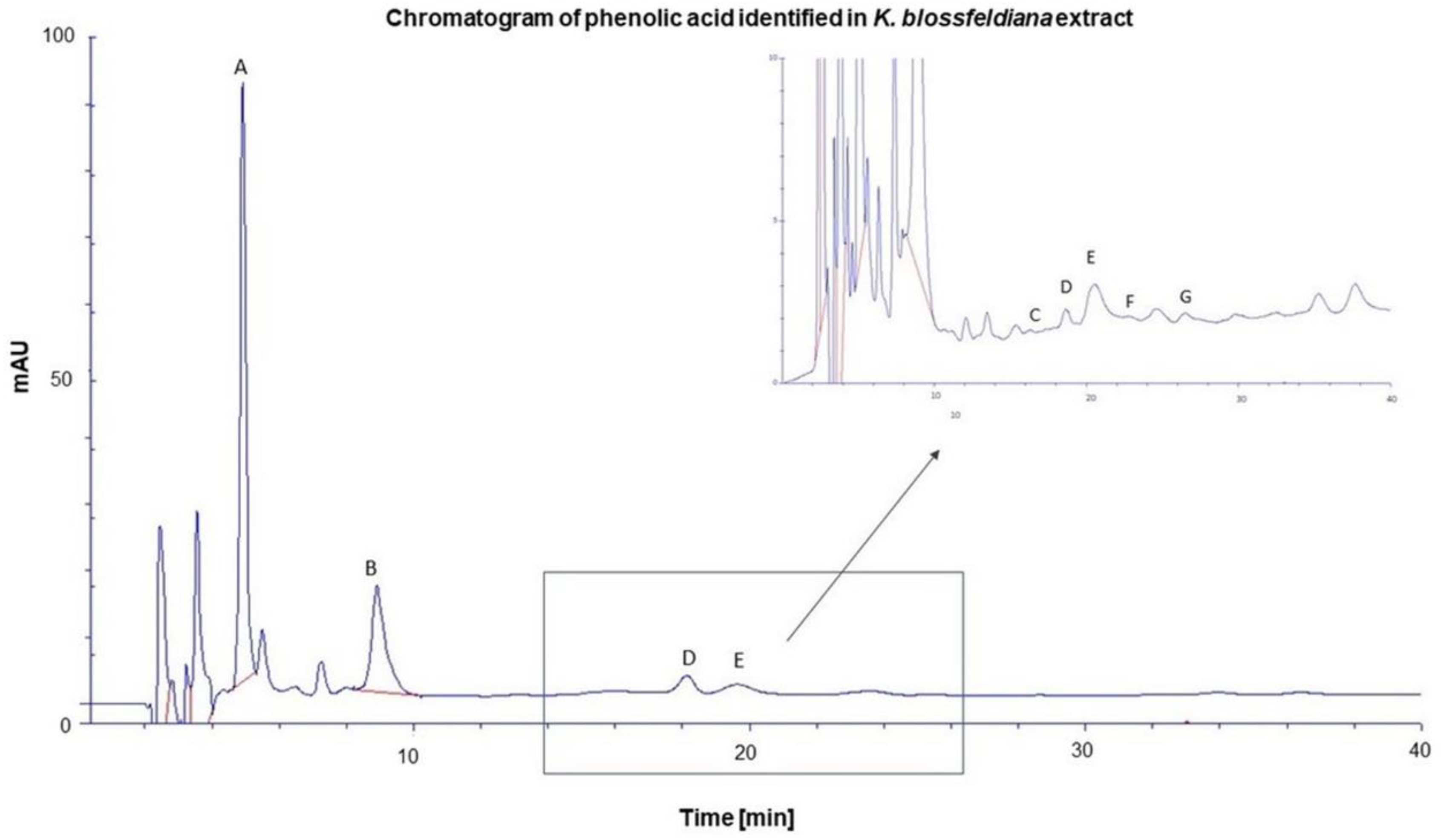
Figure 2.
Hyaluronidase activity in the presence of K. blossfeldiana ethanol extract. The experiment was prepared in three independent repeats (in three repetitions, n=9). Error bars represent standard deviations.
Figure 2.
Hyaluronidase activity in the presence of K. blossfeldiana ethanol extract. The experiment was prepared in three independent repeats (in three repetitions, n=9). Error bars represent standard deviations.
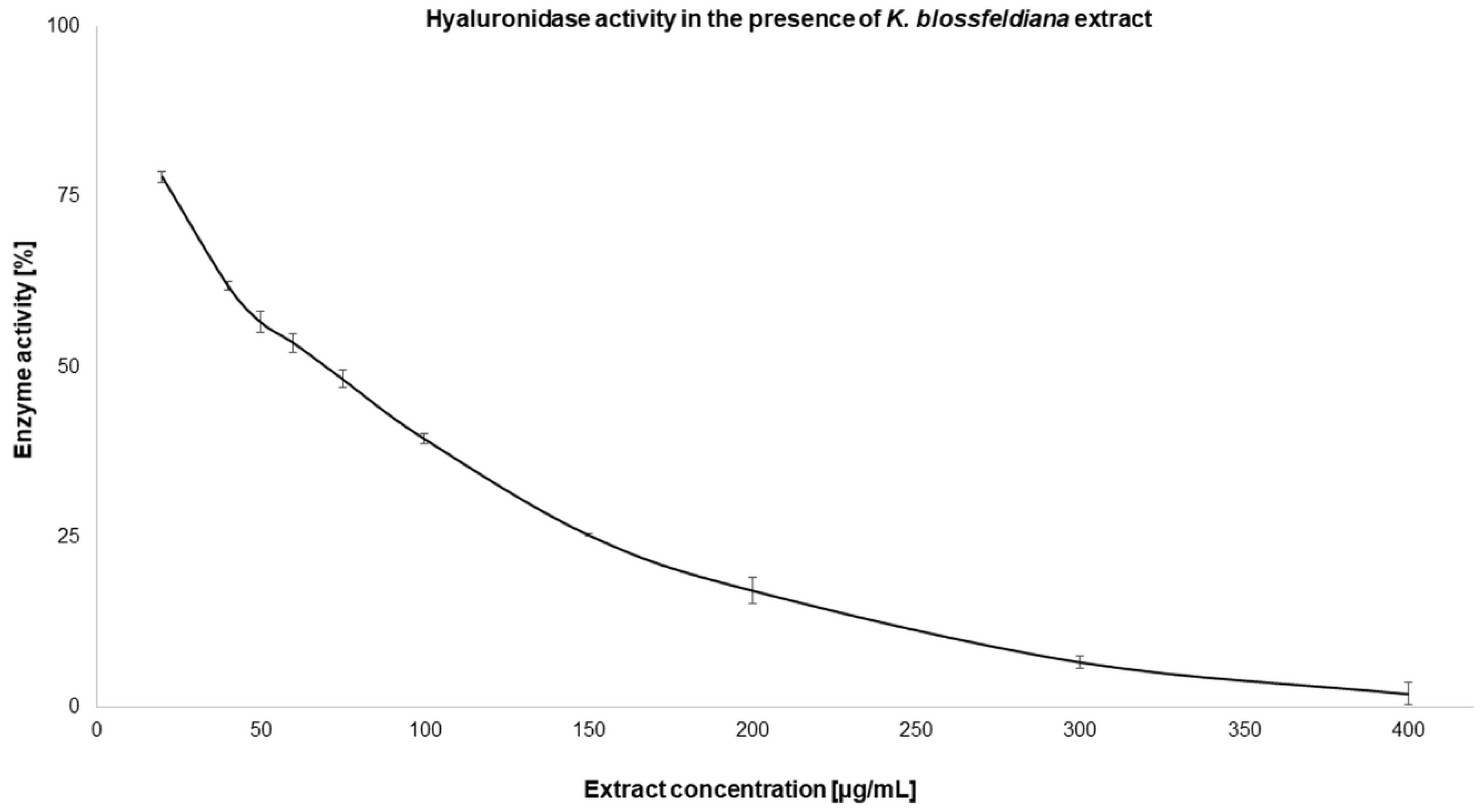
Figure 3.
Elastase activity in the presence of K. blossfeldiana ethanol extract. The experiment was prepared in three independent repeats (in three repetitions, n=9). Error bars represent standard deviations.
Figure 3.
Elastase activity in the presence of K. blossfeldiana ethanol extract. The experiment was prepared in three independent repeats (in three repetitions, n=9). Error bars represent standard deviations.
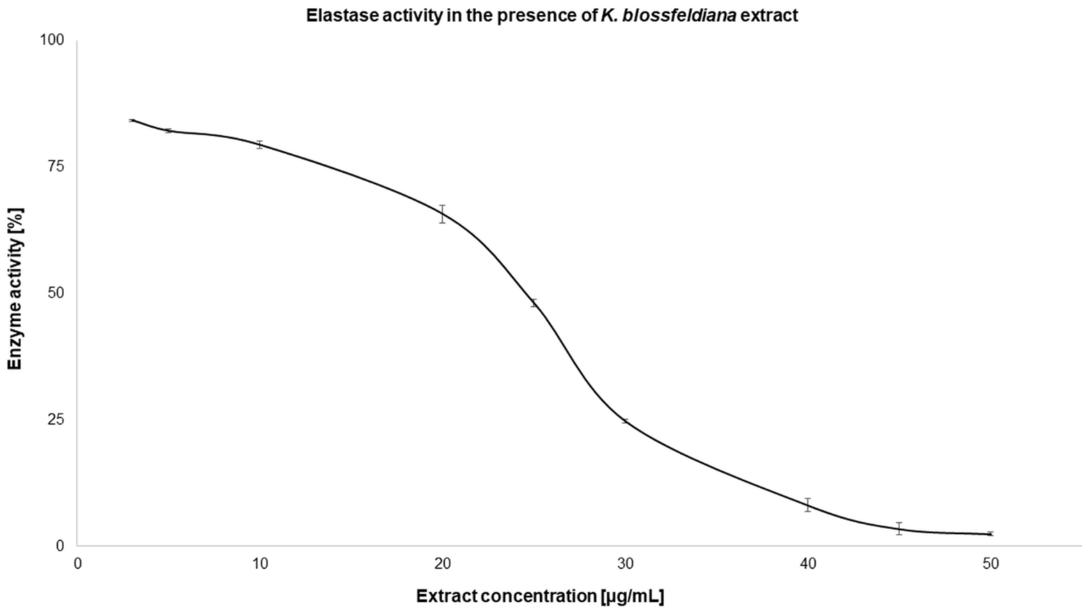
Figure 4.
Accumulation of phenolic acids in the skin. The content of individual phenolic acids was determined in the skin extraction fluid collected after the 24-hour penetration study. All values are presented as mean ± SD, where n = 3; n.d. – not detected.
Figure 4.
Accumulation of phenolic acids in the skin. The content of individual phenolic acids was determined in the skin extraction fluid collected after the 24-hour penetration study. All values are presented as mean ± SD, where n = 3; n.d. – not detected.
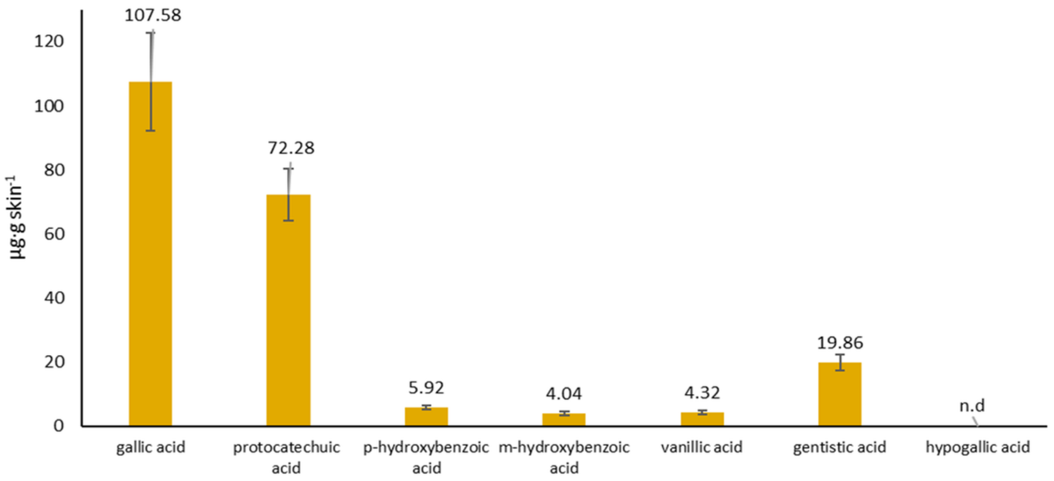

Table 1.
Compounds categories detected in the ethanol extract of K. blossfeldiana by LC-QTOF-MS.
| Compounds categories | Total number of compounds in each group | |
|---|---|---|
| 1 | Carbohydrate | 1 |
| 2 | Organic acid | 8 |
| 3 | Acyclic alcohol glycoside | 9 |
| 4 | Acyclic nitrile glycoside | 7 |
| 5 | Gallic acid derivative | 10 |
| 6 | Aminoacid | 3 |
| 7 | Acyclic acid glycoside | 2 |
| 8 | Benzoic acid derivative | 9 |
| 9 | Acetophenone derivative | 1 |
| 10 | Phenylpropanoid derivative | 12 |
| 11 | Phenol derivative | 3 |
| 12 | Flavanole | 6 |
| 13 | Sesquiterpenoid derivative | 1 |
| 14 | Dimeric proanthocyanidin | 5 |
| 15 | Megastigmane glycoside | 3 |
| 16 | Dimeric iridoid derivative | 1 |
| 17 | Monoterpene derivative | 1 |
| 18 | Bicyclo[3.1.1] glycoside | 1 |
| 19 | Phenylethane glycoside | 1 |
| 20 | Flavonole glycoside | 5 |
| 21 | Fatty acid glycoside | 1 |
| 22 | Iridoid glycoside | 1 |
| 23 | Lipid | 2 |
| 24 | Unidentified | 50 |
| Total identified compounds | 93 | |
Table 2.
Content of phenolic acids in HKB.
| Phenolic acid | µg·mL extract-1 |
|---|---|
| gallic acid | 284.74 ± 15.64 |
| protocatechuic acid | 74.35 ± 4.30 |
| p-hydroxybenzoic acid | 11.10 ± 0.43 |
| m-hydroxybenzoic acid | 0.85 ± 0.11 |
| vanillic acid | 12.63 ± 0.33 |
| gentistic acid | 9.10 ± 0.39 |
| hypogallic acid | 1.95 ± 0.07 |
The results are presented as mean values ± standard deviation (± SD) obtained from three experiments.
Table 3.
The antioxidant activity of K. blossfeldiana ethanol extract determined with DPPH, ABTS, and FRAP test. .
Table 3.
The antioxidant activity of K. blossfeldiana ethanol extract determined with DPPH, ABTS, and FRAP test. .
| Test | IC50 [µg/mL] | |
|---|---|---|
| K. blossfeldiana ethanol extract | Ascorbic acid | |
| DPPH | 7.72 ± 0.09* | 15.23 ± 0.76 |
| ABTS | 4.21 ± 0.32* | 7.38 ± 0.09 |
| FRAP | 11.25 ± 0.17* | 5.29 ± 0.21 |
The values were obtained from three experiments performed in three repetitions (n=9). Significant differences relative to ascorbic acid (as a standard) are marked with an asterisk “*” (p < 0.05).
Table 4.
The activity of K. blossfeldiana ethanol extract against hyaluronidase and elastase. .
| Enzymatic inhibition assay | IC50 [µg/mL] | |
|---|---|---|
| K. blossfeldiana ethanol extract | Oleanolic acid | |
| Hyaluronidase | 77.31 ± 2.44* | 49.33 ± 1.35 |
| Elastase | 26.8 ± 0.13* | 17.25 ± 0.27 |
The values were obtained from three experiments performed in three repetitions (n=9). Significant differences relative to oleanolic acid (as a standard) are marked with an asterisk “*” (p < 0.05).
Table 5.
Phenolic acids concentration in the acceptor fluid while 24 hours permeation study after application of HKB on the skin.
Table 5.
Phenolic acids concentration in the acceptor fluid while 24 hours permeation study after application of HKB on the skin.
| Time (h) |
Gallic acid | Protocatechuic acid | p-Hydroxybenzoic acid | m-Hydroxybenzoic acid | Vanillic acid | Gentistic acid | Hypogallic acid | |||
|---|---|---|---|---|---|---|---|---|---|---|
| (µg·cm-2) | ||||||||||
| 1 | 5.59 ± 0.52 |
n.d. |
n.d. | n.d. | n.d. | n.d. | n.d. | |||
| 2 | 7.97 ± 0.87 |
8.01 ± 1.68 | n.d. |
n.d. |
n.d. | n.d. | n.d. | |||
| 3 | 12.31 ± 0.56 | 9.57 ± 3.06 | n.d. | n.d. | n.d. | n.d. |
n.d. |
|||
| 5 | 24.35 ± 1.36 | 23.38 ± 2.27 | n.d. |
n.d. |
n.d. |
n.d. | n.d. | |||
| 8 | 51.70 ± 3.44 | 40.96 ± 2.67 | 4.27 ± 1.27 | 7.19 ± 0.36 | 8.95 ± 1.00 |
n.d. |
n.d. |
|||
| 24 | 249.73 ± 13.69 | 97.55 ± 5.31 | 9.96 ± 2.31 | 4.13 ± 0.56 | 15.41 ± 0.55 | 7.18 ± 0.28 | n.d. |
|||
n.d. – no identified in acceptor fluid. The results are presented as mean values ± standard deviation (± SD) obtained from three experiments.
Disclaimer/Publisher’s Note: The statements, opinions and data contained in all publications are solely those of the individual author(s) and contributor(s) and not of MDPI and/or the editor(s). MDPI and/or the editor(s) disclaim responsibility for any injury to people or property resulting from any ideas, methods, instructions or products referred to in the content. |
© 2024 by the authors. Licensee MDPI, Basel, Switzerland. This article is an open access article distributed under the terms and conditions of the Creative Commons Attribution (CC BY) license (http://creativecommons.org/licenses/by/4.0/).
Copyright: This open access article is published under a Creative Commons CC BY 4.0 license, which permit the free download, distribution, and reuse, provided that the author and preprint are cited in any reuse.


