Submitted:
18 September 2024
Posted:
19 September 2024
You are already at the latest version
Abstract
Ribavirin and its analogues exhibit an in vitro antiproliferative effect in cancer cells. In this work we studied the biological activities of a number of oxymethyl derivatives of ribavirin’s aglycon – 1,2,4-triazole-3-carboxamide. Oxymethyl derivatives of 1,2,4-triazole-3-carboxamide with substitu-tions at the fifth or first position of the triazole ring, were synthesized and their antiproliferative and antimicrobial effects were assessed. For both series the presence of an antiproliferative effect was inves-tigated and in the case of 1-oxymethyl derivatives was shown an antimicrobial potential against a Gram-positive bacteria Micrococcus luteus and Gram-negative bacterium Pseudomonas aeruginosa. The obtained results showed that the n-decyloxymethyl derivatives induced leukemia cell death at low mi-cromolar concentrations. We confirmed that n-decyloxymethyl derivatives of ribavirin inhibited cell cy-cle progression and induced accumulation of leukemia cells in subG1-phase. The molecular docking results suggest that oxymethyl derivatives may act by inhibiting translation initiation due to interfering with eIF4E assembly. The outcome results relived that active derivatives (1- or 5-n-decyloxymethyl-1,2,4-triazole-3-carboxamides) can be considered as a lead compound for anti-cancer treatments.
Keywords:
1
; 2
; 4-triazole-3-carboxamides
; ribavirin
; acute lymphoblastic leukemia
; chronic myeloid leukemia
; cancer treatment
; antimicrobial effect
1. Introduction
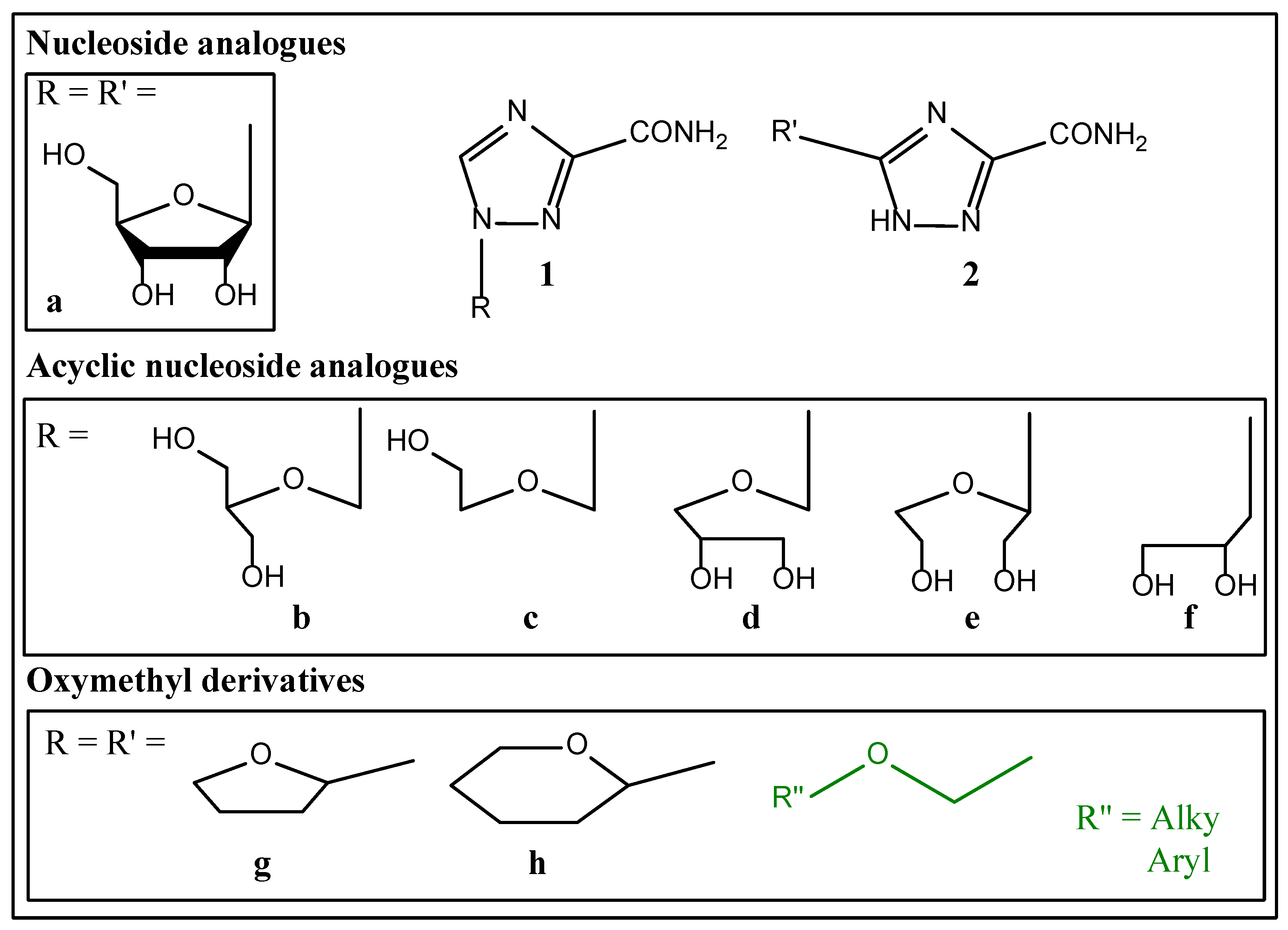
Synthetic analogues of natural nucleosides have wide spectrum of applications as antiviral, anticancer and antibacterial compounds. An example is ribavirin (1-(β-D-ribofuranosyl)-1,2,4-triazole-3-carboxamide, 1a), which is not only used as an antiviral drug, but also shows significant potential in a therapy of cancer including blood cancer [1,2], as well as a moderate antimicrobial effect [3]. Thus, the multivalency of ribavirin applications makes it an interesting parent structure for new drug candidate design.
However, numerous studies have shown that ribavirin has teratogenic and genotoxic effects, which significantly limit its therapeutic application [4]. This feature is common to most of the other nucleoside analogues, probably due to their involvement into the basic metabolic pathways. It is therefore of interest to search for alternatives of compound 1a that have meaningful structural differences from nucleosides. For some ribavirin analogues with different fragments at the 1- and/or 5-positions of triazole ring show specific activity in the cells of several cancer type. For example, several triazoles with biphenyl group in 1-position are cytotoxic in breast cancer cells in micromolar concentrations [5]. Several hybrid molecules containing secoestroides and triazole fragments demostrate cytotoxicity in cervical cancer and breast cancer cell lines [6,7,8]. However, despite significant progress in bioorganic chemistry of nucleoside analogues, identification of the most promising ways of their modification to maintain high efficacy and simultaneously reduce the significant side effects is highly in demand. Therefore, the traditional approach in the synthesis of new agents, followed by the identification of their biological effects, remains relevant to the solution of this problem.
Recently we showed antiproliferative effects of the ribavirin aglycon in acute lymphoblastic leukemia and chronic myeloid leukemia cell lines [9]. We obtained ribavirin analogues by a replacement of ribavirin ribose fragment with tetrahydropyran and tetrahydrofuran groups in 5- and 1-positions of the triazole ring. We showed the accumulation of cancer cells treated with 1,2,4-triazol-3-carboxamides in the G1 phase of the cell cycle, and the induction of caspase-3 cleavage resulting in apoptosis in leukemia cells [9]. Therefore, we assume that the ribavirin analogs with non-sugar fragment in 1- or 5-position may act like nucleoside analogs.
The present study was undertaken to prepare a series of oxymethyl derivatives of 1,2,4-triazole-3-carboxamide as well as evaluation of its anticancer actions in leukemia cell lines and antimicrobial activities in Gram-positive and Gram-negative bacteria.
2. Results and Discussions
Biologically active nucleosides analogues were obtained by replacing the carbohydrate fragment with its acyclic analogue, mostly with the exclusion of some hydroxyl groups. In the case of ribavirin analogues obtained using this approach, there should be distinguished several derivatives with acyclic carbohydrate fragment and substitution at the position 1 – 1,2,4-triazole-3-carboxamides 1b-f. Another approach to the nucleoside analogues modification was the replacement of the N-glycoside bond with a C-glycoside for easier exclusion of biolabile bond from the structure of the analogue without change in the main pharmacophore fragments. The ribavirin C-nucleoside analogue 2a was obtained by this method applied to modification 1a [10,11]. The authors noted the importance of the hydroxyethoxymethyl fragment presence in the structure of the molecule for an acyclic analogue to retain antiviral activity. Later, the other 1-hydroxyethoxymethyl derivatives of 5-substituted 1,2,4-triazole-3-carboxamides were synthesized, some of which showed antiviral activity against hepatitis C virus and anticancer effect on cell models [9,12,13]. Thus, the reduction of the carbohydrate to a hydroxyethoxymethyl moiety does not prevent molecular recognition of ribavirin analogues by a significant number of enzymes.
In our previous work on modification of the carbohydrate moiety, we have shown that derivatives of 1,2,4-triazole-3-carboxamide 1g-h and 2g-h substituted at the position 5 as well as at the position 1 of the triazole ring with 2-tetrahydrofuranyl or 2-tetrahydropyranyl groups, analogues of the carbohydrate backbone lacking hydroxyl groups, inhibit proliferation of chronic myeloid (K562) and acute lymphoblastic (CCRF-SB) leukemia cells [9]. However, the detailed mechanism of the biological activity of compounds 1g-h and 2g-h remains unclear. Therefore, we assumed that the ribavirin analogs with non-sugar fragment in 1- or 5-position may in some aspects act like a nucleoside analogs, in particular reveal anticancer and antimicrobial effects.
Figure 1.
Ribavirin 1a and its analogues.
2.1. Synthesis
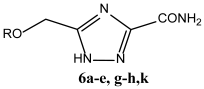
Two series of compounds were synthesized: 5-oxymethyl-1,2,4-triazole-3-carboxamides 6a-e, g-h, k and 1-oxymethyl-1,2,4-triazole-3-carboxamides 1c and 11a-k. The methods of their synthesis differed for each series: the introduction of 5-oxymethyl fragment was carried out by cyclization of triazole fragment, while the introduction of 1-oxymethyl fragment was carried out by alkylation of methyl 1,2,4-triazole-3-carboxylate.
5-oxymethyl analogues of ribavirin 6a-e, g-h, k were synthesized by the previously described method [15] consisting in ammonolysis of ethyl esters of 5-oxymethyl-1,2,4-triazole-3-carboxylic acids 5a-e, g-h, k obtained by treatment of β-N-t-butyloxycarbonyloxalamidrazone 11 with oxyacetic acids chlorohydrides followed by one-pot cyclization of intermediates (Figure 2).
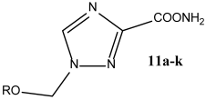
In the case of oxymethyl substituents introduction into the 1,2,4-triazole ring, which is necessary step in compounds 11 synthesis, an alkylation can occure at any nitrogen of the triazole ring, leading to a formation of three regioisomers [16,17]. According to literature sources, the method of introducing an oxymethyl substituent via triazole carboxylic acid esters N-silyl derivatives by oxymethylacetates is considered as the most regioselective, for example, the only product of such alkylation for methyl 1,2,4,-triazole-3-carboxylate is methyl 1-alkoxymethyl 1,2,4-triazole-3-carboxylate [17].
In our study, methyl 1-oxymethyl-1,2,4-triazole-3-carboxylates 10a-j were prepared in two steps: first, we obtained the silyl derivatives of methyl 1,2,4-triazole-3-carboxylate 7 by its treatment with 1,1,1,1,3,3,3,3-hexamethyldisilazane (HMDS) followed by an addition of oxymethylacetates 9a-j in the presence of Lewis acid – tin tetrachloride (Figure 3). Compoundes 9a-j were synthesized from dialkoxymethanes 14a-j [18] which, in turn, were obtained by a known method [19]. In the case of 1-([2-hydroxyethoxy]methyl)-1,2,4-triazole-3-carboxamide 1c, compound 8 was treated with [2-(acetyloxy)ethoxy]methyl acetate 9l [20] obtained from 1,3-dioxalane, the acetate protecting group of the ethyloxymethyl moiety was removed by ammonolysis. Methyl 1-methoxymethyl-1,2,4-triazole-3-carboxylate 10a was isolated by column chromatography resulting 38.5% yield. The esters 10b-j were used at the next stage without further purification. The amides 11a-j were obtained by ammonolysis of the esters 10a-j (Figure 3) and were purified by recrystallization from an ethanol-ethyl acetate mixture in yields ranging from 23 to 91%.
1-(Phenoxymethyl)-1,2,4-triazole-3-carboxamide 11k was prepared using diphenoxymethane 14k synthesized according to a procedure described in the literature [21]. Methyl 1,2,4-triazole-3-carboxylate hydrochloride 13 was treated with 14k to give the ester 1-(phenoxymethyl)-1,2,4-triazole-3-carboxylate 10k followed by its ammonolysis (Figure 4). Amide 11k was purified in a similar to the previous amides 16a-j manner resulting 52% yield.
The structures of the obtained compounds were established using a set of physicochemical methods: 1H and 13C NMR, HRMS. A combination of APT and 1H-13C HMBC NMR experiments was used to establish the position of the oxymethyl substituent in the case of compound 10a. We identified structures 11b-j and 1c as the position 1 isomers based on the similarity of their NMR characteristics to those of 11a (11a was obtained by ammonolysis of 10a). In the case of 11k, the position of the phenoxymethyl radical was established by DEPT-135 and 1H-13C HMBC NMR.

Table 2.
Synthesized 1-oxymethyl-1,2,4-triazole-3-carboxamides 11.
 |
||
| S. No. | R | Yield |
| 11a | Me | 87% |
| 11b | Et | 78% |
| 11c | n-Pr | 78% |
| 11d | i-Pr | 91% |
| 11e | n-Bu | 81% |
| 11f | t-Bu | 49% |
| 11g | n-C10H21 | 78% |
| 11h | Bn | 89% |
| 11i | cyclo-C5H10 | 82% |
| 11j | cyclo-C6H12 | 53% |
| 11k | Ph | 52% |
| 1c | HO(CH2)2 | 83% |
2.2. In Vitro Stu
2.2.1. Anti-Cancer Activity In Vitro
The in vitro cytotoxic activities of the synthesized compounds were evaluated on CCRF-SB and K562 cells using MTT assay. Compounds 11g and 6g showed the highest cytotoxic activity in the leukemia cell lines after 24 h exposition, as shown in Table 3. CC50 values for 11g were calculated as 13.6±0.3 µM in the K562 cells and 112±19 µM in the CCFR-SB cells, respectively. CC50 for 6g were 391±15 µM in K562 cell line.
CC50 values for 11g were about 20-fold lower than ribavirin and about 4-fold lower than Cyt in the K562 cells. Most notably, that one of the derivatives 6g showed activity against acute lymphoblastic leikemia (CCRF-SB), an aggressive form of pediatric leukemia.
For other compounds CC50 values were not determined. However, a number of compounds showed a dose-response cytostatic effect on leukemia cells at 72 h exposure assuming that newly synthesized 1,2,4-triazole-3-carboxamide derivatives may possess antiproliferative effect aassociated with low toxicity. MTT assay after 72 h revealed the cytotoxic effects of 11e, 6g, and 6k in acute lymphoblastic leikemia cell line, and cytotoxic effects of 11i, 11h, 11f, 6e and 6k in chronic myeloid leikemia cells (data not shown). Consequently, compounds 11e, 11g, 11i, 11h, 11f, 6e, 6g and 6k were selected for study of its antiproliferative activity.
To evaluate the effect of compounds on non-transformed cells, human peripheral blood mononuclear cells (PBMC) were isolated from the whole blood of 3 healthy volunteers. Then PBMCs were incubated 72 h with 1,2,4-triazole-3-carboxamide derivatives in the highest concentrations (500 µM). The normal cells were less sensitive to compounds than cancer cells (Figure 6), highlighting the selectivity of action of novel compounds on malignant cells.
We conducted in vitro trypan blue exclusion assay to test the cytostatic (antiproliferative) activity of newly synthesized 1,2,4-triazole-3-carboxamide derivatives. The cells were incubated with the active compounds for 24-72 h at a concentrations equal to the calculated CC20 value. Compound 11g significantly reduced cell proliferation in chronic myeloid leikemia cells cells and caused minimal cell death in acute lymphoblastic leikemia cell line. Compounds 6g and 11e showed dose-depending antiproliferative action in CCRF-SB cell line (Figure 7a, b).
To investigate the mechanism underlying the cell growth inhibition induced by 1,2,4-triazole-3-carboxamide derivatives, the cell cycle profile was analyzed by flow cytometry with PI staining. The K562 and CCRF-SB cells were exposed to compounds for 72h. In Figure 6c it is demonstrated that the compound 6g caused an increase in the cell population in the G0 phase, indicating cell death in CCRF-SB culture. The population of the G2/M and S phases of CCRF-SB cells reduced after the treatment with 7 µm 6g compared with control. At the same time, all compounds increased accumulation of cells in G1 phase and caused a decrease in the percentage of cells in the G2/M phase in K562 cell line. The treatment of K562 cells with 7 µM 11g significantly reduced the fraction of cells in the S and G2/M phases and increased the proportion of cells in the G1 phase. Furthermore, 6g and 11g significantly increased accumulation of cells in the subG1 phase corresponding to apoptotic cells by 7 and 8 times respectively in K562 cells. Compounds 6g and 11g ability to induce cell death is concordant with cytotoxicity determinated by the MTT assay.
2.2.2. Antimicrobial Effects Studies
The multivalency of the biological effects of ribavirin – the parent structure of the 1- or 5-oxymethyl-1,2,4-triazole-3-carboxamides – prompted us to study their antimicrobial properties. The antimicrobial potential of compounds 6a-e, g-h, k and 11a-k was investigated in comparison with that of reference molecules 1a and 1c against the following series of microorganisms: Micrococcus luteus ATCC 9341, Staphylococcus aureus INA 00985, Pseudomonas aeruginosa ATCC 27853, Candida albicans ATCC 14053 on agarose nutrient medium at concentrations of 25 mM (Table 4).
Compounds 6 have not showed any suppression of microorganism growth. Compounds 11i, j and 1c showed bacteriostatic activity against the Gram-positive organism M. luteus, but not against S. aureus, in contrast to ribavirin 1a, which showed no antimicrobial activity against such organisms. In the case of the Gram-negative microorganism P. aeruginosa, moderate activity compared to that of 1a was observed for compound 11c. In relation to C. albicans, the studied compounds 1c, 11 showed no activity, ribavirin 1a showed the highest activity.
2.3. Molecular Docking
Due to the revealed effects of the synthesized compounds 6a-e, g-h, k and 11a-k, 1с towards acute lymphoblastic leukemia and chronic myeloid leukemia cell lines, we became interested in trying to assume the mechanism underlying the action. As known, ribavirin undergoes phosphorylation in cells to form ribavirin 5-monophosphate (RMP) [22]. A number of cellular targets have been shown for RMP, including inosine-5’-phosphate dehydrogenase (IMPDH) and eukaryotic translation initiation factor 4E (elF4F) [23,24,25]. Inhibition of IMPDH occurs due to insertion into the inosine monophosphate binding site, and elF4E is presumably according to various sources either due to insertion into the 5′-cap mRNA binding site or by interfering with the assembly of protein subunits of the factor [26,27,28,29,30]. Оxymethyl derivatives of TCA 6a-e,g,h,k and 11a-k do not have a hydroxyl substituent and therefore cannot be phosphorylated. Therefore, the main opportunity for them to participate in these biochemical pathways remains to block the interaction of the eIF4E and eIF4G subunits of factor 4E by binding to at least one of them [31]. An example of such an effect of low molecular weight compounds is a inhibitor of this interaction, 4EGI-1 [32]. 4EGI-1 disrupts the eIF4E/eIF4G association in vitro and in vivo, and reduces viability of a wide range of cancer cells such as breast cancer and multiple myeloma [33]. 4EGI-1 inhibits tumor growth in in vivo models of acute myeloid leukemia and chronic lymphocytic leukemia. [34,35,36,37]. Therefore, we used a region of the protein surface characteristic of 4EGI-1 binding as a target for modeling. The structure of elF4E protein (PDB: 4TPW) and the structures of low molecular weight ligands 1g, h and 2g, h, 6, 11 optimized with OPLS3e force field were used for molecular docking which was performed in Schrodinger Maestro.
According to the simulation results, several newly synthesized compounds 6 and 11 as well as ribavirin 1a and its C-nucleoside analogue 2a demonstrate a preferential localization in the binding site similar to the known inhibitor 4EGI-1. Compound 1g in simulation binds mirrorly to ribavirin 1a binding. 1g and 1a occupis the same cavities in the binding site (Figure 8a). Compound 6g with its carboxamide fragment falls into the binding region of ribose fragment 1a, similarly to 1g, h and 2g, h, and its lipophilic tail binds in the cavity characteristic for 4EGI-1 inhibitor formed by Asp 51, Asn 59 and Lys 49. It is in accordance with the results of higher cytotoxity of 6g compared to compounds 1g, h and 2g, h (Figure 8b). Compound 11g demonstrating higher toxicity in cancer cells in vitro, is located at the binding site in a manner similar to the known 4EGI-1 inhibitor. The lipophilic tail of 11g enters the hydrophobic cavity of the binding site formed by amino acid residues Phe 47, Tyr 91, Ile 79 and 63, while the formation of hydrogen bonds occurs via triazole fragment in the cavity formed by Asp 51, Asn 59 and Lys 49, similarly to 4EGI-1 (Figure 8c).
Thus, as a result of the modeling, it was possible to trace the correlation between the pattern of location of hydroxymethyl analogues of ribavirin on the surface of eIF4E and their in vitro toxicity to cancer cells. This suggests that a possible mechanism of action of the synthesized compounds may be associated with inhibition of RNA translation due to disruption of the assembly of the elF4F complex.
3. Materials and Methods
3.1. Synthetic Section
3.1.1. 5-(n-Propoxymethyl)-1,2,4-Triazole-3-Carboxamide 6c
1.88 g (11.02 mmol) of n-propyloxyacetic acid chloroanhydride was added dropwise to a suspension of 1.19 g (5.15 mmol) of β-N-(t-butyloxycarbonyl)ethyloxalamidrazone 4 in anhydrous pyridine while cooling the reaction mixture to 0°C. The reaction mass was brought to boiling point and stirred for 20 h. After the reaction was completed (control was carried out using TLC), the solvent was removed on a vacuum rotary evaporator. The residue was dissolved in 1 M aqueous HCl solution and extracted 3 times with ethyl acetate in equal portions. The organic phases were combined and dried using Na2SO4, the solvent was removed on a vacuum rotary evaporator. The crude product 5c was isolated by flash chromatography on silica gel using chloroform/methanol system (with a methanol gradient from 0 to 7%) as an eluent. Crude 5c was dissolved in 2 ml of a 10 M ammonia methanol solution and heated to boiling under reflux for 12 hours, the solvent was removed using a vacuum rotary evaporator. Residue was suspended in anhydrous acetone, filtered and dried in a desiccator under reduced pressure above NaOH for 12 hours to yield 0.23 g (24%) 6c as white crystals.
Rf = 0.61 (1% CH3OH in CHCl3), mp 110-111°C. 1H NMR spectrum (DMSO-d6) δ: 0.83 (t, 3H, J=7.09, CH3CH2CH2); 1.50 (se, 2H, J=7.09, CH3CH2CH2); 3.40 (t, 2H, J=6.09, CH3CH2CH2); 4.51 (s, 2H, OCH2); 7.70 and 8.01 (2s, 2H, NH2). 13C NMR spectrum (DMSO-d6) δ: 10.54; 22.38; 63.66; 72.13; 153.22; 156.84; 159.39. HRMS: for C7H12N4O2 m/z [M+H]+ calculated: 185.0960; found: 185.0981; LC 5-(n-propoxymethyl)-1,2,4-triazole-3-carboxamide content: spectrophotometric detection 235 nm no less than 98%.
3.1.2. Methyl 1-(Methoxymethyl)-1,2,4-Triazole-3-Carboxylate (10a)
0.5 g (3.9 mmol) of methyl 1,2,4-triazole-3-carboxylate was suspended in 4 ml (19 mmol) of HMDS and stirred under reflux for 1 h. After cooling the excess of HMDS was removed using a rotary evaporator. 5 ml of anhydrous acetonitrile, 1.70 ml (18 mmol) of 9a, 0.45 ml (3.9 mmol) of SnCl4 were added to the residue and the reaction was stirred under reflux until the starting ester was no longer detectable by TLC. The reaction mass was poured into 10 ml of saturated sodium bicarbonate solution and the precipitates formed were filtered off. The filtrate was extracted with chloroform (4x10 ml), the combined chloroform extracts were washed with water (10 ml) and dried over CaCl2. Volatile components were evaporated. 0.25 g (38.5%) of the product 10a was isolated by column chromatography on silica gel, eluent: toluene-acetone, modified with 1% triethylamine (acetone gradient from 5 to 7%), as transparent oil.
Rf = 0.26 (30% acetone in toluene). 1H NMR spectrum (CDCl3) δ: 3.40 (s, 3H, OCH3); 4.99 (s, 3H, COOCH3); 5.53 (s, 2H, OCH2); 8.34 (s, 1H, CH). 13C NMR spectrum (CDCl3) δ: 52.86; 57.66; 80.39; 145.89; 154.89; 159.93. For C6H9N3O3 m/z [M+H]+ calculated: 172.1; found: 172.0.
3.1.3. 1-(Methoxymethyl)-1,2,4-Triazole-3-Carboxamide (11a)
0.2 g (1.2 mmol) of methyl 1-(methoxymethyl)-1,2,4-triazole-3-carboxylate was dissolved in 1.5 ml of 10 M ammonia solution in methanol and stirred at room temperature to conversion of the starting material (control by TLC). Volatile components were removed on a rotary evaporator, 0.15g (87%) of the product 11a was isolated by recrystallization from a solvent mixture: ethanol-ethyl acetate as white crystals.
Rf = 0.53 (1% CH3OH in CHCl3), mp 146-147°C. 1H NMR spectrum (DMSO-d6) δ: 3.29 (s, 3H, OCH3); 5.51 (s, 2H, OCH2); 7.51 and 7.67 (2s, 2H, NH2); 8.80 (s, 1H, CH). 13C NMR spectrum (DMSO-d6) δ: 56.58; 79.16; 146.21; 157.48; 160.41. HRMS: for C5H8N4O2 m/z [M+H]+ calculated: 157.0726; found: 157.0733; LC 1-(methoxymethyl)-1,2,4-triazole-3-carboxamide content: spectrophotometric detection 235 nm no less than 98%.
3.1.4. General Procedure for the Preparation of 1-Substituted of 1,2,4-Triazole-3-Carboxamides 11b-j, 1c
Methyl 1,2,4-triazole-3-carboxylate was suspended in 5 eq. HMDS and stirred under reflux for 1 hour in an anhydrous atmosphere. After cooling the excess of HMDS was removed using a rotary evaporator. Anhydrous acetonitrile, 5 eq. 9a, 1 eq. SnCl4 were added to the residue and the reaction was stirred under reflux until the starting ester was no longer detectable by TLC. The reaction mass was poured into saturated sodium bicarbonate solution and the precipitates formed were filtered off. The filtrate was extracted with chloroform, the combined chloroform extracts were washed with water (10 ml) and dried over CaCl2. Volatile components were evaporated. The product was isolated by column chromatography on silica gel, eluent: toluene-acetone, modified with 1% triethylamine (acetone gradient from 5 to 7%).
1-(ethoxymethyl)-1,2,4-triazole-3-carboxamide (11b).
From 0.5 g (2.5 mmol) of methyl 1,2,4-triazole-3-carboxylate, 0.38 mg (78%) of product 11b was obtained as white crystals.
Rf = 0.52 (1% CH3OH in CHCl3), mp 127°C. 1H NMR spectrum (DMSO-d6) δ: 1.08 (t, J=7.03, 2H, CH3CH2); 3.29 (s, 3H, OCH3); 3.54 (q, J=7.03, 2H, CH3CH2); 5.55 (s, 2H, OCH2); 7.61 and 7.75 (2s, 2H, NH2); 8.79 (s, 1H, CH). 13C NMR spectrum (DMSO-d6) δ: 14.57; 64.50; 77.64; 146.01; 157.37; 160.37. HRMS: for C6H10N4O2 m/z [M+H]+ calculated: 171.0882; found: 171.0893; LC 1-(ethoxymethyl)-1,2,4-triazole-3-carboxamide content: spectrophotometric detection 235 nm no less than 98%.
1-(n-propyloxymethyl)-1,2,4-triazole-3-carboxamide (11c).
From 1 g (7.8 mmol) of methyl 1,2,4-triazole-3-carboxylate, 0.36 mg (78%) of product 11c was obtained as white crystals.
Rf = 0.69 (1% CH3OH in CHCl3), mp 125-126°C. 1H NMR (DMSO-d6) δ: 0.80 (t, 3H, J = 7.41, ОCH2CH2CH3); 1.41-1.53 (m, 2H, ОCH2CH2CH3); 3.44 (t, 2H, J = 6.60, ОCH2CH2CH3); 5.55 (s, 2H, OCH2); 7.57 and 7.79 (2s, 2H, NH2); 8.79 (s, 1H, CH). 13C NMR (DMSO-d6) δ: 10.19; 22.00; 70.61; 77.88; 145.99; 157.35; 160.35. HRMS: for C5H8N4O2 m/z [M+H]+ calculated: 185.1038; found: 185.1048. LC 1-(n-propyloxymethyl)-1,2,4-triazole-3-carboxamide content: spectrophotometric detection 235 nm no less than 97%.
1-(isopropyloxymethyl)-1,2,4-triazole-3-carboxamide (11d).
From 1 g (7.8 mmol) of methyl 1,2,4-triazole-3-carboxylate, 0.42 mg (91%) of product 11d was obtained as white crystals.
Rf = 0.65 (1% CH3OH in CHCl3), mp 145°C. 1H NMR (DMSO-d6) δ: 1.06 (d, 3H, J = 6.12, OCHCH3); 3.77-3.81 (m, 1H, J = 6.11, ОCH); 5.56 (s, 2H, OCH2); 7.57 and 7.79 (2s, 2H, NH2); 8.79 (s, 1H, CH). 13C NMR (DMSO-d6) δ: 21.94; 70.36; 75.75; 145.98; 157.33; 160.47. HRMS: for C5H8N4O2 m/z [M+H]+ calculated: 185.1039; found: 185.1058. LC 1-(isopropyloxymethyl)-1,2,4-triazole-3-carboxamide content: spectrophotometric detection 235 nm no less than 98%.
1-(n-butyloxymethyl)-1,2,4-triazole-3-carboxamide (11e).
From 1 g (7.8 mmol) of methyl 1,2,4-triazole-3-carboxylate, 0.53 mg (81%) of product 11e was obtained as white crystals.
Rf = 0.5 (1% CH3OH in CHCl3), mp 123-126°C. 1H NMR spectrum (300 MHz, DMSO-d6) δ: 0.80 (t, J=7.41, 2H, CH3CH2); 1.47 (q, J=6.85, 2H, CH3CH2); 3.44 (t, J=6.60, 2H, CH2CH2); 5.55 (s, 2H, OCH2); 7.58 and 7.79 (2s, 2H, NH2); 8.79 (s, 1H, CH). 13C NMR spectrum (75 MHz, DMSO-d6) δ: 10.57; 22.00; 70.61; 77.88; 145.99; 157.35; 160.35. HRMS: for C8H14N4O2 m/z [M+H]+ calculated: 199.1195; found: 199.1205. LC 1-(n-butyloxymethyl)-1,2,4-triazole-3-carboxamide content: spectrophotometric detection 235 nm no less than 97%.
1-(tert-butoxymethyl)-1,2,4-triazole-3-carboxamide (11f).
From 1 g (7.8 mmol) of methyl 1,2,4-triazole-3-carboxylate, 0.39 mg (49%) of product 11f was obtained as white crystals.
Rf = 0.65 (1% CH3OH in CHCl3), mp 194-195°C. 1H NMR (DMSO-d6) δ: 1.18 (s, 9H, O(CH3)3); 5.57 (s, 2H, OCH2); 7.55 and 7.75 (2s, 2H, NH2); 8.76 (s, 1H, CH). 13C NMR (DMSO-d6) δ: 27.28; 72.94; 73.51; 132.74; 157.63; 159.84. HRMS: for C8H14N4O2 m/z [M+H]+ calculated: 199.1195; found: 199.1208. LC 1-(tert-butoxymethyl)-1,2,4-triazole-3-carboxamide content: spectrophotometric detection 235 nm no less than 96%.
1-(n-decyloxymethyl)-1,2,4-triazole-3-carboxamide (11g).
From 1 g (7.8 mmol) of methyl 1,2,4-triazole-3-carboxylate, 0.36 mg (78%) of product 11g was obtained as white crystals.
Rf = 0.60 (1% CH3OH in CHCl3), mp 122-124°C. 1H NMR (DMSO-d6) δ: 0.84 (t, 3H, J = 6.83, O(CH2)9CH3; 1.20 (s, 14H, OCH2CH2(CH2)7CH3); 1.42-1.46 (m, 2H, OCH2CH2(CH2)7CH3); 0.37 (t, 2H, J = 6.50, OCH2CH2(CH2)7CH3); 7.57 and 7.77 (2s, 2H, NH2); 8.78 (s, 1H, CH). 13C NMR (DMSO-d6) δ: 22.00; 28.59; 28.84; 68.97; 145.98; 160.34. HRMS: for C14H26N4O2 m/z [M+H]+ calculated: 283.2134; found: 283.2150. LC 1-n-decyloxymethyl-1,2,4-triazole-3-carboxylic acid amide content: spectrophotometric detection 235 nm no less than 96%.
1-(benzyloxymethyl)-1,2,4-triazole-3-carboxamide (11h).
From 1 g (7.8 mmol) of methyl 1,2,4-triazole-3-carboxylate, 0.63 mg (89%) of product 11h was obtained as white crystals.
Rf = 0.65 (1% CH3OH in CHCl3), mp 168-169°C. 1H NMR (DMSO-d6) δ: 4.60 (s, 2H, CH2C6H5); 5.67 (s, 2H, OCH2); 7.26-7.37 (m, 2H, C6H5); 7.60 and 7.82 (2s, 2H, NH2); 8.83 (s, 1H, CH). 13C NMR (DMSO-d6) δ: 70.69; 77.47; 127.65; 128.28; 136.87; 146.19; 157.46; 160.38. HRMS: for C11H12N4O2 m/z [M+H]+ calculated: 233.1039; found: 233.1089. LC 1-(benzyloxymethyl)-1,2,4-triazole-3-carboxamide content: spectrophotometric detection 235 nm no less than 97%.
1-(cyclopentyloxymethyl)-1,2,4-triazole-3-carboxamide (11i).
From 1 g (7.8 mmol) of methyl 1,2,4-triazole-3-carboxylate, 0.31 mg (53%) of product 11i was obtained as white crystals.
Rf = 0.66 (1% CH3OH in CHCl3), mp 153-154°C. 1H NMR (DMSO-d6) δ: 1.46-1.66 (m, 8H, OC5H9); 4.08 (s, 1H, OCH); 5.54 (s, 2H, OCH2); 7.57 and 7.79 (2s, 2H, NH2); 8.79 (s, 1H, CH). 13C NMR (DMSO-d6) δ: 22.90; 31.78; 76.42; 79.97; 145.99; 157.31; 160.40. HRMS: for C9H10N4O2 m/z [M+H]+ calculated: 211.1195; found: 211.1208. LC 1-(cyclopentyloxymethyl)-1,2,4-triazole-3-carboxamide content: spectrophotometric detection 235 nm no less than 98%.
1-(cyclohexyloxymethyl)-1,2,4-triazole-3-carboxamide (11j).
From 1 g (7.8 mmol) of methyl 1,2,4-triazole-3-carboxylate, 0.58 mg (82%) of product 11j was obtained as white crystals.
Rf = 0.46 (1% CH3OH in CHCl3), mp 155-156°C. 1H NMR (DMSO-d6) δ: 1.70-1.17 (m, 10H, C5H10); 3.49-3.51 (m, 1H, OCH); 5.59 (s, 2H, OCH2); 7.62 and 7.85 (2s, 2H, NH2); 8.81 (s, 1H, CH). 13C NMR (DMSO-d6) δ: 23.26; 25.06; 31.61; 75.64; 145.97; 157.33; 160.48. HRMS: for C10H16N4O2 m/z [M+H]+ calculated: 225.1352; found: 225.1380. LC 1-(cyclohexyloxymethyl)-1,2,4-triazole-3-carboxamide content: spectrophotometric detection 235 nm no less than 96%.
1-([2-hydroxyethoxy]methyl)-1,2,4-triazole-3-carboxamide (1c).
From 0.2 g (0.99 mmol) of methyl 1,2,4-triazole-3-carboxylate, 0.58 mg (83%) of product 1c was obtained as white crystals.
Rf = 0.35 (5% CH3OH in CHCl3), mp 154-156°C. 1H NMR spectrum (DMSO-d6) δ: 3.44-3.55 (m, 4H, -ОCH2CH2О-); 5.59 (s, 2H, OCH2); 7.57 and 7.79 (2s, 2H, NH2); 8.79 (s. 1H, CH). 13C NMR spectrum (DMSO-d6) δ: 59.79; 70.98; 78.09; 146.01; 158.36; 160.39. HRMS: for C6H10N4O3 m/z [M+H]+ calculated: 187.0831; found: 187.0838; LC 1-([2-hydroxyethoxy]methyl) )-1,2,4-triazole-3-carboxamide content: spectrophotometric detection 235 nm no less than 97%.
3.1.5. Methyl 1-(Phenoxymethyl)-1,2,4-Triazole-3-Carboxylate (10k)
2 g (16 mmol) methyl 1,2,4-triazole-3-carboxylate was suspended in 5 ml of a 3.4 M hydrogen chloride solution in 1,4-dioxane and stirred under reflux for 1 hour. The excess of 1,4-dioxane was removed using a rotary evaporator. 3.2 ml (16 mmol) of diphenoxymethane 14k, 5 ml of 1,4-dioxane were added to the residue and the reaction was stirred under reflux until the starting ester was no longer detectable by TLC. Volatile components were evaporated. 1.2 g (32%) of the product 10k was isolated by column chromatography on silica gel, eluent: toluene-acetone, modified with 1% triethylamine (acetone gradient from 5 to 7%), as transparent oil.
Rf = 0.62 (30% acetone in toluene). 1H NMR spectrum (CDCl3) δ: 3.99 (s, 3H, COOCH3); 6.46 (s, 2H, OCH2); 6.85-7.34 (m, 5H, Ph); 8.07 (s, 1H, CH). 13C NMR spectrum (75 MHz, CDCl3) δ: 53.33; 81.77; 117.63; 122.93; 130.34; 148.57; 156.56; 157.06; 160.41. For C6H9N3O3 m/z [M+H]+ calculated: 234.2; found: 234.1.
3.1.6. 1-(Phenoxymethyl)-1,2,4-Triazole-3-Carboxamide (11k)
Prepared as 11a from 0.74 g (3.18 mmol) of methyl 1-(phenoxymethyl)-1,2,4-triazole-3-carboxylate 10k in 2 ml of methanolic ammonia. The yield was 0.31 g (52%) as white crystals.
Rf = 0.75 (1% CH3OH in CHCl3), mp 188-192°C. 1H NMR spectrum (300 MHz, DMSO-d6) δ: 6.23 (s, 2H, OCH2); 7.03-7.35 (m, 5H, Ph); 7.67 and 7.90 (2s, 2H, NH2); 8.79 (s, 1H, CH). 13C NMR spectrum (75 MHz, DMSO-d6) δ: 75.02; 116.03; 122.63; 129.82; 146.75; 155.74; 160.26. HRMS: for C10H10N4O2 m/z [M+H]+ calculated: 219.0882; found: 219.0896; 1-(Phenoxymethyl)-1,2,4-triazole-3-carboxamide content: spectrophotometric detection 235 nm no less than 95%.
3.2. Antiproliferative Assays
3.2.1. Cell Cultures
Acute lymphoblastic leukemia (CCRF-SB) and chronic myeloid leukemia K562 cell lines were obtained from Bioresource collection of cell lines of N.N. Blokhin National Medical Research Center of Oncology. Cells were cultured in RPMI-1640 media (“Paneco”, Russia) supplemented with 10% fetal bovine serum (“Biowest”, France), 2 mM L-glutamine, 5 ME/ml penicillin and 5 µg/ml streptomycin (“Paneco”, Russia) at 37°С and 5% СО2.
3.2.2. MTT Assay
Cells were seeded in 96-well plates (15 000 cells/well) and treated with various concentrations (5 nM - 1 mM) of ribavirin or its derivatives or 0.1% solvent DMSO for 24 h. Cell viability was determined using the MTT assay. Cells were incubated at 37°C for 3 h with a solution of 3-(4,5-dimethylthiazol-2-yl)-2,5-diphenyltetrazolium bromide (“Paneco”, Russia) in PBS, final concentration 0.25 mg/ml in well. The supernatant was discarded and formazan was dissolved in 100 μl of DMSO. The absorbance values were measured at 570 nm on a Microplate Photometer Multiskan FC (“Thermo Fisher Scientific”, USA). The percentage of viable cells were calculated as a percentage of solvent treated control. Each concentration was tested in three technical and three biological replicates.
3.2.3. Cell Proliferation
Cells were seeded in 24-well plates (30 000 cells/well), treated with ribavirin or its derivatives or 0.1% solvent (DMSO) and incubated for 24 h 48 or 72 h. Cytarabine (Cyt, “SelleckChem”, USA) used as a positive control (at 10 nM). Then cells stained with 0.4% trypan blue in PBS (pH 7.4) solution (1:1 v/v) and immediately counted using TC20 automatic cell counter (“Bio-Rad”, USA). Each point was tested in two technical and three biological replicates.
3.2.4. Cell Cycle
Cells were cultured in 24-well plates (30 000 cells/well) and treated with 0.1% DMSO (solvent control), 10 nM Cyt (positive control), ribavirin or its derivatives for 72 h. Then cells were fixed in 70% ethanol for 2 h at 4˚C. Cells were then washed twice with cold PBS, pH 7.4, then staining with 500 µL cold propidium iodide (PI) solution (50 µg/mL PI, 1% Triton X-100 and 100 µg/mL RNase A in PBS). Cell cycle distribution of cells in samples were analyzed using FACSCalibur Flow Cytometer (“BD Biosciences”, San Jose, CA, USA). Each point was tested in two technical and three biological replicates.
3.2.5. Human Peripheral Blood Mononuclear Cells (PBMCs) Isolation and Culture
Peripheral blood samples were collected from 3 healthy volunteers (21-28-year-old, non-smocking). Monocytes were isolated by centrifugation with Ficoll-Isopaque (“Paneco”, Russia) and then cultured in the RPMI-1640 media (“Paneco”, Russia) supplemented with 20% FBS (“Biowest”, France), 2 mM L-glutamine, 0.5 ME/ml penicillin and 0.5 µg/ml streptomycin (“Paneco”, Russia), 10 mg/L phytohaemagglutinin (“Paneco”, Russia). Cells were incubated with 0.1% DMSO (solvent control), 10 nM Cyt or 500 µM of 1a or its derivatives for 72 h at 37°С and 5% СО2. Each point was tested in three technical replicates.
3.3. Antimicrobial Assays
Antimicrobial activity was determined with standard method of agar wells measuring the diameter of the inhibition zones. The following microorganisms from the collection of cultures of the Gause Institute of New Antibiotics were used as test cultures: Staphylococcus aureus INA 00985, Micrococcus luteus ATCC 9341, Pseudomonas aeruginosa ATCC 27853, Candida albicans ATCC 14053. The cultures grown at 35°C on the following media: Mueller-Hinton agar (Staphylococcus aureus INA 00985, Micrococcus luteus ATCC 9341, Pseudomonas aeruginosa ATCC 27853), Sabouraud agar (Candida albicans ATCC 14053) for 24 h before assay preparation. Preparation of inoculum: the cell density of the bacterial suspension in sterile saline was 0.5 McFarland standard, completely suspend by shaking on a vortex mixer by 10-15 sec and applied to petri dishes with Mueller-Hinton agar and Mueller-Hinton agar with 2% glucose for Candida albicans. Plates incubated at 35°C. Growth inhibition zones size were measured after 24 h of incubation.
3.4. Statistical Analysis
All data were calculated as the mean ± standard error of mean (S.E.M.). The data were analyzed using GraphPad v8.2.1 software (San Diego, CA, USA). The treatment effects in each experiment were compared by one-way Student’s t-test. Differences between groups were considered significant at p < 0.05. All in vitro experiments were repeated three times in 2-3 technical replications.
4. Conclusions
In the present work, we synthesized two series of fully deoxy acyclic analogues of ribavirin – 5-oxymethyl 6 and 1-oxymethyl 11 derivatives of 1,2,4-triazole-3-carboxamide, and compared their anticancer and antimicrobial properties. Derivatives of series 6 apparently lose even the weak antimicrobial potential characteristic of ribavirin 1a, while 1-oxymethyls of series 11 show antimicrobial activity against gram-positive bacteria.
Novel derivatives of 1,2,4-triazole-3-carboxamide 6g and 11g exhibited high cytostatic effect and antiproliferative activities in leukemia cell lines. The effect of the new compounds was comparable to ribavirin or Cyt (in the K562 line), and revealed specific cytotoxicity to leukemia cells compared to PBMC. Thus it was shown that compounds with n-decyloxymethyl radicals, regardless of the triazole ring substitution position, exhibit anticancer activity. Molecular docking results suggest that cell cycle arrest and suppression of cell proliferation may be mediated by inhibition of eIF4E like in case of ribavirin.
These results implies that 1-oxymethyl-1,2,4-triazole-3-carboxamides have potential to further development and application as anticancer agents.
Supplementary Materials
The following supporting information can be downloaded at the website of this paper posted on Preprints.org.
Author Contributions
Conceptualization, E.Z. and A.M.; methodology, A.M. and E.Z.; investigation, E.M., D.S., V.M., O.S.; writing—original draft preparation, A.M., E.M., E.Z., L.G.; writing—review and editing, M.Y., N.M., E.L.; visualization, E.M., E.Z.; supervision, E.L.; project administration, E.Z.; funding acquisition, E.Z. All authors have read and agreed to the published version of the manuscript.
Funding
This work (except antimicrobial tests) was supported by the Russian science foundation (RSCF) grant № 23-25-00382.
Institutional Review Board Statement
No animals were used in this research. The study was conducted in accordance with the Declaration of Helsinki, and the protocol of the study was approved by the Local N.N. Blokhin National Medical Research Center Ethical committee on 06.04.2023, approval number 2023-15.
Informed Consent Statement
Written informed consent has been obtained from the volunteers to publish this paper. The samples were pre-collected and treated anonymously.
Data Availability Statement
We encourage all authors of articles published in MDPI journals to share their research data. In this section, please provide details regarding where data supporting reported results can be found, including links to publicly archived datasets analyzed or generated during the study. Where no new data were created, or where data is unavailable due to privacy or ethical restrictions, a statement is still required. Suggested Data Availability Statements are available in section “MDPI Research Data Policies” at https://www.mdpi.com/ethics.
Acknowledgments
Current work in it synthetic part was performed using the equipment of the Shared Science and Training Center for Collective Use RTU MIREA and supported by the Ministry of Science and Higher Education of the Russian Federation.
Conflicts of Interest
The authors declare no conflicts of interest.
References
- Chudinov, M.V. Ribavirin and its analogues: Сan you teach an old dog new tricks? Fine Chemical Technologies. 2019, 14, 7–23. [Google Scholar] [CrossRef]
- Gonzalez, S. Anti-HCV and Zika activities of ribavirin C-nucleosides analogues. Bioorg. Med. Chem. 2022, 68, 116850–116858. [Google Scholar] [CrossRef] [PubMed]
- Sardushkin, M.V.; Shiryaeva, Y.K.; Donskaya, L.; Vifor, R.; Donina, M.V. Colloid-Chemical and Antimicrobial Properties of Ribavirin Aqueous Solutions. Sys Rev Pharm. 2020, 11, 2050–2053. [Google Scholar]
- Assouline, S.; Culjkovic-Kraljacic, B.; Bergeron, J.; Caplan, S.; Cocolakis, E.; Lambert, C.; Borden, K.L.B. A phase I trial of ribavirin and low-dose cytarabine for the treatment of relapsed and refractory acute myeloid leukemia with elevated eIF4E. Haematologica. 2014, 100, e7–e9. [Google Scholar] [CrossRef]
- Way, H.; Roh, J.; Venteicher, B.; Chandra, S.; Thomas, A.A. Synthesis of ribavirin 1,2,3- and 1,2,4-triazolyl analogs with changes at the amide and cytotoxicity in breast cancer cell lines. Nucleosides, Nucleotides & Nucleic Acids. 2020, 42, 38–64. [Google Scholar]
- Bózsity, N.; Minorics, R.; Szabó, J.; Mernyák, E.; Schneider, G.; Wölfling, J.; Wang, Н.С.; Wuc, С.С.; Ocsovszki, I.; Zupkó, I. Mechanism of antiproliferative action of a new d -secoestrone-triazole derivative in cervical cancer cells and its effect on cancer cell motility. J. Steroid Biochem. Mol. Biol. 2017, 165, 247–257. [Google Scholar] [CrossRef]
- Szabó, J.; Bacsa, I.; Wölfling, J.; Schneider, G.; Zupkó, I.; Varga, M.; Hermab, B.E.; Kalmar, L.; Szecsi, M.; Mernyák, E. Synthesis and in vitro pharmacological evaluation of N-[(1-benzyl-1,2,3-triazol-4-yl)methyl]-carboxamides on d-secoestrone scaffolds. J. Enzym Inhib. 2015, 31, 574–579. [Google Scholar] [CrossRef] [PubMed]
- Ilovaisky, A.I.; Scherbakov, A.M.; Chernoburova, E.A.; Povarov, A.A.; Shchetinina, M.A.; Merkulova, V.M.; Salnikova, D.I.; Sorokin, D.V.; Bozhenko, E.I.; Zavarzin, I.V.; Terentev, A.O. Secosteroid thiosemicarbazides and secosteroid–1,2,4-triazoles as antiproliferative agents targeting breast cancer cells: Synthesis and biological evaluation. J. Steroid Biochem. Mol. Biol. 2023, 234, e1–e12. [Google Scholar] [CrossRef] [PubMed]
- Zhidkova, E.; Stepanycheva, D.; Grebenkina, L.; Mikhina, E.; Maksimova, V.; Grigoreva, D.; Matveev, A.; Lesovaya, E. Synthetic 1,2,4-triazole-3-carboxamides Induce Cell Cycle Arrest and Apoptosis in Leukemia Cells. Current Pharmaceutical Design. 2023, 29, 3478–3487. [Google Scholar]
- Wittine, K.; Stipković, B.M.; Makuc, D.; Plavec, J.; Kraljević, P.S.; Sedić, M.; Pavelić, K.; Leyssen, P.; Neyts, J.; Balzarini, J.; Mintas, M. Novel 1,2,4-triazole and imidazole derivatives of L-ascorbic and imino-ascorbic acid: Synthesis, anti-HCV and antitumor activity evaluations. Bioorg. Med. Chem. 2012, 20, 3675–3785. [Google Scholar] [CrossRef] [PubMed]
- Shen, G.Y.; Robins, R.K.; Revankar, G.R. Synthetic Studies on the Isomeric N-Methyl Derivatives of C-Ribavirin. Nucleosides and Nucleotides. 1991, 10, 1707–1717. [Google Scholar] [CrossRef]
- Wan, J.Q.; Xia, Y.; Liu, Y.; Wang, M.H.; Rocchi, P.; Yao, J.H.; Qu, F.Q.; Neyts, J.; Iovanna, J.L.; Peng, L. Discovery of novel arylethynyltriazole ribonucleosides with selective and effective antiviral and antiproliferative activity. J. Med. Chem. 2009, 52, e1144–e1155. [Google Scholar] [CrossRef] [PubMed]
- Zhu, R.Z.; Wang, M.H.; Xia, Y.; Qu, F.Q.; Neytsc, J.; Peng, L. Arylethynyltriazole acyclonucleosides inhibit hepatitis C virus replication. Bioorg. Med. Chem. Lett. 2008, 18, e3321–e3327. [Google Scholar] [CrossRef]
- Xia, L.; Chunxian, L.; Lianjia, Z. Advance of structural modification of 13 nucleosides scaffold. Eur. J. Med. Chem. 2021, 214, e1–e22. [Google Scholar]
- Grebenkina, L.E.; Prutkov, A.N.; Matveev, A.V.; Chudinov, M.V. Synthesis of 5-oxymethyl-1,2,4-triazole-3-carboxamides. Fine Chemical Technologies 2022, 17, 311–322. [Google Scholar] [CrossRef]
- Tsilevich, T.L.; Shchaveleva, I.L.; Nosach, L.N.; Zhovnovataia, V.L.; Smirnov, I.P. Acyclic analogues of ribavirine. Synthesis and antiviral activity. Bioorg. Chem. 1988, 14, 689–693. [Google Scholar]
- Tsilevich, T.L.; Zavgorodniĭ, S.G.; Marks, U.; Ionova, L.V.; Florentev, V.L. Synthesis of ribavirin acyclic analogues. Bioorg. Chem. 1986, 12, 819–827. [Google Scholar]
- Hughes, W.B.; Kleene, R.D. Reaction of Methylal with Some Acid Anhydrides. Service Research and Development Company. 1954, 31, 5159–5161. [Google Scholar] [CrossRef]
- Thenet, K.; Beydoun, K.; Wiesenthal, J.; Leitner, W.; Klankermayer, J. Ruthenium-Catalyzed Synthesis of Dialkoxymethane Ethers Utilizing Carbon Dioxide and Molecular Hydrogen. Angew. Chem. Int. Ed. 2016, 55, 1–5. [Google Scholar]
- Matsumoto, H.; Kaneko, C.; Yamada, K.; Takeuchi, T.; Mori, T.; Mizuno, Y. A Convenient Synthesis of 9-(2-Hydroxyethoxymethyl)guanine (Acyclovir) and Related Compounds. Chem. Pharm. Bull. 1988, 36, 1153–1157. [Google Scholar] [CrossRef] [PubMed]
- Wenming, L.; Szewczy, J.; Liladhar, W. Practical Synthesis of Diaryloxymethanes. J. Synth. Org. Chem Jpn. 2003, 33, 2719–2723. [Google Scholar]
- Cruz-Hernandez, E.; Medina-Franco, J.; Trujillo, J.; Chavez-Blanco, A.; Dominguez-Gomez, G.; Perez-Cardenas, E.; Gonzalez-Fierro, A.; Taja-Chayeb, L.; Dueïas-Gonzalez, A. Ribavirin as a tri-targeted antitumor repositioned drug. Oncology Reports. 2015, 33, 2384–2392. [Google Scholar] [CrossRef] [PubMed]
- Hedstrom, L. IMP Dehydrogenase: Structure, Mechanism, and Inhibition. Chem. Rev. 2009, 109, 2903–2928. [Google Scholar] [CrossRef]
- Konno, Y.; Natsumeda, Y.; Nagai, M.; Yamaji, Y.; Ohno, S.; Suzuki, K.; Weber, G. Expression of human IMP dehydrogenase types I and II in Escherichia coli and distribution in human normal lymphocytes and leukemic cell lines. J Biol Chem. 1991, 266, 506–509. [Google Scholar] [CrossRef]
- Hongxia, T.; Li, H.; Zhenshun, C. Inhibition of eIF4E signaling by ribavirin selectively targets lung cancer and angiogenesis. Biochem. Biophys. Res. Commun. 2020, 529, 519–525. [Google Scholar]
- Nagai, M.; Natsumeda, Y.; Konno, Y.; Hoffman, R.; Irino, S.; Weber, G. Selective up-regulation of type II inosine 5’-monophosphate dehydrogenase messenger RNA ex-pression in human leukemias. Cancer Res. 1991, 51, 3886–3890. [Google Scholar]
- Urtishak, K.A.; Wang, L.S.; Culjkovic-Kraljacic, B.; Davenport, J.W.; Porazzi, P.; Vincent, T.L.; Teachey, D.T.; Tasian, S.K.; Moore, J.S.; Seif, A.E.; Jin, S.; Barrett, J.S.; Robinson, B.W.; Chen, I.L.; Harvey, R.C.; Carroll, M.P.; Carroll, A.J.; Heerema, N.A.; Devidas, M.; Dreyer, Z.E.; Hilden, J.M.; Hunger, S.P.; Willman, C.L.; Borden, K.L.B.; Felix, C.A. Targeting EIF4E signaling with ribavirin in infant acute lymphoblastic leukemia. Oncogene. 2019, 38, 2241–2262. [Google Scholar] [CrossRef] [PubMed]
- Volpin, F.; Casaos, J.; Sesen, J.; Mangraviti, A.; Choi, J.; Gorelick, N.; Frikeche, J.; Lott, T.; Felder, R.; Scotland, S.J.; Eisinger-Mathason, T.S.K.; Brem, H.; Tyler, B.; Skuli, N. Use of an anti-viral drug, Ribavirin, as an anti-glioblastoma therapeutic. Oncogene. 2017, 36, 3037–3047. [Google Scholar] [CrossRef] [PubMed]
- Kraljaci, B.C.; Arguello, M.; Amri, A.; Cormack, G.; Borden, K. Inhibition of eIF4E with ribavirin cooperates with common chemotherapies in primary acute myeloid leukemia specimens. Leukemia. 2011, 25, 1197–1200. [Google Scholar] [CrossRef]
- Kentsis, A.; Volpon, L.; Topisirovic, I.; Soll, C.E.; Culjkovic, B.; Shao, L.; Borden, K.L. Further evidence that ribavirin interacts with eIF4E. RNA. 2005, 11, 1762–1766. [Google Scholar] [CrossRef] [PubMed]
- Silvera, D.; Formenti, S.C.; Schneider, R.J. Translational control in cancer. Nat. Rev. Cancer. 2010, 10, 254–266. [Google Scholar] [CrossRef] [PubMed]
- Papadopoulos, E.; Jenni, S.; Kabha, E.; Wagner, G. Structure of the eukaryotic translation initiation factor eIF4E in complex with 4EGI-1 reveals an allosteric mechanism for dissociating eIF4G. Proc. Natl. Acad. Sci. USA. 2014, 111, E3187–E3195. [Google Scholar] [CrossRef] [PubMed]
- Fan, S.; Li, Y.; Yue, P.; Khuri, F.R.; Sun, S.Y. The eIF4E/eIF4G interaction inhibitor 4EGI-1 augments TRAIL-mediated apoptosis through c-FLIP Down-regulation and DR5 induction independent of inhibition of cap-dependent protein translation. Neoplasia. 2010, 12, 346–356. [Google Scholar] [CrossRef] [PubMed]
- Moerke, N.J.; Aktas, H.; Chen, H.; Cantel, S.; Reibarkh, M.Y.; Fahmy, A.; Gross, J.D.; Degterev, A.; Yuan, J.; Chorev, M.; Halperin, J.A.; Wagner, G. Small-molecule inhibition of the interaction between the translation initiation factors eIF4E and eIF4G. Cell. 2007, 128, 257–267. [Google Scholar] [CrossRef]
- Chen, L.; Aktas, B.H.; Wang, Y.; He, X.; Sahoo, R.; Zhang, N.; Denoyelle, S.; Kabha, E.; Yang, H.; Freedman, R.Y.; Supko, J.G.; Chorev, M.; Wagner, G.; Halperin, J.A. Tumor suppression by small molecule inhibitors of translation initiation. Oncotarget. 2012, 3, 869–881. [Google Scholar] [CrossRef] [PubMed]
- Tamburini, J.; Green, A.S.; Bardet, V.; Chapuis, N.; Park, S.; Willems, L.; Uzunov, M.; Ifrah, N.; Dreyfus, F.; Lacombe, C.; Mayeux, P.; Bouscary, D. Protein synthesis is resistant to rapamycin and constitutes a promising therapeutic target in acute myeloid leukemia. Blood. 2009, 114, 1618–1627. [Google Scholar] [CrossRef] [PubMed]
- Descamps, G.; Gomez-Bougie, P.; Tamburini, J.; Green, A.; Bouscary, D.; Maiga, S.; Moreau, P.; Gouill, S.L.; Pellat-Deceunynck, C.; Amiot, M. The cap-translation inhibitor 4EGI-1 induces apoptosis in multiple myeloma through Noxa induction. Br. J. Cancer. 2012, 106, 1660–1667. [Google Scholar] [CrossRef]
Figure 2.
5-oxymethyl-1,2,4-triazole-3-carboxamides preparation.
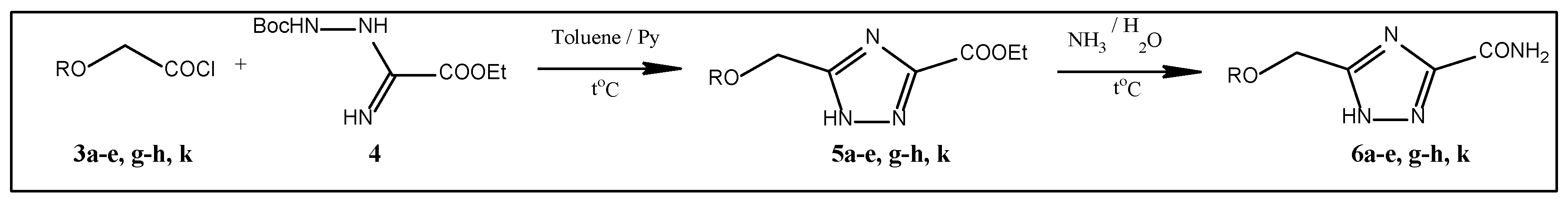
Figure 3.
Introduction of a 1-alkoxymethyl moiety.
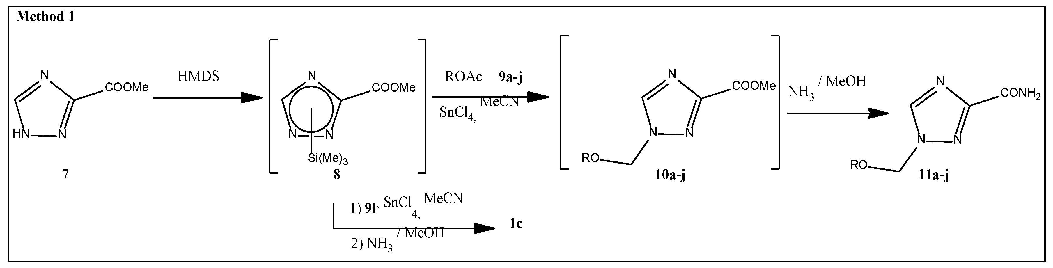
Figure 4.
Introduction of a 1-phenoxymethyl moiety.
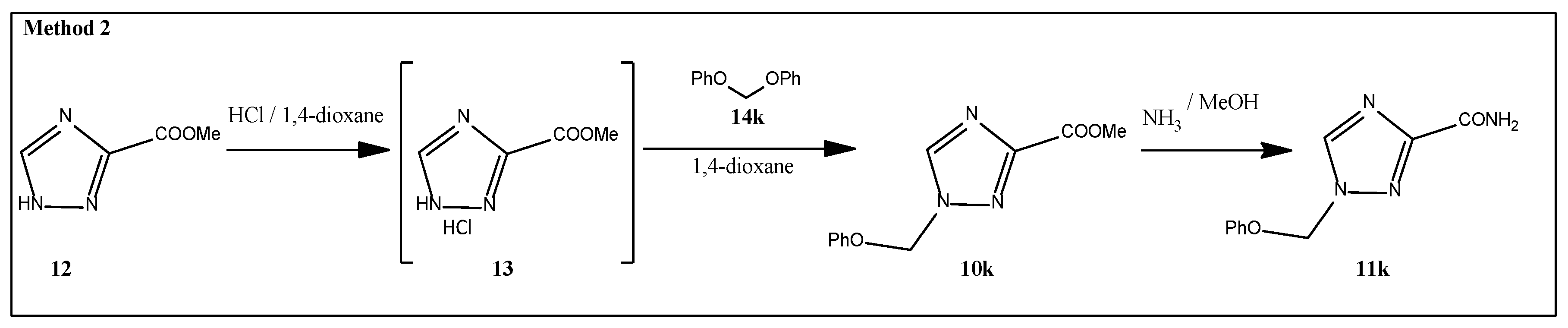
Figure 5.
APT, DEPT-135 and 1H-13C HMBC NMR spectra fragments: (a) methyl 1-(methoxymethyl)-1,2,4-triazole-3-carboxylate and (b) 1-(phenoxymethyl)-1,2,4-triazole-3-carboxamide.
Figure 5.
APT, DEPT-135 and 1H-13C HMBC NMR spectra fragments: (a) methyl 1-(methoxymethyl)-1,2,4-triazole-3-carboxylate and (b) 1-(phenoxymethyl)-1,2,4-triazole-3-carboxamide.
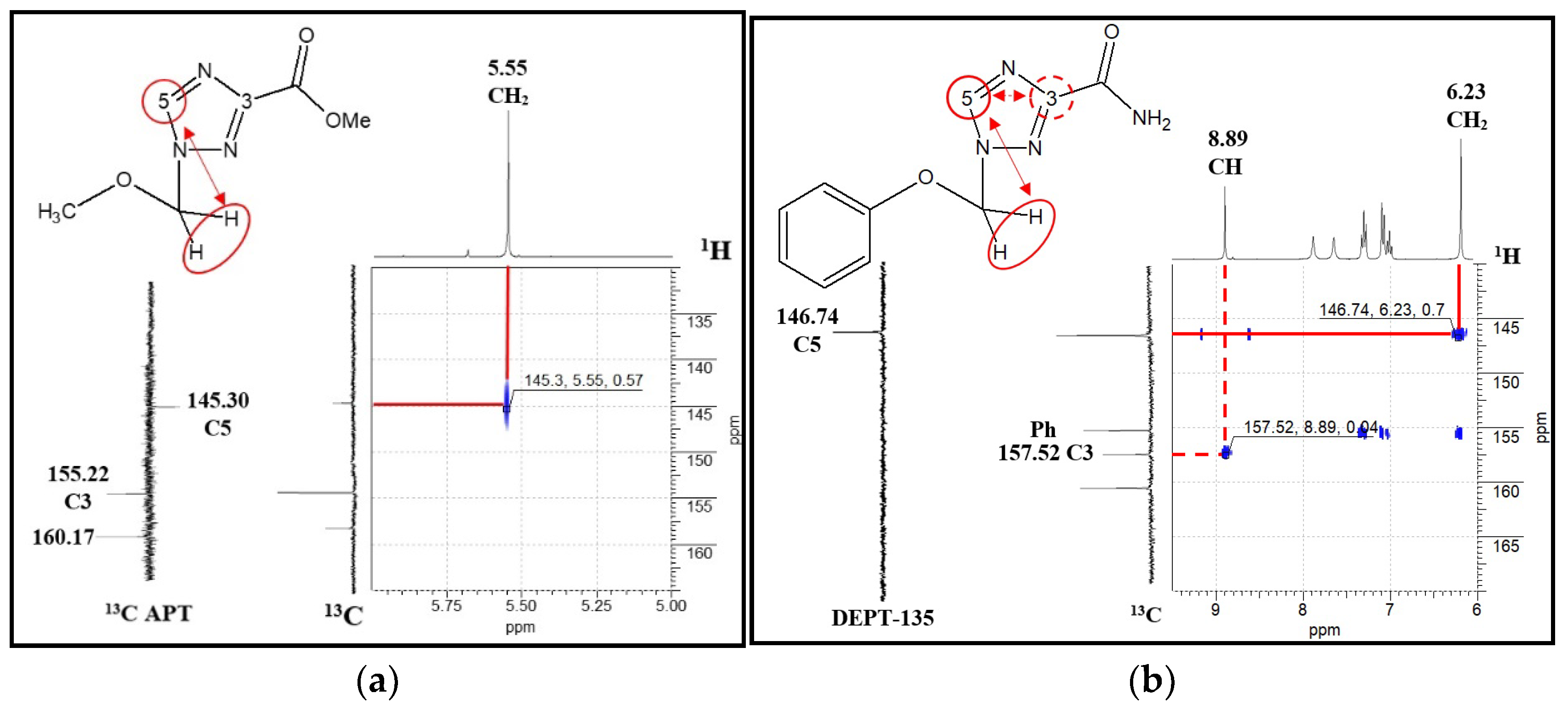
Figure 6.
Effect of compounds on human PBMCs proliferation. Cells were treated with the DMSO, Cyt, 1a or its derivatives for 72 h and then were counted using trypan blue exclusion test. V - volunteer. All data are expressed as percent of DMSO treated control. Significant differences were analyzed by the one-way ANOVA test. *- significant differences from the control (p<0.05).
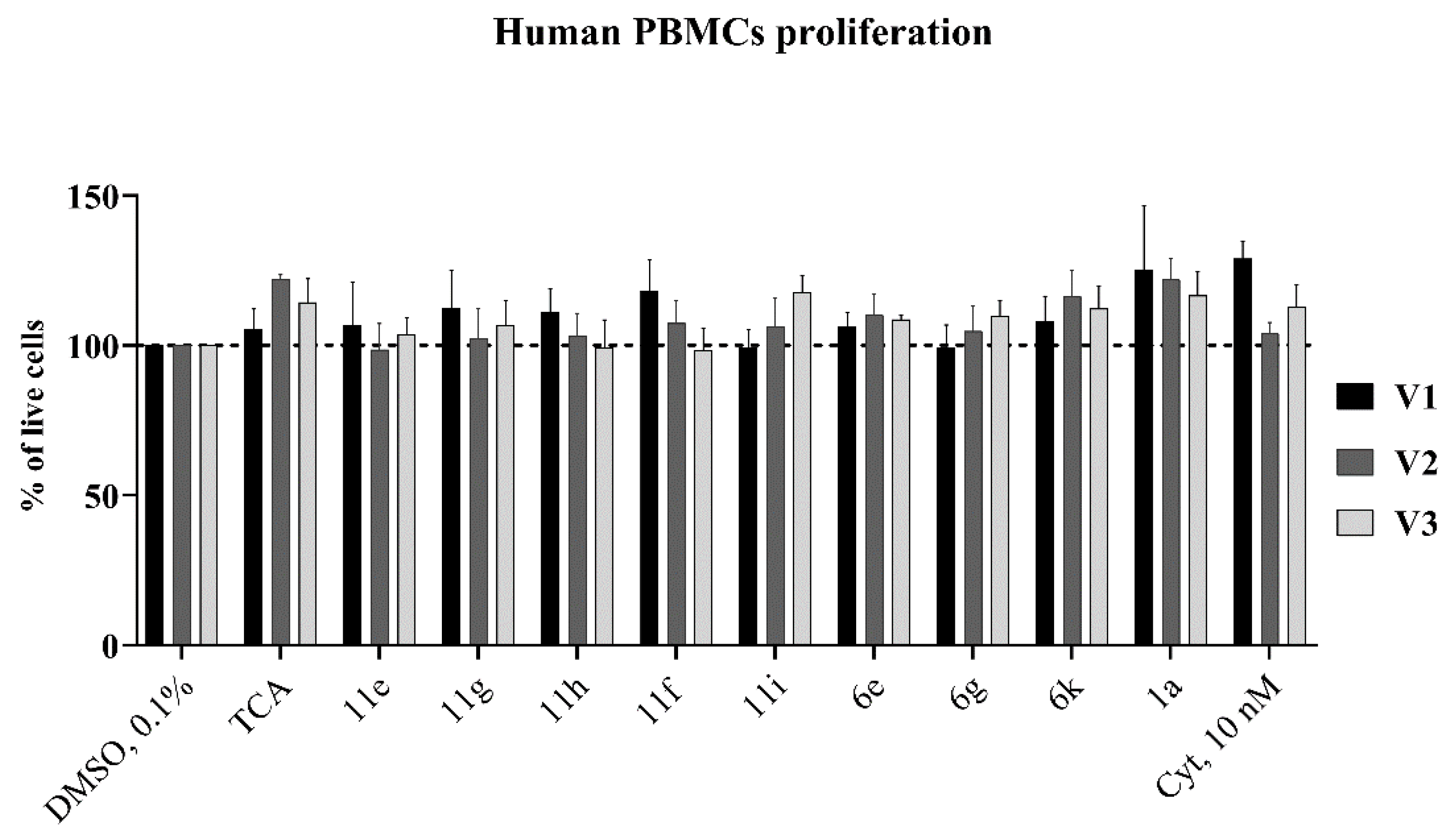
Figure 7.
(a-b) Antiproliferative effects of compounds in CCRF-SB and K562 cancer cells. The cells were cultured with the solvent (DMSO), cytarabine (Cyt), or ribavirin (1a), or its derivatives. Cells were stained with trypan blue and counted after 24, 48 and 72 hours of the treatment. (с) Effect of selected compounds on cell cycle progression in K562 and CCRF-CEM cells after 72 h of incubation with the DMSO, Cyt, 1a or its derivatives. Cells were fixed with ethanol and then stained with propidium iodide and analyzed by flow cytometry. All data are expressed as percent of DMSO treated control. Significant differences were analyzed by the one-way ANOVA test. *- significant differences from the control (p<0.05).
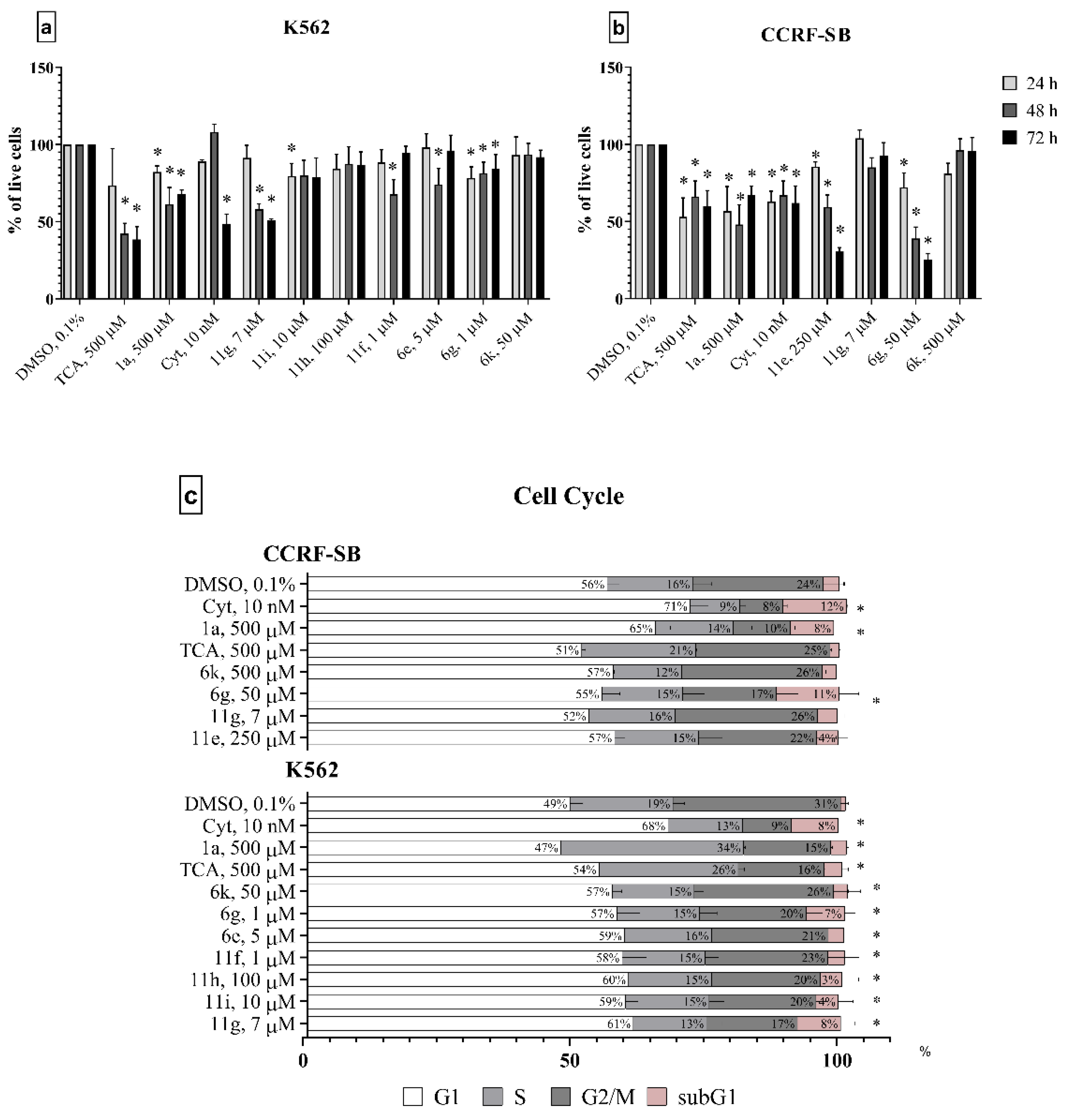
Figure 8.
Binding of test compounds to protein elF4E: (a) ribavirin 1a – green and 1g – yellow; (b) known inhibitor 4EGI-1 – yellow and 6g – green; (c) known inhibitor 4EGI-1 – green and 11g – yellow.
Figure 8.
Binding of test compounds to protein elF4E: (a) ribavirin 1a – green and 1g – yellow; (b) known inhibitor 4EGI-1 – yellow and 6g – green; (c) known inhibitor 4EGI-1 – green and 11g – yellow.
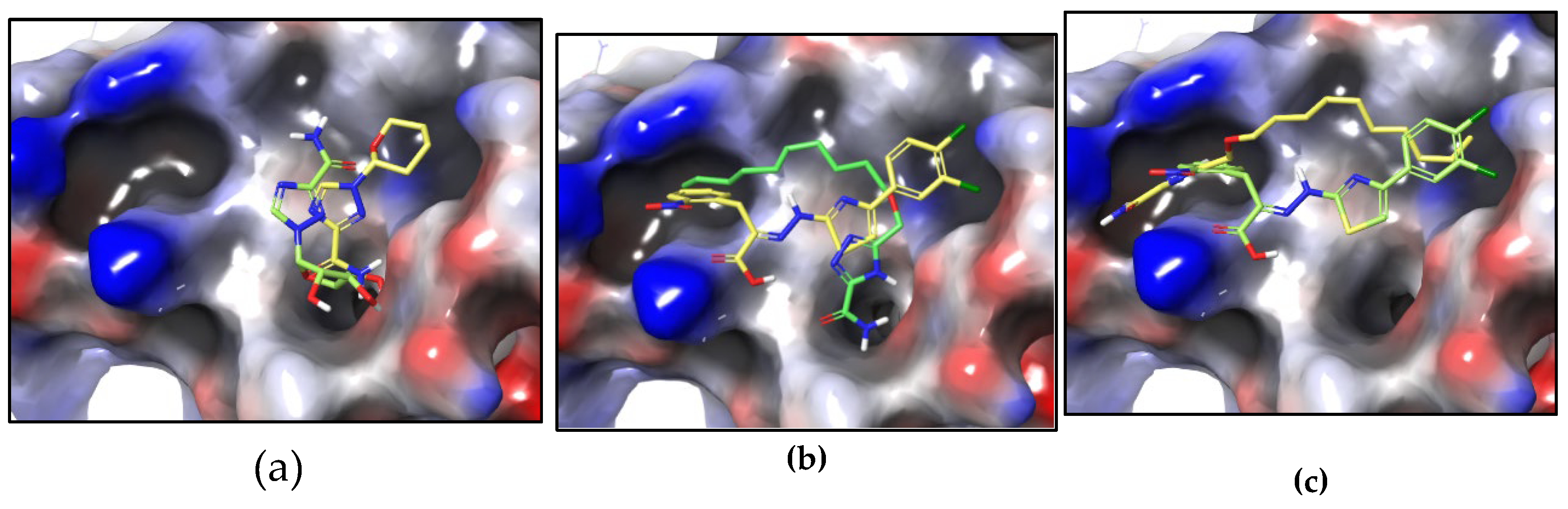
Table 1.
Synthesized 5-oxymethyl-1,2,4-triazole-3-carboxamides 6.
 |
||
| S. No. | R | Yield |
| 6a | Me | 60% |
| 6b | Et | 53% |
| 6c | n-Pr | 62% |
| 6d | i-Pr | 24% |
| 6e | n-Bu | 33% |
| 6g | n-C10H21 | 43% |
| 6h | Bn | 68% |
| 6k | Ph | 76% |
Table 3.
CC50 values for compounds after incubation with cells for 24 and 72h (MTT assay, n = 3).
| 5-oxymethyl-1,2,4-triazole-3-carboxamides | 1-oxymethyl-1,2,4-triazole-3-carboxamides | ||||||||
|---|---|---|---|---|---|---|---|---|---|
| CC50, µM | CC50, µM | ||||||||
| S. No. | K562 | CCRF-SB | S. No. | K562 | CCRF-SB | ||||
| 24h | 72h | 24h | 72h | 24h | 72h | 24h | 72h | ||
| 1a(ribavirin) | 270±11 | 10±1 | - | 188±31 | |||||
| 2g | 260±12* | 240±16* | 1g | 240±22* | 230±18* | ||||
| 2h | 240±21* | 250±13* | 1h | 230±13* | 270±25* | ||||
| 6a | - | - | - | - | 11a | - | - | - | - |
| 6b | - | - | - | - | 11b | - | - | - | - |
| 6c | - | - | - | - | 11c | - | - | - | - |
| 6d | - | - | - | - | 11d | - | - | - | - |
| 6e | - | - | - | - | 11e | - | - | - | - |
| 11f | - | - | - | - | |||||
| 6g | 391±15 | 43±7 | 500±100 | - | 11g | 14±0 | 13±3 | 112±19 | 62±2 |
| 6h | - | - | - | - | 11h | - | - | - | - |
| 11i | - | - | - | - | |||||
| 11j | - | - | - | - | |||||
| 6k | - | - | - | - | 11k | - | - | - | - |
| 1c | - | - | - | - | |||||
| Сyt | 59.4±14.0 | 58.1±16.9 | 15.8±4.1 | 0.1±0.1 | |||||
*Data was previously obtain and published in [9].
Table 4.
1-Oxymethyl-1,2,4-triazole-3-carboxamides antimicrobial effects.
| S. No. | Zone of growth inhibition, mm | |||
| S. aureus | M. luteus | P. aeruginosa | C. albicans | |
| 1-oxymethyl-1,2,4-triazole-3-carboxamides | ||||
| 1a | - | - | 25±1 | 30±1 |
| 11a | - | - | - | - |
| 11b | - | - | - | - |
| 11c | - | - | 12±1 | - |
| 11d | - | - | - | - |
| 11e | - | - | - | - |
| 11f | - | - | - | - |
| 11g | - | - | - | - |
| 11h | - | - | - | - |
| 11i | - | 12±1 | - | - |
| 11j | - | 12±1 | - | - |
| 11k | - | - | - | - |
| 1c | - | 12±1 | - | - |
Disclaimer/Publisher’s Note: The statements, opinions and data contained in all publications are solely those of the individual author(s) and contributor(s) and not of MDPI and/or the editor(s). MDPI and/or the editor(s) disclaim responsibility for any injury to people or property resulting from any ideas, methods, instructions or products referred to in the content. |
© 2024 by the authors. Licensee MDPI, Basel, Switzerland. This article is an open access article distributed under the terms and conditions of the Creative Commons Attribution (CC BY) license (http://creativecommons.org/licenses/by/4.0/).
Copyright: This open access article is published under a Creative Commons CC BY 4.0 license, which permit the free download, distribution, and reuse, provided that the author and preprint are cited in any reuse.


