Submitted:
22 August 2024
Posted:
23 August 2024
You are already at the latest version
Abstract
Biomaterials have been the subject of extensive research, with their applications in medicine and pharmacy expanding rapidly. Collagen and its derivatives stand out as valuable biomaterials due to their high biocompatibility, biodegradability, and lack of toxicity and immunogenicity. This review comprehensively examines collagen from various sources, its extraction and processing methods, and its structural and functional properties. Preserving the native state of collagen is crucial for maintaining its beneficial characteristics. The challenges associated with chemically modifying collagen to tailor its properties for specific clinical needs are also addressed. The review discusses various collagen-based biomaterials, including solutions, hydrogels, powders, sponges, scaffolds, and thin films. These materials have broad applications in regenerative medicine, tissue engineering, drug delivery, and wound healing. Additionally, the review highlights current research trends related to collagen and its derivatives. These trends may significantly influence future developments, such as using collagen-based bioinks for 3D bioprinting or exploring new collagen nanoparticle preparation methods and drug delivery systems.
Keywords:
biomaterial
; biocompatibility
; collagen
; derivatives of collagen
; medical applications
; tissue engineering and regeneration
; drug delivery systems
; bioinks for 3D bioprinting
1. Introduction
Since ancient times, humans have sought ways to accelerate wound healing and restore lost bodily functions. Modern medicine, driven by increasing clinical demands, has rapidly developed advanced materials with potential therapeutic applications [1].
While the definition of a biomaterial evolves with scientific progress, the most commonly accepted one is proposed by the European Society for Biomaterials Consensus Conference II: "A biomaterial is a material intended to interface with biological systems to evaluate, treat, augment or replace any tissue, organ or function of the body” [2,3]. The prefix bio- emphasizes the material’s compatibility with human tissues and the body's response rather than indicating a biological origin [4].
For a material to be considered a biomaterial, it must meet several criteria. These include biocompatibility (the ability to coexist with body tissues without causing harmful reactions), biofunctionality, bioinertness, and bioactivity. Additionally, the biomaterial should be sterilizable and must not induce inflammatory, toxic, or allergic reactions [5].
Biomaterials can be natural (e.g., collagen, chitin) or synthetic. Based on material properties, they can be divided into metallic (e.g., titanium, stainless steel), ceramic (e.g., zirconia, hydroxyapatite), polymeric (e.g., silicon, nylon), and composite materials (combining the properties of various materials, e.g., carbon fibers, ceramic particles) [3,6]. Polymeric materials are gaining popularity due to their diverse properties and ease of processing. The global medical polymer market is estimated to be around 1 billion dollars [7].
Due to their specific characteristics, biomaterials are primarily used for medical purposes. The applications in medicine are extensive, including orthopedics (e.g., implants such as artificial hip or artificial knee), ophthalmology (e.g., intraocular lenses), cardiology (e.g., heart valve), dentistry, plastic surgery, urology, drug delivery, and many other fields [4,8]. Thanks to ongoing research and the development of new materials, even more significant advances in this field can be expected.
One of the most widespread and studied natural biomaterials is collagen. Its biocompatibility, biodegradability, and common availability make it an attractive material for many biomedical applications. The diverse types of collagen, differing in structure and properties, enable its use in various applications [9,10].
Collagen's ability to self-organize and form three-dimensional fiber networks through cross-linking provides appropriate mechanical strength, making it an excellent material for constructing tissue scaffolds [10,11]. Moreover, collagen may interact with cell surface receptors, enabling strong cell adhesion, proliferation and migration, which are crucial for healing and tissue regeneration [12]. Through chemical modifications of collagen, its properties can be precisely adjusted to specific needs. For example, introducing functional groups can increase hydrophilicity, bioactivity, or the ability to form bonds with other materials [13].
This review presents a detailed description of the structure of collagen and its fundamental properties that determine its wide use in regenerative medicine. Various forms of collagen biomaterials, their medical applications, and research trends in collagen and its derivatives are also discussed.
2. Characteristics of the Collagen
3.1. Structure and Properties
Collagen (CLG) is an extracellular structural protein, constituting almost 1/3 of the total protein mass in the human body. As the most important component of the extracellular skin matrix, it is responsible for its elasticity and durability [14,15]. Collagen exhibits a hierarchical structure, from its primary amino acid sequence to its organization into complex fibers. Also, CLG belongs to the group of fibrillar proteins responsible for the structure and biochemical properties of connective tissue in mammals. There are many types of collagens (28 types, numbered with Roman numbers), and they are the most complex natural polymers [15-17]. The structure of these protein molecules is based on more than 20 amino acids, and the most abundant are proline (Pro), hydroxyproline (Pro-OH), glycine (Gly), and hydroxylysine (Lys-OH) [14,18]. Its molecular composition is fundamental to its structural integrity and function. The collagen structure is based on the superhelix, which is held together by hydrogen bonds, electrostatic interactions, and van der Waals forces [19,20]. Also, Gly, the smallest amino acid, appears at every third position in the sequence, allowing for the close packing necessary for the triple helix formation. Pro and Pro-OH play crucial roles in stabilizing the helical structure through their ring structures, which impose conformational constraints [14,18]. The unique structure of collagen consists of three left-handed polypeptide α chains, the so-called procollagen (Figure 1). They are wrapped around each other and have a common axis, thus forming a right-handed superhelix conformation called tropocollagen [14,18].
The primary structure of CLG refers to the specific sequence of amino acids in collagen polypeptide chains. It is characterized by repeating Gly-X-Y triplets, where X and Y are often PRO and PRO-OH, respectively (Figure 1b). Furthermore, the triple helix is stabilized thanks to Gly, high Pro content, and Pro-OH forming hydrogen bonds and electrostatic interactions between lysine and aspartic acid [14,15,22]. The unique structure of the triple helix and strong bonds between amino acids help collagen fibers maintain their flexibility and resistance to stretching. The triple helix is 300 nm long and 1.5 nm in diameter [21,23]. Telopeptides are responsible for the protein's immunogenicity on both structure ends. The α chains range from 662 (human α 1(X)) to as many as 3152 (human α 3(VI)) amino acids in length [16]. The triple helix chains can be identical or different and form homotrimers (collagen types II, III, VII, VIII, and X) or heterotrimers (Collagen types I, IV, V, VI, IX, and XI), respectively. Collagen fibrils (Figure 1a) are formed by the staggered arrangement of collagen molecules [21,22], creating a repeating banding pattern visible under electron microscopy. Further, CLG fibrils aggregate to form larger collagen fibers. These fibers contribute significantly to the mechanical properties of tissues, providing tensile strength and flexibility [20,21]. Covalent cross-links are formed between lysine (Lys) or hydroxylysine (Lys-OH) amino acids, further stabilizing the fibrils. The extent of cross-linking increases with age, enhancing the tensile strength of the CLG fibers but also making them more rigid and less elastic [24,25]. There are at least 28 different types of CLG [14,15,21]), each serving distinct functions and present in various tissues. Type I CLG is the most abundant in skin, tendon, and bone. Type II CLG is predominant in cartilage, where it resists compressive forces. Type III CLG is often found alongside Type I, particularly in the skin, blood vessels, and internal organs, contributing to tissue elasticity and repair. Other types of collagen, such as Type IV and Type V, play specialized roles in basement membranes and cell surfaces, respectively [14-16,26]. The elasticity of collagen refers to its ability to return to its original shape after being stretched or deformed [15,26]. This critical property allows various tissues to withstand mechanical stress and strain and is essential for tissues that require flexibility and the ability to absorb mechanical forces without sustaining damage [14,15].
The triple helical structure of CLG fibers contributes to its tensile strength and elasticity. The helical shape allows the fibers to stretch and return to their original form [15,21]. Also, covalent bonds, or cross-links, between CLG molecules, as mentioned above, provide structural integrity and enhance the elastic properties by preventing the fibers from sliding past each other too easily [24,25]. CLG exhibits a non-linear stress-strain curve. Initially, there is a low-stiffness region (toe region) where the crimped fibrils straighten. This is followed by a linear region where the collagen fibers are directly stretched. It also displays viscoelastic properties, meaning it has viscous and elastic characteristics. This dissipates energy and provides damping, essential for tissues undergoing cyclic loading [27]. Age, disease, and environmental exposure can affect collagen's elasticity, impacting tissue function and integrity. With aging, collagen undergoes changes such as increased cross-linking and glycation, reducing elasticity and making tissues stiffer [28,29]. Certain diseases, like osteoarthritis, can affect the quality and organization of collagen, leading to reduced elasticity and compromised function of the affected tissues [30]. UV radiation and environmental factors can degrade collagen, leading to a loss of elasticity, particularly in the skin [25,31].
2.2. Types of Collagen and Their Origin
Collagen can be sourced from various animals, and the most common sources [32-35] [36,37] include pigs (porcine collagen), cows (bovine collagen), and fish (marine collagen). Porcine Collagen is usually derived from porcine skin, bones, and connective tissues, while bovine collagen sources are skin, bones, and cartilage. Both types of CLG (porcine and bovine) are rich in Gly, Pro, and Pro-OH, which are essential for CLG synthesis and structure. Porcine CLG has high compatibility with human collagen due to structural similarity, making it useful for medical and cosmetic applications [33,35]. Bovine CLG is generally well-tolerated, though some individuals may have sensitivities or dietary restrictions related to bovine products. In medicine, pork CLG is widely used in wound dressings, skin grafts, and surgical sutures due to its biocompatibility and ability to promote healing [38]. In comparison, bovine CLG is utilized in tissue scaffolding, bone grafts, and joint health supplements [39]. Regarding cosmetics, porcine and bovine CLG are incorporated into anti-aging products, moisturizers, and serums to improve skin elasticity and hydration [29,40,41]. In dietary supplements, CLGs are available in powder form or capsules to support joint health skin, hair, and nail strength [42]. As far as the food industry is concerned, collagen derivatives are used mainly in the form of gelatin as food additives in the production of desserts, gummy candies, and other food products. Marine collagen (fish) is also received from fish's skin, scales, bones, and fins, especially from cold-water species like cod, salmon, and pollock [15,43]. This type of CLG contains a high concentration of amino acids (Gly, Pro, and Pro-OH). However, it also has smaller peptide molecules, which leads to better bioavailability [43,44]. Smaller peptide size often makes it more effective for quick absorption and utilization in the body. Marine CLG has a higher absorption rate than bovine and porcine CLG, making it highly effective in supplements and skincare products. It is also considered more eco-friendly, using fish by-products that would otherwise be discarded. Marine CLG is less common in the food industry than bovine and porcine gelatin, but it is used in some niche products targeting health-conscious consumers. In addition, it is also often chosen by those with sensitivities to mammalian collagen sources, though fish allergies must be considered.
2.3. Methods of Obtaining
To obtain collagen, extraction and hydrolysis are needed. The extraction process depends on the source material; for example, enzymes or acids must hydrolyze extracted proteins from pigs or cows [45,46]. Hydrolysis is commonly used to break the CLG into gelatin or hydrolyzed collagen peptides. Purification follows hydrolysis, the CLG is purified to remove non-collagenous proteins and other impurities [32-35]. Marine CLG is hydrolyzed to produce smaller peptides that are more easily absorbed by the body. Also, extensive purification processes ensure that received CLG is free from allergens and impurities [36,43,47]. The recovery process typically involves three steps: pretreatment of the source tissue, collagen extraction, and purification (Figure 2). The extraction temperature is controlled; a low value (4 ◦C) minimizes collagen degradation.
The pretreatment stages are usually preceded by soaking the skin in cold water for a few days and washing it this way by changing the water every few hours [21]. The skin is then cut into pieces that are easier to process. During the pretreatment stage, covalent intermolecular crosslinks between collagen molecules are broken. This process is prolonged, even in boiling water, so various mild chemical treatments are necessary. Diluted acids and alkalis are typically used for partial hydrolysis of the CLG [48,49]. For acid pretreatment, washed and chopped skin pieces are immersed in dilute acid at a controlled temperature. Acid pretreatment is suitable for relatively fragile skins [21] with a lower degree of fiber intertwinement, such as porcine and fish skins. Dilute alkalis such as sodium hydroxide (NaOH) and calcium hydroxide (Ca(OH)2) are used for pretreatment, and the duration of this step depends on the thickness of the material being treated. Alkalis are particularly effective in extracting CLG from thick and hard materials and hydrolyzing undesirable non-collagenous proteins, lipids, pigments, and other organic materials [50]. The temperature, duration, and concentration of alkali significantly influence the effectiveness of the removal of unwanted non-collagen materials [37]. Conventional extraction of CLG from different sources is typically based on chemical hydrolysis using acid, alkali, or salt solubilization. Organic acids: acetic, chloroacetic, citric, or lactic acids are used. Inorganic acids (hydrochloric, sulfuric, and nitric) can also be used [21]. Under acidic conditions, collagen molecules have a net positive charge, and the resulting electrostatic repulsive force between them facilitates molecular separation. During acid-based extraction, raw material, such as porcine or fish skin, is soaked in 0.5 M acetic acid at a controlled temperature until swelling, usually between 24 and 72 h, followed by precipitation [21,49]). In the next step, the precipitate is centrifuged, and the supernatant is then salted out for 12 h with NaCl [37,50]. The supernatant is re-dissolved in 0.5 M of acetic acid and then dialyzed for a few days against cold distilled water, lyophilized, and stored until further use [48]. Sodium hydroxide or potassium hydroxide are the most common bases used, although calcium oxide, calcium hydroxide, or sodium carbonate can also be used as extractants [37,51], and the process can take several days to several weeks [50]. Instead, this method is used for thick materials such as leather waste such as bovine shavings because it penetrates more aggressively to swell the material [21,48]. Usually, for soaking, 0.05-0.1M NaOH is used. The received CLG is then precipitated by dialysis in 50 mM Tris-HCl buffer (pH 7.4), and the collected precipitate is dissolved in 5 mM acetic acid [37]. Organic acids are more effective at cleaving CLG crosslinks, resulting in higher extraction efficiency [38,39] than mineral acids, and can solubilize non-crosslinked collagens. While alkalis tend to hydrolyze collagen fibrils, the amino acids cysteine, histidine, serine, and threonine may be destroyed [37,46]. Sometimes, ultrasounds, microwaves, and enzymes are added to the method. Pepsin, tryptase, and papain [50] are the most common enzymes used for CLG extraction [21]. They relax cross-linking in CLG by cleaving aminotelopeptides from the tropocollagen molecule. This is most useful in adult tissue, where strong intermolecular bonds are formed due to ketoimine cross-links [37,52]. In enzymatic hydrolysis, the raw material or residue is soaked in 0.5 M acetic acid or 0.01 M HCl containing selected enzymes such as 0.1 percent (w/v) pepsin. The mixture is continuously stirred at 4 °C for approximately 48 h. After this time, the solution is filtered or purified, and the supernatant is salted out to precipitate. For all processes, the last step involves separation by centrifugation, re-dissolution in acid, and dialysis to obtain acid-soluble CLG [37]. Extraction methods can be adjusted depending on the final product's properties and the process's effectiveness [37,47]. The features of the final product, such as the average length of the polypeptide chains, solubility, viscosity, thermal stability, emulsifying capacity, and water retention, are influenced by the specifics of the extraction method.
2.4. Native Collagen vs. Cross-Linked Collagen
Native and cross-linked collagen represent two different forms used in various biomedical applications. Native CLG means natural, unmodified form, with a triple-helix structure preserved, which is crucial for its biological function and mechanical properties [38,53]. Some researchers claim that to provide an optimal environment for cellular infiltration, morphology, and viability, the nativeness of collagen in a biomaterial and its cellular binding sides need to be retained [54]. It has to be emphasized that to preserve all these native properties, the processes of collagen preparation and purification have to be carefully chosen. Cross-linked CLG, on the other hand, undergoes chemical or physical treatment to introduce covalent bonds between its molecules [55-57]. This process enhances its stability and mechanical properties and can be obtained by agents such as glutaraldehyde, carbodiimides, or genipin (chemical cross-linking) or by the use of UV irradiation or dehydrothermal treatment (physical cross-linking) [56,57]. The crosslinking process enhances collagen resistance to enzymatic and thermal degradation compared to native CLG [58]. Cross-linked CLG possesses greater tensile strength and durability, which makes it suitable for load-bearing applications [22,55,59]. What is also exciting is that crosslinking can be adjusted to control the degradation rate for applications requiring long-term stability. Rasouli et al. have provided helpful guidance for collagen crosslinking methods. UVC, as a physical crosslinking method, preserves the integrin binding sites on collagen, resulting in a dose-dependent improvement in mechanical performance up to a dose of 5 J cm-2. At higher doses, it leads to collagen fragmentation. Genipin, a non-cytotoxic plant-based crosslinker, results in moderate improvements in mechanical performance comparable to UVC. EDC-NHS (carbodiimide and N-hydroxysuccinimide) also results in modest improvements, with the possibility of losing some integrin binding sites on the collagen. Glutaraldehyde dramatically improves ultimate tensile strength compared to human and rat tendons, preserving the native collagen molecular structure. However, cytotoxicity is an inherent disadvantage of using glutaraldehyde as a crosslinker [58]. Native CLG has superior biocompatibility and bioactivity due to the preservation of natural binding sites and minimal modifications, and that is why it serves significant attention, which it very often lacks [38,53].
The functionality of native collagen becomes obvious in clotting experiments. It is assumed that a fast and reliable triggering of platelet aggregation indicates that the quaternary structure of collagen is intact. Wiegand et al. compared three different collagen wound dressings in vitro. During the evaluation of platelet aggregation, only the dressing made from native collagen showed an activation of the clotting cascade. Fibroblasts seeded on the native collagen showed exponential growth over 14 days, whereas those seeded on the other two dressings exhibited meager proliferation rates. It has been shown that native collagen allows for more efficient angiogenesis and more significant fibroblast chemotaxis than denatured collagen in vitro. In intensely processed or degraded collagens, the bioactive potential of cells involved in wound healing is likely lost or attenuated [60]. Native collagen also shows a faster degradation rate due to natural enzymatic breakdown, which can be advantageous for applications requiring rapid tissue regeneration. Some authors even show that the importance of the manufacturing process and the ability to save the nativeness of collagen is more critical for the performance of collagen matrices than the animal source of collagen [61]. Cross-linked CLG can sometimes show reduced bioactivity and slower, controlled degradation, and that is why it can be suitable for long-term applications where prolonged structural support is needed. Good mechanical properties make it suitable for load-bearing and long-term applications, while native CLG is more appropriate for applications where high mechanical strength is not a primary requirement [22,38,53,59]. The choice between native and cross-linked CLG depends on the application's requirements, including mechanical properties, degradation rate, biocompatibility, and bioactivity.
3. Biomedical Properties of Collagen
3.1. Biocompatibility and Immunogenicity
Collagen’s biocompatibility has gained significant interest due to its various biomedical applications. This biocompatibility is rooted in several key characteristics, including its natural presence in the human body, its structural properties, and its ability to support cellular functions essential for tissue repair and regeneration [62]. The immune system recognizes CLG as a natural component, minimizing the risk of adverse immune responses when collagen-based biomaterials are introduced into the body. This inherent compatibility reduces the probability of rejection and inflammation, which are common issues with synthetic biomaterials [63,64]. However, the safety of CLG sourced from cattle and pigs is a significant concern due to disease risks [65]. The potential health concerns have been raised due to the incidence of bovine spongiform encephalopathy (BSE) and transmissible spongiform encephalopathy (TSE) of terrestrial animal-derived CLG and related products [66]. Allergic reactions are another critical issue. Individuals with allergies to bovine or porcine products may experience adverse reactions when exposed to CLG derived from these sources. This has been particularly noted with bovine gelatin, a common ingredient in measles, mumps, and rubella (MMR) vaccines. Some children have shown sensitivity to these vaccines, potentially due to the bovine CLG content [67]. Additionally, there have been reported cases of allergic reactions to bovine CLG used in medical devices, highlighting the potential for adverse immune responses in sensitive individuals [66].
Marine CLG is a potential alternative to bovine and porcine CLG due to the lower probability of disease transmission. A material obtained from fish and other aquatic organisms (i.e., marine CLG) is considered safer, which is why it is increasingly used in biomedical applications. However, literature references show that individuals with shellfish allergies may also react adversely to marine CLG, including potential anaphylaxis [67].
The biocompatibility of CLG is also exhibited in minimal immunogenicity. The source of collagen and the processing methods used can influence this property. Numerous studies have shown that marine CLG derived from fish (e.g., tilapia skin CLG) indicates minimal immunogenicity, making it a viable alternative to CLG of mammalian origin [68]. CLG processing methods like electrospinning can impact its immunogenic properties [69]. The triple helical structure, critical for low immunogenicity, can be preserved in self-assembled CLG nanofibers but may be altered in electrospun collagen nanofibers [70,71]. However, thorough evaluations, including cytotoxicity, hemolysis, skin sensitization, and systemic toxicity tests, confirmed low immunogenicity and biocompatibility of collagen-based materials [72].
3.2. Biodegradability of Collagen
Collagen's biodegradability significantly supports its application in the biomedical field. The degradation process of collagen-based materials depends on the CLG source, preparation method, and the crosslinking chemical used. Naturally derived CLG exhibits inherent biodegradability due to the presence of enzymatic and non-enzymatic pathways in the body that specifically target CLG fibers.
3.1.1. Enzymatic Degradation of Collagen
Enzymatic degradation is crucial to collagen's biodegradability, especially in biomedical applications where controlled cleavage is essential for tissue remodeling and healing. Although the peptide bonds in the triple helix are occluded from enzymatic active sites, single-stranded regions are affected by matrix metalloproteinases (MMPs) [73]. Key collagenases, such as MMP-1 (collagenase-1) and MMP-8 (collagenase-2), initiate collagen degradation by cleaving its triple helical structure at specific Gly-Ile or Gly-Leu bonds, resulting in the unwinding and fragmentation of CLG fibrils into smaller peptides [74]. These fragments are further broken down by gelatinases, specifically MMP-2 and MMP-9, which target denatured CLG (gelatin) to produce even smaller peptides and amino acids [75]. The amino acids released during such an enzymatic process are utilized in various metabolic pathways that integrate CLG degradation products into normal physiological processes [74]. The activity of these enzymes is precisely regulated, maintaining tissue homeostasis and ensuring efficient turnover of the extracellular matrix (ECM) [76].
3.1.1. Non-Enzymatic Degradation of Collagen
Non-enzymatic degradation of CLG involves mechanisms of fiber breakdown under physiological or pathological conditions. The primary mechanism is hydrolytic degradation, where water molecules under acidic or basic conditions cleave peptide bonds of CLG molecules [77]. Oxidative degradation, another important pathway, involves reactive oxygen species (ROS) interacting with triple helix structure, leading to the breakdown of peptide bonds. This is particularly relevant in inflammatory conditions where ROS levels are increased [78]. Mechanical degradation also plays a role, as physical forces can cause CLG fiber fragmentation, often observed in load-bearing tissues subjected to repetitive stress and strain [79].
4. Collagen-Derived Biomaterials
Collagen-derived biomaterials are available in many forms and can be used in tissue engineering, regenerative medicine, and pharmacy (Table 1, Figure 3). The most common forms include membranes, scaffolds, gels, sponges, and films, with some commercially available (Table 2).
4.1. Membranes
Membranes, usually formed as thin, flexible sheets, are used in guided bone (GBR) and tissue (GTR) regeneration. They are divided into two groups: absorbable and non-absorbable. Membranes made of polymers of natural origin (mainly collagen and chitosan) are absorbable [118]. However, a distinction can be made between cross-linked and non-cross-linked (native) collagen membranes (CLG-MB) [119,120]. They are mainly obtained from bovine or porcine tissues such as pericardium, dermis mesenchymal tissue, or, in some cases, equine tendons. Various structures and thicknesses characterize collagen membranes. The latter does not exceed 0.5 mm. The most common forms of membranes are single-layer and two-layer materials, which consist of a layer of differently oriented CLG fibers, allowing for tissue integration and a denser, permeable layer [120].
Absorbable membranes used in GBR face challenges due to their fast degradation rate, leading to early loss of barrier function that can hinder bone formation. CLG-MBs are commonly used for regeneration and exhibit varying degradation times, with histomorphometric analysis showing a significant membrane thickness decrease within weeks to months post-surgery. Crosslinking techniques have been developed to prolong absorption time by stiffening CLG-MBs and slowing down enzymatic degradation. While cross-linked membranes are generally more resistant to degradation compared to non-cross-linked membranes, excessive crosslinking may lead to compromised healing. On the other hand, native CLG-MBs can promote bone regeneration without inflammatory or foreign body reaction only in the first 2 weeks after the surgical procedure, later exhibiting signs of degradation [121]. CLG-MBs are also used in the oral cavity, where they are especially susceptible to increased degradation due to bacterial enzymes, which affect barrier function and bone regeneration. Hence, cross-linked CLG biomaterials with enhanced membrane durability are being used for this application.
Several studies have compared the properties of cross-linked and non-cross-linked collagen membranes. Those studies have shown a correlation between longer degradation times of cross-linked membranes, which helps find the right balance between the properties of such biomaterials and ensuring they exhibit the desired requirements. Research on antibacterials and antibiotics to inhibit membrane breakdown during GBR is ongoing [91,122,123].
4.2. Scaffolds
A collagen scaffold (CLG-SC) is a three-dimensional structure used in tissue engineering and regenerative medicine to support the growth and repair of various types of tissues. Research has shown that the mean pore size of a scaffold is highly significant as it greatly influences the behavior of seeded cells. Scaffolds with small pore sizes (100 μm) are more effective in promoting new bone formation. Smaller pores create hypoxic conditions that stimulate cartilage formation, while larger pores (300 μm) are essential for capillary ingrowth, which is necessary for mature bone development [80].
Despite its significant quantitative presence in bone tissue and excellent biocompatibility, CLG alone does not have osteoinductive properties [124]. The combination of CLG and apatite allows for the obtaining of such properties [125]. Hence, the current research includes multi-component scaffolds, which, in addition to CLG, mainly contain hydroxyapatite (HAP), chitosan, or non-collagen proteins.
Various sources of CLG are being utilized in scaffold fabrication. Liu Y. et al. prepared a scaffold of human-like collagen with the addition of HAP. The results of in vitro and in vivo experiments and the relatively easy production process allowed to obtain a biomaterial with great application potential [81,126].
A two-phase scaffold containing fish CLG (sourced from tilapia skin), HAP, and chondroitin sulphate was developed by Zhou et al. The material was composed of two layers with different pore sizes adapted to bone and cartilage regeneration, and it showed good biocompatibility and low cytotoxicity. However, it is worth mentioning that the production process did not include sterilization of the material, which may have a negative impact on the tested properties [80].
Scaffold of mineralized CLG with antibacterial peptides and poly(D,L-lactide-co-glycolic acid) (PLGA). The goal was to obtain material for use in the treatment of infective bone defects. The created composite material allowed for the long-term release of the anti-inflammatory agent at the target application site up to over 35 days [82].
4.3. Gels
Collagen gels and hydrogels (CLG-G and CLG-HG) constitute a group of materials with an extensive range of current and potential medical applications. These include wound care, tissue engineering, and drug delivery systems. Tissues of porcine, bovine, or rat origin are most often used to produce CLG-Gs, but marine CLG is becoming more and more popular [127,128].
According to the latest edition of The International Union of Pure and Applied Chemistry (IUPAC) Gold Book, gels are described as non-fluid colloidal or polymer networks that are swollen with a fluid throughout their entire volume. Hydrogel is a gel that uses water as the swelling agent [129].
The advantages of CLG-Gs include their biocompatibility, ability to mimic the extracellular matrix, and potential for promoting tissue regeneration. The high water content in hydrogels helps provide a moist wound environment, a crucial factor influencing the speed of wound healing. Moreover, the gel structure is flexible and adapts to the shape of the treated area without causing any discomfort to the patient. Gels can absorb excess wound exudate and remove toxins and water-soluble waste [130]. CLG-Gs also have antioxidant properties, as Shen Z. et al. demonstrated for gels obtained from fish tissues [96]. However, some disadvantages of gels may include risks of uncrosslinked collagen gels' degradation products following metabolic pathways [130].
In order to obtain gels with better mechanical properties (for use as, e.g., dressing materials), additives are used in the form of synthetic polymers, e.g., polyethylene glycol, and natural polymers such as chitosan or hyaluronic acid [131]. Various crosslinking methods are also used to obtain the desired mechanical, optical, or biological properties.
One of the research directions is collagen hydrogels for injection. Hydrogels are an excellent material for carrying drugs and cells used in regenerative medicine (they may contain stem cells, growth factors, and drugs). Additives delivered in the hydrogel do not dissolve easily in the tissues adjacent to the application site, so their pharmacological effect is focused on the site [98]. Since cross-linked collagen is unsuitable for injection, shear-thinning, in-situ crosslinking, or enzymatic crosslinking hydrogels are being developed. Those prepared using enzymatic (biological) crosslinking methods eliminate the disadvantage of toxic compounds, and according to scientific studies, cells show a high capacity for proliferation within these hydrogels, proving their excellent biocompatibility [132].
CLG-Gs can also be used to regenerate corneal damage. The challenge in creating a biomaterial for such an application, in addition to biocompatibility, is also physical properties, in this case, maintaining the transparency and clarity of the material. Hydrogels cross-linked by riboflavin photoactivated with UV-A light showed greater transparency than physical CLG-G [104]. Rosenquist et al. synthesized CLG chemically cross-linked with thiol. The resulting material was fully transparent, degradable by enzymes, and supported the adhesion and proliferation of human corneal cells [105]. Xeroudaki et al. proposed a more complex system. A collagen hydrogel implant reinforced with nanocellulose was developed. Chemical and physical (UV light) cross-linking methods were used, and dexamethasone was introduced as an anti-inflammatory agent, with satisfactory drug release results (80% release within 60 days). The light transmittance of the obtained implants was comparable to the light transmittance of the human cornea [106].
4.4. Sponges
Collagen sponges (CLG-SP) are 3-dimensional biomaterials used in medicine for several decades. Due to their hemostatic properties, they are commonly used in wound treatment and dentistry (for tooth extraction). Sponges have a structure similar to a scaffold but are more flexible, used commonly as wound dressings and as a material for soft tissue repair.
Most commercially available CLG-SP are of mammalian origin (bovine, equine). However, marine CLG-SP is also currently being developed. In the work by Wang et al. [110], a collagen sponge made from tilapia skin was shown to support acute wound healing efficiently. Additional results indicated that sponge degradation products were used to create new tissue.
A collagen sponge with the addition of taurine (Tau®Col) was developed by Wu et al. [111] as a material with antioxidant and anti-inflammatory properties. In vivo data indicated the fastest wound closure time, the best granulation process, and the best CLG deposition for the sponge among all the tested materials.
CLG-SP can also be used in more complex systems, like the example - a composite of collagen sponge, polydopamine, and plasma-rich platelet. It was implanted in mouse wounds and covered with a skin graft [112]. The authors indicated porosity and biocompatibility as the main advantages of the composite.
4.5. Films
Collagen films (CLG-F) are thin layers often obtained by evaporating a solvent from a collagen solution. They are used in tissue engineering and are a potential material for wound treatment. Unfortunately, they have an essential disadvantage - poor mechanical properties, especially in wet environments [107]. Therefore, the current direction of research is to improve those properties. This can be done by using additives and/or crosslinking the films. Adamiak et al. examined the effect of adding salicin on the mechanical properties of CLG-F. They obtained a film with a tensile strength 50% higher than the material without salicin. The authors emphasized the interaction between CLG and salicin - a probable crosslinking process [107]. Tenorova et al. showed that non-cross-linked CLG-F with plasticizers and carboxymethylcellulose were produced as potential wound dressings. The obtained materials showed differences in mechanical properties depending on the source of CLG [108].
CLG-F, with the addition of nanoparticles, is also the research subject. An example can be the presence of 1% iron oxide and graphene oxide nanoparticles that significantly improved mechanical properties (from 0.2 to 0.8 MPa), increased antioxidant activity by about 20%, and showed antibacterial properties [109].
4.6. Other Forms
Materials in the form of microcapsules and tubes are also produced and tested. However, such forms are less frequently studied than scaffolds, gels, or membranes.
A collagen-based microencapsulation platform was developed to encapsulate living cells in a reconstituted nanofibrous CLG meshwork. This platform for potential osteoarthritis therapy provided a biocompatible and physiologically relevant microenvironment that supports cell attachment, proliferation, migration, and differentiation [114].
A material with excellent application potential is collagen tubes (inner lumen diameter app. 0,5 mm) produced by Ishibashi et al.. The proposed form may be used in vascular networks and nerve fibers in artificial organ fabrication and regenerative medicine technology [115]. A similar form of collagen tubes and rods with an outer diameter of 1 mm was developed by Iwamoto et al.. The material's advantages are relatively good mechanical properties (comparable to nylon) and excellent cell culture ability, making it possible to use in regenerative medicine [116].
5. Applications of Collagen in Medicine
5.1. Collagen Hydrolysates and Oral Ingestion Of Collagen
Collagen-derived materials of low molecular weight are collagen hydrolysates (CLG-Hs), also known as collagen peptides. They result from breaking down collagen proteins into smaller peptide chains through hydrolysis. These peptides are rich in amino acids like glycine, proline, and hydroxyproline, essential for maintaining connective tissue's structural integrity and regeneration. The reduced size of CLG molecules enhances their absorption and bioavailability in the body. Unlike native CLG, which is insoluble in water, CLG-Hs are soluble, making them easier to incorporate into various formulations [133].
When taken orally, CLG-Hs offer a promising therapeutic option for managing the health issues connected with bone tissue degeneration, such as osteoarthritis [134]. Their ability to improve bone mineral density, enhance bone formation, and maintain joint and skin health underscores their potential as a multifaceted supplement for age-related degenerative conditions [135,136]. The mechanism of action for orally ingested CLG peptides involves breaking down the CLG into smaller peptides and amino acids due to digestion in the gastrointestinal tract and their absorption into the bloodstream. Animal model studies have shown that hydroxyproline peptides could be quantified at high concentrations in human blood following the oral administration of gelatin hydrolysate, and these specific peptides can accumulate in body tissues (bones and skin) [137,138]. Wang et al. (2015), in their studies on post-cesarean section wounds in rats, proved that oral intake of CLG peptides could improve the wound-healing process [139].
There are examples of clinical trials implementing CLG-Hs to treat osteoarthritis and other bone-related diseases [140-142]. Clinical studies have demonstrated that the daily oral administration of CLG peptides can significantly increase bone mineral density in postmenopausal women [141]. In other studies, the impact of post-exercise CLG supplementation on improving the symptoms of sarcopenia has been presented [143,144]. Further research and long-term clinical trials could solidify their role in osteoporosis treatment and prevention strategies.
5.2. Tissue Regeneration and Implantology
Tissue regeneration techniques develop materials that restore, maintain, or improve tissue functions. This is often based on the use of a scaffold that will support the growth of new viable tissues [145]. Although many tissue regeneration materials are based on CLG of different origins, its long-term performance still lacks mechanical properties and has other limitations. For this reason, CLG is often combined/blended, and cross-linked with different additives, enriching the new composite material with the features that pure CLG lacks.
5.2.1. Oral Mucosa
Oral mucosa equivalents reconstruct oral mucosa defects derived from oncologic resection, vestibuloplasty, and periodontal therapy. Terada et al. [83] developed biomaterial composed of chitosan and fish-scale CLG obtained from fishery waste. Chitosan is a linear polysaccharide characterized by biodegradability, biocompatibility, low antigenicity, and bioresorbability. It is often combined with CLG to create materials for tissue regeneration. Chitosan-collagen composite scaffolds obtained in this study were seeded with oral keratinocytes from human oral mucosa samples. Those keratinocytes produced a multi-layered, polarized, stratified epithelial layer with superficial keratinization on the scaffolds, proving their potential usefulness in epithelial tissue engineering and clinical applications.
5.2.2. Vascular Tissue
Wang et al. [93] used type I CLG from the scales of giant snakehead (Channa Micropeltes) to obtain lyophilized patches. The authors did methylation modification and 1,4-butanediol diglycidyl ether (BDE) crosslinking to improve the mechanical and degradation stability of the patches. After in vitro cytocompatibility studies, the different CLG patches were subcutaneously implanted into the hind limbs of mice, resulting in the growth of blood and lymphatic vessels around and within the patches. The authors concluded that the BDE-crosslinked methylated CLG patch promoted spontaneous lymphatic reconnection and the restoration of lymphatic flow.
5.2.3. Wound Healing
Wang et al. [146] fabricated collagen sponges from Tilapia skin without chemical cross-linking agents or other chemically synthesized macromolecules in order to develop medical dressings for hemostasis and wound healing. In vivo evaluation of wound healing was performed in rats. The size of the wounds reduced significantly after 7 days of collagen sponge implantation. Histological observation 21 days after wounding demonstrated that the number of capillaries in the wounds had increased significantly, with red granulation tissue gradually transforming into connective tissue with the thickening of the epidermis. In another work [84], the fish collagen/alginate (FCA) sponge scaffold improved cell adhesion and proliferation and exhibited the best cellular compatibility in human dermal cells. The scaffold was functionalized by chitooligosaccharides (COSs) and using 1-ethyl-3-(3-dimethylaminopropyl) carbodiimide hydrochloride as a cross-linking agent. Ge et al. [102] prepared hydrogel from pepsin-soluble collagen obtained from Nile tilapia skin. After extensive characterization of the hydrogel, the authors used it as a wound dressing to promote the healing of deep second-degree burns on rat skin. They observed faster formation of the epidermis layer and higher maturity of skin appendages for the group treated with collagen hydrogel. The outcomes suggest that the hydrogel may promote CLG proliferation and differentiation of epithelial cells.
5.2.4. Bone
Due to the complex implant requirements and limited applicability of alternative devices, there is a growing need for new substitutes for bone tissue reconstruction. Natural bone tissue mainly comprises inorganic (hydroxyapatite) and organic components (collagen). Based on this, Liu et al. designed scaffolds made from nano-hydroxyapatite (n-HAP) and human-like collagen (HL-CLG). HL-CLG, expressed by recombinant Escherichia coli from a human mRNA sequence, has excellent water retention ability and osteoinductivity, which are useful when developing artificial bone graft substitutes. However, the insufficient mechanical strength of pure CLG scaffolds induces the addition of various additives. The n-HAP used by the authors mentioned above has the advantages of good biocompatibility, high plasticity, and remarkable mechanical properties, as its chemical and crystalline properties are analogous to bone apatite. Its ultrafine structure and high surface area are advantageous for cell-biomaterial interactions. The authors evaluated the biodegradability and histocompatibility of the scaffolds in vivo via the subcutaneous injection and the bone repair effect in New Zealand rabbits as an experimental animal model. The scaffolds were implanted into the radius defects of rabbits’ legs. The materials were completely degraded 12 weeks after implantation, and one of the few tested scaffolds visibly repaired the bone defect. The results showed that a new bone formed gradually as the scaffold degraded. The authors concluded that the tested materials could lead to bone regeneration, and the degradation products participated in new bone formation [81].
Double network collagen-based hydrogels were developed to be artificially supportive tissue in implantation experiments. Swim bladder CLG and chemically cross-linked poly(N,N’-dimethylacrylamide) were used to form the double network gels. The materials were implanted in vivo into the bone defects of rabbit knees, which showed excellent biomechanical performance. 4 weeks after implantation, the bonding strength of the gels was measured, showing strong osteointegration ability [103].
5.2.5. Cartilage
The biomaterials used in cartilage engineering should provide mechanical support, shape, and cell-scale architecture for new tissue formation. In addition, a scaffold used for such a purpose provides the microenvironment for regenerative cell recruitment, support, proliferation, differentiation, and, finally, neo-tissue formation [88]. Due to the limited self-repair capacity of cartilage, new replacement strategies are still needed as an alternative to autologous grafts. Bertmueller et al. [86] prepared cross-linked marine CLG scaffolds for nasal cartilage repair. The scaffolds were seeded with nasal septum chondrocytes before being used in vivo. Then, the authors removed nasal septal cartilage and reconstructed that cartilage during the same procedure in rats using seeded and unseeded scaffolds. The materials did not induce any cytotoxic reactions in vitro. Chondrocytes could adhere to marine CLG and produce cartilaginous matrix proteins, such as CLG type II. Marine CLG-SCs could prevent septal perforations in an autologous, orthotopic rat model, showing excellent properties for cartilage tissue engineering [86].
Autologous chondrocyte transplantation (ACT) means the patient’s cartilage is harvested and cultivated to produce the implant. The cultivated cells are usually distributed on a three-dimensional scaffold. Once the damaged cartilage is removed, the scaffold and cells may be implanted into the articular cartilage. The study by Zak et al. [87] revealed the results 2 years after matrix-associated autologous chondrocyte transplantation using the Novocart 3D scaffold. The scaffold is a bilayered CLG type I sponge containing chondroitin sulfate, with more or less parallel plates interconnected with tiny fibers. Patients’ chondrocytes are isolated from a few small biopsy specimens arthroscopically harvested from a knee and then sent to the manufacturer, where chondrocytes are multiplied and seeded onto the CLG-SC. Then, a patient undergoes implantation (mini-arthrotomy surgical procedure), during which the scaffold is shaped to match the exact contours of the prepared lesion. The aforementioned authors used magnetic resonance imaging for the non-invasive assessment of the status of cartilage repair tissue and its surrounding structures and early postoperative complications. After 2 years, the authors observed, in most cases, results within the range of complete filling of the lesion, between slight incomplete filling and slight hypertrophy. Most transplants showed complete integration to the adjacent bone and cartilage, and in the detailed presentation of the cartilage surface, an irregularity occurred within the superior zone [87].
5.2.6. Cornea
The healthy cornea is a rigid, transparent anterior eye surface essential for visual acuity [145]. CLG is a significant component of the corneal stroma that provides structural support and maintains corneal transparency. Indications for corneal transplantation include a range of diseases, from degenerative and dystrophic conditions to infectious and inflammatory corneal disorders, where the cornea, particularly the corneal stroma, loses its transparency [100]. Collagen hydrogel can replace a significant portion of the corneal stroma damaged or affected by disease. Collagen-based hydrogel facilitates the regeneration of the cornea by creating a framework for the migration and colonization of host cells and tissues [147]. Xeroudaki et al. [100] have recently developed bioengineered porcine collagen (BPC) platform based on high-purity medical-grade collagen extracted from porcine skin. They used porcine collagen (type-I atelo-collagen) crosslinked by 1-[3-(Dimethylamino) propyl]77-3-ethylcarbodiimide (EDC) and N-hydroxysuccinimide (NHS) to prepare curved implants of pre-defined thicknesses, suitable for replacing a substantial portion of a damaged or diseased corneal stroma. The porous collagen hydrogel permitted migration and population by host cells while maintaining transparency and thickness six months after surgical implantation in an in vivo model of human corneal surgery in rabbits [100].
5.2.7. Dental and Periodontal Tissue
The periodontium, a hierarchically organized organ comprising intercalated hard mineralized (bone and cementum) and soft unmineralized (periodontal ligament) tissues, provides mechanical and physical support to the teeth. Its regeneration remains challenging because it is difficult to reconstruct such a sophisticated mineralized/unmineralized hierarchical architecture [89]. Using biomimetic self-assembly and microstamping techniques, Yu et al. constructed a hierarchical bilayer architecture consisting of intrafibrillarly mineralized CLG resembling bone and cementum and unmineralized parallel-aligned fibrils mimicking periodontal ligament. Those biphasic scaffolds were implanted into a complete periodontal defect model in rats. At 4 weeks after implantation, micro-CT results showed relatively continuous and intact newly formed bone in the periodontal defect area. After extending the implantation time to 8 weeks, the biphasic scaffold reconstructed a complete and functional periodontium by inserting periodontal ligament fibers into the newly formed cementum and alveolar bone. The above results indicate that the biphasic scaffolds with CLG could successfully induce stem cell multilineage differentiation into soft and hard periodontal tissues, similar to cell regulation results in vitro [89].
Allan et al. [92] developed a new collagen membrane, CelGro™, for guided bone regeneration (GBR). In GBR, bone substitute is used to fill the defect, which is then overlaid by a barrier membrane, which creates a favorable microenvironment for the repopulation of osteoprogenitor cells into bone substitute to guide bone repair and prevent non-osseous tissue ingrowth from the gingival. Bio-resorbable scaffolds are favored for GBR as they eliminate the need for post-operative retrieval. The authors proved that the developed CLG-MB acted in an osteoconductive manner to facilitate cortical bone regeneration and significantly improved cortical bone repair in the preclinical animal study. The human clinical trial involved ten participants who received GBR using CelGro™ around 16 dental implants in a 2-stage clinical procedure. The trial demonstrated that GBR with CelGro™ resulted in the successful regeneration of sufficient mature bone to stabilize the dental implants and process crown placement. The results showed satisfactory biosafety and clinical efficacy, indicating that CelGro™ CLG-MB can be used in dental and orthopedic applications [92].
5.2.8. Neural Tissue
The nervous system is a crucial component of the body, and restoring its damage is a great challenge due to its complex physiology and limited regenerative capacity. CLG has been extensively studied as a biomaterial for neural tissue engineering, and as a result, numerous collagen-based nerve guides are commercially available on the market for peripheral nerve regeneration [148]. One is NeuraGen® 3D - the resorbable nerve guide matrix, a product specifically engineered to create an optimized environment for peripheral nerve regeneration. Cross-linked collagen conduits are available with various inner diameters filled with collagen-glycosaminoglycan matrix developed as an analog of the Schwann cell-ECM to create an internal framework to facilitate axonal growth [95].
5.3. Hemostatic Activity
An ideal hemostatic dressing should rapidly control excessive hemorrhage and be absorbable, biodegradable, non-antigenic, and biocompatible. CLG seems to fit into this description perfectly. Moreover, it can induce platelet adhesion and aggregation and release clotting factors to promote blood coagulation [149]. Additionally, collagen sponges have great sorption ability, thus promoting a hemostatic effect. Sun et al. [149] prepared collagen sponges from Nile tilapia skin by lyophilization and cross-linking with 1-ethyl-3-(3-dimethylaminopropyl) carbodiimide in the presence of N-hydroxysuccinimide (EDC/NHS). After a broad toxicity evaluation of the obtained dressings, the authors checked its hemostatic activity both in vitro and in vivo, compared with non-crosslinked sponges and commercial products. EDC/NHS cross-linked collagen sponge showed a faster and better blood clotting ability in vitro than the commercial product. In the rat femoral artery hemorrhage model (in vivo), the cross-linked sponge exhibited the best blood clotting ability and hemostatic efficiency compared to non-crosslinked and commercial collagen sponges, exhibiting the shortest bleeding time and fewest blood loss. The work by Wang et al. [146], already mentioned in this review, evaluated collagen sponges from Tilapia skin without chemical cross-linking agents or other chemically synthesized macromolecules. The hemostatic effect in vivo was measured in rats and was superior to gauze with shorter hemostasis time.
6. Current Research Trends
Over the years, the study of collagen has expanded beyond its traditional roles in connective tissue, revealing many functions and applications. Recently, research on CLG has witnessed a surge of innovative trends propelled by advancements in biotechnology, materials science, and medical research. This chapter delves into cutting-edge research on CLG, highlighting the latest discoveries and their potential for use in tissue engineering, regenerative medicine, and drug delivery systems.
6.1. Drug Delivery Systems Based on Collagen
Collagen is an efficient carrier for various agents, such as genes, drugs, proteins, and growth factors. It is possible to alter CLG to fabricate materials of various durabilities, structures, and forms due to its adaptable nature [150]. Examples of such collagen-based systems are listed in Table 4. Collagen nanoparticles (CLG-NPs) are advantageous over other natural and synthetic polymeric nanoparticles as they have favorable biocompatibility and biodegradability, low antigenicity, high contact surface, reduced toxicity, and high cationic-charge density potential. They are small, have a large surface area and absorptive capability, and can diffuse in water to form colloidal solutions [150,151]. They are also easily sterilized, characterized by improved thermal stability and cell retention. Another advantage of collagen NPs against native CLG is their much smaller particle size (collagen Mw is up to 300 kDa) and ability to permeate, at least partially, through the skin. Nicklas et al. [152] fabricated marine sponge CLG-NPs with estradiol-hemihydrate adsorbed to their surface for transdermal drug delivery. They applied it in a hydrogel form and compared it with a commercial gel that did not contain nanoparticles. The area under the curve (AUC) for estradiol time-concentration curves over 24 hours was 2.3- to 3.4-fold higher, and estradiol levels 24 hours after administration of estradiol were at least twofold higher with the nanoparticle gel. Shalaby M. et al. presented their study results on the characterization and application of fish scale-derived collagen NPs in wound healing [153]. Siddiqui M.A. et al. presented the research results on encapsulating resveratrol in glutathione-coated CLG-NPs. This study showed that utilizing glutathione-coated CLG-NPs as DDS for resveratrol improves its effectiveness and reduces the required dose. CLG-NPs boost resveratrol's bioavailability, while glutathione targets the brain[154]. Anwar M.M. et al. (2022) have prepared theophylline-harbouring collagen-based nanoparticles as a stable and safe drug delivery system [155].
One of the advantages of drug delivery systems based on collagen is that the CLG structure is not distorted, matrices are easily recognized by the body, and contribute to the tissue repair process. CLG matrices are often used for local delivery of antibacterials, like gentamicin, providing very high local drug concentration. Such composites can be helpful in surgery, wound healing, ophthalmology, and odontology. PerioChip is an example of a second-line treatment in the care and maintenance of periodontal disease. It comes as an insert made from a biodegradable gelatin matrix containing 2.5 mg of chlorhexidine digluconate, which is released slowly for 7 days after being placed in periodontal pockets [156]. Tihan et al. [157] prepared bovine collagen sponges with ibuprofen as a potential dental delivery system. They used bovine collagen cross-linked with different amounts of glutaraldehyde to control the time of sponge degradation and drug release. The results confirmed that the higher the cross-linker concentration, the longer the sponge degradation time and slower drug release.
6.2.3. D-Printing Of Collagen
The future of collagen-based bioinks in tissue engineering looks promising, with ongoing research focusing on improving their properties and expanding their applications. CLG and its derivatives are used to create bioinks for 3D bioprinting, which allows the construction of complex tissue structures layer by layer [166,167]. These bioinks can be combined with cells and other biomaterials to mimic tissues' natural extracellular matrix (ECM) [168]. However, since the typically formulated bioinks based on CLG usually lack proper printability due to the slow gelation kinetics, the latest research focuses on improving the bioink's mechanical properties. One approach to enhance the printability and mechanical properties of CLG as a bioink is the incorporation of other synthetic and non-synthetic materials (also including cross-linking agents) such as gelatin [169], chitosan [170], alginate [167], methacrylic anhydrate [171], methacrylated hydroxybutyl chitosan [172] and many others. Also, increasing the collagen concentration in the bioink can enhance its mechanical properties. High-density CLG bioinks have been developed to improve printability and mechanical strength while maintaining cell viability [173], but high-concentration CLG can impede cell proliferation. The pH adjustment and the addition of agents like NaOH can optimize cell viability and mechanical properties [174,175].
6.3. Collagen and Stem Cells
Collagen scaffolds have gained significant attention as a supportive matrix for stem cells due to their ability to improve cell proliferation, growth, and activity [176]. Even though the first research utilizing CLG biomaterial in nerve regeneration was published over 10 years ago, there are limited examples of clinical trials on the application of CLG in stem cell therapy, specifically for spinal cord injuries. Nevertheless, new articles on this topic continue to emerge. Zou Y. et al. used an aligned CLG-SC to support a large surface area for stem cells and improve axon regeneration and remyelination [177]. In other research, CLG-SC utilization's effects on enhancing the delivery of cells to regenerated tissue and improving stem cells' effects in spinal injury therapy have been presented [178]. CLG-SCs provide a structural framework and significantly influence the behavior of seeded stem cells. The micro-architecture of the scaffold, including pore size and distribution, promotes the differentiation of stem cells. For instance, mesenchymal stem cells encapsulated within CLG-SCs exhibited enhanced angiogenic capacity and stemness [179].
6.4. Recombinant Human Collagen
Animal-derived collagen presents challenges, including potential allergic reactions, disease transmission, and ethical concerns. Due to these challenges, the latest research focuses on other CLG sources and new methods of CLG synthesis, such as biotechnological methods. Recombinant human collagen (rh-CLG) represents a significant advancement in biotechnology, offering a synthetic alternative to naturally derived CLG.
Recombinant technology enables the production of human collagen by inserting human collagen genes into host cells, such as bacteria, yeast, or mammalian cells, which then produce CLG identical to that found in the human body [180-182]. This method ensures a purer, more consistent, and safer product, free from animal-related issues. Recombinant collagen, engineered with carefully selected or designed amino acid sequences, can achieve specific bioactivity, high purity, and consistent quality across batches. However, it also has drawbacks, including limited stability, rapid degradation, thermosensitivity, and suboptimal mechanical properties, which can be improved by mixing rhCol with additives such as other naturally derived materials (i.e., chitosan [183], hyaluronic acid [184]) or crosslinking agents (i.e., amino acids [185,186], phosphonium salts [187]). Rh-CLG has broad applications in medicine, including tissue engineering [188,189], bone regeneration[190], and wound care [191], and holds promise for more innovative therapies and biomedical uses. rhCol can be used as a bio-mimicking material and substitute natural tissues in biomedical evaluations [192,193]. Also, due to its tailored biocompatibility, rhCol is gaining interest as a bioink in tissue construction via 3D bioprinting. The information proves that rh-CLG can be successfully used as a bioink when functionalized with crosslinking agents [194-196].
7. Conclusions
Understanding collagen’s structure and properties is essential to fully exploit its potential in biomaterials. Collagen can exist in its native or cross-linked forms, depending on the specific application. Proper extraction conditions are crucial for obtaining materials with desired properties.
Collagen's biocompatibility and biodegradability make it a valuable biomaterial. Its immunogenicity, influenced by the protein source, is a critical factor to consider for applications. Collagen-based materials can take various forms, including membranes, scaffolds, gels, sponges, films, microcapsules, and tubes. Additives can be used to customize their mechanical and therapeutic properties. The growing demand for collagen biomaterials, evident in the wide range of commercially available products, confirms their importance in medicine and pharmacy.
Tissue regeneration and implantology are major application areas for collagen materials. These include oral mucosa, vascular, dental, periodontal, neural, bone, cartilage, and corneal tissues. Collagen's hemostatic activity makes it ideal for wound-healing dressings. Collagen hydrolysates can be used orally for various applications, including degenerative bone diseases.
Research trends are expanding collagen's applications. One promising area is using collagen as a drug delivery system, particularly collagen nanoparticles. New, eco-friendly methods of collagen nanoparticle preparation via biosynthesis are being developed, and their properties as nanocarriers are being evaluated. Collagen-based bioinks are crucial for 3D bioprinting in tissue engineering, with research focusing on enhancing their printability and mechanical properties. Recombinant human collagen is gaining interest as an alternative to animal-derived collagen, offering potential advantages.
The future of collagen applications in medicine looks promising. Continued research and innovation will likely lead to more effective collagen-based materials, improving patient outcomes in tissue repair and regeneration.
Author Contributions
Conceptualization, B.M.; methodology, H.W.-F., S.W., and B.M.; formal analysis, H.W-F., and B.M.; investigation, H.W.-F., K.P., S.W., M.K, M.N., B.J., and B.M.; data curation, H.W.-F., K.P., S.W., M.K, M.N., B.J., and B.M.; writing—original draft preparation, H.W.-F., K.P., S.W., M.K, M.N., B.J., and B.M.; writing—review and editing, B.M. and H.W.-F.; visualization, B.J.; supervision, B.M. All authors have read and agreed to the published version of the manuscript.
Funding
This research received no external funding.
Institutional Review Board Statement
Not applicable.
Data Availability Statement
Not applicable.
Acknowledgments
H.W.-F., K.P., S.W., and B.M. are grateful for the support received within the Project No. POIR.01.01.01-00-1004/18-00 from the National Centre for Research and Development, Poland. M.N. and B.M. acknowledge the support from the National Science Centre, Poland - grant number 2023/07/X/NZ7/01059.
Conflicts of Interest
The authors declare no conflicts of interest.
References
- Todros, S.; Todesco, M.; Bagno, A. Biomaterials and Their Biomedical Applications: From Replacement to Regeneration. Processes 2021, 9, 1949.
- Williams, D.F. On the nature of biomaterials. Biomaterials 2009, 30, 5897-5909. [CrossRef]
- Biswal, T.; BadJena, S.K.; Pradhan, D. Sustainable biomaterials and their applications: A short review. Materials Today: Proceedings 2020, 30, 274-282. [CrossRef]
- Raghavendra, G.M.; Varaprasad, K.; Jayaramudu, T. Chapter 2 - Biomaterials: Design, Development and Biomedical Applications. In Nanotechnology Applications for Tissue Engineering, Thomas, S., Grohens, Y., Ninan, N., Eds. William Andrew Publishing: Oxford, 2015; pp. 21-44. [CrossRef]
- dos Santos, V.; Brandalise, R.N.; Savaris, M. Biomaterials: Characteristics and Properties. In Engineering of Biomaterials, dos Santos, V., Brandalise, R.N., Savaris, M., Eds. Springer International Publishing: Cham, 2017; pp. 5-15. [CrossRef]
- Rezaie, H.R.; Bakhtiari, L.; Öchsner, A. Introduction. In Biomaterials and Their Applications, Reza Rezaie, H., Bakhtiari, L., Öchsner, A., Eds. Springer International Publishing: Cham, 2015, pp. 1-18.; [CrossRef]
- He, W.; Benson, R. 8 - Polymeric Biomaterials. In Applied Plastics Engineering Handbook (Second Edition), Kutz, M., Ed. William Andrew Publishing: 2017; pp. 145-164. [CrossRef]
- Kulinets, I. 1 - Biomaterials and their applications in medicine. In Regulatory Affairs for Biomaterials and Medical Devices, Amato, S.F., Ezzell, R.M., Eds. Woodhead Publishing: 2015; pp. 1-10. [CrossRef]
- Sorushanova, A.; Delgado, L.M.; Wu, Z.; Shologu, N.; Kshirsagar, A.; Raghunath, R.; Mullen, A.M.; Bayon, Y.; Pandit, A.; Raghunath, M., et al. The Collagen Suprafamily: From Biosynthesis to Advanced Biomaterial Development. Adv Mater 2019, 31, e1801651. [CrossRef]
- Lim, Y.-S.; Ok, Y.-J.; Hwang, S.-Y.; Kwak, J.-Y.; Yoon, S. Marine Collagen as A Promising Biomaterial for Biomedical Applications. In Mar Drugs, 2019; Vol. 17.
- Lee, C.H.; Singla, A.; Lee, Y. Biomedical applications of collagen. International Journal of Pharmaceutics 2001, 221, 1-22. [CrossRef]
- An, B.; Lin, Y.S.; Brodsky, B. Collagen interactions: Drug design and delivery. Adv Drug Deliv Rev 2016, 97, 69-84. [CrossRef]
- Ferreira, A.M.; Gentile, P.; Chiono, V.; Ciardelli, G. Collagen for bone tissue regeneration. Acta Biomater 2012, 8, 3191-3200. [CrossRef]
- Gordon, M.K.; Hahn, R.A. Collagens. Cell Tissue Res 2010, 339, 247-257. [CrossRef]
- Felician, F.F.; Xia, C.; Qi, W.; Xu, H. Collagen from Marine Biological Sources and Medical Applications. Chem Biodivers 2018, 15, e1700557. [CrossRef]
- Prajaputra, V.; Isnaini, N.; Maryam, S.; Ernawati, E.; Deliana, F.; Haridhi, H.A.; Fadli, N.; Karina, S.; Agustina, S.; Nurfadillah, N., et al. Exploring marine collagen: Sustainable sourcing, extraction methods, and cosmetic applications. South African Journal of Chemical Engineering 2024, 47, 197-211. [CrossRef]
- Alcaide-Ruggiero, L.; Molina-Hernandez, V.; Granados, M.M.; Dominguez, J.M. Main and Minor Types of Collagens in the Articular Cartilage: The Role of Collagens in Repair Tissue Evaluation in Chondral Defects. Int J Mol Sci 2021, 22. [CrossRef]
- Engel, J.; Bächinger, H.P. Structure, stability and folding of the collagen triple helix. Topics in Current Chemistry 2005, 247, 7-33. [CrossRef]
- Ottani, V.; Raspanti, M.; Ruggeri, A. Collagen structure and functional implications. Micron 2001, 32, 251-260. [CrossRef]
- Karsdal, M.A.; Leeming, D.J.; Henriksen, K.; Bay-Jensen, A.C. Biochemistry of Collagens, Laminins and Elastin; Academic Press: 2016; 10.1016/c2015-0-05547-2pp. 1-238.
- Amirrah, I.N.; Lokanathan, Y.; Zulkiflee, I.; Wee, M.; Motta, A.; Fauzi, M.B. A Comprehensive Review on Collagen Type I Development of Biomaterials for Tissue Engineering: From Biosynthesis to Bioscaffold. Biomedicines 2022, 10. [CrossRef]
- Fan, L.; Ren, Y.; Emmert, S.; Vuckovic, I.; Stojanovic, S.; Najman, S.; Schnettler, R.; Barbeck, M.; Schenke-Layland, K.; Xiong, X. The Use of Collagen-Based Materials in Bone Tissue Engineering. Int J Mol Sci 2023, 24. [CrossRef]
- Shoulders, M.D.; Raines, R.T. Collagen structure and stability. Annu Rev Biochem 2009, 78, 929-958. [CrossRef]
- Collier, T.A.; Nash, A.; Birch, H.L.; de Leeuw, N.H. Effect on the mechanical properties of type I collagen of intra-molecular lysine-arginine derived advanced glycation end-product cross-linking. J Biomech 2018, 67, 55-61. [CrossRef]
- Tu, Y.; Quan, T. Oxidative Stress and Human Skin Connective Tissue Aging. Cosmetics 2016, 3. [CrossRef]
- Lindholm, M.; Manon-Jensen, T.; Karsdal, M.A. Chapter 14 - Type XIV collagen. Biochemistry of Collagens, Laminins and Elastin (Second Edition) 2019, 121-125.
- Fontenele, F.F.; Bouklas, N. Understanding the inelastic response of collagen fibrils: A viscoelastic-plastic constitutive model. Acta Biomater 2023, 163, 78-90. [CrossRef]
- Evans, M.; Lewis, E.D.; Zakaria, N.; Pelipyagina, T.; Guthrie, N. A randomized, triple-blind, placebo-controlled, parallel study to evaluate the efficacy of a freshwater marine collagen on skin wrinkles and elasticity. J Cosmet Dermatol 2021, 20, 825-834. [CrossRef]
- Chambers, E.S.; Vukmanovic-Stejic, M. Skin barrier immunity and ageing. Immunology 2020, 160, 116-125. [CrossRef]
- Yamauchi, M.; Sricholpech, M.; Terajima, M.; Tomer, K.B.; Perdivara, I. Glycosylation of Type I Collagen. Methods Mol Biol 2019, 1934, 127-144. [CrossRef]
- Oba, C.; Ohara, H.; Morifuji, M.; Ito, K.; Ichikawa, S.; Kawahata, K.; Koga, J. Collagen hydrolysate intake improves the loss of epidermal barrier function and skin elasticity induced by UVB irradiation in hairless mice. Photodermatol Photoimmunol Photomed 2013, 29, 204-211. [CrossRef]
- Sirbu, R.; Paris, S.; Lupașcu, N.; Cadar, E.; Mustafa, A.; Cherim, M. Collagen Sources and Areas of Use. European Journal of Interdisciplinary Studies 2016, 4, 8-13. [CrossRef]
- Avila Rodriguez, M.I.; Rodriguez Barroso, L.G.; Sanchez, M.L. Collagen: A review on its sources and potential cosmetic applications. J Cosmet Dermatol 2018, 17, 20-26. [CrossRef]
- Jafari, H.; Lista, A.; Siekapen, M.M.; Ghaffari-Bohlouli, P.; Nie, L.; Alimoradi, H.; Shavandi, A. Fish Collagen: Extraction, Characterization, and Applications for Biomaterials Engineering. Polymers (Basel) 2020, 12, 2230. [CrossRef]
- Silvipriya, K.; Kumar, K.; Bhat, A.; Kumar, B.; John, A.; Lakshmanan, P. Collagen: Animal Sources and Biomedical Application. Journal of Applied Pharmaceutical Science 2015, 5, 123-127. [CrossRef]
- Coppola, D.; Oliviero, M.; Vitale, G.A.; Lauritano, C.; D'Ambra, I.; Iannace, S.; de Pascale, D. Marine Collagen from Alternative and Sustainable Sources: Extraction, Processing and Applications. Mar Drugs 2020, 18. [CrossRef]
- Matinong, A.M.E.; Chisti, Y.; Pickering, K.L.; Haverkamp, R.G. Collagen Extraction from Animal Skin. Biology (Basel) 2022, 11. [CrossRef]
- Davison-Kotler, E.; Marshall, W.S.; Garcia-Gareta, E. Sources of Collagen for Biomaterials in Skin Wound Healing. Bioengineering (Basel) 2019, 6. [CrossRef]
- Wang, H. A Review of the Effects of Collagen Treatment in Clinical Studies. Polymers (Basel) 2021, 13, 3868. [CrossRef]
- Jadach, B.; Mielcarek, Z.; Osmalek, T. Use of Collagen in Cosmetic Products. Curr Issues Mol Biol 2024, 46, 2043-2070. [CrossRef]
- Calleja-Agius, J.; Brincat, M.; Borg, M. Skin connective tissue and ageing. Best Pract Res Clin Obstet Gynaecol 2013, 27, 727-740. [CrossRef]
- Yoon, H.S.; Cho, H.H.; Cho, S.; Lee, S.R.; Shin, M.H.; Chung, J.H. Supplementating with dietary astaxanthin combined with collagen hydrolysate improves facial elasticity and decreases matrix metalloproteinase-1 and -12 expression: a comparative study with placebo. J Med Food 2014, 17, 810-816. [CrossRef]
- Silva, T.H.; Moreira-Silva, J.; Marques, A.L.; Domingues, A.; Bayon, Y.; Reis, R.L. Marine origin collagens and its potential applications. Mar Drugs 2014, 12, 5881-5901. [CrossRef]
- Sionkowska, A.; Lewandowska, K.; Adamiak, K. The Influence of UV Light on Rheological Properties of Collagen Extracted from Silver Carp Skin. Materials (Basel) 2020, 13. [CrossRef]
- Chen, Y.; Li, L.; Yao, J.; Ma, Y.-Y.; Chen, J.-M.; Lu, T.-B. Improving the Solubility and Bioavailability of Apixaban via Apixaban–Oxalic Acid Cocrystal. Crystal Growth & Design 2016, 16, 2923-2930. [CrossRef]
- Yang, H.; Shu, Z. The extraction of collagen protein from pigskin. J. Chem. Pharm. Res. 2014, 6, 683–687.
- Sionkowska, A.; Adamiak, K.; Musial, K.; Gadomska, M. Collagen Based Materials in Cosmetic Applications: A Review. Materials (Basel) 2020, 13. [CrossRef]
- Chen, J.; Li, L.; Yi, R.; Xu, N.; Gao, R.; Hong, B. Extraction and characterization of acid-soluble collagen from scales and skin of tilapia (Oreochromis niloticus). LWT - Food Science and Technology 2016, 66, 453-459. [CrossRef]
- Schmidt, M.M.; Prestes Dornelles, R.; Mello, R.; Kubota, E.H.; Mazutti, M.; Kempka, A.; Demiate, I. Collagen extraction process. 2016, 23, 913-922.
- Pal, G.K.; Suresh, P.V. Sustainable valorisation of seafood by-products: Recovery of collagen and development of collagen-based novel functional food ingredients. Innovative Food Science & Emerging Technologies 2016, 37, 201-215. [CrossRef]
- Liu, D.; Wei, G.; Li, T.; Hu, J.; Lu, N.; Regenstein, J.M.; Zhou, P. Effects of alkaline pretreatments and acid extraction conditions on the acid-soluble collagen from grass carp (Ctenopharyngodon idella) skin. Food Chem 2015, 172, 836-843. [CrossRef]
- Ran, X.G.; Wang, L.Y. Use of ultrasonic and pepsin treatment in tandem for collagen extraction from meat industry by-products. Journal of the science of food and agriculture 2014, 94, 585-590. [CrossRef]
- Jiang, L.B.; Su, D.H.; Liu, P.; Ma, Y.Q.; Shao, Z.Z.; Dong, J. Shape-memory collagen scaffold for enhanced cartilage regeneration: native collagen versus denatured collagen. Osteoarthritis Cartilage 2018, 26, 1389-1399. [CrossRef]
- Dill, V.; Morgelin, M. Biological dermal templates with native collagen scaffolds provide guiding ridges for invading cells and may promote structured dermal wound healing. Int Wound J 2020, 17, 618-630. [CrossRef]
- Eekhoff, J.D.; Fang, F.; Lake, S.P. Multiscale mechanical effects of native collagen cross-linking in tendon. Connect Tissue Res 2018, 59, 410-422. [CrossRef]
- Jansen, K.A.; Licup, A.J.; Sharma, A.; Rens, R.; MacKintosh, F.C.; Koenderink, G.H. The Role of Network Architecture in Collagen Mechanics. Biophys J 2018, 114, 2665-2678. [CrossRef]
- Adamiak, K.; Sionkowska, A. Current methods of collagen cross-linking: Review. Int J Biol Macromol 2020, 161, 550-560. [CrossRef]
- Rasouli, R.; Yaghoobi, H.; Frampton, J. A Comparative Study of the Effects of Different Crosslinking Methods on the Physicochemical Properties of Collagen Multifilament Bundles. Chemphyschem 2024, 25, e202400259. [CrossRef]
- Sallent, I.; Capella-Monsonis, H.; Zeugolis, D.I. Production and Characterization of Chemically Cross-Linked Collagen Scaffolds. Methods Mol Biol 2019, 1944, 23-38. [CrossRef]
- Wiegand, C.; Buhren, B.A.; Bunemann, E.; Schrumpf, H.; Homey, B.; Frykberg, R.G.; Lurie, F.; Gerber, P.A. A novel native collagen dressing with advantageous properties to promote physiological wound healing. J Wound Care 2016, 25, 713-720. [CrossRef]
- Bohm, S.; Strauss, C.; Stoiber, S.; Kasper, C.; Charwat, V. Impact of Source and Manufacturing of Collagen Matrices on Fibroblast Cell Growth and Platelet Aggregation. Materials (Basel) 2017, 10. [CrossRef]
- Chen, Q.; Pei, Y.; Tang, K.; Albu-Kaya, M.G. Structure, extraction, processing, and applications of collagen as an ideal component for biomaterials - a review. Collagen and Leather 2023, 5, 20. [CrossRef]
- Joyce, K.; Fabra, G.T.; Bozkurt, Y.; Pandit, A. Bioactive potential of natural biomaterials: identification, retention and assessment of biological properties. Signal Transduct Target Ther 2021, 6, 122. [CrossRef]
- Lin, K.; Zhang, D.; Macedo, M.H.; Cui, W.; Sarmento, B.; Shen, G. Advanced Collagen-Based Biomaterials for Regenerative Biomedicine. Advanced Functional Materials 2018, 29, 1804943. [CrossRef]
- Colchester, A.C.; Colchester, N.T. The origin of bovine spongiform encephalopathy: the human prion disease hypothesis. Lancet 2005, 366, 856-861. [CrossRef]
- Jin, S.; Sun, F.; Zou, Q.; Huang, J.; Zuo, Y.; Li, Y.; Wang, S.; Cheng, L.; Man, Y.; Yang, F., et al. Fish Collagen and Hydroxyapatite Reinforced Poly(lactide- co-glycolide) Fibrous Membrane for Guided Bone Regeneration. Biomacromolecules 2019, 20, 2058-2067. [CrossRef]
- Sakaguchi, M.; Toda, M.; Ebihara, T.; Irie, S.; Hori, H.; Imai, A.; Yanagida, M.; Miyazawa, H.; Ohsuna, H.; Ikezawa, Z., et al. IgE antibody to fish gelatin (type I collagen) in patients with fish allergy. J Allergy Clin Immunol 2000, 106, 579-584. [CrossRef]
- Li, D.; Gao, Y.; Wang, Y.; Yang, X.; He, C.; Zhu, M.; Zhang, S.; Mo, X. Evaluation of biocompatibility and immunogenicity of micro/nanofiber materials based on tilapia skin collagen. J Biomater Appl 2019, 33, 1118-1127. [CrossRef]
- Peng, Y.Y.; Glattauer, V.; Ramshaw, J.A.; Werkmeister, J.A. Evaluation of the immunogenicity and cell compatibility of avian collagen for biomedical applications. J Biomed Mater Res A 2010, 93, 1235-1244. [CrossRef]
- Hassanbhai, A.M.; Lau, C.S.; Wen, F.; Jayaraman, P.; Goh, B.T.; Yu, N.; Teoh, S.H. In Vivo Immune Responses of Cross-Linked Electrospun Tilapia Collagen Membrane<sup/>. Tissue Eng Part A 2017, 23, 1110-1119. [CrossRef]
- Zhou, T.; Wang, N.; Xue, Y.; Ding, T.; Liu, X.; Mo, X.; Sun, J. Electrospun tilapia collagen nanofibers accelerating wound healing via inducing keratinocytes proliferation and differentiation. Colloids Surf B Biointerfaces 2016, 143, 415-422. [CrossRef]
- Wang, C.; Hu, C.; Gao, D.; Zhao, Z.; Chen, X.; Hu, X.; Gong, S.; Li, L.; Zhang, L. Pharmacokinetics and bioequivalence of generic and branded abiraterone acetate tablet: a single-dose, open-label, and replicate designed study in healthy Chinese male volunteers. Cancer Chemother Pharmacol 2019, 83, 509-517. [CrossRef]
- Hulmes, D.J.S. Collagen Diversity, Synthesis and Assembly. In Collagen, Fratzl, P., Ed. Springer US: Boston, MA, 2008; 10.1007/978-0-387-73906-9_2pp. 15-47.
- Nagase, H.; Woessner, J.F., Jr. Matrix metalloproteinases. J Biol Chem 1999, 274, 21491-21494. [CrossRef]
- Fields, G.B. Mechanisms of Action of Novel Drugs Targeting Angiogenesis-Promoting Matrix Metalloproteinases. Front Immunol 2019, 10, 1278. [CrossRef]
- Costello, L.; Dicolandrea, T.; Tasseff, R.; Isfort, R.; Bascom, C.; von Zglinicki, T.; Przyborski, S. Tissue engineering strategies to bioengineer the ageing skin phenotype in vitro. Aging Cell 2022, 21, e13550. [CrossRef]
- Bohlender, J.M.; Franke, S.; Stein, G.; Wolf, G. Advanced glycation end products and the kidney. Am J Physiol Renal Physiol 2005, 289, F645-659. [CrossRef]
- Reiser, K.M. Nonenzymatic glycation of collagen in aging and diabetes. Proc Soc Exp Biol Med 1991, 196, 17-29. [CrossRef]
- Bailey, A.J.; Paul, R.G.; Knott, L. Mechanisms of maturation and ageing of collagen. Mech Ageing Dev 1998, 106, 1-56. [CrossRef]
- Zhou, H.; Chen, R.; Wang, J.; Lu, J.; Yu, T.; Wu, X.; Xu, S.; Li, Z.; Jie, C.; Cao, R., et al. Biphasic fish collagen scaffold for osteochondral regeneration. Mater Design 2020, 195, 108947. [CrossRef]
- Liu, Y.; Gu, J.; Fan, D. Fabrication of High-Strength and Porous Hybrid Scaffolds Based on Nano-Hydroxyapatite and Human-Like Collagen for Bone Tissue Regeneration. Polymers (Basel) 2020, 12, 61. [CrossRef]
- He, Y.; Jin, Y.; Ying, X.; Wu, Q.; Yao, S.; Li, Y.; Liu, H.; Ma, G.; Wang, X. Development of an antimicrobial peptide-loaded mineralized collagen bone scaffold for infective bone defect repair. Regenerative Biomaterials 2020, 7, 515-525. [CrossRef]
- Terada, M.; Izumi, K.; Ohnuki, H.; Saito, T.; Kato, H.; Yamamoto, M.; Kawano, Y.; Nozawa-Inoue, K.; Kashiwazaki, H.; Ikoma, T., et al. Construction and characterization of a tissue-engineered oral mucosa equivalent based on a chitosan-fish scale collagen composite. J Biomed Mater Res B Appl Biomater 2012, 100, 1792-1802. [CrossRef]
- Chandika, P.; Ko, S.C.; Oh, G.W.; Heo, S.Y.; Nguyen, V.T.; Jeon, Y.J.; Lee, B.; Jang, C.H.; Kim, G.; Park, W.S., et al. Fish collagen/alginate/chitooligosaccharides integrated scaffold for skin tissue regeneration application. Int J Biol Macromol 2015, 81, 504-513. [CrossRef]
- Tsai, S.W.; Huang, S.S.; Yu, W.X.; Hsu, Y.W.; Hsu, F.Y. Collagen Scaffolds Containing Hydroxyapatite-CaO Fiber Fragments for Bone Tissue Engineering. Polymers (Basel) 2020, 12, 1174. [CrossRef]
- Bermueller, C.; Schwarz, S.; Elsaesser, A.F.; Sewing, J.; Baur, N.; von Bomhard, A.; Scheithauer, M.; Notbohm, H.; Rotter, N. Marine collagen scaffolds for nasal cartilage repair: prevention of nasal septal perforations in a new orthotopic rat model using tissue engineering techniques. Tissue Eng Part A 2013, 19, 2201-2214. [CrossRef]
- Zak, L.; Albrecht, C.; Wondrasch, B.; Widhalm, H.; Vekszler, G.; Trattnig, S.; Marlovits, S.; Aldrian, S. Results 2 Years After Matrix-Associated Autologous Chondrocyte Transplantation Using the Novocart 3D Scaffold: An Analysis of Clinical and Radiological Data. Am J Sports Med 2014, 42, 1618-1627. [CrossRef]
- Szychlinska, M.A.; Calabrese, G.; Ravalli, S.; Dolcimascolo, A.; Castrogiovanni, P.; Fabbi, C.; Puglisi, C.; Lauretta, G.; Di Rosa, M.; Castorina, A., et al. Evaluation of a Cell-Free Collagen Type I-Based Scaffold for Articular Cartilage Regeneration in an Orthotopic Rat Model. Materials (Basel) 2020, 13, 2369. [CrossRef]
- Yu, M.; Luo, D.; Qiao, J.; Guo, J.; He, D.; Jin, S.; Tang, L.; Wang, Y.; Shi, X.; Mao, J., et al. A hierarchical bilayer architecture for complex tissue regeneration. Bioact Mater 2022, 10, 93-106. [CrossRef]
- Wu, Y.; Chen, S.; Luo, P.; Deng, S.; Shan, Z.; Fang, J.; Liu, X.; Xie, J.; Liu, R.; Wu, S., et al. Optimizing the bio-degradability and biocompatibility of a biogenic collagen membrane through cross-linking and zinc-doped hydroxyapatite. Acta Biomater 2022, 143, 159-172. [CrossRef]
- Ahn, J.J.; Kim, H.J.; Bae, E.B.; Cho, W.T.; Choi, Y.; Hwang, S.H.; Jeong, C.M.; Huh, J.B. Evaluation of 1-Ethyl-3-(3-Dimethylaminopropyl) Carbodiimide Cross-Linked Collagen Membranes for Guided Bone Regeneration in Beagle Dogs. Materials (Basel) 2020, 13. [CrossRef]
- Allan, B.; Ruan, R.; Landao-Bassonga, E.; Gillman, N.; Wang, T.; Gao, J.; Ruan, Y.; Xu, Y.; Lee, C.; Goonewardene, M., et al. Collagen Membrane for Guided Bone Regeneration in Dental and Orthopedic Applications. Tissue Eng Part A 2021, 27, 372-381. [CrossRef]
- Wang, J.K.; Yeo, K.P.; Chun, Y.Y.; Tan, T.T.Y.; Tan, N.S.; Angeli, V.; Choong, C. Fish scale-derived collagen patch promotes growth of blood and lymphatic vessels in vivo. Acta Biomater 2017, 63, 246-260. [CrossRef]
- Wang, C.-H.; Hsieh, D.-J.; Periasamy, S.; Chuang, C.-T.; Tseng, F.-W.; Kuo, J.-C.; Tarng, Y.-W. Regenerative porcine dermal collagen matrix developed by supercritical carbon dioxide extraction technology: Role in accelerated wound healing. Materialia 2020, 9, 100576. [CrossRef]
- Lee, J.Y.; Giusti, G.; Friedrich, P.F.; Archibald, S.J.; Kemnitzer, J.E.; Patel, J.; Desai, N.; Bishop, A.T.; Shin, A.Y. The effect of collagen nerve conduits filled with collagen-glycosaminoglycan matrix on peripheral motor nerve regeneration in a rat model. J Bone Joint Surg Am 2012, 94, 2084-2091. [CrossRef]
- Shen, Z.; Zhang, Q.; Li, L.; Li, D.; Takagi, Y.; Zhang, X. Properties of Grass Carp (Ctenopharyngodon idella) Collagen and Gel for Application in Biomaterials. In Gels, 2022; Vol. 8.
- Shang, Y.; Yao, S.; Qiao, X.; Wang, Z.; Zhao, X.; Huang, Z.; Gu, Q.; Wang, N.; Peng, C. Evaluations of Marine Collagen Peptides from tilapia skin on experimental oral ulcer model of mice. Materials Today Communications 2021, 26, 101893. [CrossRef]
- Moeinzadeh, S.; Park, Y.; Lin, S.; Yang, Y.P. In-situ stable injectable collagen-based hydrogels for cell and growth factor delivery. Materialia (Oxf) 2021, 15. [CrossRef]
- Fernandes-Cunha, G.M.; Chen, K.M.; Chen, F.; Le, P.; Han, J.H.; Mahajan, L.A.; Lee, H.J.; Na, K.S.; Myung, D. In situ-forming collagen hydrogel crosslinked via multi-functional PEG as a matrix therapy for corneal defects. Sci Rep 2020, 10, 16671. [CrossRef]
- Xeroudaki, M.; Thangavelu, M.; Lennikov, A.; Ratnayake, A.; Bisevac, J.; Petrovski, G.; Fagerholm, P.; Rafat, M.; Lagali, N. A porous collagen-based hydrogel and implantation method for corneal stromal regeneration and sustained local drug delivery. Sci Rep 2020, 10, 16936. [CrossRef]
- Lazurko, C.; Khatoon, Z.; Goel, K.; Sedlakova, V.; Eren Cimenci, C.; Ahumada, M.; Zhang, L.; Mah, T.F.; Franco, W.; Suuronen, E.J., et al. Multifunctional Nano and Collagen-Based Therapeutic Materials for Skin Repair. ACS Biomater Sci Eng 2020, 6, 1124-1134. [CrossRef]
- Ge, B.; Wang, H.; Li, J.; Liu, H.; Yin, Y.; Zhang, N.; Qin, S. Comprehensive Assessment of Nile Tilapia Skin (Oreochromis niloticus) Collagen Hydrogels for Wound Dressings. Mar Drugs 2020, 18, 178. [CrossRef]
- Mredha, M.T.I.; Kitamura, N.; Nonoyama, T.; Wada, S.; Goto, K.; Zhang, X.; Nakajima, T.; Kurokawa, T.; Takagi, Y.; Yasuda, K., et al. Anisotropic tough double network hydrogel from fish collagen and its spontaneous in vivo bonding to bone. Biomaterials 2017, 132, 85-95. [CrossRef]
- Fernandes-Cunha, G.M.; Brunel, L.G.; Arboleda, A.; Manche, A.; Seo, Y.A.; Logan, C.; Chen, F.; Heilshorn, S.C.; Myung, D. Collagen Gels Crosslinked by Photoactivation of Riboflavin for the Repair and Regeneration of Corneal Defects. ACS Applied Bio Materials 2023, 6, 1787-1797. [CrossRef]
- Rosenquist, J.; Folkesson, M.; Höglund, L.; Pupkaite, J.; Hilborn, J.; Samanta, A. An Injectable, Shape-Retaining Collagen Hydrogel Cross-linked Using Thiol-Maleimide Click Chemistry for Sealing Corneal Perforations. ACS Applied Materials & Interfaces 2023, 15, 34407-34418. [CrossRef]
- Xeroudaki, M.; Rafat, M.; Moustardas, P.; Mukwaya, A.; Tabe, S.; Bellisario, M.; Peebo, B.; Lagali, N. A double-crosslinked nanocellulose-reinforced dexamethasone-loaded collagen hydrogel for corneal application and sustained anti-inflammatory activity. Acta Biomaterialia 2023, 172, 234-248. [CrossRef]
- Adamiak, K.; Lewandowska, K.; Sionkowska, A. The Infuence of Salicin on Rheological and Film-Forming Properties of Collagen. In Molecules, 2021; Vol. 26.
- Tenorová, K.; Masteiková, R.; Pavloková, S.; Kostelanská, K.; Bernatonienė, J.; Vetchý, D. Formulation and Evaluation of Novel Film Wound Dressing Based on Collagen/Microfibrillated Carboxymethylcellulose Blend. In Pharmaceutics, 2022; Vol. 14.
- Abdullah, J.A.; Yemişken, E.; Guerrero, A.; Romero, A. Marine Collagen-Based Antibacterial Film Reinforced with Graphene and Iron Oxide Nanoparticles. In International Journal of Molecular Sciences, 2023; Vol. 24.
- Wang, L.; Qu, Y.; Li, W.; Wang, K.; Qin, S. Effects and metabolism of fish collagen sponge in repairing acute wounds of rat skin. Front Bioeng Biotechnol 2023, 11, 1087139. [CrossRef]
- Wu, L.; Zhang, Q.; Li, Y.; Song, W.; Chen, A.; Liu, J.; Xuan, X. Collagen sponge prolongs taurine release for improved wound healing through inflammation inhibition and proliferation stimulation. Ann Transl Med 2021, 9, 1010. [CrossRef]
- Zheng, Z.; Li, M.; Shi, P.; Gao, Y.; Ma, J.; Li, Y.; Huang, L.; Yang, Z.; Yang, L. Polydopamine-modified collagen sponge scaffold as a novel dermal regeneration template with sustained release of platelet-rich plasma to accelerate skin repair: A one-step strategy. Bioact Mater 2021, 6, 2613-2628. [CrossRef]
- Chen, J.; Gao, K.; Liu, S.; Wang, S.; Elango, J.; Bao, B.; Dong, J.; Liu, N.; Wu, W. Fish Collagen Surgical Compress Repairing Characteristics on Wound Healing Process In Vivo. Mar Drugs 2019, 17, 33. [CrossRef]
- Yeung, P.; Cheng, K.H.; Yan, C.H.; Chan, B.P. Collagen microsphere based 3D culture system for human osteoarthritis chondrocytes (hOACs). Sci Rep 2019, 9, 12453. [CrossRef]
- Ishibashi, Y.; Haraguchi, R.; Aoki, S.; Oishi, Y.; Narita, T. Effect of UV Irradiation of Pre-Gel Solutions on the Formation of Collagen Gel Tubes. Gels 2023, 9. [CrossRef]
- Iwamoto, Y.; Haraguchi, R.; Nakao, R.; Aoki, S.; Oishi, Y.; Narita, T. One-Pot Preparation of Collagen Tubes Using Diffusing Gelation. ACS Omega 2022, 7, 22872-22878. [CrossRef]
- Hu, Z.; Yang, P.; Zhou, C.; Li, S.; Hong, P. Marine Collagen Peptides from the Skin of Nile Tilapia (Oreochromis niloticus): Characterization and Wound Healing Evaluation. Mar Drugs 2017, 15, 102. [CrossRef]
- Ren, Y.; Fan, L.; Alkildani, S.; Liu, L.; Emmert, S.; Najman, S.; Rimashevskiy, D.; Schnettler, R.; Jung, O.; Xiong, X., et al. Barrier Membranes for Guided Bone Regeneration (GBR): A Focus on Recent Advances in Collagen Membranes. Int J Mol Sci 2022, 23. [CrossRef]
- Sbricoli, L.; Guazzo, R.; Annunziata, M.; Gobbato, L.; Bressan, E.; Nastri, L. Selection of Collagen Membranes for Bone Regeneration: A Literature Review. In Materials, 2020; Vol. 13.
- Mizraji, G.; Davidzohn, A.; Gursoy, M.; Gursoy, U.; Shapira, L.; Wilensky, A. Membrane barriers for guided bone regeneration: An overview of available biomaterials. Periodontol 2000 2023, 93, 56-76. [CrossRef]
- Calciolari, E.; Ravanetti, F.; Strange, A.; Mardas, N.; Bozec, L.; Cacchioli, A.; Kostomitsopoulos, N.; Donos, N. Degradation pattern of a porcine collagen membrane in an in vivo model of guided bone regeneration. J Periodontal Res 2018, 53, 430-439. [CrossRef]
- Radenković, M.; Alkildani, S.; Stoewe, I.; Bielenstein, J.; Sundag, B.; Bellmann, O.; Jung, O.; Najman, S.; Stojanović, S.; Barbeck, M. Comparative In Vivo Analysis of the Integration Behavior and Immune Response of Collagen-Based Dental Barrier Membranes for Guided Bone Regeneration (GBR). In Membranes, 2021; Vol. 11.
- Wu, Y.; Chen, S.; Luo, P.; Deng, S.; Shan, Z.; Fang, J.; Liu, X.; Xie, J.; Liu, R.; Wu, S., et al. Optimizing the bio-degradability and biocompatibility of a biogenic collagen membrane through cross-linking and zinc-doped hydroxyapatite. Acta Biomater 2022, 143, 159-172. [CrossRef]
- Rico-Llanos, G.A.; Borrego-Gonzalez, S.; Moncayo-Donoso, M.; Becerra, J.; Visser, R. Collagen Type I Biomaterials as Scaffolds for Bone Tissue Engineering. Polymers (Basel) 2021, 13. [CrossRef]
- Kolodziejska, B.; Kaflak, A.; Kolmas, J. Biologically Inspired Collagen/Apatite Composite Biomaterials for Potential Use in Bone Tissue Regeneration-A Review. Materials (Basel) 2020, 13. [CrossRef]
- Liu, Y.; Gu, J.; Fan, D. Fabrication of High-Strength and Porous Hybrid Scaffolds Based on Nano-Hydroxyapatite and Human-Like Collagen for Bone Tissue Regeneration. In Polymers, 2020; Vol. 12.
- Furtado, M.; Chen, L.; Chen, Z.; Chen, A.; Cui, W. Development of fish collagen in tissue regeneration and drug delivery. Engineered Regeneration 2022, 3, 217-231. [CrossRef]
- Mandal, A.; Clegg, J.R.; Anselmo, A.C.; Mitragotri, S. Hydrogels in the clinic. Bioengineering & Translational Medicine 2020, 5, e10158. [CrossRef]
- International Union of Pure and Applied, C. reference method. In The IUPAC Compendium of Chemical Terminology, 2014; 10.1351/goldbook.R05231.
- Ribeiro, M.; Simões, M.; Vitorino, C.; Mascarenhas-Melo, F. Hydrogels in Cutaneous Wound Healing: Insights into Characterization, Properties, Formulation and Therapeutic Potential. In Gels, 2024; Vol. 10.
- Zhang, Y.; Wang, Y.; Li, Y.; Yang, Y.; Jin, M.; Lin, X.; Zhuang, Z.; Guo, K.; Zhang, T.; Tan, W. Application of Collagen-Based Hydrogel in Skin Wound Healing. Gels 2023, 9. [CrossRef]
- Xie, C.; Liu, G.; Wang, L.; Yang, Q.; Liao, F.; Yang, X.; Xiao, B.; Duan, L. Synthesis and Properties of Injectable Hydrogel for Tissue Filling. In Pharmaceutics, 2024; Vol. 16.
- Hong, H.; Fan, H.; Chalamaiah, M.; Wu, J. Preparation of low-molecular-weight, collagen hydrolysates (peptides): Current progress, challenges, and future perspectives. Food Chem 2019, 301, 125222. [CrossRef]
- T, W.; L, L.; N, C.; P, C.; K, T.; A, G. Efficacy of Oral Collagen in Joint Pain - Osteoarthritis and Rheumatoid Arthritis. Journal of Arthritis 2017, 06. [CrossRef]
- Liu, J.; Zhang, B.; Song, S.; Ma, M.; Si, S.; Wang, Y.; Xu, B.; Feng, K.; Wu, J.; Guo, Y. Bovine collagen peptides compounds promote the proliferation and differentiation of MC3T3-E1 pre-osteoblasts. PLoS One 2014, 9, e99920. [CrossRef]
- Y, K. Effects of Collagen Ingestion and their Biological Significance. Journal of Nutrition & Food Sciences 2016, 06. [CrossRef]
- Kawaguchi, T.; Nanbu, P.N.; Kurokawa, M. Distribution of prolylhydroxyproline and its metabolites after oral administration in rats. Biol Pharm Bull 2012, 35, 422-427. [CrossRef]
- Watanabe-Kamiyama, M.; Shimizu, M.; Kamiyama, S.; Taguchi, Y.; Sone, H.; Morimatsu, F.; Shirakawa, H.; Furukawa, Y.; Komai, M. Absorption and effectiveness of orally administered low molecular weight collagen hydrolysate in rats. J Agric Food Chem 2010, 58, 835-841. [CrossRef]
- Wang, J.; Xu, M.; Liang, R.; Zhao, M.; Zhang, Z.; Li, Y. Oral administration of marine collagen peptides prepared from chum salmon (Oncorhynchus keta) improves wound healing following cesarean section in rats. Food Nutr Res 2015, 59, 26411. [CrossRef]
- McAlindon, T.E.; Nuite, M.; Krishnan, N.; Ruthazer, R.; Price, L.L.; Burstein, D.; Griffith, J.; Flechsenhar, K. Change in knee osteoarthritis cartilage detected by delayed gadolinium enhanced magnetic resonance imaging following treatment with collagen hydrolysate: a pilot randomized controlled trial. Osteoarthritis Cartilage 2011, 19, 399-405. [CrossRef]
- Konig, D.; Oesser, S.; Scharla, S.; Zdzieblik, D.; Gollhofer, A. Specific Collagen Peptides Improve Bone Mineral Density and Bone Markers in Postmenopausal Women-A Randomized Controlled Study. Nutrients 2018, 10. [CrossRef]
- Kumar, S.; Sugihara, F.; Suzuki, K.; Inoue, N.; Venkateswarathirukumara, S. A double-blind, placebo-controlled, randomised, clinical study on the effectiveness of collagen peptide on osteoarthritis. Journal of the science of food and agriculture 2015, 95, 702-707. [CrossRef]
- Centner, C.; Zdzieblik, D.; Roberts, L.; Gollhofer, A.; Konig, D. Effects of Blood Flow Restriction Training with Protein Supplementation on Muscle Mass And Strength in Older Men. J Sports Sci Med 2019, 18, 471-478.
- Zdzieblik, D.; Oesser, S.; Baumstark, M.W.; Gollhofer, A.; Konig, D. Collagen peptide supplementation in combination with resistance training improves body composition and increases muscle strength in elderly sarcopenic men: a randomised controlled trial. Br J Nutr 2015, 114, 1237-1245. [CrossRef]
- Blidi, O.; El Omari, N.; Balahbib, A.; Ghchime, R.; Ibrahimi, A.; Bouyahya, A.; Chokairi, O.; Barkiyou, M. Extraction Methods, Characterization and Biomedical Applications of Collagen: a Review. Biointerface Research in Applied Chemistry 2021, 11, 13587-13613. [CrossRef]
- Wang, T.; Yang, L.; Wang, G.; Han, L.; Chen, K.; Liu, P.; Xu, S.; Li, D.; Xie, Z.; Mo, X., et al. Biocompatibility, hemostatic properties, and wound healing evaluation of tilapia skin collagen sponges. Journal of Bioactive and Compatible Polymers 2020, 36, 44-58. [CrossRef]
- Rana, D.; Desai, N.; Salave, S.; Karunakaran, B.; Giri, J.; Benival, D.; Gorantla, S.; Kommineni, N. Collagen-Based Hydrogels for the Eye: A Comprehensive Review. Gels 2023, 9, 643. [CrossRef]
- Boni, R.; Ali, A.; Shavandi, A.; Clarkson, A.N. Current and novel polymeric biomaterials for neural tissue engineering. J Biomed Sci 2018, 25, 90. [CrossRef]
- Sun, L.; Li, B.; Song, W.; Zhang, K.; Fan, Y.; Hou, H. Comprehensive assessment of Nile tilapia skin collagen sponges as hemostatic dressings. Mater Sci Eng C Mater Biol Appl 2020, 109, 110532. [CrossRef]
- Arun, A.; Malrautu, P.; Laha, A.; Luo, H.; Ramakrishna, S. Collagen Nanoparticles in Drug Delivery Systems and Tissue Engineering. In Applied Sciences, 2021; Vol. 11.
- El-Sawah, A.A.; El-Naggar, N.E.; Eldegla, H.E.; Soliman, H.M. Bionanofactory for green synthesis of collagen nanoparticles, characterization, optimization, in-vitro and in-vivo anticancer activities. Sci Rep 2024, 14, 6328. [CrossRef]
- Nicklas, M.; Schatton, W.; Heinemann, S.; Hanke, T.; Kreuter, J. Preparation and characterization of marine sponge collagen nanoparticles and employment for the transdermal delivery of 17beta-estradiol-hemihydrate. Drug Dev Ind Pharm 2009, 35, 1035-1042. [CrossRef]
- Shalaby, M.; Hamouda, D.; Khedr, S.M.; Mostafa, H.M.; Saeed, H.; Ghareeb, A.Z. Nanoparticles fabricated from the bioactive tilapia scale collagen for wound healing: Experimental approach. PLoS One 2023, 18, e0282557. [CrossRef]
- Siddiqui, M.A.; Akhter, J.; Aarzoo; Junaid Bashir, D.; Manzoor, S.; Rastogi, S.; Arora, I.; Aggarwal, N.B.; Samim, M. Resveratrol loaded nanoparticles attenuate cognitive impairment and inflammatory markers in PTZ-induced kindled mice. Int Immunopharmacol 2021, 101, 108287. [CrossRef]
- Anwar, M.M.; Shalaby, M.A.; Saeed, H.; Mostafa, H.M.; Hamouda, D.G.; Nounou, H. Theophylline-encapsulated Nile Tilapia fish scale-based collagen nanoparticles effectively target the lungs of male Sprague-Dawley rats. Sci Rep 2022, 12, 4871. [CrossRef]
- Ruszczak, Z.; Friess, W. Collagen as a carrier for on-site delivery of antibacterial drugs. Adv Drug Deliv Rev 2003, 55, 1679-1698. [CrossRef]
- Tihan, G.T.; Rău, I.; Zgârian, R.G.; Ghica, M.V. Collagen-based biomaterials for ibuprofen delivery. Comptes Rendus. Chimie 2016, 19, 390-394. [CrossRef]
- Voicu, G.; Geanaliu, R.; Ghiţulică, C.; Ficai, A.; Grumezescu, A.; Bleotu, C. Synthesis, characterization and bioevaluation of irinotecan-collagen hybrid materials for biomedical applications as drug delivery systems in tumoral treatments. Open Chemistry 2013, 11, 2134-2143. [CrossRef]
- Constantin Barbaresso, R.; Rău, I.; Gabriela Zgârian, R.; Meghea, A.; Violeta Ghica, M. Niflumic acid-collagen delivery systems used as anti-inflammatory drugs and analgesics in dentistry. Comptes Rendus. Chimie 2013, 17, 12-17. [CrossRef]
- Petrisor, G.; Ion, R.M.; Brachais, C.H.; Boni, G.; Plasseraud, L.; Couvercelle, J.P.; Chambin, O. In VitroRelease of Local Anaesthetic and Anti-Inflammatory Drugs from Crosslinked Collagen Based Device. Journal of Macromolecular Science, Part A 2012, 49, 699-705. [CrossRef]
- Wang, X.; Ronsin, O.; Gravez, B.; Farman, N.; Baumberger, T.; Jaisser, F.; Coradin, T.; Helary, C. Nanostructured Dense Collagen-Polyester Composite Hydrogels as Amphiphilic Platforms for Drug Delivery. Adv Sci (Weinh) 2021, 8, 2004213. [CrossRef]
- Anandhakumar, S.; Krishnamoorthy, G.; Ramkumar, K.M.; Raichur, A.M. Preparation of collagen peptide functionalized chitosan nanoparticles by ionic gelation method: An effective carrier system for encapsulation and release of doxorubicin for cancer drug delivery. Mater Sci Eng C Mater Biol Appl 2017, 70, 378-385. [CrossRef]
- Anghel, N.; Dinu, V.M.; Verestiuc, L.; Spiridon, I.A. Transcutaneous Drug Delivery Systems Based on Collagen/Polyurethane Composites Reinforced with Cellulose. Polymers (Basel) 2021, 13. [CrossRef]
- Lee, D.; Wufuer, M.; Kim, I.; Choi, T.H.; Kim, B.J.; Jung, H.G.; Jeon, B.; Lee, G.; Jeon, O.H.; Chang, H., et al. Sequential dual-drug delivery of BMP-2 and alendronate from hydroxyapatite-collagen scaffolds for enhanced bone regeneration. Sci Rep 2021, 11, 746. [CrossRef]
- Rathore, P.; Arora, I.; Rastogi, S.; Akhtar, M.; Singh, S.; Samim, M. Collagen Nanoparticle-Mediated Brain Silymarin Delivery: An Approach for Treating Cerebral Ischemia and Reperfusion-Induced Brain Injury. Front Neurosci 2020, 14, 538404. [CrossRef]
- Khiari, Z. Recent Developments in Bio-Ink Formulations Using Marine-Derived Biomaterials for Three-Dimensional (3D) Bioprinting. Mar Drugs 2024, 22. [CrossRef]
- Cavallo, A.; Al Kayal, T.; Mero, A.; Mezzetta, A.; Pisani, A.; Foffa, I.; Vecoli, C.; Buscemi, M.; Guazzelli, L.; Soldani, G., et al. Marine Collagen-Based Bioink for 3D Bioprinting of a Bilayered Skin Model. Pharmaceutics 2023, 15. [CrossRef]
- Marques, C.F.; Diogo, G.S.; Pina, S.; Oliveira, J.M.; Silva, T.H.; Reis, R.L. Collagen-based bioinks for hard tissue engineering applications: a comprehensive review. J Mater Sci Mater Med 2019, 30, 32. [CrossRef]
- Nagaraj, A.; Etxeberria, A.E.; Naffa, R.; Zidan, G.; Seyfoddin, A. 3D-Printed Hybrid Collagen/GelMA Hydrogels for Tissue Engineering Applications. Biology (Basel) 2022, 11. [CrossRef]
- Suo, H.; Zhang, J.; Xu, M.; Wang, L. Low-temperature 3D printing of collagen and chitosan composite for tissue engineering. Mater Sci Eng C Mater Biol Appl 2021, 123, 111963. [CrossRef]
- Sanz, B.; Albillos Sanchez, A.; Tangey, B.; Gilmore, K.; Yue, Z.; Liu, X.; Wallace, G. Light Cross-Linkable Marine Collagen for Coaxial Printing of a 3D Model of Neuromuscular Junction Formation. Biomedicines 2020, 9. [CrossRef]
- Liu, Y.; Luo, X.; Wu, W.; Zhang, A.; Lu, B.; Zhang, T.; Kong, M. Dual cure (thermal/photo) composite hydrogel derived from chitosan/collagen for in situ 3D bioprinting. Int J Biol Macromol 2021, 182, 689-700. [CrossRef]
- Matejkova, J.; Kanokova, D.; Supova, M.; Matejka, R. A New Method for the Production of High-Concentration Collagen Bioinks with Semiautonomic Preparation. Gels 2024, 10. [CrossRef]
- Stepanovska, J.; Otahal, M.; Hanzalek, K.; Supova, M.; Matejka, R. pH Modification of High-Concentrated Collagen Bioinks as a Factor Affecting Cell Viability, Mechanical Properties, and Printability. Gels 2021, 7. [CrossRef]
- Lee, A.; Hudson, A.R.; Shiwarski, D.J.; Tashman, J.W.; Hinton, T.J.; Yerneni, S.; Bliley, J.M.; Campbell, P.G.; Feinberg, A.W. 3D bioprinting of collagen to rebuild components of the human heart. Science 2019, 365, 482-487. [CrossRef]
- Kou, Z.; Li, B.; Aierken, A.; Tan, N.; Li, C.; Han, M.; Jing, Y.; Li, N.; Zhang, S.; Peng, S., et al. Mesenchymal Stem Cells Pretreated with Collagen Promote Skin Wound-Healing. Int J Mol Sci 2023, 24. [CrossRef]
- Zou, Y.; Ma, D.; Shen, H.; Zhao, Y.; Xu, B.; Fan, Y.; Sun, Z.; Chen, B.; Xue, W.; Shi, Y., et al. Aligned collagen scaffold combination with human spinal cord-derived neural stem cells to improve spinal cord injury repair. Biomater Sci 2020, 8, 5145-5156. [CrossRef]
- Kourgiantaki, A.; Tzeranis, D.S.; Karali, K.; Georgelou, K.; Bampoula, E.; Psilodimitrakopoulos, S.; Yannas, I.V.; Stratakis, E.; Sidiropoulou, K.; Charalampopoulos, I., et al. Neural stem cell delivery via porous collagen scaffolds promotes neuronal differentiation and locomotion recovery in spinal cord injury. NPJ Regen Med 2020, 5, 12. [CrossRef]
- Pietrucha, K.; Zychowicz, M.; Podobinska, M.; Buzanska, L. Functional properties of different collagen scaffolds to create a biomimetic niche for neurally committed human induced pluripotent stem cells (iPSC). Folia Neuropathol 2017, 55, 110-123. [CrossRef]
- Wang, J.; Hu, H.; Wang, J.; Qiu, H.; Gao, Y.; Xu, Y.; Liu, Z.; Tang, Y.; Song, L.; Ramshaw, J., et al. Characterization of recombinant humanized collagen type III and its influence on cell behavior and phenotype. Journal of Leather Science and Engineering 2022, 4, 33. [CrossRef]
- Ma, L.; Liang, X.; Yu, S.; Zhou, J. Expression, characterization, and application potentiality evaluation of recombinant human-like collagen in Pichia pastoris. Bioresour Bioprocess 2022, 9, 119. [CrossRef]
- Guo, X.; Ma, Y.; Wang, H.; Yin, H.; Shi, X.; Chen, Y.; Gao, G.; Sun, L.; Wang, J.; Wang, Y., et al. Status and developmental trends in recombinant collagen preparation technology. Regen Biomater 2024, 11, rbad106. [CrossRef]
- Deng, A.; Yang, Y.; Du, S.; Yang, X.; Pang, S.; Wang, X.; Yang, S. Preparation of a recombinant collagen-peptide (RHC)-conjugated chitosan thermosensitive hydrogel for wound healing. Mater Sci Eng C Mater Biol Appl 2021, 119, 111555. [CrossRef]
- He, Y.; Hou, Z.; Wang, J.; Wang, Z.; Li, X.; Liu, J.; XiaolinYang; Liang, Q.; Zhao, J. Assessment of biological properties of recombinant collagen-hyaluronic acid composite scaffolds. Int J Biol Macromol 2020, 149, 1275-1284. [CrossRef]
- Hou, Y.; Zhang, M.; Xu, F. [Optimization of unnatural amino acid incorporation in collagen and the cross-linking through thioether bond]. Sheng Wu Gong Cheng Xue Bao 2021, 37, 3231-3241. [CrossRef]
- He, H.; Wei, N.; Xie, Y.; Wang, L.; Yao, L.; Xiao, J. Self-Assembling Triple-Helix Recombinant Collagen Hydrogel Enriched with Tyrosine. ACS Biomater Sci Eng 2024, 10, 3268-3279. [CrossRef]
- Wang, Q.; Yan, H.; Yao, L.; Xie, Y.; Liu, P.; Xiao, J. A highly bioactive THPC-crosslinked recombinant collagen hydrogel implant for aging skin rejuvenation. Int J Biol Macromol 2024, 266, 131276. [CrossRef]
- Yang, Y.; Ritchie, A.C.; Everitt, N.M. Using type III recombinant human collagen to construct a series of highly porous scaffolds for tissue regeneration. Colloids Surf B Biointerfaces 2021, 208, 112139. [CrossRef]
- Kong, B.; Sun, L.; Liu, R.; Chen, Y.; Shang, Y.; Tan, H.; Zhao, Y.; Sun, L. Recombinant human collagen hydrogels with hierarchically ordered microstructures for corneal stroma regeneration. Chem Eng J 2022, 428, 131012. [CrossRef]
- Fushimi, H.; Hiratsuka, T.; Okamura, A.; Ono, Y.; Ogura, I.; Nishimura, I. Recombinant collagen polypeptide as a versatile bone graft biomaterial. Communications Materials 2020, 1, 87. [CrossRef]
- Wang, J.; Qiu, H.; Xu, Y.; Gao, Y.; Tan, P.; Zhao, R.; Liu, Z.; Tang, Y.; Zhu, X.; Bao, C., et al. The biological effect of recombinant humanized collagen on damaged skin induced by UV-photoaging: An in vivo study. Bioact Mater 2022, 11, 154-165. [CrossRef]
- Ramirez-Rodriguez, G.B.; Montesi, M.; Panseri, S.; Sprio, S.; Tampieri, A.; Sandri, M. (*) Biomineralized Recombinant Collagen-Based Scaffold Mimicking Native Bone Enhances Mesenchymal Stem Cell Interaction and Differentiation. Tissue Eng Part A 2017, 23, 1423-1435. [CrossRef]
- Mostert, D.; Jorba, I.; Groenen, B.G.W.; Passier, R.; Goumans, M.T.H.; van Boxtel, H.A.; Kurniawan, N.A.; Bouten, C.V.C.; Klouda, L. Methacrylated human recombinant collagen peptide as a hydrogel for manipulating and monitoring stiffness-related cardiac cell behavior. iScience 2023, 26, 106423. [CrossRef]
- Tytgat, L.; Dobos, A.; Markovic, M.; Van Damme, L.; Van Hoorick, J.; Bray, F.; Thienpont, H.; Ottevaere, H.; Dubruel, P.; Ovsianikov, A., et al. High-Resolution 3D Bioprinting of Photo-Cross-linkable Recombinant Collagen to Serve Tissue Engineering Applications. Biomacromolecules 2020, 21, 3997-4007. [CrossRef]
- Gibney, R.; Patterson, J.; Ferraris, E. High-Resolution Bioprinting of Recombinant Human Collagen Type III. Polymers (Basel) 2021, 13. [CrossRef]
- Yang, Y.; Wang, Z.; Xu, Y.; Xia, J.; Xu, Z.; Zhu, S.; Jin, M. Preparation of Chitosan/Recombinant Human Collagen-Based Photo-Responsive Bioinks for 3D Bioprinting. Gels 2022, 8. [CrossRef]
Figure 1.
(a) Structure of collagen fibers; (b) Molecular structures of the triple helix (Reprinted from [21]).
Figure 1.
(a) Structure of collagen fibers; (b) Molecular structures of the triple helix (Reprinted from [21]).
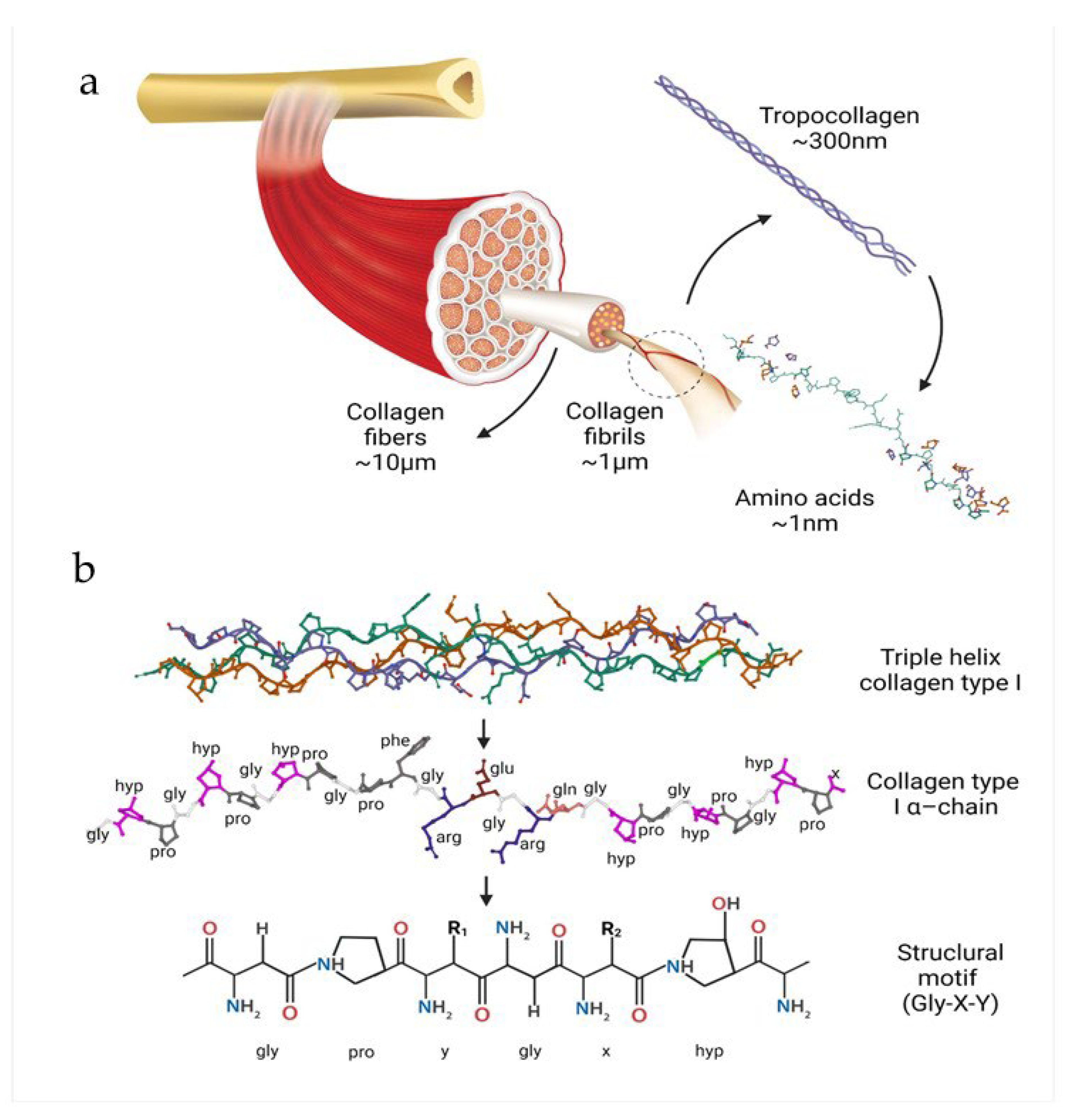
Figure 2.
Steps of collagen isolation process (based on the [21]).
Figure 2.
Steps of collagen isolation process (based on the [21]).
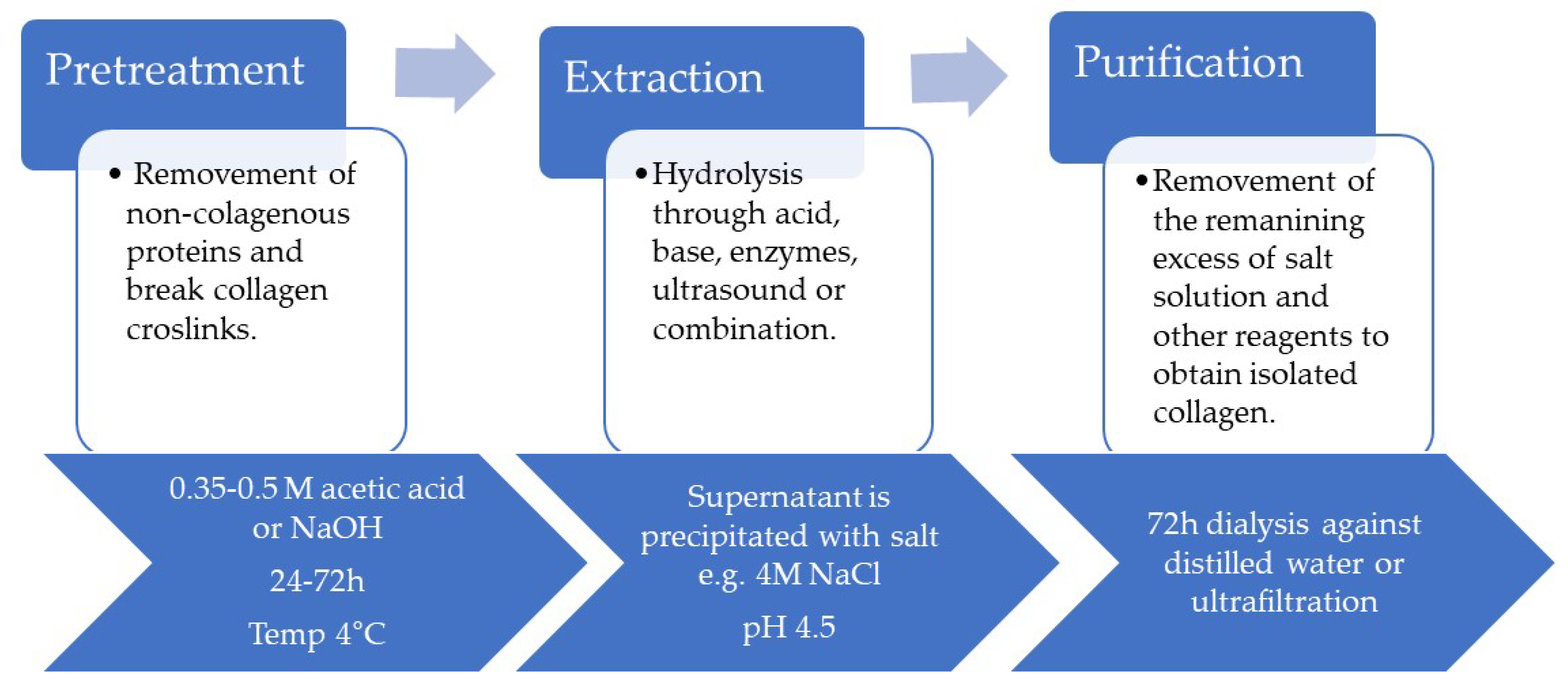
Figure 3.
Examples of forms of collagen derivatives and medical applications.
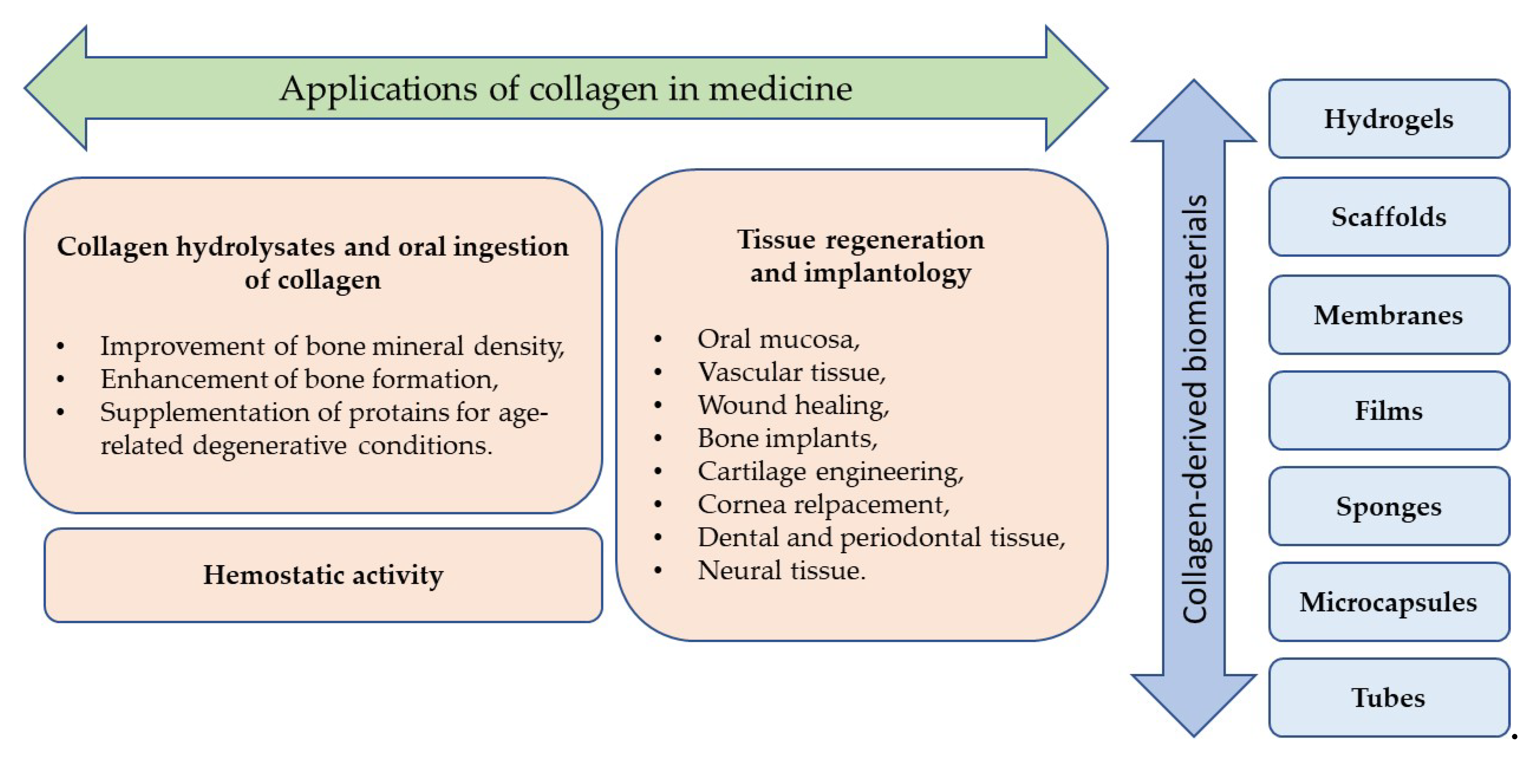

Table 1.
Typical forms of collagen-derived biomaterials.
| Material form | Collagen source | Additives | Cross-linking | In vivo test | In vitro test | Application | Result | Reference |
|---|---|---|---|---|---|---|---|---|
| Scaffold | Fish collagen | Chondroitin sulfate, hydroxyapatite | Yes | Yes | Yes | Osteochondral regeneration | After 6 weeks, the scaffold-treated defects were well-filled with smooth, integrated tissue, unlike the empty group with irregular surfaces. By 12 weeks, the scaffold group showed complete filling with cartilage-like tissue and superior integration compared to the empty group. | [80] |
| Scaffold | Human-like collagen | Nano-hydroxyapatite | Yes | Yes | Yes | Bone regeneration | 12 weeks after implantation, one of the tested scaffolds degraded completely and visibly repaired the bone defect | [81] |
| Scaffold | NA | PLGA, antibacterial synthetic peptides | Yes | No | Yes | Bone regeneration | Obtaining collagen-based scaffolds with osteogenic activity and sustained release of antibacterial peptides creates an environment that promotes cell differentiation and inhibits bacteria | [82] |
| Scaffold | Fish scale collagen (tilapia) | Chitosan | Yes | No | Yes | Oral mucosa therapeutic device | Oral keratinocytes from human oral mucosa produced a multi-layered, polarized, stratified epithelial layer | [83] |
| Scaffold | Fish collagen (flatfish) | Chitooligosaccharides, carbodiimide derivative | Yes | No | Yes | Skintissue regeneration | Induced cell adhesion and proliferation, promotion of well-spread cell morphology | [84] |
| Scaffold | Calf skin | Hydroxyapatite, CaO fibers | Yes | Yes | Yes | Bone regeneration | 8 weeks after implantation, the condylar bone defect was wholly regenerated, and the scaffold had been completely absorbed | [85] |
| Scaffold | Jellyfish | - | Yes | Yes | Yes | Nasa cartilage repair | Excellent biocompatibility with only slight evidence of local inflammatory reactions; prevention of septal perforations | [86] |
| Scaffold | N/A | Chondrocytes | N/A | Yes | No | Matrix associated autologous | Partial or complete filling of the lesion in knee joint cartilage | [87] |
| Scaffold | Equine | - | Yes | Yes | No | Cartilage repair | Integration into the host articular cartilage and promotion of the new cartilage-like tissue development by recruiting the host cells and driving them towards the chondrogenic differentiation, total biodegradation, and replacement of the biomaterial with the newly formed cartilage-like tissue at 16 weeks post-implantation | [88] |
| Scaffold | Type I collagen (corning) | Concentrated growth factor | No | Yes | Yes | Periodontal defects healing | 8 weeks after implantation, the scaffold reconstructed a complete and functional periodontium with the insertion of periodontal ligament fibers into newly formed cementum and alveolar bone | [89] |
| Membrane | Porcine Peritonea | Zinc-doped nanohydroxyapatite | Yes | Yes | Yes | Guided bone regeneration | Obtaining a membrane that preserves the triple-helical structure of collagen fibers and their native 3D network and has a satisfactory biodegradation rate | [90] |
| Membranes | Porcine (Bio-Gide®), Bovine (Colla-D®) | 1-ethyl-3-(3-dimethylaminopropyl) carbodiimide | Yes | Yes | Yes | Guided bone regeneration | Membranes integrated well with surrounding tissues and achieved good osseointegration, without cytotoxic effect, with no membrane exposure observed and no complications | [91] |
| Membrane | Type I porcine collagen (CelGro™) | - | No | Yes | No | Cortical bone regeneration | CelGro™ significantly improved cortical bone repair in the preclinical animal study; in dental implant placement, GBR with CelGro™ resulted in successful regeneration of sufficient mature bone to stabilize the dental implants and process to crown placement | [92] |
| Patches | Scales of snakehead (Channa Micropeltes) | 1,4-butanediol diglycidyl ether | Yes | Yes | Yes | Subcutaneous implantation in mice | Improved cell attachment, proliferation, an infiltration favorable growth of blood and lymphatic vessels | [93] |
| Collagen matrix | Porcine skin | - | No | Yes | Yes | Wound healing | The collagen matrix supports the migration of cells through the matrix, accelerating the healing process | [94] |
| Collagen matrix | Type I bovine | - | Yes | Yes | No | Nerve defect regeneration | Significant improvement of the nerve gap bridging and functional motor recovery in a rat model | [95] |
| Gel | Fish collagen | Genipin | Yes | No | Yes | Biomaterial | Obtained collagen gels exhibit high thermal stability, antioxidant capacity, and characteristic FTIR peaks of type I collagen, indicating their potential for biomaterial applications | [96] |
| Gel | Fish skin (tilapia) – collagen peptides applied in a gel form | - | No | Yes | Yes | Oral ulcer healing on dorsum tongue of mice | Healing promotion: decreased inflammatory cell infiltration, reduced TNF-αand IL-1β expression, increased fibroplasia, angiogenesis, and collagenesis trend | [97] |
| Hydrogel | Type I rat tail collagen | Alginate, CaSO4 | Yes | Yes | Yes | Cells / biomolecules delivery in surgeries | A simple method for creating pre-crosslinked injectable collagen-based hydrogels was developed, and significant results of cell viability compared to similar hydrogels were achieved | [98] |
| Hydrogel | Bovine | Polyethylene glycol | Yes | Yes | Yes | Corneal defects repairment | PEG-collagen hydrogels were able to fill the defect area, remained transparent over one week, and supported multi-layered epithelial growth | [99] |
| Hydrogel | Porcine | - | Yes | Yes | Yes | Corneal implantation | Positive replacement of the portion of a native corneal stroma with rapid wound healing in vivo, the implant permitted host stroma cell migration, epithelial and nerve regeneration while maintaining corneal shape and thickness during a 6-month postoperative period | [100] |
| Hydrogel | Porcine skin | Chondroitin sulfate, poly-d-lysine | Yes | Yes | Yes | Wound healing | Induced fast and superior skin regeneration in a non-healing wound model in diabetic mice | [101] |
| Hydrogel | Fish skin (Nile tilapia) | - | No | Yes | Yes | Healing of deep second-degree burns on rat skin | Significant acceleration of healing of deep second-degree burn wounds | [102] |
| Hydrogel | Swim bladder of Bester sturgeon fish – Type I atelocollagen | Hydroxyapatite, poly(N,N’-dimethylacrylamide) | Yes | Yes | Yes | Implantation into the osteochondral defect | 4 weeks after implantation, the collagen-based gel did not degrade and maintained high strength, indicating its strong osteointegration ability | [103] |
| Gel | Type I Bovine telocollagen | Riboflavin | Yes | No | Yes | Sealant for Corneal Perforation | A highly transparent gel with high adhesion between endo and exogenous collagen was obtained | [104] |
| Hydrogel | Type I Porcine | DL-N-acetylhomocysteine thiolactone | Yes | No | Yes | Sealant for Corneal Perforation | Fully transparent enzyme degradable hydrogel was obtained, and the manufacturing method allows for tuning of the mechanical properties of the gel | [105] |
| Hydrogel | Type I porcine dermal collagen | Dexamethasone, 1-[3-(Dimethylamino) propyl] −3-ethylcarbodiimide me- thiodide, N-hydroxysuccinimide, riboflavin | Yes | Yes | Yes | Corneal application | A transparent hydrogel composed of collagen reinforced by nanocellulose fibers and strengthened through chemical and photochemical crosslinking. Sustainable biomaterial, offering abundance, renewability, and biocompatibility, was obtained, which can be loaded with dexamethasone to reduce inflammation for at least two months post-implantation effectively | [106] |
| Film | Fish collagen (silver carp) | Salicin | Yes | No | Biomaterial application, cosmetics | Addition of salicin increases the viscosity of the solution (intermediate product) and improves the mechanical properties of collagen films | [107] | |
| Film | Porcine, bovine, equine | Carboxymethylcellulose, glycerine, macrogol 300 | No | No | Wound treatment | Films made of equine collagen showed the highest mechanical strength and the lowest swelling ratio compared to the porcine and bovine collagen | [108] | |
| Film | Type 1 marine collagen | Nanoparticles Iron Oxide, Nanoparticles Graphene oxide | No | No | Medicine, Food packaging | Adding iron oxide and graphene oxide improves collagen films' antioxidant, antibacterial and mechanical properties. | [109] | |
| Sponge | Fish collagen, Bovine, Rat tail | No additives | No | Yes | Yes | Wound healing material | The porosity and structure of tilapia skin sponge can be tuned by changing the final concentration. The obtained material can induce blood vessel ingrowth in the wound | [110] |
| Sponge | Rat tail | Taurine | No | Yes | Yes | Wound healing material | The fastest growth of epidermis increased level of TGF- and VEGF protein secretion was observed for collagen sponges with taurine compared to collagen alone and the control sample | [111] |
| Sponge | Type I bovine | Polydopamine, platelet rich plasma | Yes | Yes | Yes | Full thickness skin defect healing | Collagen sponge with polydopamine and PRP showed the highest cell adhesion and proliferation and the fastest wound healing compared to materials without polydopamine | [112] |
| Sponge | Fish collagen (tilapia); bovine collagen | Polyethylene oxide, chitosan | - | Yes | No | Evaluation of wound healing in rats | Increasing the percentage of wound contraction, reducing the inflammatory infiltration, and accelerating the epithelization and healing also enhanced the total protein and hydroxyproline levels in the wound bed | [113] |
| Microcapsule | Type I rat tail | Osteoarthritis chondrocytes | Yes | No | Yes | Osteoarthritis | UV-treated collagen pre-gels form hollow tubes with high stability, adjustable viscoelasticity, and controlled pore structure, ideal for separate endothelial and ectodermal cell cultures | [114] |
| Collagen tube | Type I | Riboflavin | Yes | No | Yes | Vascular networks and nerve fibers in artificial organ fabrication and regenerative medicine | A method for producing collagen gel tubes for potential biomedical applications has been developed |
[115] |
| Tube / rod | Type I porcine atelocollagen | Carbonate buffer | Yes | No | Yes | Regenrative medicine | A collagen-based material with excellent mechanical properties, biocompatibility, and patentability has been developed | [116] |
| Powder | Fish skin (Nile tilapia) collagen polypeptides | - | No | Yes | Yes | Evaluation of wound healing activity | High capacity to induce HaCaT cell migration; healing improvement in the deep partial-thickness scald model in rabbits | [117] |
Table 2.
Commercial examples of collagen biomaterials.
| Product / Manufacturer | Collagen type | Additive | Material type |
|---|---|---|---|
| Bio-Gide® / Geistlich Pharma | Type I, III | - | Membrane |
| Jason® / Botiss | Type III | - | Membrane |
| OssixPlus® / Datum Dental Ltd. | Type I | - | Membrane |
| Biomend® Zimmer | Type I | - | Membrane |
| Collagraft® / Zimmer | Type I >95%, Type II <5% | HAP 1 - 65% β-TCP 2 - 35% |
Scaffold |
| GingivAid®/ Maxigen Biotech Inc. | Type I | HAP, β-TCP | Scaffold |
| Integra Mozaik / Integra Life Sciences | Type I | TCP 3 | Scaffold |
| FormaGraft / Maxigen Biotech Inc. | Type I | HAP, TCP | Scaffold |
| Orthoss Collagen / Geistlich | N/A | Bovine HAP | Scaffold |
| OP-1 Implant / Stryker | Type I | BMP-7 4 | Scaffold |
| SilvaKollagen®Gel/ DermaRite®, | Type I | 1% silver oxide | Gel |
| WounDres® / Coloplast | N/A | Panthenol, alantoine | Gel |
| RatuZel / Regional Health Center Ltd. | Type I | Lactic acid | Gel |
| Parasorb® / Resorba | N/A | - | Sponge |
| Hemocollagene / Septodont | Type I | - | Sponge |
| Surgispon® / AegisLifeSciences | N/A | - | Sponge |
1 HAP - Hydroxyapatite; 2 β-TCP - β-Tricalcium Phosphate, 3 TCP - Tricalcium Phosphate, 4 BMP-7 Bone Morphogenetic Protein
Table 4.
Examples of drug delivery systems based on collagen.
| Collagen origin and the type of drug delivery system |
Drug (and its activity) |
Route of administration | Application | Reference |
|---|---|---|---|---|
| bovine type I collagen cross-linked by glutaraldehyde, hybrid lyophilizates | irinotecan (topoisomerase I inhibitor) | transdermal | treatment of bone and skin cancer | [158] |
| bovine type I collagen cross-linked by glutaraldehyde, lyophilized sponges | niflumic acid (an analgesic and anti-inflammatory agent) | local - teeth | pain management in dentistry and medicine |
[159] |
| type I collagen from calf skin, cross-linked by glutaraldehyde | lidocaine hydrochloride (local anesthetic), diclofenac sodium salt (anti-inflammatory), caffeic acid (anti-inflammatory, antioxidant) |
dermal | potential dermal application for anesthetic or anti-inflammatory action | [160] |
| amphiphilic composite platform associating dense collagen hydrogels and up to 50 wt% polyesters |
spironolactone (an antagonist against mineralocorticoid receptor) | not specified | potential application in cardiovascular and renal diseases, cutaneous chronic wounds, age-related macular degeneration, chorioretinal disorders |
[161] |
| collagen peptide and chitosan nanoparticles (pH-responsive) | doxorubicin hydrochloride (antineoplastic activity) | not specified | significant anti-proliferative properties against HeLa (human cervical carcinoma) cells, potential innovative drug delivery carriers in advanced cancer therapy | [162] |
| biomaterials made from cellulose, collagen, and polyurethane formed into thin films | ketoconazole (antifungal agent) | transcutaneous | controlled drug release and biocidal activity | [163] |
| collagen (rat tail-derived) type I -hydroxyapatite scaffolds functionalized using BMP-2 and loaded with biodegradable microspheres with ALN encapsulated |
bone morphogenic protein-2 (BMP-2; osteoinductive growth factor) and alendronate (ALN; treatment of bone loss and osteoporosis) | bone implantation | the initial release of BMP-2 for a few days, followed by the sequential release of ALN, after two weeks, provides enhanced bone regeneration |
[164] |
| ovine collagen-based micellar nanoparticles (3-ethyl carbodiimide-hydrochloride and malondialdehyde as crosslinkers) |
silymarin (neuroprotective activity) | intraperitoneal | enhanced neuroprotection by increasing drug bioavailability and targeting (in rats) | [165] |
| PerioChip – gelatin insert | chlorhexidine digluconate (antibacterial) | periodontal pockets | enhanced reduction of pocket depth by approx. 0.4 mm within 6 months | [156] |
| bovine collagen sponge (cross-linked with glutaraldehyde) | ibuprofen (anti-inflammatory, analgesic) | dental | dental problems | [157] |
| collagen (from the marine sponge Chondrosia reniformis) nanoparticles | 17β-estradiol-hemihydrate (hormone replacement therapy) | transdermal | prolonged release and enhanced absorption of estradiol through human skin | [152] |
Disclaimer/Publisher’s Note: The statements, opinions and data contained in all publications are solely those of the individual author(s) and contributor(s) and not of MDPI and/or the editor(s). MDPI and/or the editor(s) disclaim responsibility for any injury to people or property resulting from any ideas, methods, instructions or products referred to in the content. |
© 2024 by the authors. Licensee MDPI, Basel, Switzerland. This article is an open access article distributed under the terms and conditions of the Creative Commons Attribution (CC BY) license (http://creativecommons.org/licenses/by/4.0/).
Copyright: This open access article is published under a Creative Commons CC BY 4.0 license, which permit the free download, distribution, and reuse, provided that the author and preprint are cited in any reuse.


