Submitted:
12 August 2024
Posted:
13 August 2024
Read the latest preprint version here
Abstract
This study analyzed methodologies for identifying β-casein genotypes in dairy cows and the purity of A2 fermented milk. The research was justified by the need for quick and accurate techniques for herd screening and A2 milk quality control. The methodology included the development of an ELISA test to detect β-casein A1 in fermented milk and the evaluation of a commercial Lateral Flow Immuno Assay (LFIA) to identify A2A2 genotypes and the purity of A2 milk. The results showed that the ELISA test had high sensitivity and specificity (100%) in detecting β-casein A1. The LFIA test was 100% effective in identifying A2A2 genotypes and verifying the purity of A2 milk with a minimum detectable contamination of 5% for raw milk and 10% for fermented milk. Compared to traditional genotyping, these methods proved to be highly reliable and more practical for large-scale screening. It was concluded that the ELISA and LFIA were valuable tools for ensuring the quality and authenticity of A2 milk, meeting the demands of producers and consumers for safe and healthy dairy products.
Keywords:
A2A2 dairy cows
; Genotyping
; ELISA
; LFIA
; Comparison of methods
; Facilitated analysis
1. Introduction
Millions of people around the world consume milk and its derivatives daily, taking advantage of its nutritional richness as an excellent source of proteins, fats, and micronutrients [1,2,3]. Milk is composed of two main groups of proteins: caseins and whey proteins [4]. The four types of casein (αs1-casein, αs2-casein, β-casein, and κ-casein) are encoded by the genes CSN1S1, CSN1S2, CSN2, and CSN3, respectively, and are located in a cluster within a 250 kb region on chromosome 6 [5]. Among them, β-casein is the main protein found in milk [6]. Different mutations in the bovine CSN2 gene have resulted in 12 genetic variants of β-casein, including A1, A2, A3, B, C, D, E, F, G, H1, H2, and I, with A1 and A2 being the most common variants [5,7,8]. Cows with the A1/A1 homozygous genotype and A1/A2 heterozygous genotype produce A1 milk, while cattle with the A2/A2 genotype produce A2 milk [2,8,9].
The A1 and A2 variants do not differ nutritionally; the distinction between them lies in a single nucleotide polymorphism (SNP) at the amino acid residue at position 67 of the peptide chain. In A1 milk, this residue is histidine (His67), while in A2 milk it is proline (Pro67) [5,10,11,12,13]. It is believed that the A2 variant was initially present in all herds; however, a few thousand years ago, a natural mutation from proline to histidine occurred in European dairy cattle, resulting in the presence of the A1 allele in various breeds as we know them today [4,14,15].
During gastrointestinal digestion of dietary proteins, enzymatic action can result in the release of biologically active peptides [9,16,17]. During the digestion of A1 bovine milk, the presence of the amino acid histidine allows the proteolytic cleavage of β-casein by human gastrointestinal enzymes, releasing a bioactive peptide known as β-casomorphin-7 (BCM-7) [12,18]. Studies indicate that the BCM-7 peptide has opioid activity, with affinity for µ-opioid receptors (MORs), found in various regions of the body [14,19].
Several studies have also reported correlations between the consumption of A1 cow’s milk and dairy products with various human health conditions, due to the release of the BCM-7 peptide during digestion, including type 1 diabetes, cardiovascular disorders, gastrointestinal problems, inflammatory responses, and autism spectrum disorders [3,14,15,20,21,22,23,24,25,26,27,28,29]. Awareness of the adverse effects of BCMs on human health has generated a growing demand for exclusively A2 dairy products, both among companies and dairy consumers [2,10,12]. As a result, producers have started selecting females with the A2/A2 genotype [4,9,30], leading to a steady increase in the global A2 milk market in recent years [3], with estimates that this market will grow exponentially until 2029 [4]. Thus, labeling indicating the presence of β-casein A2 has become common in various dairy products, often associated with nutritional benefits [5].
In this context, a variety of analytical methods have been employed to study and identify the genetic variants of β-casein [18]. This includes techniques such as liquid chromatography-tandem mass spectrometry (LC-MS/MS) [31], reverse-phase high-performance liquid chromatography (RP-HPLC) [32], electrophoresis and high-resolution accurate mass spectrometry (HRAMS) [33], as well as mid-infrared spectroscopy and chemometrics [10]. In addition to these, various DNA-based genotyping methods have been described, including allele-specific polymerase chain reaction (AS-PCR) [18,22], allele-specific competitive replication system (ACRS-PCR) [18,34], restriction fragment length polymorphism (RFLP-PCR) [18,32,35], single-strand conformation polymorphism analysis (SSCP-PCR) [7], amplification refractory mutation system (ARMS-PCR) [32], and gene sequencing [36,37]. Although current tests are highly accurate, they are expensive, complex, and time-consuming [6,32].
Genotyping is the gold standard for identifying A2A2 animals due to its high accuracy, but it has drawbacks such as cost, complexity, and the need for specific biological samples like blood, buccal swabs, hair bulbs, or ear tissue. Non-invasive methods like hair bulbs and buccal swabs are less invasive but often yield lower DNA quality, increasing error chances. Invasive methods like blood and ear tissue provide higher DNA quality, making them more reliable despite added complexity and cost. Laboratory genotyping requires specialized personnel and centralized testing, complicating mass screening. Point-of-care (POC) methodologies enable decentralized, mass screening and should be developed for global herd testing and quick A2 milk genetic selection. Ensuring A2 milk purity is crucial for quality control in the dairy industry, necessitating accurate methods to verify milk and dairy products. This growing demand for precise evaluation methods is driven by the increasing focus on A2 milk production and the need for stringent quality control. Therefore, the aim of this study was to evaluate different rapid methods for identifying A2A2 animals, as well as to assess various methods for verifying the purity of A2 milk (absence of β-casein A1) in raw milk and fermented milk. In the present study, an ELISA test was developed for the detection of β-casein A1 in fermented milk, creating an important methodology for the quality control of this type of food. Additionally, this work evaluated the sensitivity and specificity of a commercial Lateral Flow Immuno Assay (LFIA) specific for A2 milk detection for the identification of A2A2 animals and verification of A2 milk purity. The data were compared with other techniques, such as traditional genotyping and other ELISA-based methodologies, and the results were analyzed and discussed. This study aims to benefit the management of A2 milk-producing herds, provide an industrial solution for batch analysis of milk, and offer a reliable alternative for consumers to identify milk and dairy products according to their nutritional preferences.
2. Materials and Methods
2.1. Material
Chemicals were purchased from Sigma-Aldrich® (Cotia-SP, Brazil) and Rapid test (LFIA) A2-MiLK TEST® was provided by Scienco Biotech (Lages-SC, Brazil).
2.1. Selection of Dairy Cow Samples for Genotyping
Milk samples were collected from dairy cows on different farms in the state of Santa Catarina (Brazil). The collection was carried out individually and manually from each animal, without using milking machines, to avoid contamination, using clean and dry bottles. For each collection, a stream from each teat of the animal was mixed. The collected milk was immediately labeled and frozen for later analysis. Milk samples from 34 previously genotyped animals from a rural property located at the geographical coordinates -28.46926833441343, -49.306805225131185 were analyzed. The milk samples were provided by the farm owner, who also made available the genotyping reports previously performed on the herd animals. The genotyping of these animals was carried out through hair collection and genotypic analysis by the company Zoetis. Additionally, milk samples from 28 animals from a rural property located at the geographical coordinates -28.581369733535674, -49.18977477310808 were analyzed. The animals were previously genotyped by the company STgenetics, using ear cartilage collection. The milk samples from these animals were donated by the farm owner, who also provided the genotyping reports.
2.2. ELISA Test for Identification of β-Casein A1 Dairy Products
ELISA (Enzyme-Linked Immunosorbent Assay) tests were performed on the collected milk samples to identify A2A2 animals. For this analysis, the protocol described by de Jesus et al. [6] was used. ELISA tests were also performed to detect β-casein A1 contamination in A2 milk. In this case, milk from A2A2 genotyped animals was mixed with varying proportions of milk from A1A1 genotyped animals, and the assay followed the previously described protocol [6]. The absorbance results were used to generate a calibration curve to determine the percentage of β-casein A1 in A2 milk, aiming to develop a protocol for analyzing A2 milk mixtures.
ELISA assays were also conducted to detect β-casein A1 contamination in fermented milk samples. The objective was to create a methodology for quality control of A2 fermented milk, ensuring its purity. In this case, commercial A2 fermented milk (Letticoa2 (L20240711:36)) was used, mixed in known proportions with regular fermented milk (Yakult (L25.06.240837C)). The samples were subjected to ELISA analysis according to the following protocol. Polystyrene plates were coated with 96 μL of coating buffer, to which 4 μL of the fermented milk sample, previously diluted in NaOH at a 1:10 ratio, were added. The plates were then incubated overnight at 4°C to allow for the immobilization of A1 casein in the wells. After immobilization, the plates were blocked for 1 hour at 37°C with 200 μL of blocking buffer containing 1% BSA in PBS to prevent nonspecific binding. Subsequently, the anti-β-casein A1 IgY antibody was diluted in PBS containing 1% BSA and 0.05% Tween-20, at a 1:1500 dilution. This solution was added to the wells and incubated for 45 minutes at 37°C, allowing the specific antibody to bind to the captured A1 casein. After incubation with the primary antibody, the plates were washed three times with washing buffer (PBS containing 0.05% Tween-20) to remove unbound antibodies. Then, the secondary anti-IgY-HRP antibody (Sigma-Aldrich) was diluted in PBS + 1% BSA + 0.05% Tween-20, at a 1:7500 dilution, and added to the wells. The plates were again incubated for 45 minutes at 37°C, protected from light. After incubation with the secondary antibody, the plates were washed five times with washing buffer to remove any unbound secondary antibody. To develop the reaction, 50 μL of tetramethylbenzidine (TMB) substrate were added to each well and incubated for 15 minutes at room temperature. The reaction was then stopped by the addition of 50 μL of H2SO4. Finally, the optical density (OD) of each well was measured in an ELISA plate reader at 450 nm. The color intensity developed is directly related to the amount of A1 casein present in the fermented milk samples, thus allowing quantification through this sensitive and specific immunological method. A calibration curve with the obtained absorbances was generated.
2.3. LFIA Test for Identification of A2A2 Genotype
To evaluate the A2A2 genotype using a Point-of-Care method, the A2-MiLK TEST® from Scienco Biotech was employed. The procedure followed the manufacturer’s protocol. First, milk was manually collected in a separate, clean flask. Then, ten drops of the collected milk were added to a diluent tube using a plastic pipette provided in the kit. The tube was shaken to ensure homogeneity of the solution. Next, the test cassette was placed on a flat surface, and two drops of the diluted milk solution were pipetted onto the cassette. The solution was allowed to migrate along the device for 20 minutes at room temperature. The results were interpreted by observing the lines on the cassette: according to the manufacturer’s the appearance of one pink line in the test area (T) and another in the control area (C) indicated A2 milk, produced by an A2A2 animal. Conversely, the appearance of only one pink line in the control area (C) indicated the milk was not A2, produced by animals of genotype A1A1 or A1A2.
2.4. LFIA Test for Purity Evaluation
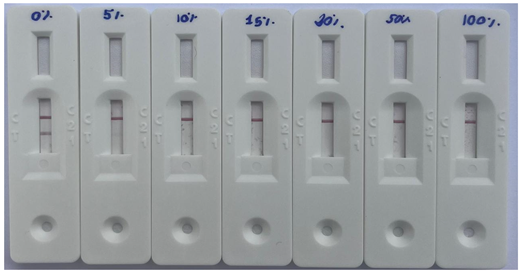
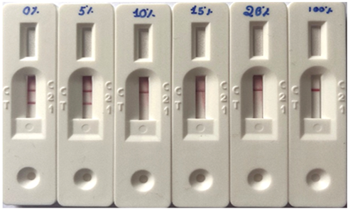
To verify the purity in milk and fermented milk samples, the A2-MiLK TEST® from Scienco Biotech was also used. Samples from cows with confirmed genotyping for A1A1 and A2A2 milk were selected, as well as commercial fermented milks, including A1 fermented milk from the Yakult brand (L25.06.240837C) and A2 fermented milk from the Letticoa2 brand (L20240711:36). Formulations were then prepared with varying percentages of A1A1 milk added to A2A2 milk (0%, 5%, 10%, 15%, 20%, 50%, and 100% (v/v)), and A1 fermented milk added to A2 fermented milk (0%, 5%, 10%, 15%, 20%, and 100% (v/v)). The procedure followed the manufacturer’s protocol. Ten drops of the milk mixture were added to a diluent tube using a sterile plastic pipette provided in the kit. The tube was shaken to ensure homogeneity of the solution. Next, the test cassette was placed on a flat surface, and two drops of the diluted milk solution were pipetted onto the cassette. The solution was allowed to migrate along the device for 20 minutes at room temperature. The results were interpreted, according to the manufacturer´s protocol, by observing the lines on the cassette: the appearance of one pink line in the test area (T) and another in the control area (C) indicated 100% A2 milk. Conversely, the appearance of only one pink line in the control area (C) indicated the presence of A1 β-casein, meaning the A2 milk was not pure, containing a percentual of contamination with A1 β-casein.
2.5. Statistical Analysis
All experiments were conducted in duplicate. ANOVA and Tukey tests with a 95% confidence interval were used to verify the existence of statistical differences between the samples. The presence of outliers was also checked using the PAST 4.3 software.
3. Results
3.1. Identification of β-Casein Phenotypes by ELISA and LFIA Tests
A total of 62 raw milk samples from cows previously genotyped for β-casein A1A1, A1A2, and A2A2 were used. The milk samples had their β-casein phenotypes analyzed by ELISA test, and the results are shown in Figure 1. The ELISA test (Figure 1a) showed a clear differentiation in absorbance values between the groups with A1 phenotype (A1A1 and A1A2) and A2 phenotype (A2A2). The absorbances of the A1A1 and A1A2 raw milk samples ranged from 0.33 to 0.975 and 0.104 to 0.609, respectively. For the A2A2 raw milk samples, the absorbances ranged from 0.038 to 0.081, with one sample showing an absorbance of 0.195, characterized as an outlier (absorbance above the calculated upper limit of 0.105). The ELISA absorbance histogram (Figure 1b) shows that most samples have values concentrated between 0.04 and 0.08, indicating a predominantly low absorbance in the A2 raw milk samples. In contrast, a smaller number of samples show higher absorbance values, between 0.30 and 0.60, representing the A1 raw milk samples with higher absorbance response. The highest peak of the histogram is around 0.07, reflecting the high frequency of A2 raw milk samples with this absorbance value. Some raw milk samples, with absorbance values above 0.5, are considered outliers and may indicate significant differences in the composition or response of A1 raw milks compared to A2 raw milks.
Table 1 presents a comparison between different methods for identifying A2 raw milk, including animal genotyping by gene sequencing, the ELISA test, and the LFIA test. The results show that, out of a total of 62 raw milk samples from cows genotyped by gene sequencing, 9.68% have the A1A1 genotype, 38.71% have the A1A2 genotype, and 51.61% have the A2A2 genotype. The tests used showed high consistency, with a sensitivity and specificity of 100% for A2A2 genotype detection. A representative image of the LFIA results from A1A1, A1A2, and A2A2 cows is shown in Figure 2. The LFIA results from the 62 genotyped animals demonstrated that all A2A2 animals were correctly identified, with no false positives. This yielded a specificity and sensitivity of 100% (Table 1). Both the ELISA test and LFIA test corroborate the results of gene sequencing, correctly identifying the A2A2 genotypes. This demonstrates that these methods are highly reliable for identifying cows that produce A2 milk.
3.2. ELISA and LFIA Tests on Raw Milk Samples to Assess Purity
The applicability of the ELISA method in identifying the purity of raw A2 milk was evaluated and compared with the LFIA test. For this, A2A2 milk samples were contaminated with 0% (negative control), 5%, 10%, 15%, 20%, 50%, and 100% (v/v) (positive control) of raw A1A1 milk. The ELISA test absorbance results of the samples can be seen in Figure 3, and the LFIA test results can be seen in Table 2. For the ELISA test, the initial average absorbance of A2 milk was 0.046 ± 0.001, progressively increasing with the contamination of A1 milk (P ≤ 0.05), reaching 0.124 ± 0.007 with 15% contamination. The average absorbance for A1 milk was 0.338 ± 0.002. The ELISA method demonstrated efficacy in detecting A2 milk contamination with A1 milk when the latter was present in a proportion of 15% or higher. The LFIA test detected the presence of A1 milk when added to A2 milk in concentrations of 5% or higher.
3.3. ELISA and LFIA Tests on Fermented Milk Samples to Assess Purity
The applicability of the ELISA method in identifying the purity of A2 fermented milk was evaluated and compared with the LFIA test. For this, A2 fermented milk samples were contaminated with 0% (negative control), 5%, 10%, 15%, 20% and 100 % (positive control) of A1 fermented milk. The ELISA test absorbance results of the samples can be seen in Figure 4, and the LFIA test results can be seen in Table 3. The A2 fermented milk samples showed a significant increase in average absorbance (P ≤ 0.05) starting from 10% contamination with A1 fermented milk, rising from 0.044 ± 0.003 in the negative control to 0.105 ± 0.001 in the fermented milk contaminated with 10% A1 fermented milk. With contamination at 15% and 20% of A1 fermented milk, the average absorbance values increased to 0.148 ± 0.018 and 0.162 ± 0.013, respectively, while the sample with 100% A1 fermented milk showed absorbance values of 0.348 ± 0.008. The LFIA test detected the presence of A1 fermented milk when added to A2 fermented milk at concentrations of 10% or higher. These results demonstrate that the ELISA method is capable of detecting the presence of A1 fermented milk in A2 fermented milk when added in a proportion of 10% or higher, a value equal to that obtained with the LFIA test.
4. Discussion
4.1. Importance of Non Invasive ELISA Test and LFIA Test in Detection of A1 β-Casein and Expansion to Other Dairy Products
In identifying phenotypes using the ELISA test, the skewed distribution with a bias toward low absorbance values suggests that most samples have a low absorbance response. This clear separation between the absorbance values of A1 and A2 raw milk allows for rapid classification, with A2 raw milk samples concentrated in a narrow range and A1 samples showing greater variability. The uniform distribution and defined peaks indicate that the ELISA test is sensitive and specific enough to distinguish the different phenotypes of raw milk. This precision facilitates the quick identification of A2 raw milk, reduces classification errors, speeds up screening processes, ensures the consistency of the ELISA test, and provides a reliable basis for future studies. One advantage of the ELISA test is the ability to use the raw milk itself as a sample, which, combined with the quick turnaround of results, ensures the purity of the analyzed milk. This is advantageous because it eliminates the need to collect other samples from the animals, such as blood or cartilage, which are generally used for genetic sequencing tests. Additionally, the samples can be easily collected during conventional milking. Due to its efficient differentiation of β-casein A1 and A2 phenotypes, the ELISA method can be used for phenotyping dairy herds from milk samples collected from each animal. Accurate identification of animals with the A2A2 genotype is crucial for the production of A2 milk, preferred for its potential health benefits. The consistency of the methods tested in Table 1 reinforces the feasibility of implementing these techniques on a large scale to optimize A2 milk production. Since the phenotype is directly related to the genotype, this method is applicable for selecting animals with the A2A2 genotype and offers advantages by being less invasive and faster compared to DNA-based genotyping methods [44]. Identifying animals that produce milk with β-casein A2 allows producers to direct breeding to increase the frequency of this genotype in the herd [13]. For example, cattle that produce A2 milk, and consequently have the A2A2 genotype, can be prioritized in mating programs during selection, leading to successful selective breeding [8,18]. In recent years, selecting the A2A2 allele has become a desirable trend in animal management to meet the growing market demand for milk and dairy products containing β-casein A2, referred to as “A2 protein”, “A2 milk”, or “β-casein A2 protein” [9,30]. The A2 milk market is increasing due to heightened consumer awareness of health and well-being, particularly in light of reports associating A1 casein with diseases such as type 1 diabetes, gastrointestinal symptoms, heart disease, and autism [3,15]. Additionally, the expanding market for A2 dairy products can provide new business opportunities and incentives for producers who invest in selecting a 100% tested herd [13].
The use of LFIA test and ELISA test to detect A2 milk is an effective tool to prevent fraud in the A2 milk market, as demonstrated by the results of A2 raw milk contaminated with A1 raw milk. Due to the increasing demand and added value of A2 milk, there is a significant risk of adulteration, where A1 milk may be improperly mixed and sold as A2 milk. With its high sensitivity and specificity, the evaluated tests allows for precise identification of β-casein phenotypes, ensuring that only genuine A2 milk is marketed. By implementing the analysized tests, producers and regulators can ensure the authenticity of A2 milk, protect consumer confidence, and maintain market integrity. Additionally, using raw milk samples facilitates continuous monitoring of milk purity throughout the production chain, from milking to final distribution. Milk adulteration generally aims to increase the volume of the product delivered to meet demand [45]. Additionally, fraud is one of the most serious problems faced by the dairy industry, as it not only results in significant financial losses for the industry, affecting producers and distributors, but can also pose health risks to consumers [46]. By analyzing A2 milk samples in Austria over a six-month period for the presence of β-casein A1, Mayer et al. [18] found that four out of five A2 milk samples analyzed were adulterated, containing β-casein A1.
The importance of extending adulteration detection to other dairy products, such as fermented milk, is crucial. Dairy derivatives, including yogurts and cheeses, are highly popular and widely consumed, making them susceptible to similar frauds as liquid milk. Implementing rigorous β-casein A1 detection tests in these products would not only ensure the authenticity and purity of A2 fermented milk but also protect the health of consumers who choose these products for their purported health benefits. Furthermore, extending these tests across the entire dairy product chain would enhance consumer confidence and maintain market integrity, ensuring that all products labeled as A2 meet the expected standards. The ELISA test successfully detected the presence of the A1 phenotype in fermented milks, demonstrating its effectiveness in identifying adulterations. The importance of extending adulteration detection to other dairy products, such as fermented milk, is crucial. Dairy derivatives, including yogurts and cheeses, are highly popular and widely consumed, making them susceptible to similar frauds as liquid milk. Implementing rigorous β-casein A1 detection tests in these products would not only ensure the authenticity and purity of A2 fermented milk but also protect the health of consumers who choose these products for their purported health benefits. Furthermore, extending these tests across the entire dairy product chain would enhance consumer confidence and maintain market integrity, ensuring that all products labeled as A2 meet the expected standards. The growing diversity and quantity of dairy products, such as fermented milks, now labeled to highlight the presence of β-casein A2, reflect the current trend [4]. These A2 products are often marketed at a significantly higher price compared to conventional products [18]. In this context, it is essential to develop and standardize analytical methods that can detect the presence of β-casein A1 in dairy products sold as A2, given the lack of category definitions, clear specifications, and consistent regulatory methods for the quality control of these products [41,47,48]. Thus, such measures are crucial to prevent adulteration, whether intentional or not, and to ensure their authenticity [18].
4.2. Advantages, Limitations, Considerations for Practical Application and Cost-Effectiveness of the ELISA and LFIA Methodologies
Immunossays are recognized for their high sensitivity, precision, and broad applicability [48,49,50]. It provides faster results than DNA-based genotyping assays, which require a significant amount of time for the extraction and analysis of genetic material [32,44]. Additionally, milk sample collection for the ELISA test and LFIA test is less invasive compared to blood and tissue sample collection for DNA genotyping tests [34,38]. Despite the possibility of using milk samples for qPCR tests, the extraction of DNA from somatic cells in milk is challenging due to the presence of inhibitors such as fat, protein, and calcium, as well as the low number of these cells in milk from healthy herds, making it difficult to achieve a high yield of DNA [48]. In addition to simplified protocols for sample collection and preparation [47], ELISA and LFIA are recognized as one of the most economical and cost-effective methods for direct measurements of β-casein A1 in milk samples [47,48]. Despite numerous advantages, the ELISA method has limitations, such as the fact that is not a POC assay, the fact it needs sample dilutions to fit the dynamic range and the requirement for strong alkaline solutions [44]. As advantage, the ELISA method described here is quantitiative, and therefore, can determine the percentage of A1 casein contamination in A2 milk, while LFIA is not a quantitative method and cannot determine precisely the level of contamination. Similarly to the ELISA test, the LFIA test used for comparison in this study offers results in a short time, usually within minutes, allowing for quick analysis compared to traditional genotyping methods, which are known to be time-consuming [51]. Additionally, these tests are designed to be easily used by non-specialized personnel, benefiting both producers and dairy farmers as well as consumers, especially since they can be conducted directly on farms, industries, or home. They are more cost-effective and less labor-intensive compared to more complex laboratory techniques, such as PCR with restriction RFLP, high-resolution melting (HRM) genotyping, and SNP rhAmp genotyping, which involve extensive DNA extraction, sample pre-treatment, and data analysis procedures [44]. Among the limitations of the method, it is important to mention that it identifies A2A2 animals, but does not distinguish between A1A2 and A1A1 animals, nor can it analyze changes in the amino acid sequence of the target region, as is possible with DNA sequencing [37]. As mentioned earlier, due to the relatively low cost, effectiveness, and ease of use, the ELISA method and the commercial LFIA test are suitable for routine and large-scale screening of β-casein variants in milk and fermented milk.
4.3. Assessment of Sensitivity and Specificity of ELISA and LFIA Test
In comparing the methods for detecting milk purity, LFIA (Lateral Flow Immunoassay) and ELISA (Enzyme-Linked Immunosorbent Assay), it is evident that each has distinct sensitivity levels. The LFIA method can detect contamination levels above 5%, whereas the ELISA method detects contaminations above 10%. Although both ELISA test and LFIA test possess high sensitivity and specificity in detecting β-casein A1 and A2 in milk, only ELISA can determine quantitativelly the level of A1 contamination. Therefore, ELISA, with its high precision and detailed quantification capability, is ideal for applications requiring extremely reliable results [52]. Furthermore, both methods demonstrated 100% accuracy in identifying A2A2 animals, with no false positives recorded. This indicates that both LFIA and ELISA are highly reliable for detecting A2A2 animals and can effectively replace genotyping for this purpose. These findings underscore the practical utility of both methods in the dairy industry, providing reliable alternatives for ensuring A2 milk purity and enhancing the efficiency of contamination detection and animal selection. To identify A2A2 animals traditional genotyping methods, such as direct DNA analysis, are accurate but costly and time-consuming. Analyzed methos are faster, more practical, and less costly, making it ideal for applications requiring frequent monitoring and quick decisions, such as in the dairy industry. In terms of cost, traditional genotyping involves higher expenses due to the complexity of the process and the need for specialized infrastructure, whereas ELISA and LFIA is more economical and simpler options. Table 4 presents a detailed comparison between traditional genotyping methods, ELISA test, LFIA test for detecting β-casein A1 in milk.
5. Conclusions
The methodologies analyzed for identifying β-casein genotypes in dairy cows and the purity of A2 fermented milk demonstrated significant effectiveness. The research was justified by the need for quick and accurate techniques for herd screening and A2 milk quality control. The methodology included the development of an ELISA test to detect beta-casein A1 in fermented milk and the evaluation of ELISA test and LFIA test to identify A2A2 genotypes and the purity of A2 milk. The results showed that the ELISA test had high sensitivity and specificity (100%) in detecting beta-casein A1. The LFIA test (A2-MiLK TEST®) were effective in identifying A2A2 genotypes woth 100% accuraacy and also in verifying the purity of A2 milk with a minimum detectable contamination of 5% for raw milk and 10% for fermented milk. Compared to traditional genotyping, these rapid methods proved to be highly reliable and more practical for large-scale screening. Therefore, ELISA and LFIA tests are valuable tools for ensuring the quality and authenticity of A2 milk, providing an efficient, cost-effective solution that meets the demands of producers and consumers for safe and healthy dairy products, while also supporting the growth of the A2 milk market.
Data Availability Statement
The raw data supporting the conclusions of this article will be made available by the authors on request.
References
- Kaskous, S. A1- and A2-Milk and Their Effect on Human Health. Journal of Food Engineering and Technology 2020, 9, 15–21. [Google Scholar] [CrossRef]
- Ardicli, S.; Aldevir, O.; Aksu, E.; Gumen, A. The Variation in the Beta-Casein Genotypes and Its Effect on Milk Yield and Genomic Values in Holstein-Friesian Cows. Anim Biotechnol 2023, 34, 4116–4125. [Google Scholar] [CrossRef] [PubMed]
- Jeong, H.; Park, Y.S.; Yoon, S.S. A2 Milk Consumption and Its Health Benefits: An Update. Food Sci Biotechnol 2024, 33, 491–503. [Google Scholar] [CrossRef] [PubMed]
- Dantas, A.; Kumar, H.; Prudencio, E.S.; de Avila, L.B.; Orellana-Palma, P.; Dosoky, N.S.; Nepovimova, E.; Kuča, K.; Cruz-Martins, N.; Verma, R.; et al. An Approach on Detection, Quantification, Technological Properties, and Trends Market of A2 Cow Milk. Food Research International 2023, 167. [Google Scholar] [CrossRef] [PubMed]
- Li, X.; Spencer, G.W.K.; Ong, L.; Gras, S.L. Beta Casein Proteins – A Comparison between Caprine and Bovine Milk. Trends Food Sci Technol 2022, 121, 30–43. [Google Scholar] [CrossRef]
- de Jesus, B.A.P.; Echeverri, L.M.S.; Magalhães, M. de L.B.; Silva, G.F. da Generation and Characterization of Avian IgY Antibodies for Detecting Beta-Casein A1 in Bovine Milk. Anal Biochem 2023, 678, 115283. [Google Scholar] [CrossRef]
- Jann, O.; Ceriotti, G.; Caroli, A.; Erhardt, G. A New Variant in Exon VII of Bovine β-Casein Gene (CSN2) and Its Distribution among European Cattle Breeds. Journal of Animal Breeding and Genetics 2002, 119, 65–68. [Google Scholar] [CrossRef]
- Khan, R.; De, S.; Dewangan, R.; Tamboli, R.; Gupta, R. Potential Status of A1 and A2 Variants of Bovine Beta-Casein Gene in Milk Samples of Indian Cattle Breeds. Anim Biotechnol 2023, 34, 4878–4884. [Google Scholar] [CrossRef]
- Borş, A.; Borş, S.I.; Floriștean, V.C. Health-Related Outcomes and Molecular Methods for the Characterization of A1 and A2 Cow’s Milk: Review and Update. Vet Sci 2024, 11. [Google Scholar] [CrossRef] [PubMed]
- Xiao, S.; Wang, Q.; Li, C.; Liu, W.; Zhang, J.; Fan, Y.; Su, J.; Wang, H.; Luo, X.; Zhang, S. Rapid Identification of A1 and A2 Milk Based on the Combination of Mid-Infrared Spectroscopy and Chemometrics. Food Control 2022, 134, 108659. [Google Scholar] [CrossRef]
- Navarro, N.S.; Albanell, E.; De Marchi, M.; Manuelian, C.L. An Attempt to Identify Milk Protein Fraction Genotypes Using Unsupervised and Supervised Near-Infrared Spectroscopy Methods. Ital J Anim Sci 2024, 23, 313–319. [Google Scholar] [CrossRef]
- Giribaldi, M.; Lamberti, C.; Cirrincione, S.; Giuffrida, M.G.; Cavallarin, L. A2 Milk and BCM-7 Peptide as Emerging Parameters of Milk Quality. Front Nutr 2022, 9. [Google Scholar] [CrossRef]
- Arens, S.C.; Sharpe, K.T.; Schutz, M.M.; Hardie, L.C.; Dechow, C.C.; Heins, B.J. Relationships of Beta-Casein Genetics with Production, Fertility, and Survival of Purebred Organic Holstein Dairy Cows Production, Fertility, and Survival of Purebred Organic Holstein Dairy Cows. JDS Communications 2023, 4, 458–463. [Google Scholar] [CrossRef] [PubMed]
- Bolat, E.; Eker, F.; Yılmaz, S.; Karav, S.; Oz, E.; Brennan, C.; Proestos, C.; Zeng, M.; Oz, F. BCM-7: Opioid-like Peptide with Potential Role in Disease Mechanisms. Molecules 2024, 29, 1–18. [Google Scholar] [CrossRef] [PubMed]
- Prasad, M.; Gourkhede, D.P.; Hb, V.; Shinde, B.; Mishra, B.P.; Wankhade, P.R.; Belore, B.; Lalthanmawii, J.; Koneti, P.B. Delving into the A1/A2 Milk Hypothesis: A Analysis of Milk Proteins and Their on Human Health. International Journal of Veterinary Sciences and Animal Husbandry, -9.
- Morais, A.T. do B.; Morais, S.T.B.; Feitor, J.F.; Santos, W.G.; Gomes da Silva Catunda, L.; Walkling-Ribeiro, M.; Ahrne, L.; Cardoso, D.R. Impact of Physicochemical Modifications in Casein Promoted by UV-C on the Peptide Profile of Gastric Digestion and the Transepithelial Transport of Peptides. J Agric Food Chem 2023, 71, 7495–7507. [Google Scholar] [PubMed]
- Hiago Bellaver, E.; Eliza Redin, E.; Militão da Costa, I.; Schittler Moroni, L.; Pinto Kempka, A. Food Peptidomic Analysis of Bovine Milk Fermented by Lacticaseibacillus Casei LBC 237: In Silico Prediction of Bioactive Peptides and Anticancer Potential. Food Research International 2024, 180. [Google Scholar] [CrossRef]
- Mayer, H.K.; Lenz, K.; Halbauer, E.M. “A2 Milk” Authentication Using Isoelectric Focusing and Different PCR Techniques. Food Research International 2021, 147. [Google Scholar] [CrossRef] [PubMed]
- Brantl, V.; Teschemacher, H.; Bl, J.; Henschen, A.; Lottspeich, F. Opioid Activities of β-Casomorphins. Life Sci 1981, 28, 1903–1909. [Google Scholar] [CrossRef]
- Summer, A.; Frangia, F. Di; Marsan, P.A.; Noni, I. De; Malacarne, M.; Summer, A.; Frangia, F. Di; Marsan, P.A.; Noni, I. De Occurrence, Biological Properties and Potential Effects on Human Health of β -Casomorphin 7: Current Knowledge and Concerns. Crit Rev Food Sci Nutr 2020, 60, 3705–3723. [Google Scholar] [CrossRef] [PubMed]
- Park, Y.W.; Haenlein, G.F.W. A2 Bovine Milk and Caprine Milk as a Means of Remedy for Milk Protein Allergy. Dairy 2021, Vol. 2, Pages 191-201 2021, 2, 191–201. [Google Scholar] [CrossRef]
- Rahimi, Z.; Gholami, M.; Rahimi, Z.; Yari, K. Evaluation of Beta-Casein Locus for Detection of A1 and A2 Alleles Frequency Using Allele Specific PCR in Native Cattle of Kermanshah, Iran. Biharean Biol 2015, 9, 85–87. [Google Scholar]
- Aslam, H.; Ruusunen, A.; Berk, M.; Loughman, A.; Rivera, L.; Pasco, J.A.; Jacka, F.N.; Aslam, H.; Ruusunen, A.; Berk, M.; et al. Unravelled Facets of Milk Derived Opioid Peptides: A Focus on Gut Physiology, Fractures and Obesity. Int J Food Sci Nutr 2020, 71, 36–49. [Google Scholar] [CrossRef]
- Bao, X.; Wu, J. Impact of Food-Derived Bioactive Peptides on Gut Function and Health. Food Research International 2021, 147, 110485. [Google Scholar] [CrossRef]
- Fernández-tomé, S.; Martínez-maqueda, D.; Tabernero, M.; Largo, C.; Recio, I.; Miralles, B. Effect of the Long-Term Intake of a Casein Hydrolysate on Mucin Secretion and Gene Expression in the Rat Intestine. 2017, 33, 176–180.
- Barnett, M.P.G.; Mcnabb, W.C.; Roy, N.C.; Woodford, K.B.; Clarke, A.J. Dietary A1 β-Casein Affects Gastrointestinal Transit Time, Dipeptidyl Peptidase-4 Activity, and Inflammatory Status Relative to A2 β-Casein in Wistar Rats. Int J Food Sci Nutr 2014, 65, 720–727. [Google Scholar] [CrossRef]
- Tailford, K.A.; Berry, C.L.; Thomas, A.C.; Campbell, J.H. A Casein Variant in Cow’s Milk Is Atherogenic. Atherosclerosis 2003, 170, 13–19. [Google Scholar] [CrossRef]
- Elliott, R.B.; Harris, D.P.; Hill, J.P.; Bibby, N.J.; Wasmuth, H.E. Type I (Insulin-Dependent) Diabetes Mellitus and Cow Milk: Casein Variant Consumption. Diabetologia 1999, 42, 292–296. [Google Scholar] [CrossRef]
- Sokolov, O.; Kost, N.; Andreeva, O.; Korneeva, E.; Meshavkin, V.; Tarakanova, Y.; Dadayan, A.; Zolotarev, Y.; Grachev, S.; Mikheeva, I.; et al. Peptides Autistic Children Display Elevated Urine Levels of Bovine Casomorphin-7 Immunoreactivity. Peptides 2014, 56, 68–71. [Google Scholar] [CrossRef] [PubMed]
- Scott, B.A.; Haile-Mariam, M.; MacLeod, I.M.; Xiang, R.; Pryce, J.E. Evaluating the Potential Impact of Selection for the A2 Milk Allele on Inbreeding and Performance in Australian Holstein Cattle. Frontiers in Animal Science 2023, 4, 1–10. [Google Scholar] [CrossRef]
- Singh, M.K.; Kumar, A.; Nimmanapalli, R.; Malik, M.; Aggarwal, A.; Kumar, V.; Kumar, M. High-Resolution Mass Spectrometer-Based Identification of β-Casein Variant (A2/A1) in the Milk of Indian Holstein Friesian Crossed Cows. Journal of Food Composition and Analysis 2024, 128, 106002. [Google Scholar] [CrossRef]
- Vigolo, V.; Franzoi, M.; Cendron, F.; Salvadore, G.; Penasa, M.; Cassandro, M.; De Marchi, M. Characterization of the Genetic Polymorphism Linked to the β-Casein A1/A2 Alleles Using Different Molecular and Biochemical Methods. J Dairy Sci 2022, 105, 8946–8955. [Google Scholar] [CrossRef] [PubMed]
- Singh, M.K.; Kumar, A.; Rai, D.C.; Aggarwal, A.; Malik, M. Identification of β-Casein Phenotypes (A1/A2) in the Milk of the Indian Jersey Crossbreed Bovine Using the High-Resolution Accurate Mass Spectrometer. Int J Food Sci Technol 2023, 1–5. [Google Scholar] [CrossRef]
- Şahin, Ö.; Boztepe, S. Assessment of A1 and A2 Variants in the CNS2 Gene of Some Cattle Breeds by Using ACRS-PCR Method. Anim Biotechnol 2023, 34, 1505–1513. [Google Scholar] [CrossRef] [PubMed]
- Kumar, A.; Kumar, S.; Singh, R.V.; Chauhan, A.; Kumar, A.; Sonwane, A.; K, I.; Singh, R. Investigation of Genetic Polymorphism at β-Casein A1/A2 Loci and Association Analysis with Production & Reproduction Traits in Vrindavani Crossbred Cows. Anim Biotechnol 2022, 33, 1562–1570. [Google Scholar] [PubMed]
- Sebastiani, C.; Arcangeli, C.; Ciullo, M.; Torricelli, M.; Cinti, G.; Fisichella, S.; Biagetti, M. Frequencies Evaluation of β-Casein Gene Polymorphisms in Dairy Cows Reared in Central Italy. Animals 2020, 10, 1–7. [Google Scholar] [CrossRef] [PubMed]
- Ayaz; Suhail, S. M.; Ahmad, I.; Zeb, M.T.; Khan, R.; Ijaz, A.; Ahmad, I.; Riaz, M.H.; Ali, F.; Khan, K.; et al. Detection of A2A2 Genotype of Beta Casein Protein (CSN2) Gene in Local, Exotic and Cross Bred Cattle in Pakistan. Anim Biotechnol 2023, 34, 1462–1473. [Google Scholar] [CrossRef] [PubMed]
- Antonopoulos, D.; Vougiouklaki, D.; Laliotis, G.P.; Tsironi, T.; Valasi, I.; Chatzilazarou, A.; Halvatsiotis, P.; Houhoula, D. Identification of Polymorphisms of the CSN2 Gene Encoding β-Casein in Greek Local Breeds of Cattle. Vet Sci 2021, 8. [Google Scholar] [CrossRef] [PubMed]
- Miluchová, M.; Gábor, M.; Candrák, J. The Effect of the Genotypes of the CSN2 Gene on Test-Day Milk Yields in the Slovak Holstein Cow. Agriculture 2023, 13. [Google Scholar] [CrossRef]
- Reiche, A.-M.; Martín-Hernández, M.C.; Spengler Neff, A.; Bapst, B.; Fleuti, C.; Dohme-Meier, F.; Hess, H.D.; Egger, L.; Portmann, R. The A1/A2 β-Casein Genotype of Cows, but Not Their Horn Status, Influences Peptide Generation during Simulated Digestion of Milk. J Dairy Sci 2024. [Google Scholar] [CrossRef] [PubMed]
- Giglioti, R.; Gutmanis, G.; Katiki, L.M.; Okino, C.H.; de Sena Oliveira, M.C.; Vercesi Filho, A.E. New High-Sensitive RhAmp Method for A1 Allele Detection in A2 Milk Samples. Food Chem 2020, 313, 126167. [Google Scholar] [CrossRef] [PubMed]
- Kulibaba, R.O.; Liashenko, Y.V.; Sakhatskyi, M.I. Polymorphism of CSN2 and TNF-α Genes in the Population of Holstein Cattle Bred in Ukraine. Cytol Genet 2024, 58, 29–38. [Google Scholar] [CrossRef]
- Hammer, D.A.T.; Ryan, P.D.; Hammer, Ø.; Harper, D.A.T. Past: Paleontological Statistics Software Package for Education and Data Analysis. Palaeontologia Electronica 2001, 4, 178. [Google Scholar]
- Elferink, A.J.W.; Entiriwaa, D.; Bulgarelli, P.; Smits, N.G.E.; Peters, J. Development of a Microsphere-Based Immunoassay Authenticating A2 Milk and Species Purity in the Milk Production Chain. Molecules 2022, 27. [Google Scholar] [CrossRef] [PubMed]
- Ionescu, A.D.; Cîrîc, A.I.; Begea, M. A Review of Milk Frauds and Adulterations from a Technological Perspective. Applied Sciences 2023, 13, 9821. [Google Scholar] [CrossRef]
- Handford, C.E.; Campbell, K.; Elliott, C.T. Impacts of Milk Fraud on Food Safety and Nutrition with Special Emphasis on Developing Countries. Compr Rev Food Sci Food Saf 2016, 15, 130–142. [Google Scholar] [CrossRef] [PubMed]
- Oglobline, A.N.; Padula, M.P.; Doble, P.A. Quality Control of A1-Free Dairy ☆. Food Control 2022, 135, 108685. [Google Scholar] [CrossRef]
- Jiménez-Montenegro, L.; Mendizabal, J.A.; Alfonso, L.; Urrutia, O. DNA Extraction Procedures and Validation Parameters of a Real-Time PCR Method to Control Milk Containing Only A2 β-Casein. Food Control 2022, 142. [Google Scholar] [CrossRef]
- Perestam; Fujisaki, K. K.; Nava, O.; Hellberg, R.S. Comparison of Real-Time PCR and ELISA-Based Methods for the Detection of Beef and Pork in Processed Meat Products. Food Control 2017, 71, 346–352. [Google Scholar] [CrossRef]
- Yayla, M.E.A. Detection and Validation of A2 Milk Suitable for Consumers Having Milk Intolerance by ELISA Method. Journal of Advanced Research in Natural and Applied Sciences 2023, 9, 881–892. [Google Scholar] [CrossRef]
- Xiao, S.; Wang, Q.; Li, C.; Liu, W.; Zhang, J.; Fan, Y.; Su, J.; Wang, H.; Luo, X.; Zhang, S. Rapid Identification of A1 and A2 Milk Based on the Combination of Mid-Infrared Spectroscopy and Chemometrics. Food Control 2022, 134, 108659. [Google Scholar] [CrossRef]
- Minic, R.; Zivkovic, I. Optimization, Validation and Standardization of ELISA. In Norovirus, 1st ed.; Mózsik, G., Ed.; IntechOpen: London, UK, 2021; pp. 9–27. [Google Scholar]
Figure 1.
Raw milk samples had their β-casein phenotypes analyzed by ELISA test: (a) Absorbance values (mean ± SD) of raw milk samples obtained by the ELISA test designed to detect A1 and A2 β-casein. Different lowercase letters indicate significant difference (P ≤ 0.05) by Tukey test; (b) ELISA absorbance histogram.
Figure 2.
Results of the LFIA test from the comparison stage between tests to identify A2A2/raw milk cows.
Figure 2.
Results of the LFIA test from the comparison stage between tests to identify A2A2/raw milk cows.
Figure 3.
Absorbance values (mean ± SD) of A2A2 raw milk samples contaminated with A1A1 raw milk, obtained by the ELISA test designed to detect β-casein A1 and A2. Note: different lowercase letters indicate a significant difference (p≤0.05) by Tukey’s test.
Figure 3.
Absorbance values (mean ± SD) of A2A2 raw milk samples contaminated with A1A1 raw milk, obtained by the ELISA test designed to detect β-casein A1 and A2. Note: different lowercase letters indicate a significant difference (p≤0.05) by Tukey’s test.
Figure 4.
Absorbance values (mean ± SD) of A2 fermented milk samples contaminated with A1 fermented milk, obtained by the ELISA test designed to detect β-casein A1 and A2. Note: different lowercase letters indicate a significant difference (P ≤ 0.05) by Tukey’s test.
Figure 4.
Absorbance values (mean ± SD) of A2 fermented milk samples contaminated with A1 fermented milk, obtained by the ELISA test designed to detect β-casein A1 and A2. Note: different lowercase letters indicate a significant difference (P ≤ 0.05) by Tukey’s test.
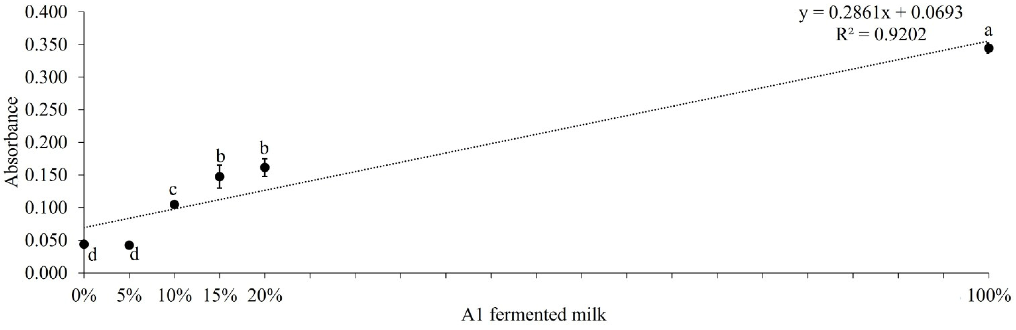

Table 1.
Comparison between tests for identification of A2A2 cows/raw milk.
| Number of cows1 | Genotype – Gene sequencing2 |
ELISA test3 | A2-MiLK TEST®3 | Sensitivity | Specificity |
|---|---|---|---|---|---|
| 6 | A1A1 | No-A2 | No-A2 | 100% | 100% |
| 24 | A1A2 | No-A2 | No-A2 | 100% | 100% |
| 32 | A2A2 | A2 | A2 | 100% | 100% |
1 Total of 62 cows/raw milk samples. 2 Sample used in the test – hair bulb or ear cartilage. 3 Sample used in the ELISA and LFIA– raw milk.
Table 2.
Results of the LFIA test for detection of A2 β-casein in A2A2 milk contaminated with A1A1 milk.
Table 2.
Results of the LFIA test for detection of A2 β-casein in A2A2 milk contaminated with A1A1 milk.
| A1A1 (%)1 | |||||||
|---|---|---|---|---|---|---|---|
| 0% | 5% | 10% | 15% | 20% | 50% | 100% | |
 |
|||||||
| Result - Milk A2 presence | Positive | Negative | Negative | Negative | Negative | Negative | Positive |
1 % of A1A1 raw milk added to A2A2 raw milk.
Table 3.
Results of the LFIA test for detection of A2 β-casein in A2 fermented milk contaminated with A1 fermented milk.
Table 3.
Results of the LFIA test for detection of A2 β-casein in A2 fermented milk contaminated with A1 fermented milk.
| A1A1 (%)1 | ||||||
|---|---|---|---|---|---|---|
| 0% | 5% | 10% | 15% | 20% | 100% | |
 |
||||||
| Result - Milk A2 presence | Positive | Positive | Negative | Negative | Negative | Negative |
1 % of A1 fermented milk added to A2 fermented milk.
Table 4.
Comparison between traditional genotyping methods, ELISA test, and LFIA test.
| Aspect | Traditional genotyping | ELISA (Enzyme-Linked Immunosorbent Assay) |
LFIA (Lateral Flow ImmunoAssay) A2 Milk Test |
|---|---|---|---|
| Method | Direct DNA analysis | Detection of A1 β-casein using immunological reactions | Detection of milk proteins |
| Process | Use of laboratory techniques to extract and analyze DNA | Use of specific antibodies to detect and quantify A1 β-casein | Point of care testing, no need of lab equipment |
| Result | Identification of genotypes A1A1, A1A2 or A2A2 | Quantification of A1 β-casein concentration in milk | Identification of A2A2 animals and milk mixture purity |
| Precision | High accuracy in determining genotypes | High accuracy in measuring A1 β-casein concentration | 100% A2A2 detection 95% purity detection |
| Application | Genetic studies, heredity analyzes | Milk quality control, selection for human consumption | Milk quality control, heredity analyzes |
| Benefits | Specific and direct; useful for detailed genetic studies | Fast, practical and suitable for quality monitoring | Point of Care, fast, cost-effective, suitable for quality monitoring |
| Disadvantages | Costly and time-consuming; requires specialized equipment and knowledge | Less specific compared to direct genotyping | Does not differentiate among A1A1 and A1A2 genotypes |
Disclaimer/Publisher’s Note: The statements, opinions and data contained in all publications are solely those of the individual author(s) and contributor(s) and not of MDPI and/or the editor(s). MDPI and/or the editor(s) disclaim responsibility for any injury to people or property resulting from any ideas, methods, instructions or products referred to in the content. |
© 2024 by the authors. Licensee MDPI, Basel, Switzerland. This article is an open access article distributed under the terms and conditions of the Creative Commons Attribution (CC BY) license (https://creativecommons.org/licenses/by/4.0/).
Copyright: This open access article is published under a Creative Commons CC BY 4.0 license, which permit the free download, distribution, and reuse, provided that the author and preprint are cited in any reuse.


