
Submitted:
28 July 2024
Posted:
30 July 2024
You are already at the latest version
Abstract
This article aims to analyze the relationship between non-obstructive coronary artery disease (NOCAD) and atrial fibrillation (AF), exploring the underlying pathophysiological mechanisms and implications for clinical management. NOCAD and AF are prevalent cardiovascular conditions that often coexist, yet their interrelation is not well understood. NOCAD, characterized by the presence of atherosclerotic plaques without significant luminal stenosis, can lead to ischemic necrosis of cardiomyocytes and their replacement with fibrous tissue, in this way sustaining the focal ectopic activity in atrial myocardium. Atrial fibrillation, on the other hand, is the most common sustained cardiac arrhythmia, associated with an increased risk of stroke and heart failure, which may accelerate atherosclerosis and may increase oxygen consumption in the myocardium, creating a mismatch between supply and demand, thus promoting the development or worsening of coronary ischemia. Therefore, NOCAD and AF seems to be a complex Interplay with one begets another.
Keywords:
coronary ischemia
; non-obstructive coronary artery disease
; INOCA
; MINOCA
; ANOCA
; atrial fibrillation
1. Introduction
Non-obstructive coronary artery disease (NOCAD) and atrial fibrillation (AF) are two prevalent cardiovascular conditions that pose significant challenges in clinical practice due to their complex interplay and shared risk factors. Non-obstructive coronary artery disease, characterized by coronary artery stenosis of less than 50%, has emerged as a distinct entity with important implications for cardiovascular health. On the other hand, AF, the most frequent arrhythmia, is associated with an increased risk of stroke, heart failure, and mortality. The coexistence of NOCAD and AF presents a unique clinical scenario that warrants further investigation to optimize patient management and outcomes [1].
The prevalence of NOCAD has been steadily increasing, particularly in patients with symptoms suggestive of myocardial ischemia but without significant coronary artery stenosis on angiography. This subset of patients often presents with chest pain, dyspnea and other anginal symptoms, posing a diagnostic challenge for clinicians. Recent advances in imaging modalities, such as coronary computed tomography angiography (CCTA) and invasive physiologic assessments, have improved the ability to identify and characterize NOCAD, shedding light on its pathophysiology and clinical significance [2].
Atrial fibrillation, characterized by rapid and irregular atrial electrical activity, is a common arrhythmia affecting millions of individuals worldwide. It is associated with an increased risk of thromboembolic events, including stroke, as well as heart failure and cardiovascular mortality. The presence of AF in patients with NOCAD further complicates the management of these individuals, as it may influence treatment decisions and outcomes [3].
There is a strong dual relationship between NOCAD and AF: the first one may promote through ischemia the development of focal ectopic activity in the left atrium, sustaining in this way AF. On the other hand, AF is able to generate an inflammatory response by mechanisms not yet fully understood which will lead to the acceleration of atherosclerosis and moreover, by increasing oxygen consumption, it may generate a mismatch between demands and blood supply in myocardium, generating angina in patients with NOCAD [4,5].
Understanding the complex relationship between NOCAD and AF is essential for optimizing patient care and improving clinical outcomes. This article aims to explore the dual relationship and the implications of the coexistence of NOCAD and AF. By elucidating the mechanisms underlying these conditions and their interplay, more effective strategies may be developed for risk stratification, treatment and follow-up in this unique patient population.
2. Non-Obstructive Coronary Artery Disease: ANOCA, INOCA and MINOCA
Coronary artery disease (CAD) is traditionally associated with significant obstructive lesions in the coronary arteries. However, many patients present with symptoms of myocardial ischemia or even myocardial infarction (MI) without significant coronary artery obstruction. This has led to the classification of NOCAD, which is a clinical condition characterized by the presence of coronary atherosclerosis without significant luminal obstruction, typically defined as less than 50% stenosis on CCTA or invasive angiogram. Unlike obstructive CAD, NOCAD presents a unique diagnostic and therapeutic challenge due to its subtler manifestation and complex pathophysiology [6,7].
NOCAD is increasingly recognized in clinical practice, particularly due to the widespread use of advanced imaging techniques. It is more prevalent among women than men and is often observed in patients with risk factors such as hypertension, diabetes and hyperlipidemia. Studies indicate that NOCAD may account for up to 20-30% of patients presenting with chest pain and evidence of ischemia but without significant obstructive lesions on coronary angiography [8].
The pathophysiology of NOCAD involves several mechanisms, including endothelial dysfunction, microvascular dysfunction and diffuse atherosclerotic changes. Endothelial dysfunction impairs the ability of the coronary arteries to dilate, leading to reduced blood flow and myocardial ischemia. Microvascular dysfunction involves the small coronary vessels, which may contribute to ischemic symptoms despite the absence of large vessel obstruction. Additionally, diffuse atherosclerotic changes may cause a gradual reduction in coronary flow reserve, exacerbating ischemic conditions [9,10].
Patients with NOCAD often present with symptoms similar of those with obstructive CAD, including chest pain (angina), dyspnea and fatigue. However, the symptoms may be more variable and less predictable. Importantly, NOCAD can lead to significant morbidity, including recurrent hospitalizations and reduced quality of life. It is also associated with an increased risk of adverse cardiovascular events, such as MI and heart failure [8,11].
The diagnosis of NOCAD typically involves a combination of clinical assessment, non-invasive imaging and invasive coronary angiography. Key diagnostic tools include:
- Coronary Angiography: this remains the gold standard for visualizing coronary anatomy. In NOCAD, angiography reveals less than 50% stenosis in the coronary arteries [12].
- Non-Invasive Imaging: techniques such as stress echocardiography, cardiac MRI, Positron Emission Tomography and CCTA may identify ischemia and assess coronary anatomy without the need of invasive procedures [17].
There are more subtypes of NOCAD depending on clinical presentation, documented ischemia and the presence of myocardial injury, defined by elevated myocardial cytolysis enzymes: ANOCA (Angina with Non-Obstructive Coronary Arteries), INOCA (Ischemia with Non-Obstructive Coronary Arteries) and MINOCA (Myocardial Infarction with Non-Obstructive Coronary Arteries) (Figure 1) [18].
Both ANOCA and INOCA refers to patients who experience angina (chest pain or dyspnea- these symptoms are often exertional and relieved by rest or nitrates, mirroring the presentation of obstructive CAD), but have non-obstructive coronary arteries on angiography and without signs of myocardial injury such as troponin elevation. The difference between them is that the first one presents without electrocardiogram (ECG) changes, stress echocardiography or cardiac MRI suggesting ischemia, while in the second one, non-invasive tests will show signs of ischemia [19,20].
The pathophysiological mechanisms of ANOCA and INOCA include:
- microvascular dysfunction: impaired regulation of blood flow in the small coronary vessels, leading to insufficient oxygen supply to the myocardium caused by structural remodeling of the microvasculature, which will lead to fixed reduced microcirculatory conductance or vasomotor disorders affecting the coronary arterioles, which will cause dynamic arteriolar obstruction (the mechanism for microvascular angina) [21].
- vasospasm: transient constriction of the epicardial coronary arteries, leading to reduced blood flow and ischemia (the mechanism for epicardial vasospastic angina) [22].
The diagnosis involves non-invasive tests to detect ischemia (will differentiate ANOCA from INOCA) and confirming the absence of obstructive coronary arteries via angiography (Table 1).
FFR and IFR are used to differentiate borderline stenoses and classify them as obstructive CAD or NOCAD. FFR is the ratio of mean distal coronary pressure to mean aortic pressure at maximal hyperemia and an abnormal FFR is defined as <=0.80. IFR is a non-hyperemic pressure ratio during diastole, an abnormal value being <=0.89 [16].
Diagnostic options for coronary function testing include the inducing steady-state hyperemia (by using adenosine to achieve endothelium-independent vasodilation) and the calculation of coronary flow reserve (through thermodilution using a pressure–temperature sensor guidewire in the left anterior descending artery or through Doppler flow velocity). Most studies show the prognostic value of thermodilution-based CFR have used a cut-off value of 2 [27,28].
To evaluate the microvascular resistance, two indexes may be calculated using the methods presented before: the index of microvascular resistance (IMR), which may be calculated as the product of distal coronary pressure at maximal hyperemia multiplied by the hyperemic mean transit time (a value>=25 is representative of microvascular dysfunction) and the hyperemic myocardial velocity resistance (HMR) index, a Doppler-based index which may be calculated by dividing intracoronary pressure by hyperemic flow velocity (a value> 1.9 was an independent predictor of recurrent chest pain) [29,30,31].
MINOCA patients, on the other hand, present with signs and symptoms consistent with an acute myocardial infarction. This includes chest pain at rest, often with accompanying symptoms such as sweating and nausea. Importantly, these patients exhibit elevated cardiac biomarkers and may show ischemic changes on an ECG [32].
MINOCA encompasses a broader range of pathophysiological mechanisms, including:
- coronary artery spasm: severe transient constriction of a coronary artery, potentially leading to myocardial infarction [33].
- microvascular dysfunction: similar to INOCA, impaired function of the microvasculature may contribute to ischemia and infarction [36].
- coronary embolism or thrombosis: embolic events or thrombus formation in non-obstructive coronary arteries [37].
The diagnosis starts with fulfilling the criteria for myocardial infarction (elevated cardiac biomarkers, symptoms and ECG changes) and confirming non-obstructive coronary arteries on angiography. Additional diagnostic steps may include cardiac MRI (to identify myocardial scarring, inflammation or other structural abnormalities), intravascular imaging (techniques like IVUS and OCT to detect subtle plaque disruptions or thrombi) and laboratory tests (to evaluate potential causes like thrombophilia or autoimmune disorders) (Figure 2) [32,37].
MINOCA must be differentiated from takotsubo cardiomyopathy (also known as stress-induced cardiomyopathy, which is characterized by transient left ventricular dysfunction due to a transient increased release of catecholamines) and from myocarditis (inflammation of the myocardium due to infectious or autoimmune causes that may mimic MI) [39,40,41].
Management of NOCAD is tailored to the underlying cause and it includes: antiplatelet therapy (aspirin and/or other antiplatelet agents such as P2Y12 inhibitors, which may be used in patients with plaque disruption to prevent thrombotic events), statins (these agents help stabilize atherosclerotic plaques and reduce the risk of cardiovascular events), beta-blockers, ACE inhibitors (which may improve endothelial function and reduce cardiovascular risk in patients with hypertension or diabetes) and specific interventions (such as calcium channel blockers and nitrates for coronary artery spasm). Moreover, lifestyle modifications should be done such as adopting a heart-healthy diet, eliminating tobacco use to reduce endothelial damage and atherosclerosis progression, weight and stress management, a regular physical activity should be promoted to improve cardiovascular fitness and endothelial function. Not at least, the control of risk factors is very important (hypertension, diabetes, dyslipidemia) [42,43,44].
3. Coronary Ischemia as Substrate in Atrial Fibrillation
Coronary ischemia, even in microvascular circulation specific for NOCAD, may be a significant promotor for AF, the most common sustained cardiac arrhythmia. Coronary ischemia has been identified as a critical substrate in the development and perpetuation of AF, that is why understanding the relationship between coronary ischemia and AF is crucial for developing effective treatment strategies and improving patient outcomes [45].
Coronary ischemia results from an imbalance between myocardial oxygen supply and demand and may be caused by atherosclerosis (in case of obstructive CAD) and other factors such as coronary artery spasm, microvascular dysfunction, and thromboembolism (in case of NOCAD). Regardless of mechanism, the reduced oxygen supply to the myocardium can cause myocardial injury, inflammation, and fibrosis, creating a substrate conducive to AF. Ischemic episodes cause myocardial cell apoptosis and their replacement with fibrous tissue. This fibrotic tissue disrupts the normal myocardial architecture, creating areas of slow conduction and re-entry circuits, which are critical in the development of AF. Ischemia-induced inflammation further contributes to this process by promoting structural and electrical remodeling of the atria [46].
Moreover, ischemia may also alter the balance of the autonomic nervous system, increasing sympathetic activity and reducing vagal tone. This imbalance can precipitate AF by enhancing automaticity, increasing triggered activity, and promoting re-entry phenomena in the atria. Not at least, the complications of coronary ischemia such as heart failure, diastolic disfunction and mitral regurgitation will lead to atrial dilatation, promoting in this way AF, because as atrial cardiomyocytes stretch, they will be replaced with fibroblasts, which stimulate collagen synthesis, in this way being favorized the genesis of reentry circuits [47].
Finally, coronary ischemia plays also an important role in the maintaining of AF, not only in its pathogenesis. A study, which involved 700 subjects undergoing a strategy of pulmonary vein isolation for AF, showed that patients with coronary ischemia have a higher recurrence of AF after ablation and moreover, the interventional treatment of coronary lesions can reduce the rate of AF recurrence [48].
Inflammation and oxidative stress are pivotal mechanisms that link coronary ischemia to AF.
3.1. Inflammation due to coronary ischemia leading to AF
When coronary ischemia occurs, it triggers a complex inflammatory response. Reduced blood flow to the myocardium leads to hypoxia, which subsequently causes cellular stress and injury, which will initiate an inflammatory cascade: the first step is the release of cytokines: ischemic myocardial cells release pro-inflammatory cytokines such as interleukin-6 (IL-6), tumor necrosis factor-alpha (TNF-α), and interleukin-1 beta (IL-1β). These cytokines will accelerate fibrosis in atrial cardiomyocytes by modulating MMP2 (matrix metalloproteinase 2) expression and will also recruit inflammatory cells to the site of injury, leading to the second step, the activation of leukocytes: neutrophils, monocytes and macrophages are among the first responders to ischemic injury, which will release proteolytic enzymes, reactive oxygen species (ROS) and additional cytokines, which further amplify the inflammatory response. Finally, inflammation damages the endothelial cells, leading to increased vascular permeability, reduced nitric oxide availability, and enhanced adhesion of leukocytes to the endothelium (endothelial dysfunction- the final step) [49,50].
The inflammatory cascade will lead to structural and electrical remodeling of the atria, creating a substrate for AF, the main factors being the fibrosis, atrial dilation and gap junction remodeling (Figure 3) [51].
On one hand, prolonged inflammation promotes the activation of fibroblasts and the deposition of extracellular matrix proteins, leading to atrial fibrosis. Fibrotic tissue disrupts normal conduction pathways, creating areas of slow conduction and re-entry circuits that facilitate AF (electrical remodeling). On the other hand, inflammation can lead to atrial dilation, which stretches atrial myocytes and alters their electrophysiological properties. This dilation further predisposes the atria to the development of AF (structural remodeling) [52].
Moreover, inflammatory mediators may also alter the expression and function of connexins, such as connexin 40 and 43, the proteins that form gap junctions between cardiac cells. This disruption of intercellular communication can contribute to the heterogeneous conduction properties seen in AF [53].
3.2. Oxidative stress due to coronary ischemia leading to atrial fibrillation
Reactive oxygen species, which are chemically reactive molecules containing oxygen, play a crucial role in the pathogenesis of AF, particularly in the context of coronary ischemia. During coronary ischemia, the production of ROS increases significantly due to some mechanisms such as mitochondrial dysfunction (ischemia impairs mitochondrial function, leading to an increased production of ROS such as superoxide anions) and activation of NADPH Oxidase and Xanthine Oxidase, two enzymes that generate ROS [54,55].
Reactive oxygen species may contribute to the initiation and perpetuation of AF through several mechanisms: direct myocyte damage, calcium handling abnormalities, ion channel dysfunction and gap junction dysfunction [56].
Firstly, ROS can directly damage atrial myocytes by oxidizing lipids, proteins and DNA. ROS-induced cellular damage may lead to apoptosis or necrosis, contributing to atrial fibrosis and inflammation (damaged cells release inflammatory mediators that further propagate inflammation and remodeling in the atria) [57,58].
Calcium ions (Ca²⁺) play a crucial role in cardiac myocyte contraction and electrical activity. Secondly, ROS can disrupt normal calcium handling by sarcoplasmic reticulum (SR) dysfunction. They can alter the function of SR Ca²⁺-ATPase (SERCA) and ryanodine receptors (RyR), leading to abnormal Ca²⁺ release and reuptake, this dysfunction being the cause of Ca²⁺ overload in the cytoplasm. Moreover, oxidative modification of RyR can cause a "leaky" SR, resulting in spontaneous Ca²⁺ release, which can trigger afterdepolarizations and ectopic activity, promoting AF [59].
Then, ROS can also modify the function of various ion channels, which are critical for maintaining normal cardiac electrophysiology, such as:
- sodium channels (Na⁺ Channels): ROS can reduce sodium current (INa) by modifying channel proteins, leading to slowed conduction and increased susceptibility to re-entry circuits [60].
- potassium channels (K⁺ Channels): ROS can affect several potassium channels involved in repolarization, such as IKs, IKr, and Ito. This can prolong or shorten action potential duration, creating a substrate for AF [61].
- calcium channels (Ca²⁺ Channels): ROS can increase L-type calcium current (ICa,L), contributing to abnormal calcium influx and triggered activity [62].
Finally, ROS may impair gap junction function by altering the expression and function of connexins (such as connexin 43), leading to impaired intercellular communication, in this way creating a decreased conductance (dysfunctional gap junctions result in heterogeneous conduction and the formation of re-entry circuits, which are key mechanisms in AF) [63].
4. Atrial Fibrillation as Substrate for Microvascular Dysfunction
While AF is well recognized for its potential to cause thromboembolic events, its role as a substrate for microvascular dysfunction and coronary ischemia is less understood. Traditionally, the focus has been on the management of stroke risk and rhythm control. However, emerging evidence suggests that AF may also contribute to microvascular dysfunction and coronary ischemia [64].
The pathophysiology of AF involves a complex interplay of various factors, being associated with a concept using Coumel’s Triangle that requires a trigger for initiation, a catalyst agent and a substrate (arrhythmogenic and structural) for the perpetuation and maintenance of the trigger (Figure 4) [46]:
- Triggers and Drivers: AF may be triggered by ectopic beats originating from the pulmonary veins or other locations in the atria, such as: the left atria posterior wall, the superior vena cava, the left atrial appendage, the coronary sinus and the ligament of Marshall. These triggers, combined with areas of slowed conduction and functional reentry circuits, create a substrate for the initiation and perpetuation of AF. In some cases, rapid firing of ectopic foci or localized reentrant circuits may also drive the arrhythmia [65].
- Catalyst: The catalyst may change refractory periods, in this way increasing autonomic activity and it is represented by autonomic nervous system, thyroid hormones and illicit drugs. Sympathetic activation can increase the likelihood of AF episodes, while parasympathetic stimulation may promote the termination of AF; in this way, imbalances in autonomic tone can influence the susceptibility to AF [66].
- Substrate: The substrate is essential in maintaining the action of trigger and catalyst and it can be structural or electrical. The first one consists in structural modifications in the atria, such as fibrosis (excessive deposition of collagen and other extracellular matrix proteins) and dilation. These changes can disrupt normal electrical conduction pathways in the atrium and create conditions that are conducive to sustain AF. The second one is created by abnormal electrical activity in the atria, characterized by alterations in ion channel function and intracellular signaling pathways. Modifications in the action potential duration, refractoriness, and conduction velocity can promote the reentry of electrical impulses and the chaotic electrical activity seen in AF [67].
There are other factors that sustain the activity of the Coumel’s Triangle, such as inflammation and oxidative stress, which may promote atrial structural remodeling, electrical instability and fibrosis, creating a proarrhythmic environment in the atria and comorbidities (hypertension, heart failure, diabetes, obesity and sleep apnea) that can contribute to atrial remodeling [56].
Coronary ischemia in AF patients may occur even in the absence of significant epicardial CAD. AF may promote coronary ischemia through a lot of mechanisms such as: an increased myocardial oxygen demand (the rapid and irregular ventricular rate during AF increases myocardial oxygen consumption, generating a mismatch between demand and supply in oxygen), reduced myocardial perfusion (AF can lead to reduced diastolic filling time and impaired coronary perfusion, especially during tachycardia) and coronary microembolization (the formation and dislodgement of microthrombi can lead to microvascular obstruction and ischemia) [68].
4.1. Microvascular Dysfunction in AF
Despite coronary ischemia, AF may also induce microvascular dysfunction through several mechanisms: endothelial dysfunction, neurohormonal activation, microthrombi formation and impaired coronary flow reserve [69].
4.2. Endothelial Dysfunction Due to Atrial Fibrillation
Endothelial dysfunction, which plays a significant role in the pathophysiology of microvascular dysfunction and coronary ischemia, may be promoted by AF through many ways [70]:
- Systemic inflammation and oxidative stress: On one hand, AF is associated with elevated levels of inflammatory markers such as C-reactive protein (CRP), IL-6 and TNF-α. These cytokines may cause direct damage to endothelial cells, impairing their function. On the other hand, the arrhythmic nature of AF leads to increased production of ROS, which are able to lead to cellular dysfunction and apoptosis [71,72].
- Hemodynamic shear stress: The irregular and rapid heart rate in AF results in abnormal shear stress on the blood vessel walls. This mechanical stress can disrupt the normal function of endothelial cells, reducing their ability to regulate vascular tone and blood flow [73].
- Endothelial nitric oxide synthase (eNOS) dysfunction: In AF, the bioavailability of NO, which is a critical vasodilator produced by endothelial cells, is reduced due to increased oxidative stress and inflammation, leading to impaired vasodilation and endothelial dysfunction. Moreover, under normal conditions, eNOS produces NO, but in the presence of oxidative stress and reduced tetrahydrobiopterin (BH4), a cofactor for eNOS, the enzyme becomes uncoupled and produces superoxide instead of NO, further exacerbating oxidative stress and endothelial dysfunction [74].
Endothelial dysfunction contributes also to the pro-thrombotic state in AF. The damaged endothelium expresses more adhesion molecules, promoting platelet aggregation and clot formation, which can lead to thromboembolic events. Finally, endothelial dysfunction can exacerbate heart failure by impairing myocardial perfusion and contributing to adverse ventricular remodeling. The reduced NO availability and increased oxidative stress may affect myocardial contractility and promote fibrosis [74,75,76].
4.3. Neurohormonal Activation due to Atrial Fibrillation
Neurohormonal activation plays a crucial role as catalyst in the pathophysiology of AF and contributes significantly to microvascular dysfunction. This complex interplay involves the activation of the sympathetic nervous system (SNS) and the renin-angiotensin-aldosterone system (RAAS), which collectively exacerbate vascular and cardiac abnormalities [77].
On one hand, AF is associated with heightened sympathetic activity, leading to increased levels of catecholamines (epinephrine and norepinephrine), which bind to adrenergic receptors on endothelial cells and vascular smooth muscle cells, causing vasoconstriction and reduced endothelial-dependent vasodilation. Moreover, catecholamines may also stimulates the production of ROS within endothelial cells, contributing to oxidative stress, which impairs eNOS activity and reduces NO bioavailability. Then, SNS activation promotes the expression of pro-inflammatory cytokines and adhesion molecules on endothelial cells, facilitating leukocyte adhesion and infiltration. This inflammatory response further damages the endothelium and impairs its function [78].
On the other hand, AF stimulates the RAAS, leading to increased production of angiotensin II, which is a potent vasoconstrictor that directly affects endothelial cells by promoting oxidative stress, inflammation and cellular apoptosis. It also stimulates the production of endothelin-1, another vasoconstrictor that exacerbates endothelial dysfunction. An increased level of aldosterone contributes to endothelial dysfunction by promoting fibrosis, oxidative stress, and inflammation, as it able to induce the expression of pro-inflammatory cytokines and adhesion molecules, which facilitate leukocyte adhesion and vascular inflammation. In this way, angiotensin II and aldosterone promote vascular remodeling, characterized by increased smooth muscle cell proliferation, fibrosis and reduced elasticity of blood vessels. This remodeling impairs endothelial function and may contribute to the progression of atherosclerosis. Finally, angiotensin II and aldosterone may also activate NADPH oxidase, an enzyme complex responsible for the production of ROS [79,80].
Not at least, chronic RAAS activation leads to sustained vasoconstriction and sodium retention, contributing to the development and maintenance of hypertension, a common risk factor for both AF and coronary ischemia [81].
4.4. Microthrombi Formation due to Atrial Fibrillation
Microthrombi formation is a critical and often underappreciated mechanism by which AF contributes to microvascular dysfunction and coronary ischemia because they are able to obstruct the microvasculature, leading to impaired tissue perfusion and ischemic damage [82].
It is well known that AF, being associate with irregular atrial contraction, is associated with blood stasis, particularly in the left atrial appendage. Then, the irregular ventricular response in AF further contributes to turbulent blood flow, promoting conditions favorable for thrombus formation, but AF is also associated with endothelial injury, which disrupts the balance between pro-coagulant and anticoagulant factors, favoring thrombus formation. Patients with AF often exhibit elevated levels of coagulation factors such as fibrinogen, factor VIII and von Willebrand factor. Moreover, AF may lead to increased platelet activation and aggregation, releasing pro-thrombotic substances like thromboxane A2 and adenosine diphosphate, promoting further clot formation [83].
In this way, the microthrombi formed can lodge in the coronary microvasculature, obstructing blood flow and leading to localized ischemia, which is particularly detrimental in the myocardial tissue, that requires high oxygen levels. Recurrent formation and dissolution of microthrombi may also lead to cycles of ischemia and reperfusion injury, causing further endothelial damage and perpetuating a vicious cycle of thrombosis and dysfunction [84,85,86].
The microthrombi formed are responsible for thromboembolic events, not only in the coronary tree, but also in other systemic arteries, leading to ischemia in all territories and being one of cause for the cryptogenic stroke [87].
4.5. Impaired CFR Due to Atrial Fibrillation
Atrial fibrillation can significantly impact coronary circulation, leading to impaired CFR by several mechanisms. Firstly, the irregular ventricular rate in AF leads to variations in cardiac output and coronary perfusion. The inconsistent filling times and rapid heart rates reduce diastolic filling, a critical period for coronary blood flow, particularly to the left ventricle. Secondly, the loss of atrial systole reduces stroke volume, leading to decreased perfusion pressure in the coronary arteries, which further impairs CFR [88,89].
Then, as discussed previously, AF promotes the formation of microthrombi in the coronary microvasculature, in this way being limited the ability of coronary vessels to increase blood flow during periods of increased demand. Moreover, AF-induced neurohormonal activation and inflammation can lead to structural changes in the coronary microvasculature, including increased stiffness and reduced compliance, these changes impairing the ability of the vessels to dilate appropriately, reducing CFR [90,91].
Vice versa, impaired myocardial perfusion can contribute to the recurrence and persistence of AF as ischemia-induced atrial remodeling and fibrosis may create a substrate for AF perpetuation, but severe ischemia and impaired CFR can also increase the risk of ventricular arrhythmias, posing a risk for sudden cardiac death [92].
Most patients with AF and impaired CFR will experience symptoms of angina, particularly during periods of increased cardiac demand, such as physical exertion or emotional stress, but some of them can have ischemia without typical anginal symptoms, which can go unnoticed and lead to more severe cardiac events [93].
5. NOCAD and AF: challenges in clinical practice
NOCAD and AF represent significant and often overlapping cardiovascular pathologies whom coexistence presents unique challenges in clinical practice, encompassing diagnostic complexities, therapeutic dilemmas and prognostic uncertainties.
5.1. Diagnostic Challenges
The diagnosis of NOCAD in patients with AF is particularly challenging due to the overlapping symptomatology and the limitations of traditional diagnostic modalities. Both conditions can present with angina, dyspnea and palpitations, making it difficult to distinguish between ischemic symptoms and arrhythmia-related discomfort [94].
While ECG is a cornerstone in the diagnosis of both conditions, its utility is limited in the presence of AF because this arrhythmia can mask ischemic changes such as ST-segment depression or T-wave inversions, complicating the diagnosis of NOCAD. Then, standard coronary angiography may reveal non-obstructive lesions, but it does not provide information on microvascular dysfunction or endothelial dysfunction, which are often seen in NOCAD. Advanced imaging techniques such as IVUS or OCT can be useful but are not routinely performed due to cost and accessibility issues [95,96].
Finally, non-invasive imaging modalities like CCTA and CMR can provide detailed anatomical and functional information, but their interpretation can be complicated by AF-related motion artifacts [97].
5.2. Therapeutic Challenges
The treatment of patients with both NOCAD and AF requires a multifaceted approach that addresses both the ischemic and arrhythmic components of their disease. However, the therapeutic strategies for these conditions can sometimes be conflicting.
Regarding the antithrombotic therapy, patients with AF are at increased risk of thromboembolic events and usually require anticoagulation. However, those with NOCAD may also require antiplatelet therapy. The concomitant use of anticoagulants and antiplatelets increases the risk of bleeding, necessitating a careful balancing act [98].
The primary anticoagulants used in AF include direct oral anticoagulants (DOACs) such as apixaban, rivaroxaban, dabigatran and edoxaban, which have taken the place of warfarin. For NOCAD, antiplatelet agents such as aspirin or P2Y12 inhibitors (e.g., clopidogrel, ticagrelor) are commonly prescribed. The challenge lies in determining the necessity and duration of SAPT (single antiplatelet therapy)/DAPT in addition to anticoagulation because the concurrent use of anticoagulants and antiplatelets significantly increases the risk of bleeding, but the AUGUSTUS trial sustains that in case of both AF and coronary ischemia, apixaban and a P2Y12 inhibitor should be used instead of warfarin or dual antiplatelet agents. Moreover, individualized patient assessment using tools like the HAS-BLED score to estimate bleeding risk should be used and balances against the thrombotic risks. Strategies to mitigate bleeding risk include the use of proton pump inhibitors (PPIs) to prevent gastrointestinal bleeding and close monitoring of coagulation parameters [99,100,101].
Managing AF involves also strategies to rate vs. rhythm control and both approaches have implications for patients with NOCAD. If it is possible, the conversion to sinus rhythm using antiarrhythmic drugs, electrical cardioversion or catheter ablation is vital in reducing oxygen consumption, thus preventing the evolution of coronary ischemia. Rhythm control involves the use of antiarrhythmic drugs (AADs) like amiodarone, dofetilide, flecainide or sotalol. However, the use of flecainide is limited in patients with coronary ischemia (especially post myocardial infarction) as it may increase mortality in this case, but there is in a study with only 78 patients, which claims that treatment with flecainide appears not to increase mortality in patients with AF, preserved left ventricle function and occult CAD indicated by PET Stress Testing with Coronary Flow Capacity. Moreover, another study sustains the effectiveness and safety of dofetilide in patients with AF and heart failure or CAD [102,103].
If AF is declared permanent, rate control is typically achieved using beta-blockers, calcium channel blockers or digoxin. Betablockers are also the first-line treatments included in antianginal medications while calcium channel blockers are the first option for vasospastic angina. Digoxin, while effective in controlling heart rate, has a narrow therapeutic index and potential proarrhythmic effects [99].
Catheter ablation is an option for rhythm control in patients with symptomatic AF refractory to medical therapy. However, the procedure itself carries risks and its impact on coronary microvascular function in NOCAD patients is not fully understood. Moreover, the recurrence of AF post-ablation remains a concern, necessitating ongoing monitoring and potential repeat procedures [104].
5.3. Prognostic Challenges
Understanding the prognosis in patients with coexisting NOCAD and AF is complex, as each condition independently influences cardiovascular outcomes.
Risk stratification tools for AF and NOCAD may not fully capture the interplay between these conditions. For example, in CHA2DS2-VASc score it is included obstructive CAD but not NOCAD. There are studies which claims that CHA2DS2-VASc score may be a useful predictor for selecting patients to undergo coronary angiographic exploration for the diagnosis of obstructive CAD or that it may be an independent predictor for SYNTAX score, in patients with non-ST-segment elevation acute myocardial infarction and without AF, but no correlations with NOCAD have been studied yet [105,106].
Comprehensive risk models that integrate both ischemic and arrhythmic risks are needed. Then, studies suggest that patients with NOCAD and AF have a higher incidence of adverse cardiovascular events compared to those with either condition alone. The mechanisms underlying this increased risk are multifactorial, involving endothelial dysfunction, chronic inflammation, and neurohormonal activation [107].
Finally, both NOCAD and AF significantly impact patients' quality of life. The recurrent symptoms, frequent hospitalizations and the psychological burden of managing multiple medications can lead to reduced adherence and poorer outcomes [108].
6. Conclusions
NOCAD and AF seems to be a complex interplay with one begets another. The coexistence of NOCAD and AF presents a myriad of challenges in clinical practice. Diagnostic accuracy is hindered by overlapping symptoms and limitations of current imaging modalities. Therapeutic strategies must balance the risks of bleeding and thromboembolism, while also managing ischemic and arrhythmic components effectively. Prognostic evaluation is complicated by the intricate interplay between the two conditions, necessitating more sophisticated risk models. Addressing these challenges requires a multidisciplinary approach, integrating cardiology, electrophysiology, and patient-centered care to optimize outcomes for this complex patient population. Future research should focus on developing targeted therapies and refining diagnostic tools to better manage.
Author Contributions
Conceptualization, A.F.O. and M.F.; methodology, P.C.M., A.M.B. and R.M.; software, O.M. and A.J.; validation, D.E.F., A.T. and A.V.; formal analysis, L.M., G.B. and D.V.S.; investigation, D.M and M.C.; resources, L.M, G.B. and D.M.; data curation, A.B. and I.C.; writing—original draft preparation, A.F.O; writing—review and editing, A.F.O. and M.F.; visualization, R.M., O.M. and D.E.F.; supervision D.M.T. and I.C.; project administration, A.B., A.V. and C.P.; funding acquisition, A.T. and A.J. All authors have read and agreed to the published version of the manuscript.
Funding
This research received no external funding.
Institutional Review Board Statement
Not applicable.
Informed Consent Statement
Not applicable.
Data Availability Statement
The data is contained within this article.
Conflicts of Interest
The authors declare no conflicts of interest.
References
- Gautier, A.; Picard, F.; Ducrocq, G.; Elbez, Y.; Fox, K.M.; Ferrari, R.; Ford, I.; Tardif, J.-C.; Tendera, M.; Steg, P.G.; et al. New-Onset Atrial Fibrillation and Chronic Coronary Syndrome in the CLARIFY Registry. European Heart Journal 2024, 45, 366–375. [Google Scholar] [CrossRef] [PubMed]
- Hwang, I.-C.; Lee, H.; Yoon, Y.E.; Choi, I.-S.; Kim, H.-L.; Chang, H.-J.; Lee, J.Y.; Choi, J.A.; Kim, H.J.; Cho, G.-Y.; et al. Risk Stratification of Non-Obstructive Coronary Artery Disease for Guidance of Preventive Medical Therapy. Atherosclerosis 2019, 290, 66–73. [Google Scholar] [CrossRef] [PubMed]
- Floria, M.; Oancea, A.F.; Morariu, P.C.; Burlacu, A.; Iov, D.E.; Chiriac, C.P.; Baroi, G.L.; Stafie, C.S.; Cuciureanu, M.; Scripcariu, V.; et al. An Overview of the Pharmacokinetics and Pharmacodynamics of Landiolol (an Ultra-Short Acting Β1 Selective Antagonist) in Atrial Fibrillation. Pharmaceutics 2024, 16, 517. [Google Scholar] [CrossRef] [PubMed]
- Yan, T.; Zhu, S.; Xie, C.; Zhu, M.; Weng, F.; Wang, C.; Guo, C. Coronary Artery Disease and Atrial Fibrillation: A Bidirectional Mendelian Randomization Study. JCDD 2022, 9, 69. [Google Scholar] [CrossRef] [PubMed]
- Cappello, I.A.; Pannone, L.; Della Rocca, D.G.; Sorgente, A.; Del Monte, A.; Mouram, S.; Vetta, G.; Kronenberger, R.; Ramak, R.; Overeinder, I.; et al. Coronary Artery Disease in Atrial Fibrillation Ablation: Impact on Arrhythmic Outcomes. Europace 2023, 25, euad328. [Google Scholar] [CrossRef] [PubMed]
- Yang, E.H.; Lerman, A. Angina Pectoris with a Normal Coronary Angiogram. Herz 2005, 30, 17–25. [Google Scholar] [CrossRef] [PubMed]
- Ahmadzadeh, K.; Roshdi Dizaji, S.; Kiah, M.; Rashid, M.; Miri, R.; Yousefifard, M. The Value of Coronary Artery Disease – Reporting and Data System (CAD-RADS) in Outcome Prediction of CAD Patients; a Systematic Review and Meta-Analysis. Archives of Academic Emergency Medicine 2023, 11, e45. [Google Scholar] [CrossRef] [PubMed]
- Najib, K.; Boateng, S.; Sangodkar, S.; Mahmood, S.; Whitney, H.; Wang, C.E.; Racsa, P.; Sanborn, T.A. Incidence and Characteristics of Patients Presenting with Acute Myocardial Infarction and Non-obstructive Coronary Artery Disease. Cathet Cardio Intervent 2015, 86. [Google Scholar] [CrossRef] [PubMed]
- Shaw, J.; Anderson, T. Coronary Endothelial Dysfunction in Non-Obstructive Coronary Artery Disease: Risk, Pathogenesis, Diagnosis and Therapy. Vasc Med 2016, 21, 146–155. [Google Scholar] [CrossRef]
- Tunc, E.; Eve, A.A.; Madak-Erdogan, Z. Coronary Microvascular Dysfunction and Estrogen Receptor Signaling. Trends in Endocrinology & Metabolism 2020, 31, 228–238. [Google Scholar] [CrossRef]
- for the APPROACH investigators; Kissel, C. K.; Chen, G.; Southern, D.A.; Galbraith, P.D.; Anderson, T.J. Impact of Clinical Presentation and Presence of Coronary Sclerosis on Long-Term Outcome of Patients with Non-Obstructive Coronary Artery Disease. BMC Cardiovasc Disord 2018, 18, 173. [Google Scholar] [CrossRef]
- Rahman, H.; Corcoran, D.; Aetesam-ur-Rahman, M.; Hoole, S.P.; Berry, C.; Perera, D. Diagnosis of Patients with Angina and Non-Obstructive Coronary Disease in the Catheter Laboratory. Heart 2019, 105, 1536–1542. [Google Scholar] [CrossRef] [PubMed]
- Ono, M.; Kawashima, H.; Hara, H.; Gao, C.; Wang, R.; Kogame, N.; Takahashi, K.; Chichareon, P.; Modolo, R.; Tomaniak, M.; et al. Advances in IVUS/OCT and Future Clinical Perspective of Novel Hybrid Catheter System in Coronary Imaging. Front. Cardiovasc. Med. 2020, 7, 119. [Google Scholar] [CrossRef] [PubMed]
- Reiber, J.H.C.; Tu, S.; Tuinenburg, J.C.; Koning, G.; Janssen, J.P.; Dijkstra, J. QCA, IVUS and OCT in Interventional Cardiology in 2011. Cardiovasc Diagn Ther 2011, 1, 57–70. [Google Scholar] [CrossRef]
- Tajeddini, F.; Nikmaneshi, M.R.; Firoozabadi, B.; Pakravan, H.A.; Ahmadi Tafti, S.H.; Afshin, H. High Precision Invasive FFR, low-cost Invasive iFR, or non-invasive CFR ?: Optimum Assessment of Coronary Artery Stenosis Based on the patient-specific Computational Models. Numer Methods Biomed Eng 2020, 36, e3382. [Google Scholar] [CrossRef] [PubMed]
- Ghorbanniahassankiadeh, A.; Marks, D.S.; LaDisa, J.F. Correlation of Computational Instantaneous Wave-Free Ratio With Fractional Flow Reserve for Intermediate Multivessel Coronary Disease. J Biomech Eng 2021, 143, 051011. [Google Scholar] [CrossRef] [PubMed]
- Liu, A.; Wijesurendra, R.S.; Liu, J.M.; Forfar, J.C.; Channon, K.M.; Jerosch-Herold, M.; Piechnik, S.K.; Neubauer, S.; Kharbanda, R.K.; Ferreira, V.M. RETRACTED: Diagnosis of Microvascular Angina Using Cardiac Magnetic Resonance. Journal of the American College of Cardiology 2018, 71, 969–979. [Google Scholar] [CrossRef] [PubMed]
- Almeida, A.G. MINOCA and INOCA: Role in Heart Failure. Curr Heart Fail Rep 2023, 20, 139–150. [Google Scholar] [CrossRef] [PubMed]
- Woudstra, J.; Vink, C.E.M.; Schipaanboord, D.J.M.; Eringa, E.C.; Den Ruijter, H.M.; Feenstra, R.G.T.; Boerhout, C.K.M.; Beijk, M.A.M.; De Waard, G.A.; Ong, P.; et al. Meta-Analysis and Systematic Review of Coronary Vasospasm in ANOCA Patients: Prevalence, Clinical Features and Prognosis. Front. Cardiovasc. Med. 2023, 10, 1129159. [Google Scholar] [CrossRef]
- Mehta, P.K.; Huang, J.; Levit, R.D.; Malas, W.; Waheed, N.; Bairey Merz, C.N. Ischemia and No Obstructive Coronary Arteries (INOCA): A Narrative Review. Atherosclerosis 2022, 363, 8–21. [Google Scholar] [CrossRef]
- Chen, W.; Ni, M.; Huang, H.; Cong, H.; Fu, X.; Gao, W.; Yang, Y.; Yu, M.; Song, X.; Liu, M.; et al. Chinese Expert Consensus on the Diagnosis and Treatment of Coronary Microvascular Diseases (2023 Edition). MedComm 2023, 4, e438. [Google Scholar] [CrossRef] [PubMed]
- Mileva, N.; Nagumo, S.; Mizukami, T.; Sonck, J.; Berry, C.; Gallinoro, E.; Monizzi, G.; Candreva, A.; Munhoz, D.; Vassilev, D.; et al. Prevalence of Coronary Microvascular Disease and Coronary Vasospasm in Patients With Nonobstructive Coronary Artery Disease: Systematic Review and Meta-Analysis. JAHA 2022, 11, e023207. [Google Scholar] [CrossRef] [PubMed]
- Mayala, H.A.; Yan, W.; Jing, H.; Shuang-ye, L.; Gui-wen, Y.; Chun-xia, Q.; Ya, W.; Xiao-li, L.; Zhao-hui, W. Clinical Characteristics and Biomarkers of Coronary Microvascular Dysfunction and Obstructive Coronary Artery Disease. J Int Med Res 2019, 47, 6149–6159. [Google Scholar] [CrossRef] [PubMed]
- Kunadian, V.; Chieffo, A.; Camici, P.G.; Berry, C.; Escaned, J.; Maas, A.H.E.M.; Prescott, E.; Karam, N.; Appelman, Y.; Fraccaro, C.; et al. An EAPCI Expert Consensus Document on Ischaemia with Non-Obstructive Coronary Arteries in Collaboration with European Society of Cardiology Working Group on Coronary Pathophysiology & Microcirculation Endorsed by Coronary Vasomotor Disorders International Study Group. European Heart Journal 2020, 41, 3504–3520. [Google Scholar] [CrossRef] [PubMed]
- De Lima, J.J.G.; W. Gowdak, L.H.; De Paula, F.J.; Muela, H.C.S.; David-Neto, E.; Bortolotto, L.A. Evaluation of a Protocol for Coronary Artery Disease Investigation in Asymptomatic Elderly Hemodialysis Patients. IJNRD 2018, Volume 11, 303–311. [Google Scholar] [CrossRef]
- Fanning, J.P.; Nyong, J.; Scott, I.A.; Aroney, C.N.; Walters, D.L. Routine Invasive Strategies versus Selective Invasive Strategies for Unstable Angina and Non-ST Elevation Myocardial Infarction in the Stent Era. Cochrane Database of Systematic Reviews 2016, 2016. [Google Scholar] [CrossRef] [PubMed]
- Barbato, E.; Aarnoudse, W.; Aengevaeren, W.R.; Werner, G.; Klauss, V.; Bojara, W.; Herzfeld, I.; Oldroyd, K.G.; Pijls, N.H.J.; De Bruyne, B.; et al. Validation of Coronary Flow Reserve Measurements by Thermodilution in Clinical Practice. Eur Heart J 2004, 25, 219–223. [Google Scholar] [CrossRef] [PubMed]
- Pijls, N.H.J.; De Bruyne, B.; Smith, L.; Aarnoudse, W.; Barbato, E.; Bartunek, J.; Bech, G.J.W.; Van De Vosse, F. Coronary Thermodilution to Assess Flow Reserve: Validation in Humans. Circulation 2002, 105, 2482–2486. [Google Scholar] [CrossRef] [PubMed]
- Lee, J.M.; Jung, J.-H.; Hwang, D.; Park, J.; Fan, Y.; Na, S.-H.; Doh, J.-H.; Nam, C.-W.; Shin, E.-S.; Koo, B.-K. Coronary Flow Reserve and Microcirculatory Resistance in Patients With Intermediate Coronary Stenosis. J Am Coll Cardiol 2016, 67, 1158–1169. [Google Scholar] [CrossRef]
- Usui, E.; Murai, T.; Kanaji, Y.; Hoshino, M.; Yamaguchi, M.; Hada, M.; Hamaya, R.; Kanno, Y.; Lee, T.; Yonetsu, T.; et al. Clinical Significance of Concordance or Discordance between Fractional Flow Reserve and Coronary Flow Reserve for Coronary Physiological Indices, Microvascular Resistance, and Prognosis after Elective Percutaneous Coronary Intervention. EuroIntervention 2018, 14, 798–805. [Google Scholar] [CrossRef]
- Everaars, H.; De Waard, G.A.; Driessen, R.S.; Danad, I.; Van De Ven, P.M.; Raijmakers, P.G.; Lammertsma, A.A.; Van Rossum, A.C.; Knaapen, P.; Van Royen, N. Doppler Flow Velocity and Thermodilution to Assess Coronary Flow Reserve. JACC: Cardiovascular Interventions 2018, 11, 2044–2054. [Google Scholar] [CrossRef] [PubMed]
- Yildiz, M.; Ashokprabhu, N.; Shewale, A.; Pico, M.; Henry, T.D.; Quesada, O. Myocardial Infarction with Non-Obstructive Coronary Arteries (MINOCA). Front. Cardiovasc. Med. 2022, 9, 1032436. [Google Scholar] [CrossRef] [PubMed]
- Bryniarski, K.; Gasior, P.; Legutko, J.; Makowicz, D.; Kedziora, A.; Szolc, P.; Bryniarski, L.; Kleczynski, P.; Jang, I.-K. OCT Findings in MINOCA. JCM 2021, 10, 2759. [Google Scholar] [CrossRef] [PubMed]
- Jigoranu, R.-A.; Roca, M.; Costache, A.-D.; Mitu, O.; Oancea, A.-F.; Miftode, R.-S.; Haba, M. Ștefan C.; Botnariu, E.G.; Maștaleru, A.; Gavril, R.-S.; et al. Novel Biomarkers for Atherosclerotic Disease: Advances in Cardiovascular Risk Assessment. Life 2023, 13, 1639. [Google Scholar] [CrossRef] [PubMed]
- Zhukova, N.S.; Shakhnovich, R.M.; Merkulova, I.N.; Sukhinina, T.S.; Pevzner, D.V.; Staroverov, I.I. [Spontaneous Coronary Artery Dissection]. Kardiologiia 2019, 59, 52–63. [Google Scholar] [CrossRef] [PubMed]
- Del Buono, M.G.; Montone, R.A.; Camilli, M.; Carbone, S.; Narula, J.; Lavie, C.J.; Niccoli, G.; Crea, F. Coronary Microvascular Dysfunction Across the Spectrum of Cardiovascular Diseases. Journal of the American College of Cardiology 2021, 78, 1352–1371. [Google Scholar] [CrossRef] [PubMed]
- Cheema, A.N.; Yanagawa, B.; Verma, S.; Bagai, A.; Liu, S. Myocardial Infarction with Nonobstructive Coronary Artery Disease (MINOCA): A Review of Pathophysiology and Management. Curr Opin Cardiol 2021, 36, 589–596. [Google Scholar] [CrossRef] [PubMed]
- Hayes, S.N.; Tweet, M.S.; Adlam, D.; Kim, E.S.H.; Gulati, R.; Price, J.E.; Rose, C.H. Spontaneous Coronary Artery Dissection: JACC State-of-the-Art Review. J Am Coll Cardiol 2020, 76, 961–984. [Google Scholar] [CrossRef] [PubMed]
- Amin, H.Z.; Amin, L.Z.; Pradipta, A. Takotsubo Cardiomyopathy: A Brief Review. J Med Life 2020, 13, 3–7. [Google Scholar] [CrossRef]
- Kogan, E.A.; Berezovskiy, Y.S.; Blagova, O.V.; Kukleva, A.D.; Bogacheva, G.A.; Kurilina, E.V.; Kalinin, D.V.; Bagdasaryan, T.R.; Semeyonova, L.A.; Gretsov, E.M.; et al. [Miocarditis in Patients with COVID-19 Confirmed by Immunohistochemical]. Kardiologiia 2020, 60, 4–10. [Google Scholar] [CrossRef]
- Timpau, A.-S.; Miftode, R.-S.; Leca, D.; Timpau, R.; Miftode, I.-L.; Petris, A.O.; Costache, I.I.; Mitu, O.; Nicolae, A.; Oancea, A.; et al. A Real Pandora’s Box in Pandemic Times: A Narrative Review on the Acute Cardiac Injury Due to COVID-19. Life 2022, 12, 1085. [Google Scholar] [CrossRef] [PubMed]
- Minha, S.; Gottlieb, S.; Magalhaes, M.A.; Gavrielov-Yusim, N.; Krakover, R.; Goldenberg, I.; Vered, Z.; Blatt, A. Characteristics and Management of Patients with Acute Coronary Syndrome and Normal or Non-Significant Coronary Artery Disease: Results from Acute Coronary Syndrome Israeli Survey (ACSIS) 2004-2010. J Invasive Cardiol 2014, 26, 389–393. [Google Scholar] [PubMed]
- Turgeon, R.D.; Sedlak, T. Use of Preventive Medications in Patients With Nonobstructive Coronary Artery Disease: Analysis of the PROMISE Trial. CJC Open 2021, 3, 159–166. [Google Scholar] [CrossRef] [PubMed]
- Oancea, A.F.; Chipăilă, E.D.; Iov, E.D.; Morariu, P.; Tănase, D.M.; Floria, M. Stem Cell Therapy in Myocardial Infarction: Still Therapeutic Hope? Romanian Journal of Cardiology 2022, 32, 132–137. [Google Scholar] [CrossRef]
- Frederiksen, T.C.; Dahm, C.C.; Preis, S.R.; Lin, H.; Trinquart, L.; Benjamin, E.J.; Kornej, J. The Bidirectional Association between Atrial Fibrillation and Myocardial Infarction. Nat Rev Cardiol 2023, 20, 631–644. [Google Scholar] [CrossRef] [PubMed]
- Oancea, A.F.; Jigoranu, R.A.; Morariu, P.C.; Miftode, R.-S.; Trandabat, B.A.; Iov, D.E.; Cojocaru, E.; Costache, I.I.; Baroi, L.G.; Timofte, D.V.; et al. Atrial Fibrillation and Chronic Coronary Ischemia: A Challenging Vicious Circle. Life 2023, 13, 1370. [Google Scholar] [CrossRef]
- Carrick, R.T.; Benson, B.E.; Bates, O.R.J.; Spector, P.S. Competitive Drivers of Atrial Fibrillation: The Interplay Between Focal Drivers and Multiwavelet Reentry. Front Physiol 2021, 12, 633643. [Google Scholar] [CrossRef] [PubMed]
- Hiraya, D.; Sato, A.; Hoshi, T.; Watabe, H.; Yoshida, K.; Komatsu, Y.; Sekiguchi, Y.; Nogami, A.; Ieda, M.; Aonuma, K. Impact of Coronary Artery Disease and Revascularization on Recurrence of Atrial Fibrillation after Catheter Ablation: Importance of Ischemia in Managing Atrial Fibrillation. Cardiovasc electrophysiol 2019, 30, 1491–1498. [Google Scholar] [CrossRef] [PubMed]
- da Silva, R.M.F.L. Influence of Inflammation and Atherosclerosis in Atrial Fibrillation. Curr Atheroscler Rep 2017, 19, 2. [Google Scholar] [CrossRef]
- Dobrev, D.; Heijman, J.; Hiram, R.; Li, N.; Nattel, S. Inflammatory Signalling in Atrial Cardiomyocytes: A Novel Unifying Principle in Atrial Fibrillation Pathophysiology. Nat Rev Cardiol 2023, 20, 145–167. [Google Scholar] [CrossRef]
- Harada, M.; Nattel, S. Implications of Inflammation and Fibrosis in Atrial Fibrillation Pathophysiology. Card Electrophysiol Clin 2021, 13, 25–35. [Google Scholar] [CrossRef] [PubMed]
- Hu, Y.-F.; Chen, Y.-J.; Lin, Y.-J.; Chen, S.-A. Inflammation and the Pathogenesis of Atrial Fibrillation. Nat Rev Cardiol 2015, 12, 230–243. [Google Scholar] [CrossRef] [PubMed]
- Guo, Y.; Lip, G.Y.H.; Apostolakis, S. Inflammation in Atrial Fibrillation. J Am Coll Cardiol 2012, 60, 2263–2270. [Google Scholar] [CrossRef] [PubMed]
- Pahimi, N.; Rasool, A.H.G.; Sanip, Z.; Bokti, N.A.; Yusof, Z.; W. Isa, W.Y.H. An Evaluation of the Role of Oxidative Stress in Non-Obstructive Coronary Artery Disease. JCDD 2022, 9, 51. [Google Scholar] [CrossRef] [PubMed]
- Youn, J.-Y.; Zhang, J.; Zhang, Y.; Chen, H.; Liu, D.; Ping, P.; Weiss, J.N.; Cai, H. Oxidative Stress in Atrial Fibrillation: An Emerging Role of NADPH Oxidase. J Mol Cell Cardiol 2013, 62, 72–79. [Google Scholar] [CrossRef] [PubMed]
- Karam, B.S.; Chavez-Moreno, A.; Koh, W.; Akar, J.G.; Akar, F.G. Oxidative Stress and Inflammation as Central Mediators of Atrial Fibrillation in Obesity and Diabetes. Cardiovasc Diabetol 2017, 16, 120. [Google Scholar] [CrossRef]
- Korantzopoulos, P.; Letsas, K.; Fragakis, N.; Tse, G.; Liu, T. Oxidative Stress and Atrial Fibrillation: An Update. Free Radic Res 2018, 52, 1199–1209. [Google Scholar] [CrossRef] [PubMed]
- Ping, Z.; Fangfang, T.; Yuliang, Z.; Xinyong, C.; Lang, H.; Fan, H.; Jun, M.; Liang, S. Oxidative Stress and Pyroptosis in Doxorubicin-Induced Heart Failure and Atrial Fibrillation. Oxid Med Cell Longev 2023, 2023, 4938287. [Google Scholar] [CrossRef] [PubMed]
- Ren, X.; Wang, X.; Yuan, M.; Tian, C.; Li, H.; Yang, X.; Li, X.; Li, Y.; Yang, Y.; Liu, N.; et al. Mechanisms and Treatments of Oxidative Stress in Atrial Fibrillation. Curr Pharm Des 2018, 24, 3062–3071. [Google Scholar] [CrossRef]
- Avula, U.M.R.; Dridi, H.; Chen, B.; Yuan, Q.; Katchman, A.N.; Reiken, S.R.; Desai, A.D.; Parsons, S.; Baksh, H.; Ma, E.; et al. Attenuating Persistent Sodium Current–Induced Atrial Myopathy and Fibrillation by Preventing Mitochondrial Oxidative Stress. JCI Insight 2021, 6, e147371. [Google Scholar] [CrossRef]
- Sovari, A.A. Cellular and Molecular Mechanisms of Arrhythmia by Oxidative Stress. Cardiology Research and Practice 2016, 2016, 1–7. [Google Scholar] [CrossRef]
- Nattel, S.; Dobrev, D. The Multidimensional Role of Calcium in Atrial Fibrillation Pathophysiology: Mechanistic Insights and Therapeutic Opportunities. European Heart Journal 2012, 33, 1870–1877. [Google Scholar] [CrossRef] [PubMed]
- Yuan, M.; Gong, M.; He, J.; Xie, B.; Zhang, Z.; Meng, L.; Tse, G.; Zhao, Y.; Bao, Q.; Zhang, Y.; et al. IP3R1/GRP75/VDAC1 Complex Mediates Endoplasmic Reticulum Stress-Mitochondrial Oxidative Stress in Diabetic Atrial Remodeling. Redox Biol 2022, 52, 102289. [Google Scholar] [CrossRef] [PubMed]
- Corban, M.T.; Toya, T.; Ahmad, A.; Lerman, L.O.; Lee, H.-C.; Lerman, A. Atrial Fibrillation and Endothelial Dysfunction. Mayo Clinic Proceedings 2021, 96, 1609–1621. [Google Scholar] [CrossRef]
- Krummen, D.E.; Hebsur, S.; Salcedo, J.; Narayan, S.M.; Lalani, G.G.; Schricker, A.A. Mechanisms Underlying AF: Triggers, Rotors, Other? Curr Treat Options Cardio Med 2015, 17, 14. [Google Scholar] [CrossRef]
- Carnagarin, R.; Kiuchi, M.G.; Ho, J.K.; Matthews, V.B.; Schlaich, M.P. Sympathetic Nervous System Activation and Its Modulation: Role in Atrial Fibrillation. Front. Neurosci. 2019, 12, 1058. [Google Scholar] [CrossRef]
- Corradi, D.; Callegari, S.; Maestri, R.; Benussi, S.; Alfieri, O. Structural Remodeling in Atrial Fibrillation. Nat Rev Cardiol 2008, 5, 782–796. [Google Scholar] [CrossRef] [PubMed]
- Cameron, A.; Schwartz, M.J.; Kronmal, R.A.; Kosinski, A.S. Prevalence and Significance of Atrial Fibrillation in Coronary Artery Disease (CASS Registry). The American journal of cardiology 1988, 61, 714–717. [Google Scholar] [CrossRef] [PubMed]
- Endothelial Dysfunction Due to Atrial Fibrillation - Google Academic Available online:. Available online: https://scholar.google.ro/scholar?hl=ro&as_sdt=0%2C5&q=Endothelial+Dysfunction+Due+to++atrial+fibrillation&btnG= (accessed on 27 July 2024).
- Okawa, K.; Sogo, M.; Morimoto, T.; Tsushima, R.; Sudo, Y.; Saito, E.; Ozaki, M.; Takahashi, M. Relationship Between Endothelial Dysfunction and the Outcomes After Atrial Fibrillation Ablation. JAHA 2023, 12, e028482. [Google Scholar] [CrossRef]
- Black, N.; Mohammad, F.; Saraf, K.; Morris, G. Endothelial Function and Atrial Fibrillation: A Missing Piece of the Puzzle? Cardiovasc electrophysiol 2022, 33, 109–116. [Google Scholar] [CrossRef]
- Maida, C.D.; Vasto, S.; Di Raimondo, D.; Casuccio, A.; Vassallo, V.; Daidone, M.; Del Cuore, A.; Pacinella, G.; Cirrincione, A.; Simonetta, I. Inflammatory Activation and Endothelial Dysfunction Markers in Patients with Permanent Atrial Fibrillation: A Cross-Sectional Study. Aging (Albany NY) 2020, 12, 8423. [Google Scholar] [CrossRef] [PubMed]
- Guazzi, M.; Arena, R. Endothelial Dysfunction and Pathophysiological Correlates in Atrial Fibrillation. Heart 2009, 95, 102–106. [Google Scholar] [CrossRef] [PubMed]
- Khan, A.A.; Thomas, G.N.; Lip, G.Y.H.; Shantsila, A. Endothelial Function in Patients with Atrial Fibrillation. Annals of Medicine 2020, 52, 1–11. [Google Scholar] [CrossRef] [PubMed]
- Miftode, R.-S.; Costache, I.-I.; Constantinescu, D.; Mitu, O.; Timpau, A.-S.; Hancianu, M.; Leca, D.-A.; Miftode, I.-L.; Jigoranu, R.-A.; Oancea, A.-F.; et al. Syndecan-1: From a Promising Novel Cardiac Biomarker to a Surrogate Early Predictor of Kidney and Liver Injury in Patients with Acute Heart Failure. Life 2023, 13, 898. [Google Scholar] [CrossRef] [PubMed]
- Qin, S.; Boidin, M.; Buckley, B.J.R.; Lip, G.Y.H.; Thijssen, D.H.J. Endothelial Dysfunction and Vascular Maladaptation in Atrial Fibrillation. Eur J Clin Investigation 2021, 51, e13477. [Google Scholar] [CrossRef] [PubMed]
- Linz, D.; Elliott, A.D.; Hohl, M.; Malik, V.; Schotten, U.; Dobrev, D.; Nattel, S.; Böhm, M.; Floras, J.; Lau, D.H. Role of Autonomic Nervous System in Atrial Fibrillation. International Journal of Cardiology 2019, 287, 181–188. [Google Scholar] [CrossRef] [PubMed]
- Olshansky, B. Interrelationships between the Autonomic Nervous System and Atrial Fibrillation. Progress in cardiovascular diseases 2005, 48, 57–78. [Google Scholar] [CrossRef] [PubMed]
- Parthenakis, F.I.; Patrianakos, A.P.; Skalidis, E.I.; Diakakis, G.F.; Zacharis, E.A.; Chlouverakis, G.; Karalis, I.K.; Vardas, P.E. Atrial Fibrillation Is Associated with Increased Neurohumoral Activation and Reduced Exercise Tolerance in Patients with Non-Ischemic Dilated Cardiomyopathy. International journal of cardiology 2007, 118, 206–214. [Google Scholar] [CrossRef] [PubMed]
- Pfenniger, A.; Geist, G.E.; Arora, R. Autonomic Dysfunction and Neurohormonal Disorders in Atrial Fibrillation. Cardiac electrophysiology clinics 2021, 13, 183–190. [Google Scholar] [CrossRef]
- Aroor, A.R.; DeMarco, V.G.; Jia, G.; Sun, Z.; Nistala, R.; Meininger, G.A.; Sowers, J.R. The Role of Tissue Renin-Angiotensin-Aldosterone System in the Development of Endothelial Dysfunction and Arterial Stiffness. Frontiers in endocrinology 2013, 4, 161. [Google Scholar] [CrossRef]
- El-Maraghi, N.; Genton, E. The Relevance of Platelet and Fibrin Thromboembolism of the Coronary Microcirculation, with Special Reference to Sudden Cardiac Death. Circulation 1980, 62, 936–944. [Google Scholar] [CrossRef]
- Kell, D.B.; Lip, G.Y.; Pretorius, E. Fibrinaloid Microclots and Atrial Fibrillation. Biomedicines 2024, 12, 891. [Google Scholar] [CrossRef]
- Heusch, G.; Skyschally, A.; Kleinbongard, P. Coronary Microembolization and Microvascular Dysfunction. International journal of cardiology 2018, 258, 17–23. [Google Scholar] [CrossRef]
- Kei, C.Y.; Singh, K.; Dautov, R.F.; Nguyen, T.H.; Chirkov, Y.Y.; Horowitz, J.D. Coronary “Microvascular Dysfunction”: Evolving Understanding of Pathophysiology, Clinical Implications, and Potential Therapeutics. International Journal of Molecular Sciences 2023, 24, 11287. [Google Scholar] [CrossRef]
- Camici, P.G.; d’Amati, G.; Rimoldi, O. Coronary Microvascular Dysfunction: Mechanisms and Functional Assessment. Nature Reviews Cardiology 2015, 12, 48–62. [Google Scholar] [CrossRef]
- Bray, M.A.; Sartain, S.E.; Gollamudi, J.; Rumbaut, R.E. Microvascular Thrombosis: Experimental and Clinical Implications. Translational Research 2020, 225, 105–130. [Google Scholar] [CrossRef]
- Pintea Bentea, G.; Berdaoui, B.; Samyn, S.; Morissens, M.; van de Borne, P.; Castro Rodriguez, J. Particularities of Coronary Physiology in Patients with Atrial Fibrillation: Insights from Combined Pressure and Flow Indices Measurements. Frontiers in Cardiovascular Medicine 2023, 10, 1206743. [Google Scholar] [CrossRef]
- Kochiadakis, G.E.; Skalidis, E.I.; Kalebubas, M.D.; Igoumenidis, N.E.; Chrysostomakis, S.I.; Kanoupakis, E.M.; Simantirakis, E.N.; Vardas, P.E. Effect of Acute Atrial Fibrillation on Phasic Coronary Blood Flow Pattern and Flow Reserve in Humans. European heart journal 2002, 23, 734–741. [Google Scholar] [CrossRef]
- Range, F.T.; Schäfers, M.; Acil, T.; Schäfers, K.P.; Kies, P.; Paul, M.; Hermann, S.; Brisse, B.; Breithardt, G.; Schober, O. Impaired Myocardial Perfusion and Perfusion Reserve Associated with Increased Coronary Resistance in Persistent Idiopathic Atrial Fibrillation. European heart journal 2007, 28, 2223–2230. [Google Scholar] [CrossRef]
- Scarsoglio, S.; Gallo, C.; Saglietto, A.; Ridolfi, L.; Anselmino, M. Impaired Coronary Blood Flow at Higher Heart Rates during Atrial Fibrillation: Investigation via Multiscale Modelling. Computer Methods and Programs in Biomedicine 2019, 175, 95–102. [Google Scholar] [CrossRef]
- Sugimoto, Y.; Kato, S.; Fukui, K.; Iwasawa, T.; Utsunomiya, D.; Kimura, K.; Tamura, K. Impaired Coronary Flow Reserve Evaluated by Phase-Contrast Cine Magnetic Resonance Imaging in Patients with Atrial Fibrillations. Heart Vessels 2021, 36, 775–781. [Google Scholar] [CrossRef] [PubMed]
- Taqueti, V.R.; Everett, B.M.; Murthy, V.L.; Gaber, M.; Foster, C.R.; Hainer, J.; Blankstein, R.; Dorbala, S.; Di Carli, M.F. Interaction of Impaired Coronary Flow Reserve and Cardiomyocyte Injury on Adverse Cardiovascular Outcomes in Patients Without Overt Coronary Artery Disease. Circulation 2015, 131, 528–535. [Google Scholar] [CrossRef] [PubMed]
- Widmer, R.J.; Samuels, B.; Samady, H.; Price, M.J.; Jeremias, A.; Anderson, R.D.; Jaffer, F.A.; Escaned, J.; Davies, J.; Prasad, M. The Functional Assessment of Patients with Non-Obstructive Coronary Artery Disease: Expert Review from an International Microcirculation Working Group. EuroIntervention 2019, 14, 1694–1702. [Google Scholar] [CrossRef] [PubMed]
- Rahman, H.; Corcoran, D.; Aetesam-ur-Rahman, M.; Hoole, S.P.; Berry, C.; Perera, D. Diagnosis of Patients with Angina and Non-Obstructive Coronary Disease in the Catheter Laboratory. Heart 2019, 105, 1536–1542. [Google Scholar] [CrossRef] [PubMed]
- Ferdinand, K.C.; Samson, R. Nonobstructive Coronary Artery Disease in Women: Risk Factors and Noninvasive Diagnostic Assessment. Cardiovascular Innovations and Applications 2019, 3, 349. [Google Scholar] [CrossRef]
- Rottländer, D.; Saal, M.; Degen, H.; Gödde, M.; Horlitz, M.; Haude, M. Diagnostic Role of Coronary CT Angiography in Paroxysmal or First Diagnosed Atrial Fibrillation. Open Heart 2021, 8, e001638. [Google Scholar] [CrossRef] [PubMed]
- Lv, W.-H.; Dong, J.-Z.; Du, X.; Hu, R.; He, L.; Long, D.-Y.; Sang, C.-H.; Jia, C.-Q.; Feng, L.; Li, X.; et al. Antithrombotic Strategy and Its Relationship with Outcomes in Patients with Atrial Fibrillation and Chronic Coronary Syndrome. J Thromb Thrombolysis 2022, 53, 868–877. [Google Scholar] [CrossRef] [PubMed]
- Cheung, C.C.; Nattel, S.; Macle, L.; Andrade, J.G. Management of Atrial Fibrillation in 2021: An Updated Comparison of the Current CCS/CHRS, ESC, and AHA/ACC/HRS Guidelines. Canadian Journal of Cardiology 2021, 37, 1607–1618. [Google Scholar] [CrossRef] [PubMed]
- Kany, S.; Schnabel, R. Adding to the Evidence or to the Confusion: Dual Antithrombotic Therapy in Chronic Coronary Syndrome and Atrial Fibrillation. Heart 2021, 107, 1690–1691. [Google Scholar] [CrossRef]
- Lopes, R.D.; Vora, A.N.; Liaw, D.; Granger, C.B.; Darius, H.; Goodman, S.G.; Mehran, R.; Windecker, S.; Alexander, J.H. An Open-Label, 2 × 2 Factorial, Randomized Controlled Trial to Evaluate the Safety of Apixaban vs. Vitamin K Antagonist and Aspirin vs. Placebo in Patients with Atrial Fibrillation and Acute Coronary Syndrome and/or Percutaneous Coronary Intervention: Rationale and Design of the AUGUSTUS Trial. American Heart Journal 2018, 200, 17–23. [Google Scholar] [CrossRef]
- Manocha, P.; Bavikati, V.; Langberg, J.; Lloyd, M.S. Coronary Artery Disease Potentiates Response to Dofetilide for Rhythm Control of Atrial Fibrillation. Pacing Clinical Electrophis 2012, 35, 170–173. [Google Scholar] [CrossRef] [PubMed]
- Pantlin, P.G.; Bober, R.M.; Bernard, M.L.; Khatib, S.; Polin, G.M.; Rogers, P.A.; Morin, D.P. Class 1C Antiarrhythmic Drugs in Atrial Fibrillation and Coronary Artery Disease. Cardiovasc electrophysiol 2020, 31, 607–611. [Google Scholar] [CrossRef]
- Haegeli, L.M.; Calkins, H. Catheter Ablation of Atrial Fibrillation: An Update. European heart journal 2014, 35, 2454–2459. [Google Scholar] [CrossRef] [PubMed]
- Akboga, M.K.; Yilmaz, S.; Department of Cardiology, Faculty of Medicine, Pamukkale University; Denizli-Turkey; Yalcin, R. ; Department of Cardiology, Faculty of Medicine, Gazi University; Ankara-Turkey Prognostic Value of CHA2DS2-VASc Score in Predicting High SYNTAX Score and in-Hospital Mortality for Non-ST Elevation Myocardial Infarction in Patients without Atrial Fibrillation. The Anatolian Journal of Cardiology 2021, 25, 789–795. [Google Scholar] [CrossRef] [PubMed]
- Wojszel, Z.B.; Kuźma, Ł.; Rogalska, E.; Kurasz, A.; Dobrzycki, S.; Sobkowicz, B.; Tomaszuk-Kazberuk, A. A Newly Defined CHA2DS2-VA Score for Predicting Obstructive Coronary Artery Disease in Patients with Atrial Fibrillation—A Cross-Sectional Study of Older Persons Referred for Elective Coronary Angiography. JCM 2022, 11, 3462. [Google Scholar] [CrossRef] [PubMed]
- Worme, M.D.; Tan, M.K.; Armstrong, D.W.; Yan, A.T.; Tan, N.S.; Brieger, D.; Budaj, A.; Gore, J.M.; López-Sendón, J.; Van de Werf, F. Previous and New Onset Atrial Fibrillation and Associated Outcomes in Acute Coronary Syndromes (from the Global Registry of Acute Coronary Events). The American Journal of Cardiology 2018, 122, 944–951. [Google Scholar] [CrossRef]
- C. K.; Chen, G.; Southern, D.A.; Galbraith, P.D.; Anderson, T.J. Impact of Clinical Presentation and Presence of Coronary Sclerosis on Long-Term Outcome of Patients with Non-Obstructive Coronary Artery Disease. BMC Cardiovasc Disord 2018, 18, 173. [Google Scholar] [CrossRef]
Figure 1.
The differences between NOCAD subtypes: ANOCA, INOCA and MINOCA. ANOCA: Angina with Non-Obstructive Coronary Arteries; INOCA: Ischemia with Non-Obstructive Coronary Arteries; MINOCA: Myocardial Infarction with Non-Obstructive Coronary Arteries.
Figure 1.
The differences between NOCAD subtypes: ANOCA, INOCA and MINOCA. ANOCA: Angina with Non-Obstructive Coronary Arteries; INOCA: Ischemia with Non-Obstructive Coronary Arteries; MINOCA: Myocardial Infarction with Non-Obstructive Coronary Arteries.
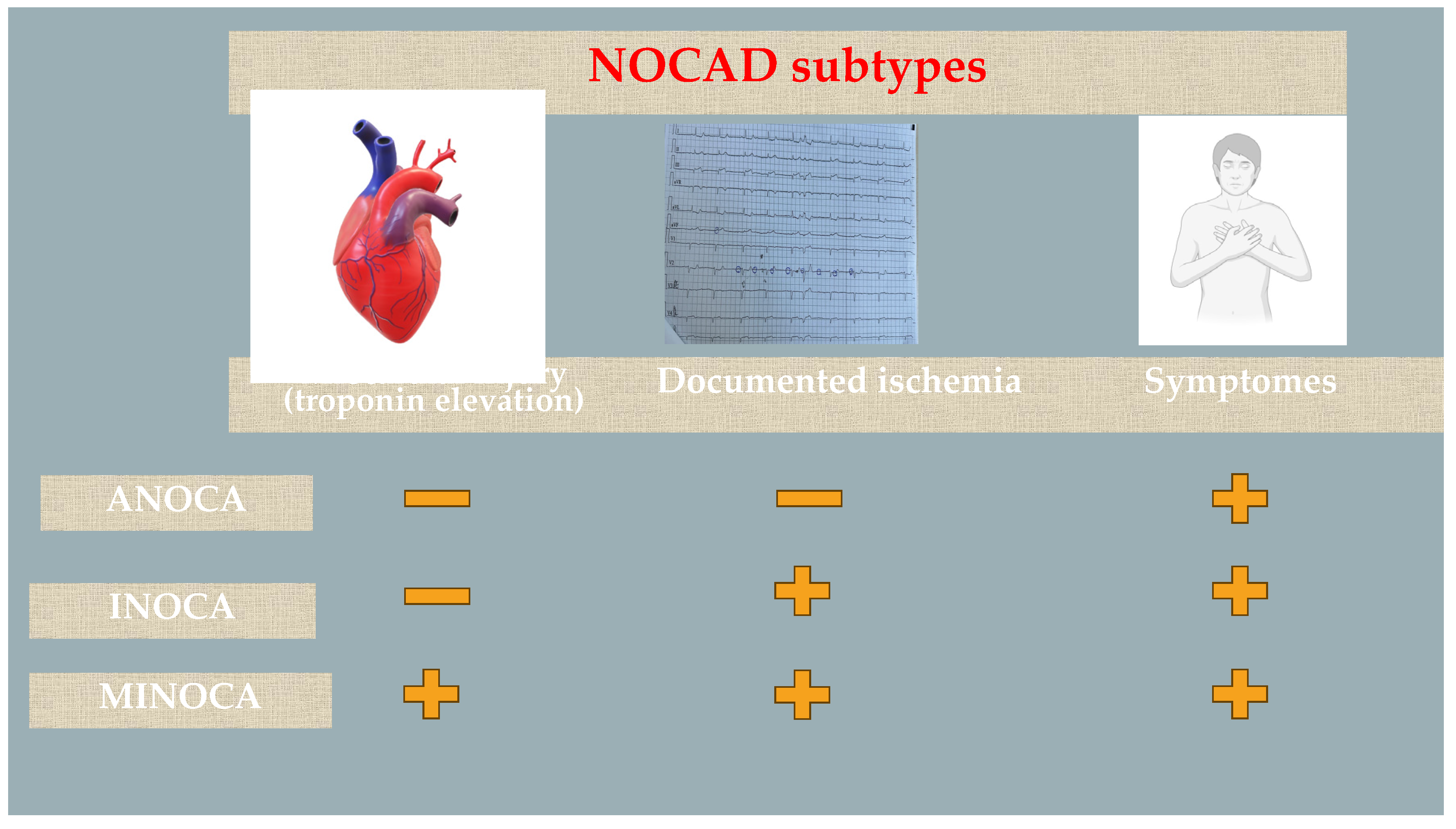
Figure 2.
Diagnostic tools in patients suspected with MINOCA. CAD: coronary artery disease; CMR: cardiac magnetic resonance; IVUS: intravascular ultrasound; OCT: optical coherence tomography; SCAD: spontaneous coronary artery dissection.
Figure 2.
Diagnostic tools in patients suspected with MINOCA. CAD: coronary artery disease; CMR: cardiac magnetic resonance; IVUS: intravascular ultrasound; OCT: optical coherence tomography; SCAD: spontaneous coronary artery dissection.
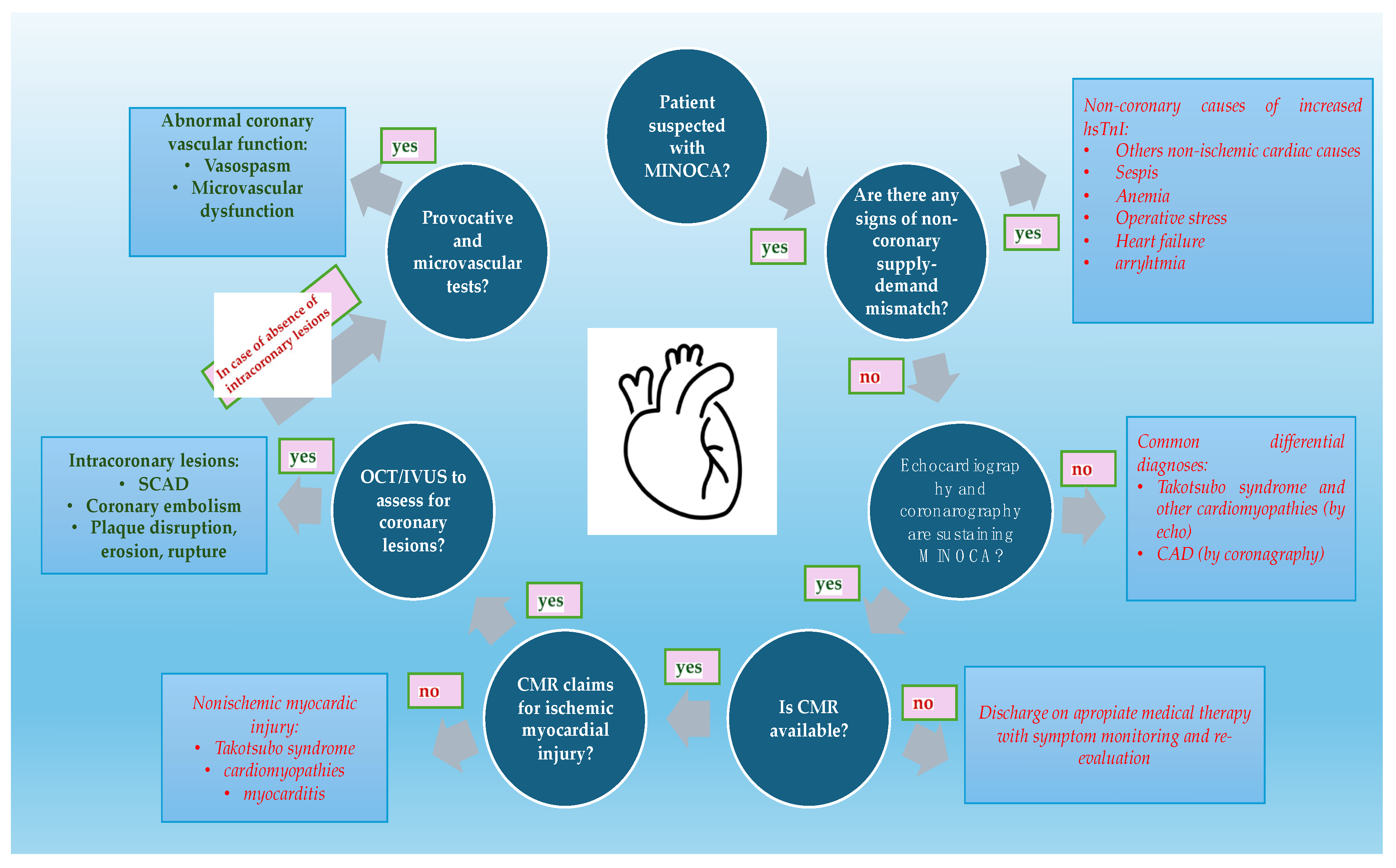
Figure 3.
The inflammatory cascade due to coronary ischemia leading to atrial fibrillation. AF: atrial fibrillation; IL: interleukin; TNF: tumor necrosis factor.
Figure 3.
The inflammatory cascade due to coronary ischemia leading to atrial fibrillation. AF: atrial fibrillation; IL: interleukin; TNF: tumor necrosis factor.
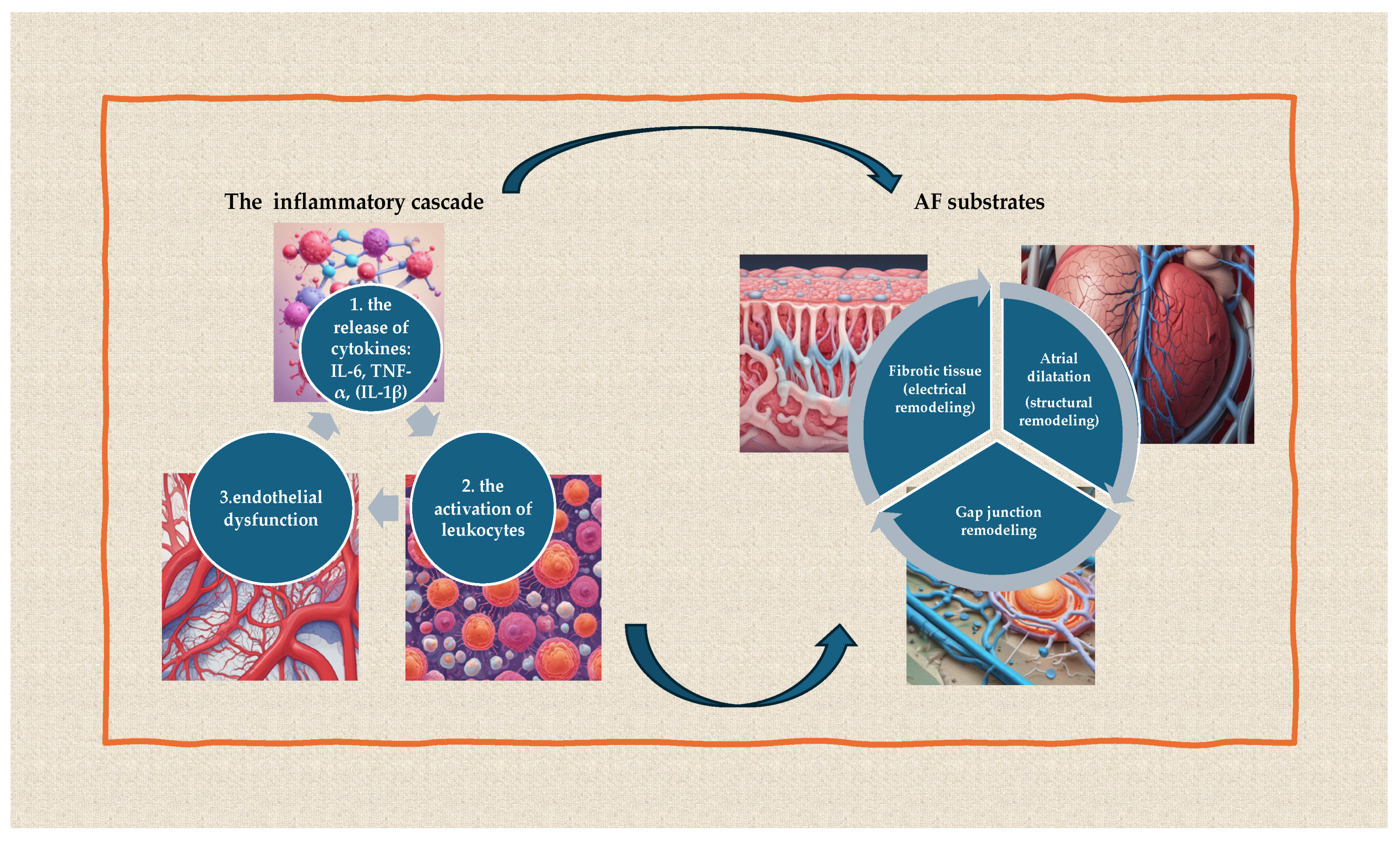
Figure 4.
The pathophysiology of atrial fibrillation.
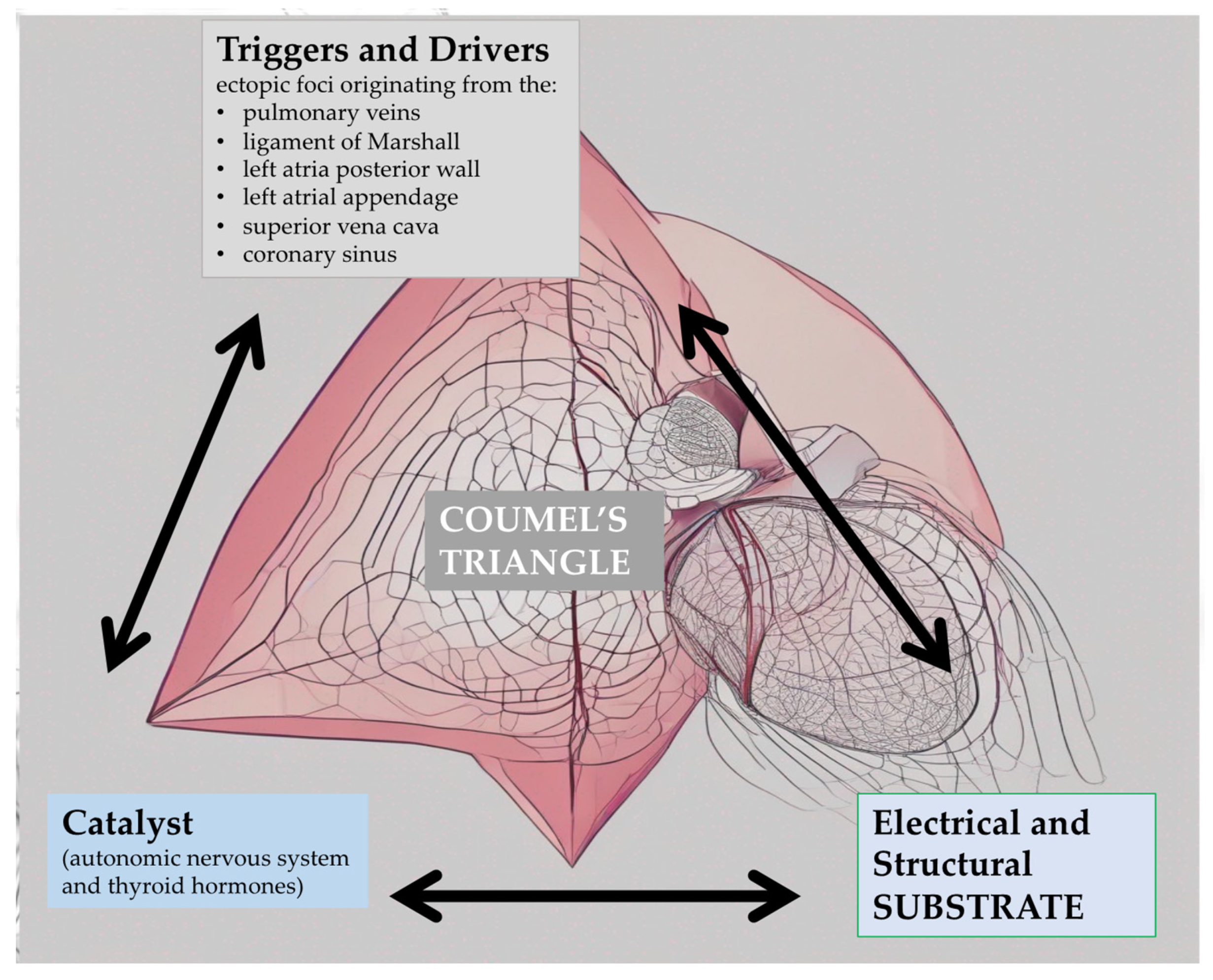

Table 1.
Diagnostic tools used to identify ANOCA and INOCA. ANOCA: Angina with Non-Obstructive Coronary Arteries; CAD: coronary artery disease; FFR: Fractional Flow Reserve; HMR: hyperemic myocardial velocity resistance; IFR: Instantaneous Wave-Free Ratio; IMR: index of microvascular resistance; INOCA: Ischemia with Non-Obstructive Coronary Arteries; MINOCA: Myocardial Infarction with Non-Obstructive Coronary Arteries; CFR: Coronary Flow Reserve.
| Diagnostic tool | Role of the diagnostic tool |
|---|---|
| Cardiac biomarkers (especially high-sensitive troponin) [23] | May differentiate ANOCA and INOCA from MINOCA by showing the status of myocardial injury |
| Non-Invasive Tests to detect ischemia: exercise tolerance test, transthoracic doppler echocardiography, myocardial contrast echocardiography, myocardial perfusion imaging, positron emission tomography, stress echocardiography and cardiac MRI [24] | May differentiate ANOCA (without signs of ischemia) from INOCA (the signs of ischemia are present) |
| Coronary Angiography [25] | Gold standard to exclude obstructive CAD |
| Invasive tests during coronary angiography: vasoreactivity test using intracoronary acetylcholine, FFR, IFR, CFR, IMR, HMR [26] | May differentiate the type of NOCAD regarding its physiopathology |
Table 2.
Types of SCAD based on angiography [38].
Table 2.
Types of SCAD based on angiography [38].
| Type of SCAD | Description |
|---|---|
| Type 1 (contrast staining of false lumen) | It has contrast stains in the arterial wall with multiple radiolucent lumens with or without slow contrast clearing. |
| Type 2 (long diffuse and smooth narrowing) | It shows diffuse, smooth, usually, 20–30 mm narrowing with varying severity. |
| Type 3 (focal/ tubular stenosis) | It shows focal or tubular stenosis that mimics atherosclerosis. |
| Type 4 (occlusion of the vessel) | There is no antegrade flux distal to the lesion |
Disclaimer/Publisher’s Note: The statements, opinions and data contained in all publications are solely those of the individual author(s) and contributor(s) and not of MDPI and/or the editor(s). MDPI and/or the editor(s) disclaim responsibility for any injury to people or property resulting from any ideas, methods, instructions or products referred to in the content. |
© 2024 by the authors. Licensee MDPI, Basel, Switzerland. This article is an open access article distributed under the terms and conditions of the Creative Commons Attribution (CC BY) license (https://creativecommons.org/licenses/by/4.0/).
Copyright: This open access article is published under a Creative Commons CC BY 4.0 license, which permit the free download, distribution, and reuse, provided that the author and preprint are cited in any reuse.


