Submitted:
15 July 2024
Posted:
16 July 2024
You are already at the latest version
Abstract
The aim of this study is to assess of rare earth elements (REE) content in hair samples in children, living in Lovozero village, near REE mining site, and possible effects of REEs on the prevalence of nervous system diseases in the Lovozersky district (Murmansk region, Kola Peninsula). School-age children (53 persons) were recruited for analysis of REE content in their hair samples. REEs (Y, La, Ce, Pr, Nd, Sm, Eu, Gd, Tb, Dy, Ho, Er, Tm, Yb, Lu) content was estimated by inductively coupled plasma mass spectrometry (ICP-MS). Analysis of REE content in the hair of children living in the Russia, Kazakhstan and China indicates REEs intake from environment. A possible contribution of REEs to the nervous system disorders is supported by the link between REE content in the hair samples of children, living near REE mining (China) and by manifesting cognitive disorders in these children. It also turn out that highest prevalence of the nervous system diseases in children 15-17 years old manifested in the Lovozersky district then in other area in the Murmansk region. The paper discusses the possible contribution of REEs to the prevalence of episodic paroxysmal disorders (G40-G47), cerebral palsy (G80-G83), epilepsy and status epilepticus (G40-G41).
Keywords:
earth elements
; nervous system
; paroxysmal disorders
; cerebral palsy
; epilepsy
; children
1. Introduction
Nowadays, rare earth elements (REEs) are widely used in various fields of human activity and the demand for them is constantly increasing. Mining of REEs is limited to certain countries, with China being the leader. Russia also makes a certain contribution to the global REE mining, where the main REE mining is concentrated on the Kola Peninsula and in Zabaykalsky region. In view of the increasing load of REEs on the environment, the extraction accompanied by the spread of REEs in the air, soil, water, and agricultural products, the biological effectiveness of REEs and the assessment their possible negative effects on the human body are gaining more attention [1,2,3].
REEs refer to a series of elements, including lanthanides (Ln), Scandium (Sc) and Yttrium (Y), which are made up of seventeen metallic elements in the periodic table [3]. REEs are usually divided into two groups based on their different structures and characteristics. Lanthanum (La), Cerium (Ce), Praseodymium (Pr), Neodymium (Nd), Promethium (Pm), Samarium (Sm), Europium (Eu) are referred to as light rare earth elements (LREEs), while (Gd), Terbium (Tb), Dysprosium (Dy), Holmium (Ho), Erbium (Er), Thulium (Tm), Ytterbium (Yb), and Lutetium (Lu), Sc, and Y are referred to as heavy rare earth elements (HREEs) [1,2,3]. The human body can exposed to REEs through various pathways such as inhalation, ingestion, dermal contact, and iatrogenic exposure, and this causes deposition, which in turn destroys the structure and function of various organs of the human body and induces multi-system diseases [1,2,4]. The main results evidenced that REEs can enter human body via several routes, are associated with numerous diseases, can cause ROS production, DNA damage and cell death, and are more toxic to cancer cells than normal cells [2]. The studies in vitro and in vivo studies have shown that REEs exert these adverse effects mainly by affecting genetics and epigenetics, altering the activation of signaling pathways [1,2,5,6,7,8,9,10].
Epidemiological investigations of residents living in mining areas have shown that long-term exposure to REEs can cause neurological diseases, such as motor and sensory impairments, neurodegeneration, or neurosis [11,12]. Observational studies in special populations, such as children and pregnant women in mining areas, have shown that REEs can lead to reduced intelligence and motor ability in children, can deposit in the fetal brain, and can affect neural tube development [13,14]. These studies have shown that REEs can deposit in the brain, impair the development of the central nervous system (CNS), and even pass through the placental barrier to generate passage or transgenerational inheritance [1].
The research of REEs content in hair samples in young children aged 0-3 years whose mothers live in a REEs mining area of Jiangxi Province shown that REEs level in hair can be used as a biomarker to reflect body’s level of exposure to REEs [15]. Children aged from 6 to 9 years, living at the REEs-high regions, have manifested the mental age and intelligence quotients (IQ) lower than those at normal regions [16]. Yongwei [17] found relationship between REE, Lead and intelligence of children aged 6 to 16 years. The IQ of children aged 7–10 years living near a REEs ore area was significantly lower, than at normal regions [18]. The increase in the daily intake of REEs was associated with the decrease in intelligence quotient, including the total score of intelligence quotient, intelligence age, short-term memory, and the score of attention [19]. The individual and joint effects of prenatal exposure to 13 REEs manifested by decrease of the mental and psychomotor development indices (MDI/PDI) in 24- month’s children [20]. These studies show that REE can do damage to the CNS, but its underlying mechanism and targets are still unclear.
The study of toxicological effects of REEs is most relevant in those regions, where REE ore is mined. In this respect, the Kola Peninsula is a natural laboratory that allows studying the effects of REE on human health. The Kola Peninsula is the most industrial territory of the Arctic region. The high density of enterprises and cities are locating in the central part of the peninsula that creates conditions for the wide distribution of pollutants [21,22]. The enterprise where ore containing REEs is extracted is the Lovozersky Mining and Processing Plant (Lovozerskiy GOK LLC), located near the Revda settlement, where miners, mine workers and their families mainly live. In 18.5 kilometres from the mining Revda is the Lovozero village (Figure 1), where the indigenous Saami people of the Kola Peninsula is mainly inhabited and where it has no industrial enterprises.
The ores contain predominantly cerium lanthanides. When loparite ores are concentrated, processing wastes formed, including concentration tailings, which are sent as part of the slurry to a tailings storage facility. Concentration tailings are known to pose a threat to the environment due to the processes of weathering and transition into a soluble form of environmentally hazardous elements [2,23,24,25,26]. The impact of the Lovozersky mining and processing plant on the environment, it has been assessed in separate studies [23,27,28]. The investigations evident, that dusting in the tailings of the processing plant is a main source of environmental contamination near the Lovozersky Mining and Processing Plant [28].
Workers in recycling facilities expose to significant amounts of REEs through inhalation, ingestion, and skin contact [29,30,31]. In the study of [32], scalp hair of people living near a mining site in Inner Mongolia, contained significantly higher REE levels compared to people from the control area. In a similar study, the hair of children living in Shang and Liao villages, near a mining site, contained higher REE levels versus children from reference areas [33]. The blood and hair of people living near a large-scale mining site in southwest Fujian Province, China, contented of REEs [34]. A correlation between the concentration of REEs in soil and human samples was found in Hezhang County, China [35]. All these studies indicated that the REE mining process influenced the environment in mining areas and the people living nearby [2,36]. The human body expose to various chemicals simultaneously, usually with inherent correlations to one another [37,38]. Therefore, human health in the Lovozersky district is the result of mixed effects of the LREEs and HREEs, where exposure children to REEs could cause the certain morbidity.
The aim of this study is to assess of REE content in hair samples in children, living in the vicinity of mining site contented REEs, and a possible effects of REEs on prevalence of nerve system diseases in children of 0-14 and 15-17 years old in the Lovozersky district.
2. Experimental Procedure
2.1. Sample Preparation
School-age children 7-15 years old (53 persons), living in the vicinity (Lovozero village), of the Kola Peninsula mining site, where REE-bearing ore is mined and processed, were recruited in the study. The method of sampling hair and their preliminary preparation for RRE’s analysis was carried out according to the recommendations of the IAEA [39]. The criterion for inclusion of children in the study was the consent of the subject and the presence of the first group health. The principles of medical ethics approved by the UN General Assembly (1992), the Council of Europe Convention on Bioethics (1997) and the Council on Bioethics of the Russian Academy of Sciences (18.01.2016) were applied in the study. The all parents of the recruited children were acquainted with the purpose and conditions of the study and gave their written consent to their child’s participation in this study (Protocol of Local Bioethics Committee №2, 10.02.2017). Collected hair samples were prepared according to the procedure [40], for which the samples were washed with acetone and with deionized water (18.2 MΩ/cm2) and dried at ambient temperature. Demineralized water was obtained using the Element system (Millipore, Burlington, Massachusetts, USA), the acid was purified using the BSB-939-IR unit (Berghof, Eningen, Germany). Hair samples (0.05-0.15 g) were treated with 10 ml of 65 % pure nitric acid in a closed 50-ml polypropylene volumetric tube in a water bath at a temperature of 100° C for 1-2 hours of decomposition of the organ part. After cooling, the digested solution was diluted to 20-50 ml volumetric flask with 2 % HNO3. Working solutions were freshly prepared daily for analysis and then stored in 15-ml polypropylene centrifugal tubes for measurements.
2.1. Sample Analysis
All samples were analyzed for REEs (Y, La, Ce, Pr, Nd, Sm, Eu, Gd, Tb, Dy, Ho, Er, Tm, Yb, Lu) and other elements in the Laboratory of Optical and Chemical Analysis methods of the Tananaev Institute of Chemistry Russian Academy of Sciences (ICT KSC RAS). Inductively coupled plasma mass spectrometry (ICP-MS) were used in the study. Elemental ICP-MS analysis (ELAN 9000 DRC-e, Perkin Elmer, USA) of samples was carried out using available methods [40,41] and methods developed in the ICT KSC RAS.
The following conditions were maintained for all measurements: plasma gas flow of 15.0 L min-1, nebuliser gas flow of 0.90 L min-1, auxiliary gas flow of 1.25 L min-1 and Radio Frequency (RF) power of 1.3 kW. Multi-element calibration solutions Inorganic Ventures (Christiansburg, VA, USA) (IV-STOCK-21, IV-STOCK-26, IV-STOCK-28, IV-STOCK-29) were used for the instrumental external calibration, linear fitting rates were 0.9999. Potential spectral interference was recognized during validation of the method, and mathematical methods of interference correction were chosen. The limit of determination of elements was in the range from 0.001 to 0.0001 mg kg-1, depending on the determined element of study object. The uncertainty level were estimated for the procedure, including sample preparation at the level of 10-15 %. Quality control the accuracy and precision of the analysis were carried out using the analysis of international and Russian reference materials certified for the content of the determined elements.
2.2. Comparison Areas
To compare the morbidity of the children in the Lovozero district, where the source of the dominant contamination is dusting of tailings containing REE, with the corresponding indices at other sites, we selected the comparison areas (Figure 1). 1. Murmansk city, where there are no mining enterprises, but there is a wide range of pollutants typical of urbanized areas. 2. Apatity city, which is in the sphere of dusting of the “ANOF-2” tailing dump - the main and one of the largest on the Kola Peninsula source of environmental pollution with suspended substances as SiO2, TiO2, Al2O3, Fe2O3, FeO, P2O5, CaO, SrO, MgO, MnO, Na2O, K2O, F2 [42]. 3. Kandalaksha city - emissions from the Kandalaksha aluminium smelter contain a significant amount of such pollutants as hydrogen fluoride, fluorides, polycyclic aromatic hydrocarbons, inorganic dust, etc. .Moreover, the priority pollutants are compounds, the presence of which in the atmosphere can have a significant impact on human health and, especially, on the morbidity of children [22]. 4. Monchegorsk city – the main pollutant is Sulphur dioxide as emissions from Kola Mining and Metallurgical Company - a division of Norilsk Nickel - Sulphur dioxide causes irritation of the skin, mucous membranes of the nose, eyes and upper respiratory tract. 5. Olenegorsk city, where Mining and Processing Plant is located: haematite-magnetite ores of the Olenegorsky deposit belong to ferruginous quartzites with a content of the following elements in magnetite - Fe - 72.1±0.08 %, Mn - 0.02-0.04 % with negligible contents of Al, Si, Cu, Mg. 6. Kovdorsky district with the ore mining and processing industrial complex is Russia’s second-largest producer of apatite concentrate in terms of production output and the world’s only producer of baddeleyite concentrate (ZrO2). Iron mine working on magnetite-rich ore, mineral composition of tailings from the Kovdorskiy GOK enrichment plant contains: % CaO -27,1; MgO - 21,1; SiO2 - 18,2; P2O5 - 10,5; Al2O3 - 2,2; Fe2O3 - 2,0; Na2O - 0,8; K2O - 0,8; SO3 - 0,5; MnO - 0,1; TiO2 - 0,1; others - 16,6. 7. Kolsky district, where mixed type of pollutions are detected; mainly caused by the transport of air masses containing toxic compounds
from the main sources of contamination on the Kola Peninsula. 8. Pechenganickel Combine: the dominant nickel concentrator in the ore is pentlandite. Its average chemical composition in massive ores are (%): Ni - 35.3, Fe - 30.8, Co - 0.7, S - 33.2, sum - 99.9, formula: (Ni4.65Fe4.26Сo0.08)8.99S8.00; in the disseminated (%): Ni - 34.1, Fe - 32.0, Co - 0.6, S - 33.0, sum - 99.7, formula: (Ni4.50Fe4.44Сo0.08)8.02S7.98.
Thus, similarities and differences in the morbidity of the children population living in these territories will make it possible to identify the common and specific possible causes of morbidity in children due to the nature of local contamination of the comparison areas.
2.3. Analysis of the Incidence of the Population in the Kola North
The Statistical Compendiums “Incidence of the population of the Murmansk region 1995-2018, as well as the data of the Murmansk Regional Medical Information and Analytical Center for 2018-2020 were used in the work.
2.4. Statistical Data Processing
The data obtained was statistically processed by using the STATISTICA 10 software package; graphing was carried out using the graphic editor ORIGIN. The average arithmetic values of the studied parameters (M), and also gave the values of the standard deviation (±δ) were calculated. Along with these statistics, medians, variation spread, percentile range (P25-P75), and coefficients of variation were also calculated.
3. Results of the Study
3.1. REE Content in the Children Hair Samples
Statistical characteristics of the content of REEs in hair samples from children living in Lovozero village in the vicinity of the mining enterprise are presented in Table 1.
In the Table 1 one can see that Sc, Dy and Yb are characterized by the highest coefficients of variability of content in hair samples from children. This indicates uneven accumulation of these elements by the children body that can probably be associated with their sporadic intake from the living environment. Since the REE content in hair samples from children depends on the territory of residence, on proximity to the places of development of ore deposits containing REE, on their intake into the environment and their concentrations in soil, water, air and foodstuffs, there are no standards for assessing REE content as such. To assess the level of REE content in hair samples from children living in Lovozero village, it is possible to conduct only comparative studies with hair samples from children living in the territories with elevated REE content in the environment and territories with background values.
3.2. Comparing
Table 2 shows the values in some trace elements and in REE content in hair samples from children in Lovozero village, in Zabaykalsky region [43], in Pavlodar District of the Kazakhstan Republic [44] as well as in hair samples from the adult population of Sweden [45]. For predicting the possible effects of REE accumulation in the body, Ca content is a very important indicator, because lanthanides compete for Ca binding sites with signaling proteins (1,5,9). The lower the calcium content in the organism determines the higher the probability of its substitution by lanthanides. Table 2 shows that the Ca content in hair samples of children from Lovozero is lower than in the corresponding hair samples of children from Zabaikalsky region, Pavlodar District and hair samples from Sweden.
If we compare the mean values of Sc content, it is higher in hair samples of children from Lovozero, than in other comparison areas. However, median Sc values are lower in children from Lovozero, than in children from Zabaykalsky and Pavlodar region, and are comparable to those in hair samples from the Swedish population.
Important elements for functional activity of the brain are Fe and Zn, the distribution of which was shown to be heterogeneous on brain preparations, along with Ca content. Exposure to Ln alters their distribution and leads to a decrease in their content [46]. Table 2 shows that Fe content in the hair of children from Lovozero is almost 35 times lower than in children from Zabaykalsky region, 2 times lower than in children from Pavlodar region and 2.7 times higher than in hair samples of Swedish residents. Zn content is also lower in hair samples from children in Lovozero, than in children in Zabaykalsky and Pavlodar region, but comparable to the content in hair samples from Swedish residents.
The significant variations one can see under comparing the lanthanide content in hair samples of children from Lovozero and in hair samples of representatives of the comparison areas. However, La content is comparable in hair samples in all comparison areas. Se content in hair samples of children from Lovozero is lower than in children from Zabaykalsky and Pavlodar region, but comparable to that of Swedish residents. Median Eu values in hair samples of children from Lovozero and Zabaykalsky region are comparable, but lower than in children from Pavlodar region. Nd content is lower in hair samples of children from Lovozero, and Sm content is almost 4 times higher than in children from Zabaykalsky region, but 2 times lower than in children from Pavlodar region. Tb content in children from Lovozero village is lower than in children from Zabaikaly and Pavlodar regions; Yb content in children from Lovozero is higher than in children from Zabaikaly, but 2 times lower than in children from Pavlodar regions. Yb content in children from Lovozero is higher, than in children from Zabaykalsky, and lower than in children from Pavlodar regions. Lu content is comparable in hair samples from children in the comparison areas. Hf, Ta contents are lower in children from Lovozero, than in children from Zabaykalsky and Pavlodar regions, and comparable to the content in hair samples from Swedish residents.
A comparison in the content of other elements we will not give in the text, as we have noted the most important ones related to lanthanides. But the contents of other elements in Table 2 give an idea of the peculiarities of the comparison areas, including those associated with ore mining (Zabaykalsky region) and the prehistory of contamination of the territory with radionuclides during nuclear tests at the Semipalatinsk test site (Pavlodar District).
The data in Table 2 give an idea of the differences in the elemental composition of hair samples from children due to the peculiarities of the territories of residence and testify that the elemental content of hair reflects the content of elements in the environment. For example, the uranium content in hair samples of children from Pavlodar region is 66 times higher than in children from Lovozero, 33 times higher than in children from Zabaykalsky region and 9.2 times higher than in residents of Sweden, indicating the prehistory of the area associated with radionuclide contamination.
To compare the REE content in hair samples of children living in more unified territories on the distribution of REE in the environment, we compared the REE content in hair samples of children living in Lovozero and in China, in the territories where REE are mined [47]. The peculiarity of the territories in China was that they were associated with the mining of ore containing LREEs. In a LREEs mining area in southern China, Shang village (referred to as M1) and Liao village (referred to as M2) were selected as sampling sites. There were many open-casting mining surfaces around and near M1, and M2 was relatively farther away from a few mining surfaces. Two other villages, Dong village (referred to as C1) and Ping village (referred to as C2) in the no mining area in the same county were selected as control. C1 village was nearer to the mining area (with a direct distance of 8 km) than C2, which was 14 km from the mining area and separated by high mountains. The 323 fourth-year healthy pupils aged 11–15 years at the primary schools in the above-mentioned villages were chosen as the subjects of the study. The contents of REEs in hair samples of children from all the sites together with children in Lovozero are shown in Table 3. In Table 3, it can be seen that the content of all light elements in hair samples in children from Lovozero is lower than in the comparison areas of children from China (Table 3, Figure 2).
The diagram (Figure 2) shows the distribution of REE concentrations (geometric mean of the samples) in the hair samples from children, living in the Lovozero village (Murmansk region) and children from different villages in China. The hair samples from children, living in China is enriched with LREE, while the concentration of light REE in the hair samples of children from Lovozero is significantly lower. But the concentrations of HREEs (from Gd to Yb) in the all samples are comparable, and the concentration of Lu in the hair samples of children from Lovozero exceeds all corresponding values. One can see (Figure 2), that the content of almost all LREEs is higher in Shang village (M1), where many open-casting mining surfaces around and near the site were observed. The presented results once again show that living in the vicinity to the ore mining sites containing REE leads to REE accumulation in the body of the inhabitants of the neighboring territories and, especially, in the children’s body. In this connection, it can be seen that the REE content in hair samples of children from Lovozero corresponds the elemental composition of the ore mined and processed by the Lovozersky Mining and Processing Plant. In addition, the predominance of HREEs in hair samples from children in Lovozero in compare with children hair samples from China, demonstrates the difference between ore mined contenting REE in Lovozersky region and in corresponding territories in China.
Further, we compared the REE content in hair samples in children from the Lovozero village and from the REE mining area, namely Maoniuping rare-earth deposit, was located in the Mianning City of Sichuan Province, China. Four villages around the mining area of rare earth metals were as the field for research. Exposure to REEs and lead was assessed by analysis the scalp hair samples of children aged 6-16 years who lived in a REE mining area [48]. This study has shown that both light and heavy REEs, as well as lead in hair had negative effect on intelligence of children aged 6 to 16-years-old.
Comparison of the REEs content in children hair samples from Lovozero village and in hair samples of 95 children from four villages around the mining area in the Mianning City is presented in Table 4.
In Table 4, it can be seen that the content of LREEs in the hair samples of children from the mining area in the Mianning City of Sichuan Province is higher than in the hair samples of children from Lovozero. The content of Eu in hair samples of children from Lovozero is slightly lower, and the content of Gd is almost identical. However the content of HREEs is higher in the hair samples of children from Lovozero, than in the corresponding hair samples of children from Maoniuping rare-earth deposit. Thus, the mean values of Tb, Ho, Er, Tm, Yb, Lu in children from Lovozero exceed the corresponding values in children from the mining area (China) by 1.6, 1.65, 1.5, 3.0, 1.9, 4.6 times, respectively. And in 25 percent of children from Lovozero (P25) the content of elements Tb, Dy, Ho, Er, Tm, Yb, Lu is 2.8, 1.2, 1.9, 2.4, 4.6, 2.4, 7.3 times higher, respectively, than in the corresponding percent of children from the mining area (China).
Since HREEs, independently LREEs and lead, contribute to lower intelligence in children from the mining area (China) [48], one can assume that the REE content in the organism of children from Lovozero might also effect on cognitive and other functions of nervous system.
3.3. Nervous System Diseases
Comparison of the prevalence of the nervous system diseases in children 0-14 with adolescents 15-17 years old in the same territory can indirectly indicate the accumulation of REEs with age, in the event that it occurs (Figure 3).
One can see (Figure 3:1,1A), that the prevalence of episodic paroxysmal disorders is the highest in the Murmansk city (28.5±2.9) and after then in the Lovozersky (16.9±6.2) and in the Kolsky (14.0±0.8) districts in children of 0-14 years old. However, the prevalence of this nosology is the highest in the Lovozersky district (51.7±4.4), then in the Murmansk city (42.6±2.6) and then in the Kolsky district (34.0±3.2) among the adolescents of 15-17 years old. Thus the prevalence of episodic and paroxysmal disorders (G40-G47) in adolescents 15-17 years old increases by 1.5, 3.1, 2.4 time on comparing with children 0-14 years old in the Murmansk city, in the Lovozersky and in the Kolsky districts, respectively.
The prevalence of cerebral palsy and other paralytic syndromes (G80-G83) in children of 0-14 years old is 4.4±0.2, 5.3±0.2, 3.7±0.1, 3.2±1.0 in the Murmansk, in the Kandalaksha cities, in the Kolsky and in the Lovozersky districts, respectively, and it is 4.7±0.4, 4.6±0.1, 3.0±0.8, 6.0±1.7 in adolescents of 15-17 years old from the Murmansk, the Kandalaksha cities, from in the Kolsky and in the Lovozersky districts, respectively. These data demonstrate, that prevalence of cerebral palsy and other paralytic syndromes increases by 1.1 and 1.9 times in the Murmansk city, in the Lovozersky district and it decreases by 1.2 times in the Kandalaksha city and in the Kolsky district in adolescents, relatively, in children of 0-14 years old on the corresponding areas.
The prevalence of epilepsy and status epilepticus (G40-G41) in children of 0-14 years old is highest in the Murmansk, in the Kandalaksha cities, in the Kovdorsky and in the Lovozersky districts (7.3±0.8, 6.2±0.3, 5.6±0.9, 5.9±1.2, respectively). However, prevalence these diseases in adolescents 15-17 years old (9.0±0.9, 14.1±3.0, 6.5±1.0, 15.0±3.3, respectively) on comparing areas increase by 1.2, 2.3, 1.2, 2.5 times relatively of corresponding values in children 0-14 years old. Thus, the Lovozersky district is a leader among the comparing territories in the prevalence of nervous system diseases among adolescents of 15-17 years old. The accumulation by children’s body of the neurotoxic substances from environment could be a one from reasons the increase of the prevalence of nervous system diseases with age in the Lovozersky district. Therefore, we suggest that children in the Lovozersky district more prone to nervous system diseases, than in other comparison areas, in result of chronic accumulation of lanthanides in low doses. REEs could be could be ingested to the body with water [23,28], by air particle inhalation [26,27] or even dermal absorption [7,14], or other ways.
4. Discussion
Comparison of REE content in hair samples of children from the Lovozero village with corresponding values REES in the hair samples of children living in other territories (Table 2, Table 3 and Table 4), has shown the predominate of the HREEs in the hair samples from the Lovozero. To some extent, the comparability of physiological effects to exposure of heavy and light REEs, follows from the work of Yongwei et al. [17]. Evidence for comparability of the effects of HREEs and LREEs follows from the fact that Er3+, as a heavy REE, like to La3+ [49,50] and Pr3+ [51], affects on conductivity of nervous impulses in the frog neuromuscular synapse [52]. Besides, Haley et al. [52,53,54,55,56] investigated the pharmacological and toxicological effects on the animals of the light and heavy REEs, including Sc, Pr, Nd, Sm, Gd, Tb, Dy, Ho, Er, Tm, Yb. The all REEs induced the similar acute toxicity and the similar pharmacological effects, moreover the acute lethality increased with increasing REE atomic number [53]. Hence, the biological effects associated with exposure to La and other LREEs could be inducing by exposure to HREEs. Is there any reason to assume that REE accumulation in children’s bodies might induce the high prevalence of nervous system diseases in the Lovozero district ?
4.1. Accumulation REE in the Brain
REEs cross the blood-brain barrier and accumulate in brain tissue [1,4,5,9,19,44,58,59,60,61,62]. REEs can also penetrate the placental barrier and enter into the fetal body and brain tissue, and young animals, can ingest REEs via breast-feeding [15,20,60,61]. Moreover, REEs, could be are transported in the brain from the cerebrospinal fluid (CSF) [63].
The numerous studies demonstrate the effects of REEs on the nervous system. Zhu, et al. [19], has found the cumulative effect of REEs on the brain functions. The daily supplementation of REEs in the rat dietary has decreased of acetylcholinesterase (AchE) activity in the hippocampus in comparing with control group. The swelling in the cell body of the cerebral neurons and phagocytic phenomenon have manifested in the rats with a moderate dose of REEs [19]. Consequences of the REE accumulation in the cerebral cortex, in result of long-term intake of a small their amount from environment, could cause subclinical damage [19]. The study of Feng et al. [46] have revealed that La3+ has accumulated in brain and remained for long time after exposition of rats to LaCl3 through oral administration. The obviously higher concentrations of La accumulated in the cerebral cortex, hippocampus and cerebellum [46].
The high affinity of Ln for calcium ions leads to Ca2+ substitution in Ca2+-binding sites of proteins and to changes in neuronal conductivity. The changes in Ca2+ metabolism under the influence of Ln can have serious consequences for brain function [64]. La3+ can inhibit calcium-dependent neurotransmitter release [65], bind with the Ca2+ binding sites on the axon membrane and alter on membrane conductivity [66]. La3+ reduces both the influx and efflux of Ca2+ in the squid giant axon [67] and inhibits Ca2+ binding with brain synaptosomes simultaneously with depression in the activities of neural ATPase and AchE [68]. The rate of high-affinity uptake of [14C] glutamate by brain synaptosomes has depleted under acute LaCl3 toxicity in chicks [69]. La3+ also prevents the electrically stimulated release of transmitter from the squid giant synapse [70], the release of neurotransmitter amino acids from the spinal-medullary synaptosomes of rats [71]. La3+ causes a large increase in miniature end plate potential discharge from the frog neuromuscular junction [49]. Like La3+, Er3+ and Pr3+ also have a dual action on transmitter release at the frog neuromuscular synapse [72].
The significant increases in the La contents in the serum, in hippocampus and cerebral cortex, the rise in intracellular free calcium in hippocampal cells, a significant decrease in the activity of Ca2þ-ATPases, the inhibition of activities protein kinase-1 and the signaling pathways in the hippocampus under long-term LaCl3 exposure in rats were found by He et al. [73]. Yang et al. [74] have found that LaCl3 negatively affected on synaptic ultrastructure in hippocampus. Heuser J.E. [75] also revealed altered synaptic ultrastructure in hippocampal neuronal cell cultures under exposure to LaCl3. The study of LaCl3, CeCl3, and NdCl3 in the mouse brain showed that injury of the brain and oxidative stress, arising by Ln, trigger a cascade of reactions, such as lipid peroxidation, decreases of the total antioxidative capacity and activities of antioxidative enzymes, excessive release of nitric oxide, increase of glutamic acid, and downregulated level of acetylcholinesterase activities [76]. The accumulation Ce in the mouse hippocampus caused hippocampal apoptosis and the impairment of spatial recognition memory of mice [77].
The heavy REEs manifest a similar with La distribution in the brain [57]. Intravenous injection of 169Yb in rats has allowed counting the 169Yb distribution in five brain regions (hypothalamus, cerebellum, hippocampus, corpus striatum and cerebral cortex). It turn, out, that Yb did enter into the brain where remains for long time. The highest specific activities was in the hypothalamus, hippocampus and cerebellum. The 153Sm distribution in the brain was similar with 169Yb [78]. However unlike of 169YbCl3, the highest amounts and lowest concentrations of 153Sm were found in the cerebral cortex, and the highest concentrations of 153Sm was in the hypothalamus.
Gadolinium (Gd) also accumulates in the brain, along with other HREEs. Usually gadolinium-based contrast agents (GBCAs) are used in MRI [8]. The autopsy specimens shown that GBCA accumulates in the dentate nucleus, Globus pallidus, cerebellar white matter, frontal lobe white matter and frontal lobe cortical matter. A higher signal intensity of Gd detected in the dentate nucleus and Globus pallidus [79,80,81,82].
In addition to soluble REE forms, micro-sized Ln particles can also penetrate to the brain. Intravenous injection to rats of Sc2O3 with particle diameter ~3 µm has revealed distribution of Sc particles in the brain [83]. And even though Yokel et al. [84] showed minor penetration of Ce nanoparticles into brain parenchyma, Baranovskaya et al. [58] has found good evidence of Ce penetration into the brain as the presence of cerium-containing mineral phases in the brain of the Far East deer by using electron microscopy.
Along with the brain neurons, disorders in neuroglia (astrocytes) due to Ln exposure are of particular importance for arising of nervous system diseases [85]. LaCl3 can also induce axon abnormality in the hippocampus and primary cultured neurons [86].
The effects of REEs on the nervous system could also be through changes in gene expression. In particular, Y3+ can change the expression of some genes, which may be responsible for the toxicity of REEs on learning and memory [87].
Thus, there is no doubt that REEs, in case of their intake into the organism, can penetrate into the brain by various ways, accumulate in the different brain structures responsible for thinking, long-term and short-term memory, sensory perception, and motor activity. Disturbance of these structures in the result of REE impact could initiate such diseases as paroxysmal activity, paralyses, epilepsy.
4.2. Nervous System Diseases and RREs
The increasing consumption of rare earth elements (REEs) has resulted in a considerable risk of environmental exposure [1,2,3]. The adverse effects REEs on reproductive health [88] and on prenatal REEs exposure [13,20,60], on children’s neurodevelopment are not yet fully recognized. However, there is clear evidence that REEs, among other factors, could contribute to diseases of the nervous system.
4.2.1. Episodic Paroxysmal Disorders (G40-G47)
Paroxysmal movement disorders (PMDs) are neurological diseases typically manifesting with intermittent attacks of abnormal involuntary movements [89,90]. Two main recognized categories of PMDs base on the phenomenology: Paroxysmal dyskinesias (PxDs) manifeste by transient episodes hyperkinetic movement disorders, while attacks of cerebellar dysfunction are the hallmark of episodic ataxias (EAs) [91,92]. PxDs are as a network disorder, in which either primary striatal dysfunction or aberrant cerebellar output conveyed to the striatum results in the phenotype. The striatum (cluster of neurons in the subcortical basal ganglia) is a critical component of the motor and reward systems; it receives glutamatergic and dopaminergic inputs from different sources and serves as the primary input to the rest of the basal ganglia. Disruption of synaptic neurotransmission, mostly affecting cerebellar and striatal circuitries, emerges as a common final [92]. The exact pathophysiological mechanisms underlying the PMDs remains fully understood. One suggests that alterations in synaptic neurotransmission, brain energy metabolism and ion channel function can all be involved in the pathogenesis of PMDs [91,93,94].
REEs could contribute in the certain biological processes leading to various hyperkinetic disorders and its alteration. As shown above, REEs accumulate mainly in the cerebral cortex, in the hippocampus, in the cerebellum. The main groups of neurons, where REEs detected, include dentate nucleus and Globus pallidus. The dentate nucleus is a cluster of neurons in the cerebellum, and it is the largest single structure linking the cerebellum to the rest of the brain [95]. The dorsal region of the dentate nucleus contains output channels involved in motor function [96,97]. Accumulation of REEs in the Globus pallidus (GP), as a major component of the basal ganglia, with principal inputs from the striatum, and principal direct outputs to the thalamus and to the substantia nigra, could induces multiple lesions. Figure 4 demonstrates localization of brain regions responsible for motor disorders and the deposition of the certain REEs in these structures.
4.2.2. Cerebral Palsy and Other Paralytic Syndromes (G80-G83)
Cerebral palsy (CP) is primarily a neuromotor disorder that affects the development of movement, muscle tone and posture [98,99,100]. The underlying pathophysiology is an injury to the developing brain in the prenatal through neonatal period [98]. The motor disorders of CP are often accompanied by disturbances of sensation, perception, cognition, communication, and behavior, by epilepsy, and by secondary musculoskeletal problems [96,97,98]. CP can be defined according to the anatomical site of the brain lesion (cerebral cortex, pyramidal tract, extrapyramidal system, or cerebellum), clinical symptoms and signs (spasticity, dyskinesia [dystonic and choreo-athetotic forms], or ataxia) [100,101,102], Figure 4.
In the majority of cases, there is evidence that factors present during the prenatal period play a prominent role in CP, although the causes of congenital CP are unknown. CP is a syndrome of motor impairment that results from a lesion occurring in the developing brain [100]. Prerequisites for the development of CP could lie early in the formation of the neural tube [103]. Most cases of CP result from an interference in brain development in utero. Ion channels participate in the formation of the brain and spinal cord during one of the first developmental steps known as neural tube formation [104]. The excitable nature of neurons and muscle cells is dependent on the specific expression of ion channels and their subcellular localization in these cells [105]. In the during neural tube formation, neural plate cells exhibit Ca2+ transients partly mediated by the NMDA receptor, is a glutamate receptor and predominantly Ca2+ ion channel found in neurons [104]. The propagation of activity in neural tissue is generally associated with synaptic transmission, but epileptiform activity of the CP in the hippocampus can propagate with or without synaptic transmission due to endogenous field effect transmission in the hippocampus [106]. The third to fourth week of embryogenesis is a critical period in the development of neural tube morphology. If any interference encountered during this period, neural tube may fail to close [103,107].
REEs can enter a fetus through the placental barrier [13,20,108], and exposure to REE may affect the growth and development of the fetus. Cao et al. [20] studied association between prenatal exposure to 13 REEs and the neurodevelopment of children at 24-months of age. It was observed that exposure to 7 REEs during the first trimester were significantly associated with decreased of the mental index (MDI) and to 9 REEs during third trimester were significantly associated decreased of psychomotor development (PDI) scores in children at 24-months. Tm, Er in the first trimester and Ce, Ln in the third trimester accounted most importance to joint effects on MDI and PDI, respectively. Since the third to fourth week (first trimester) is a critical period in the development of neural tube, REEs may be a factor that may promote to impaired neural tube morphogenesis and the development of CP in children.
Wei et al. [13] examined the associations between concentrations of 10 REEs in maternal serum and the risk for fetal neural tube defects (NTDs). A higher level of La in maternal serum was associated with an increased risk for fetal NTDs. When each of the 10 REEs were examined individually and when all other nine REEs were simultaneously considered, NTD risks increased with overall concentrations of the 10 REEs under considering as a co-exposure mixture [13].
Contribution of REEs to prenatal disorder leading to CP might manifest by alteration of ion channels [109,110], by abolish the “calcium response” of nerve terminals [70]. The imbalance of the Ca level and Ca2+ binding protein, caused by La accumulated in brain, can disturb the ion homeostasis and cause a series of physiological disorders in the CNC [46].
Some cases of CP that have a fetal origin associated with non-cerebral congenital anomalies (CAs) [111,112,113,114,115,116]. Pharoah [111] found an elevated risk of CAs of the eye, cardiac anomalies, cleft lip and ⁄ or palate, congenital dislocation of the hips and talipes, and atresias of the oesophagus and intestines. REE appear to be able to contribute to prenatal development and promote to CAs, associated with CP. 141Ce residues distributed in all the animal’s viscera and tissue, but were higher in the eye, bone, testis, brain, heart and adipose, and the accumulation of cerium increased with increasing dose and prolonging feed time [117]. Exposure of pregnant women to Ce and Yb has decreased thyroid-stimulating hormone (TSH) levels in infants [118], that evident about prenatal effects of Ce and Yb. The association between maternal exposure to REEs in the prenatal period and the prevalence of Orofacial clefts (OFCs), as disruptions of normal craniofacial structure, support the possible involvement of REEs in the initiation of CAs [119].
La exposure influence on learning and memory, the expression of apoptosis-related proteins in offspring rats [120]. La transmit to offspring rats through parental blood circulation and breast milk before delactation and through water drinking after delectation and affect on learning and memory and the hippocampal neurons of offspring rats [120]. LaCl3 impacts on dendritic spines in CA1 pyramidal cells, decreasing dendritic spine density during development [121,122,123]. More than 90% of the excitatory synapses in the central nervous system are located on dendritic spines [124,125], therefore effects of RREs in the neonatal period by decreasing dendritic spine density during development could induce lesion in neuron conductivity and promote to realization of PC.
4.2.3. Epilepsy and Status Epilepticus (G40-G41)
Epilepsy comprises a heterogeneous group of brain diseases, which all share in common an enduring predisposition to generate seizures [126]. Known causes of epilepsy include genetic abnormalities such as de novo mutations and/or a precipitating injury. In the majority of cases, the underlying causes remain elusive [127,128]. Epileptogenesis, a pathological process transforming a normal healthy brain into an epileptic brain, it is characterized by multiple pathological changes within the brain such as acute and ongoing cell death, aberrant synaptic reorganization and neurogenesis, blood-brain barrier (BBB) disruption, and inflammation among many others [129]. Seizures are essentially a malfunctioning of the brain, due to the ‘‘misfiring’’ of its neurons [129]. It was clear that neurons communicated through action potentials along their axons, and that those action potentials were electrical events that depended on the movements of ions, particularly sodium and potassium, across the neuronal cell membrane [129]. Zhang et al. [108] suggested a new propagation mechanism for neural activity in the hippocampus involving endogenous field effect transmission in epilepsy. The discovery that astrocytes release glutamate, which causes synchronous neuronal depolarizations, has led to the idea that a source of excitation in epileptic discharges could be these cells [130,131,132].
REEs disrupt astrocyte functions that could play a certain role in the provocation of epileptic seizures. Along with the brain neurons, disorders in neuroglia (astrocytes) due to Ln exposure are of particular importance for arising of nervous system diseases [85,86]. Astrocytes, according to current thinking, play a decisive role in nervous system disease [130,131,132]. Because many of the synapses in the CNS are tripartite in nature, disruption of astrocytic supportive functions and/or of gliotransmission has the potential to disrupt synaptic transmission, synaptic plasticity and neuronal excitability [132,133]. The terminal foot of astrocytes is one important component of blood–brain barrier and La from blood would first encounter the astrocytes [134]. It turn out that La suppresses astrocyte-neuron lactate shuttle (ANLS) in rat hippocampus and the changes of ANLS system probably involved in the neurotoxicity of La [85]. Astrocytes are the main target in toxic encephalopathies caused by environmental exposure to heavy metals [135].
5. Conclusion
The content of REEs in the hair samples of children from Lovozero village, corresponding and exceeding the values REES in miners and in children living in the ore-mining zone on the compare territories, indicates the intake of REEs by the children body from external sources. Theoretically, it is possible to assume that accumulation of REEs in children might be occur already intrauterine, in case of prolonged residence of the mother on the territory contaminated with REE. Besides, the gradual deposition of REEs in the children born and living in the contaminated territories could lead to an increase in the incidence of the nervous system diseases with age, as the REE content in the organism increases. Accumulation of REEs in the certain brain structures could lead to impaired synaptic transmission, to altered neurotransmission and cellular neuronal metabolism, to disruption of ion channels, and to other phenomena underlying of the nervous system diseases. This study suggests a possible role of REEs in nervous system dysfunction, but proof of such a role requires further research.
6. Limitations
The study of REE content is limited only to hair samples from children living in Lovozero village in the vicinity of a mining and processing plant, processing REE containing ore. In the future, the study will be continued and expanded to include analyses of REE content in hair samples of women and men living in Lovozero settlement, as well as miners extracting ore.
The study was carried out within the framework of Research and Development Programme No. 122022200516-5, 122022400094-6.
Author Contributions
Conceptualization, N.B. and S.D.; methodology, N.B. and S.D.; validation, N.B. and S.D; formal analysis, N.B. and S.D.; data curation, N.B. and S.D.; writing—original draft preparation, N.B.; writing—review and editing, N.B.; project administration, N.B.; funding acquisition, N.B. and S.D. All authors have read and agreed to the published version of the manuscript.
Funding
The study was carried out within the framework of Research and Development Programme No. 122022200516-5, 122022400094-6.
Informed Consent Statement
Not applicable.
Data Availability Statement
The original contributions presented in the study are included in the article, further inquiries can be directed to the corresponding author/s.
Conflicts of Interest
The authors declare no conflicts of interest.
References
- Wang, W.; Yang Y.; Wang D.; Huang L. Toxic Effects of Rare Earth Elements on Human Health: A Review. Toxics. 2024, 12, 5, p.317. PMID: 38787096. [CrossRef]
- Brouziotis, A.A.; Giarra, A.; Libralato, G.; Pagano, G.; Guida, M.; Trifuoggi, M. Toxicity of rare earth elements: An overview on human health impact. Front. Environ. Sci. 2022. 10, p.948041. [CrossRef]
- Balaram V. Rare earth elements: A review of applications, occurrence, exploration, analysis, recycling, and environmental impact. Geoscience Frontiers. 2019, 101285e1303. [CrossRef]
- Li Z, Liang T, Li K, Wang P. Exposure of children to light rare earth elements through ingestion of various size fractions of road dust in REEs mining areas. Sci Total Environ. 2020, 15, pp.743:140432. Epub 2020 Jun 23. PMID: 32659548. [CrossRef]
- He, X.; Zhang, Z.; Zhang, H.; Zhao, Y.; Chai, Z. Neurotoxicological evaluation of long-term lanthanum chloride exposure in rats. Toxicol Sci. 2008, 103, 2, pp.354-61. Epub 2008 Mar 3. PMID: 18319242. [CrossRef]
- Rim, K.T.; Koo, K.H.; Park, J.S. Toxicological evaluations of rare earths and their health impacts to workers: A literature review. Saf Health Work. 2013, 4, 1, pp.12-26. Epub 2013 Mar 11. PMID: 23516020; PMCID: PMC3601293. [CrossRef]
- Rim, K.T. Effects of rare earth elements on the environment and human health: A literature review. Toxicol. Environ. Health Sci. 2016, 8, pp. 189–200. [CrossRef]
- Gwenzi, W.; Mangori, L.; Danha, C.; Chaukura, N.; Dunjana, N.; Sanganyado, E. Sources, behaviour, and environmental and human health risks of high-technology rare earth elements as emerging contaminants. Sci Total Environ. 2018, 15, 636, pp. 299-313. Epub 2018 Apr 27. PMID: 29709849. [CrossRef]
- Abdelnour, S.A.; Abd El-Hack, M.E.; Khafaga, A.F.; Noreldin, A.E.; Arif, M.; Chaudhry M.T., Losacco, C.; Abdeen, A.; Abdel-Daim, M.M. Impacts of rare earth elements on animal health and production: Highlights of cerium and lanthanum. Sci Total Environ. 2019, 1, 672, pp.1021-1032. PMID: 30999219. [CrossRef]
- Chung C, Deák F, Kavalali ET. Molecular substrates mediating lanthanide-evoked neurotransmitter release in central synapses. J Neurophysiol. 2008,100, 4, pp. 2089-2100. Epub 2008 Aug 20. PMID: 18715899; PMCID: PMC2576212. [CrossRef]
- Gaman, L.; Radoi, M.P.; Delia, C.E.; Luzardo, O.P.; Zumbado, M.; Rodríguez-Hernández, Á.; Stoian, I.; Gilca, M.; Boada, L.D.; Henríquez-Hernández, L.A. Concentration of heavy metals and rare earth elements in patients with brain tumours: Analysis in tumour tissue, non-tumour tissue, and blood. Int. J. Environ. Health Res. 2021, 31, pp.741–754. [CrossRef]
- Zheng L, Zhang J, Yu S, Ding Z, Song H, Wang Y, Li Y. Lanthanum Chloride Causes Neurotoxicity in Rats by Upregulating miR-124 Expression and Targeting PIK3CA to Regulate the PI3K/Akt Signaling Pathway. Biomed Res Int. 2020, 5, p.5205142. PMID: 32461997; PMCID: PMC7222569. [CrossRef]
- Wei J., Wang C., Yin S., Pi X., Jin L., Li Z. et al.) Concentrations of rare earth elements in maternal serum during pregnancy and risk for fetal neural tube defects. Environ Int. 2020, 137, p.105542. [CrossRef]
- Li Z, Liang T, Li K, Wang P. Exposure of children to light rare earth elements through ingestion of various size fractions of road dust in REEs mining areas. Sci Total Environ. 2020, 15, 743, p.140432. PMID: 32659548. [CrossRef]
- Peng R.L., Pan X.C., Xie Q. Relationship of the hair content of rare earth elements in young children aged 0 to 3 years to that in their mothers living in a rare earth mining area of Jiangxi. Chin J PrevMed. 2003, 37, pp.20–22 (in Chinese).
- Zhu, W.F.; Xu, S. Q.; Zhang, H., Shao, P.P., Wu, D.S., Yang, W.J., Feng J. Investigation of children intelligence quotient in REE mining area: Bio-effect study of REE mining area in South Jiangxi province. Chin. Sci. Bull. 1996, 41, pp.914–916 (in Chinese).
- Yongwei, W.; Dan, W.; Na, H.Y.; Peijia, S.; Bing, Y. Relationship between Rare Earth Elements, Lead and Intelligence of Children Aged 6 to 16 years: A Bayesian Structural Equation Modelling Method. Int. Arch. Nurs. Health. Care. 2019, 5, p. 123. [CrossRef]
- Fan, G.Q.; Yuan, Z.K.; Zheng, HL.; Liu, Z.J. Study on the effects of exposure to rare earth elements and health-responses in children aged 7–10 years. J Hyg Res. 2004, 33, pp. 23–28 (in Chinese).
- Zhu, W.; Xu, S.; Shao, P.; Zhang, H.; Wu, D.; Yang, W.; Feng, J. Bioelectrical activity of the central nervous system among populations in a rare earth element area. Biol. Trace. Elem. Res. 1997, 57, 1, pp.71-7. PMID: 9258470. [CrossRef]
- Cao, Z.; Yang, M.; Gong, H.; Feng, X.; Hu, L., Li; R., Xu, S.; Wang, Y.; Xiao, H.; Zhou, A. Association between prenatal exposure to rare earth elements and the neurodevelopment of children at 24-months of age: A prospective cohort study. Environ Pollut. 2024, 15, 343, p.123201. PMID: 38135135. [CrossRef]
- Belisheva, N.K. Comparative Analysis of Morbidity and Elemental Composition of Hair Among Children Living on Different Territories of the Kola North. In Processes and Phenomena on the Boundary Between Biogenic and Abiogenic Nature, O.V. Frank-Kamenetskaya et al. (eds.), Lecture Notes in Earth System Sciences, Springer Nature, Switzerland AG. 2020, pp. 803-827. [CrossRef]
- Belisheva, N.; Martynova, A.; Mikhaylov, N. An Interdisciplinary Approach to Predicting the Effects of Trans-boundary Atmospheric Transport to Northwest European Neighboring States In Book Integration processes in the Russian and international research domain: Experience and prospects, KnE Social Sciences, 2022. pp. 158–171. [CrossRef]
- Mazukhina, S.; Krasavtseva, E.; Makarov, D.; Maksimova, V. Thermodynamic Modeling of Hypergene Processes in Loparite Ore Concentration Tailings. Minerals. 2021, 11, 996. [CrossRef]
- Stovern, M.; Guzmán, H.; Rine, K.; Felix, O.; King, M.; Ela, W.; Betterton, E.; Sáez, A. Windblown Dust Deposition Forecasting and Spread of Contamination around Mine Tailings. Atmosphere. 2016, 7, 16.
- Wang, L.; and Liang, T. Accumulation and fractionation of rare Earth elements in atmospheric particulates around a mine tailing in Baotou, China. Atmos. Environ. 2014. 88, pp. 23–29. [CrossRef]
- Krasavtseva, E.; Maksimova, V.; Makarov, D.; Potorochin, E. Modelling of the Chemical Halo of Dust Pollution Migration in Loparite Ore Tailings Storage Facilities. Minerals 2021, 11, 1077. [CrossRef]
- Krasavtseva, E.A.; Maksimova, V.V.; Gorbacheva, T.T.; Makarov, D.V.; Alfertyev, N.L. Evaluation of soils and plants chemical pollution within the area affected by storages of loparite ore processing waste. Mine Surv. Subsurf. Use 2021, 112, 52–58. (In Russian).
- Krasavtseva, E.; Sandimirov, S.; Elizarova, I.; Makarov, D. Assessment of Trace and Rare Earth Elements Pollution in Water Bodies in the Area of Rare Metal Enterprise Influence: A Case Study-Kola Subarctic. Water 2022, 14, p.3406. [CrossRef]
- Shin, S.H.; Kim, H-O.; Rim, K.T. Worker Safety in the Rare Earth Elements Recycling Process From the Review of Toxicity and Issues. Safety and Health at Work. 2019, 10, 4, pp. 409-419. [CrossRef]
- Hao, Z.; Li, Y.; Li, H.; Wei, B.; Liao, X.; Liang, T.; et al. Levels of rare Earth elements, heavy metals and uranium in a population living in Baiyun Obo, inner Mongolia, China: A pilot study. Chemosphere. 2015. 128, pp. 161–170. [CrossRef]
- Liang, Q.; Yin, H.; Li, J.; Zhang, L.; Hou, R.; Wang, S. Investigation of rare Earth elements in urine and drinking water of children in mining area. Medicine 2018. 97, e12717. [CrossRef]
- Wei, B.; Li, Y.; Li, H.; Yu, J.; Ye, B.; and Liang, T. Rare Earth elements in human hair from a mining area of China. Ecotoxicol. Environ. Saf. 2013. 96, pp. 118–123. [CrossRef]
- Tong, S.L., Zhu, W.Z., Gao, Z.H., Meng, Y.X., Peng, R.L.., Lu, G.C. Distribution characteristics of rare earth elements in children’s scalp hair from a rare earths mining area in southern China. J. Environ. Sci. Health. A Tox. Hazard. Subst. Environ. Eng. 2004, 39, 9, p.2517-32. PMID: 15478941. [CrossRef]
- Li, Xf.; Chen, Zb.; Chen, Zq. Distribution and fractionation of rare earth elements in soil–water system and human blood and hair from a mining area in southwest Fujian Province, China. Environ Earth Sci. 2014, 72, pp. 3599–3608. [CrossRef]
- Meryem, B.; Hongbing, J. I.; Yang, G.; Huajian, D.; Cai, L. (). Distribution of rare Earth elements in agricultural soil and human body (scalp hair and urine) near smelting and mining areas of Hezhang, China. J. Rare Earths. 2016, 34, pp. 1156–1167. [CrossRef]
- Edahbi, M.; Plante, B.; Benzaazoua, M. Environmental challenges and identification of the knowledge gaps associated with REE mine wastes management. J. Clean. Prod. 2019, 212, pp. 1232–1241. [CrossRef]
- Billionnet, C. et al. Estimating the health effects of exposure to multi-pollutant mixture. Ann. Epidemiol. 2012. 22, pp. 126–141. [CrossRef]
- Dominici, F. et al. Protecting human health from air pollution: Shifting from a single-pollutant to a multipollutant approach. Epidemiology 2010, 21, pp. 187–194. [CrossRef]
- Report of IAEA, Coordinated Research Programme NAH: The Significance of Hair Mineral Analysis as a Means for Assessing Internal Body Badens of Environmental Pollutants; Vienna, 1993; Res-18.
- Method for determining trace elements in diagnosed biosubstrates by inductively coupled argon plasma mass spectrometry (ICP-MS). Guidelines, Moscow, Russian, 2003, p. 34 (in Russian).
- Seregina, I.F.; Osipov, K.; Bolshov, M.A.; Filatova, D.G.; Lanskaya, S.Yu. Matrix Interference in the Determination of Elements in Biological Samples by Inductively Coupled Plasma Mass Spectrometry and Methods for Its Elimination. J. Analyt. Chem. 2019, 74, pp. 182–191. [CrossRef]
- Pashkevich, M.A..; Stryzhenok, A.V. Analysis of the landscape-geochemical situation in the area of the tailings management of ANOF-2 of JSC Apatit. Notes of the Mining Institute. 2013, 206, pp. 155-159 (in Russian).
- Mikhailova, L.A.; Baranovskaya, N.V.; Bondarevich, E.A.; Vitkovsky, Y.A.; Zhornyak, L.V.; Epova, E.S.; Eryomin, O.V.; Nimaeva, B.V.; Ageeva, E.V. Determination of elemental homeostasis of the child population of Zabaikalsky Krai by the method of multi-element instrumental neutron activation analysis. Gigiena i Sanitariya. 2023, 102, 2, pp. 123-131 (in Russian).
- Baranovskaya, N.V.; Rikhvanov, L.P.; Ignatova, T.N., Narkovich, D.V.; Denisova O.A. The human geochemistry essays: Monograph. Tomsk Polytechnic University Publishing House, Tomsk, 2015; p. 378, ISBN 978-5-4387-0581-9.
- Rodushkin, I.; Axelsson, M.D. Application of double focusing sector field ICP-MS for multielemental characterization of human hair and nails. Part II. A study of the inhabitants of northern Sweden. Sci Total Environ. 2000, 262,1-2, pp. 21-36. PMID: 11059839. [CrossRef]
- Feng, L.; Xiao, H.; He, X.; Li, Z.; Li, F.; Liu, N.; Zhao, Y.; Huang, Y.; Zhang, Z.; Chai, Z. Neurotoxicological consequence of long-term exposure to lanthanum. Toxicol Lett. 2006, 165, 2, pp. 112-20. PMID: 16542800. [CrossRef]
- Tong, S.L.; Zhu, W.Z.; Gao, Z.H.; Meng, Y.X.; Peng, R.L.; Lu G.C. Distribution characteristics of rare earth elements in children’s scalp hair from a rare earths mining area in southern China. J. Environ. Sci. Health. A Tox. Hazard. Subst. Environ. Eng. 2004, 39, 9, pp. 2517-32. PMID: 15478941. [CrossRef]
- Yongwei, W.; Dan, W.; Na, H.Y.; Peijia, S.; Bing, Y. Relationship between Rare Earth Elements, Lead and Intelligence of Children Aged 6 to 16 years: A Bayesian Structural Equation Modelling Method. Int. Arch. Nurs. Health. Care 2019, 5, p. 123. [CrossRef]
- Heuser, J.; Miledi, R. Effects of lanthanum ions on function and structure of frog neuromuscular junctions. Proc. R. Soc. Lond. B Biol Sci. 1971, 179, 1056, pp. 247-60. PMID: 4400214. [CrossRef]
- Kajimoto, N.; Kirpekar, S.M. Effect of manganese and lanthanum on spontaneous release of acetylcholine at frog motor nerve terminals. Nat. New. Biol. 1972, 235, 53, pp. 29-30. PMID: 4502408. [CrossRef]
- Alnaes, E.; Rahamimoff, R. Dual Action of Praseodymium (Pr3+) on Transmitter Release at the Frog Neuromuscular Synapse. Nature, 1974, 247, pp. 478-479.
- Haley, T.J.; Komesu, N.; Efros, M.; Koste, L.; Upham H.C. Pharmacology and toxicology of praseodymium and neodymium chlorides. Toxicol Appl Pharmacol. 1964, 6, pp. 614-20. PMID: 14217003. [CrossRef]
- Haley, T.J.; Koste, L.; Komesu, N.; Efros, M.; Upham H.C. Pharmacology and toxicology of dysprosium, holmium, and erbium chlorides. Toxicol. Appl. Pharmacol. 1966, 8, 1, pp. 37-43. PMID: 5921895. [CrossRef]
- Haley, T.J.; Komesu, N.; Fleswer, A.M.M.; Mavis, L.; Cawthorne, J.; Upham, H.C. Pharmacology and toxicology of terbium, thulium, and ytterbium chlorides. Toxicol. Appl. Pharmacol. 1963, 5, pp. 427-436. [CrossRef]
- Haley, T.J., Raymond, K., Komesu, N.., Upham, H.C. Toxicological and pharmacological effects of gadolinium and samarium chlorides. Br. J. Pharmacol. Chemother. 1961, 17, 3, pp. 526-32. PMID: 13903826; PMCID: PMC1482085. [CrossRef]
- Haley, T.J.; Komesu, N.; Mavis, L.; Cawthorne, J.; Upham, H.C. Pharmacology and toxicology of scandium chloride. J. Pharm. Sci. 1962, 51, p. 1043-5. PMID: 13952089. [CrossRef]
- Xiao, H.; Li, F.; Zhang, Z.; Feng, L.; Li, Z.; Yang, J.; Chai, Z. Distribution of ytterbium-169 in rat brain after intravenous injection. Toxicol Lett. 2005 a. 155, 2, pp. 247-52. PMID: 15603919. [CrossRef]
- Baranovskaya N.V., Mazukhina S.I., Panichev A.M., VakhЕ.А., Tarasenko I.A., Seryodkin I.V., Ilenok S.S., Ivanov V.V., Ageeva E.V., Makarevich R.A., Strepetov D.A., Vetoshkina A.V. Features of chemical elements migration in natural waters and their deposition in the form of neocrystallisations in living organisms (physico-chemical modeling with animal testing). Bulletin of the Tomsk Polytechnic University. Geo Assets Engineering, 2024, vol. 335, no. 2, pp. 187–201. [CrossRef]
- Feng, L.; Xiao, H.; He, X.; Li, Z.; Li, F.; Liu, N.; Zhao, Y.; Huang Y.; Zhang Z.; Chai, Z. Neurotoxicological consequence of long-term exposure to lanthanum. Toxicol. Lett. 2006, 165, 2, pp.112-20. PMID: 16542800. [CrossRef]
- Feng, L.; He, X.; Xiao, H.; Li, Z.; Li, F.; Liu, N.; Chai, Z.; Zhao, Y.; Zhang, Z. Ytterbium and trace element distribution in brain and organic tissues of offspring rats after prenatal and postnatal exposure to ytterbium. Biol. Trace Elem. Res. 2007, 117, 1-3, pp. 89-104. PMID: 17873395. [CrossRef]
- He, X.; Zhang, Z.; Zhang, H.; Zhao, Y.; Chai, Z. Neurotoxicological evaluation of long-term lanthanum chloride exposure in rats. Toxicol Sci. 2008, 103, 2, pp. 354-61. Epub 2008 Mar 3. PMID: 18319242. [CrossRef]
- Zheng, L.; Yang, J.; Liu, Q.; Yu, F.; Wu, S.; Jin, C.; Lu, X.; Zhang L.; Du, Y.; Xi Q.; Cai Y. Lanthanum chloride impairs spatial learning and memory and down regulates NF-κB signalling pathway in rats. Arch Toxicol. 2013, 87, 12, pp. 2105-17. PMID: 23670203. [CrossRef]
- Iliff, J.J.; Lee, H.; Yu, M.; Feng, T.; Logan, J.; Nedergaard, M. et al. Brain-wide pathway for waste clearance captured by contrast enhanced MRI. J. Clin. Invest. 2013, 123, pp. 1299–309. [CrossRef]
- Evans, C.H. Biochemistry of the Lanthanides. Plenum Press, New York, USA, 1990; pp. 25–31.
- Przywara, D.A.; Bhave, S.V.; Bhave, A.; Chowdhury P.S.; Wakade, T.D.; Wakade, A.R. Activation of K+ channels by lanthanum contributes to the block of transmitter release in chick and rat sympathetic neurons. J. Membr. Biol. 1992, 125, 2, pp. 155-62. PMID: 1552563. [CrossRef]
- Blaustein, M.P.; Goldman, D.E. The action of certain polyvalent cations on the voltage-clamped lobster axon. J. Gen. Physiol. 1968, 51, 3, pp. 279-91. PMID: 5648828; PMCID: PMC2201132. [CrossRef]
- Van Breemen, C.; De Weer, P. Lanthanum inhibition of 45Ca efflux from the squid giant axon. Nature 1970, 226, 5247, pp. 760-1. PMID: 5443255. [CrossRef]
- Basu, A.; Chakrabarty, K.; Chatterjee, G.C. Neurotoxicity of lanthanum chloride in newborn chicks. Toxicol Lett. 1982, 14, 1-2, pp. 21-5. PMID: 6130644. [CrossRef]
- Basu, A.; Chakrabarty, K.; Haldar, S.; Addya, S.; Chatterjee, G.C. The effects of lanthanum chloride administration in newborn chicks on glutamate uptake and release by brain synaptosomes. Toxicol. Lett. 1984, 20, 3, pp. 303-8. PMID: 6701916. [CrossRef]
- Miledi, R. Lanthanum ions abolish the “calcium response” of nerve terminals. Nature. 1971, 229, 5284, pp. 410-411. PMID: 4323456. [CrossRef]
- Osborne, R.H.; Bradford, H.F. The influence of sodium, potassium and lanthanum on amino acid release from spinal-medullary synaptosomes. J Neurochem. 1975, 25, 1, pp. 35-41. PMID: 166144. [CrossRef]
- Metral, S.; Bonneton, C.; Hort-Legrand, C.; Reynes, J. Dual action of erbium on transmitter release at the frog neuromuscular synapse. Nature. 1978, 271, 5647, pp. 773-5. PMID: 24184. [CrossRef]
- He, X.; Zhang, Z.; Zhang, H.; Zhao, Y.; Chai, Z. Neurotoxicological evaluation of long-term lanthanum chloride exposure in rats. Toxicol Sci. 2008, 103, 2, pp. 354-61. Epub 2008 Mar 3. PMID: 18319242. [CrossRef]
- Yang, J.; Liu, Q.; Zhang, L.; Wu, S.; Qi, M.; Lu, S.; Xi, Q.; Cai, Y. Lanthanum chloride impairs memory, decreases pCaMK IV, pMAPK and pCREB expression of hippocampus in rats. Toxicol. Lett. 2009, 190, 2, pp. 208-14. PMID: 19643171. [CrossRef]
- Heuser, J.E. The Structural Basis of Long-Term Potentiation in Hippocampal Synapses, Revealed by Electron Microscopy Imaging of Lanthanum-Induced Synaptic Vesicle Recycling. Front. Cell Neurosci. 2022, 16, pp. 920360. PMID: 35978856; PMCID: PMC9376242. [CrossRef]
- Zhao, H., Cheng, Z., Hu, R., Chen, J., Hong, M., Zhou, M., Gong, X., Wang, L., Hong, F. Oxidative injury in the brain of mice caused by lanthanid. Biol. Trace Elem. Res. 2011, 142, 2, pp. 174-89. PMID: 20614199. [CrossRef]
- Cheng, Z., Li, N., Cheng, J., Hu, R., Gao, G., Cui, Y., Gong, X., Wang, L., Hong, F. Signal pathway of hippocampal apoptosis and cognitive impairment of mice caused by cerium chloride. Environ. Toxicol. 2012, 27, 12, pp. 707-18. PMID: 21384496. [CrossRef]
- Xiao, H.; Zhang, Z.; Li, F. et al. Accumulation and distribution of samarium-153 in rat brain after intraperitoneal injection. Biol. Trace Elem. Res. 2005. 104, pp. 33–40. [CrossRef]
- Kanda, T., Ishii, K., Kawaguchi, H., Kitajima, K., Takenaka D. High signal intensity in the dentate nucleus and globus pallidus on unenhanced T1-weighted MR images: Relationship with increasing cumulative dose of a gadolinium-based contrast material. Radiology 2014, 270, pp. 834–41. [CrossRef]
- Kanda, T., Nakai, Y., Aoki, S., Oba, H., Toyoda, K., Kitajima, K., Furui, S. Contribution of metals to brain MR signal intensity: Review articles. Jpn. J. Radiol. 2016, 34, 4, pp. 258-66. PMID: 26932404. [CrossRef]
- Kanda, T.; Nakai, Y.; Hagiwara, A.; Oba, H.; Toyoda, K.; Furui, S. Distribution and chemical forms of gadolinium in the brain: A review. Br. J. Radiol. 2017, 90, 1079, pp. 20170115. PMID: 28749164; PMCID: PMC5963376. [CrossRef]
- McDonald, R.J., McDonald, J.S., Kallmes, D.F., Jentoft, M.E., Paolini, M.A., Murray, D.L., Williamson, E.E., Eckel, L.J. Gadolinium Deposition in Human Brain Tissues after Contrast-enhanced MR Imaging in Adult Patients without Intracranial Abnormalities. Radiology. 2017, 285, 2, p. 546-554. PMID: 28653860. [CrossRef]
- Nnomo, A., Dieme, D., Jomaa, M. et al. Toxicokinetic study of scandium oxide in rats. Toxicology Letters. 2024. 392. pp. 56-63. [CrossRef]
- Yokel RA, Au TC, MacPhail R; et al. Distribution, elimination, and biopersistence to 90 days of a systemically introduced 30 nm ceria-engineered nanomaterial in rats. Toxicol Sci. 2012, 27, 1, pp. 256–268. [CrossRef]
- Jin, C., Gao, L., Li, Y., Wu, S., Lu, X., Yang, J., Cai, Y. Lanthanum damages learning and memory and suppresses astrocyte-neuron lactate shuttle in rat hippocampus. Exp. Brain. Res. 2017, 235, 12, pp. 3817-3832. PMID: 28993860. [CrossRef]
- Song, Z.; Mao, H.; Liu, J.; Sun, W.; Wu, S.; Lu, X.; Jin, C.; Yang, J. Lanthanum Chloride Induces Axon Abnormality Through LKB1-MARK2 and LKB1-STK25-GM130 Signaling Pathways. Cell. Mol. Neurobiol. 2023, 43, 3, pp. 1181-1196. [CrossRef]
- Yang, W.; Zhang, P.; Liu, J.; Xue, Y. Effect of long-term intake of Y3+ in drinking water on gene expression in brains of rats. Journal of Rare Earths, 2006, 24, 3, pp. 369-373.
- Chen J, Xiao HJ, Qi T, Chen DL, Long HM, Liu SH. Rare earths exposure and male infertility: The injury mechanism study of rare earths on male mice and human sperm. Environ SciPollut Res Int. 2015, 22, 3, pp. 2076-2086. Epub 2014 Aug 30. PMID: 25167826. [CrossRef]
- Fahn, S.; Jankovic, J.; Hallett, M. Principles and Practice of Movement Disorders. 2nd ed. Elsevier: Philadelphia, PA, 2011.
- Delorme, C.; Giron, C.; Bendetowicz, D.; Méneret, A.; Mariani, L.L.; Roze, E. Current challenges in the pathophysiology, diagnosis, and treatment of paroxysmal movement disorders. Expert Rev Neurother. 2021, 21, 1, pp. 81-97. PMID: 33089715. [CrossRef]
- Zhang, X.J.; Xu, Z.Y.; Wu, Y.C.; Tan, E.K. Paroxysmal movement disorders: Recent advances and proposal of a classification system. Parkinsonism Relat. Disord. 2019, 59, pp. 131-139. PMID: 30902529. [CrossRef]
- Garone, G.; Capuano, A.; Travaglini, L.; Graziola, F.; Stregapede, F.; Zanni, G.; Vigevano, F.; Bertini, E.; Nicita, F. Clinical and Genetic Overview of Paroxysmal Movement Disorders and Episodic Ataxias. Int. J. Mol. Sci. 2020, 21, 10, pp. 3603. PMID: 32443735; PMCID: PMC7279391. [CrossRef]
- Mark, M.D.; Maejima, T.; Kuckelsberg, D.; Yoo, J.W.; Hyde, R.A.; Shah, V.; Gutierrez, D.; Moreno, R.L.; Kruse, W.; Noebels, J.L.; Herlitze, S. Delayed postnatal loss of P/Q-type calcium channels recapitulates the absence epilepsy, dyskinesia, and ataxia phenotypes of genomic Cacna1a mutations. J Neurosci. 2011, 31, 11, pp. 4311-26. PMID: 21411672; PMCID: PMC3065835. [CrossRef]
- Tan, G.H.; Liu, Y.Y.; Wang, L.; Li, K.; Zhang, Z.Q.; Li, H.F.; Yang, Z.F.; Li, Y.; Li, D.; Wu, M.Y.; Yu, C.L.; Long, J.J.; Chen, R.C.; Li, L.X.; Yin, L.P.; Liu, J.W.; Cheng, X.W.; Shen Q.; Shu, Y.S.; Sakimura, K.; Liao, L.J.; Wu, Z.Y.; Xiong, Z.Q. PRRT2 deficiency induces paroxysmal kinesigenic dyskinesia by regulating synaptic transmission in cerebellum. Cell Res. 2018, 28, 1, pp. 90-110. PMID: 29056747; PMCID: PMC5752836. [CrossRef]
- Sultan, F.; Hamodeh, S.; Baizer, J.S. The human dentate nucleus: A complex shape untangled. Neuroscience. 2010, 167, 4, pp. 965-8. PMID: 20223281. [CrossRef]
- Saab, C.Y.; Willis, W.D. The cerebellum: Organization, functions and its role in nociception. Brain. Res. Brain. Res. Rev. 2003, 42, 1, pp. 85-95. PMID: 12668291. [CrossRef]
- Dum, R.P.; Strick, P.L. An unfolded map of the cerebellar dentate nucleus and its projections to the cerebral cortex. J. Neurophysiol. 2003, 89, 1, pp. 634-9. PMID: 12522208. [CrossRef]
- Rosenbaum, P.; Paneth N.; Leviton, A.; Goldstein, M.; Bax M.; Damiano, D.; Dan, B.; Jacobsson, B. A report: The definition and classification of cerebral palsy. Dev. Med. Child. Neurol. Suppl. 2007, 109, pp. 8-14. Erratum in: Dev. Med. Child. Neurol. 2007, 49, 6, pp. 480. PMID: 17370477.
- Graham, H.K.; Rosenbaum, P.; Paneth, N.; Dan, B.; Lin J.P.; Damiano, D.L.; Becher, J.G.; Gaebler-Spira, D.; Colver, A.; Reddihough, D.S.; Crompton, K.E.; Lieber, R.L. Cerebral palsy. Nat Rev Dis Primers. 2016, 2, pp.15082. PMID: 27188686; PMCID: PMC9619297. [CrossRef]
- Colver, A.; Fairhurst, C.; Pharoah, P.O. Cerebral palsy. Lancet. 2014, 383, 9924, pp. 1240-9. PMID: 24268104. [CrossRef]
- Pharoah, P.O. Prevalence and pathogenesis of congenital anomalies in cerebral palsy. Arch. Dis. Child Fetal. Neonatal. Ed. 2007, 92, 6, pp. F489-93. PMID: 17428819; PMCID: PMC2675398. [CrossRef]
- Patel, D.R.; Neelakantan, M.; Pandher, K.; Merrick, J. Cerebral palsy in children: A clinical overview. Transl. Pediatr. 2020, 9, (Suppl 1), pp. S125-S135. PMID: 32206590; PMCID: PMC7082248. [CrossRef]
- Copp, A.J.; Stanier, P.; Greene, N.D. Neural tube defects: Recent advances, unsolved questions, and controversies. Lancet Neurol. 2013, 12, 8, pp. 799-810. PMID: 23790957; PMCID: PMC4023229. [CrossRef]
- Goyal, R.; Spencer, K.A.; Borodinsky, L.N. From Neural Tube Formation Through the Differentiation of Spinal Cord Neurons: Ion Channels in Action During Neural Development. Front. Mol. Neurosci. 2020, 13, p. 62. PMID: 32390800; PMCID: PMC7193536. [CrossRef]
- McCobb, D.P.; Best, P.M.; Beam, K.G. The differentiation of excitability in embryonic chick limb motoneurons. J. Neurosci. 1990, 10, pp. 2974–2984. [CrossRef]
- Zhang, M.; Ladas, T.P.; Qiu, C.; Shivacharan, R.S.; Gonzalez-Reyes, L.E.; Durand, D.M. Propagation of Epileptiform Activity Can Be Independent of Synaptic Transmission, Gap Junctions, or Diffusion and Is Consistent with Electrical Field Transmission. Journal of Neuroscience 2014, 34, 4, pp. 1409-1419. [CrossRef]
- Greene, N.D.; Copp, A.J. Neural tube defects. Annu Rev Neurosci. 2014, 37, pp. 221-42. PMID: 25032496; PMCID: PMC4486472. [CrossRef]
- Zhang, W.; Ren, A.; Yang, Z.; Pei, L.; Hao, L.; Xie, Q.; Jiang, Y. Correlation studies of trace elements in mother’s hair, venous blood and cord blood. Chinese J. Reprod. Health, 2005.
- Das, T.; Sharma, A.; Talukder, G. Effects of lanthanum in cellular systems. A review. Biol. Trace Elem. Res. 1988, 18, pp. 201-28. PMID: 2484565. [CrossRef]
- Wang, L.; He, J.; Xia, A.; Cheng, M.; Yang, Q.; Du, C.; Wei, H.; Huang, X.; Zhou, Q. Toxic effects of environmental rare earth elements on delayed outward potassium channels and their mechanisms from a microscopic perspective. Chemosphere. 2017, 181, pp. 690-698. PMID: 28476009. [CrossRef]
- Pharoah, P.O. Causal hypothesis for some congenital anomalies. Twin Res Hum Genet. 2005, 8, 6, pp. 543-50. PMID: 16354495. [CrossRef]
- Pharoah, P.O., Dundar, Y. Monozygotic twinning, cerebral palsy and congenital anomalies. Hum Reprod Update. 2009, 15, 6, pp. 639-48. PMID: 19454557. [CrossRef]
- Rankin, J.; Cans, C.; Garne, E.; Colver, A.; Dolk, H.; Uldall, P.; Amar E.; Krageloh-Mann I. Congenital anomalies in children with cerebral palsy: A population-based record linkage study. Dev Med Child Neurol. 2010, 52, 4, pp. 345-51. PMID: 19737295. [CrossRef]
- Bahtiyar MO, Dulay AT, Weeks BP, Friedman AH, Copel JA. Prevalence of congenital heart defects in monochorionic/diamniotic twin gestations: A systematic literature review. J. Ultrasound Med. 2007, 26, 11, pp. 1491-8. PMID: 17957043. [CrossRef]
- Croen, L.A.; Grether, J.K.; Curry, C.J.; Nelson, K.B. Congenital abnormalities among children with cerebral palsy: More evidence for prenatal antecedents. J Pediatr. 2001, 138, 6, pp. 804-10. PMID: 11391320. [CrossRef]
- Garne, E.; Dolk, H.; Krägeloh-Mann, I.; Holst Ravn, S.; Cans, C. SCPE Collaborative Group. Cerebral palsy and congenital malformations. Eur. J. Paediatr. Neurol. 2008, 12, 2, pp. 82-8. PMID: 17881257. [CrossRef]
- Chen, Z.; Liu, Y.; Wang, Y. Study on distributions and accumulations of rare earth element cerium (141Ce) in mice. J. Nanjing Agric. Univ. 2000, [accessed Jun 14 2012]. Available online: http://en.cnki.com.cn/Article_en/CJFDTOTALNJNY200003025.htm. Chinese.
- Liu, Y.; Wu, M.; Zhang, L.; Bi, J.; Song, L.; Wang, L.; Liu, B.; Zhou, A.; Cao, Z.; Xiong, C.; Yang, S.; Xu, S.; Xia, W.; Li, Y.; Wang, Y. Prenatal exposure of rare earth elements cerium and ytterbium and neonatal thyroid stimulating hormone levels: Findings from a birth cohort study. Environ. Int. 2019, 133 (Pt B), pp. 105222. PMID: 31655275. [CrossRef]
- Liu, L.; Wang, L.; Ni, W.; Pan Y.; Chen, Y.; Xie, Q.; Liu, Y.; Ren, A. Rare earth elements in umbilical cord and risk for orofacial clefts. Ecotoxicol Environ Saf. 2021, 207, pp. 111284. PMID: 32942100. [CrossRef]
- Wang, J.; Wu, T.; Ma, L. et al. Action of Akt Pathway on La-Induced Hippocampal Neuron Apoptosis of Rats in the Growth Stage. Neurotox. Res. 2020, 38, pp. 434-446. [CrossRef]
- Sun, W., Yang, J., Hong, Y. et al. Lanthanum Chloride Impairs Learning and Memory and Induces Dendritic Spine Abnormality by Down-Regulating Rac1/PAK Signaling Pathway in Hippocampus of Offspring Rats. Cell Mol Neurobiol. 2020, 40, pp. 459–475. [CrossRef]
- Latremoliere, A.; Woolf, C. Central sensitization: A generator of pain hypersensitivity by central neural plasticity. J. Pain. 2009, 10, 9, pp. 895–926. [CrossRef]
- Malenka R. The long-term potential of LTP. Nat. Rev. Neurosci. 2003, 4, 11, pp. 923-926. [CrossRef]
- Yuste, R.; Bonhoeffer, T. Genesis of dendritic spines: Insights from ultrastructural and imaging studies. Nat. Rev. Neurosci. 2004, 5, 1, pp. 24–34. [CrossRef]
- Tada, T.; Sheng, M. Molecular mechanisms of dendritic spine morphogenesis. Curr. Opin. Neurobiol. 2006, 16, 1, pp. 95–101. [CrossRef]
- Allers, K.; Essue, B.M.; Hackett, M.L.; Muhunthan, J.; Anderson, C.S.; Pickles, K.; Scheibe, F.; Jan, S. The economic impact of epilepsy: A systematic review. BMC Neurol. 2015, 15, pp. 245. PMID: 26607561; PMCID: PMC4660784. [CrossRef]
- Pitkanen, A.; Engel, J.Jr. (2014). Past and present definitions of epileptogenesis and its biomarkers. Neurotherapeutics, 2014, 11, pp. 231–241. [CrossRef]
- Engel, T.; Brennan, G.P.; Soreq, H. Editorial: The molecular mechanisms of epilepsy and potential therapeutics. Front. Mol. Neurosci. 2022, 15, pp. 1064121. [CrossRef]
- Jefferys, J.G. Advances in understanding basic mechanisms of epilepsy and seizures. Seizure. 2010, 19, 10, pp.638-46. PMID: 21095139. [CrossRef]
- Fellin, T.; Pascual, O.; Gobbo, S.; Pozzan, T.; Haydon, P.G.; Carmignoto, G. Neuronal synchrony mediated by astrocytic glutamate through activation of extrasynaptic NMDA receptors. Neuron. 2004, 43, 5, pp. 729-43. Erratum in: Neuron. 2005, 45, 1, pp. 177. PMID: 15339653. [CrossRef]
- Guthrie, P.B.; Knappenberger, J.; Segal, M.; Bennett, M.V.; Charles, A.C.; Kater, S.B. ATP released from astrocytes mediates glial calcium waves. J Neurosci. 1999, 19, 2, pp. 520-8. PMID: 9880572; PMCID: PMC6782195. [CrossRef]
- Angulo, M.C.; Kozlov, A.S.; Charpak, S.; Audinat, E. Glutamate released from glial cells synchronizes neuronal activity in the hippocampus. J Neurosci. 2004, 24, 31, pp. 6920-7. Erratum in: J Neurosci. 2004, 24, 34, pp. 2p following 7575. PMID: 15295027; PMCID: PMC6729611. [CrossRef]
- Halassa, M.M.; Fellin, T., Haydon, P.G. The tripartite synapse: Roles for gliotransmission in health and disease. Trends Mol Med. 2007, 13, 2, pp. 54-63. PMID: 17207662. [CrossRef]
- Yang, J.; Liu, Q.; Qi, M.; Lu, S.; Wu, S.; Xi, Q.; Cai, Y. Lanthanum chloride promotes mitochondrial apoptotic pathway in primary cultured rat astrocytes. Environ. Toxicol. 2013, 28, 9, pp. 489-97. PMID: 21793157. [CrossRef]
- Verkhratsky, A.; Parpura, V. Astrogliopathology in neurological, neurodevelopmental and psychiatric disorders. Neurobiol Dis. 2016, 85, pp. 254-261. PMID: 25843667; PMCID: PMC4592688. [CrossRef]
Figure 1.
Main sources of environmental pollution in the Kola Peninsula, including the Revda settlement with mining site and the Lovozero village in the vicinity of mining area.
Figure 1.
Main sources of environmental pollution in the Kola Peninsula, including the Revda settlement with mining site and the Lovozero village in the vicinity of mining area.
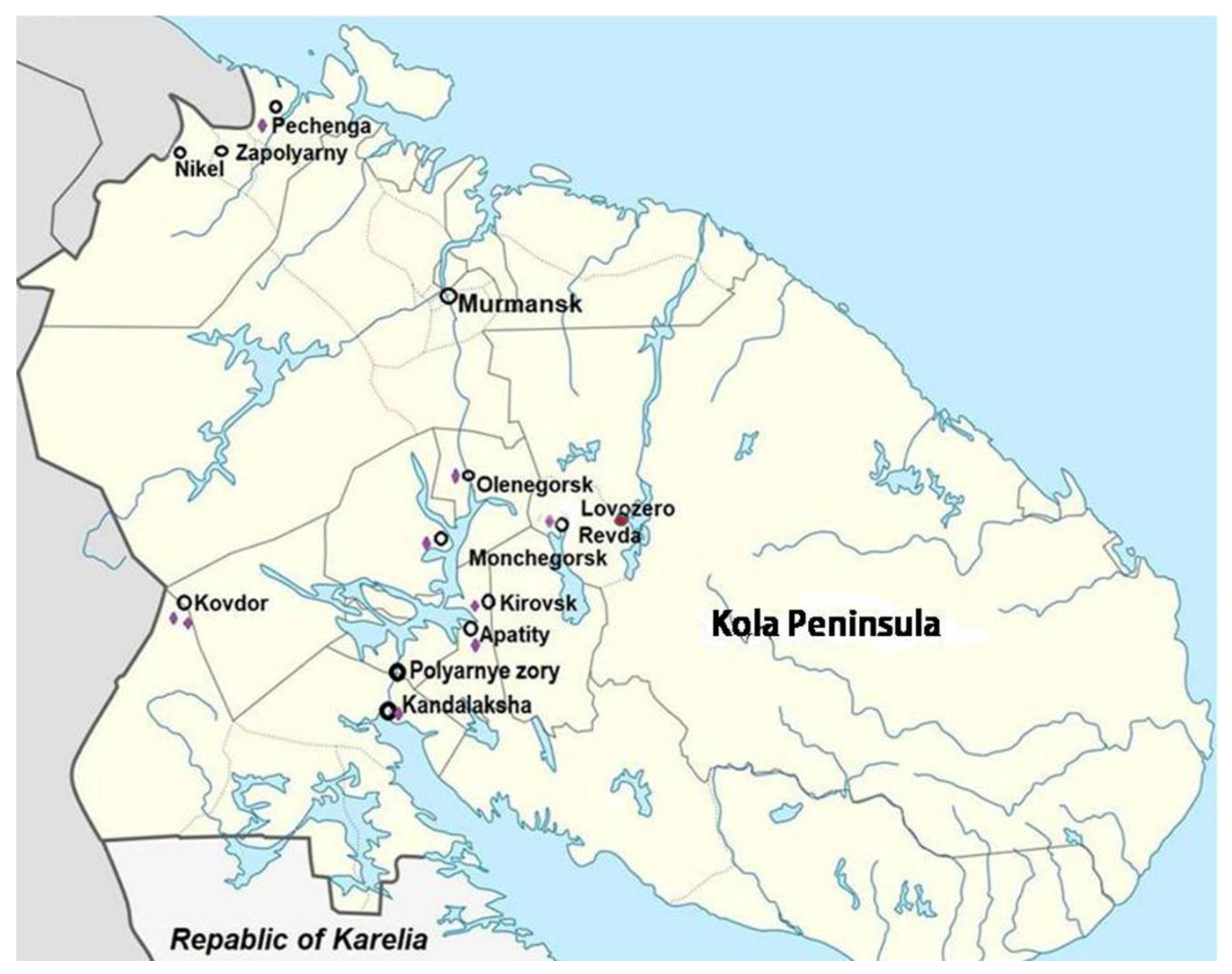
Figure 2.
REE (G.M.) in hair samples from different villages in China: M1 - Shang village, M2 - Liao village, C1 - Dong village, C2 – Ping village, look in text) and from Lovozero village (L) in Russia (Murmansk region), µg/g.
Figure 2.
REE (G.M.) in hair samples from different villages in China: M1 - Shang village, M2 - Liao village, C1 - Dong village, C2 – Ping village, look in text) and from Lovozero village (L) in Russia (Murmansk region), µg/g.
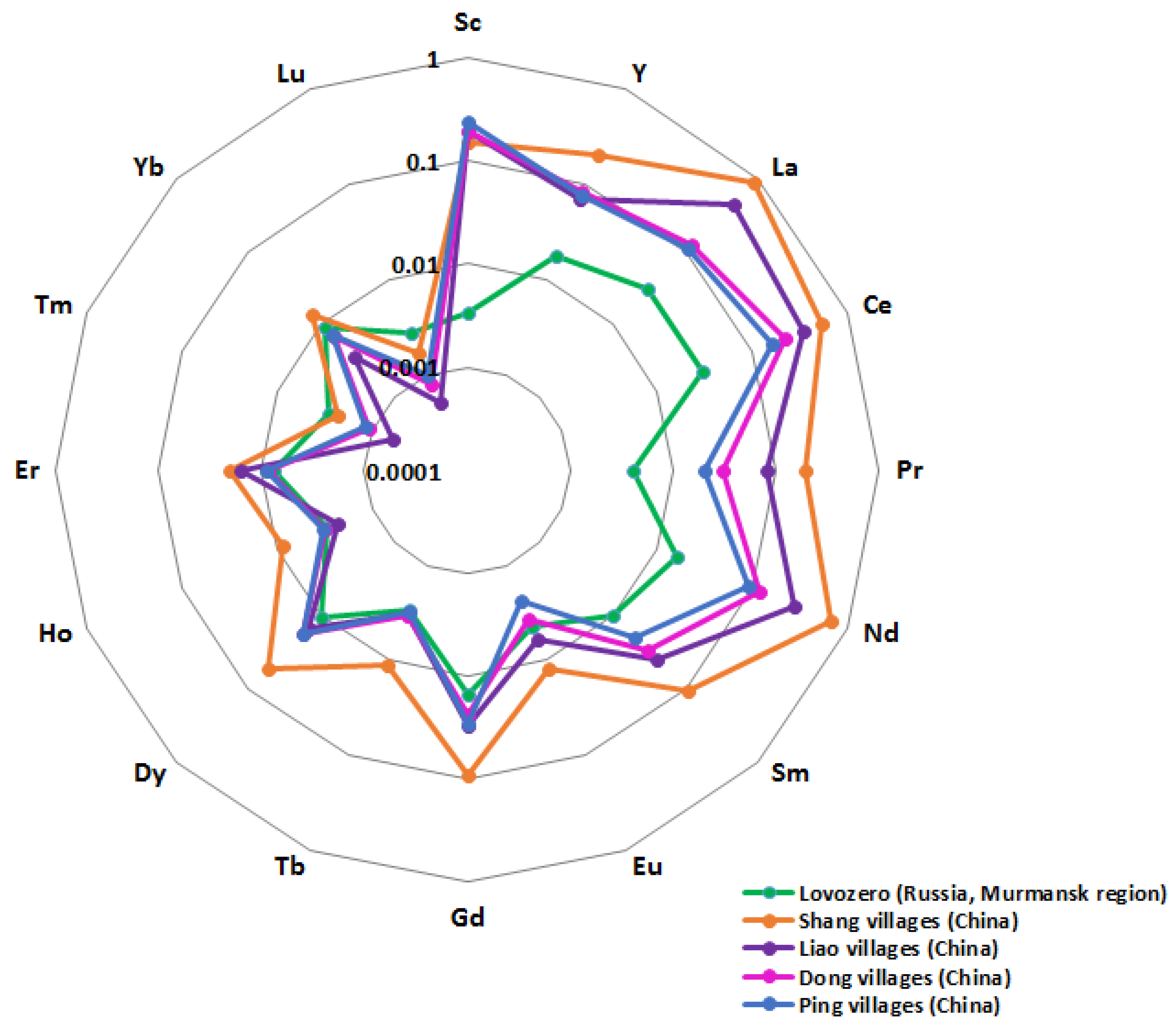
Figure 3.
Average annual values (M±SE) of the prevalence of nervous system disease in children (left column - 0-14, right column -15-17 years old) for period 2018-2020 years: 1, 1 A - Episodic paroxysmal disorders (G40-G47); 2, 2A - Cerebral palsy and other paralytic syndromes (G80-G83); 3, 3A - epilepsy and status epilepticus (G40-G41). Abscissa axis – compare territories, cities: 1 - Murmansk; 2- Apatity; 3 - Kandalaksha; 4 - Monchegorsk; 5 - Olenegorsk; districts: 6 - Kovdorskiy, 7 - Kola; 8 - Lovozerskiy; 9 - Pechenga; 10 - Murmansk Region as a whole. Ordinate axis – values of prevalence of nervous system disease (per 1000 corresponding age population).
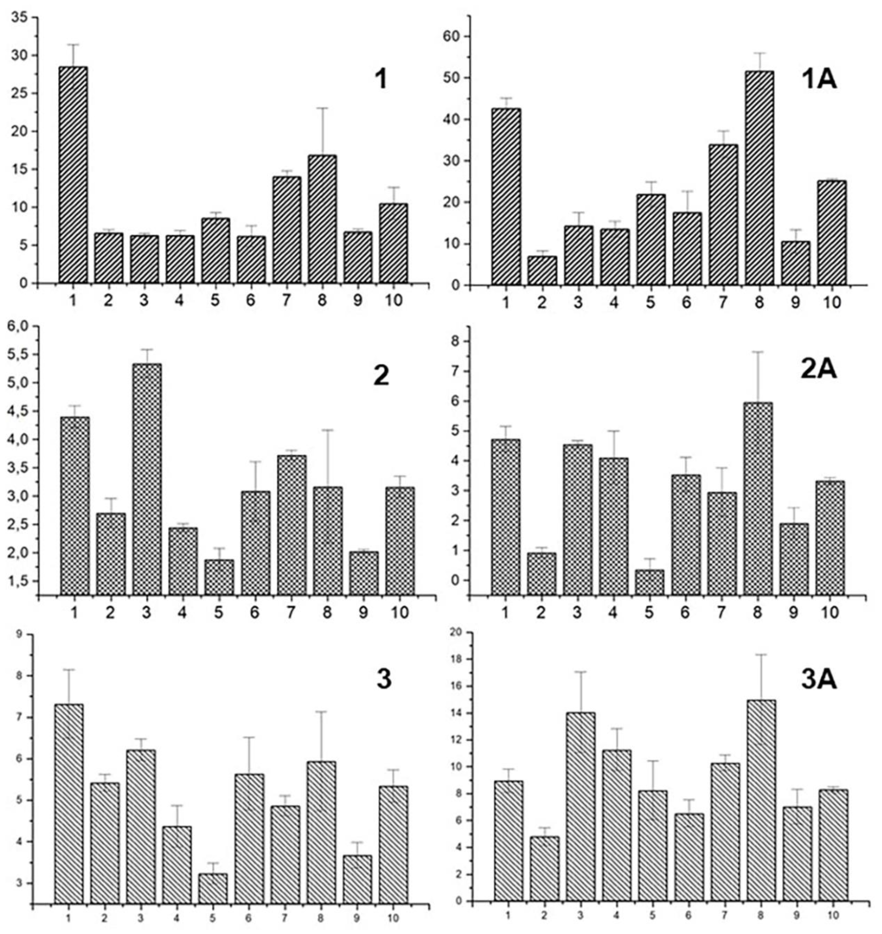
Figure 4.
The localization of brain regions responsible for motor disorders and the accumulation of the certain REEs in these structures. The elements whose presence in the corresponding structures is hypothetical are marked by violet. Figure modified by the authors based on a model of the brain https://i.pinimg.com/originals/f3/22/b0/f322b02e21b31540ff702e91ad4db722.jpg.
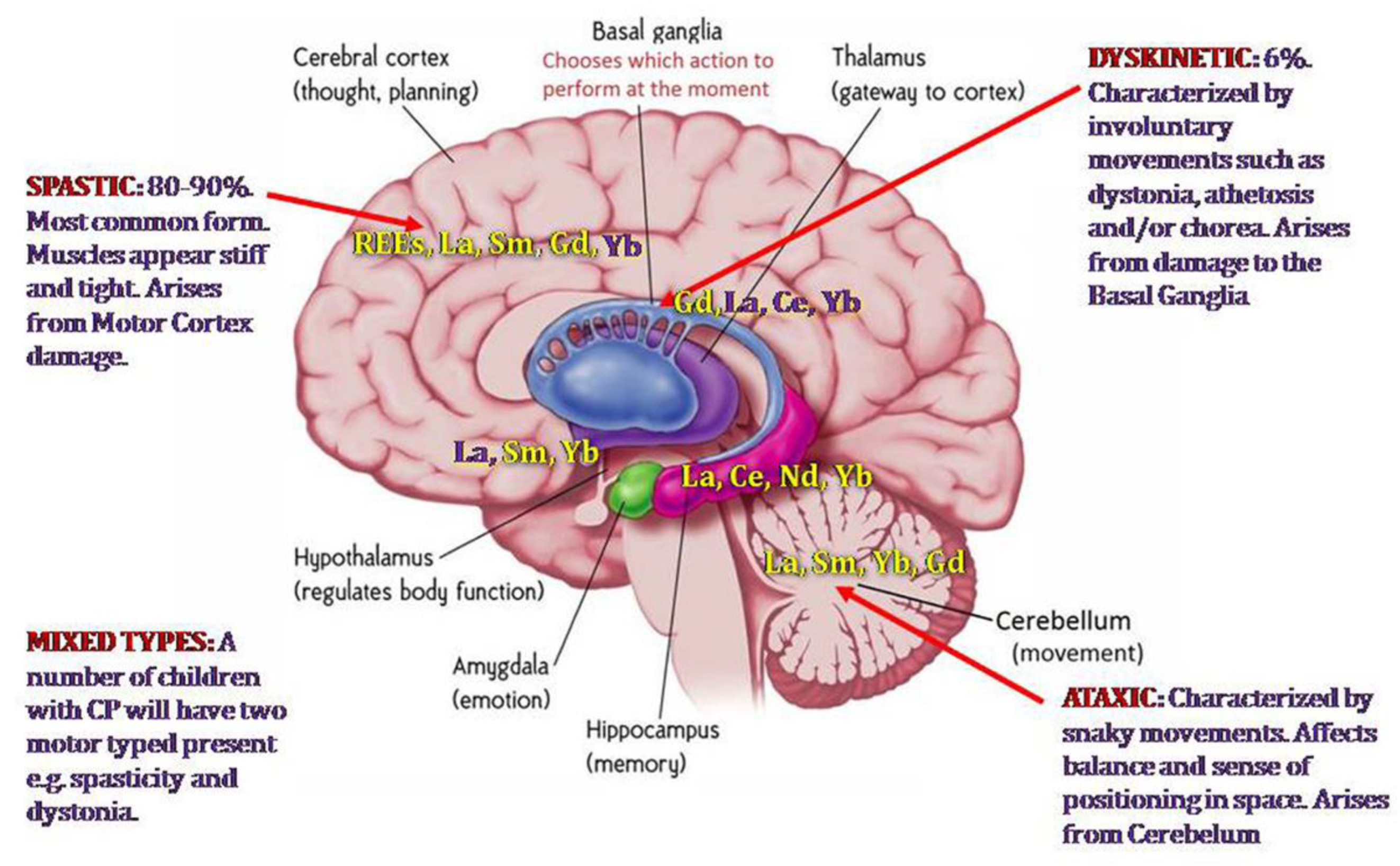

Table 1.
Statistical characteristics of the content of REEs in hair samples from children living in Lovozero village in the vicinity of the mining enterprise.
Table 1.
Statistical characteristics of the content of REEs in hair samples from children living in Lovozero village in the vicinity of the mining enterprise.
| n=53 | Mean | SD | Min | Max. | Median | P25 | P75 | C. V. |
|---|---|---|---|---|---|---|---|---|
| Sc | 0,0536 | 0,1238 | 0,0010 | 0,5544 | 0,0010 | 0,0010 | 0,0010 | 230,9 |
| Y | 0,0204 | 0,0152 | 0,0090 | 0,0782 | 0,0157 | 0,0130 | 0,0193 | 74,6 |
| La | 0,0336 | 0,0222 | 0,0124 | 0,1288 | 0,0269 | 0,0198 | 0,0362 | 66,2 |
| Ce | 0,0351 | 0,0219 | 0,0053 | 0,1073 | 0,0297 | 0,0200 | 0,0412 | 62,3 |
| Pr | 0,0047 | 0,0019 | 0,0020 | 0,0131 | 0,0044 | 0,0038 | 0,0051 | 39,3 |
| Nd | 0,0161 | 0,0048 | 0,0089 | 0,0383 | 0,0154 | 0,0132 | 0,0174 | 29,7 |
| Sm | 0,0124 | 0,0069 | 0,0031 | 0,0548 | 0,0116 | 0,0092 | 0,0141 | 55,7 |
| Eu | 0,0049 | 0,0030 | 0,0017 | 0,0233 | 0,0045 | 0,0036 | 0,0054 | 61,9 |
| Gd | 0,0214 | 0,0024 | 0,0013 | 0,0630 | 0,0163 | 0,0101 | 0,0277 | 76,7 |
| Tb | 0,0032 | 0,0009 | 0,0015 | 0,0046 | 0,0032 | 0,0025 | 0,0040 | 27,3 |
| Dy | 0,0129 | 0,0199 | 0,0055 | 0,1531 | 0,0095 | 0,0080 | 0,0118 | 154,2 |
| Ho | 0,0031 | 0,0010 | 0,0015 | 0,0058 | 0,0033 | 0,0023 | 0,0038 | 30,3 |
| Er | 0,0077 | 0,0017 | 0,0032 | 0,0118 | 0,0076 | 0,0067 | 0,0091 | 22,4 |
| Tm | 0,0029 | 0,0009 | 0,0012 | 0,0050 | 0,0027 | 0,0023 | 0,0035 | 31,3 |
| Yb | 0,0121 | 0,0223 | 0,0037 | 0,1697 | 0,0091 | 0,0072 | 0,0103 | 184,0 |
| Lu | 0,0028 | 0,0012 | 0,0015 | 0,0089 | 0,0027 | 0,0022 | 0,0033 | 43,5 |
Table 2.
Comparisons of some trace and REE contents (µg/g) in children hair samples from Russia (Lovozero village, Murmansk region (1), Zabaykalsky region (2); from Kazakhstan (Pavlodar District, 3) and in adult hair samples from Sweden (4).
Table 2.
Comparisons of some trace and REE contents (µg/g) in children hair samples from Russia (Lovozero village, Murmansk region (1), Zabaykalsky region (2); from Kazakhstan (Pavlodar District, 3) and in adult hair samples from Sweden (4).
| Elements | 1 | 2 | 3 | 4 | |||
| M ± S.D. | Ме (Q25-Q75) | M ± S.D. | Ме (Q25-Q75) | M ± SE | M (S.D.) ME | Range | |
| Na | 642 ± 599 | 431 (275-783) | 728 ± 1198 | 389(143-940) | 223±17 | 147 (149) 94 | 17-670 |
| Ca | 287 ± 175 | 220 (186-349) | 960 ± 2844 | 228(100-673) | 1403±90 | 750 (660) 590 | 113-2890 |
| Sc | 0.054 ±0.124 | 0.001 (0.001-0.001) | 0.02 ± 0.01 | 0.02(0.01-0.02) | 0.007±0.0004 | 0.0014 (0.001) 0.0011 | 0.0004-0.0045 |
| Cr | 4.21 ±13.64 | 4.43 (1.32-6.5) | 5.23 ± 5.69 | 4.68(0.46-6.11) | 0.5±0.07 | 0.167 (0.118) 0.131 | 0.046-0.527 |
| Fe | 24.5 ±18.6 | 23.0 (13.6-29.8) | 853 ± 872 | 793(300-1018) | 45±3.7 | 9.6( 4.4) 8.4 | 4.9-23 |
| Co | 0.030 ±0.043 | 0.008 (0.002-0.047) | 1.05 ± 1.42 | 0.66(0.53-0.9) | 0.07±0.01 | 0.013 ( 0.011) 0.01 | 0.002-0.063 |
| Zn | 120 ±89 | 106 (77-130) | 210 ± 348 | 152(117-194) | 207±8 | 142 (29) 144 | 68-198 |
| As | 0.61 ±0.70 | 0.45 (0.001-0.96) | 0.42 ± 1.02 | 0.24(0.16-0.5) | < 0.8 | 0.085 (0.054) 0.067 | 0.034-0.319 |
| Se | 0.71±0.28 | 0.68 (0.50-0.87) | - | - | 0.8±0.03 | 0.83 (0.28) 0.79 | 0.48-1.84 |
| Br | - | - | 8.82 ± 17.23 | 3.96(2.07-7.91) | 6.5±0.6 | 37 (33) 26 | 5.6-221 |
| Rb | 0.72 ± 0.79 | 0.48 (0.27-1.05) | 0.79 ± 1.24 | 0.3(0.2-0.81) | < 3 | 0.093 (0.085) 0.06 | 0.012-0.482 |
| Sr | 1.38 ±1.65 | 0.59 (0.32-2.06) | 4.53 ± 1.23 | 5(2.5-5) | < 15 | 1.2 (1) 0.97 | 0.14-5.54 |
| Ag | 0.28 ±0.16 | 0.23 (0.19-0.35) | 2.34 ± 17.67 | 0.11(0.05-0.26) | 0.3±0.03 | 0.231 (0.298) 0.132 | 0.025-1.96 |
| Sb | 0.06 ±0.06 | 0.044 (0.028-0.07) | 0.11 ± 0.15 | 0.06(0.03-0.12) | 0.07±0.01 | 0.022 (0.017) 0.017 | 0.007-0.122 |
| Cs | 0.014 ±0.022 | 0.01 (0.005-0.014) | 0.03 ± 0.14 | 0.005(0.001-0.02) | < 0.05 | 0.00067 (0.00046) 0.00051 | 0.00017-0.0019 |
| Ba | 0.58 ±0.88 | 0.41 (0.21-0.60) | 4.07 ± 12.24 | 2(1.97-2.43) | < 10 | 0.64 (0.49) 0.46 | 0.16-1.92 |
| Y | 0.020 ±0.015 | 0.016 (0.013-0.019) | - | - | - | - | - |
| La | 0.034 ±0.022 | 0.027(0.02-0.036) | 0.04 ± 0.09 | 0.02(0.01-0.02) | 0.05±0.006 | 0.035 (0.046) 0.018 | 0.0046-0.106 |
| Ce | 0.035 ±0.022 | 0.03 (0.02-0.041) | 0.39 ± 1.79 | 0.1(0.05-0.1) | < 0.08 | 0.039 (0.05) 0.019 | 0.007-0.164 |
| Pr | 0.005 ±0.002 | 0.004 (0.004-0.005) | - | - | - | ||
| Nd | 0.016 ±0.005 | 0.015 (0.013-0.017) | 0.25 ± 0.46 | 0.09(0.09-0.22) | - | ||
| Sm | 0.0124 ±0.007 | 0.012 (0.009-0.014) | 0.006 ± 0.02 | 0.003(0-0.003) | 0.02±0.003 | ||
| Eu | 0.005 ±0.003 | 0.004 (0.004-0.005) | 0.02 ± 0.07 | 0.004(0.001-0.01) | < 0.03 | ||
| Gd | 0.0214 ±0.016 | 0.016 (0.010-0.028) | - | - | - | ||
| Tb | 0.003 ±0.001 | 0.003 (0.002-0.004) | 0.01 ± 0.02 | 0.005(0.005-0.02) | 0.01±0.0003 | ||
| Dy | 0.013 ±0.02 | 0.01 (0.008-0.012) | - | - | - | ||
| Ho | 0.003 ±0.001 | 0.003 (0.002-0.004) | - | - | - | ||
| Er | 0.008 ±0.002 | 0.008 (0.007-0.009) | - | - | - | ||
| Tm | 0.003±0.001 | 0.003 (0.002-0.004) | - | - | - | ||
| Yb | 0.012 ±0.022 | 0.009 (0.007-0.010) | 0.007 ± 0.009 | 0.005(0-0.009) | 0.03±0.0005 | ||
| Lu | 0.003 ±0.001 | 0.003 (0.002-0.003) | 0.003 ± 0.01 | 0.001(0-0.003) | 0.002±0.0001 | ||
| Hf | 0.004 ±0.005 | 0.004 (0.001-0.007) | 0.05 ± 0.12 | 0.037(0.001-0.05) | 0.02±0.002 | 0.005 (0.008) 0.0017 | 0.0004-0.037 |
| Ta | 0.006 ±0.015 | 0.006 (0.001-0.001) | 0.02 ± 0.04 | 0.008(0-0.008 | < 0.03 | 0.004 (0.003) 0.0031 | <0.002-0.020 |
| Au | 0.012 ±0.021 | 0.012 (0.001-0.017) | 0.03 ± 0.14 | 0.001(0-0.01) | 0.11±0.03 | 0.03 (0.028) 0.017 | 0.003-0.200 |
| Hg | 0.42±0.197 | 0.42 (0.31-0.5) | - | - | 0.4±0.04 | 0.261 (0.145) 0.249 | 0.053-0.927 |
| Th | <0.001 <0.001 | <0.001 | 0.23 ± 0.29 | 0.14(0.05-0.27) | 0.02±0.001 | 0.0013 (0.001) 0.001 | 0.0003-0.0044 |
| U | 0.005 ±0.003 | 0.005 (0.003-0.006) | 0.01 ± 0.01 | 0.01(0.01-0.016) | 0.33±0.07 | 0.057 (0.065) 0.036 | 0.006-0.436 |
Table 3.
Comparisons of REE contents, ng/g, except where noted (*) in children hair samples from Lovozero village (L) in Russia (Murmansk region) and children hair samples from different villages in China (M1, M2, C1, C2 –look in text).
Table 3.
Comparisons of REE contents, ng/g, except where noted (*) in children hair samples from Lovozero village (L) in Russia (Murmansk region) and children hair samples from different villages in China (M1, M2, C1, C2 –look in text).
| L-G.M.(R) | L-M±SE | M1-G.M.(R) | M2-G.M.(R) | C1-G.M.(R) | C2-G.M.(R) | |
| La* | 0.03(0.01-0.13) | 0.033±0.003 | 0.89(0.17-6.93) | 0.45(0.14-0.37) | 0.12(0.05-0.30) | 0.11(0.04-0.40) |
| Ce* | 0.03(0.01-0.11) | 0.035±0.003 | 0.53(0.20-1.37) | 0.34(0.16-0.82) | 0.22(0.09-0.57) | 0.16(0.07-0.44) |
| Pr* | 0.004(0.00-0.01) | 0.005±0.000 | 0.19(0.04-1.55) | 0.08(0.03-0.26) | 0.03(0.01-0.09) | 0.02(0.01-0.09) |
| Nd* | 0.016(0.01-0.04) | 0.016±0.001 | 0.67(0.13-5.27) | 0.28(0.09-0.95) | 0.12(0.04-0.32) | 0.09(0.04-0.32) |
| Sm* | 0.01(0.00-0.05) | 0.012±0.001 | 0.11(0.02-0.91) | 0.04(0.03-0.12) | 0.03(0.01-0.06) | 0.02(0.01-0.07) |
| Eu | 4.4(1.70-23.33) | 4.9±0.04 | 12.1(2.9-95.0) | 6.0(2.6-21.2) | 3.7(1.8-8.7) | 2.4(1.9-6.6) |
| Gd | 15.1(1.29-63.22) | 21.4±0.2 | 90.5(19.4-645.6) | 30.6(12.2-119.0) | 23.7(8.3-59.3) | 29.5(9.8-64.6) |
| Tb | 3.0(1.51-4.65) | 3.17±0.012 | 11.1(2.6-73.8) | 3.3(1.4-13.2) | 3.4(1.3-8.3) | 3.1(1.6-9.7) |
| Dy | 10.3(5.51-153.10) | 12.9±0.27 | 53.5(13.6-338.6) | 15.1(6.4-63.0) | 18.2(7.5-44.1) | 17.8(9.8-54.5) |
| Ho | 3.0(1.53-5.83) | 3.14±0.013 | 8.5(2.2-55.63) | 2.3(1.0-10.7) | 3.1(1.2-7.3) | 3.2(1.7-10.4) |
| Er | 7.5(3.23-11.79) | 7.74±0.024 | 20.7(5.7-134.3) | 15.9(2.7-27.3) | 8.5(3.8-20.1) | 8.8(5.2-28.9) |
| Tm | 2.8(1.24-4.96) | 2.9±0.012 | 2.3(0.6-16.2) | 0.6(0.3-3.1) | 1.1(0.4-2.5) | 1.2(0.6-4.1) |
| Yb | 9.1(3.68-169.73) | 12.1±0.307 | 13.5(4.4-90.5) | 3.6(1.6-18.3) | 6.6(3..6-15.0) | 7.1(3.9-22.3) |
| Lu | 2.7(1.47-8.93) | 2.8±0.017 | 1.7(0.4-13.3) | 0.5(0.2-2.6) | 0.8(0.4-2.0) | 1.0(0.4-3.3) |
| Y | 17.5(0.009-0.0078)* | 20.0±2.0 | 203.5(0.06-1.27)* | 71.2(0.03-0.30)* | 82.3(0.03-0.19)* | 76.5(0.04-0.29)* |
| Sc | 3.4(1.0-554.0) | 53.6±17.0 | 154 | 195.5 | 194.7 | 243.1 |
| *Unit µg/g; other - ng/g; G.M.: geometric mean; R: minimum and maximum values | ||||||
Table 4.
Comparisons of the REEs content (μg/g) in hair samples of children from Lovozero village (Murmansk region, Russia) and in hair samples of 95 children from four villages around the mining area (Maoniuping rare-earth deposit in the Mianning City of Sichuan Province, China).
Table 4.
Comparisons of the REEs content (μg/g) in hair samples of children from Lovozero village (Murmansk region, Russia) and in hair samples of 95 children from four villages around the mining area (Maoniuping rare-earth deposit in the Mianning City of Sichuan Province, China).
| Lovozero village, Russia | 4 village, China | ||||||
|---|---|---|---|---|---|---|---|
| M | P25 | P75 | M | P25 | P75 | ||
| La | 0,0269 | 0,0198 | 0,0362 | 0.3907 | 0.2399 | 1.1505 | |
| Ce | 0,0297 | 0,0200 | 0,0412 | 0.5958 | 0.3811 | 1.6591 | |
| Pr | 0,0044 | 0,0038 | 0,0051 | 0.0489 | 0.0337 | 0.1271 | |
| Nd | 0,0154 | 0,0132 | 0,0174 | 0.1646 | 0.1098 | 0.3574 | |
| Sm | 0,0116 | 0,0092 | 0,0141 | 0.0226 | 0.0150 | 0.0364 | |
| Eu | 0,0045 | 0,0036 | 0,0054 | 0.0060 | 0.0042 | 0.0097 | |
| Gd | 0,0163 | 0,0101 | 0,0277 | 0.0184 | 0.0118 | 0.0296 | |
| Tb | 0,0032 | 0,0025 | 0,0040 | 0.0020 | 0.0009 | 0.0030 | |
| Dy | 0,0095 | 0,0080 | 0,0118 | 0.0112 | 0.0066 | 0.0151 | |
| Ho | 0,0033 | 0,0023 | 0,0038 | 0.0020 | 0.0012 | 0.0027 | |
| Er | 0,0076 | 0,0067 | 0,0091 | 0.0051 | 0.0028 | 0.0071 | |
| Tm | 0,0027 | 0,0023 | 0,0035 | 0.0009 | 0.0005 | 0.0015 | |
| Yb | 0,0091 | 0,0072 | 0,0103 | 0.0049 | 0.0030 | 0.0064 | |
| Lu | 0,0027 | 0,0022 | 0,0033 | 0.0006 | 0.0003 | 0.0010 | |
| Y | 0,0157 | 0,0130 | 0,0193 | 0.0497 | 0.0312 | 0.0672 | |
Disclaimer/Publisher’s Note: The statements, opinions and data contained in all publications are solely those of the individual author(s) and contributor(s) and not of MDPI and/or the editor(s). MDPI and/or the editor(s) disclaim responsibility for any injury to people or property resulting from any ideas, methods, instructions or products referred to in the content. |
© 2024 by the authors. Licensee MDPI, Basel, Switzerland. This article is an open access article distributed under the terms and conditions of the Creative Commons Attribution (CC BY) license (http://creativecommons.org/licenses/by/4.0/).
Copyright: This open access article is published under a Creative Commons CC BY 4.0 license, which permit the free download, distribution, and reuse, provided that the author and preprint are cited in any reuse.


