
Submitted:
03 July 2024
Posted:
04 July 2024
You are already at the latest version
Abstract
Knee osteoarthritis (KOA) is a progressive and multifactorial disease that leads to joint pain, muscle weakness, physical disability, and decreased quality of life. In KOA, hyaluronic acid (HA) quantity and molecular weight (MW) are decreased, leading to joint pain due to increased wear of the knee articular cartilage. Arthrogenic muscle inhibition, which is usually found in patients with KOA, is associated with joint inflammation, pain, and swelling, also causing muscle atrophy, primarily of the anterior thigh muscles, and hindering the rehabilitation process. The aim of our work was to determine if a single HA infiltration could minimize the effects of arthrogenic muscle inhibition in patients with KOA in the short term, using isokinetic dynamometry to evaluate the strength of the knee extensor and flexor muscles of the thigh. Thirty patients with KOA who underwent both clinical and isokinetic assessment, and that received a single injection of HA, were retrospectively included. Our results showed that a single intra-articular injection of HA significantly reduces pain and improves joint function at four weeks, while non-statistically significant improvements were observed for the reference isokinetic parameter (maximum torque) at both 90°/s and 180°/s. Further high-quality studies are necessary to confirm the results of our study.
Keywords:
osteoarthritis
; knee osteoarthritis
; hyaluronic acid
; injection
; isokinetic dynamometer
1. Introduction
Osteoarthritis (OA) is a chronic and degenerative disease affecting the joints, with the knee being the most frequently affected site (about 50% of all OA cases) [1,2,3,4].
KOA is a progressive and multifactorial disease that leads to joint pain, muscle weakness, physical disability, and decreased quality of life. The pathogenic process of OA is characterized by mechanical, inflammatory, and metabolic factors leading to an imbalance between the repair and destruction of joint structures [5,6]. Risk factors for KOA include age, genetics, body weight, sex, ethnicity, and previous injuries (work/sport activities) [7,8,9,10].
During KOA development, the balance between the synthesis and degradation of articular cartilage is mainly influenced by pro-degenerative cytokines such as matrix metalloproteinases (MMPs), interleukins (IL) (e.g., IL-1β, -6, -15), tumor necrosis factor-α (TNF-α), and reactive oxygen species (ROS). These factors degrade the extracellular cartilage matrix (ECM), inhibit the proliferation of articular chondrocytes, and promote their apoptosis, causing an imbalance in the synthesis and degradation of collagen and proteoglycans (PG) in the cartilage matrix, leading to loss of cartilage elasticity, deterioration, and breakdown, eventually resulting in the erosion of the joint surface [11,12].
KOA is mainly diagnosed based on signs and symptoms (pain, functional limitation, hypertrophy, and swelling). According to the new National Institute for Health and Care Excellence (NICE) guidelines, KOA can be diagnosed clinically without imaging in patients aged 45 and older who present with activity-related joint pain [13]. Imaging tests (such as weight-bearing radiographs or magnetic resonance imaging) are indicated only when the presentation is atypical (e.g., prolonged joint stiffness, rest pain) or when there is an unexpected rapid progression of symptoms or a change in clinical characteristics (e.g., swollen and hyperemic knee with nocturnal pain) [14].
The first-line treatment for KOA is conservative and includes non-pharmacological approaches such as physical exercise (strength, aerobic and aquatic, stretching and proprioceptive) [15,16,17,18,19], physiotherapy and physical therapies (transcutaneous electrical nerve stimulation TENS, diathermy, etc.) [20,21,22,23], weight loss (if obese or overweight), use of walking aids or braces (if indicated), education and self-management (maintaining good lifestyle habits, regular exercise, weight control, and avoiding heavy weight-bearing activities), and pharmacological approaches such as paracetamol, non-steroidal anti-inflammatory drugs (NSAIDs) [1,24], chondroprotective agents (glucosamine sulfate and chondroitin sulfate) [25,26,27], and intra-articular therapies using corticosteroids (CS) [15], hyaluronic acid (HA), and platelet-rich plasma (PRP) [28,29,30,31,32].
HA is one of the main components of synovial fluid and plays several key roles in maintaining cartilage health and intra-articular homeostasis [33]. Its rheological and viscoelastic properties provide significant lubricating and shock-absorbing capabilities to the synovial fluid, while its macromolecular size and hydrophilicity help retain fluid in the joint cavity during movements and even after the removal of forces due to mechanical loading [34].
Additionally, HA interacts with pro-inflammatory mediators and, by binding to its cellular receptors, modulates cell proliferation, migration, and gene expression within the joint. The progression of KOA results from the reduction of the molecular weight (MW) (from 6,500-10,900 kDa to 2,700-4,500 kDa) [35] and the concentration of HA in the synovial fluid [34]. This occurs due to hyaluronidases (enzymes that degrade HA) [36,37], ROS, and nitric oxide synthase (NOS) [38]. This degradation of synovial fluid causes joint pain due to increased wear of the knee articular cartilage [35].
Due to the direct association between HA degradation and KOA progression, the intra-articular administration of exogenous HA represents a well-established therapeutic option [39]. Intra-articular HA treatment aims to fill the joint space to counteract the reduction in HA concentration and distribution that occurs with KOA progression [35]. The binding of HA to its CD44 receptor leads to numerous beneficial effects such as chondroprotection, anti-inflammatory effects (thanks to the reduction of IL-1β expression and subsequent reduction of MMPs release), and increased synthesis of PG and glycosaminoglycans (GAGs) [35].
Joint pain and the consequent alteration of daily activities often lead to an overall reduction in motor activity levels. This can result in the weakening of the extensor and flexor muscles of the thigh, with a secondary increase in joint instability and degeneration [40]. Specifically, in KOA, there is up to a 50% reduction in quadriceps strength [41,42]. This occurs partly due to a neural activation deficit of the quadriceps known as arthrogenic muscle inhibition associated with joint inflammation, pain, and swelling [43,44]. Arthrogenic muscle inhibition causes muscle atrophy, primarily of the anterior thigh muscles, and hinders the rehabilitation process [33,44,45,46].
There are various options for assessing lower limb muscle strength in the presence of KOA, including manual muscle testing, handheld dynamometry, and isokinetic dynamometry [47]. A recent literature review has demonstrated that isokinetic testing is a useful and valid tool for assessing muscle strength in patients with KOA [47].
In our study, we hypothesized that a single HA infiltration could minimize the effects of arthrogenic muscle inhibition in patients with KOA in the short term, using isokinetic dynamometry to evaluate the strength of the knee extensor and flexor muscles. Given that scientifically validated questionnaires and rating scales are primarily based on patients' subjective perceptions, our goal was to make them more objective through the use of isokinetic methodology.
2. Materials and Methods
All records containing clinical and radiological information about patients admitted with KOA to the Rehabilitation and Spinal Deformities Unit of the University of Naples Federico II and at the outpatient clinics of the Gruppo Forte in Salerno between May and November 2023 were retrieved from the centers’ databases.
The inclusion criteria were: age between 40 and 70 years and diagnosis of primary KOA according to the American College of Rheumatology (ACR) guidelines [48].
The exclusion criteria were: inflammatory diseases (e.g., rheumatoid arthritis, etc.), joint infections, acute synovitis, neuropathic arthropathy (Charcot), gout, intra-articular tumors, varus or valgus deformities >15°, ligamentous instability, previous major traumas (such as ligament injuries or fractures) and knee surgeries, avascular necrosis, Paget's disease, major dysplasia or congenital anomalies, osteonecrosis, acromegaly, hemochromatosis, Wilson's disease, primary osteochondromatosis, Ehlers-Danlos syndrome, hyperparathyroidism, and hypothyroidism. Additionally, having received intra-articular injections in the knee in the six months prior to the study was also an exclusion criterion.
The patients included in the study received, at the time of assessment, clinical evaluation, followed by an isokinetic strength test, and finally a HA injection.
Age, sex, weight (in kilograms), height (in meters), BMI (kg/m²), employment status (or former employment if retired), physical activity, comorbidities and medications taken, dominant limb, imaging exams performed, knee(s) affected by KOA, and the severity of KOA were retrieved.
Radiological evidence of KOA was assessed using the Kellgren-Lawrence (K-L) classification for radiographs [49] and a classification highly correlated with K-L grades for MRIs [50].
Pain intensity was assessed using the Visual Analogue Scale (VAS) [51], while functional assessment was performed using the Knee injury and Osteoarthritis Outcome Score (KOOS) using its Italian version [52]. Both assessments were conducted at T0 (the day of the first isokinetic test and injection) and at T1 (four weeks after T0).
The isokinetic evaluation of the strength of the extensor and flexor muscles was performed using the Easytech “Prima Doc”® isokinetic dynamometer.
Subjects, after a brief theoretical explanation regarding the isokinetic test, were seated on the machine's chair with their torso and legs well extended, adjusting the inclination (approximately 110°) and depth of the backrest to ensure maximum comfort during the test. The straps at the backrest and along each thigh were applied to help maintain stability and overall safety during the examination (Figure 1).
The evaluation of quadriceps strength was therefore carried out in a seated position with the hip flexed and the leg flexed at 90° on the thigh, with the dynamometer probe placed three cm from the medial malleolus. The ROM used for the test is 0°-90° and was calculated by taking as a reference for the ninetieth degree the position of maximum active extension of each individual subject (then, by flexing the leg at 90° on the knee, the 0 point was obtained, i.e., the starting point of each repetition of the test) (Figure 2).
Before starting the test, gravity compensation was performed according to the machine's software instructions. The protocol, shared and implemented at both centers where the study was conducted, included a warm-up phase followed by a testing phase, which comprised both a strength test (five maximal repetitions - 5RM) and an endurance test (20 maximal repetitions - 20RM).
The warm-up consisted of two sets of 15 repetitions, with the first set "empty" and the second set performed at an angular velocity of 180°/s, advising the examinee to apply sub-maximal contractions to progressively get used to the isokinetic muscle effort.
Five minutes after the warm-up phase, the actual isokinetic test began, which included the strength test (5RM) at an angular velocity of 90°/second and, after a 30-second pause, the endurance test (20RM) at an angular velocity of 180°/second.
Each patient was encouraged in a standardized manner to exert maximum voluntary effort throughout the execution of the tests. If the same patient was a candidate for isokinetic evaluation and HA infiltration in both knees, a five-minute rest was observed between the two isokinetic tests. The order of test execution (e.g., right knee first and then left) at T0 was recorded to repeat it in the same manner at T1.
Among the many parameters that can be analyzed, for this study, we chose to consider the maximum torque (Max MDF) for both the strength and endurance tests. Each test was recorded by the isokinetic software with the relevant graph showing the trend of Max MDF for individual repetitions in relation to the degree of extension.
Patients were treated with a single injection of 60 mg of medium MW HA (1500-2000 kDa)/4 mL (Hyalubrix® 60, Fidia Farmaceutici S.p.A.) [53,54]. The injection was performed approximately 15 minutes after the isokinetic test ended. The injection was performed without ultrasound guidance, with the patient in a supine position and the knee flexed, using a 21G needle (0.8 mm x 40 mm) through the infero-patellar lateral portal (Figure 3) after appropriate skin disinfection with chlorhexidine and povidone-iodine.
In case of post-injection pain, patients were advised to apply ice two to three times a day in the days following the injection and, only in cases of severe pain, to take NSAIDs or pain relievers for no more than three days.
Patients were also recommended not to undergo any physiotherapy and/or therapeutic exercise and to avoid taking NSAIDs or pain relievers (except as mentioned above) between T0 and T1 to avoid affecting the study results.
Data were analyzed using SPSS statistical software (v.29). Results are presented as mean values and standard deviation (SD). Comparisons for VAS, KOOS and Max MDF values between T0 and T1 were performed using Student's t-test [55]. A P-value <0.05 was considered statistically significant.
It was not possible to compare knees with different degrees of radiological severity due to the disparity in sample sizes, as the majority of the evaluated knees (34 out of 46, 74%) had a K-L grade II, with only two knees of grade I and 10 of grade III.
3. Results
Thirty patients and a total of 46 knees were included in the study, with two knees classified as K-L II, 34 as K-L II, and 10 as K-L III. The anthropometric characteristics of the included patients are reported in Table 1 and Table 2.
What can be inferred from Table 1 and Table 2 is that the sample consists predominantly of women (16 out of 30) who have higher average age and BMI compared to men.
Sixteen patients exhibited bilateral knee osteoarthritis (6 M, 10 F), and in 10 of these patients with bilateral osteoarthritis but different K-L grades, the dominant limb was the one with the higher grade.
The changes in VAS between T0 and T1 are reported in Table 3, while the changes in KOOS between T0 and T1 are reported in Table 4.
What can be inferred from Table 3 and Table 4 is that both VAS and KOOS improved significantly between T0 and T1 in all K-L groups, both when patients were evaluated as a whole and within various radiological severity subgroups according to K-L.
4. Discussion
In patients with KOA, the reduction of muscle strength known as arthrogenic muscle inhibition is associated with inflammation, pain, and joint swelling, resulting in reduced joint mobility and muscle atrophy [43,44].
In our study, we hypothesized that a single HA injection could minimize the effects of quadriceps muscle arthrogenic inhibition and help improve conservative treatment of KOA. We designed a prospective study to test the efficacy of intra-articular HA for KOA using the VAS and KOOS scales for pain and joint function, and the isokinetic test to evaluate the strength of knee extensor and flexor muscles.
Our results showed that a single intra-articular HA injection significantly reduced pain and improved joint function at four weeks. These results are comparable to those obtained in other studies using the same type of HA (Hyalubrix®) for KOA [54,56,57,58,59,60,61], even though they followed a protocol of three injections (30 mg/2 mL, one injection per week for three consecutive weeks) instead of a single one as in our study.
The reduction in pain and improvement in joint function could be attributed to the restoration of viscoelasticity and maintenance of knee joint lubrication [62]. Additionally, HA improves chondrocyte survival and stimulates the production of PGs and endogenous HA by suppressing pro-inflammatory factors [63]. The overall result of these effects from intra-articular HA administration is the inhibition of joint inflammation and synovial hypertrophy.
HA also reduces pain through a direct effect on nerve afferents by modulating the sensitivity of nociceptor nerves [63,64]. Furthermore, in some studies, HA has improved proprioception and muscle strength in the isokinetic test in KOA patients [65].
In our study, non-statistically significant improvements were observed for Max MDF at both 90°/s and 180°/s. The exact reason for the (albeit non-statistically significant) improvement in muscle strength in the isokinetic test after intra-articular HA injection is unknown. This could be due to the reduction in friction related to the increased viscosity induced by HA, leading to reduced joint inflammation and pain, and improved joint function and muscle activation [40,65,66].
Only six studies in the literature have evaluated the efficacy of intra-articular HA administration on muscle strength in KOA patients using an isokinetic dynamometer [33,40,65,66,67,68], and a direct comparison of our results with published studies is difficult mainly due to different isokinetic protocols, HA administration protocols, and types of HA used. Additionally, in some studies, the efficacy of HA was not evaluated alone but combined with other therapies, making it impossible to discern the effects of each individual treatment in the combined therapeutic protocol.
In four studies [40,65,66,68], the efficacy of HA alone was evaluated either alone or compared to control groups, while in two studies [33,67], the efficacy of intra-articular HA administration was not evaluated alone but in combination with other treatments.
In these six studies in the literature, pain and functional limitation improved significantly, as also reported in the results of our study. Muscle strength evaluated by isokinetic testing improved in all parameters in three studies [33,40,66], while in the study by Tang et al. [68], strength improved globally at 80°/s but not at 240°/s for the extensor muscles, and in the study by Diracoglu et al. [65], strength improved at 60°/s but not at 180°/s and 240°/s. In the study by Bayramoğlu et al. [67], however, strength did not improve significantly at either 60°/s or 90°/s.
Comparing our results with those in the literature, the only study [67] that included an isokinetic test at 90°/s (5RM, as in our study) did not report significant improvements at short-term follow-up (three weeks, whereas our study's follow-up was four weeks) using both medium and high MW HA. This result is nearly identical to that of our study, although the exact degree of clinical and radiological severity of patients in the referenced study is unknown.
Three studies [33,40,65] included an isokinetic test at 180°/s, with significant strength improvements in two studies at six [40] and eight [33] weeks using the same injection protocol (five injections, one per week) and the same low MW HA. In one study, patients had K-L grades I and II [40], while in the other, they had moderate severity according to Altman [33]. One study [65] did not report significant strength improvements in the isokinetic test at 180°/s at four weeks (as in our study) using three weekly high MW HA injections in patients with K-L II and III. This latter result is nearly identical to that of our study, despite using a different HA and injection protocol.
What can be concluded from the literature is the inconsistency of results regarding changes in muscle strength in isokinetic testing. Indeed, even in studies that evaluated Max MDF at the same angular velocities, using the same type of HA and the same injection protocol, the results were contrasting [65,66,67].
5. Conclusions
Our results showed that a single intra-articular injection of HA significantly reduces pain and improves joint function at four weeks, while non-statistically significant improvements were observed for Max MDF at both 90°/s and 180°/s. Further high-quality studies (such as randomized clinical trials) evaluating the effects of HA administration on muscle strength in comparison with other therapies, with a larger number of patients and longer follow-up periods, are necessary to confirm the results of our study.
Author Contributions
Conceptualization, D.T. and C.R.; methodology, D.T., A.F.M., F.S.; software, A.F.M., A.P., F.S.; validation, D.T., F.S. and C.R.; formal analysis, D.T.; investigation, D.T.; resources, D.T., A.F.M.; data curation, D.T.; writing—original draft preparation, D.T.; writing—review and editing, C.R.; visualization, A.P.; supervision, F.S. All authors have read and agreed to the published version of the manuscript.
Funding
This research received no external funding.
Institutional Review Board Statement
This is a retrospective study, ethical approval not applicable.
Informed Consent Statement
Informed consent was obtained from all subjects involved in the study. Written informed consent has been obtained from the patient(s) to publish this paper.
Conflicts of Interest
The authors declare no conflicts of interest.
References
- Mintarjo, J.A.; Poerwanto, E.; Tedyanto, E.H. Current Non-Surgical Management of Knee Osteoarthritis. Cureus 2023, 15. [Google Scholar] [CrossRef] [PubMed]
- Uivaraseanu, B.; Vesa, C.M.; Tit, D.M.; Abid, A.; Maghiar, O.; Maghiar, T.A.; Hozan, C.; Nechifor, A.C.; Behl, T.; Patrascu, J.M.; et al. Therapeutic Approaches in the Management of Knee Osteoarthritis (Review). Exp Ther Med 2022, 23, 328. [Google Scholar] [CrossRef] [PubMed]
- Hamood, R.; Tirosh, M.; Fallach, N.; Chodick, G.; Eisenberg, E.; Lubovsky, O. Prevalence and Incidence of Osteoarthritis: A Population-Based Retrospective Cohort Study. J Clin Med 2021, 10, 4282. [Google Scholar] [CrossRef] [PubMed]
- Wu, Z.; Zhou, R.; Zhu, Y.; Zeng, Z.; Ye, Z.; Wang, Z.; Liu, W.; Xu, X. Self-Management for Knee Osteoarthritis: A Systematic Review and Meta-Analysis of Randomized Controlled Trials. Pain Res Manag 2022, 2022, 2681240. [Google Scholar] [CrossRef] [PubMed]
- Geng, R.; Li, J.; Yu, C.; Zhang, C.; Chen, F.; Chen, J.; Ni, H.; Wang, J.; Kang, K.; Wei, Z.; et al. Knee Osteoarthritis: Current Status and Research Progress in Treatment (Review). Exp Ther Med 2023, 26, 481. [Google Scholar] [CrossRef] [PubMed]
- Michael, J.W.-P.; Schlüter-Brust, K.U.; Eysel, P. The Epidemiology, Etiology, Diagnosis, and Treatment of Osteoarthritis of the Knee. Dtsch Arztebl Int 2010, 107, 152–162. [Google Scholar] [CrossRef] [PubMed]
- Fuller-Thomson, E.; Stefanyk, M.; Brennenstuhl, S. The Robust Association between Childhood Physical Abuse and Osteoarthritis in Adulthood: Findings from a Representative Community Sample. Arthritis Rheum 2009, 61, 1554–1562. [Google Scholar] [CrossRef] [PubMed]
- Rossignol, M.; Leclerc, A.; Allaert, F.; Rozenberg, S.; Valat, J.; Avouac, B.; Coste, P.; Litvak, E.; Hilliquin, P. Primary Osteoarthritis of Hip, Knee, and Hand in Relation to Occupational Exposure. Occup Environ Med 2005, 62, 772–777. [Google Scholar] [CrossRef] [PubMed]
- Ni, J.; Wang, P.; Yin, K.-J.; Huang, J.-X.; Tian, T.; Cen, H.; Sui, C.; Xu, Z.; Pan, H.-F. Does Smoking Protect against Developing Osteoarthritis? Evidence from a Genetically Informed Perspective. Semin Arthritis Rheum 2022, 55, 152013. [Google Scholar] [CrossRef] [PubMed]
- Morales-Ivorra, I.; Romera-Baures, M.; Roman-Viñas, B.; Serra-Majem, L. Osteoarthritis and the Mediterranean Diet: A Systematic Review. Nutrients 2018, 10, 1030. [Google Scholar] [CrossRef] [PubMed]
- Hsu, H.; Siwiec, R.M. Knee Osteoarthritis. In StatPearls; StatPearls Publishing: Treasure Island (FL), 2023. [Google Scholar]
- Sutipornpalangkul, W.; Morales, N.P.; Harnroongroj, T. Free Radicals in Primary Knee Osteoarthritis. J Med Assoc Thai 2009, 92 Suppl 6, S268-274.
- Overview | Osteoarthritis in over 16s: Diagnosis and Management | Guidance | NICE. Available online: https://www.nice.org.uk/guidance/ng226 (accessed on 19 September 2023).
- Duong, V.; Oo, W.M.; Ding, C.; Culvenor, A.G.; Hunter, D.J. Evaluation and Treatment of Knee Pain: A Review. JAMA 2023, 330, 1568–1580. [Google Scholar] [CrossRef] [PubMed]
- Pesare, E.; Vicenti, G.; Kon, E.; Berruto, M.; Caporali, R.; Moretti, B.; Randelli, P.S. Italian Orthopaedic and Traumatology Society (SIOT) Position Statement on the Non-Surgical Management of Knee Osteoarthritis. J Orthop Traumatol 2023, 24, 47. [Google Scholar] [CrossRef] [PubMed]
- Zeng, C.-Y.; Zhang, Z.-R.; Tang, Z.-M.; Hua, F.-Z. Benefits and Mechanisms of Exercise Training for Knee Osteoarthritis. Front Physiol 2021, 12, 794062. [Google Scholar] [CrossRef] [PubMed]
- Wellsandt, E.; Golightly, Y. Exercise in the Management of Knee and Hip Osteoarthritis. Curr Opin Rheumatol 2018, 30, 151–159. [Google Scholar] [CrossRef] [PubMed]
- Li, Y.; Su, Y.; Chen, S.; Zhang, Y.; Zhang, Z.; Liu, C.; Lu, M.; Liu, F.; Li, S.; He, Z.; et al. The Effects of Resistance Exercise in Patients with Knee Osteoarthritis: A Systematic Review and Meta-Analysis. Clin Rehabil 2016, 30, 947–959. [Google Scholar] [CrossRef] [PubMed]
- Tarantino, D.; Theysmans, T.; Mottola, R.; Verbrugghe, J. High-Intensity Training for Knee Osteoarthritis: A Narrative Review. Sports (Basel) 2023, 11, 91. [Google Scholar] [CrossRef] [PubMed]
- van Doormaal, M.C.M.; Meerhoff, G.A.; Vliet Vlieland, T.P.M.; Peter, W.F. A Clinical Practice Guideline for Physical Therapy in Patients with Hip or Knee Osteoarthritis. Musculoskeletal Care 2020, 18, 575–595. [Google Scholar] [CrossRef] [PubMed]
- Peter, W.F.; Jansen, M.J.; Hurkmans, E.J.; Bloo, H.; Dekker, J.; Dilling, R.G.; Hilberdink, W.; Kersten-Smit, C.; de Rooij, M.; Veenhof, C.; et al. Physiotherapy in Hip and Knee Osteoarthritis: Development of a Practice Guideline Concerning Initial Assessment, Treatment and Evaluation. Acta Reumatol Port 2011, 36, 268–281. [Google Scholar] [PubMed]
- Berteau, J.-P. Knee Pain from Osteoarthritis: Pathogenesis, Risk Factors, and Recent Evidence on Physical Therapy Interventions. Journal of Clinical Medicine 2022, 11, 3252. [Google Scholar] [CrossRef] [PubMed]
- Pietrosimone, B.; Luc-Harkey, B.A.; Harkey, M.S.; Davis-Wilson, H.C.; Pfeiffer, S.J.; Schwartz, T.A.; Nissman, D.; Padua, D.A.; Blackburn, J.T.; Spang, J.T. Using TENS to Enhance Therapeutic Exercise in Individuals with Knee Osteoarthritis. Medicine & Science in Sports & Exercise 2020, 52, 2086. [Google Scholar] [CrossRef]
- Nelson, A.E.; Allen, K.D.; Golightly, Y.M.; Goode, A.P.; Jordan, J.M. A Systematic Review of Recommendations and Guidelines for the Management of Osteoarthritis: The Chronic Osteoarthritis Management Initiative of the U.S. Bone and Joint Initiative. Semin Arthritis Rheum 2014, 43, 701–712. [Google Scholar] [CrossRef] [PubMed]
- Sawitzke, A.D.; Shi, H.; Finco, M.F.; Dunlop, D.D.; Harris, C.L.; Singer, N.G.; Bradley, J.D.; Silver, D.; Jackson, C.G.; Lane, N.E.; et al. Clinical Efficacy and Safety of Glucosamine, Chondroitin Sulphate, Their Combination, Celecoxib or Placebo Taken to Treat Osteoarthritis of the Knee: 2-Year Results from GAIT. Ann Rheum Dis 2010, 69, 1459–1464. [Google Scholar] [CrossRef] [PubMed]
- Bishnoi, M.; Jain, A.; Hurkat, P.; Jain, S.K. Chondroitin Sulphate: A Focus on Osteoarthritis. Glycoconj J 2016, 33, 693–705. [Google Scholar] [CrossRef] [PubMed]
- Tarantino, D.; Sirico, F.; Corrado, B.; Ruosi, C. Intra-Articular Hyaluronic Acid Injections and Oral Collagen Supplementation for Knee OA: A Case Report of an Elite Female Soccer Player. Journal of Human Sport and Exercise: JHSE 2021, 16, 1384–1394. [Google Scholar]
- Szwedowski, D.; Szczepanek, J.; Paczesny, Ł.; Zabrzyński, J.; Gagat, M.; Mobasheri, A.; Jeka, S. The Effect of Platelet-Rich Plasma on the Intra-Articular Microenvironment in Knee Osteoarthritis. International Journal of Molecular Sciences 2021, 22, 5492. [Google Scholar] [CrossRef] [PubMed]
- Zhang, H.-F.; Wang, C.-G.; Li, H.; Huang, Y.-T.; Li, Z.-J. Intra-Articular Platelet-Rich Plasma versus Hyaluronic Acid in the Treatment of Knee Osteoarthritis: A Meta-Analysis. Drug Des Devel Ther 2018, 12, 445–453. [Google Scholar] [CrossRef] [PubMed]
- Duymus, T.M.; Mutlu, S.; Dernek, B.; Komur, B.; Aydogmus, S.; Kesiktas, F.N. Choice of Intra-Articular Injection in Treatment of Knee Osteoarthritis: Platelet-Rich Plasma, Hyaluronic Acid or Ozone Options. Knee Surg Sports Traumatol Arthrosc 2017, 25, 485–492. [Google Scholar] [CrossRef] [PubMed]
- Tarantino, D.; Mottola, R.; Palermi, S.; Sirico, F.; Corrado, B.; Gnasso, R. Intra-Articular Collagen Injections for Osteoarthritis: A Narrative Review. Int J Environ Res Public Health 2023, 20, 4390. [Google Scholar] [CrossRef] [PubMed]
- de Sire, A.; Lippi, L.; Mezian, K.; Calafiore, D.; Pellegrino, R.; Mascaro, G.; Cisari, C.; Invernizzi, M. Ultrasound-Guided Platelet-Rich-Plasma Injections for Reducing Sacroiliac Joint Pain: A Paradigmatic Case Report and Literature Review. J Back Musculoskelet Rehabil 2022, 35, 977–982. [Google Scholar] [CrossRef] [PubMed]
- Huang, M.-H.; Yang, R.-C.; Lee, C.-L.; Chen, T.-W.; Wang, M.-C. Preliminary Results of Integrated Therapy for Patients with Knee Osteoarthritis. Arthritis Rheum 2005, 53, 812–820. [Google Scholar] [CrossRef] [PubMed]
- Nicholls, M.; Manjoo, A.; Shaw, P.; Niazi, F.; Rosen, J. Rheological Properties of Commercially Available Hyaluronic Acid Products in the United States for the Treatment of Osteoarthritis Knee Pain. Clin Med Insights Arthritis Musculoskelet Disord 2018, 11, 1179544117751622. [Google Scholar] [CrossRef] [PubMed]
- Altman, R.D.; Manjoo, A.; Fierlinger, A.; Niazi, F.; Nicholls, M. The Mechanism of Action for Hyaluronic Acid Treatment in the Osteoarthritic Knee: A Systematic Review. BMC Musculoskelet Disord 2015, 16, 321. [Google Scholar] [CrossRef] [PubMed]
- Kaul, A.; Short, W.D.; Wang, X.; Keswani, S.G. Hyaluronidases in Human Diseases. Int J Mol Sci 2021, 22, 3204. [Google Scholar] [CrossRef] [PubMed]
- Stern, R.; Jedrzejas, M.J. Hyaluronidases: Their Genomics, Structures, and Mechanisms of Action. Chem Rev 2006, 106, 818–839. [Google Scholar] [CrossRef] [PubMed]
- Belcher, C.; Yaqub, R.; Fawthrop, F.; Bayliss, M.; Doherty, M. Synovial Fluid Chondroitin and Keratan Sulphate Epitopes, Glycosaminoglycans, and Hyaluronan in Arthritic and Normal Knees. Annals of the Rheumatic Diseases 1997, 56, 299–307. [Google Scholar] [CrossRef] [PubMed]
- Ferkel, E.; Manjoo, A.; Martins, D.; Bhandari, M.; Sethi, P.; Nicholls, M. Intra-Articular Hyaluronic Acid Treatments for Knee Osteoarthritis: A Systematic Review of Product Properties. Cartilage 2023, 19476035231154530. [Google Scholar] [CrossRef] [PubMed]
- Miltner, O.; Schneider, U.; Siebert, C.H.; Niedhart, C.; Niethard, F.U. Efficacy of Intraarticular Hyaluronic Acid in Patients with Osteoarthritis--a Prospective Clinical Trial. Osteoarthritis Cartilage 2002, 10, 680–686. [Google Scholar] [CrossRef] [PubMed]
- Al-Johani, A.H.; Kachanathu, S.J.; Ramadan Hafez, A.; Al-Ahaideb, A.; Algarni, A.D.; Meshari Alroumi, A.; Alenazi, A.M. Comparative Study of Hamstring and Quadriceps Strengthening Treatments in the Management of Knee Osteoarthritis. J Phys Ther Sci 2014, 26, 817–820. [Google Scholar] [CrossRef]
- Stevens, J.E.; Mizner, R.L.; Snyder-Mackler, L. Quadriceps Strength and Volitional Activation before and after Total Knee Arthroplasty for Osteoarthritis. J Orthop Res 2003, 21, 775–779. [Google Scholar] [CrossRef]
- Rice, D.A.; McNair, P.J. Quadriceps Arthrogenic Muscle Inhibition: Neural Mechanisms and Treatment Perspectives. Semin Arthritis Rheum 2010, 40, 250–266. [Google Scholar] [CrossRef] [PubMed]
- Rice, D.A.; McNair, P.J.; Lewis, G.N.; Dalbeth, N. Quadriceps Arthrogenic Muscle Inhibition: The Effects of Experimental Knee Joint Effusion on Motor Cortex Excitability. Arthritis Res Ther 2014, 16, 502. [Google Scholar] [CrossRef] [PubMed]
- Demeco, A.; de Sire, A.; Marotta, N.; Palumbo, A.; Fragomeni, G.; Gramigna, V.; Pellegrino, R.; Moggio, L.; Petraroli, A.; Iona, T.; et al. Effectiveness of Rehabilitation through Kinematic Analysis of Upper Limb Functioning in Wheelchair Basketball Athletes: A Pilot Study. Applied Sciences 2022, 12, 2929. [Google Scholar] [CrossRef]
- Brindisino, F.; Garzonio, F.; DI Giacomo, G.; Pellegrino, R.; Olds, M.; Ristori, D. Depression, Fear of Re-Injury and Kinesiophobia Resulted in Worse Pain, Quality of Life, Function and Level of Return to Sport in Patients with Shoulder Instability: A Systematic Review. J Sports Med Phys Fitness 2023, 63, 598–607. [Google Scholar] [CrossRef] [PubMed]
- Myers, B.J. Isokinetic Testing of Muscle Strength in Older Adults with Knee Osteoarthritis: An Integrative Review. Isokinetics and Exercise Science 2020, 28, 269–290. [Google Scholar] [CrossRef]
- Peat, G.; Thomas, E.; Duncan, R.; Wood, L.; Hay, E.; Croft, P. Clinical Classification Criteria for Knee Osteoarthritis: Performance in the General Population and Primary Care. Ann Rheum Dis 2006, 65, 1363–1367. [Google Scholar] [CrossRef] [PubMed]
- Kellgren, J.H.; Lawrence, J.S. Radiological Assessment of Osteo-Arthrosis. Ann Rheum Dis 1957, 16, 494–502. [Google Scholar] [CrossRef] [PubMed]
- Park, H.-J.; Kim, S.S.; Lee, S.-Y.; Park, N.-H.; Park, J.-Y.; Choi, Y.-J.; Jeon, H.-J. A Practical MRI Grading System for Osteoarthritis of the Knee: Association with Kellgren–Lawrence Radiographic Scores. European Journal of Radiology 2013, 82, 112–117. [Google Scholar] [CrossRef] [PubMed]
- Åström, M.; Thet Lwin, Z.M.; Teni, F.S.; Burström, K.; Berg, J. Use of the Visual Analogue Scale for Health State Valuation: A Scoping Review. Qual Life Res 2023, 32, 2719–2729. [Google Scholar] [CrossRef] [PubMed]
- Monticone, M.; Ferrante, S.; Salvaderi, S.; Rocca, B.; Totti, V.; Foti, C.; Roi, G.S. Development of the Italian Version of the Knee Injury and Osteoarthritis Outcome Score for Patients with Knee Injuries: Cross-Cultural Adaptation, Dimensionality, Reliability, and Validity. Osteoarthritis and Cartilage 2012, 20, 330–335. [Google Scholar] [CrossRef] [PubMed]
- Lu, K.-H.; Lu, P.W.-A.; Lin, C.-W.; Lu, E.W.-H.; Yang, S.-F. Different Molecular Weights of Hyaluronan Research in Knee Osteoarthritis: A State-of-the-Art Review. Matrix Biol 2023, 117, 46–71. [Google Scholar] [CrossRef] [PubMed]
- Guidolin, D.; Franceschi, F. Viscosupplementation with High Molecular Weight Native Hyaluronan. Focus on a 1500-2000 KDa Fraction (Hyalubrix®). Eur Rev Med Pharmacol Sci 2014, 18, 3326–3338. [Google Scholar] [PubMed]
- Mishra, P.; Singh, U.; Pandey, C.M.; Mishra, P.; Pandey, G. Application of Student’s t-Test, Analysis of Variance, and Covariance. Ann Card Anaesth 2019, 22, 407–411. [Google Scholar] [CrossRef] [PubMed]
- Foti, C.; Cisari, C.; Carda, S.; Giordan, N.; Rocco, A.; Frizziero, A.; Della Bella, G. A Prospective Observational Study of the Clinical Efficacy and Safety of Intra-Articular Sodium Hyaluronate in Synovial Joints with Osteoarthritis. Eur J Phys Rehabil Med 2011, 47, 407–415. [Google Scholar] [PubMed]
- Filardo, G.; Kon, E.; Di Martino, A.; Di Matteo, B.; Merli, M.L.; Cenacchi, A.; Fornasari, P.M.; Marcacci, M. Platelet-Rich Plasma vs Hyaluronic Acid to Treat Knee Degenerative Pathology: Study Design and Preliminary Results of a Randomized Controlled Trial. BMC Musculoskelet Disord 2012, 13, 229. [Google Scholar] [CrossRef] [PubMed]
- Filardo, G.; Di Matteo, B.; Di Martino, A.; Merli, M.L.; Cenacchi, A.; Fornasari, P.; Marcacci, M.; Kon, E. Platelet-Rich Plasma Intra-Articular Knee Injections Show No Superiority Versus Viscosupplementation: A Randomized Controlled Trial. Am J Sports Med 2015, 43, 1575–1582. [Google Scholar] [CrossRef] [PubMed]
- Galluccio, F.; Gazar, Y.A.; Negm, A.A.; Fajardo Perez, M.; Yamak Altinpulluk, E.; Ergönenç, T.; Chang, K.-V.; Pan, J.L.; Allam, A.E.-S. The Booster Effect of a Single Quarterly Dose of Hyaluronic Acid in Knee Osteoarthritis: Five-Year Results of a Registry-Based Study. Cureus 2022, 14, e31592. [Google Scholar] [CrossRef] [PubMed]
- Giarratana, L.S.; Marelli, B.M.; Crapanzano, C.; De Martinis, S.E.; Gala, L.; Ferraro, M.; Marelli, N.; Albisetti, W. A Randomized Double-Blind Clinical Trial on the Treatment of Knee Osteoarthritis: The Efficacy of Polynucleotides Compared to Standard Hyaluronian Viscosupplementation. Knee 2014, 21, 661–668. [Google Scholar] [CrossRef] [PubMed]
- Cudoni, S.; Zedde, P. Trattamento conservativo della gonartrosi: Platelet Rich Plasma vs acido ialuronico. LO SCALPELLO 2019, 33, 226–229. [Google Scholar] [CrossRef]
- Gupta, R.C.; Lall, R.; Srivastava, A.; Sinha, A. Hyaluronic Acid: Molecular Mechanisms and Therapeutic Trajectory. Front Vet Sci 2019, 6, 192. [Google Scholar] [CrossRef] [PubMed]
- Moreland, L.W. Intra-Articular Hyaluronan (Hyaluronic Acid) and Hylans for the Treatment of Osteoarthritis: Mechanisms of Action. Arthritis Res Ther 2003, 5, 54–67. [Google Scholar] [CrossRef]
- Migliore, A.; Procopio, S. Effectiveness and Utility of Hyaluronic Acid in Osteoarthritis. Clin Cases Miner Bone Metab 2015, 12, 31–33. [Google Scholar] [CrossRef] [PubMed]
- Diracoglu, D.; Vural, M.; Baskent, A.; Dikici, F.; Aksoy, C. The Effect of Viscosupplementation on Neuromuscular Control of the Knee in Patients with Osteoarthritis. J Back Musculoskelet Rehabil 2009, 22, 1–9. [Google Scholar] [CrossRef] [PubMed]
- Maia, P.A.V.; Cossich, V.R.A.; Salles-Neto, J.I.; Aguiar, D.P.; de Sousa, E.B. Viscosupplementation Improves Pain, Function and Muscle Strength, but Not Proprioception, in Patients with Knee Osteoarthritis: A Prospective Randomized Trial. Clinics (Sao Paulo) 2019, 74, e1207. [Google Scholar] [CrossRef] [PubMed]
- Bayramoğlu, M.; Karataş, M.; Cetin, N.; Akman, N.; Sözay, S.; Dilek, A. Comparison of Two Different Viscosupplements in Knee Osteoarthritis -- a Pilot Study. Clin Rheumatol 2003, 22, 118–122. [Google Scholar] [CrossRef] [PubMed]
- Tang, S.F.T.; Chen, C.P.C.; Chen, M.J.L.; Hong, W.-H.; Yu, T.-Y.; Tsai, W.-C. Improvement of Muscle Strength in Osteoarthritic Knee Patients after Intraarticular Knee Injection of Hyaluronan. Am J Phys Med Rehabil 2005, 84, 274–277. [Google Scholar] [CrossRef] [PubMed]
Figure 1.
Patient's position on the isokinetic dynamometer.
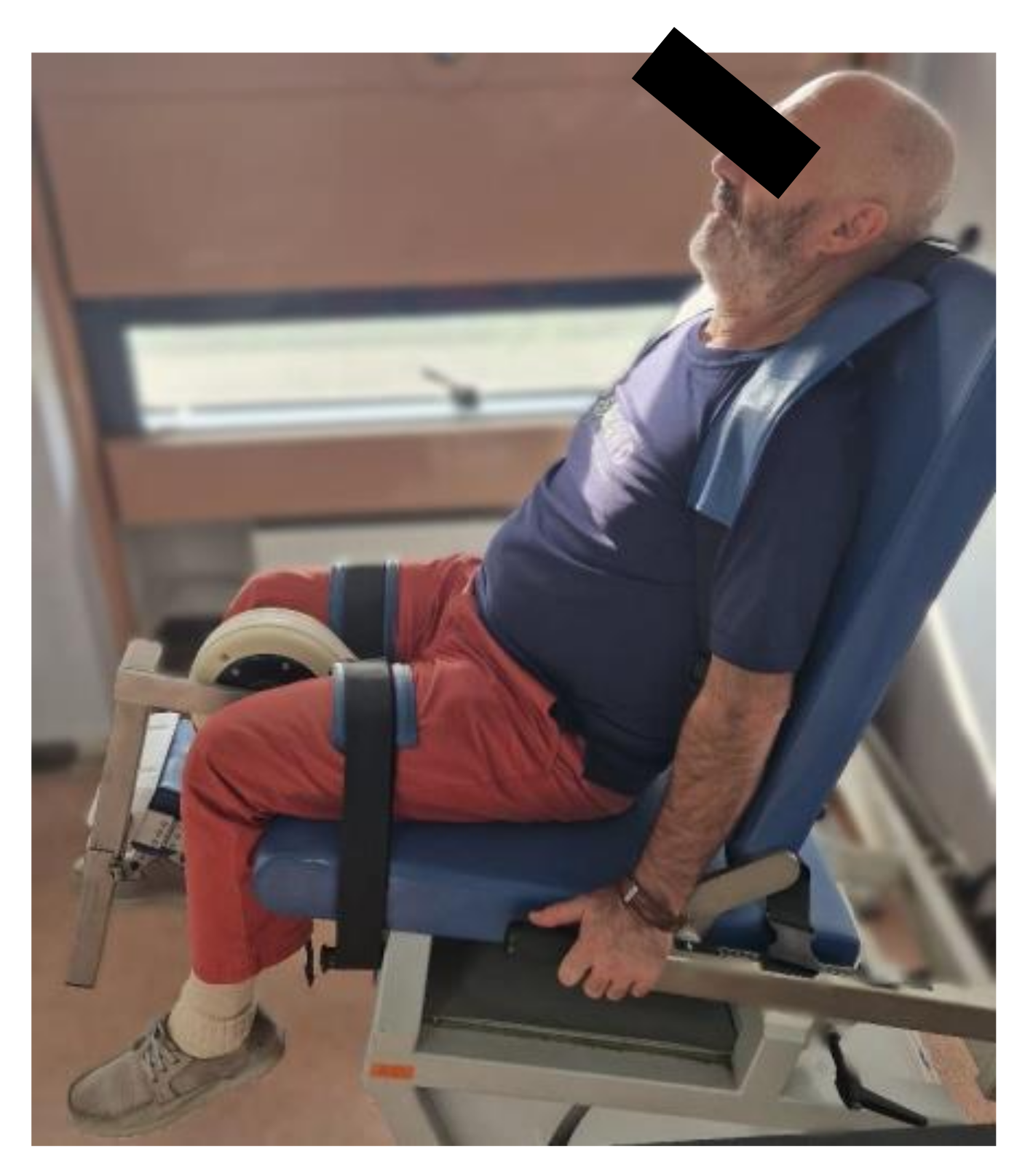
Figure 2.
Positioning of the limb to be tested for isokinetic evaluation at the 0 point (maximum extension).
Figure 2.
Positioning of the limb to be tested for isokinetic evaluation at the 0 point (maximum extension).
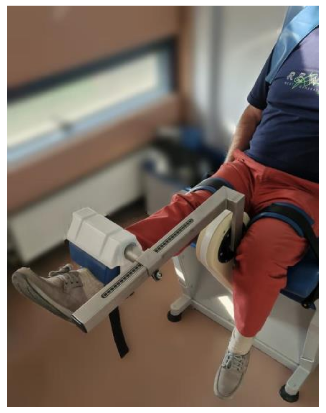
Figure 3.
Injection procedure through the infero-patellar lateral portal.
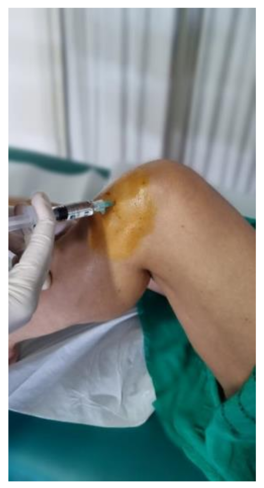

Table 1.
Anthropometric Data. The values are presented as mean and standard deviation (in parentheses).
Table 1.
Anthropometric Data. The values are presented as mean and standard deviation (in parentheses).
| Total sample (n=30) | Males (n=14) | Females (n=16) | |
|---|---|---|---|
| Age | 58.35 (8.82) | 52.57 (8.6) | 62.63 (5) |
| BMI (kg/m2) | 26.42 (2.71) | 25.34 (2.11) | 27.37 (2.95) |
| Knees (DX:SX) | 26:20 | 12:8 | 14:12 |
| Active workers | 22/30 | 14/14 | 8/16 |
| Physical exercise | 14/30 | 6/14 | 8/16 |
Table 2.
Anthropometric Data and Correlation with K-L Severity Grades. The values are presented as mean and standard deviation (in parentheses).
Table 2.
Anthropometric Data and Correlation with K-L Severity Grades. The values are presented as mean and standard deviation (in parentheses).
| K-L I | K-L II | K-L III | |
|---|---|---|---|
| Knees (DX:SX) | 0:2 | 16:18 | 10:0 |
| Age | 40 | 58.29 (8.64) | 62.2 (5.12) |
| Sex (M:F) | 2:0 | 12:16 | 4:6 |
| BMI (kg/m2) | 26.47 | 26.65 (2.61) | 27.38 (3.14) |
| Active workers | 2/2 | 20/28 | 8/10 |
| Physical exercise | 0/2 | 14/28 | 4/10 |
Table 3.
Changes in VAS between T0 and T1. The values are reported as mean and standard deviation (in parentheses). An asterisk indicates the presence of a statistically significant difference.
Table 3.
Changes in VAS between T0 and T1. The values are reported as mean and standard deviation (in parentheses). An asterisk indicates the presence of a statistically significant difference.
| T0 | T1 | p-value | |
|---|---|---|---|
| VAS (all K-L groups) | 4.7 (2.1) | 1.6 (1.34) * | <0,001 |
| VAS (K-L I) | 2 | 0 | / |
| VAS (K-L II) | 4.6 (1.9) | 1.6 (1.37) * | <0,001 |
| VAS (K-L III) | 5.8 (2) | 1.8 (1.3) * | 0,006 |
Table 4.
Changes in KOOS between T0 and T1. The values are reported as mean and standard deviation (in parentheses). An asterisk indicates the presence of a statistically significant difference.
Table 4.
Changes in KOOS between T0 and T1. The values are reported as mean and standard deviation (in parentheses). An asterisk indicates the presence of a statistically significant difference.
| T0 | T1 | p-value | |
|---|---|---|---|
| KOOS (all K-L groups) | 75% (0,1) | 91% (0,1) * | <0,001 |
| KOOS (K-L I) | 96% | 100% | / |
| KOOS (K-L II) | 76% (0,1) | 91% (0,1) * | <0,001 |
| KOOS (K-L III) | 70% (0,1) | 91% (0,1) * | 0,001 |
Table 5.
Changes in Max MDF of extensor (EXT) and flexor (FLX) muscles at 5RM (90°/s). The values are reported as mean and standard deviation (in parentheses).
Table 5.
Changes in Max MDF of extensor (EXT) and flexor (FLX) muscles at 5RM (90°/s). The values are reported as mean and standard deviation (in parentheses).
| T0 | T1 | p-values | |
|---|---|---|---|
| EXT:FLX (N/m) (all K-L groups) | 88.7(22.8):50.1 (18.5) | 92.3 (31.7):51 (22.5) | 0.66:0.87 |
| EXT:FLX (N/m) (K-L I) | 142:101 | 171:119 | / |
| EXT:FLX (N/m) (K-L II) | 88.8 (21.7):48.7 (15.7) | 89.6 (27.6):49.2 (18.7) | 0.93:0.93 |
| EXT:FLX (N/m) (K-L III) | 77.4 (10.8):44.6 (14.2) | 85.8 (29):43.2 (11.3) | 0.58:0.87 |
Table 6.
Changes in Max MDF of extensor (EXT) and flexor (FLX) muscles at 20RM (180°/s). The values are reported as mean and standard deviation (in parentheses).
Table 6.
Changes in Max MDF of extensor (EXT) and flexor (FLX) muscles at 20RM (180°/s). The values are reported as mean and standard deviation (in parentheses).
| T0 | T1 | p-values | |
|---|---|---|---|
| EXT:FLX (N/m) (all K-L groups) | 68.3 (17.7):42.5 (16) | 69.7(18.7):41.7 (12.7) | 0.87:0.8 |
| EXT:FLX (N/m) (K-L I) | 77:37 | 90:44 | / |
| EXT:FLX (N/m) (K-L II) | 69 (18.2):43.5 (15.8) | 68.4(17.5):42.5 (13.3) | 0.93:0.85 |
| EXT:FLX (N/m) (K-L III) | 64.2 (17.2):40 (18) | 70.2 (24.3):38.4(12.7) | 0.67:0.87 |
Disclaimer/Publisher’s Note: The statements, opinions and data contained in all publications are solely those of the individual author(s) and contributor(s) and not of MDPI and/or the editor(s). MDPI and/or the editor(s) disclaim responsibility for any injury to people or property resulting from any ideas, methods, instructions or products referred to in the content. |
© 2024 by the authors. Licensee MDPI, Basel, Switzerland. This article is an open access article distributed under the terms and conditions of the Creative Commons Attribution (CC BY) license (http://creativecommons.org/licenses/by/4.0/).
Copyright: This open access article is published under a Creative Commons CC BY 4.0 license, which permit the free download, distribution, and reuse, provided that the author and preprint are cited in any reuse.


