
Submitted:
04 June 2024
Posted:
05 June 2024
Read the latest preprint version here
Abstract
The thymus, a central lymphoid organ in animals serves as the site for T cell development, differentiation and maturation, vital to adaptive immunity. Thymus is critical for maintaining tissue homeostasis providing protection against tumors and tissue damage. Overactive or prolonged immune response can lead to oxidative stress due to increased production of reactive oxygen species. Heat stress induces oxidative stress and overwhelms the natural antioxidant defense mechanisms. The objectives of the study were to investigate the protective properties of astaxanthin against heat-induced oxidative stress and apoptosis in the chicken thymus, by comparing the growth performance and gene signaling pathways among three groups- thermal neutral, heat stress and heat stress with astaxanthin, under two temperature conditions of 21-22°C and 32-35°C. Both treatments under heat stress experienced reduced growth performance, while the group treated with astaxanthin showed a slightly lesser decline. The inflammatory response and antioxidant defense system were activated by the upregulation of the NF-kB, NFE2L2, PPARα, cytoprotective capacity and apoptotic gene pathways in the heat stress compared to the thermal neutral group. Conversely, expression levels were without significance between the thermal neutral and heat stress with antioxidant groups, suggesting astaxanthin antioxidant effectiveness to mitigate inflammation and oxidative stress damage.
Keywords:
oxidative stress
; astaxanthin
; NF-kB
; NFE2L2
; cytoprotective capacity
; thymus
; broilers
1. Introduction
The thymus is a crucial central lymphoid organ in animals, playing an integral role in the immune response by serving as the site for T cell development, differentiation, and maturation [1,2,3]. These mature T cells subsequently colonize secondary lymphoid organs to combat invading pathogens [4]. Beyond its role in adaptive immunity, the thymus is also a critical mediator of innate immune responses, providing protection against tumors, pathogens, and tissue damage [5]. The integrity of the thymus is essential for maintaining tissue homeostasis and a fully functional immune system. Thymic injury can lead to immune impairment, resulting in significant consequences due to the development of an immature immune system, which can leave the organism immunocompromised [6,7,8].
Stress represents a physiological and biochemical defense mechanism through which the body responds to adverse environmental effects. This response helps the organism adapt to its environment and maintain internal equilibrium [9]. While moderate stress can enhance immunity, excessive stress can negatively impact growth, development, and production performance in animals. Importantly, it can also lead to immune suppression, increasing susceptibility to diseases and potentially resulting in death [10]. Persistent oxidative stress (OS), in particular, impairs immune function through mechanisms such as cellular DNA damage and biomolecule fragmentation [11,12,13]. Normally, the body's oxidation-antioxidant system maintains a dynamic balance, but OS occurs when the antioxidant (AOX) defenses are overwhelmed [14,15]. This condition can lead to slower growth rates, decreased feed conversion efficiency, reduced production performance, and in severe cases, significant economic losses in industries like poultry farming [16,17]. OS initiates various signaling pathways and inflammatory responses, further compounding its impact on health [18].
The nuclear factor kappa-light-chain-enhancer of activated B cells (NF-kB) signaling pathway is a key mediator of OS-induced inflammation [19,20,21,22]. Activation of NF-kB leads to the production of inflammatory cytokines, which are part of the body's response to harmful stimuli. Inflammation, closely linked to the immune system, is a pathological response to such stimuli [23]. Additionally, the transcription factor nuclear factor, erythroid 2-like-2 (NFE2L2/NRF2) plays a significant role in cytoprotection by stimulating the expression of AOX and detoxifying enzymes, including NAD(P)H:quinone oxidoreductase-1 (NQO-1), glutathione S-transferase (GST), and heme-oxygenase-1 (HO-1) [24]. The AOX defense system comprises both enzymatic AOXs (such as SOD, peroxidase, CAT, and GPx1) and non-enzymatic AOXs (including vitamin E, carotenoids, and vitamin C). An imbalance between oxidative and AOX systems can lead to excessive reactive oxygen species (ROS) and reactive nitrogen species (RNS) accumulation, resulting in the destruction of lipids, proteins, and nucleic acids, ultimately causing cellular damage and cell death. OS is thus implicated in a variety of pathological conditions, including inflammatory reactions and tumor development [25,26,27]. Peroxisome proliferator-activated receptor alpha (PPARα) is a nuclear receptor that regulates the genes involved in lipid metabolism, fatty acid oxidation and inflammation, and plays a crucial role in maintaining metabolic homeostasis. PPARα has been shown to produce significant anti-inflammatory effects [28,29] and inhibit the activation of NF-kB [30].
Heat-induced OS poses a particular challenge in animals, affecting their ability to metabolize carotenoids, which necessitates dietary supplementation [31]. Astaxanthin (AST), a potent lipid-soluble AOX from the carotenoid family, has demonstrated exceptional free radical scavenging and anti-inflammatory properties [32,33,34,35]. Its unique structure, featuring hydroxyl and keto groups on each ionone ring, enhances its AOX activity, thereby protecting cellular membranes from oxidation [36]. Despite its presence in aquatic organisms and birds, AST cannot be synthesized by animals and must be obtained from dietary sources such as green algae, red yeast, and crustacean byproducts, with Haematococcus pluvialis being a particularly rich source [37,38]. Supplementation with AST has shown numerous health benefits, including anti-inflammatory, immunomodulatory, cardiovascular, neuroprotective, and anticancer effects [39]. In poultry farming, particularly under heat-stress conditions, AST holds promise as a natural AOX feed supplement, potentially mitigating the adverse effects of heat stress and improving overall health and productivity.
The main objectives of the present study were to investigate the protective properties of AST against heat-induced oxidative stress and apoptosis in the chicken thymus. Our research question is, “How does heat-induced oxidative stress affect the NF-kB, NFE2L2, PPARα, cytoprotective capacity genes and apoptotic pathways in the broiler thymus, and can astaxanthin help mitigate the stress?” We hypothesize that heat-induced oxidative stress in the broiler thymus correlates with poor growth performance, disrupting the physiological and biochemical defense mechanisms; and H. pluvialis-derived astaxanthin dietary supplementation mitigates the effects through modulation of transcription pathways.
2. Materials and Methods
2.1. Ethics Statement
Animal protocol (Protocol No. 17-2605) used in this study was approved by the University of Hawaii Institutional Animal Care and Use Committee (IACUC). Animals were raised under animal welfare guidelines and euthanized in accordance with humane protocols in preparation for necropsy.
2.2. Experimental Animal Design
Cob 500 unsexed broiler chicks were obtained from Asagi Hatchery (Honolulu, Hawaii, USA). Several mitigation strategies were simultaneously tested in parallel and the findings reported separately based on treatment. In this trial the feed additive treatment is AST. The animals were raised from Day 0 to 6 weeks in deep litter pens with pine shavings on concrete flooring, four birds in each pen. The animals were reared under two temperature conditions: TN (n=24) at 21-22°C and 50% RH, and HS (n=36) at 32-35°C and 42-50% RH. Animals were provided a normal starter feed from 0 to 21 days, and a normal finisher feeds on 22-42 days with free access to feed and water. Nutritional compositions of the supplemented diets are listed in Table 1. After 14 days, the HS group was further divided into two dietary regimens, i.e., basal diet HS (n=18, treatment 1), and basal diet with 1.33 mg/kg AX supplement HSAX (n=18, treatment 2). The light cycle was set at 1:23 dark:light cycles throughout the trial. The broilers were euthanized on day 42, and ileum tissue samples from randomly selected 6 birds of each group were collected at necropsy.
2.3. Astaxanthin-rich Dietary Supplement
The diet was supplemented with P25HB provided by AstaReal®, Inc. (Burlington, New Jersey, USA). PH25B contains 2.5% (w/w) dried Haematococcus pluvialis-algae, and other components including modified starch, gum Arabic, mixed tocopherols, L-ascorbyl palmitate, silicon dioxide, xanthan gum, γ –cyclodextrin, polysorbate 80, rosemary extract and ferulic acid. A comparable nutritional composition of H. pluvialis-algae is listed in Appendix A (Table A1). The natural forms of astaxanthin comprise of mainly mono-esterified, followed by di-esterified and free forms, 3,3’ –dihydroxy-β, β-carotene-4, 4’ –dione (C40H5204 free form).
2.4. Growth Performance
Weekly feed intake was recorded, and the average daily feed intake (ADFI), average daily gain ratio (ADGR), and feed conversion ratio (FCR) were calculated. The body weight (BW) of each bird was recorded using a Mettler Toledo scale before heat stress treatment and at the end of the heat treatment.
2.5. Tissue Sample Collection
Immediately after euthanizing, ileum tissues were collected from randomly selected 6 birds of each group, snapped-frozen in liquid nitrogen, and stored at −80°C.
2.6. Total RNA extraction and cDNA preparation
Total RNA was isolated from frozen tissues (50–100mg) using TRIzol reagent (Invitrogen, Carlsbad, CA) according to the manufacturer’s instructions. The concentration of total RNA was determined using NanoPhotometer® P330 (IMPLEN, Los Angeles, CA). Complementary DNA (cDNA) was synthesized from 1 µg of total RNA (20 µl reaction of RT mixture) using a High-Capacity cDNA Reverse Transcription Kit (Applied Biosystems, Foster City, CA), and further diluted with nuclease free–water (1:25) for the qPCR reaction below.
2.7. Bioinformatics: Genome Assembly and Gene Primer Design
The National Center for Biotechnology Information (NCBI) genome browser was used to search and compile genes for Gallus gallus domesticus related to heat stress, oxidative stress, cytoprotective, epithelial integrity, transcription factors and housekeeping genes. The NCBI-Basic Local Alignment Search Tool (BLAST) was used to design primers for polymerase chain reaction (PCR) from the accession numbers obtained from the list of genes (Table 2). The primer parameters were set for a PCR product size between a minimum of 100 and maximum of 250 for 5 primers to return. The primer melting temperatures were set for a minimum of 55°C, optimum of 57°C, and maximum of 60°C with a maximum Tm difference of 3°C. The exon junction span was set that the primers must span an exon-exon junction. The organism specified was Gallus gallus (taxid 9031). The forward and reverse primer sequences (5’->3’) were then submitted to Integrated DNA Technologies (Coralville, IA, USA) for synthesis.
2.8. Quantitative Real-time RT-PCR (qPCR)
The qPCR was performed using PerfeCTa SYBR Green FastMix (Quantabio, Beverly, MA, USA) on a Q - qPCR instrument (Quantabio, Beverly, MA, USA). The qPCR reaction mixture consisted of 2 µl of cDNA, 10 µl PerfeCTa SYBR Green FastMix, 1 µl of each forward and reverse primers (5 µmol concentration), and 6 µl of sterile deionized water to make a final reaction mixture of 20 µl. Specific primer pairs for the detection of each gene were designed using the NCBI Primer-Blast tool. The qPCR reaction was carried out following standard cycling mode. The amplification conditions were 50°C for 2 min (hold), 95°C for 2 min (hold), followed by 40 repeat cycles of 95°C for 15 s (denaturation), 60°C for 15 s (annealing), and 72°C for 1 min (extension). A melting curve was also generated to confirm SYBR Green-based objective amplicon, and further qPCR products were confirmed using 2% agarose gel electrophoresis. Three house-keeping genes Glyceraldehyde 3-phosphate dehydrogenase (GAPDH), Beta-actin (β-actin), and TATA-Box Binding Protein (TBP) were analyzed in triplicates in each bird to determine the most stable house-keeping gene. Based on the uniformity of expression level across samples, β-actin was chosen as the housekeeping gene. Gene expression level was determined using cycle threshold (Ct) values following the standard curve method after normalization with housekeeping genes. Fold change for each gene was calculated using the 2−ΔΔCt method and presented as mean ± standard error [40].
2.9. Gene Ontology
Significantly differentially expressed genes identified from the qPCR procedure were searched in the Ensembl genome database for chicken (GRCg6a) species to obtain Gene ontologies (GO) information (https://uswest.ensembl.org/Gallus_gallus/Info/Index) [41]. The GO included the cellular component, molecular function and biological process of these genes identified with ENSGAL Transcript IDs.
2.10. Statistical Analysis
The statistical analysis was performed using the Kruskal-Wallis rank sum test with statistical significance set at p<0.05, followed by the Dunn post hoc test for comparison between three groups: TN, HS, and HSAX, and the p-value adjusted using the Bonferroni method. Growth performance measurements were calculated based on the data collected at the end of the 42-day trial period. Analysis was conducted using the R open source program, libraries ‘FSA’, ‘dunn.test’, and ‘gplots’, R Core Team (2023)(https://www.R-project.org/) [42].
3. Results
3.1. Growth Performance
At the end of the 42-day poultry trial, the TN group growth performance indicators were found to be significantly higher for BW, ADFI, ADGR, compared to the HS and HSAX groups by conducting a Kruskal-Wallis test to evaluate differences among the three groups (p<0.01). Although the results clearly showed negative impacts of heat on the HS and HSAX groups, when performing the post hoc pairwise comparisons using Dunn’s test and applying the Bonferroni correction to control increased risk of error due to multiple comparisons, the negative impacts were more statistically significant comparing HS vs TN (p<0.01), rather than HSAX vs TN (p<0.05). In addition, the FCR showed the TN group was significantly lower in feed conversion, requiring less feed to maintain body weight compared to the HS vs TN, and HSAX vs TN (p<0.05) (Table 3, Figure 1).
3.2. Quantitative Real-time RT-PCR (qPCR) Gene Expression
For gene expression studies, three genes were considered for housekeeping genes, GAPDH (glyceraldehyde-3-phosphate dehydrogenase), ACTB (β-actin), and TBP (TATA-box binding protein). β-actin was selected for its high and relatively stable expression under experimental conditions performed, making it a reliable reference for normalizing the gene expression data collected.
3.2.1. Statistically Significant Genes Identified in Differential Expression Analysis

Table 4.
Genes exhibiting significant differential expression for the thymus tissue of Cob-500 broilers with β-actin as the housekeeping gene for normalization. Three groups were compared: TN, HS, and HSAX. Statistical analysis based on the Kruskal-Wallis rank sum test, followed with the Dunn post hoc test for comparisons, and p-value adjusted with the Bonferroni method, and where “ns” is considered “not significant”. * 0.05>p>0.01, ** 0.01>p>0.001.
| Kruskal-Wallis | Dunn/Bonferroni p-value | |||
| Gene | p-value | TN vs HS | TN vs HSAX | HS vs HSAX |
| NFKB1 | 0.0112 | 0.0283 | ns | 0.0283 |
| RELA | 0.0119 | 0.0088 | ns | ns |
| IKBKB | 0.0090 | 0.0105 | 0.0694 | ns |
| NFE2L2 | 0.0394 | 0.0331 | ns | ns |
| KEAP1 | 0.0112 | 0.0283 | 0.0283 | ns |
| MAF | 0.0298 | 0.0241 | ns | ns |
| PPARA | 0.0644 | 0.0601 | ns | ns |
| RXRA | 0.0041 | 0.0029 | ns | ns |
| PPARG | 0.0034 | 0.0105 | 0.0105 | ns |
| CD36 | 0.0417 | 0.0448 | ns | ns |
| SOD1 | 0.1284 | ns | ns | ns |
| SOD2 | 0.0489 | ns | ns | 0.0694 |
| SOD3 | 0.0446 | 0.0386 | ns | ns |
| CAT | 0.2911 | ns | ns | ns |
| GPX1 | 0.2086 | ns | ns | ns |
| GPX2 | 0.0050 | 0.0061 | ns | 0.0448 |
| GPX3 | 0.0852 | ns | ns | ns |
| GPX4 | 0.2359 | ns | ns | ns |
| PRDX1 | 0.1169 | ns | ns | ns |
| PRDX3 | 0.1345 | ns | ns | ns |
| PRDX4 | 0.0260 | 0.0283 | ns | ns |
| PRDX6 | 0.0060 | 0.0061 | ns | 0.0694 |
| BCL2 | 0.3123 | ns | ns | ns |
| CASP3 | 0.0005 | ns | ns | 0.0002 |
| TP53 | 0.5671 | ns | ns | ns |
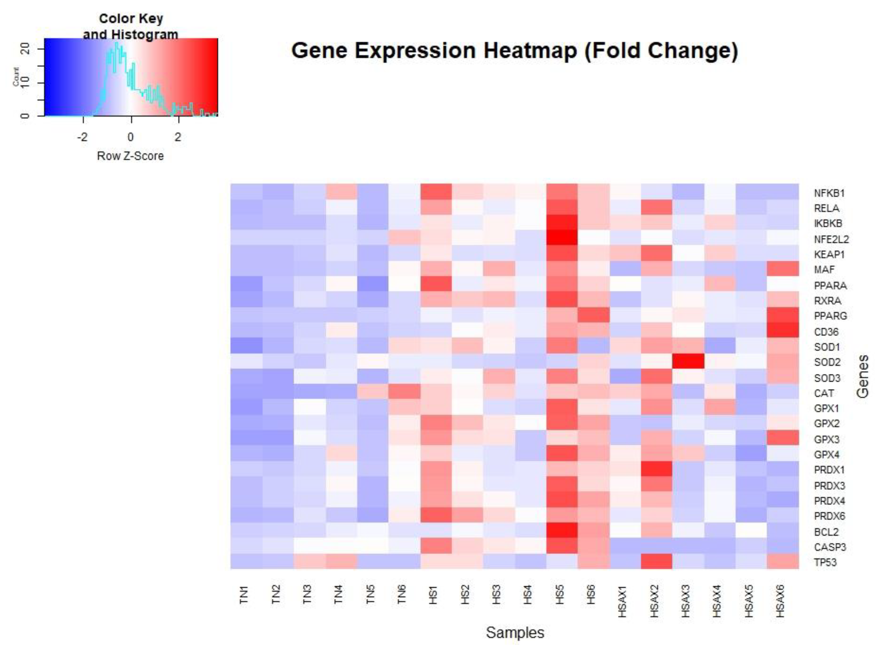
Figure 2.
Gene expression fold change heatmap for thymus tissue of Cob-500 broilers. Three groups were compared: TN, HS, and HSAX. The HS group expression values show shades of red with significant higher expression and the HSAX to a lesser extent, whereas, blue indicates a lower level of expression in the TN group.
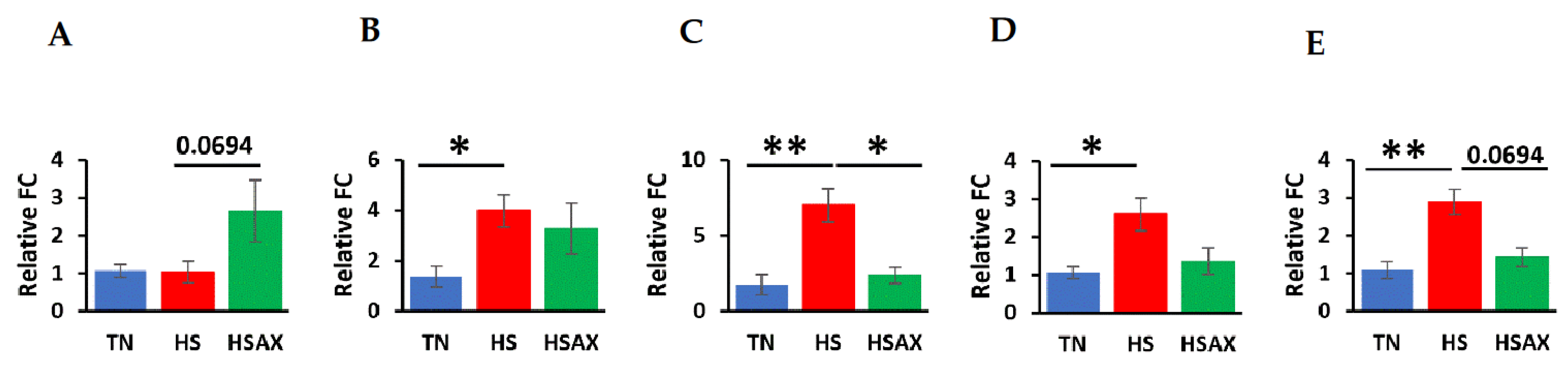
3.2.2. NF-kB Transcription Signaling Pathway Genes
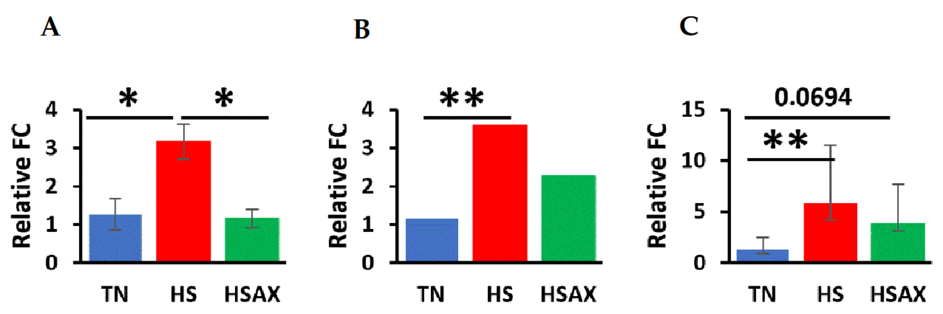
The studies showed the impact of HS and AOX treatment expressed through the NF-kB transcription factor signaling pathway and pairwise comparisons. Gene expression results where HS was significantly upregulated higher than TN were found in NF-kB, TN vs HS (p=0.0283), proto-oncogene, NF-kB subunit (RELA), TN vs HS (p=0.0088) and inhibitor of nuclear factor kappa B kinase subunit beta (IKBKB), TN vs HS (p=0.0105). HS was also significantly higher as compared to HSAX in the expression of NF-kB, HS vs HSAX (p=0.0283). There was a marginal difference between HSAX elevated in expression over TN of IKBKB, TN vs HSAX (p=0.0694).
Figure 3.
Effects of heat stress and astaxanthin treatment in the NF-kB transcription factor signaling pathway in the thymus tissue of Cob-500 broilers. Three groups were compared: TN, HS, and HSAX. Kruskal-Wallis test and Dunn post hoc test for statistical significance represented: (A) NFKB1, KW p=0.0112, TN vs HS (p=0.0283), HS vs HSAX (p=0.0283); (B) RELA, KW p=0.0119, TN vs HS (p=0.0088); and (C) IKBKB, KW p=0.0090, TN vs HS (p=0.0105), TN vs HSAX (p=0.0694).
3.2.3. NFE2L2-Mediated Signaling Pathway Genes
The results of the impact of HS and AOX treatment on the expression of the NFE2L2-mediated signaling pathway showed the HS treatment group upregulated as compared to the TN group, NFE2L2, TN vs HS (p=0.0331), kelch like ECH associated protein 1 (KEAP1), TN vs HS (p=0.0283), and musculoaponeurotic fibrosarcoma (MAF), TN vs HS (p=0.0241). Additionally, the AST treatment was shown to be significantly upregulated over the TN group, KEAP1, TN vs HSAX (p=0.0283).
Figure 4.
Effects of heat stress and astaxanthin treatment in the NFE2L2-mediated signaling pathway in the thymus tissue of Cob-500 broilers. Three groups were compared: TN, HS, and HSAX. Kruskal-Wallis test and Dunn post hoc test for statistical significance represented: (A) NFE2L2, KW p=0.0394, TN vs HS (p=0.0331); (B) KEAP1, KW p=0.0112, TN vs HS (p=0.0283), TN vs HSAX (p=0.0283); and (C) MAF, KW p=0.0298, TN vs HS (p=0.0241).
3.2.4. PPARα Signaling Pathway Genes
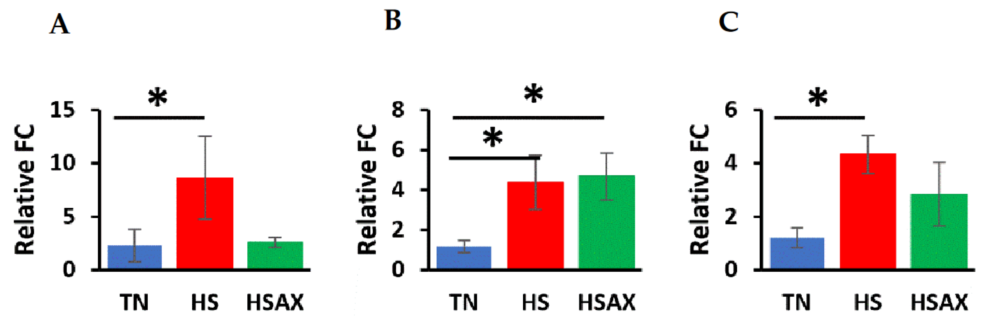
Consistent with the findings for the NF-kB and NFE2L2 pathways, the HS treatments resulted in upregulation over the TN groups in the PPARα pathway genes. The findings showed retinoid X receptor alpha (RXRA), TN vs HS (p=0.0029), peroxisome proliferator-activated receptor gamma (PPARγ), TN vs HS (p=0.0105), cluster of differentiation 36 (CD36), TN vs HS (p=0.0448), and a marginal difference in PPARα, TN vs HS (p=0.0601). Significant upregulation was also found in PPARγ, TN vs HSAX (p=0.0105).
Figure 5.
Effects of heat stress and astaxanthin treatment in the PPARα signaling pathway in the thymus tissue of Cob-500 broilers. Three groups were compared: TN, HS, and HSAX. Kruskal-Wallis test and Dunn post hoc test for statistical significance represented: (A) PPARα, KW p=0.0644, TN vs HS (p=0.0601)(not significant); (B) RXRA, KW p=0.0041, TN vs HS (p=0.0029); (C) PPARγ, KW p=0.0034, TN vs HS (p=0.0105), TN vs HSAX (p=0.0105); and (D) CD36, KW p=0.0417, TN vs HS (p=0.0448).
3.2.5. Cytoprotective Capacity Genes
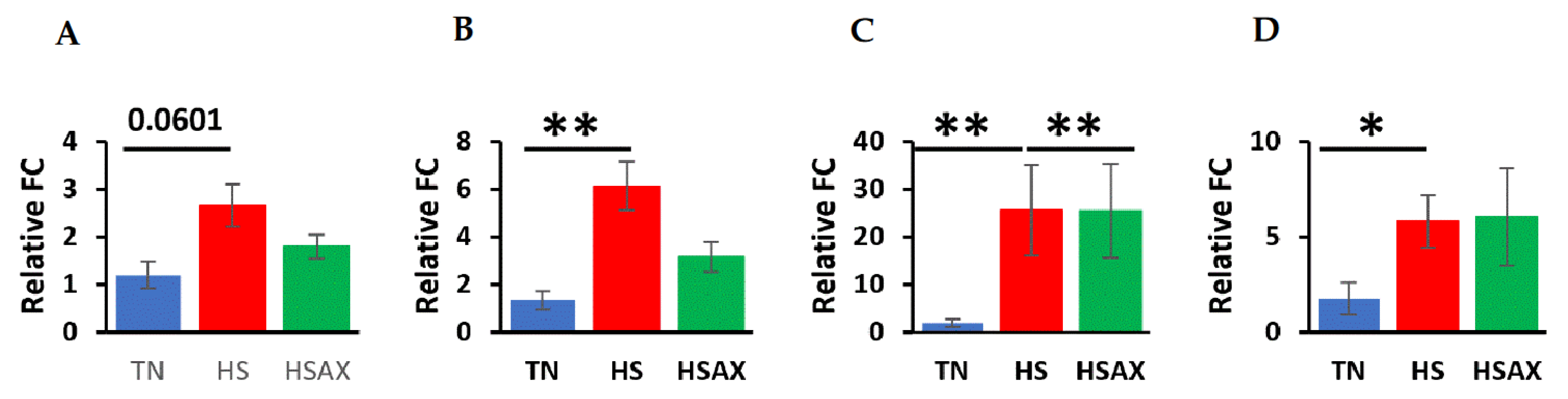
Results of the cytoprotective capacity genes provided a variation of expression. Upregulation of the HS over the TN was significant for superoxide dismutase 3 (SOD3), TN vs HS (p=0.0386), glutathione peroxidase 2 (GPX2), TN vs HS, (p=0.0061), peroxiredoxin 4 (PRDX4), (p=0.0283) and peroxiredoxin 6 (PRDX6), (p=0.0061). Comparing the HS and HSAX groups, GPX2 was significantly upregulated, HS vs HSAX (p=0.0448). Marginal differences were found in SOD2, HS vs HSAX (p=0.0694) and PRDX6, HS vs HSAX (p=0.0694), but with opposing outcomes.
Figure 6.
Effects of heat stress and astaxanthin treatment in the cytoprotective capacity gene expression in the thymus tissue of Cob-500 broilers. Three groups were compared: TN, HS, and HSAX. Kruskal-Wallis test and Dunn post hoc test for statistical significance represented: (A) SOD2, KW p=0.0489, HS vs HSAX (p=0.0694); (B) SOD3, KW p=0.0446, TN vs HS (p=0.0386); (C) GPX2, KW p=0.0050, TN vs HS (p=0.0061), HS vs HSAX (p=0.0448); (D) PRDX4, KW p=0.0260, TN vs HS (p=0.0283); and (E) PRDX6, KW p=0.0060, TN vs HS (p=0.0061), HS vs HSAX (p=0.0694).
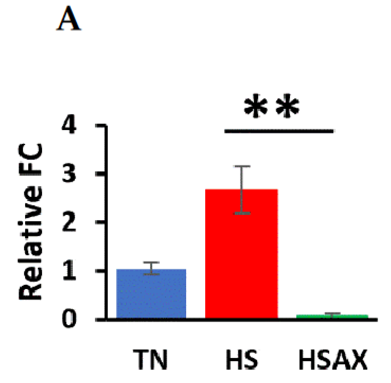
3.2.6. Apoptotic Gene
The results of the apoptotic pathway showed a clear indicator in the upregulation of caspase 3 (CASP3), HS vs HSAX (p=0.0002).
Figure 7.
Effects of heat stress and astaxanthin treatment in the apoptotic pathways in the thymus tissue of Cob-500 broilers. Three groups were compared: TN, HS, and HSAX. Kruskal-Wallis test and Dunn post hoc test for statistical significance represented: (A) CASP3, KW p=0.0005, HS vs HSAX (p=0.0002).
3.2.7. Gene Ontology (GO) Gene Enrichment
4. Discussion
4.1. Growth Performance
Growth performance is anticipated to suffer under any stress condition, and in this study under HS. Although the growth performance indexes did not show any significant benefit from an AOX supplement, the health of the poultry was further elucidated through the molecular mechanisms of gene expression providing insights affecting the health and wellbeing of the broilers.
4.2. Gene Ontology Enrichment and Expression Analysis
4.2.1. NF-kB Transcription Signaling Pathway Genes
The NF-kB transcription signaling pathway is a critical regulator of immune and inflammatory responses. NF-kB can be found throughout the cell in the nucleus, mitochondrion, chromatin and cytoplasm. When not upregulated, NF-kB is generally sequestered in the cytoplasm by IkB inhibitor proteins such as IKBKB, located in the cytoplasm and cytosol. RELA found in the cytoplasm is a key subunit of the NF-kB transcription factor complex to form heterodimers of NF-kB for translocation into the nucleus upon activation for DNA-binding transcription activity. When activated by stimuli, such as stress, free radicals, pathogens and cytokines, IkBs are phosphorylated by a kinase enzyme for ubiquitination and degradation, freeing the NF-kB to translocate into the nucleus to promote the transcription of target genes involved in inflammation, immune response, cell proliferation, and survival. The NF-kB pathway plays a significant role in responding to cellular stress and homeostasis (Table 5).
The outcome of our studies indicates that the upregulation of the HS group over the TN group is consistent with the function of the NF-kB complex to respond to cellular stress. While there is no significance between the TN and HSAX groups, it would indicate that AST is having some effect in reducing cellular stress, which coincides with the results where there is a significant difference between the upregulation of the HS over the HSAX group.
4.2.2. NFE2L2-Mediated Signaling Pathway Genes
The NFE2L2 (NRF2) plays a crucial role in cellular defense against oxidative stress. NFE2L2 is found throughout the cell in the nucleus, Golgi apparatus, chromatin, plasma membrane and cytoplasm. Under normal conditions, NFE2L2 is bound by KEAP1 in the cytoplasm, and in response to OS or electrophilic stimuli, NFE2L2 is released to translocate to the nucleus where it heterodimerizes with small MAF proteins. This NFE2L2-MAF complex binds to antioxidant response elements (AREs) in the promoters of target genes leading to transcription of a various cytoprotective genes (Table 5).
The results of our study show the HS group upregulated in comparison to the TN group in response to OS throughout the NFE2L2 signaling pathway. Similarly, HSAX is upregulated over the TN group in the KEAP1 gene expression, a critical regulator of the NFE2L2 signaling pathway, acting as a sensor for OS and controlling the activity of NFE2L2, likely a homeostatic feedback mechanism to maintain a balanced redox environment.
4.2.3. PPARα Signaling Pathway Genes
The PPARα signaling pathway plays a vital role in lipid metabolism, energy homeostasis and inflammation. Although PPARα which is predominantly found in the nucleus has only a marginal significance in upregulation of the HS over the TN group, RXRA, PPARγ and CD36 is significantly expressing. PPARγ in the HSAX group is also upregulated in comparison to the TN group. PPARα is generally in an inactive state in the nucleus and is activated by fatty acids or specific ligands to form a heterodimer with RXRA, and further undergo conformational changes for DNA-binding transcription factor activity in the promoter of target genes. Among the target genes is CD36 which facilitates fatty acid uptake and enzymes for lipid metabolism. PPARγ can interact with PPARα to help regulate lipid metabolism, reduce inflammation and maintain energy homeostasis (Table 5).
The results from our study indicate that although there is a subtle response of PPARα to heat stress, PPARγ appears to be involved in the cellular response to heat stress and suggests a role in AOX-mediated response to stress. RXRA forms heterodimers with PPARα and is essential for transcriptional activity. The upregulation of CD36 in the HS group compared to the TN suggests increased lipid metabolism or fatty acid utilization, placing a demand on energy due to heat-induced OS.
4.2.4. Cytoprotective Capacity Genes
Cytoprotective capacity genes safeguard cells from various stressors including oxidative damage by enhancing AOX defense mechanisms to promote cell survival. SOD2 is found in the mitochondrion and responds to OS through the oxidation-reduction process for removal of superoxide radicals. SOD3 is found in the extracellular space and plays a crucial role in scavenging superoxide radicals outside the cell. GPX2 is found in the cytosol, intercellular bridge and mitotic spindle, involved in detoxifying peroxides, such as hydrogen peroxide. PRDX4 is found in the cytoplasm and endoplasmic reticulum, and PRDX6 is also found in the cytoplasm, and also the nucleus. Both peroxiredoxins are involved in scavenging for peroxides, providing peroxide detoxification and protection of cells from OS (Table 5).
The results showed significant upregulation of the HS compared to the TN groups in the expression of SOD3, GPX2, PRDX4 and PRDX6. There was also significant upregulation of the HS group over the HSAX group for GPX2, but only marginally for PRDX6. In addition, when analyzing the expression of SOD3, there was marginal upregulation of HSAX over the HS group. Overall, our findings highlight the dynamic regulation of AOX genes in response to heat-induced OS and the potential modulatory effects of AST-AOX supplementation on cellular antioxidant defense mechanisms.
4.2.5. Apoptotic Pathway Genes
The CASP3 can be found in the cytoplasm and nucleus where it encodes for a cysteine protease involved in the execution phase of apoptosis. BCL2 encodes for an anti-apoptotic protein that inhibits cell death, and TP53 encodes for a tumor suppressor protein involved in cell cycle regulation and apoptosis (Table 5).
Our findings show significant upregulation of CASP3 in the HS group over the HSAX group, which suggests an activation of apoptotic pathways, potentially indicating cellular damage or heat-induced OS. The lack of significant expression of BCL2 and TP53 may suggest that heat stress alone may not induce significant alterations in apoptotic regulators, or that the antioxidant supplementation may be effective in mitigating the response to heat stress.
5. Conclusions
Overall, our experimental findings highlight the dynamic regulation of gene expression in the thymus related to the NF-kB, NFE2L2, PPARα, cytoprotective capacity and apoptotic pathways. Our main objective of the present study was to investigate the protective properties of AST against heat-induced OS and apoptosis in the chicken thymus. Our research provided insights into the molecular regulatory mechanisms that respond to heat-induced OS, and the potential therapeutic implementation of AST-AOX supplementation to mitigate the effects through modulation of transcription pathways. The complexities of such mechanisms, and the varied responses of AOX whether endogenous or applied leaves us with knowledge gaps that require further research to understand the therapeutic potentials of AST-AOX.
Supplementary Materials
The following supporting information can be downloaded at the website of this paper posted on Preprints.org. Supplementary data is available in an open-source repository Github as referenced in the Data Availability Statement below.
Author Contributions
DLK conceived the study, collected samples, performed laboratory assays, analyzed the data, performed data preparation and figures and wrote the manuscript. YF, MN, HY and VSK conceived the study and performed analysis. YD conceived the study and contributed resources. All authors edited the manuscript and approved the final draft.
Funding
This project was supported by grants from the National Institute of Health (NIH) grants T32DK137523, U54GM138062, U54MD007601, P30GM114737, P20GM103466, P20GM139753, U54HG013243, National Science Foundation (NSF) 1745130, American Indian Science and Engineering Society, Advancing Agricultural Science Opportunities for Native Americans (AASONA), and University of Hawaii, Hilinehu Educational Leadership Advancement (S362A210110). The content is solely the responsibility of the authors and does not necessarily represent the official views of the NIH.
Institutional Review Board Statement
The study was conducted according to the guidelines and approved by the University of Hawaii Institutional Animal Care and Use Committee (IACUC) under protocol number 17-2605.
Informed Consent Statement
“Not applicable.”.
Data Availability Statement
The original data presented in this study are openly available in Github at (https://github.com/sweetiek/Broiler_thymus_astaxanthin).
Acknowledgments
Special acknowledgments and sincere gratitude to Socorro Tauyan, Facility Manager of the Small Animal Facility, Magoon Research Station, Honolulu, Hawaii, for her support and oversight throughout the animal nutrition experiment, and coordination with the Animal Physiology Lab at the University of Hawaii, Human Nutrition, Food and Animal Sciences.
Conflicts of Interest
The authors declare no conflict of interest.
References
- Vos, J.G. , Moore, J.A. Immune suppression as related to toxicology. CRC Crit. Rev. Toxicol., 1977, 5, 67–101. [Google Scholar] [CrossRef]
- Murray, J.M. , Kaufmann, G.R., Hodgkin, P.D., Lewin, S.R., Kelleher, A.D., et al. Naive T cells are maintained by thymic output in early ages but by proliferation without phenotypic change after age twenty. Immunol. Cell Biol., 2003, 81, 487–495. [Google Scholar]
- Gameiro, J. , Nagib, P., Verinaud, L. The thymus microenvironment in regulating thymocyte differentiation. Cell Adhes. Migr., 2010, 4, 382–390. [Google Scholar] [CrossRef] [PubMed]
- Savino, W. The thymus is a common target organ in infectious diseases. PLoS Pathog 2006, 2, e62. [Google Scholar] [CrossRef] [PubMed]
- Thapa, P. , Farber, D.L. The role of the thymus in the immune response. Thorac. Surg. Clin., 2019, 29, 123–131. [Google Scholar] [CrossRef] [PubMed]
- Raviola, E.; Karnovsky, M.J. Evidence for a blood-thymus barrier using electronopaque tracers. J Exp Med 1972, 136, 466–498. [Google Scholar] [CrossRef] [PubMed]
- Lynch, H.E. , Goldberg, G.L., Chidgey, A., Van den Brink, M.R., Boyd, R., et al. Thymic involution and immune reconstitution. Trends Immunol., 2009, 30, 366–373. [Google Scholar] [CrossRef] [PubMed]
- Dai, X. , Zhang, D., Wang, C., Wu, Z., Liang, C. The pivotal role of thymus in atherosclerosis mediated by immune and inflammatory response. Int. J. Med. Sci., 2018, 15, 1555–1563. [Google Scholar] [CrossRef] [PubMed]
- Chrousos, G. P. Stress and disorders of the stress system. Nat. Rev. Endocrinol., 2009, 5, 374. [Google Scholar] [CrossRef]
- Kumar, B. Stress and its impact on farm animals. Front. Biosci 2012, E4, 1759. [Google Scholar] [CrossRef]
- Li, W.J., Nie, S.P., Peng, X.P., Liu, X.Z., Li, C., et al. Ganoderma atrum polysaccharide improves age-related oxidative stress and immune impairment in mice. J Agric Food Chem 2012, 60, 1413–1418. [Google Scholar] [CrossRef] [PubMed]
- Gostner, J.M., Becker, K., Ueberall, F., Fuchs, D. The good and bad of antioxidant foods: an immunological perspective. Food Chem Toxicol 2015, 80, 72–79. [Google Scholar] [CrossRef]
- Lessard, M., Savard, C., Deschene, K., Lauzon, K., Pinilla, V.A., et al. Impact of deoxynivalenol (DON) contaminated feed on intestinal integrity and immune response in swine. Food Chem Toxicol 2015, 80, 7–16. [Google Scholar] [CrossRef] [PubMed]
- Saeidnia, S., Abdollahi, M. Toxicological and pharmacological concerns on oxidative stress and related diseases. Toxicol Appl Pharmacol 2013, 273, 442–55. [Google Scholar] [CrossRef] [PubMed]
- Zhang, X.Y., Yao, J.K. Oxidative stress and therapeutic implications in psychiatric disorders. Prog Neuropsychopharmacol Biol Psychiatry 2013, 46, 197–9. [Google Scholar] [CrossRef]
- Neri, M., Fineschi, V., Di Paolo, M., Pomara, C., Riezzo, I., et al. Cardiac Oxidative Stress and Inflammatory Cytokines Response after Myocardial Infarction. Curr Vasc Pharmacol 2015, 13, 26–36. [Google Scholar] [CrossRef]
- Nunes, V.A., Gozzo, A.J., Cruz-Silva, I., Juliano, M.A., Viel, T.A., et al. Vitamin E prevents cell death induced by mild oxidative stress in chicken skeletal muscle cells. Comp Biochem Physiol C Toxicol Pharmacol 2005, 141, 225–40. [Google Scholar] [CrossRef]
- Singhal, A., Morris, V.B., Labhasetwar, V., Ghorpade, A. Nanoparticle-mediated catalase delivery protects human neurons from oxidative stress. Cell Death Dis 2013, 4, e903. [Google Scholar] [CrossRef]
- Rao, M., Yang, W., Seifalian, A.M., Winslet, M.C. Role of cyclooxygenase-2 in the angiogenesis of colorectal cancer. Int J Color Dis 2004, 19, 1–11. [Google Scholar] [CrossRef]
- Ashton, K.J., Reichelt, M.E., Mustafa, S.J., Teng, B., Ledent, C., et al. Transcriptomic effects of adenosine 2A receptor deletion in healthy and endotoxemic murine myocardium. Purinergic Signal 2016, 13, 1–23. [Google Scholar]
- Goodman, W.A., Omenetti, S., Date, D., Di, M.L., De, S.C. et al. KLF6 contributes tomyeloid cell plasticity in the pathogenesis of intestinal inflammation. Mucosal Immunol 2016, 9, 1250–1262. [Google Scholar] [CrossRef] [PubMed]
- Hu, X., Chi, Q., Liu, Q., Wang, D., Zhang, Y., et al. Atmospheric H2S triggers immune damage by activating the TLR-7/MyD88/NF-κB pathway and NLRP3 inflammasome in broiler thymus. Chemosphere 2019, 237, 124427. [Google Scholar] [CrossRef]
- Di Prisco, G., Iannaccone, M., Ianniello, F., Ferrara, R., Caprio, E., et al. The neonicotinoid insecticide Clothianidin adversely affects immune signaling in a human cell line. Sci. Rep. 2017, 7, 13446. [Google Scholar] [CrossRef]
- Bajpai, V.K., Alam, M.B., Ju, M.K., Kwon, K.R., Huh, Y.S., et al. Antioxidant mechanism of polyphenol-rich Nymphaea nouchali leaf extract protecting DNA damage and attenuating oxidative stress-induced cell death via Nrf2-mediated heme-oxygenase-1 induction coupled with ERK/p38 signaling pathway. Biomed Pharmacother 2018, 103, 1397–1407. [Google Scholar] [CrossRef]
- Lam. G.Y., Huang, J., Brumell, J.H. The many roles of NOX2 NADPH oxidasederived ROS in immunity. Semin Immunopathol 2010, 32, 415–30. [Google Scholar] [CrossRef]
- Kotsias, F., Hoffmann, E., Amigorena, S., Savina, A. Reactive oxygen species production in the phagosome: impact on antigen presentation in dendritic cells. Antioxid Redox Signal 2013, 18, 714–29. [CrossRef] [PubMed]
- Yang, Y., Bazhin, A.V., Werner, J., Karakhanova, S. Reactive oxygen species in the immune system. Int Rev Immunol 2013, 32, 249–70. [CrossRef]
- Moraes, L. A., Piqueras, L., and Bishop-Bailey, D. Peroxisome proliferator-activated receptors and inflammation. Pharmacol. Ther. 2006, 110, 371–385. [CrossRef] [PubMed]
- Mounier, R., Theret, M., Arnold, L., Cuvellier, S., Bultot, L. O., et al. AMPKalpha1 regulates macrophage skewing at the time of resolution of inflammation during skeletal muscle regeneration. Cell Metab 2013, 18, 251–264. [CrossRef]
- Okamoto, H., T. Iwamoto, S. Kotake, S. Momohara, H. Yamanaka, et al. Inhibition of NF-kappaB signaling by fenofibrate, a peroxisome proliferator-activated receptor-alpha ligand, presents a therapeutic strategy for rheumatoid arthritis. Clin. Exp. Rheumatol 2005, 23, 323–330.
- Eggersdorfer, M., A. Wyss, A. Carotenoids in human nutrition and health. Archives of Biochemistry and Biophysics 2018, 652, 18–26. [CrossRef] [PubMed]
- Destefanis, S. , Giretto, D., Muscolo, M.C., Di Cerbo, A., Guidetti, G., et al. Clinical evaluation of a nutraceutical diet as an adjuvant to pharmacological treatment in dogs affected by Keratoconjunctivitis sicca. BMC Vet. Res. 2016, 12, 214. [Google Scholar]
- Guidetti, G. Guidetti, G., Di Cerbo, A., Giovazzino, A., Rubino, V., Palatucci, A.T., et al. In Vitro Effects of Some Botanicals with Anti-Inflammatory and Antitoxic Activity. J. Immunol. Res 2016, 5457010. [Google Scholar]
- Di Cerbo, A. , Morales-Medina, J.C., Palmieri, B., Pezzuto, F., Cocco, R., et al. Functional foods in pet nutrition: Focus on dogs and cats. Res. Vet. Sci. 2017, 112, 161–166. [Google Scholar] [CrossRef] [PubMed]
- Fakhri, S. , Abbaszadeh, F., Dargahi, L., Jorjani, M. Astaxanthin: A mechanistic review on its biological activities and health benefits. Pharmacol. Res. 2018, 136, 1–20. [Google Scholar] [CrossRef] [PubMed]
- Astaxanthin. (n.d.). Retrieved from https://pubchem.ncbi.nlm.nih.gov/compound/Astaxanthin. Accessed 02 Apr 2024.
- Heaney, R. P. Factors Influencing the Measurement of Bioavailability, Taking Calcium as a Model. The Journal of Nutrition 2001, 131. [Google Scholar] [CrossRef] [PubMed]
- Mularczyk, M., Michalak, I., Marycz, A. Astaxanthin and other Nutrients from <italic>Haematococcus pluvialis</italic>-Multifunctional Applications. Mar Drugs 2020, 18, 459. [CrossRef] [PubMed]
- Shahidi, F., Barrow, C. J. Marine nutraceuticals and functional foods. Boca Raton. CRC Press 2008.
- Livak, K. J., Schmittgen, T. D. Analysis of relative gene expression data using real-time quantitative PCR and the 2−ΔΔCt method. Methods 2001, 25, 402–408. [CrossRef]
- <italic>Ensembl</italic>.org, https://uswest.ensembl.org/Gallus_gallus/Info/Index Accessed 04 Apr 2024.
- R Core Team (2023) https://www.R-project.org/ Accessed 04 Apr 2024.
Figure 1.
Growth performance indicators of Cob-500 broilers. Three groups were compared: TN, HS, and HSAX. Kruskal-Wallis test and Dunn post hoc test for statistical significance represented: (A) BW(g), KW p=0.0047, TN vs HS (p=0.0095) and TN vs HSAX (p=0.0278); (B) ADFI(g), KW p=0.0038, TN vs HS (p=0.0047) and TN vs HSAX (p=0.0500); (C) ADGR(g), KW p=0.0047, TN vs HS (p=0.0095) and TN vs HSAX (p=0.0278); and (D) FCR, KW p=0.0049, TN vs HS (p=0.0226) and TN vs HSAX (p=0.0119).
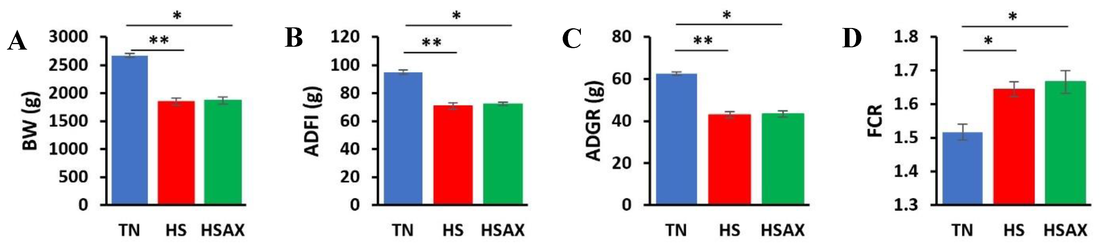
Table 1.
Basal diet ingredients and calculated analysis of broiler diets used in the mixture design for thermal neutral and heat stress including astaxanthin supplement.
Table 1.
Basal diet ingredients and calculated analysis of broiler diets used in the mixture design for thermal neutral and heat stress including astaxanthin supplement.
| Ingredients (%) | Starter | Finisher |
|---|---|---|
| Corn | 54.86 | 63.14 |
| Soybean Meal | 39.99 | 30.09 |
| Soybean oil | 2.00 | 4.50 |
| Limestone | 1.27 | 0.85 |
| Monocalcium phosphate | 0.75 | 0.50 |
| Lysine (%) | 0.23 | 0.18 |
| Methionine (%) | 0.14 | 0.12 |
| Threonine (%) | 0.20 | 0.16 |
| Sodium chloride (%) | 0.43 | 0.35 |
| Sodium bicarbonate | 0.12 | 0.10 |
| Vitamin-mineral premix | 0.50 | 0.50 |
| Astaxanthin supplement¹ | 0.01 | 0.01 |
| Total | ||
| Calculated analysis | ||
| ME (kcal/kg) | 2909 | 3203 |
| CP (%) | 22.09 | 18.07 |
| Calcium (%) | 0.75 | 0.52 |
| Total Phosphorus (%) | 0.57 | 0.47 |
| digPhosphorous (%) | 0.30 | 0.23 |
| Lysine (%) | 1.39 | 1.10 |
| dig Lysine (%) | 1.25 | 0.99 |
| Methionine (%) | 0.48 | 0.41 |
| dig Methionine (%) | 0.45 | 0.39 |
| Cysteine (%) | 0.43 | 0.38 |
| Threonine (%) | 1.03 | 0.85 |
| dig Threonine (%) | 0.85 | 0.69 |
| Tryptophan (%) | 0.33 | 0.26 |
| Methionine + cysteine (%) | 0.91 | 0.80 |
| Arginine (%) | 1.61 | 1.31 |
| Valine (%) | 1.22 | 1.03 |
| Isoleucine (%) | 0.93 | 0.76 |
| Leucine (%) | 1.89 | 1.63 |
| Neutral detergent fiber | 9.13 | 8.78 |
| Crude fiber | 3.97 | 3.46 |
| Sodium | 0.22 | 0.18 |
| Chloride | 0.30 | 0.25 |
| Choline (mg/kg) | 1419 | 1200 |
| Astaxanthin (mg/kg) | --- | 1.33 |
¹Astaxanthin was mixed with the soybean oil and supplemented in the diet during feed mixing.
Table 2.
Gallus gallus oligonucleotide primers used for real-time RT-PCR analysis.
| Gene | NCBI Accession No. | Primer set (5’-3’) |
| β-ACTIN | NM_205518.2 | F:5' - AATTGTGCGTGACATCAAGG |
|
TBP GAPDH |
NM_205103.2 NM_204305.2 |
R:3' -CACAGGACTCCATACCCAAG F:5' -GCGGCAGGCTCTGTT R:3' -ACCGAAAAGGTTTTTGACCC F:5' -AAGTCGGAGTCAACGGATTT R:3' -TCACAAGTTTCCCGTTCTCA |
| NFKB1 RELA IKBKB |
NM_205134.2 NM_001396038.1 NM_001395965.1 |
F:5’ -GGACGGCGAAAGGACTCT R:3’ -CCATTGCAAACATTTGGGGAT F:5’ -CGGTTCCGCTATAAGTGTGA R:3’ -GTAATGGTTTACGCGGATGG F:5’ -TCCCTGGGAGATGAAGGAG R:3’ -TTTGGATGGTTCAGCCTCTT |
| NFE2L2 KEAP1 MAF PPARA RXRA PPARG CD36 |
XM_015287264.4 GenBank: KU321503.1 NM_001044671.1 NM_001001464.1 XM_003642291.6 NM_001001460.2 NM_001030731.1 |
F:5’ –CAGGGGTAGCAAGGTATGAG R:3’ -TGCCTCCAAAGGATGTCAAT F:5’ –GATCGACGGGATGATCTACG R:3’ –GGCGTACAGCAGTATGTTCA F:5’ –CCAGAGTTTTTCATGTACCCG R:3’ -CTTTGTAGCTGTCTTCGTGC F:5’ – AGCCACTTGCTATCACCAAT R:3’ – ACTTAAACTCCTTTATGATTCTGGT F:5’ –CTTCCTGCCACTGGATTTCT R:3’ –CTGATGACGGAGAAGGGTG F:5’ – CTTGACAGCGCCAGAGATTA R:3’ –GATTGCACTTTGGCAATCCT F:5’ –TTTCTTGCAAAGCAGGAGGTT R:3’ –CTGATCTTCGTGAGAGAAGCTGTA |
| SOD1 | NM_205064.2 | F:5' -AAAAGATGCAGATAGGCACG |
| R:3' -TTATCTCCCCCTCTACCCAG | ||
| SOD2 SOD3 |
NM_204211.2 XM_040699307.2 |
F:5' -CCTTCGCAAACTTCAAGGAG R:3' -AGCAATGGAATGAGACCTGT F:5’ –CAACTCGCAAACAACGCT R:3’ –CTGGTGAGTGAGAACCTGC |
| CAT | NM_001031215.2 | F:5' -TTCCACGTTAAGACCGATCA |
| R:3' -CAATCTTGCCCACTGGAATG | ||
| GPX1 | NM_001277853.3 | F:5' -AATTCGGGCACCAGGAGAA |
| R:3' - CTCGAACATGGTGAAGTTGG | ||
| GPX2 | NM_001277854.3 | F:5' -AGTTCGGCTACCAGGAGAA |
| R:3' -CTTCTGGAACAGGGTGAAGT | ||
| GPX3 | NM_001163232.3 | F:5' - AGGTGAAATGCTACGACTCC |
|
GPX4 PRDX1 PRDX3 PRDX4 PRDX6 BCL2 CASP3 TP53 |
NM_204220.3 NM_001271932.2 XM_426543.6 XM_046912353.1 NM_001039329.3 NM_205339.3 NM_204725.1 NM_205264.1 |
R:3' - AGTGCATTCAGTTCGAGGTA F:5’ –AATGTGCGCTCAGGCG R:3’ –AGACGAAGCCCCTGTACT F:5’ –TGCGGGGCTCTTTGTATTAAA R:3’ –ATTGCCCATCTGGCATTACA F:5’ –CGTTGTCAATGGGGAGTTC R:3’ –GGGGCACACAAAGGTGAAAT F:5’ –ATCCCCTTGACTTCACGTTT R:3’ –ATCTTCATTGGTCCGAGTCC F:5’ –GACATCAACGCCTACAATGG R:3’ –GGCCAAATATGAACACCACA F:5’ –GAGGATGGGATGCCTTTGTG R:3’ –CCACGATAAACTGGGTGACT F:5’ – GGTGGAGGTGGAGGAGC R:3’ –TGAGCGTGGTCCATCTTTTA F:5’ –GTTACCACGACGAGCCACCAA R:3’ –TGCAGCGCCTCATTGATCTCCTT |
Table 3.
Growth performance indicators of Cob-500 broilers for body weight, adjusted daily feed intake, adjusted daily growth rate, and feed conversion. Three groups were compared: TN, HS, and HSAX as the mean ± standard deviation. Statistical analysis was based on the Kruskal-Wallis rank sum test, followed with the Dunn post hoc test for comparisons, and p-value adjusted with the Bonferroni method, and where “ns” is considered “not significant”. * 0.05>p>0.01, ** 0.01>p>0.001.
| Kruskal-Wallis |
Dunn/Bonferroni p-value |
||||||
| Measurements | TN | HS | HSAX | p-value | TN vs HS | TN vs HSAX | HS vs HSAX |
| BW (g) | 2673.68 ± 35.71 | 1848.85 ± 61.56 | 1867.83 ± 60.82 | 0.0047 | 0.0095 | 0.0278 | ns |
| ADFI (g) | 94.98 ± 1.45 |
70.68 ± 72.24 |
72.24 ± 1.14 |
0.0038 | 0.0047 | 0.0500 | ns |
| ADGR (g) | 62.65 ± 0.86 |
42.99 ± 1.46 |
43.45 ± 1.44 |
0.0047 | 0.0095 | 0.0278 | ns |
| FCR | 1.52 ± 0.02 |
1.64 ± 0.02 |
1.67 ± 0.03 |
0.0049 | 0.0226 | 0.0119 | ns |
Table 5.
Gene ontology of differentially expressed genes in the thymus of 6-week Cob-500 broilers exposed to 21 days of heat stress and astaxanthin treatment (Ensembl) [41].
Table 5.
Gene ontology of differentially expressed genes in the thymus of 6-week Cob-500 broilers exposed to 21 days of heat stress and astaxanthin treatment (Ensembl) [41].
| Gene | Cellular component | Molecular function | Biological process | Transcript IDs | ||||
|---|---|---|---|---|---|---|---|---|
| NFKB1 (nuclear factor kappa B subunit 1) | nucleus, cytoplasm, mitochondrion, chromatin | DNA binding transcription factor activity, RNA polymerase II specific DNA binding, chromatin, protein and actinin binding | regulation of transcription by RNA polymerase II, MAPK cascade, JNK cascade, NIK/NF-kappaB signaling | ENSGAL00010005476 | ||||
| RELA (proto-oncogene, NF-kB subunit) | cytoplasm | DNA-binding transcription factor activity | regulation of DNA-templated transcription | ENSGALG00010001277 | ||||
| IKBKB (inhibitor of nuclear factor kappa B kinase subunit beta) | cytoplasm, cytosol | protein kinase activity, identical protein binding, protein homodimerization activity, scaffold protein binding, transferrin receptor binding | Protein phosphorylation, I-kappaB kinase/NF-kappaB signaling, cellular response to tumor necrosis factor, regulation of establishment of endothelial barrier, negative regulation of bicellular tight junction assembly | ENSGALG00010017955 | ||||
| NFE2L2 (nuclear factor, erythroid 2 like 2) | nucleus, cytoplasm, Golgi apparatus, chromatin, centrosome, plasma membrane, RNA polymerase II transcription regulator complex | DNA binding transcription factor activity, RNA polymerase II specific DNA-binding transcription factor binding, ubiquitin protein ligase binding | response to oxidative stress, inflammatory response, regulation of gene expression, protein ubiquitination, cell redox homeostasis, positive regulation of glutathione biosynthetic process, regulation of removal of superoxide radicals | ENSGAL00010024107 | ||||
| KEAP1 (kelch like ECH associated protein 1) | nucleus, cytoplasm, endoplasmic reticulum, Cul3-RING ubiquitin ligase complex, centriolar satellite | RNA polymerase II-specific DNA-binding transcription factor binding, ubiquitin ligase-substrate adaptor activity, disordered domain specific binding, identical protein binding | cellular response to oxidative stress, ubiquitin-dependent protein catabolic process, regulation of DNA-templated transcription, regulation of autophagy | ENSG00000079999 | ||||
| MAF (MAF bZIP transcription factor) | nucleus, cytoplasm, RNA polymerase II transcription regulator complex | DNA-binding transcription factor activity, RNA polymerase II sequence-specific DNA binding | regulation of DNA templated transcription, regulation of transcription by RNA polymerase II, cell development | ENSGALG00010007800 | ||||
| RXRA (retinoid X receptor alpha) | nucleus, RNA polymerase II transcription regulator complex, receptor complex | RNA polymerase II transcription regulatory region sequence-specific DNA binding, enzyme, peptide, metal ion binding, retinoic acid-responsive element binding, Vitamin D response element binding | peroxisome proliferator activated receptor signaling pathway, retinoic acid receptor signaling pathway, positive regulation of Vitamin D receptor signaling pathway, cell differentiation | ENSGALG00010028422 | ||||
| PPARG (peroxisome proliferator-activated receptor gamma) | nucleus, cytoplasm, intracellular membrane-bounded organelle | DNA-binding transcription factor activity, nuclear receptor activity, zinc and metal ion binding | Regulation of DNA-templated transcription, transcription by RNA polymerase II, intracellular receptor signaling pathway | ENSGALG00010027917 | ||||
| CD36 (CD36 molecule) | external side of plasma membrane, receptor complex, membrane raft | low-density lipoprotein particle receptor activity, high-density lipoprotein particle binding, Toll-like receptor binding, scavenger receptor activity | MAPK cascade, cell surface receptor signaling pathway, positive regulation of cytosolic calcium ion concentration, nitric oxide mediated signal transduction, intestinal cholesterol absorption, positive regulation of reactive oxygen species metabolic process, lipid transport across blood-brain barrier | ENSGALG000010008392 | ||||
| SOD2 (superoxide dismutase 2, mitochondrial) | mitochondrion | superoxide dismutase activity, oxidoreductase activity, manganese ion binding, metal ion binding, identical protein binding | response to oxidative stress, oxidation-reduction process, negative regulation of oxidative stress-induced intrinsic apoptotic signaling pathway, response to hydrogen peroxide, removal of superoxide radicals | ENSGALT00000019062 | ||||
| SOD3 (superoxide dismutase 3) | extracellular space, collagen- containing extracellular matrix | superoxide dismutase activity, copper and metal ion binding | superoxide metabolic process, response to hypoxia | ENSGALG00010009833 | ||||
| GPX2 (glutathione peroxidase 2) | cytosol, intercellular bridge, mitotic spindle | glutathione peroxidase activity, phospholipid-hydroperoxide glutathione peroxidase activity | response to oxidative stress | ENSGALG00010021537 | ||||
| PRDX4 (peroxredoxin 4) | cytoplasm, endoplasmic reticulum | antioxidant activity, thioredoxin peroxidase activity, oxidoreductase activity, peroxiredoxin activity, molecular sequestering activity | response to oxidative stress, cell redox homeostasis, hydrogen peroxide catabolic process, reactive oxygen species metabolic process, cellular oxidant detoxification | ENSGALG00010003214 | ||||
| PRDX6 (peroxredoxin 6) | nucleus, cytoplasm | antioxidant activity, glutathione peroxidase activity, oxidoreductase activity, peroxiredoxin activity, ubiquitin protein ligase binding | response to oxidative stress, cellular oxidant detoxification, glycerophospholipid catabolic process | ENSGALG00010013528 | ||||
| CASP3 (caspase 3) | nucleus, cytoplasm | endopeptidase activity, hydrolase activity, cyclin-dependent protein serine/threonine kinase inhibitor activity, cysteine-type endopeptidase activity involved in execution phase of apoptosis | T and B cell homeostasis, negative regulation of cytokine production, intrinsic apoptotic signaling pathway, negative regulation of cell cycle, neuron apoptotic process, epithelial cell apoptotic process | ENSGALG00010007067 | ||||
Disclaimer/Publisher’s Note: The statements, opinions and data contained in all publications are solely those of the individual author(s) and contributor(s) and not of MDPI and/or the editor(s). MDPI and/or the editor(s) disclaim responsibility for any injury to people or property resulting from any ideas, methods, instructions or products referred to in the content. |
© 2024 by the authors. Licensee MDPI, Basel, Switzerland. This article is an open access article distributed under the terms and conditions of the Creative Commons Attribution (CC BY) license (http://creativecommons.org/licenses/by/4.0/).
Copyright: This open access article is published under a Creative Commons CC BY 4.0 license, which permit the free download, distribution, and reuse, provided that the author and preprint are cited in any reuse.


