
Submitted:
22 May 2024
Posted:
22 May 2024
You are already at the latest version
Abstract
Background: Intracavitary contrast-enhanced ultrasound is widely accepted as a highly informative, safe and easily reproducible technique for diagnosis, treatment and follow- up of different pathologies of the biliary tree. Methods: This article reports the combined conclusions of personal experience with pictorial examples and literature review of applications for CEUS in intracavitary biliary scenarios. Different methodologies, choices of equipment, indications for the procedure and expected complications were identified and described from the authors’ own experience as well as from a review of published studies with total of 208 patients for the period between 2009 and 2015. Results: Indications were biliary obstruction by various etiologies, including postoperative strictures, evaluation of the biliary tree of liver donors (n=12) and evaluation of localization of a drainage catheter. One of the studies found a statistically significant difference in the diagnostic accuracy of PTC and PUSC. Conclusion: Intracavitary CEUS brings all the positive features of US together with the virtues of contrast- enhanced imaging, proving comparable accuracy to the listed techniques for diagnostics of biliary tree diseases.
Keywords:
percutaneous transhepatic cholangiodrainage
; intracavitary
; contrast enhanced ultrasound
; biliary obstruction
1. Introduction
Ultrasonography (US) is a proven diagnostic method with a long list of generally accepted advantages. It is a non-ionizing and cheap method which can be performed at the patient’s bedside, easily reproducible, and in addition a modality from which physicians gain ample information for various organs [1]. The method is commonly utilized as a diagnostic tool for biliary tree pathology, because of its satisfactory evaluation [2]. In case of focal lesions, contrast-enhanced ultrasound (CEUS) may give additional information often leading to conclusive diagnosis [3]. A CEUS examination is relatively inexpensive, easy to perform and non-invasive of nature except for the intravenous access and often provides supplementary insights of decisive character [2]. Contrast agents (UCAs), used for CEUS contain microbubbles of gas enveloped by protein, lipid or polymer [4]. The most commonly used UCA in Europe, SonoVue ®, consists of sulphur hexafluoride, enveloped in a shell of phospholipids. The excretion of the gas is pulmonary, while the excretion of the phospholipid component is by the liver [5]. Their lack of renal impact makes them appropriated for use in kidney impairment [6]. CEUS can detect blood flow in far smaller vessels, compared to Doppler US i.e. diameter of 40 μm and 100 μm, respectively [2].
In contrast to the liver, the blood supply to the extrahepatic biliary ducts is solely arterial [7]. Every analysis of a biliary lesion consists of an early arterial, a portal and a late parenchymal phase [8]. The common limitations of US to scan the distal common bile duct and ampulla cannot be overcome by CEUS, but CEUS may be useful to differentiate between calculi (especially with low content of calcium) and mass formations, since tumors demonstrate enhancement [3].
At low acoustic pressure microbubbles resonate forming non- linear signals, which can be detected on US, and after removing the linear signals from the surrounding tissue a contrast image is formed, which can be followed in dynamics. These features render CEUS as an attractive alternative to fluoroscopic imaging for endocavitary use due to its non-ionizing nature plus in addition non-iodinated contrast agent formulation [9]. Much smaller amount of contrast is needed in endocavitary CEUS, because the substance is not washed out with the blood stream and thus remains in the cavity longer compared to when applied in the systemic circulation [10,11]. UCA is considered safe, with rarely reported adverse event, when used intravenously, which may indicate even a safer profile in intracavitary applications [12,13]. The introduction of UCAs into physiological or pathological cavities of the body is very useful for evaluation of its inner structure and shape, potential fistula, position of a drainage system and patency of a hollow organ or duct (e.g., fallopian tubes, biliary system, or reflux detection), etc. [14,15,16,17,18,19,20,21].
This is also embedded in the EFSUMB guidelines and recommendations for the clinical practice of CEUS in non-hepatic applications from 2018, which assume that injection of ultrasound contrast media into physiological or non-physiological cavities is aiding in managing different clinical problems: identification of needle or catheter position or delineation of any cavity or duct or support tracking of a fistula. No fixed dose of US contrast agent is suggested for intracavitary use and the range varies between 0.1 mL – 1mL SonoVue™ (or a few drops) diluted in ≥ 10 mL 0.9 % normal saline [22].
These indications are very important, since they provide potential alternative to contrast fluoroscopy [23]. Microbubbles can be used as echo-enhancers into any nonvascular cavity which allows detailing and clarification thus facilitating essential ultrasound-mediated therapy [24]. Among the list of application of intracavitary CEUS some of the most important are percutaneous transhepatic cholangiography (PTC), endoscopic ultrasound (EUS) with endoscopic retrograde cholangiography (ERCP), tracing peritoneal-pleural communication and evacuation of abscesses [22]. Overabundance of UCA leads to lower imaging quality by posterior acoustic shadowing and it is mandatory to emphasize that lesser dosage of ultrasound contrast agent (UCA) is needed compared with intravenous use [25].
In the case of abscess formation intravenous UCA delineates the avascular content and helps determine the appropriate access for drainage [25,26]. Moreover, after the drainage catheter is placed a better control of positioning of the tube and precise demarcation of the abscess wall plus potential communications and fistulae can be achieved by intracavitary CEUS. Finally, the perhaps most valuable feature of abscess intracavitary CEUS is its reproducibility for monitoring the effect of the treatment [25,26,27].
In the scenario of cholangiography CEUS can be utilized to identify post-procedural complications such as e.g. hepato-biliary fistula formation where contrast enhancement is observed not only in the biliary tree, but also in the liver [5]. The method also is applicable in determining the position of a drain and to evaluate a biliary obstruction or leakage [15,28,29,30]
Percutaneous transhepatic cholangiography and drainage (PTCD) is a widely utilized procedure for diagnosing and treating benign and malignant biliary pathology [31,32,33]. As a rule, ERCP is the modality of choice for biliary diagnostic and therapeutic interventions [34]. Endosonographically (EUS) - guided cholangiography and drainage is gaining wide popularity among endoscopists and is practiced at many specialized centers around the world. Indications for PTC and PTCD are cases where an easier and less invasive endoscopic procedure is not possible for diagnosis or treatment [35,36] (e.g., biliary–enteric anastomosis, a Billroth II operation, gastrectomy, hepaticojejunostomy, a Roux-en-Y choledochojejunostomy with a failed afferent limb, duodenal peripapillary diverticula, etc.), or where endoscopic passing through a stenosis has failed [34].
Real-time imaging with ultrasound (US) is useful for the guidance of PTCD (US-PTCD), especially in difficult approach for non-dilated ducts and for left-sided bile duct branches [31,37].
In terms of diagnostics for biliary tree the available approaches are MRCP, endoscopic ERCP, PTC, Endosonographically guided cholangiography and drainage (EUS-CD). The non-MRI-depending above mentioned techniques are used more commonly with therapeutic than with diagnostic aim, because of the availability of magnetic resonance cholangiopancreatography (MRCP). PTC is a second line procedure in cases of unsuccessfully performed ERCP [38], mainly when endoscopic approach is not possible. A combined technique between PTC and ERCP is used – the rendezvous technique. PTC is by far preferable by some authors in case of interventions in the hilar region and in the strictures of the intrahepatic biliary ducts [39,40,41]. Among the list of advantages of PTC before ERCP are the higher success rate for beginners, possibility of real- time three- dimension imaging, usage of color Doppler for a higher dependability on the approach [42,43,44,45] and less exposure to radiation. The rendezvous technique is possible, during which, first a wire percutaneously is placed by PTC, to the duodenum, serving as a guide for placement of a prosthesis. The technique makes it possible to perform sequencing endoscopic procedures even in cases of Billroth II gastrectomy [46].
The procedure can be utilized in all biliary diseases – benign, malignant (including brachytherapy) and anomalies in the development of the tree [47,48]. Following iatrogenic injury of the biliary tree, application of PTC is comparable with ERCP to determine the anatomy, the location of leakage and to prove a possible stricture [49]. PTC is helpful for localization and for placement of percutaneous transhepatic biliary drainage (PTBD). Redirection of bile, facilitation of the localization of the injury and balloon dilatation of strictures are all possible because of PTBD [50]. Despite being first line, ERCP neither can show an injury of the right segmental duct, nor in case of major bile duct occlusion or transection, may provide any additional information beyond identifying remnants of the bile duct post-injury [51]. Although relatively rare, special attention should be given to PTBT in cases of non-dilated ducts, most commonly after resection of the liver [52]. The absolute and relative contraindications for PTCD are not different from every other invasive procedures [38].
2. Materials and Methods
The materials and methods section is divided in two separate entities. Firstly, and most important a literature review was conducted and secondly, we added a lesser part with personal experience and pictorial examples to demonstrate the usefulness of some important indications.
Literature review: a literature search of PubMed database and manual search was conducted between 01 March and 20 March 2024. The following search terms were used: “intracavitary”, “extravascular”, “endocavitary”, “intrabiliary” and “CEUS” and “contrast enhanced ultrasound”, “percutaneous transhepatic cholangiography”, “PTCD” and “biliary drainage”. A total number of 6 studies on the topic were identified in a total of 208 patients. All of the studies were conducted between 2009 and 2015 (Table 1.).
Indications for PTC and PTCD were in all cases where an easier and less invasive endoscopic procedure was not possible for diagnosis or treatment [35,36], or where endoscopic passing through a stenosis had failed. As a rule, the indication for PTC is mainly therapeutic for the decompression of an obstructed biliary tract [34]. Injecting intra-biliary contrast can aid in identifying leaks, strictures, and the position of drains. [9].
PTCD Technique
The literature holds a variety of different procedures for placement of PTC drainage catheter and there may be both advantages and disadvantages of one over the other. Our standard procedure utilizes a loop catheter of 40 cm and 6F to 8 F diameter. The catheter is placed under ultrasound guidance using Seldinger technique and initial puncture with 1,2 mm lumbar needle. When the lumbar needle is visualized with its tip inside selected dilated bile duct and bile can be aspirated with a syringe, then a 0.035-inch guidewire is inserted and at this point, if x-ray is available, it may be advantageous to visualize the passage of guidewire to deeper bile duct sections. When guidewire is deemed correctly in position the lumbar needle is exchanged with the pigtail catheter over the guidewire. Potentially 6F to 10F dilators can be used before inserting the catheter, however, when using Seldinger technique often there is no need for a dilatator if only the inner needle is taken out from the pig-tail catheter leaving the outer needle shaft to stabilize the catheter during insertion over the guidewire. When the catheter supposedly is in correct position, final confirmation is obtained by CEUS cholangiography and may also be supplemented with X-ray contrast cholangiography. Finally, the catheter is securely fixed to the skin with a suture or dedicated fixture device to ensure inadvertent displacement and before sending off the patient the catheter is connected to a drainage bag.
Drainage sets are available in different lengths with a wide range of diameter from 6F up to even 18F, (the latter being rarely indicated) all featuring side holes (with a distance from the skin of around 7.5 cm). These sets come complete with a connecting tube, sealing cap, and skin plate. The PTCD catheter includes a pigtail end and multiple side holes and internal string fixture that holds the pig tail shape in position. When pulling out the catheter it is crucial to cut the string to avoid scratching the liver surface thereby causing potential peritoneal bleeding. Numerous additional treatment options exist, spanning from the rendezvous technique for ongoing drainage via minimally invasive endoscopic methods to the percutaneous placement of a metal stent [34].
3. Results
3.1. Studies – See Table 1
The first available study is by Ignee et al. on a small group of patients with various obstructive biliary pathology and intrahepatic duct dilatation of 2 mm and more. A right sided approach was used in the majority of the cases. The initial experience with CEUS-PTCD intervention was successful in all patients for identification of stenosis. Dislodgement of the catheter occurred in 1 patient and the catheter was re-inserted. In this study fluoroscopy was used, but only for guidance. Repeated injections of the diluted US contrast media were necessary in 2 patients [58]
The study by Zheng et al. included 12 living liver donors for evaluation of the biliary tree. The approach was via cannulation of the cystic duct and common bile ducts intraoperatively. The intervention was successful for adequate evaluation of the biliary anatomy in all patients and as a result in 3 of the donors were diagnosed biliary variations and the rest of the donors were with normal biliary pattern. No complications were described. Intraoperative cholangiography was used for comparison. The accuracy of 3D ultrasound cholangiography was excellent for first and second order biliary branches [62].
In a larger cohort of 58 patients again with various obstructive biliary pathology, percutaneous ultrasound cholangiography (PUSC) was compared with percutaneous transhepatic cholangiography (PTC). For 8 patients bilateral approach was necessary for drainage catheter placement. The patients were divided according to the level of obstruction into hilar obstruction and extrahepatic obstruction group, respectively. The strength of PUSC was in defining the level of obstruction as hilar with accuracy 100%. Determination of CBD obstruction was weaker with accuracy of 93.8%. The accuracy of PUSC in determining the level of obstruction was 96.6% (56/58). The accuracy of PTC in both groups was 100% (P=1.000, P=0.492, respectively). In general, the accuracy of PUSC in determining the cause of obstruction was 93.1% (54/58) while that of PTC was 79.3% (46/58). The difference between them was statistically significant (P=0.031). 3 patients in the PUSC group had experienced pain in the right upper quadrant and 25 patients in the PTC group had epigastric pain [63].
Xu et al. conducted a study performing intracavitary CEUS during PTCD in the largest number of patients (n=80) so far. The indications for performing the intrabiliary CEUS were to localize the drainage catheter, to localize the distal tip of the catheter and/or to evaluate the level and the degree of obstruction. The US contrast agent injections were performed in a two-step manner- first injection to track the distal tip of the catheter and second injection to evaluate the obstruction of the biliary tract. Four catheters were not properly placed and were repositioned. The intervention was successful in all 80 patients (100%). CT- or fluoroscopy guided cholangiography was used as reference method. The patients were defined as having intrahepatic or extrahepatic obstruction according to the level of obstruction and complete or incomplete, according to the degree of obstruction. The authors report an excellent accuracy for intrabiliary CEUS for determination of the level of obstruction and 96.3% accuracy for detecting the degree of obstruction. The accuracy for locating the proper position of the tip of the catheter was 100% [30].
Chopra et al. evaluated 12 patients after orthotopic liver transplantation with side-to-side biliary anastomosis with intracavitary CEUS for assessment of postoperative complications. On the fifth postoperative day they performed conventional cholangiography and CEUS cholangiography via a T-tube placed intraoperatively. Both techniques showed similar success rate. Pathology was found in 4 patients - one anastomotic stenosis, one anastomotic leakage and delayed duodenal outflow in 2 cases. Fluoroscopy showed superiority in detecting anastomotic stricture and leakage. Along with the detection of pathology, the visualization of the extra- and intrahepatic bile duct was assessed. The results showed that CEUS was inferior in visualization of extrahepatic bile ducts [64].
As a continuation of their initial experience, Ignee et al. conducted another study for extravascular use of CEUS in a larger patient cohort (n=38) with various primary or secondary biliary obstructive pathology during PTCD after failed or potentially impossible endoscopic retrograde cholangiography (ERC). The intervention was performed in several stages. The diluted US contrast agent was injected before the X-ray contrast agent. In the first stage both contrast agents were injected to confirm the intraductal position of the needle and to detect the level of obstruction. Then repeated injections were necessary to complete the PTCD and in cases of malignant stenosis were indicated for catheter or stent position confirmation. Right sided approach was used in the majority of the cases. The puncture and catheter insertion were successful in all patients (100%). A few complications occurred i.e. one subcutaneous hematoma, two catheter dislodgement and pleural effusion and pleural-peritoneal fistula in one patient. Fluoroscopy was used as standard method. The accuracy of extravascular CEUS was 100% for correctly diagnosing the level of obstruction as hilar or extrahepatic and 97.4% for defining the degree of obstruction (complete/incomplete) [19].
Although still “off-label”, a well-known application of the CEUS in children, is the administration of microbubble-based US contrast agents for real-time evaluation of their distribution in body cavities [65] Chen et al. demonstrated successful utilization of intracavitary CEUS in children with biliary diseases. The authors present their experience in performing percutaneous ultrasound cholangiography and the use of sulfur hexafluoride microbubbles as contrast agent in children with neonatal hepatitis, biliary atresia, choledochal cysts with pancreaticobiliary malfunction and postoperative complications after hepatobiliary surgery [60].
3.2. Case Reports from the Review
Two interesting cases of intracavitary CEUS application are worth mentioning (Table 2).
The first case is of a male patient with bile leakage after T-tube removal. The T-tube was placed during cholecystectomy and CBD exploration. After the operation the patient developed mild fever, jaundice and abdominal pain which indicated the performance of US-guided PTCD via right sided approach. After an injection of diluted US contrast agent, a leakage of the contrast from the common bile duct was observed and was confirmed by percutaneous transhepatic cholangiogram [66].
The second case is of a young male patient with postoperative biliary complications after liver transplantation for familial amyloid polyneuropathy type 1. On the 35 posttransplant day a PTCD was performed due to continuing bile leak at the hepaticojejunostomy site. Seven days after the biliary drain placement an intermittent haemobilia occurred. A CEUS cholangiogram was performed and after demonstration of the correctly sited distal aspect of the drain within the hepaticojejunostomy with leakage around the liver surface at the site of proximal percutaneous insertion, the presence of an occult biliary-arterial fistula was identified. The latter was confirmed by conventional tubogram [29].
3.3. Intracavitary CEUS Technique
The necessary equipment for intracavitary CEUS into drainage tube for PTC includes a second-generation ultrasound contrast agent (SonoVue® Bracco Milan, Italy), a 10 cc syringe and standard saline solution. The injection is performed directly into the connecting tube. It is important not to use any additional tools with membrane and not from the side arm of a 3 way-screwcock (Figure 1).
No accurate and scientifically validated numbers are available but some authors recommend a very high dilution of US contrast agent (0.1–0.2 mL SonoVue per 20 mL physiologic saline solution) injected into the bile ducts after the initial puncture is performed to confirm proper intraductal placement or identify a possible stenosis [11,22,29,34]. Our personal experience, though, is that a few droplets in a 10 or 20 cc will suffice and care should always be taken to avoid overabundance of contrast which will result in shadowing of deeper structures. It is also very important to use recommended (as always with CEUS) low mechanical index (less than 0.2). Roberts et al. were among the first who demonstrated injection of perflutren lipid microspheres (Definity), an ultrasound contrast agent, in various dilutions through intraoperatively cannulated porcine common bile duct. The utilization of micro-bubble contrast in the biliary system resulted in outstanding visualization of the bile duct, extending down to ducts as small as fifth-order branches, and revealed bile ducts measuring less than 2 mm in diameter [57]. In one of the first reported human studies after insertion of the drain, SonoVue® 1mL was injected followed by another 5-mL saline flush [58]. According to the experience of Ignee et al. as an approach for diagnostic cholangiography after sufficient local anesthesia and sedation, a suitable biliary duct (mainly in segment 5 or 6) is targeted under US guidance and punctured by means of a 20 G Chiba needle. 1-3 ml of ultrasound contrast agent mixture (again 0.1 ml in 20 ml of physiological saline solution) is injected for correct positioning of the needle in the bile ducts followed by insertion of a thin catheter (5F). In case of therapeutical PTC placement there is no need to inject UCA until the end to confirm correct placement of the catheter. In critically ill patients a drain (mostly 8 – 10 F) might be used for external bile drainage. If the procedure results in improvement of the patient’s general condition, the internalization of the drain into intestine can be done either via a rendezvous procedure with ERC, PTCD with application of an internal-external drain or via the same PTC catheter with placement of a permanent metal stent. The PTC may simply be left in position in terminal patients if it is placed for palliation to alleviate symptoms from bile duct obstruction such as skin itching, general malaise, or cholangitis. In oncology treatment the strategy could also be to place PTC in order to bring down elevated bilirubin blood level allowing for chemotherapy and in such scenarios the catheter can be discontinued after treatment if successful opening of the stricture results from the chemotherapy. Another very important utility of the injection of US contrast agent through the drainage tube is the possibility to detect complications of PTCD like bile leakage, drain dislocation and fistulas in the pleural cavity [59].
Xu et al. commenced by injecting 1 mL of UCAs into the drainage catheter to initially pinpoint the tip location. They undertook this approach to prevent the overflow signal of UCAs from obscuring the drainage catheter and inundating the entire biliary tract. Once the positions of the drainage catheter were verified, additional UCAs were introduced gradually through the drainage catheter to illustrate the level and extent of obstruction within the biliary tract. In certain patients with severe incomplete obstruction, enhancing the dosage and pressure of UCA injection could potentially enhance diagnostic accuracy. As a result, the authors proposed a novel PTBD strategy. Rather than employing US-guided PTBD in conjunction with fluoroscopic cholangiography, Xu et al. conducted IB-CEUS concurrently with conventional US-guided PTBD, omitting the need for fluoroscopic cholangiography. This innovative approach offers time- and cost-savings, as the entire procedure can be conducted at the bedside without the need for radiation exposure. It represents a comprehensive "one-station" strategy for PTBD [30].
In a very recent pictorial essay Chen et al. report their experience in percutaneous ultrasound cholangiography with microbubbles in children with biliary diseases. To disperse sulfur hexafluoride and phosphatide, the authors inject 5 mL of 0.9% sodium chloride solution into the bottle. Subsequently, the microbubble dispersion is mixed with 30 mL of 0.9% sodium chloride solution in a 35-mL injection syringe. The mixed US microbubble contrast agents are then injected into the drainage tube. The distribution of the microbubbles in the biliary system is observed under the contrast imaging mode [60].
The impact of UCAs on the biliary system remains largely unexplored due to limited literature on the subject. In one of the largest studies on the topic, no instances of chemical cholangitis were reported [30]. However, there is a potential risk of cholangitis associated with overdistention, particularly in dilated systems. Therefore, it is advisable to minimize the amount of contrast injected. Additionally, the long-term effects of sulfur hexafluoride on biliary epithelium have yet to be thoroughly investigated [61].
In the study of Luao et al., after depicting the level of biliary obstruction, the contrast agent liquid remaining in the bile duct was aspirated to the greatest extent possible. This procedure was undertaken to minimize the risk of any unexpected damage to the bile duct [15].
3.4. Complications
Despite the diversity of the published data on PTCD complication rate and serious adverse events have been observed less in recent years [34]. In a recent observational study by Turan et al. during a 5-year period, a total of 331 patients underwent PTCD of whom 205 (61.9%) developed PTCD-related complications. Of the patients without a pre-existent infection, 40.6% developed infectious complications, i.e., cholangitis in 26.3%, sepsis in 24.6%, abscess formation in 2.7%, and cholecystitis in 1.3%. Non-infectious complications developed in 34.4%. 30-day mortality was 17.2% [54]. The use of smaller size needles is associated with a significantly lower complication rate [55,56]. Important to emphasize is the fact that most of these patients have end-stage malignant disease of a variety of etiologies and the procedure most often is performed with a palliative purpose.
3.5. Pictorial Examples
We present three cases of personal experience where intracavitary CEUS during or after PTCD played a pivotal role and provided decisive solution to a critical clinical scenario.
First case was a palliative PTCD in a patient with terminal stage gallbladder cancer with spread into the intrahepatic bile ducts (Figure 2). An 8 Fr pigtail catheter was inserted in the left liver lobe for biliary decompression. Three months later the patient presented with nonfunctioning PTC drain. Due to the fact that the injection of diluted US contrast agent (SonoVue) was initially impossible, a higher pressure on the syringe’s plug was exerted. A possible clot was the reason for obstruction of the drainage tube, which was overcome. Then the diluted US contrast agent entered the biliary ducts unhindered and confirmed the proper position of the patent drainage tube.
Second case was a patient with carcinoma of the antrum of the stomach with malignant infiltration of the pancreatic head and compression of biliary tract (Figure 3). The biliary drainage was placed for palliative purposes. The pictures show intracavitary injection of diluted ultrasound contrast agent into biliary drainage catheter inserted in the left liver lobe.
Third case was a female patient with inoperable Klatskin tumor (Figure 4). ERCP showed >90% malignant stenosis of d. choledochus at the hilum with length 20 mm. The patient was referred for PTCD and the proper drainage catheter position in the bile ducts of the left liver lobe was confirmed by intracavitary CEUS:
4. Discussion
Since the approval of the use of US contrast agents more than 20 years back in some parts of the world [67] CEUS has proven its excellent diagnostic accuracy comparable with contrast-enhanced CT or MRI, especially in focal liver lesions [68,69,70,71,72,73,74,75]. In addition to this well-accepted usage endocavitary CEUS represents a dynamic modality assessment in real time and can provide high-quality additional information to the US, without further considerations as usage of iodinated contrast and radiation [9].
The current review and our personal experience including the pictorial cases presented herein plus all of the available studies for intracavitary application of CEUS in PTCD, present excellent examples of the benefits and added information from this novel technique. All studies, cited in this review, show excellent accuracy of intracavitary CEUS for determining the level of obstruction, locating the drainage catheter and its distal tip, identifying biliary leaks and in addition very good ability to define the degree of obstruction as well as delineating the biliary tree anatomy in living liver donors. All these clinical scenarios represent excellent indications and demonstrate the non-inferiority of intracavitary CEUS compared to conventional fluoroscopy.
PTBD in non-dilatated biliary ducts is indicated in symptomatic fistulas (incidence after surgery of the liver, biliary tree and pancreas 3-10%)[76,77,78,79,80,81], in cases of bad performance status, long-lasting fistula or inaccessible leaks by ERCP. PTBT is with relatively low risk and a considerably positive result. The procedure has proven to be less intricate in dilated than in nondilated ducts [52,82,83,84,85]. The reason for absence of dilatation is the presence of leak, despite the distal stenosis. This leads to the need of smaller catheters, to be placed in small-caliber ducts [52,82,85]. Interventions close to the hepatic hilum increase the risk for complications such as haemobilia [86]. It is presumed that draining bile outside rather than into the bowel favors fistula healing and reduces the risk of superinfection [87].
This relatively new application of US contrast agents into a cavity or drainage catheter is gaining more and more popularity. It is cost-effective, available for bed-side use, possesses all of the advantages of the intravascular use i. e. excellent safety profile, no nephrotoxicity and avoidance of ionizing radiation. Additionally, a very important advantage is the opportunity for repeated examination during the intervention and for follow up.
Despite these advantages, there are some limitations of the intracavitary CEUS. First of all, like all clinical ultrasound, it is operator dependent and lower quality of visualization must be expected in obese patients and in the presence of abundant bowel gas [9]. Another still unexplored area is the investigation of the chemical effect of the US contrast agent on the biliary epithelium. So far, however, there are no reported cases of chemical cholangitis or complications due to increased intraductal pressure after intrabiliary administration of US contrast agent.
As number of studies on the topic is very limited and some report small numbers of patients with big variation of obstructive biliary pathology, further randomized studies with larger patient cohorts and more homogeneous biliary pathology are warranted.
In the future, there may be a focus on developing materials for extravascular contrast-enhanced ultrasound imaging, with an emphasis on using specific compounds such as cell-penetrating peptides. For instance, a disulfide-bridged cyclic RGD (Arg-Gly-Asp) peptide, named iRGD (internalizing RGD), which is a tumor-homing peptide with high affinity and specificity for a certain integrin, could be utilized to construct targeted materials. Integrating iRGD peptide into materials and thereby potentially enhance penetration of blood vessels and the extracellular matrix, facilitating accumulation and increasing the likelihood of enhanced imaging [88,89]. Proteins incorporating the Arg-Gly-Asp (RGD) attachment site, along with integrins functioning as their receptors, represent a fundamental recognition system crucial for cell adhesion [90].
Additionally, there are strategies for further increase the accumulation of targeted materials and enhance the probability of improved imaging through the use of NO-releasing agents [91].
There has been a surge of interest in utilizing microbubbles as carriers for drugs, aiming to deliver them to specific sites and achieve localized release through the disruption of microbubbles using high-frequency ultrasound waves. This localized release strategy enhances drug efficiency while concurrently reducing systemic side effects. Particularly notable is its success in facilitating the targeted release of chemotherapeutic agents, effectively mitigating their systemic adverse effects [92].
Another future direction of research is the oral contrast-enhanced ultrasound for delineating a fistula, as there are reports of the stability of UCA within the stomach despite acidic conditions [93].
5. Conclusions
The intracavitary CEUS has proven to be comparable to fluoroscopy in aiding the PTCD procedure and delineating the normal biliary anatomy or detect pathology. It is safe to use, radiation free, reproducible and could be performed for several times during the intervention and for follow-up. Whenever available, intracavitary CEUS should be in the list of physician’s arsenals to address various diagnostic and therapeutic challenges assisted by imaging modalities.
Author Contributions
Conceptualization: CN and EA; methodology: CN; Literature search: EA and CP; software: CP; writing—original draft preparation, EA and CP; writing—review and editing, CN; visualization: EA, CN and CP; supervision: CN. All authors have read and agreed to the published version of the manuscript.
Funding
This research received no external funding.
Institutional Review Board Statement
Not applicable.
Informed Consent Statement
Patient consent was waived due to death of the patient from a terminal illness and due to the data and images being unidentifiable and not relating to the patient plus considered quality control of nature.
Data Availability Statement
No new data were created or analyzed in this study. Data sharing is not applicable to this article.
Acknowledgments
none.
Conflicts of Interest
The authors declare no conflict of interest.
References
- Y. Chen et al., “Model for End-Stage Liver Disease Score Predicts the Mortality of Patients with Coronary Heart Disease Who Underwent Percutaneous Coronary Intervention,” Cardiol Res Pract, vol. 2021, 2021. [CrossRef]
- D. D. Cokkinos et al., “Contrast-enhanced ultrasound examination of the gallbladder and bile ducts: A pictorial essay,” Journal of Clinical Ultrasound, vol. 46, no. 1, pp. 48–61, Jan. 2018. [CrossRef]
- F. Piscaglia et al., “The EFSUMB guidelines and recommendations on the clinical practice of contrast enhanced ultrasound (CEUS): Update 2011 on non-hepatic applications,” Ultraschall in der Medizin, vol. 33, no. 1, pp. 33–59, 2012. [CrossRef]
- S. H. Morin, A. K. Lim, J. F. Cobbold, S. D. Taylor-Robinson, and R. Steiner, “Use of second generation contrast-enhanced ultrasound in the assessment of focal liver lesions,” World J Gastroenterol, vol. 13, no. 45, pp. 5963–5970, 2007, [Online]. [CrossRef]
- C. Greis, “Technology overview: SonoVue (Bracco, Milan),” Eur Radiol, vol. 14 Suppl 8, pp. P11-5, Nov. 2004. [CrossRef]
- Badea R, Zaro R, Tantau M, and Chiorean L., “ Ultrasonography of the biliary tract–up to date. The importance of correlation between imaging methods and patients’ signs and symptoms,” Med Ultrason., no. 17, pp. 383–391, 2015. [CrossRef]
- Z. Sparchez, R. Pompilia, S. Bota, R. Sirli, and A. Popescu, “Role of contrast enhanced ultrasound in the assessment of biliary duct disease,” Med Ultrason, vol. 16, pp. 41–47, Mar. 2014. [CrossRef]
- H.-X. Xu, “Contrast-enhanced ultrasound in the biliary system: Potential uses and indications,” World J Radiol, vol. 1, no. 1, p. 37, 2009. [CrossRef]
- G. T. Yusuf, C. Fang, D. Y. Huang, M. E. Sellars, A. Deganello, and P. S. Sidhu, “Endocavitary contrast enhanced ultrasound (CEUS): a novel problem solving technique,” Insights Imaging, vol. 9, no. 3, pp. 303–311, 2018. [CrossRef]
- Goddi, R. Novario, F. Tanzi, R. Di Liberto, and P. Nucci, “In vitro analysis of ultrasound second generation contrast agent diluted in saline solution.,” Radiol Med, vol. 107 5–6, pp. 569–79, 2004, [Online]. Available: https://api.semanticscholar.org/CorpusID:33565326.
- Heinzmann, T. Müller, J. Leitlein, B. Braun, S. Kubicka, and W. Blank, “Endocavitary Contrast Enhanced Ultrasound (CEUS) – Work in Progress,” Ultraschall in der Medizin - European Journal of Ultrasound, vol. 33, no. 01, pp. 76–84, Feb. 2012. [CrossRef]
- F. Piscaglia and L. Bolondi, “The safety of Sonovue® in abdominal applications: Retrospective analysis of 23188 investigations,” Ultrasound Med Biol, vol. 32, no. 9, pp. 1369–1375, Sep. 2006. [CrossRef]
- G. T. Yusuf, M. E. Sellars, A. Deganello, D. O. Cosgrove, and P. S. Sidhu, “Retrospective Analysis of the Safety and Cost Implications of Pediatric Contrast-Enhanced Ultrasound at a Single Center,” American Journal of Roentgenology, vol. 208, no. 2, pp. 446–452, Feb. 2017. [CrossRef]
- L. Zhou et al., “Value of three-dimensional hysterosalpingo-contrast sonography with SonoVue in the assessment of tubal patency,” Ultrasound in Obstetrics & Gynecology, vol. 40, no. 1, pp. 93–98, Jul. 2012. [CrossRef]
- Z. Luyao, X. Xiaoyan, X. Huixiong, X. Zuo-feng, L. Guang-jian, and L. Ming-de, “Percutaneous ultrasound-guided cholangiography using microbubbles to evaluate the dilated biliary tract: initial experience,” Eur Radiol, vol. 22, no. 2, pp. 371–378, Feb. 2012. [CrossRef]
- K. Darge, “Voiding urosonography with ultrasound contrast agents for the diagnosis of vesicoureteric reflux in children,” Pediatr Radiol, vol. 38, no. 1, pp. 40–53, Jan. 2008. [CrossRef]
- X.-W. Cui, A. Ignee, T. Maros, B. Straub, J.-G. Wen, and C. F. Dietrich, “Feasibility and Usefulness of Intra-Cavitary Contrast-Enhanced Ultrasound in Percutaneous Nephrostomy,” Ultrasound Med Biol, vol. 42, no. 9, pp. 2180–2188, Sep. 2016. [CrossRef]
- X.-W. Cui, A. Ignee, U. Baum, and C. F. Dietrich, “Feasibility and Usefulness of Using Swallow Contrast-Enhanced Ultrasound to Diagnose Zenker’s Diverticulum: Preliminary Results,” Ultrasound Med Biol, vol. 41, no. 4, pp. 975–981, Apr. 2015. [CrossRef]
- Ignee, X. Cui, G. Schuessler, and C. Dietrich, “Percutaneous transhepatic cholangiography and drainage using extravascular contrast enhanced ultrasound,” Z Gastroenterol, vol. 53, no. 05, pp. 385–390, May 2015. [CrossRef]
- Ignee, NathanS. S. Atkinson, G. Schuessler, and C. Dietrich, “Ultrasound contrast agents,” Endosc Ultrasound, vol. 5, no. 6, p. 355, 2016. [CrossRef]
- Y. N. Patrunov, I. Apolikhina, E. Peniaeva, A. N. Sencha, and A. Saidova, “CEUS for minimally invasive procedures: Intracavitary CEUS,” in Contrast-Enhanced Ultrasound From Simple to Complex, Sencha A. N. and Patrunov Y. N, Eds., Springer International Publishing, 2022, pp. 327–337. [CrossRef]
- P. Sidhu et al., “The EFSUMB Guidelines and Recommendations for the Clinical Practice of Contrast-Enhanced Ultrasound (CEUS) in Non-Hepatic Applications: Update 2017 (Long Version),” Ultraschall in der Medizin - European Journal of Ultrasound, vol. 39, no. 02, pp. e2–e44, Apr. 2018. [CrossRef]
- P. Nolsøe et al., “Use of Ultrasound Contrast Agents in Relation to Percutaneous Interventional Procedures: A Systematic Review and Pictorial Essay,” Journal of Ultrasound in Medicine, vol. 37, no. 6, pp. 1305–1324, Jun. 2018. [CrossRef]
- Deganello et al., “Intravenous and intracavitary use of contrast-enhanced ultrasound in the evaluation and management of complicated pediatric pneumonia,” Journal of Ultrasound in Medicine, vol. 36, no. 9, pp. 1943–1954, Sep. 2017. [CrossRef]
- Y. Huang et al., “Contrast-enhanced US–guided Interventions: Improving Success Rate and Avoiding Complications Using US Contrast Agents,” RadioGraphics, vol. 37, no. 2, pp. 652–664, Mar. 2017. [CrossRef]
- T. V. Bartolotta, A. Taibbi, M. Midiri, and R. Lagalla, “Focal liver lesions: contrast-enhanced ultrasound,” Abdom Imaging, vol. 34, no. 2, pp. 193–209, Apr. 2009. [CrossRef]
- Ignee, C. Jenssen, X.-W. Cui, G. Schuessler, and C. F. Dietrich, “Intracavitary contrast-enhanced ultrasound in abscess drainage – feasibility and clinical value,” Scand J Gastroenterol, vol. 51, no. 1, pp. 41–47, Jan. 2016. [CrossRef]
- R. Mao, E. Xu, K. Li, and R. Zheng, “Usefulness of contrast-enhanced ultrasound in the diagnosis of biliary leakage following T-tube removal,” Journal of Clinical Ultrasound, vol. 38, no. 1, pp. 38–40, Jan. 2010. [CrossRef]
- M. Daneshi, B. Rajayogeswaran, P. Peddu, and P. S. Sidhu, “Demonstration of an occult biliary-arterial fistula using percutaneous contrast-enhanced ultrasound cholangiography in a transplanted liver,” Journal of Clinical Ultrasound, vol. 42, no. 2, pp. 108–111, Feb. 2014. [CrossRef]
- Xu, R. Zheng, Z. Su, K. Li, J. Ren, and H. Guo, “Intra-biliary contrast-enhanced ultrasound for evaluating biliary obstruction during percutaneous transhepatic biliary drainage: A preliminary study,” Eur J Radiol, vol. 81, no. 12, pp. 3846–3850, Dec. 2012. [CrossRef]
- Dietrich et al., “EFSUMB Guidelines on Interventional Ultrasound (INVUS), Part III – Abdominal Treatment Procedures (Long Version),” Ultraschall in der Medizin - European Journal of Ultrasound, vol. 37, no. 01, pp. E1–E32, Dec. 2015. [CrossRef]
- M. Nagino et al., “Preoperative biliary drainage for biliary tract and ampullary carcinomas,” J Hepatobiliary Pancreat Surg, vol. 15, no. 1, pp. 25–30, Jan. 2008. [CrossRef]
- G. Cozzi et al., “Percutaneous Transhepatic Biliary Drainage in the Management of Postsurgical Biliary Leaks in Patients with Nondilated Intrahepatic Bile Ducts,” Cardiovasc Intervent Radiol, vol. 29, no. 3, pp. 380–388, Jun. 2006. [CrossRef]
- F. Dietrich, B. Braden, X. W. Cui, A. Ignee, and P. M. Rodgers, “Percutaneous Transhepatic Cholangiodrainage,” in Interventional Ultrasound – A Practical Guide and Atlas, no. 2, 2015, pp. 198–214. [CrossRef]
- J. Albert, F. Ulrich, S. Zeuzem, and C. Sarrazin, “Endoskopisch-retrograde Cholangiopankreatografie (ERCP) bei Patienten mit operativ veränderter Anatomie des Gastrointestinaltrakts,” Z Gastroenterol, vol. 48, no. 08, pp. 839–849, Aug. 2010. [CrossRef]
- H. P. JANDER, J. GALBRAITH, and J. S. ALDRETE, “Percutaneous Transhepatic Cholangiography Using the Chiba Needle,” South Med J, vol. 73, no. 4, pp. 415–421, Apr. 1980. [CrossRef]
- P. V. Kavanagh, E. van Sonnenberg, G. R. Wittich, B. W. Goodacre, and E. M. Walser, “Interventional Radiology of the Biliary Tract,” Endoscopy, vol. 29, no. 06, pp. 570–576, Aug. 1997. [CrossRef]
- Wenz W., “Percutaneous Transhepatic Cholangiography,” Radiologe, vol. 13, pp. 41–46, 1973.
- KROMER MU et al., “ Bile duct stenoses and leakage after cholecystectomy: endoscopic diagnosis, therapy and treatment outcome- [Article in German],” Zeitschrift fur Gastroenterologie , vol. 34, no. 3, pp. 167–172, 1996.
- R. Jakobs, U. Weickert, D. Hartmann, and J. F. Riemann, “Interventionelle Endoskopie bei benignen und malignen Gallengangsstenosen,” Z Gastroenterol, vol. 43, no. 3, pp. 295–303, Mar. 2005. [CrossRef]
- S. Misra, G. B. Melton, F. J. Geschwind, A. C. Venbrux, J. L. Cameron, and K. D. Lillemoe, “Percutaneous management of bile duct strictures and injuries associated with laparoscopic cholecystectomy: a decade of experience,” J Am Coll Surg, vol. 198, no. 2, pp. 218–226, Feb. 2004. [CrossRef]
- T. Tsuyuguchi et al., “Techniques of biliary drainage for acute cholangitis: Tokyo Guidelines,” J Hepatobiliary Pancreat Surg, vol. 14, no. 1, pp. 35–45, Jan. 2007. [CrossRef]
- J. Ferrucci, J. Wittenberg, R. Sarno, and Dreyfuss, “Fine needle transhepatic cholangiography: a new approach to obstructive jaundice,” American Journal of Roentgenology, vol. 127, no. 3, pp. 403–407, Sep. 1976. [CrossRef]
- U. Laufer, J. Kirchner, R. Kickuth, S. Adams, M. Jendreck, and D. Liermann, “A Comparative Study of CT Fluoroscopy Combined with Fluoroscopy Versus Fluoroscopy Alone for Percutaneous Transhepatic Biliary Drainage,” Cardiovasc Intervent Radiol, vol. 24, no. 4, pp. 240–244, Jul. 2001. [CrossRef]
- K. Koito, T. Namieno, T. Nagakawa, and K. Morita, “Percutaneous transhepatic biliary drainage using color Doppler ultrasonography.,” Journal of Ultrasound in Medicine, vol. 15, no. 3, pp. 203–206, Mar. 1996. [CrossRef]
- P. J. Shorvon, P. B. Cotton, R. R. Mason, J. H. Siegel, and A. R. Hatfield, “Percutaneous transhepatic assistance for duodenoscopic sphincterotomy.,” Gut, vol. 26, no. 12, pp. 1373–1376, Dec. 1985. [CrossRef]
- Nuernberg, A. Ignee, and C. Dietrich, “Aktueller Stand der Sonografie in der Gastroenterologie - Darm und oberer Gastrointestinaltrakt Teil 2,” Z Gastroenterol, vol. 46, no. 4, pp. 355–366, Apr. 2008. [CrossRef]
- M. E. Alden and M. Mohiuddin, “The impact of radiation dose in combined external beam and intraluminal IR-192 brachytherapy for bile duct cancer,” International Journal of Radiation Oncology*Biology*Physics, vol. 28, no. 4, pp. 945–951, Mar. 1994. [CrossRef]
- L. Stewart, “Bile Duct Injuries During Laparoscopic Cholecystectomy,” Archives of Surgery, vol. 130, no. 10, p. 1123, Oct. 1995. [CrossRef]
- K. D. Lillemoe, “Current management of bile duct injury,” British Journal of Surgery, vol. 95, no. 4, pp. 403–405, Mar. 2008. [CrossRef]
- K. D. Lillemoe, J. A. Petrofski, M. A. Choti, A. C. Venbrux, and J. L. Cameron, “Isolated right segmental hepatic duct injury: a diagnostic and therapeutic challenge,” Journal of Gastrointestinal Surgery, vol. 4, no. 2, pp. 168–177, Mar. 2000. [CrossRef]
- B. Funaki et al., “Percutaneous biliary drainage in patients with nondilated intrahepatic bile ducts.,” American Journal of Roentgenology, vol. 173, no. 6, pp. 1541–1544, Dec. 1999. [CrossRef]
- P. M. Rodgers, “Interventional Ultrasound – A Practical Guide and Atlas,” Ultrasound, vol. 23, no. 2, pp. 198–198, May 2015. [CrossRef]
- S. Turan et al., “Complications of percutaneous transhepatic cholangiography and biliary drainage, a multicenter observational study,” Abdominal Radiology, vol. 47, no. 9, pp. 3338–3344, Aug. 2021. [CrossRef]
- R. Burke et al., “Quality improvement guidelines for percutaneous transhepatic cholangiography and biliary drainage.,” J Vasc Interv Radiol, vol. 14, no. 9 Pt 2, pp. S243-6, Sep. 2003.
- W. P. Harbin, P. R. Mueller, and J. T. Ferrucci, “Transhepatic cholangiography: complicatons and use patterns of the fine-needle technique: a multi-institutional survey.,” Radiology, vol. 135, no. 1, pp. 15–22, Apr. 1980. [CrossRef]
- J. P. Roberts, A. Neill, and R. Goldstein, “The use of a micro-bubble contrast agent to allow visualization of the biliary tree,” Clin Transplant, vol. 20, no. 6, pp. 740–742, Nov. 2006. [CrossRef]
- Ignee, U. Baum, G. Schuessler, and C. Dietrich, “Contrast-enhanced ultrasound-guided percutaneous cholangiography and cholangiodrainage (CEUS-PTCD),” Endoscopy, vol. 41, no. 08, pp. 725–726, Aug. 2009. [CrossRef]
- Ignee, G. Schuessler, X.-W. Cui, and C. Dietrich, “Der endokavitäre Kontrastmittelultraschall – verschiedene Anwendungen, eine Übersicht über die Literatur und zukünftige Perspektiven,” Ultraschall in der Medizin - European Journal of Ultrasound, vol. 34, no. 06, pp. 504–528, Jun. 2013. [CrossRef]
- M. Chen, X. Zhang, W. Zhou, N. Zhang, G. Wang, and L. Zhou, “Percutaneous Ultrasound Cholangiography With Microbubbles in Children With Biliary Diseases,” Ultrasound Q, vol. 39, no. 4, pp. 228–234, Dec. 2023. [CrossRef]
- S. H. Chandrashekhara and S. Gulati, “Contrast-enhanced ultrasound-based cholangiography using percutaneous transhepatic biliary drainage tube,” Indian Journal of Gastroenterology, vol. 42, no. 5, pp. 744–745, Oct. 2023. [CrossRef]
- R. Zheng et al., “Evaluating Biliary Anatomy and Variations in Living Liver Donors by a New Technique: Three-Dimensional Contrast-Enhanced Ultrasonic Cholangiography,” Ultrasound Med Biol, vol. 36, no. 8, pp. 1282–1287, Aug. 2010. [CrossRef]
- Z. Luyao, X. Xiaoyan, X. Huixiong, X. Zuo-feng, L. Guang-jian, and L. Ming-de, “Percutaneous ultrasound-guided cholangiography using microbubbles to evaluate the dilated biliary tract: initial experience,” Eur Radiol, vol. 22, no. 2, pp. 371–378, Feb. 2012. [CrossRef]
- S. S. Chopra1abcdef et al., “Contrast enhanced ultrasound cholangiography via T-tube following liver transplantation.” [Online]. Available: http://www.annalsoftransplantation.com/fulltxt.php?ICID=883700.
- M. R. Acord et al., “Contrast-enhanced ultrasound in pediatric interventional radiology,” Pediatr Radiol, vol. 51, no. 12, pp. 2396–2407, Nov. 2021. [CrossRef]
- R. Mao, E. J. Xu, K. Li, and R. Q. Zheng, “Usefulness of contrast-enhanced ultrasound in the diagnosis of biliary leakage following T-tube removal,” Journal of Clinical Ultrasound, vol. 38, no. 1, pp. 38–40, Jan. 2010. [CrossRef]
- R. G. Barr, “The Urgent Need for <scp>FDA</scp> to Approve a <scp>Whole-Body</scp> Application of Ultrasound Contrast Agents,” Journal of Ultrasound in Medicine, vol. 42, no. 4, pp. 761–764, Apr. 2023. [CrossRef]
- S. Moudgil et al., “Comparison of Contrast Enhanced Ultrasound With Contrast Enhanced Computed Tomography for the Diagnosis of Hepatocellular Carcinoma,” J Clin Exp Hepatol, vol. 7, no. 3, pp. 222–229, Sep. 2017. [CrossRef]
- H. Trillaud et al., “Characterization of focal liver lesions with SonoVue ® -enhanced sonography: International multicenter-study in comparison to CT and MRI,” World J Gastroenterol, vol. 15, no. 30, p. 3748, 2009. [CrossRef]
- M. Friedrich-Rust et al., “Contrast-Enhanced Ultrasound for the differentiation of benign and malignant focal liver lesions: a meta-analysis,” Liver International, vol. 33, no. 5, pp. 739–755, May 2013. [CrossRef]
- M. D’Onofrio et al., “Malignant focal liver lesions at contrast-enhanced ultrasonography and magnetic resonance with hepatospecific contrast agent,” Ultrasound, vol. 22, no. 2, pp. 91–98, May 2014. [CrossRef]
- R. G. Barr, “Contrast enhanced ultrasound for focal liver lesions: how accurate is it?,” Abdominal Radiology, vol. 43, no. 5, pp. 1128–1133, May 2018. [CrossRef]
- K. Seitz et al., “Contrast-Enhanced Ultrasound (CEUS) for the Characterization of Focal Liver Lesions in Clinical Practice (DEGUM Multicenter Trial): CEUS vs. MRI – a Prospective Comparison in 269 Patients,” Ultraschall in der Medizin - European Journal of Ultrasound, vol. 31, no. 05, pp. 492–499, Jul. 2010. [CrossRef]
- P. N. Burns and S. R. Wilson, “Focal Liver Masses: Enhancement Patterns on Contrast-enhanced Images—Concordance of US Scans with CT Scans and MR Images,” Radiology, vol. 242, no. 1, pp. 162–174, Jan. 2007. [CrossRef]
- P. C. Jo, H.-J. Jang, P. N. Burns, K. W. Burak, T. K. Kim, and S. R. Wilson, “Integration of Contrast-enhanced US into a Multimodality Approach to Imaging of Nodules in a Cirrhotic Liver: How I Do It,” Radiology, vol. 282, no. 2, pp. 317–331, Feb. 2017. [CrossRef]
- C.-M. Lo, S.-T. Fan, C.-L. Liu, E. C. S. Lai, and J. Wong, “Biliary Complications After Hepatic Resection,” Archives of Surgery, vol. 133, no. 2, Feb. 1998. [CrossRef]
- T. Buanes, A. Waage, O. Mjåland, and K. Solheim, “Bile leak after cholecystectomy significance and treatment: results from the National Norwegian Cholecystectomy Registry.,” Int Surg, vol. 81, no. 3, pp. 276–9, 1996.
- Greif et al., “The Incidence, Timing, and Management of Biliary Tract Complications After Orthotopic Liver Transplantation,” Ann Surg, vol. 219, no. 1, pp. 40–45, Jan. 1994. [CrossRef]
- M. C. Parviainen, J. A. Sand, and I. H. Nordback, “Coincidence of pancreatic and biliary leakages after pancreaticoduodenal resections.,” Hepatogastroenterology, vol. 43, no. 11, pp. 1246–9, 1996.
- J. Belghiti, K. Hiramatsu, S. Benoist, P. P. Massault, A. Sauvanet, and O. Farges, “Seven hundred forty-seven hepatectomies in the 1990s: an update to evaluate the actual risk of liver resection1,” J Am Coll Surg, vol. 191, no. 1, pp. 38–46, Jul. 2000. [CrossRef]
- S. IWATSUKI and T. E. STARZL, “Personal Experience With 411 Hepatic Resections,” Ann Surg, vol. 208, no. 4, pp. 421–434, Oct. 1988. [CrossRef]
- S. Kaufman et al., “Percutaneous transhepatic biliary drainage for bile leaks and fistulas,” American Journal of Roentgenology, vol. 144, no. 5, pp. 1055–1058, May 1985. [CrossRef]
- J. P. Vaccaro, G. S. Dorfman, and R. E. Lambiase, “Treatment of biliary leaks and fistulae by simultaneous percutaneous drainage and diversion,” Cardiovasc Intervent Radiol, vol. 14, no. 2, pp. 109–112, Mar. 1991. [CrossRef]
- Ernst, G. Sergent, D. Mizrahi, O. Delemazure, and C. L’Herminé, “Biliary Leaks: Treatment by Means of Percutaneous Transhepatic Biliary Drainage,” Radiology, vol. 211, no. 2, pp. 345–348, May 1999. [CrossRef]
- V. J. Harris, K. K. Kopecky, J. T. Harman, and D. W. Crist, “Percutaneous Transhepatic Drainage of the Nondilated Biliary System,” Journal of Vascular and Interventional Radiology, vol. 4, no. 5, pp. 591–595, Sep. 1993. [CrossRef]
- L’Hermine, O. Ernst, O. Delemazure, and G. Sergent, “Arterial complications of percutaneous transhepatic biliary drainage,” Cardiovasc Intervent Radiol, vol. 19, no. 3, pp. 160–164, May 1996. [CrossRef]
- R. A. Audisio et al., “The occurrence of cholangitis after percutaneous biliary drainage: evaluation of some risk factors.,” Surgery, vol. 103, no. 5, pp. 507–12, May 1988.
- H.-J. Cho et al., “Activatable iRGD-based peptide monolith: Targeting, internalization, and fluorescence activation for precise tumor imaging,” Journal of Controlled Release, vol. 237, pp. 177–184, Sep. 2016. [CrossRef]
- Yan et al., “Tumor-penetrating Peptide-integrated Thermally Sensitive Liposomal Doxorubicin Enhances Efficacy of Radiofrequency Ablation in Liver Tumors,” Radiology, vol. 285, no. 2, pp. 462–471, Nov. 2017. [CrossRef]
- Ruoslahti, “RGD AND OTHER RECOGNITION SEQUENCES FOR INTEGRINS,” Annu Rev Cell Dev Biol, vol. 12, no. 1, pp. 697–715, Nov. 1996. [CrossRef]
- S. Wu and X. Lin, “Trials in developing a nanoscale material for extravascular contrast-enhanced ultrasound targeting hepatocellular carcinoma,” PeerJ, vol. 8, p. e10403, Dec. 2020. [CrossRef]
- S. Ajmal, “Contrast-Enhanced Ultrasonography: Review and Applications,” Cureus, Sep. 2021. [CrossRef]
- Young, G. T. Yusuf, C. Fang, A. Metafa, S. Gupta, and P. S. Sidhu, “Cholecystoduodenal fistula identified on oral contrast-enhanced ultrasound,” J Ultrasound, vol. 25, no. 2, pp. 339–342, Jun. 2022. [CrossRef]
Figure 1.
The US contrast must be applied directly into the connecting tube, not through a “chimney” or devices with a membrane and not from an angle i.e. not from a side-arm of a 3-way-screwcock.
Figure 1.
The US contrast must be applied directly into the connecting tube, not through a “chimney” or devices with a membrane and not from an angle i.e. not from a side-arm of a 3-way-screwcock.
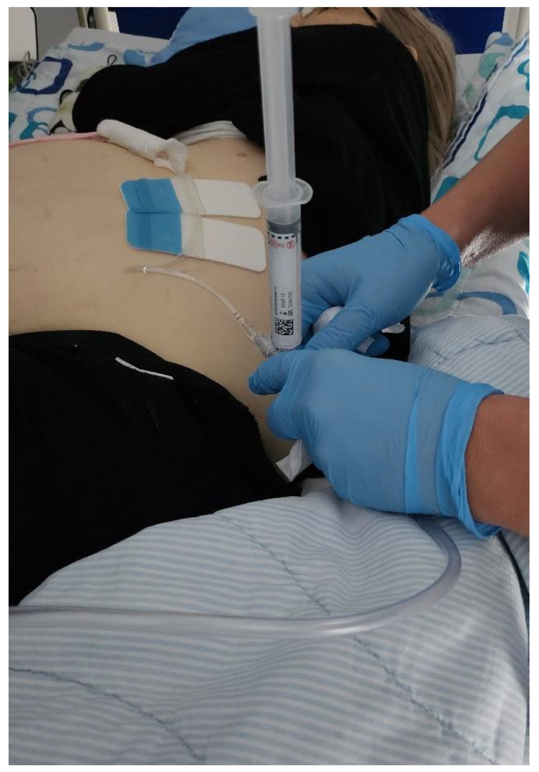
Figure 2.
(Dual image- left CEUS image, right- B-mode). Panel a – prior to US contrast injection demonstrating markedly dilated intrahepatic bile ducts in the left liver lobe; Panel b- biliary decompression and filling of the biliary catheter and bile ducts after US contrast injection.
Figure 2.
(Dual image- left CEUS image, right- B-mode). Panel a – prior to US contrast injection demonstrating markedly dilated intrahepatic bile ducts in the left liver lobe; Panel b- biliary decompression and filling of the biliary catheter and bile ducts after US contrast injection.
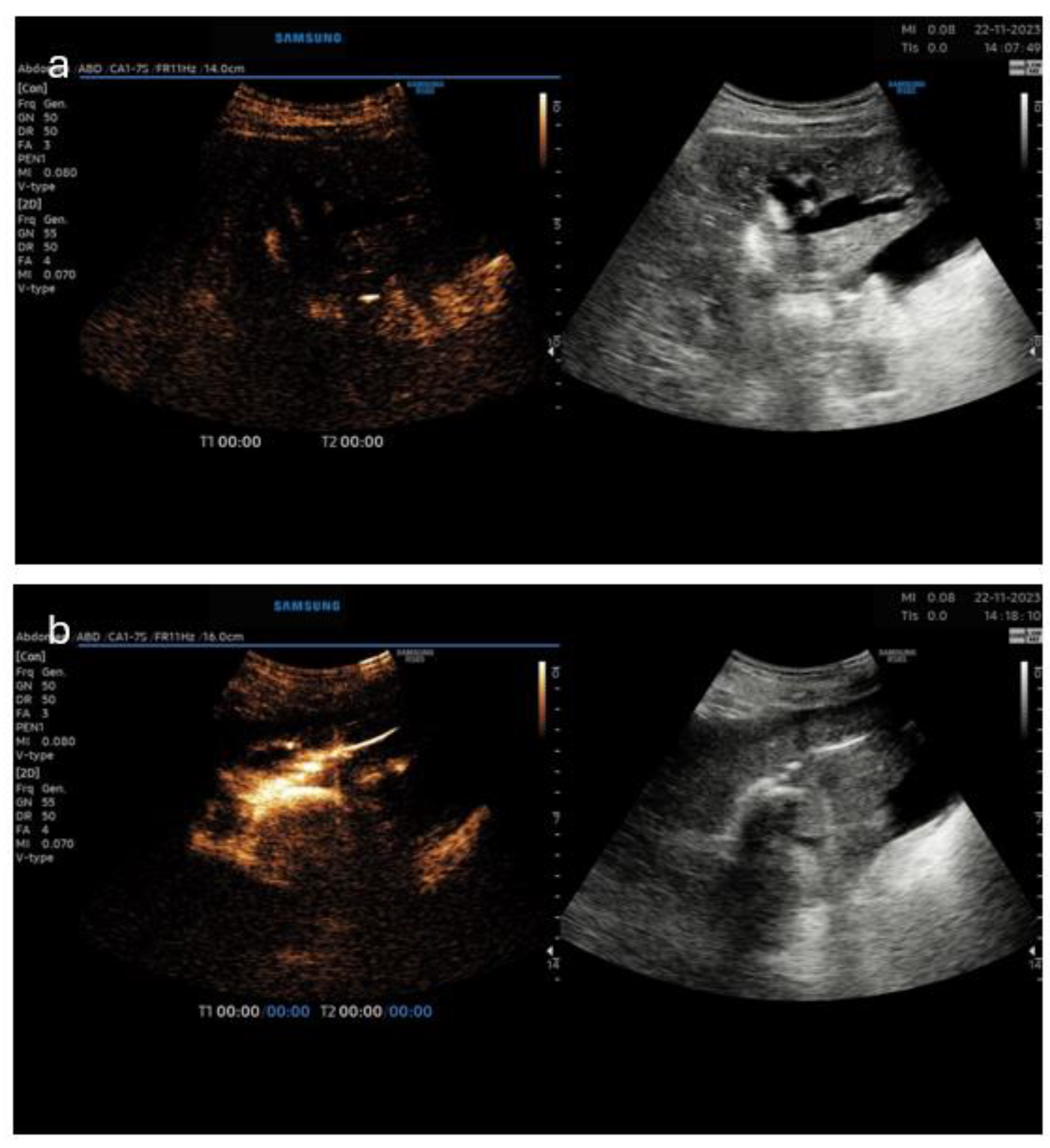
Figure 3.
Panel c- B-mode image of left-sided puncture of the dilated bile ducts; Panel d-(dual image- left CEUS image, right- B-mode) for confirmation of proper catheter position.
Figure 3.
Panel c- B-mode image of left-sided puncture of the dilated bile ducts; Panel d-(dual image- left CEUS image, right- B-mode) for confirmation of proper catheter position.
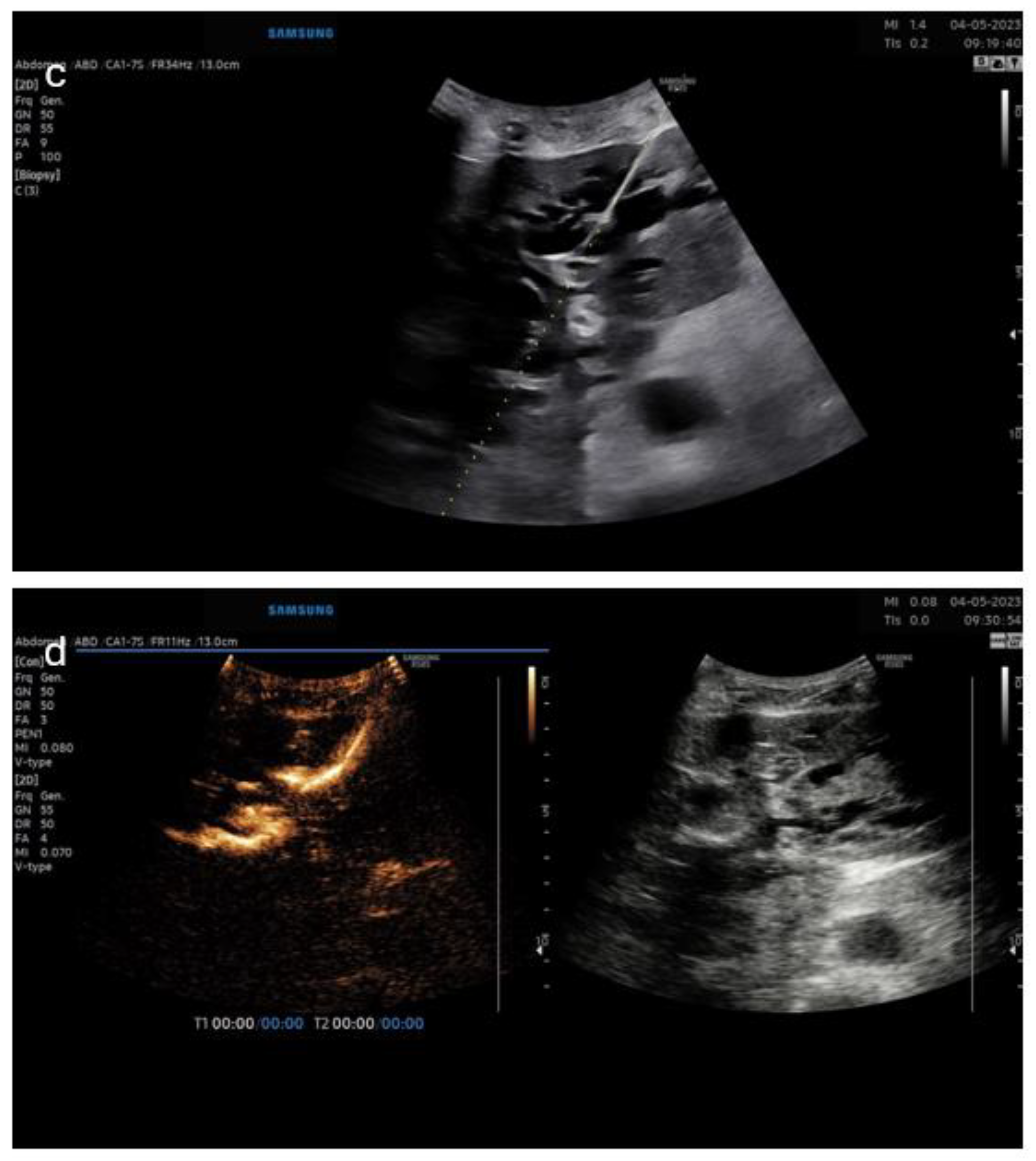
Figure 4.
Panel e- B-mode image of planning the puncture site; Panel f- dual image- left CEUS image, right- B-mode.
Figure 4.
Panel e- B-mode image of planning the puncture site; Panel f- dual image- left CEUS image, right- B-mode.
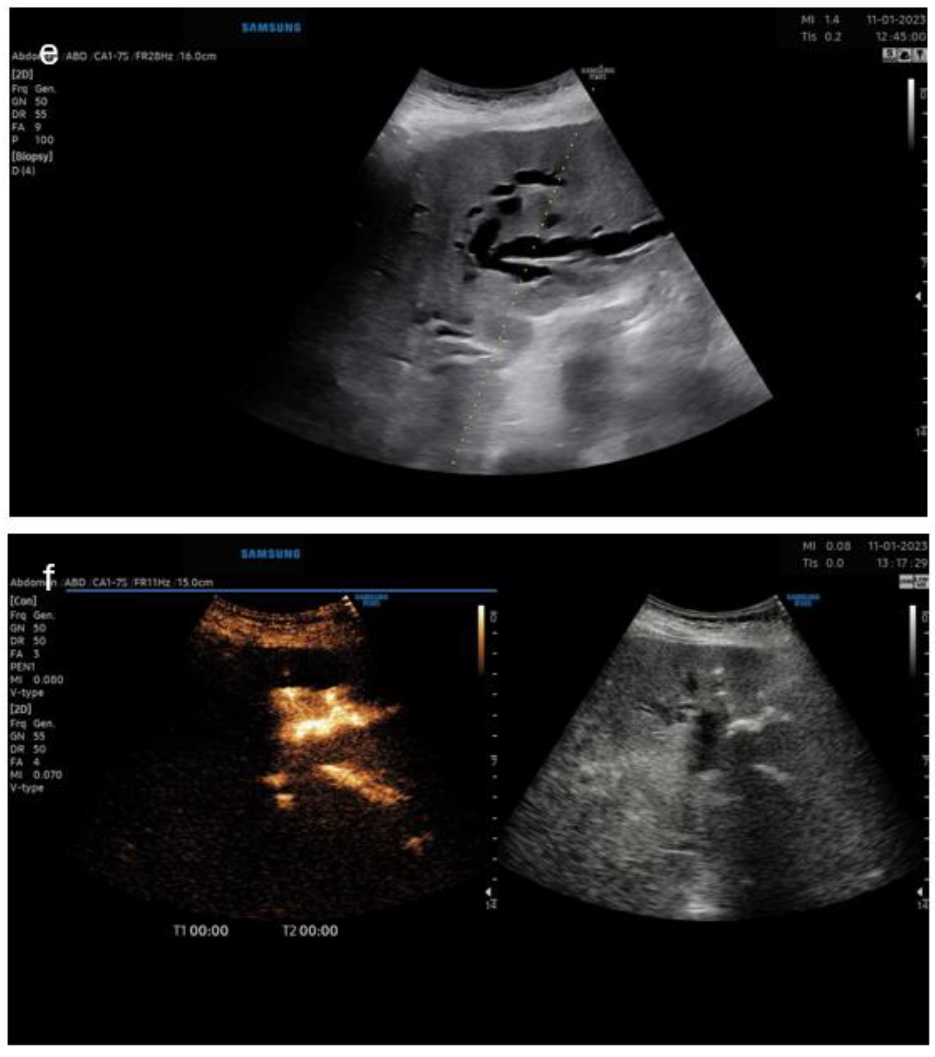

Table 1.
Studies on Intracavitary/intrabiliary CEUS during or post PTCD.
| Study | N | M/F | Indication | Groups | Success rate | Complications | Approach side | Repeated injections | Fluoroscopy comparison | Bilateral approach | Determining level of obstruction | Accuracy |
|---|---|---|---|---|---|---|---|---|---|---|---|---|
| Ignee et al. 2009 | 8 | NA | Klatskin n=5 (50%); Distal bile duct Ca n=1; Ca pancreas n=1; Chronic obstructive Pancreatitis with pseudocyst n=1; Trauma of common bile duct n=1 |
NA | n=8 (100%) | displacement of the drain n=1 | Rightsided n=6 (75%); Median subcostal n=2 (25%) |
n=2 |
Yes, but for guidance after stenosis is identified | No |
Not indicated | NA |
| Zheng et al. 2010 | 12 | 10/2 | Evaluation of biliary tree in living liver donors | Normal biliary pattern biliary variations | n=12 (100%) for adequate evaluation of biliary tree anatomy |
no |
Canulation of cystic duct and CBD intraoperatively | No | Yes-intraoperative cholangiography | NA |
NA |
Excellent for first order branches |
| Luyao et al. 2011 | 58 | 37/21 | Hilar cholangiocarcinoma n=19; HCC with invasion of common hepatic duct n=2; Hilar cholangitis n=2; Periampullary tumor n=8; Common bile duct stone n=10; postoperative stricture n=2 |
Hilar obstruction Extrahepatic obstruction |
Not indicated | PUSC- pain in the right upper quadrant n=3 PTC- epigastric pain n=25 |
Not indicated | No | Yes – after PUSC | n=8 |
PUSC –hilar 100% accuracy; PUSC – CBD obstr. 93.8% accuracy; PTC – hilar 100% accuracy; PTC- CBD obstr. 100% accuracy |
For level of obstruction: PUSC 96.6%; PTC 100% For cause: PUSC 93.1%; PTC 79.3% |
| Xu et al. 2012 | 80 | 61/19 | Localize the drainage catheter; Localize the distal tip of catheter; Evaluate level and degree of biliary obstruction |
Intrahepatic obstr. n=44; Extrahepatic obstr. n=36; Complete n=56; Incomplete n=24 |
n=80 (100%) | 4 catheters not properly placed, which required reposition | Not indicated | Yes | Yes; FC n=68; CTC n=12 |
NA |
100% accuracy (extrahepatic/intra hepatic) |
96.3 % (77/80) for complete/incomplete; 100% for tip location |
| Chopra et al. 2012 | 12 | 7/5 | Evaluation of the biliary tree via T-tube after liver transplantation | Intrahepatic; Extrahepatic |
Comparable to fluoroscopy pathology found in 4 pts anastomotic stenosis n=1; Delayed duodenal outflow n=2; Anastomotic leakage n=1 |
not indicated | Via postoperative T-tube | No | Yes fluoroscopy superior in identifying anastomotic stricture and leakage |
NA |
CEUS inferior in visualization of extra hepatic bile ducts | NA |
| Ignee et al. 2015 | 38 | 25/13 | Pancreas adenocarcinoma n=11; CBD stone n=6; Klatskin tu n=6; Inflammatory stricture n=5; Pancreatic meta n=2; Lymph node meta n=3; Duodenal Ca n=2 ICC n=1; Neuroendocrine Ca of papilla n=1; IPMN n=1 |
Hilar (above cystic duct); Extrahepatic; Complete; Incomplete |
100% |
subcutaneous hematoma n=1 catheter dislodgement n=2 pleural effusion and peural- peritoneal fistula n=1 |
Rightsided n=33; Median left hepatic n=5 |
Yes (several) plus the day after the intervention | Yes (after CEUS contrast) | NA |
Hilar obst. n=8; Extrahepatic obstr. n=30 |
97.4% for degree Incomplete 9/10 |
Table 2.
Case reports on Intracavitary/intrabiliary CEUS during or post PTCD.
| Authors | Mao | Daneshi |
| N | 1 | 1 |
| M/F | M | M |
| Indication | Bile leakage after T-tube removal after cholecystectomy and CBD exploration | Liver transplantation for familial amyloid polyneuropathy type 1 - postoperative biliary-arterial fistula |
| Successful | yes | yes |
| Complications | None | Hemobilia after internal- external drainage catheter placement |
| Approach side | Right sided segment VI | Right sided |
| Repeated injections | No | No |
| Fluoroscopy | Yes (for confirmation) | Yes (prior drainage and after CEUS cholangiogram) |
| Bilateral approach | No | No |
| Determining level | Yes (bile leakage from CBD) | Yes (microbubbles at right hepatic artery branch) |
Disclaimer/Publisher’s Note: The statements, opinions and data contained in all publications are solely those of the individual author(s) and contributor(s) and not of MDPI and/or the editor(s). MDPI and/or the editor(s) disclaim responsibility for any injury to people or property resulting from any ideas, methods, instructions or products referred to in the content. |
© 2024 by the authors. Licensee MDPI, Basel, Switzerland. This article is an open access article distributed under the terms and conditions of the Creative Commons Attribution (CC BY) license (http://creativecommons.org/licenses/by/4.0/).
Copyright: This open access article is published under a Creative Commons CC BY 4.0 license, which permit the free download, distribution, and reuse, provided that the author and preprint are cited in any reuse.


