Submitted:
07 May 2024
Posted:
07 May 2024
You are already at the latest version
Abstract
At present, magnetic selection of genetically modified cells is mainly performed with surface markers naturally expressed by cells such as LNGFR, CD4 and H-2Kk. The disadvantage of such markers is the possibility of their undesired and poorly predictable expression by unmodified cells before or after cell manipulation, which makes it essential to develop new surface markers that would not have such a drawback. Earlier, modified CD52 surface protein variants with embedded HA and FLAG epitope tags (CD52/FLAG and CD52/HA) were developed by the group of Dr. Mazurov for selection of CRISPR-modified cells using FACS. In the current study, we tested whether these markers can be used for magnetic selection of transduced cells. For this purpose, appropriate constructs were created in MigR1-based bicistronic retroviral vectors containing EGFP and DsRedExpress2 as fluorescent reporters. Cytometric analysis of the transduced NIH 3T3 cell populations after magnetic selection evaluated the efficiency of isolation and purity of the obtained populations, as well as the change in the median fluorescence intensity (MFI). The results of this study demonstrate that the surface markers CD52/FLAG and CD52/HA can be effectively used for magnetic cell selection, and their efficiencies are comparable to that of the commonly used LNGFR marker. At the same time, the significant advantage of these markers is the absence of HA and FLAG epitope sequences in cellular proteins, which rules out spurious co-isolation of negative cells.
Keywords:
HA tag
; FLAG tag
; LNGFR
; EGFP
; DsRedExpress2
; magnetic selection
; MACS selection
1. Introduction
Progress in experimental biology, which was additionally spurred by the advent of cell therapeutic applications, resulted in considerable interest in the methods of ex vivo cell separation [1,2,3,4]. The choice of a cell selection strategy is based on many parameters, however, the main criteria include the effectiveness of cell selection, the purity of the obtained populations and the preservation of cell properties after selection [5]. One of the important directions in this vast field is the specific selection of genetically modified cells. In this case, markers that include various reporter and surface proteins, as well as antibiotic resistance factors are required for cell selection [6]. Cell selection approaches depend on objectives and markers used, and range from simple antibiotic cell selection to fluorescence-activated cell sorting (FACS) and magnet-activated cell sorting (MACS). The unquestionable advantages of magnetic selection include the simplicity of the procedure and its ability to process large numbers of cells in a short time [7,8,9]. In addition, this approach makes it possible to select rare populations [10,11], as well as populations with high and low levels of surface marker expression [12]. For magnetic selection of genetically modified cells, approaches based on the use of streptavidin-biotin systems might be used, including biotinylation of the surface tag via co-expression in cells of biotin ligase BirA [13], expression of streptavidin [14] or streptavidin-binding peptide on the cell surface [15]. These approaches, however, have not become widespread; instead, certain surface markers naturally expressed by cells are primarily used for magnetic selection of modified cells, in particular CD4 [16,17] and LNGFR [10,12,18]. Commercial kits (Miltenyi Biotec) use LNGFR, CD4 and H-2Kk markers for magnetic selection of transiently modified cell populations. The disadvantage of such natural markers is that they can be expressed by unmodified cells both before and, which is especially dangerous, after manipulations with cells, which can lead to co-isolation of negative cells, reduced quality of selection and incorrect interpretation of the results. For example, LNGFR, in addition to cells of neural origin, is expressed in cells originating from all three germ layers [19], including neural crest cells [20]. LNGFR is also expressed by cells holding great promise for cell therapy, namely mesenchymal stem cells (MSCs) from various tissues [21,22], including subpopulations of MSCs with angiogenic potential [23,24]. It is important to emphasize that while MSCs originally express LNGFR, they gradually lose it during cultivation [25]. On the other hand, MSCs may re-express LNGFR during adipose differentiation [26]. Such a highly complex and dynamic expression pattern cannot a priori be excluded for other naturally expressed marker proteins.
These considerations make it highly important developing new surface markers that would not have such a disadvantage. In this regard, a promising strategy is the use of epitope tags converted to be associated with cell surface proteins. One of the most popular epitope tags are the FLAG and HA tags, which are often used to purify epitope-labeled recombinant proteins [27,28], in studies related to the structure and subcellular localization of proteins [29], as well as in genetic modification of cells [30]. The potential attractiveness of FLAG and HA tags for magnetic selection of transduced cells is, firstly, due to their small size, which makes it possible to embed them in surface proteins without changing the properties of the latter, thereby providing access of tags to antibodies associated with magnetic particles. Secondly, the epitopes of HA and FLAG tags differ from the epitopes of animal cell proteins, which excludes the selection of untransduced cells. Finally, highly specific monoclonal antibodies to HA and FLAG tags are commercially available.
Earlier, two modifications of the GPI-linked surface marker CD52 by means of embedding HA and FLAG tags within CD52 sequence were developed for effective FACS selection of CRISPR-modified cells [31]. Since both the reagents and the conditions and requirements for fluorescent and magnetic sorting differ significantly, and the applicability of the developed CD52/HA and CD52/FLAG markers for magnetic selection was not a priori guaranteed, we showed in this study the possibility of using these two markers for effective magnetic selection of transduced cells, and also compared their work with that of the LNGFR selection marker.
2. Materials and Methods
2.1. Cell Cultures
NIH 3T3 and HEK 293 cells were cultured at 37 °C and 5% CO2 in a DMEM medium (HyClone Laboratories, South Logan, UT, USA) containing 4.5 g/L glucose, 10% fetal bovine serum (FBS) (HyClone), 2 mM L-glutamine (Gibco, Gaithersburg, MD, USA), 100 units/mL penicillin and 100 µg/mL streptomycin (Gibco). Cells were detached from the culture vessels by TrypLE Express (Gibco) treatment for 2–5 min at 37 °C.
2.2. Retroviral Constructs
The two variants of the human CD52 open reading frame (ORF) containing embedded FLAG or HA epitope tags were amplified from the plasmids pUCHR-mClover-sAID_CD5Flag2 and pUCHR-mClover-sAID_CD5HA2 (kindly provided by Dr. Mazurov), and cloned into pTZ57R/T plasmid vector, followed by insert sequence verification. The hCD52/HA and hCD52/FLAG ORFs were excised by BspH I/Sal I digestion and re-cloned into the 2nd position of the bucistronic MigR1ad1 retroviral expression vector (described in [12]) via Nco I/Sal I sites to obtain MigRCD52HA and MigRCD52Fl vectors.
The final constructs used in this study, namely constructs pMigRCD52H-EGFP and pMigRCD52Fl-EGFP, were prepared by re-cloning the EGFP ORF from the pMigLNR2-EGFP construct [12] into the 1st position of MigRCD52HA and MigRCD52Fl vectors, respectively, via BamH I/EcoR I sites, while constructs pMigRCD52H-DsRex and pMigRCD52Fl-DsRex were similarly prepared by the transfer of the DsRedExpress2 ORF from the pMigLNR2-DsRex construct via BamH I/EcoR I sites of the above vectors.
The preparation of bicistronic retroviral construct with DsRedExpress2 reporter in the 1st position and hLNGFR selection marker in the 2nd position (pMigLNR2-DsRex) was described earlier [12].
2.3. Transduction of the NIH 3T3 Cells
The transfection of HEK 293 cells to obtain viral particles was performed in a 10 cm Petri dish using the calcium phosphate method when the cells reached 35% - 45% confluence. Calcium phosphate solution with 10 μg of the relevant retroviral plasmid (Figure 1) and 10 μg of pCL-Eco plasmid (Addgene#12371) was added to the culture medium. After 12 hrs, the medium was replaced with the fresh one, and after additional 24 hours, the viral medium was collected, centrifuged for 10 minutes at 400x g at 4 oC, and the supernatant passed through a 0.45-μm filter (Corning).
The transduction of NIH 3T3 cells was carried out in a 6-well plate, using a seeding density of 20.000 cells/cm2. After reaching 35-40% cell confluence, the medium was discarded and replaced with a 1:1 mixture of viral supernatant with the fresh medium. Polybrene (Sigma-Aldrich St. Louis, MO, USA) was added thereafter to a concentration of 16 µg/mL, and cells were further cultured for 72 hours, followed by the replacement of virus-containg medium with a fresh one. The efficiency of cell transduction was analyzed cytometrically 3-4 days thereafter by determining the fraction of cells in the sample expressing the reporter protein. As a result, transduced cell populations NIH 3T3-DsRedEx2-CD52/HA, NIH 3T3-DsRedEx2-CD52/FLAG, NIH 3T3-EGFP-CD52/HA, NIH 3T3-EGFP-CD52/FLAG and NIH 3T3-DsRedEx2-LNGFR were obtained; hereafter referred to as DsRedEx2-HA, DsRedEx2-FLAG, EGFP-HA, EGFP-FLAG, and DsRedEx2-LN, respectively. The transduced cells were cultured in DMEM medium with the above mentioned additives. For magnetic selection of cells with CD52/HA and CD52/FLAG markers, the cell populations transduced with construct variants differing in marker and reporter protein were combined so that the number of DsRedEx2 and EGFP cells in a mixed population was equal.
2.4. Magnetic Selection of Transduced Cells
Selection of the transduced cells was performed with equipment, consumables and reagents from Miltenyi Biotec (Germany), including OctoMACS Separator (No. 130-042-109), MS columns (No. 130-042-201), microbead-conjugated antibodies against FLAG epitope (µMACS DYKDDDDK isolation kit, No.130-101-591) and against HA epitope (µMACS HA isolation kit, No.130-091-122), as well as MACSelect anti-LNGFR Micro Beads (130-091-330). These microbead-conjugated antibodies are hereafter referred to as anti-FLAG MBs, anti-HA MBs and anti-LNGFR MBs, respectively. After incubation of cells with MBs, magnetic selection was performed in accordance with the recommendations of the manufacturer for anti-LNGFR MBs. In short, the transduced cells were detached with TripLE Express solution (Gibco) for 5 min at 37 oC. The collected cells were centrifuged for 5 min at 150x g at 4 oC, and the pellet was resuspended in phosphate buffer with 1 mM EDTA and 0.5% FBS. The cells were counted using hemocytometer, and aliquots of 0.8-1.2 x 10⁶ cells were distributed into 15-ml tubes. The cells were again centrifuged for 10 min at 150x g, and cell pellets were suspended in 380 µL of phosphate buffer with 2 mM EDTA and 0.5% FBS (PBE). Next, 20 µL of relevant MBs were added to the cell suspension and incubated for 15 min on ice. The volume of the cell suspension was then adjusted to 2 mL with PBE and applied in factions of 0.5 ml to MS column mounted on OctoMACS magnet; thereafter the column was washed with 2 mL of PBE buffer. To collect the selected cells, the column was removed from the magnet and placed in a 15 mL tube. 1 mL of PBE buffer was added to the MS column and the buffer was forced through the column with a plunger. The cells were counted using a hemocytometer. Cell viability was determined using Trypan blue.
2.5. Flow Cytometry
Cells before and after magnetic selection were collected as described above and analyzed on a BD LSRFortessa cell analyzer (BD USA) equipped with a 488 nm laser with 530±15 nm emission filter and a 561 nm laser with a 586±7.5 nm emission filter, which were used to detect EGFP and DsRedExpress2 fluorescence. EGFP and DsRedExpress2 emissions were compensated using BD FACSDiva software. Cell clusters and debris were eliminated by sequential gating of cells for size and granularity (FSC-A/SSC-A), as well as for area and peak height (FSC-A/FSC-H). The cytometric settings were kept unchanged during the measurement of all samples. The amount of data collected for each sample was at least 10.000 events. Cytometric data were processed using the Flowing Software version 2.5.1 1 (https://bioscience .fi/services/cell-imaging/flowing-software), on the basis of which the necessary calculations of the parameters of the transduced populations were carried out.
2.6. Statistical Analysis
All experiments were performed at least three times, the data were presented as average values ± standard deviation.
3. Results
3.1. Retroviral Constructs
To adequately analyze the applicability of new CD52-based markers for magnetic cell selection and compare them with the common LNGFR marker, bicistronic retroviral constructs with fluorescent protein reporters were created, which were analogous to those used by us previously [12]. It is important to note that in the mentioned work, a strict correlation was shown between the expression of reporter proteins and the LNGFR marker by transduced cells. In the retroviral vectors used in this work, the LNGFR marker in the second position of the bicistrone unit was replaced by CD52/FLAG or CD52/HA markers, while the fluorescent reporters were in the first position. Figure 1 shows the retroviral vectors that were used for subsequent transduction of NIH 3T3 cells.
3.2. Transduction of NIH 3T3 Cells
As a result of the transduction of NIH 3T3 cells by a set of vectors shown in Figure 1, five cell populations were obtained. Cytometric analysis showed that the fraction of EGFP-positive and DsRedExp2-positive cells among the variants varied, but in all cases was more than 50% (Figure 2, Figure 5). For a more unambiguous interpretation of the selection results and to ensure the possibility of selection by two markers in one cell sample, we prepared two mixed populations A and B, combining cells with different tags and reporter proteins (Figure 2). For mixed population A, DsRedEx2-HA and EGFP-FLAG variants were combined (marked in Figure 2 with numbers 1 and 3, respectively). For mixed population B, DsRedEx2-FLAG and EGFP-HA variants were used (marked in Figure 2 with numbers 2 and 4, respectively).
3.3. Magnetic Selection of NIH 3T3 Transduced Cells
3.3.1. Magnetic Selection of FLAG+ and HA+ Cells
For an adequate comparison of the effectiveness of various surface markers in selection, it is important not only to use expression constructs with similar structures, but also to employ cell populations with a similar degree of transduction, as well as use identical conditions for magnetic selection, with critical reagents procured from the same manufacturer. In addition, the concentration of antibodies, the number of cells to be selected, and the proportion of reporter protein-positive cells in populations before selection in all experiments should be similar. Finally, for the most correct comparison, the conditions of cell selection should preferably be unsaturated with intermediate concentrations of antibodies employed, thus resulting in only a fraction, but not all, of the positive cells of the original population being selected.
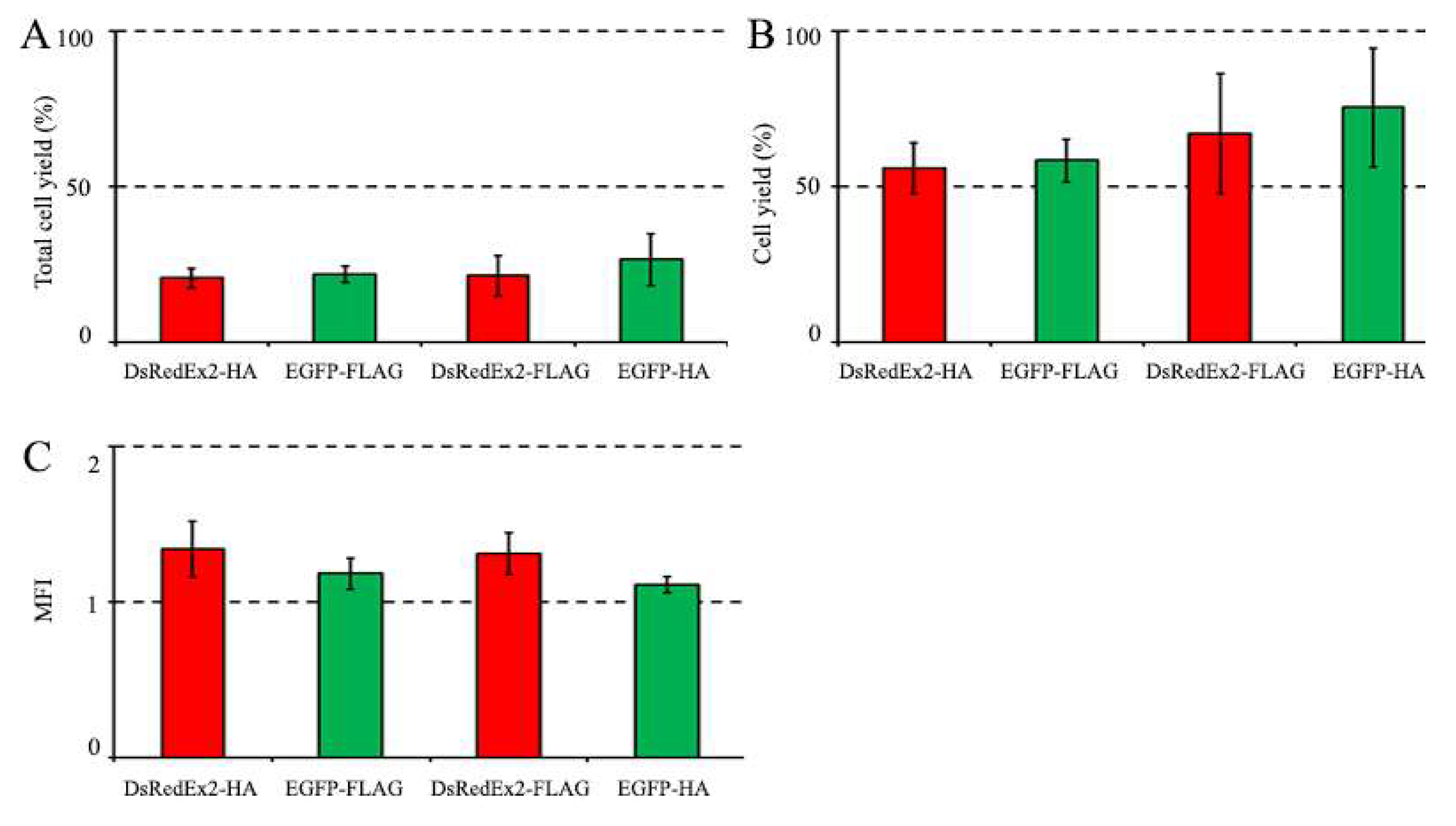
At the first stage of comparison, we performed magnetic selection of the transduced cells by FLAG and HA tags. The selection results were evaluated by changing the proportion of EGFP- and DsRedExp2-positive cells in the population after magnetic selection. Cytometry of cells of mixed populations A and B before selection showed similar amounts of EGFP and DsRedEx2 cells, and their proportions in mixed populations were about 30% (Figure 3A). Each mixed population was divided into two equal parts, one of which was used for magnetic cell selection with FLAG tag, and the other by HA tag. Magnetic cell selection was performed with 20 µL of anti-FLAG MBs or 20 µL of anti-HA MBs. Cytometry of cells before and after magnetic selection is shown in Figure 3. The percentage of cells selected from the total population differed slightly between the variants and ranged from 20.5±3.2% for DsRedEx2-HA to 26.5±8.4% for EGFP-HA (Figure 4A). The purity of selected cells was about 90% (ranging from 89.4% to 93.8%), although for the EGFP-FLAG variant was somewhat lower (83.4%) (Figure 3B). The efficiency of magnetic selection (calculated as the fraction of cells selected from the marker-positive population) ranged from 55.9±8.2% for DsRedEx2-HA to 74.5±19.2% for the EGFP-HA variant (Figure 4B). Since the efficiency of selection in all cases was essentially lower than 100%, the conditions of magnetic cell selection were adequate for appropriate comparison of selection parameters between variants. The MFI value for magnetic selection of variants was slightly higher for DsRedEx2 reporter than for EGFP, regardless of the tag by which the selection was carried out (Figure 4). In general, no significant differences between CD52/FLAG and CD52/HA markers were observed during magnetic selection.
Figure 3.
Magnetic selection of two mixed populations of transduced NIH 3T3 cells (see Figure 2). (A, B) Histograms of cells before and after magnetic selection with 20 μL of MBs. Control - untransduced NIH 3T3 cells.
Figure 3.
Magnetic selection of two mixed populations of transduced NIH 3T3 cells (see Figure 2). (A, B) Histograms of cells before and after magnetic selection with 20 μL of MBs. Control - untransduced NIH 3T3 cells.
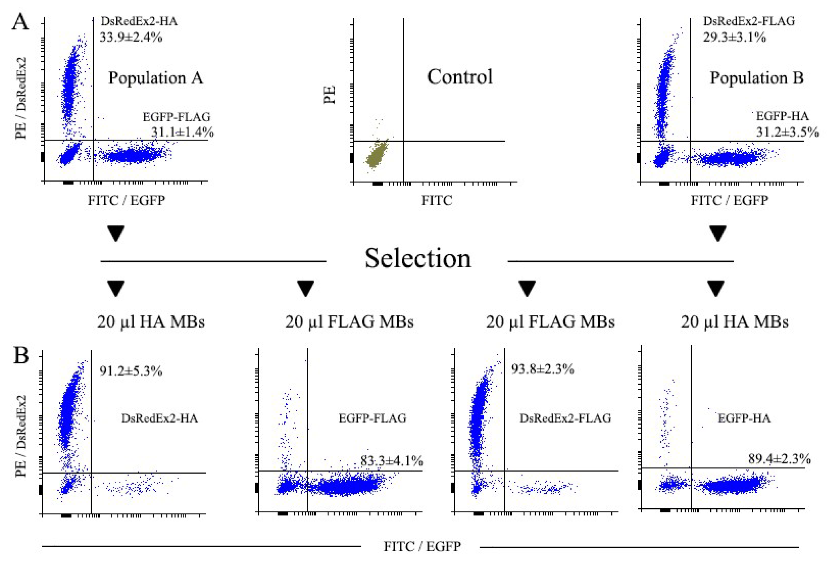
Figure 4.
Magnetic selection of two mixed populations of transduced NIH 3T3 cells (see Figure 3). (A) The yield of total cells after selection. (B) The yield of EGFP+ or DsRedExp2+ cells after selection. (C) MFI (vertical axis) depicts ratio of MFI values after and before selection. The number of cells before selection (1 x 10⁶) was taken as 100%.
3.3.2. Magnetic Selection of LNGFR+ Cells
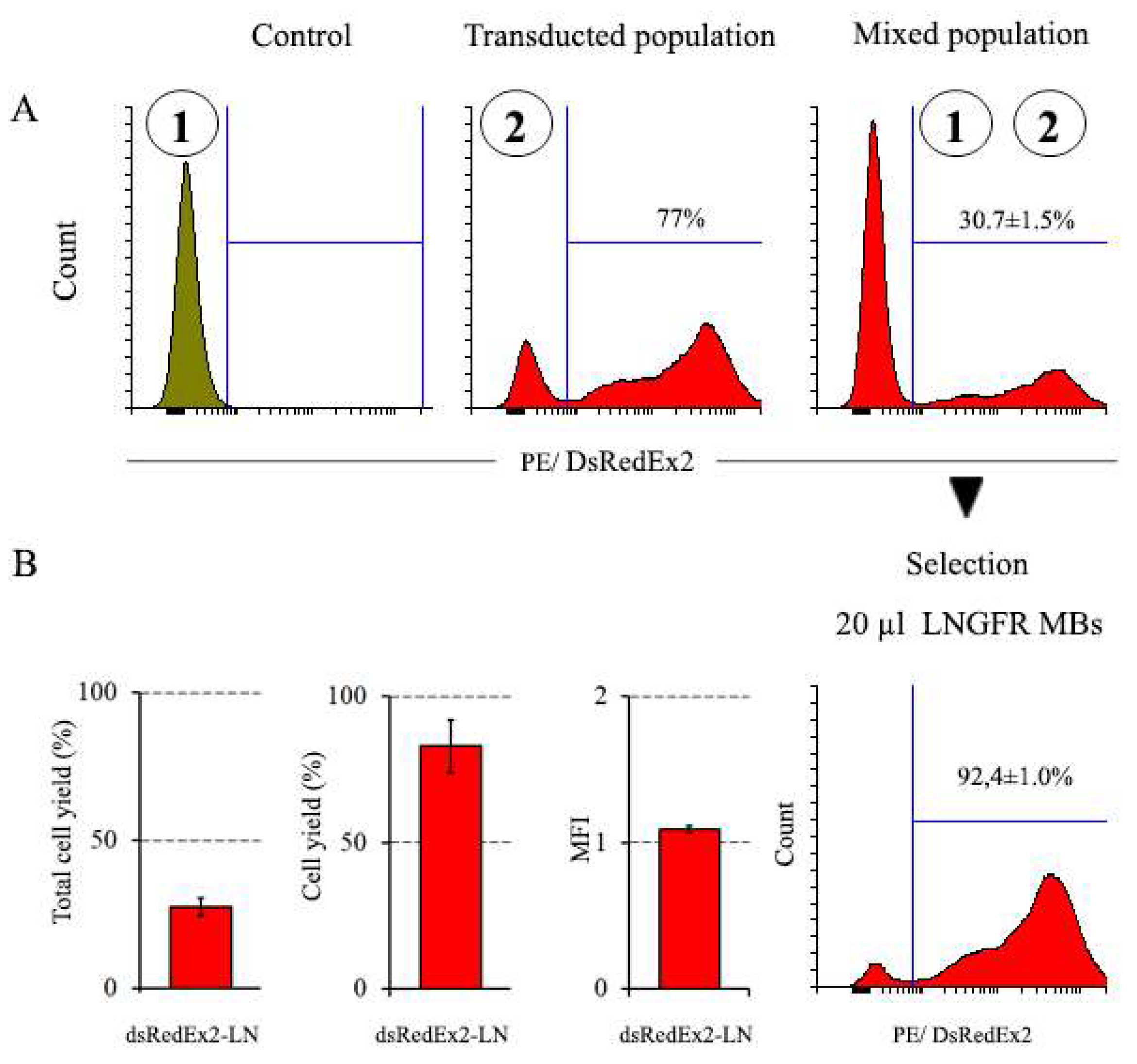
At the next stage, we compared the results of magnetic selection of cells expressing CD52/FLAG and CD52/HA markers with those of cells expressing LNGFR marker. For this, magnetic selection of LNGFR cells was carried out under conditions similar to those used for selection for CD52/FLAG and CD52/HA. After the transduction of NIH 3T3 cells by the pMigLNR2-DsRex vector, the fraction of DsRedEx2-positive cells in the population constituted more than 75% (Figure 5). To reduce the proportion of positive DsRedEx2 cells in the population to 30%, the transduced and untransduced populations of NIH 3T3 cells were mixed. Cytometry of the initial and mixed cell populations is shown in Figure 5. Magnetic selection of the mixed population was performed with 20 µL of anti-LNGFR MBs (Figure 5). As a result of selection, the proportion of DsRedExp2+ cells increased from 30.7±1.5% to 92.4±1.0%, while the proportion of selected DsRedEx2+ cells from the mixed population was close to 83% (Figure 5). The MFI value has changed slightly during selection. The results of magnetic selection of cell populations with different surface markers are shown in Table 1.
Figure 5.
Magnetic selection of a mixed population of NIH 3T3 cells expressing LNGFR surface marker. (A) Cell populations numbered 1–2 (circled) were used to create mixed population; selection was performed using 20 μL of anti-LNGFR MBs. (B) The yield of total cells and DsRedExp2+ cells after selection, and change in MFI expressed as the ratio of values MFI after and before selection. The number of cells before selection (1 x 10⁶) was taken as 100%. Control - untransduced NIH 3T3 cells.
4. Discussion
Various methods for selecting genetically modified cells have their specific advantages and drawbacks. Among them, the method of magnetic cell selection [7,32,33] stands out for its simplicity and speed, low negative impact on cells and the ability to work with large, including clinically relevant, populations. In the present work, we investigated the applicability of fused protein surface markers CD52/HA and CD52/FLAG, used initially for FACS isolation of CRISPR-edited cells, for magnetic selection of genetically modified cells. Special attention was paid to an adequate comparison of the selection efficiency attained with these markers. In addition, to analyze the work of new markers in the context of more traditional approaches, a comparison was made with the often used magnetic selection marker LNGFR. All these comparisons were carried out using bicistrone retroviral vectors identical in structure and under identical conditions of cell selection.
The results of the study show that CD52/HA and CD52/FLAG surface markers can be effectively used in magnetic cell selection. Of note, although the FLAG- and HA-specific magnetic beads used in this study are intended by the manufacturer to be used for isolation of tag-fused proteins, they apparently work adequately with cells as well. Since FLAG and HA tags are absent in vertebrate proteins, which minimizes the chances of accidental co-isolation of unwanted cells, CD52/HA and CD52/FLAG markers are a promising alternative to natural markers commonly used in magnetic selection of genetically modified cells. In addition, a significant advantage of the markers we used is their minimum size, less than 200 base pairs, which is several times smaller than the size of other markers, and is of great importance in the case of using expression vectors with limited packaging potential, such as AAV.
It is worth noting that the efficiency of CD52/HA and CD52/FLAG, although comparable to the efficiency of LNGFR, was slightly lower than the latter. A possible reason for this may be the possibility of a less than optimal configuration of HA and FLAG tags after insertion into CD52, which may lead to a certain decrease in their availability for the corresponding antibodies. As an alternative explanation, it can also be assumed that the affinity of antibodies in the case of LNGFR is slightly higher than in the case of HA and FLAG tags. It would be interesting in the future to investigate the effect of the antibody affinity on the effectiveness of magnetic selection. It should also be noted as an interesting fact that the purity of positive cell selection for cells with DsRedExp2 was slightly higher for both tags than for cells with EGFP. As we showed previously [12], a higher level of surface marker expression gives a higher purity of the population after magnetic selection. In this regard, it can be assumed that when DsRedExp2 is expressed by cells, higher expression of CD52/HA and CD52/FLAG markers is achieved, which, in turn, would lead to an increase in the degree of purity of the isolated populations. It should be noted that no complete elimination of negative cells during magnetic selection was achieved, and the purity of the selected cells did not reach 100%; however, this result is generally consistent with the data of previously published studies [9,34].
It should also be mentioned that the FLAG tag embedded in tetraspanin CD63 has recently been used for magnetic enrichment of cells [35]. According to the results of this work, however, it remains unclear how successful the use of such a marker was in terms of the yield of selected cells and the purity of the isolated population, although, by indirect indications, it was moderate. In our work, these issues were properly addressed, including the comparison with the traditional magnetic selection marker LNGFR. It is also worth mentioning the several-fold reduced size of the markers we used compared to that of CD63, which might be a significant advantage in some applications.
5. Conclusions
The results of this work demonstrate that the surface markers CD52/FLAG and CD52/HA, along with LNGFR, can be effectively used for magnetic selection of genetically modified cells. A comparison of the purity, efficiency of cell selection and MFI did not show significant differences between the three markers, however, the results of selection of cells with LNGFR turned out to be slightly better than those of cells with HA and FLAG tags. At the same time, the absence of HA and FLAG epitopes in cellular proteins, along with the small size of the markers used, eliminates the possibility of unintended co-isolation of negative cells and represents a significant advantage over LNGFR and other natural proteins used as selective markers in magnetic selection.
Author Contributions
Conceptualization, A.B., N.P. and O.K.; experimental work, N.P., A.P., A.B. and O.K.; data analysis, N.P. and O.K.; writing—original draft preparation, O.K. and N.P.; writing—review and editing, N.P., O.K. and A.B; supervision, A.B.; funding acquisition, A.B. All authors have read and agreed to the present version of the manuscript.
Funding
This work was supported by the Russian Science Foundation (grant no. 18-14-00300).
Institutional Review Board Statement
Not applicable.
Informed Consent Statement
Not applicable.
Data Availability Statement
Original data are available upon request.
Acknowledgments
Authors cordially thank Dr. Mazurov for a kind gift of the plasmids pUCHR-mClover-sAID_CD5Flag2 and pUCHR-mClover-sAID_CD5HA2.
Conflicts of Interest
The authors declare no conflict of interest.
References
- de Wynter, E.A.; Coutinho, L.H.; Pei, X.; Marsh, J.C.; Hows, J.; Luft, T.; Testa, N.G. Comparison of Purity and Enrichment of CD34+ Cells From Bone Marrow, Umbilical Cord and Peripheral Blood (Primed for Apheresis) Using Five Separation Systems. Stem Cells 1995, 13, 5. 524-532. [CrossRef]
- Dainiak, M.B.; Kumar, A.; Galaev, I.Yu.; Mattiasson, B. Methods in cell separations. Adv. Biochem. Eng. Biotechnol. 2007, 106, 1–18. [Google Scholar] [CrossRef]
- Plouffe, B.D.; Murthy, S.K.; Lewis, L.H. Fundamentals and Application of Magnetic Particles in Cell Isolation and Enrichment. Rep. Prog. Phys. 2015, 78, 016601. [Google Scholar] [CrossRef]
- Frenea-Robin, M.; Marchalot, J. Basic principles and recent advances in magnetic cell separation. Magnetochemistry 2022, 8, 11. [CrossRef]
- Amos, P.J.; Bozkulak, E.C.; Qyang, Y. Methods of Cell Purification: A Critical Juncture for Laboratory Research and Translational Science. Cells Tissues Organs 2012, 195, 26–40. [Google Scholar] [CrossRef]
- Mortensen, R.M.; Kingston, R.E. Selection of transfected mammalian cells. Curr. Protoc. Mol. Biol. 2009, 86:9.5.1-9.5.13. [CrossRef]
- Schmitz, B.; Radbruch, A.; Kümmel, T.; Wickenhauser, C.; Korb, H.; Hansmann, M.; Thiele, J.; Fischer, R. Magnetic activated cell sorting (MACS)—a new immunomagnetic method for megakaryocytic cell isolation: comparison of different separation techniques. European Journal of Haematology 1994, 5, 267–275. [Google Scholar] [CrossRef]
- Sutermaster, B.A.; Darling, E.M. Considerations for high-yield, high-throughput cell enrichment: fluorescence versus magnetic sorting. Sci. Rep. 2019, 9, 227. [Google Scholar] [CrossRef]
- Pan, J.; Wan, J. Methodological comparison of FACS and MACS isolation of enriched microglia and astrocytes from mouse brain. J. Immunol. Methods 2020, 486, 112834. [Google Scholar] [CrossRef]
- Lee, M,Y.; Lufkin, T. Development of the "Three-step MACS": a novel strategy for isolating rare cell populations in the absence of known cell surface markers from complex animal tissue. J. Biomol. Tech. 2012, 23, 2, 69-77. [CrossRef]
- Wang, Z.; Wang, H.; Lin, S.; Ahmed, S.; Angers, S.; Sargent, E.H.; Kelley, S.O. Nanoparticle Amplification Labeling for High-Performance Magnetic Cell Sorting. Nano Lett. 2022, 12, 4774–4783. [Google Scholar] [CrossRef]
- Polyakova, N.; Kandarakov, O.; Belyavsky, A. Selection of cell populations with high or low surface marker expression using magnetic sorting. Cells 2023, 12. [Google Scholar] [CrossRef]
- Han, H.; Liu, Q.; He, W.; Ong, K.; Liu, X.; Gao, B. An efficient vector system to modify cells genetically. PLoS ONE, 2638. [Google Scholar] [CrossRef]
- Gotoh, H.; Matsumoto, Y. Cell-surface streptavidin fusion protein for rapid selection of transfected mammalian cells. Gene 2007, 2, 146–153. [Google Scholar] [CrossRef]
- Matheson, N.J.; Peden, A.A.; Lehner, P.J. Antibody-free magnetic cell sorting of genetically modified primary human CD4+ T cells by one-step streptavidin affinity purification. PLoS ONE 2014, 9(10):e111437. [CrossRef]
- Gaines, P.; Wojchowski, D.M. pIRES-CD4t, a dicistronic expression vector for MACS-or FACS-based selection of transfected cells. Biotechniques 1999, 4, 683–688. [Google Scholar] [CrossRef]
- David, R.; Groebner, M.; Franz, W.M. Magnetic cell sorting purification of differentiated embryonic stem cells stably expressing truncated human CD4 as surface marker. Stem Cells 2005, 23, 477–482. [Google Scholar] [CrossRef]
- Kaufman, W.L.; Kocman, I.; Agrawal, V.; Rahn, H-P. ; Besser, D.; Gossen, M. Homogeneity and persistence of transgene expression by omitting antibiotic selection in cell line isolation. Nucleic Acids Res. 2008, 17, e111. [Google Scholar] [CrossRef]
- Thomson, T.M.; Rettig, W.J.; Chesa, P.G.; Green, S.H.; Mena, A.C.; Old, L.J. Expression of human nerve growth factor receptor on cells derived from all three germ layers. Exp. Cell Res. 1988, 2, 533–539. [Google Scholar] [CrossRef]
- Zhang, J.; Duan, X.; Zhang, H.; Deng, Z.; Zhou, Z.; Wen, N.; Smith, A.J.; Zhao, W.; Jin, Y. Isolation of neural crest-derived stem cells from rat embryonic mandibular processes. Biol. Cell 2006, 10, 567–575. [Google Scholar] [CrossRef]
- Quirici, N.; Soligo, D.; Bossolasco, P.; Servida, F.; Lumini, C.; Deliliers, G.L. Isolation of bone marrow mesenchymal stem cells by anti-nerve growth factor receptor antibodies. Exp. Hematol. 2002, 7, 783–791. [Google Scholar] [CrossRef]
- Barilani, M.; Banfi, F.; Sironi, S.; Ragni, E.; Guillaumin, S.; Polveraccio, F.; Rosso, L.; Moro, M.; Astori, G.; Pozzobon, M.; Lazzari, L. Low-affinity Nerve Growth Factor Receptor (CD271) Heterogeneous Expression in Adult and Fetal Mesenchymal Stromal Cells. Sci. Rep. 2018, 8, 9321. [Google Scholar] [CrossRef]
- Sasse, S.; Skorska, A.; Lux, C.A.; Steinhoff, G.; David, R.; Gaebel, R. Angiogenic Potential of Bone Marrow Derived CD133+ and CD271+ Intramyocardial Stem Cell Trans-Plantation Post MI. Cells 2020, 9, 78. [Google Scholar] [CrossRef]
- Smith, R.J.P.; Faroni, A.; Barrow, J.R.; Soul, J.; Reid, A.J. The angiogenic potential of CD271+ human adipose tissue-derived mesenchymal stem cells. Stem Cell Res. Ther. 2021, 12, 160. [Google Scholar] [CrossRef]
- Schäck, L.M.; Noack, S.; Weist, R.; Jagodzinski, M.; Krettek, C.; Buettner, M.; Hoffmann, A. Analysis of surface protein expression in human bone marrow stromal cells: new aspects of culture-induced changes, inter-donor differences and intracellular expression. Stem Cells Dev. 2013, 24, 3226–3235. [Google Scholar] [CrossRef]
- Walmsley, G.G.; Atashroo, D.A.; Maan, Z.N.; Hu, M.S.; Zielins, E.R.; Tsai, J.M.; Duscher, D.; Paik, K.; Tevlin, R.; Marecic, O.; Wan, D.C.; Gurtner, G.C.; Longaker, M.T. High-Throughput Screening of Surface Marker Expression on Undifferentiated and Differentiated Human-Derived Stromal Cells. Tissue Eng. Part A. 2015, 21, 2281–2291. [Google Scholar] [CrossRef]
- Zhao, X.; Li, G.; Liang, S. Several affinity tags commonly used in chromatographic purification. J. Anal. Methods Chem. 2013, 581093. [Google Scholar] [CrossRef] [PubMed]
- Lafuente-González, E.; Guadaño-Sánchez, M.; Urriza-Arsuaga, I.; Urraca, J.L. Core-Shell Magnetic Imprinted Polymers for the Recognition of FLAG-Tagpeptide. Int. J. Mol. Sci. 2023, 24, 3453. [Google Scholar] [CrossRef] [PubMed]
- Vandemoortele, G.; Eyckerman, S.; Gevaert, K. Pick a Tag and Explore the Functions of Your Pet Protein. Trends Biotechnol. 2019, 37, 10, 1078-1090.
- Zhang, J.; Zhou, T.; Shan, Y.; Pan, G. Generation of RYBP FLAG-HA knock-in human embryonic stem cell line through CRISPR/Cas9-mediated homologous recombination. Stem Cell Res. 2022, 62, 102803. [Google Scholar] [CrossRef] [PubMed]
- Zotova, A.; Pichugin, A.; Atemasova, A.; Knyazhanskaya, E.; Lopatukhina, E.; Mitkin, N.; Holmuhamedov, E.; Gottikh, M.; Kuprash, D.; Filatov, A.; Mazurov, D. Isolation of gene-edited cells via knock-in of short glycophosphatidylinositol-anchored epitope tags. Sci. Rep. 2019, 9, 3132. [Google Scholar] [CrossRef] [PubMed]
- Grützkau, A.; Radbruch, A. Small but mighty: how the MACS-technology based on nanosized superparamagnetic particles has helped to analyze the immune system within the last 20 years. Cytometry A 2010, 77, 643–647. [Google Scholar] [CrossRef] [PubMed]
- Plouffe, B.D.; Murthy, S.K.; Lewis, L.H. Fundamentals and application of magnetic particles in cell isolation and enrichment: a review. Rep. Prog. Phys. 2015, 78, 016601. [Google Scholar] [CrossRef] [PubMed]
- Wang, G.; Yu, G.; Wang, D.; Guo, S.; Shan, F. Comparison of the purity and vitality of natural killer cells with different isolation kits. Exp. Ther. Med. 2017, 13, 1875–1883. [Google Scholar] [CrossRef]
- Rufino-Ramos, D.; Lule, S.; Mahjoum, S.; Ughetto, S.; Bragg, D.C.; Pereira de Almeida, L.; Breakefield, X.O.; Breynea, K. Using genetically modified extracellular vesicles as a non-invasive strategy to evaluate brain-specific cargo. Biomaterials 2022, 281, 121366. [Google Scholar] [CrossRef]
Figure 1.
Bicistronic expression constructs used in this study. The constructs were prepared on the basis of retroviral MigR1 vector as described in Materials and methods.
Figure 1.
Bicistronic expression constructs used in this study. The constructs were prepared on the basis of retroviral MigR1 vector as described in Materials and methods.
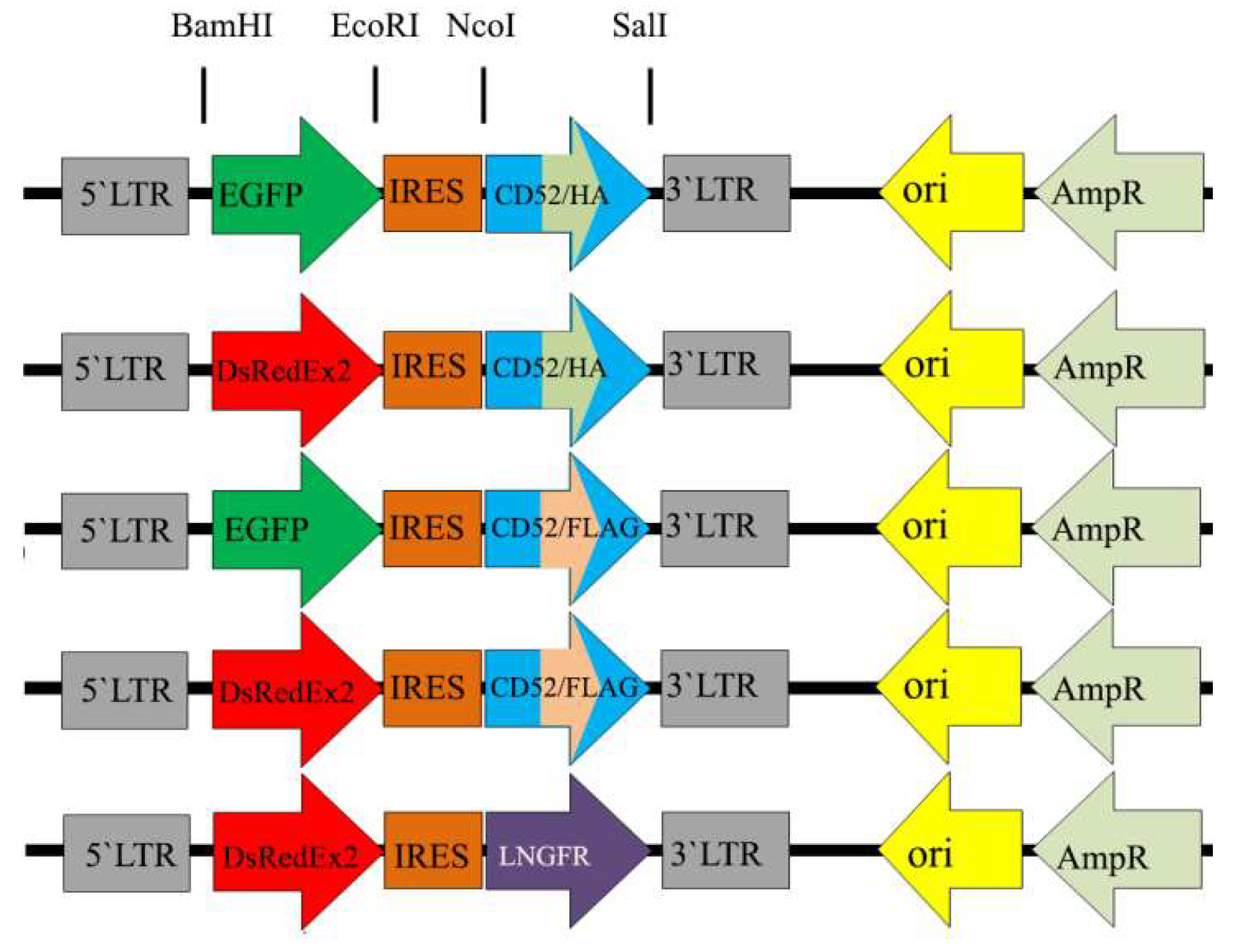
Figure 2.
Transduction of NIH 3T3 cells with the bicistronic constructs expressing DsRedExpress2 (panel A) or EGFP (panel B) reporters. Cell populations numbered 1–4 (circled) were used to create mixed populations (panel C) used subsequently for magnetic selection as depicted in Figure 3. Control - untransduced NIH 3T3 cells.
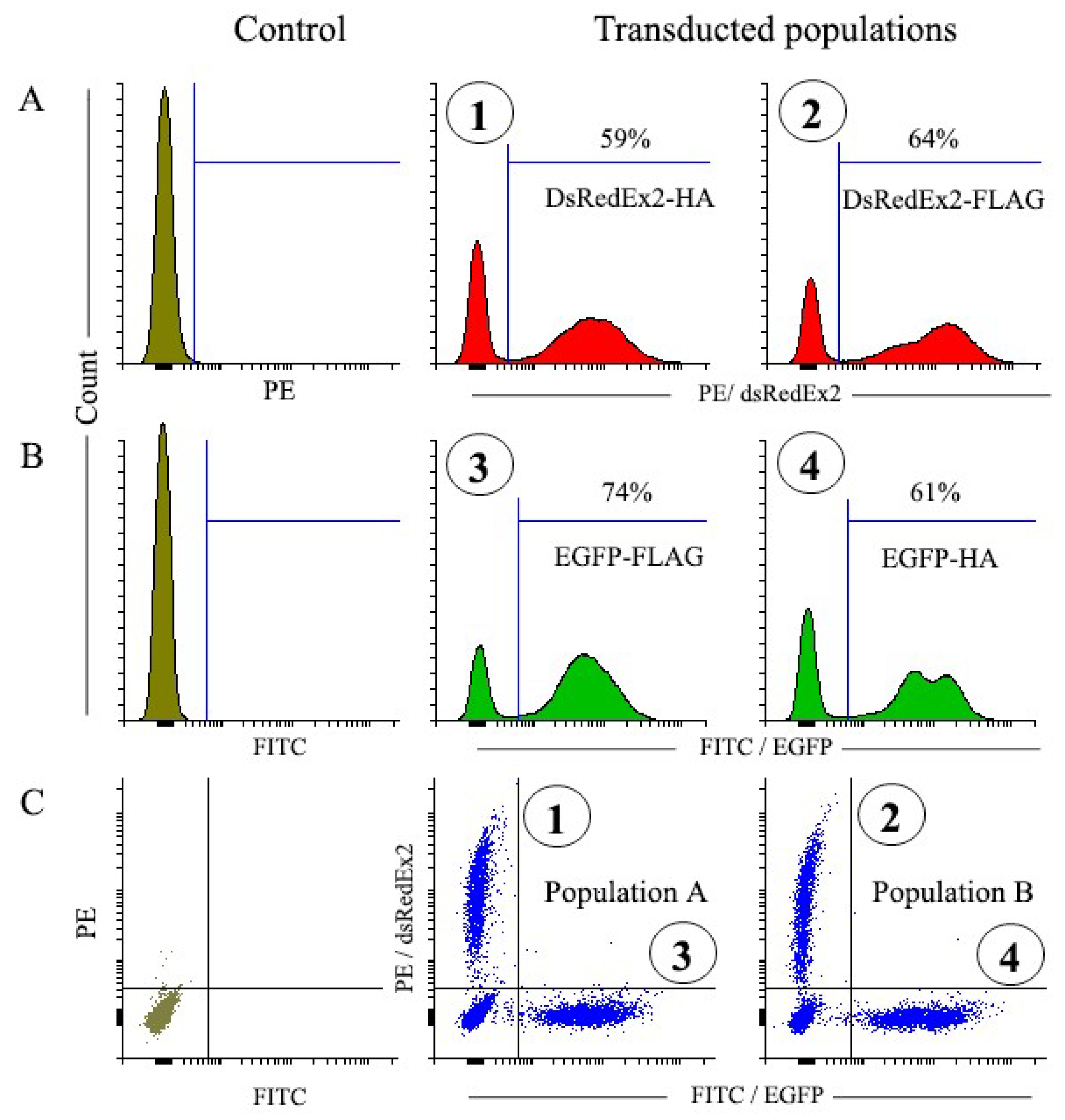

Table 1.
Characteristics of populations with different surface markers after magnetic selection.
| Scheme . | Total yield (%) |
Cell Yield (%) |
Purity (%) |
MFI change* |
|---|---|---|---|---|
| DsRedExp2-HA | 20.5±3.2 | 55.9±8.2 | 91.2±5.3 | 1.34±0.18 |
| EGFP-FLAG | 21.7±2.5 | 58.3±6.9 | 83.3±4.1 | 1.18±0.10 |
| DsRedExp2-FLAG | 21.2±6.6 | 67.0±19.3 | 93.8±2.3 | 1.31±0.14 |
| EGFP-HA | 26.5±8.4 | 74.5±19.2 | 89.4±2.3 | 1.11±0.05 |
| DsRedExp2-LNGFR | 27.5±3.0 | 82.9±9.0 | 92.4±1.0 | 1.09±0.02 |
* MFI expressed as the ratio of values MFI after and MFI before selection.
Disclaimer/Publisher’s Note: The statements, opinions and data contained in all publications are solely those of the individual author(s) and contributor(s) and not of MDPI and/or the editor(s). MDPI and/or the editor(s) disclaim responsibility for any injury to people or property resulting from any ideas, methods, instructions or products referred to in the content. |
© 2024 by the authors. Licensee MDPI, Basel, Switzerland. This article is an open access article distributed under the terms and conditions of the Creative Commons Attribution (CC BY) license (http://creativecommons.org/licenses/by/4.0/).
Copyright: This open access article is published under a Creative Commons CC BY 4.0 license, which permit the free download, distribution, and reuse, provided that the author and preprint are cited in any reuse.


