
Submitted:
16 April 2024
Posted:
16 April 2024
You are already at the latest version
Abstract
Introduction: Signet-ring cells are typically associated with mucin-secreting epithelium thus most commonly found in a gastrointestinal tract, however not exclusively. Primary signet ring cell carcinoma of the prostate is a rare and poorly differentiated, aggressive acinar adenocarcinoma variant with a grim prognosis. Clinical case: In June of 2023 a 54-year-old Caucasian male came to the hospital complaining of lower urinary tract obstructive symptoms with occasional macrohematuria, non-specific body aches, and shortness of breath. A prostate specimen obtained in transurethral resection of the prostate was sent for histopathological examination. After a series of extraprostatic diagnostic workups including fibrogastroduodenoscopy, colonoscopy computed tomography imaging, and immunohistochemical studies, the patient was diagnosed with primary prostatic signet-ring cell adenocarcinoma st. IV. Unfortunately, the patient with the disease this advanced, PE, and third-degree thrombocytopenia was not a candidate for chemotherapy and died of cardiopulmonary insufficiency later that week. Discussion: Prostatic signet-ring cell carcinoma accounts for 0.02% of all prostate adenocarcinoma cases. Due to its nature and epidemiology, a diligent extraprostatic investigation has to be carried out. The disease often presents with unremarkable clinical symptoms and variable serum prostate-specific antigen results which may contribute to its late diagnosis which together with inconsistent immunohistochemical findings and unpredictable response to hormonal treatment pose both diagnostic and therapeutic challenges that negatively affect the prognosis. Conclusion: The study highlights the importance of a multidisciplinary approach and the need for diagnostic and therapeutic consensus within the research community in search of the primary site of the disease which may positively influence the prognosis.
Keywords:
Prostate cancer
; signet-ring cell-like carcinoma
; features of mucin
1. Introduction
Prostate cancer is the most commonly diagnosed cancer in men with a rapidly growing age-related incidence and mortality each year (1). The majority of prostate cancers are not clinically evident and are of a relatively low virulence and this may be true in the case of an acinar adenocarcinoma which makes up 93% of all prostate cancer cases (2). Even though the acinar type is the most common, both signet-ring cell subtype and mucinous histological pattern are considered to be extremely rare (3,4).
Signet-ring cells acquire their histological appearance in the presence of an intracellular clear cytoplasmic vacuole which pushes the nucleus into the periphery giving it a crescent shape (5). The cells were first grossly characterized by their distinctive looks and diffuse submucosal growth pattern in the early 1950s (6). The variant is predominantly observed in the gastrointestinal tract, emphasizing the stomach and colon, thus naturally these organs are the first ones to be ruled out in suspicion of metastatic disease and are a reference point in other-organ signet ring cell carcinoma (SRCC) cases (5). However, it is not an uncomplicated process, since early gastric SRCC is nearly macroscopically invisible, and thus in most cases, it is diagnosed rather late posing a great diagnostic and therapeutic challenge (7).
Primary prostatic signet-ring cell-like adenocarcinoma is an exceptionally rare, poorly differentiated epithelial cancer with unremarkable genitourinary complaints, inconsistent immunohistochemical study findings, non-universally followed classification criteria, and unestablished diagnostic and therapeutic protocols which all together negatively influence already poor chances of 5-year survival (5,6,8).
To gain a more comprehensive understanding of the entity, we present a clinical case of prostate adenocarcinoma with signet ring cells and features of mucin observed in a single tertiary cancer center in Lithuania.
2. Case Report
In June of 2023, a 54-year-old Caucasian male with an unremarkable history presented with symptoms of episodic haematuria, severe non-specific body aches, shortness of breath, and anuria which was initially treated with cystostomy before undergoing transurethral prostate resection (TURP). Histopathological examination of the resection revealed a mass of poorly differentiated (G3) adenocarcinoma of an unknown primary site with a diffuse distribution of signet-ring cells (40%) with intracellular and extracellular mucin that constituted 20% - of the tumor found within the specimen. An immunophenotype was later determined and the tumor cells were positive for CDX2; Cadherin 17; MUC2; focally for Cytokeratin 20 (CK20) and Synaptophysin, and negative for CK7, NKX3.1, GATA3, SATB2, MUC5, MUC6. Both the visual representation and immunophenotype were suggestive of a metastatic tumor of the lower gastrointestinal tract, thus further investigation for the primary site verification was necessary. In search of a primary site fibrogastroduodenoscopy, chest and abdominal computed tomography, and lesser pelvic magnetic nuclear resonance scan were performed that disclosed erosive gastroduodenopathy, pulmonary embolism (PE), direct seminal vesicle and urinary bladder infiltration, multiple osteosclerotic metastases which lead to the conclusion that the primary site of signet-ring cell carcinoma is the prostate gland itself. A TNM class and stage were assigned accordingly - cT4N1M1c stage IV. The patient with a disease this advanced did not meet the criteria for radical prostatectomy (RP) or radiation therapy (RT), and was denied chemotherapy due to third-degree thrombocytopenia, due to which antithrombotic treatment, which first was prescribed for PE treatment, was discontinued as well. Infusions of Zoledronic acid were initiated. The patient died of cardiopulmonary insufficiency later that week.
Figure 1.
(All images stained with Hematoxylin and eosin (HE) and 200x magnification) A—Signet ring cells infiltrated in between prostate glands; B—Clusters of signet ring cells (arrow) in pools of extracellular mucin.
Figure 1.
(All images stained with Hematoxylin and eosin (HE) and 200x magnification) A—Signet ring cells infiltrated in between prostate glands; B—Clusters of signet ring cells (arrow) in pools of extracellular mucin.
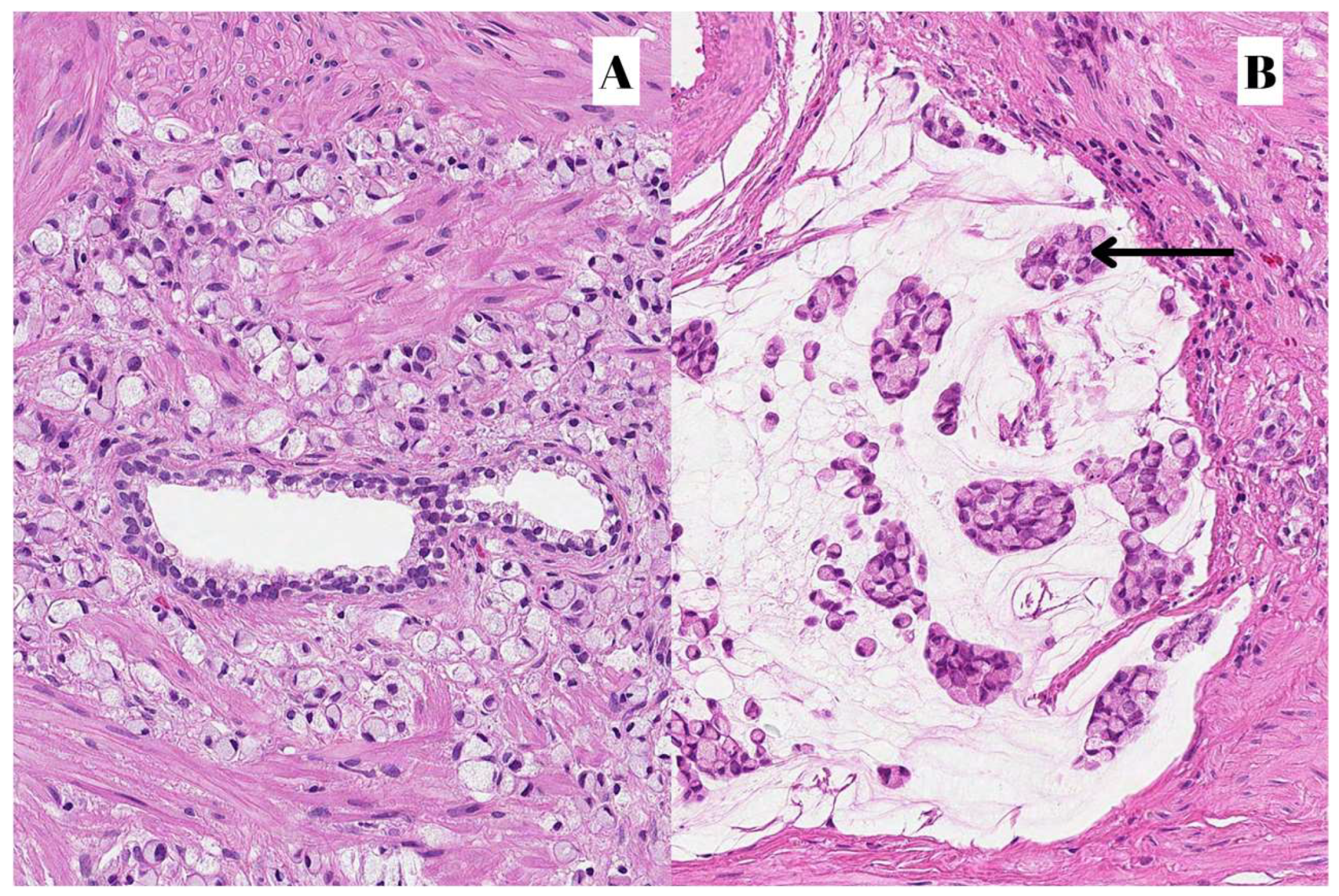
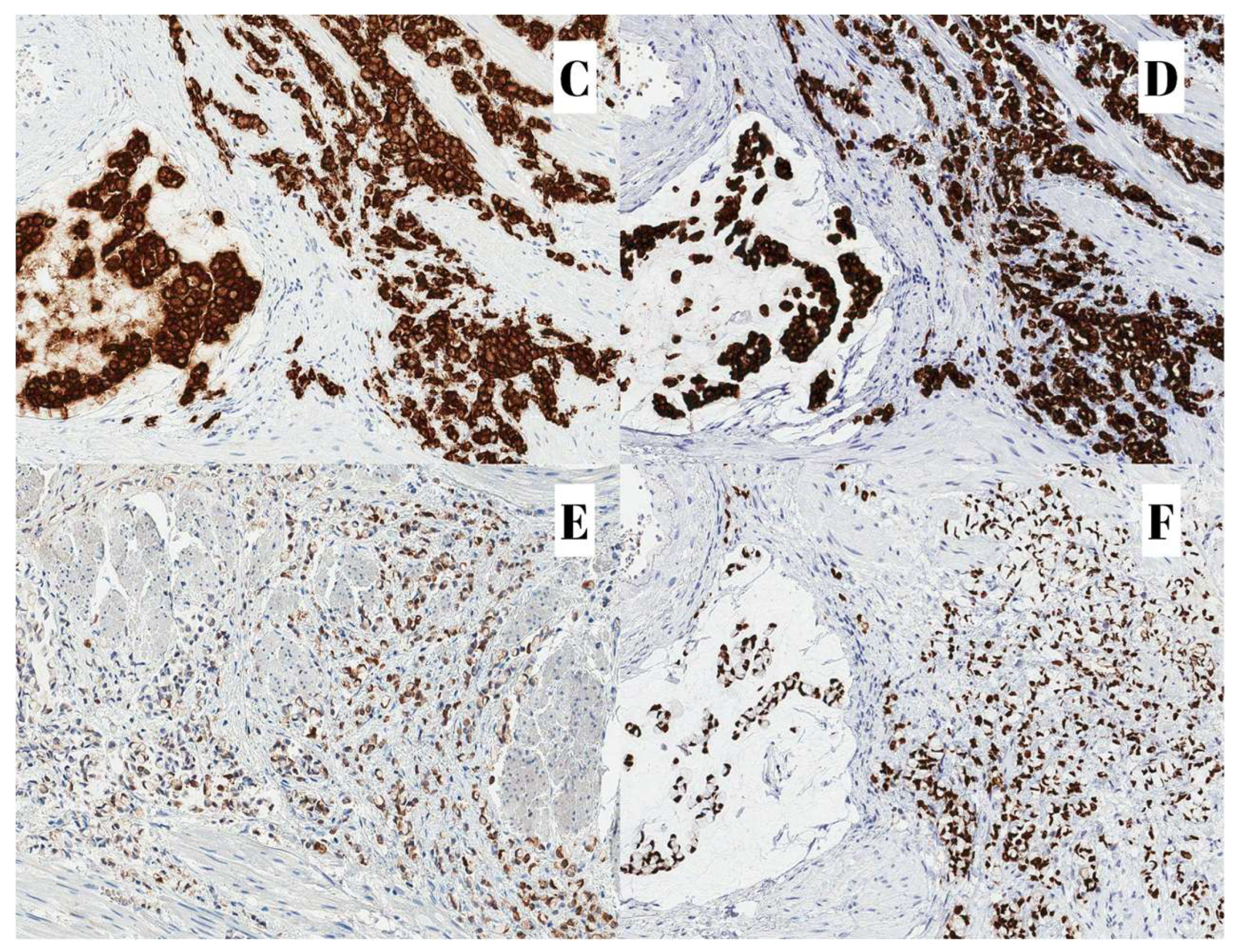
Figure 2.
(All images stained with HE and 200x magnification) C—Cadherin 17; D—Muc2; E—CK20; F—CDX2.
Figure 2.
(All images stained with HE and 200x magnification) C—Cadherin 17; D—Muc2; E—CK20; F—CDX2.
3. Literature Review
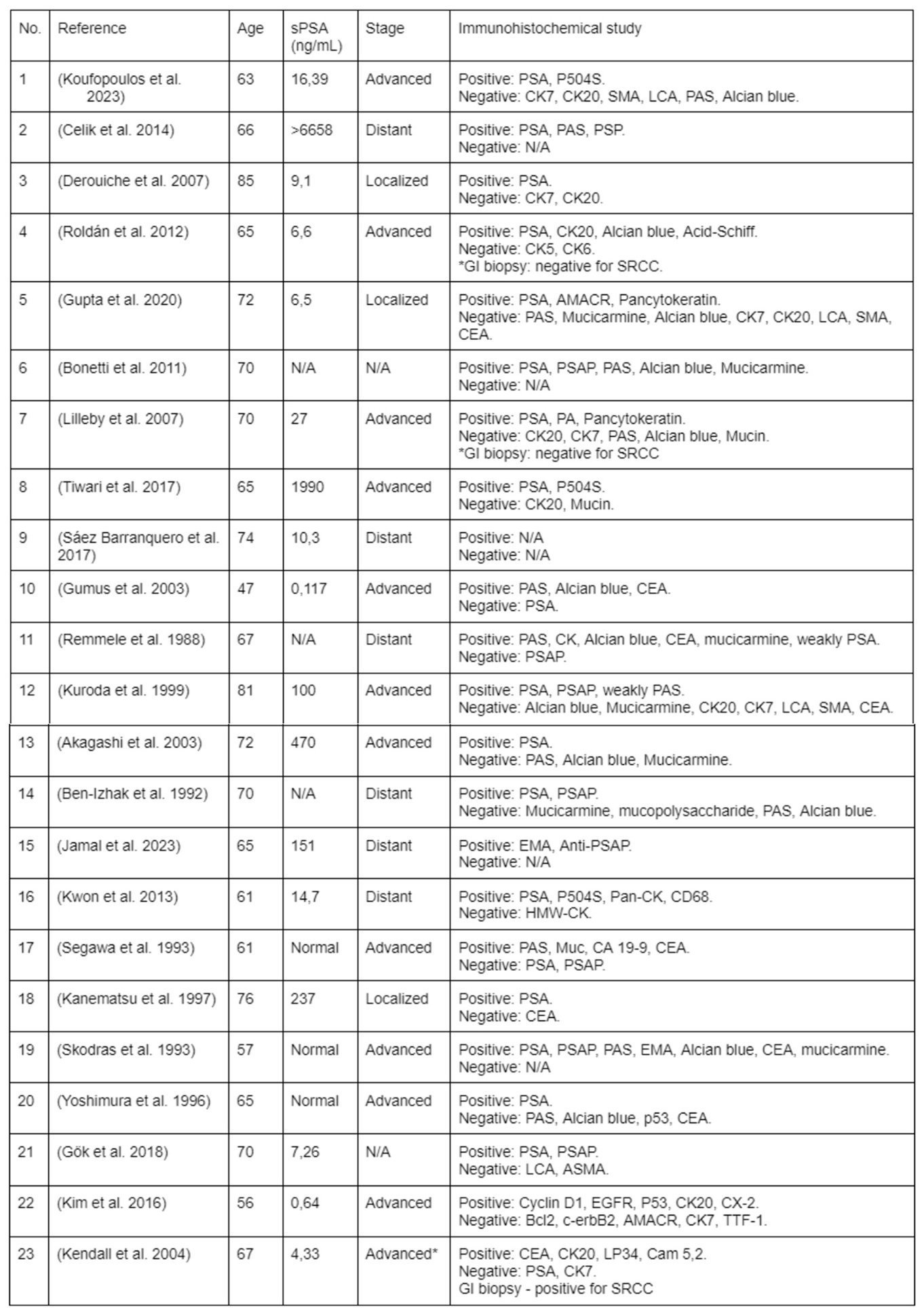
The literature review was performed on PubMed using the search words ‘’primary signet ring cell carcinoma’’, excluding prostate non-related cases. Some authors could not determine the primary site of the disease, however since the tumor was found within the prostate, the studies were not excluded from the literature review for comparative purposes. Studies with previous history of other organ-confined cancers, except that of genitourinary or gastrointestinal tracts, were excluded.
The selected clinical cases were sorted into four categories according to the stage of the disease at the time of presentation: localized, advanced, distant, or unknown (9). Other available information was collected to determine the presence of possible trends or patterns among the patients: age, main complaints, serum prostate-specific antigen (sPSA), and used immunohistochemical markers.
Out of 23 analyzed primary prostatic SRCC cases, nine were found to be advanced, nine - were distant at the time of presentation and three were localized (Table 1). The complaints often consisted of obstructive lower urinary tract symptoms, occasional gross hematuria, and symptoms that may be associated with distant metastasis. Findings including sPSA values or immunohistochemical staining were inconsistent, although most (16/23) were positive for PSA. One of the analyzed cases (Case No. 23) that was immunohistochemically negative for PSA was later found to be a metastatic disease from the upper gastrointestinal tract and was not excluded from the study for comparison.
4. Discussion
Primary signet ring cell adenocarcinoma of the prostate was first mentioned in the late 1970s, and less than 100 cases have been published in English literature ever since (10). It is a rare, high-grade (Gleason grade 5) acinar adenocarcinoma subtype characterized by its distinctive intracellular substance-containing vacuole which displaces the nucleus into the periphery of the cell giving it a crescent shape (11,12). The content of the vacuole may vary (13), however, according to the latest edition of prostate and urinary tract tumors classification by the World Health Organisation (WHO), in cases the vacuole contains mucin, the tumor should be named signet ring cell-like adenocarcinoma rather than signet ring cell adenocarcinoma. Furthermore, vacuolated cells are associated with a greater Gleason pattern which independently worsens the prognosis (4).
The diagnostic criteria suggest the diagnosis of signet ring cell adenocarcinoma of the prostate should be assigned only in cases when the vacuolated cells make up at least 25% of the entire tumor volume which may be evaluated on the whole-organ specimens obtained in surgery and not biopsy, otherwise in cases the cellular volume requirement is not met or the specimen is obtained in biopsy the entity should be referred to as a prostate adenocarcinoma with signet ring cells instead (4,11,14). The criteria of the cellular composition is not strictly followed, since cases with less than 20% have been accepted as SRCC of the prostate (40). In contrast, (15) claims that any histological specimen obtained may be used for diagnostic purposes validating those that were taken in TURP and this may be useful in cases when patients present late in the course of the disease and do not meet the criteria for radical prostatectomy. However, artifacts like lymphocytes or vacuolated smooth muscle cells mimicking SRCC in TURP specimens are not uncommon (3). Fortunately, now the confusion may be avoided by applying specific immunohistochemical studies including leukocyte common antigen and alpha-smooth muscle actin (16).
Even though frequent changes in terminology, the lack of strict diagnostic criteria, and unestablished investigative protocols possibly pose additional issues in calculating the incidence of the disease, it is clear that a true prostatic SRCC is extremely rare, with an estimated prevalence of 0,02% among all prostate adenocarcinoma cases (5). Due to its rare nature, diffuse, lateral, submucosal growth pattern, and close epidemiological relation to the gastrointestinal tract, a diligent diagnostic workup for differentials must be carried out, since the location of the primary tumor may be an independent factor for cause-related survival and virulence of the disease (7,17). The investigations should include upper gastric endoscopy, colonoscopy, cystoscopy, and abdominal computed tomography to exclude metastatic involvement of the prostate (12). It may not be a routine procedure, yet some patients may benefit from random gastric biopsies (18). Clearly, the method is not the most reliable for obvious reasons, yet it is important to recognize that early gastric SRCC may not be macroscopically visible, and late gastric SRCC may occasionally appear as mucosal erosions (7).
Additionally to the gastrointestinal tract, particular attention must be paid to exclude SRCC of organs in close anatomical proximity to the prostate like the urinary bladder or rectum (19). These organs should not stain for PSA but may be strongly positive for prostate-specific acid phosphatase (PSAP) on the immunohistochemical study, perhaps due to shared cloacal derivation (20).
Even though PSA is considered to be a highly specific marker for prostate tissue, its expression was found to be lost in poorly differentiated cells which may pose an additional diagnostic struggle in differentiating between primary and metastatic disease (21–23). Worth noting, that NKX3.1 may increase PSA sensitivity when applied in combination (24). However, before NKX3.1 stain was available, it was speculated that SRCC of the prostate can be classified into two types: tumors that react positively to PSA and simultaneously negative to carcinoembryonic antigen (CEA) and those that do not react to PSA, yet express positivity in reaction to CEA (25). The possibility of the variants has not been disproved and may be of high significance in choosing the most appropriate therapeutic approach.
Some studies claim they could not determine the primary site of the disease which is true in up to 5% of all metastatic disease cases even though immunohistochemical studies were done (8,26). Interestingly, (27) reported an alternative approach to the problem which gained great results in the case of T3b primary prostatic SRCC by applying colorectal SRCC cancer treatment based on the immunohistochemical study findings of the prostate biopsy alone. The study implies that the treatment may be applied based on the histological and molecular aspects of the disease rather than following organ-oriented treatment protocols.
SRCC of the prostate is often described as an aggressive clinical course and unpredictable response to hormonal therapy. Some publication authors argue that this presumption may have arisen from the fact that most patients were diagnosed at late stages of the disease, before the sPSA marker era, and if diagnosed early the variant is of similar prognosis to the usual acinar PA (12,13,28). Unfortunately, other articles, similar to our literature review show that sPSA values greatly vary and thus are not entirely reliable in the diagnosis of prostate SRCC (29,30), which may play a role in late diagnosis. In addition to this, another factor that is greatly contributing to the late diagnosis may be rather nonspecific clinical symptoms which often include lower urinary tract obstruction and occasional gross hematuria.
Although we lack well-established guidelines, the treatment of prostatic SRCC currently is rather similar to that of the traditional adenocarcinomas of the prostate, which includes a variable combination of surgical procedures, hormonal therapy, adjuvant radiotherapy, and/or chemotherapy (12). Even though, the multimodal aggressive approach is very reasonable, a combination of radiotherapy together with hormonal therapy may be an appropriate alternative therapy for prostate SRCC treatment (16).
Similarly to signet ring cell prostate cancer, Mucinous (aka. colloid) carcinoma (MC) is another rare variant of the usual acinar prostate adenocarcinoma. Since one-third of all prostate adenocarcinomas contain some focal differentiation of mucin it is important to stress that MC is characterized by the extracellular pools of mucin which must occupy at least 25% of the entire tumor volume on a whole organ specimen, otherwise such tumors are described as ‘’with features of mucin’’ (14). It is graded on the structural architecture of the histological view irrespective of the mucinous component and similarly to the usual prostate adenocarcinoma is associated with elevated sPSA levels before diagnosis and is of similar response to treatment and prognosis (31).
In exceptionally rare cases, MC may contain signet ring cells making the entity known as mucinous carcinoma with signet ring cells (MCSRC). The two variants need to be distinguished apart since the presence of signet ring cells seems to tremendously worsen the prognosis with 5-year survival equal to zero (22,29,32).
5. Strengths and Limitations of the Study
To our knowledge, this is the first documented case of signet ring cell adenocarcinoma of the prostate in Lithuania. The clinical case is of great importance for the scientific community and practitioners serving as a major educational tool in choosing an appropriate diagnostic approach in rare cases of prostate cancer, possibly improving overall outcomes.
Besides the strengths, the study has some limitations. We do not have enough clinical data to prove that the prostate is the primary site of the SRCC given that we lack data on specific immunohistochemical stains and the data that we have closely resembles that of the lower gastrointestinal tract. An autopsy was not performed. In addition to this, looking back, erosive gastropathy should have raised suspicion for gastric signet-ring cell carcinoma. Furthermore, minding the submucosal growth pattern and the virulence of small cluster cancers of SRCC, the CT imaging may not be sensitive enough to rule out the gastric SRCC diagnosis.
6. Conclusions
In summary, this clinical case should highlight the importance of a multidisciplinary approach in an extensive diagnostic study that is unfortunately not standardized to this day. The rarity of the entity in addition to late diagnosis poses a great challenge in choosing an appropriate treatment to improve the prognosis.
Author Contributions
AP: MS designed, performed the study, and carried out the literature search and review; AP, MS, AG, and IC helped in the literature search and wrote the paper; AP, MS, and AG helped in the acquisition and analysis of data; AP, MK, and AD made critical revision and supervision. All authors have read and approved the manuscript.
Funding
This research received no external funding.
Institutional Review Board Statement
Not applicable.
Informed Consent Statement
The consent was obtained from the family of the diseased.
Data Availability Statement
All data generated or analyzed during this study are included in this published article.
Acknowledgments
The authors would like to thank the family of the diseased for their participation and consent to the publication of the case details and associated images.
Conflicts of Interest
The authors declare no conflicts of interest.
References
- Alizadeh, M.; Alizadeh, S. Survey of Clinical and Pathological Characteristics and Outcomes of Patients With Prostate Cancer. Glob. J. Health Sci. 2014, 6, 49–57. [Google Scholar] [CrossRef] [PubMed]
- George J. Netto, Mahul B. Amin, Daniel M. Berney, Eva M. Comperat, Anthony J. Gill, Arndt Hartmann, et al. The 2022 World Health Organization Classification of Tumors of the Urinary System and Male Genital Organs—Part B: Prostate and Urinary Tract Tumors—ScienceDirect [Internet]. 2022. Available online: https://www-sciencedirect-com.ezproxy.dbazes.lsmuni.lt/science/article/pii/S0302283822024770?casa_token=exMfZeUx-RYAAAAA:wAyqbcguXV4NR3VDfCBpKGNGvQF5ck9ektDGooMWTJpzrjPu1on16RIFXiO4uVMGUON1Tvqhng (accessed on 5 February 2024).
- Laufman, H. Primary Linitis Plastica Type of Carcinoma of the Colon. Arch Surg 1951, 62, 79. [Google Scholar] [CrossRef] [PubMed]
- Al-Taee, A.; Almukhtar, R.; Lai, J.; Jallad, B. Metastatic signet ring cell carcinoma of unknown primary origin: a case report and review of the literature. Ann. Transl. Med. 2016, 4, 283. [Google Scholar] [CrossRef] [PubMed]
- Uyama, T.; Moriwaki, S. Papillary and mucus-forming adenocarcinomas of prostate. Urology 1979, 13, 432–434. [Google Scholar] [CrossRef] [PubMed]
- Koufopoulos, N.; Ieronimaki, A.-I.; Zacharatou, A.; Gouloumis, A.R.; Leventakou, D.; Boutas, I.; Dimas, D.T.; Kontogeorgi, A.; Sitara, K.; Khaldi, L.; et al. A Case of Prostatic Signet-Ring Cell-like Carcinoma with Pagetoid Spread and Intraductal Carcinoma and Long-Term Survival: PD-L1 and Mismatch Repair System Proteins (MMR) Immunohistochemical Evaluation with Systematic Literature Review. J. Pers. Med. 2023, 13, 1016. [Google Scholar] [CrossRef] [PubMed]
- Saito, S.; Iwaki, H. Mucin-producing carcinoma of the prostate: review of 88 cases. Urology 1999, 54, 141–144. [Google Scholar] [CrossRef] [PubMed]
- Gök, A.; Tuygun, C.; Akmansu, M.; Uslu, A.A.; Kartal, I.G.; Sandikçi, F.; Karabacak, O.R.; Sağnak, A.L.; Topaloğlu, H.; Ersoy, H. Primary Signet Ring Cell Carcinoma of the Prostate: A Rare Case Report. J. Clin. Med. 2018, 7, 218. [Google Scholar] [CrossRef] [PubMed]
- Kendall, A.; Corbishley, C.M.; Pandha, H.S. Signet ring cell carcinoma in the prostate. Clin Oncol R Coll Radiol G B 2004, 16, 105–107. [Google Scholar] [CrossRef] [PubMed]
- Azumi, N.; Traweek, S.T.; Battifora, H. Prostatic acid phosphatase in carcinoid tumors. Immunohistochemical and immunoblot studies. Am. J. Surg. Pathol. 1991, 15, 785–790. [Google Scholar] [CrossRef] [PubMed]
- Smith, C.; Feddersen, R.M.; Dressler, L.; McConnell, T.; Milroy, T.; Smith, A.Y. Signet ring cell adenocarcinoma of prostate. Urology 1994, 43, 397–400. [Google Scholar] [CrossRef] [PubMed]
- Kristiansen, I.; Stephan, C.; Jung, K.; Dietel, M.; Rieger, A.; Tolkach, Y.; Kristiansen, G. Sensitivity of HOXB13 as a Diagnostic Immunohistochemical Marker of Prostatic Origin in Prostate Cancer Metastases: Comparison to PSA, Prostein, Androgen Receptor, ERG, NKX3.1, PSAP, and PSMA. Int. J. Mol. Sci. 2017, 18, 1151. [Google Scholar] [CrossRef] [PubMed]
- Gregoire, C.; Muller, G.; Machiels, J.P.; Goeminne, J.C. Metastatic signet-ring cell carcinoma of unknown primary origin. Acta Clin. Belg. 2014, 69, 135–138. [Google Scholar] [CrossRef] [PubMed]
- Derouiche, A.; Ouni, A.; Kourda, N.; Belhadj, K.; Ben Jilani, S.; Chebil, M. A new case of signet ring cell carcinoma of the prostate. Clin. Genitourin. Cancer 2007, 5, 455–456. [Google Scholar] [CrossRef] [PubMed]
- Gupta, M.; Budhwar, A.; Prasad, N.; Prasad, S.K.S.; Singh, S. Primary signet ring cell carcinoma of prostate: A rare case report and review of literature. J. Cancer Res. Ther. 2023, 19, 1075. [Google Scholar] [CrossRef] [PubMed]
- Zhang, Y.; Shen, H.; Liao, K.; Wu, W.; Li, J.; Yu, H.; Wu, H.; Wang, Z. Case Report: Prostate Adenocarcinoma With Mucinous Features of Normal-Level Serum PSA, Atypical Imaging, Biopsy-Negative, and Peculiar Urethrocystoscopic Manifestation. Front. Oncol. 2020, 10. [Google Scholar] [CrossRef] [PubMed]
- Gupta, A.; Gulwani, H.V. A rare case of primary signet ring-like cell carcinoma of prostate in an elderly male. Indian J. Pathol. Microbiol. 2020, 63, 338–339. [Google Scholar] [CrossRef] [PubMed]
- Tiwari, D.; Nayak, B.; Seth, A. Good response of an aggressive rare variant of signet ring cell carcinoma of prostate with hormonal therapy. BMJ Case Rep. 2017, 2017, bcr2016217567. [Google Scholar] [CrossRef] [PubMed]
- Akagashi, K.; Tanda, H.; Kato, S.; Ohnishi, S.; Nakajima, H.; Nanbu, A.; Nitta, T.; Koroku, M. Signet-ring cell carcinoma of the prostate effectively treated with maximal androgen blockade. Int J Urol Off J Jpn Urol Assoc 2003, 10, 456–458. [Google Scholar] [CrossRef] [PubMed]
- Jamal, I.; Raman, R.B.; Kumar, S.; Agrawal, R.; Choudhary, V.N. Metastasis of primary signet ring cell carcinoma of prostate to bone marrow: A rare occurrence with review of literature. Indian J Pathol Microbiol 2023, 66, 407–410. [Google Scholar] [CrossRef] [PubMed]
- Yoshimura, K.; Fukui, I.; Ishikawa, Y.; Maeda, H.; Yamauchi, T.; Kawai, T. Locally-Confined Signet-Ring Cell Carcinoma of the Prostate: A Case Report of a Long-Term Survivor. Int. J. Urol. 1996, 3, 406–407. [Google Scholar] [CrossRef] [PubMed]

Table 1.
The literature review of the 23 analyzed cases.
 |
Disclaimer/Publisher’s Note: The statements, opinions and data contained in all publications are solely those of the individual author(s) and contributor(s) and not of MDPI and/or the editor(s). MDPI and/or the editor(s) disclaim responsibility for any injury to people or property resulting from any ideas, methods, instructions or products referred to in the content. |
© 2024 by the authors. Licensee MDPI, Basel, Switzerland. This article is an open access article distributed under the terms and conditions of the Creative Commons Attribution (CC BY) license (http://creativecommons.org/licenses/by/4.0/).
Copyright: This open access article is published under a Creative Commons CC BY 4.0 license, which permit the free download, distribution, and reuse, provided that the author and preprint are cited in any reuse.


