

Submitted:
15 April 2024
Posted:
16 April 2024
You are already at the latest version
Abstract
Considering the age of the dogs, it was expected that elderly animals would have stiffer thyroids than adults. The aim of this study was to evaluate the thyroid glands of healthy dogs using 2D shear wave elastography in order to establish qualitative and quantitative parameters of tissue stiffness in dogs of different age groups. The B-mode ultrasound and elastography findings were correlated with the weight and sex of the patients, allowing a reference range to be established for different groups of dogs. 31 dogs of different breeds, sexes and sizes were evaluated. Animals with clinical signs or ultrasound findings indicative of endocrine disease or thyroid lesions were not included in the study. Using QelaXtoTM 2D software, shear wave velocity data in m/s and color elastograms were evaluated and calculated. A healthy thyroid has a blue color elastogram, indicative of low stiffness, and there was no distinction between adult and elderly dogs, genders or sizes. The reference range for the shear wave velocity of thyroid tissue assessed by 2D shear wave elastography can be between 1.6 and 2.0 m/s with a variation of ±0.889 in adult and elderly dogs.
Keywords:
dogs
; diagnostic imaging
; elastography
; stiffness
; thyroid
; ultrasound
1. Introduction
The thyroid gland is considered to be the most important of the endocrine glands for regulating metabolism, being responsible for the proper coordination of the body's physiological processes by means of hormones [1]. Injuries to the endocrine system and its components are characterized by functional disorders and pathological changes that manifest themselves in different systems of the animal [2]. These lesions can include degeneration, necrosis, vascular disorders, immune-mediated or infectious inflammation, atrophy, hyperplasia and even neoplasia resulting from or leading to instability in the function of the gland and the secretion of its hormones. [3].
Clinical palpation can help diagnose alterations associated with hyperplasia and neoplasms of the gland, but it alone is not accurate enough to predict tissue invasion and discrete thyroid alterations [4]. In order for the diagnosis of thyroid disorders to be confirmed, it is necessary to measure the concentration of free tetraiodothyronine (free T4) and thyroid stimulating hormone (TSH), thyroid hormone supplementation, imaging exams and collection of tissue sample by biopsy or fine needle aspiration biopsy (FNAB). [5,6,7].
Different studies have shown that elastography is a superior technique to ultrasound in differentiating the character of lesions and has good diagnostic applicability [8,9]. In veterinary medicine, elastography has been studied in different organs, obtaining satisfactory results with good sensitivity and accuracy [10,11,12,13]. Despite the optimistic scenario, more studies related to shear wave elastography of the thyroid need to be carried out so that it becomes a reliable and relevant diagnostic method for the diagnosis of thyroid pathologies. Within this scenario, we discuss the use of shear wave elastography as a complementary diagnostic technique for thyroid pathologies due to its advantages over other established methods.
Thus, shear wave elastography can easily be included in the protocol of cervical ultrasound examinations for the elucidation of different thyroid pathologies as long as the technique is consolidated in the evaluation of the thyroid. The aim of this study is to evaluate the thyroid of clinically healthy dogs of different sizes (large, medium and small), sexes (females and males) and age groups (adults and the elderly) using B-mode ultrasound and 2D shear wave elastography (2D-SWE) in order to obtain qualitative and quantitative data on tissue morphology and stiffness. In addition, the aim is to obtain a reference range for the shear wave velocity of thyroid tissues in adult and elderly dogs.2.
2. Materials and Methods
2.1. Ethical Statement
The project was approved by the Ethics Committee for the Use of Animals (CEU) of the Faculdade de Ciências Agrárias e Veterinárias da Universidade Estadual “Júlio de Mesquita Filho” (FCAV/Unesp-Jaboticabal) (Protocol No. 4369/22, Jaboticabal, São Paulo, Brazil).
2.2. Animals
The study was carried out using animals from kennels and owners in the city of Jaboticabal, Brazil. Thirty-one dogs were evaluated, 11 males and 20 females, of different breeds, weights ranging from 3 to 30 kg and small, medium and large sizes.
The animals were divided into two experimental age groups: Age group 1 - made up of adult dogs aged between 1 and 6 years (n = 17); Age group 2 - made up of elderly dogs aged 7 years or more (n = 14). Mean age and standard deviation of age group 1 and 2 were 3.28 years (σ = 1.56) and 9 years (σ = 2.11) respectively. The groups were made up of different breeds, with age group 1 consisting of: Border Collie (2), French Bulldog (1), Pinscher (1), American Pit Bull (2), SRD (10) and West Highland (1); age group 2 consisting of: Dachshund (1), Golden Retriever (1), American Pit Bull (1), Poodle (3) and SRD (8).
2.3. Clinical and Ultrasound Assessment of Dogs
In order to check the health of the selected dogs, the animals were previously submitted to anamnesis, general physical examination, thyroid palpation and B-mode cervical (thyroid-focused) and abdominal ultrasound. Animals with clinical signs or ultrasound findings indicative of endocrine disease or thyroid damage were not included in the study. The clinical signs that were considered as exclusion criteria were: presence of skin lesions with or without itching, areas of alopecia, polydipsia, polyphagia, sensitivity to cold, apathy, hyperactivity, rapid weight loss/gain. In addition to these, the presence of volume increases in any region of the body was also an exclusion factor. The B-mode ultrasound findings of the thyroid that were considered exclusion criteria were: irregularity of the surface of the thyroid capsule, parenchyma with a coarse echotexture or mixed echogenicity, presence of nodular formations regardless of their characteristics, presence of areas of fibrosis. Dogs that showed inflammatory, neoplastic or increased dimensions on B-mode ultrasound in abdominal organs were not included in the study. Age-related ultrasound changes were not considered an exclusion criterion, such as renal senility. Animals with agitated behavior that did not allow imaging tests to be carried out were also excluded from the study.
All dogs with no alterations in their medical history, physical examination or ultrasound examination and considered clinically healthy were included in the study. The ultrasound and elastography examinations were carried out by the same examiner and the images were evaluated by two imagers with at least two (2) years' experience.
For the ultrasound and elastography exams, the animals underwent a wide trichotomy of the ventral cervical region from 2-3cm below the angle of the mandible to the caudal cervical region (near the entrance to the thorax). The animals were then positioned on a padded rail in dorsal decubitus with their bodies parallel to the apparatus. The animals were physically restrained by their thoracic and pelvic limbs with the help of their guardian and a trainee, without the need for sedatives and/or anesthesia. Water-based conductive gel specifically for ultrasound examinations was applied to the examination area to perform the techniques.
2.4. B-Mode Thyroid Ultrasonography
B-mode ultrasound study of the thyroid was carried out using MyLabTM X8 Platform equipment (Esaote, Italy), with a 4-15 MHz multi-frequency and linear transducer. All the gain, frequency, focus, TCG and depth settings were adapted according to the physical characteristics of each dog participating in the study. The thyroid gland lobes were located in their usual topography, between the sternocephalic and sternothyroid muscles, ventral to the trachea and caudal to the larynx. Both were assessed in a longitudinal section for their dimensions (length and width), echogenicity in relation to the adjacent muscles (hypoechogenic, isoechogenic or hyperechogenic) and surface regularity. The aforementioned characteristics were also assessed in cross-section, except for dimensions (Figure 1). Figure 2 shows the two thyroid lobes on B-mode ultrasound in a longitudinal section of dogs of different ages. Figure 2A shows the right thyroid lobe of a 3-year-old female dog, with homogeneous and hyperechogenic parenchyma, regular contours and an ellipsoid shape. Figure 2B shows the right thyroid lobe of a 7-year-old female dog, with homogeneous and hyperechogenic parenchyma, regular contours and an ellipsoid shape.
2.5. 2D Shear Wave Elastography (2D-SWE) of the Thyroid
2D shear wave elastography was performed after the B-mode ultrasound scan, using the same device and transducer. Using the QelaXtoTM 2D software (Esaote, Italy), the shear wave velocity data in m/s and the color slastogram were evaluated and calculated by analyzing the relative displacements of the tissue elements by an acoustic pressure pulse.
The elastographic images were taken in the longitudinal plane of each thyroid lobe and a scale of 0.0 to 10.0 m/s was used in all cases. The QelaXtoTM 2D software quality map was activated when one of the thyroid lobes was properly framed in the image. The map indicated the most suitable locations for acquiring the shear wave velocity, with the regions colored in green having high quality (Figure 3), yellow indicating medium quality and orange, low quality (Figure 4).
In all animals, shear wave velocity measurements were only taken in high-quality regions colored green. The stiffness scale of the color slastogram ranged from blue (soft/soft) to red (stiff/hard), passing through green and yellow (intermediate stiffness). To assess stiffness using shear wave velocity, 5 (five) samples (areas of interest - ROI's) were randomly selected, comprising different portions of the thyroid using 0.2cm diameter circles as shown in Figure 5. The shear wave velocity values of the analyzed portions were evaluated by the equipment's software and expressed in m/s.
2.6. Statistical Analysis
Statistical analyses were carried out using R software version 3.3.0 (R Foundation for Statistical Computing, Austria). Initially, the variables collected for the study were tested for normal distribution (Shapiro-Wilk test) and homoscedasticity of variance (Barrlett's test). These tests showed that the elastographic variables had a non-parametric distribution. Repeated measurements or ROIs of the shear wave velocity (SWV) and depth of each of the thyroid gland lobes assessed in all the animals were then compared using the Fiedman test. Statistical similarity was found between the SWV and depth of the right (p=0.9767; p=0.3154) and left (p=0.7406; p=0.599) ROIs. The median SWV and depth of evaluation of the right and left portions were then used.
Subsequently, aspects such as width, length, SWV and depth were compared between the right and left portions using the Wilcoxon test for paired samples. Similarities were found between the portions for width (p=0.1550), length (p=0.8734), SWV (p=0.2966) and depth (p=0.5453), so the median of these variables was used. To this end, the quantitative variables were compared between the age groups (adults and elderly), sex (males and females), size (small, medium and large) and age, and when the results showed similarity, the 95% median confidence intervals (95%CI) were constructed as the normal ranges of the objective variables of this study. For the statistical analysis of echogenicity, values were assigned to three different types of tone compared to the adjacent musculature: hyperechogenic = 3, isoechogenic = 2 and hypoechogenic = 1.
The data is presented as median ± interquartile range (IQR) and the significance value is defined as p<0.005. Width, length and depth values are presented in centimeters (cm), while shear wave velocity values are in m/s.
3. Results
The ultrasound and elastographic examinations of canines of different ages were carried out without any limitations, were easy to perform and obtained quick results on the B-mode and elastographic characteristics.
3.1. Adult and Elderly Dogs
Between the age groups of adult and elderly dogs, the width, length and echogenicity of the thyroid gland on B-mode ultrasound showed no significant difference. As for shear wave velocity (SWV) and depth in the elastographic evaluation, the age groups also showed no significant difference in values. Despite this, all the thyroids evaluated in the dogs that took part in the study showed the color elastogram colored blue in the qualitative evaluation, both in the group of adult dogs and in the group of elderly dogs. Table 1 shows the values for each age group for B-mode ultrasound and elastographic characteristics.
3.2. Females and Males
The quantitative data from B-mode ultrasound (width and length) and shear wave elastography (shear wave velocity and depth) showed no significant difference between the males and females of the species studied, as the p-value obtained was greater than 0.005 in all the aspects analyzed. Table 2 shows B-mode values for each sex and elastographic ultrasound characteristics.
3.3. Small, Medium and Large Sized Dogs
To assess whether the size of the dogs influenced the quantitative B-mode ultrasound and elastographic characteristics, the animals were separated into groups according to their size: large (G), medium (M) and small (P). The large size group was made up of dogs weighing 25 to 30 kg, medium size from 15 to 24 kg dogs and the small size from 3 to 14 kg dogs. According to this parameter, a significant difference and positive correlation was found between size and width (p=0.0031), length (p=0.004) and depth (p=0.0007) between the large size group and the medium and small size groups. Figure 6 shows B-mode ultrasound images of the thyroid lobes of dogs from different sizes with the respective width and length dimensions in a longitudinal section. Figure 6A, 6B and 6C show one of the thyroid lobes of large, medium and small size dogs. The difference between the shear wave velocities of the thyroid between the sizes of dogs was not significant between the different groups that took part in this study. Table 3 shows B-mode values for each size and elastographic ultrasound characteristics.
3.4. Correlations between Animal Characteristics, Ultrasound and Thyroid Elastography
At the end of the statistical analysis of the data focused on the age, sex and size of the dogs, we tried to correlate the variables studied with each other in order to find whether or not there was a possibility of correlation between them. The variables compared were: age, size, echogenicity, width, length, SWV and depth.
The age group only showed a positive correlation for the age factor (r-Spearman=0.868; p=0). The size of the animals showed a positive correlation with the ultrasound aspects of width (r-Spearman=0.746; p=0), length (r-Spearman=0.722; p=0) and depth (r-Spearman=0.574; p=0.001). The B-mode ultrasound characteristics of the thyroid, such as width and length, were positively correlated with each other (r-Spearman=0.695; p=0) and also when correlated with the elastographic depth data (r-Spearman=0.56; p=0.001 for width and r-Spearman=0.499; p=0.004 for length). The elastographic data for depth and shear wave velocity (SWV) did not correlate with each other, as r-Spearman=-0.271 and p=014.
4. Discussion
This study was able to characterize the thyroid gland of clinically healthy adult and elderly dogs using 2D shear wave elastography, both in terms of qualitative data (color elastogram) and quantitative data (shear wave velocity and depth). The results of this study help to consolidate 2D shear wave elastography as a potential diagnostic method for diffuse and focal thyroid pathologies in the future.
One of the main differentials of this study is the comparison of the elastographic characteristics of the thyroid between adult and elderly dogs. The hypothesis behind this study was that animals over seven years of age would have greater stiffness than younger ones. This could be justified by the longer time that the thyroid gland has been exposed to immunological and environmental challenges. Herman et al. [14] believed that in humans, in general, the stiffness of a tissue changed with advancing age. However, a comparison of shear wave velocity between adult and elderly dogs showed no significant difference between the groups studied. Thus, age is no longer a determining factor in thyroid stiffness, as the shear wave velocity values in elderly dogs were statistically similar to those of adult dogs (Table 1, where p=0.0743). Herman et al. [14] came to a similar conclusion when evaluating human thyroids from a population of 176 volunteers aged between 21 and 91 years. Furthermore, the authors state that in their study they found no effect of sex/gender on stiffness.
Table 2 shows the SWV values between females and males of the canine species found in this study, with a value of p=0.8534, corroborating the findings in humans that gender does not interfere with the elasticity of thyroid tissue [14]. Knowing that age also has no significant effect on thyroid stiffness, a single reference interval can be established for the value of shear wave velocity in 2D shear wave elastography of adult and elderly dogs. This complements the work already carried out by Jung et al. [15], who only used adult dogs aged between 1 and 5 years.
With regard to the shear wave velocity value found for adult dogs (1.63 ± 0.675 m/s), it is similar to the value established by Jung et al. (2020), who established a value of 1.57 ± 0.1 m/s for the thyroid tissue of adult Beagle dogs in 2D shear wave elastography. As the racial composition of this study varied and yet the SWV values found were similar to those found in Beagles, it can be inferred that the values can be extrapolated to different breeds and sizes of dogs. This is reinforced by the data presented in Table 3, where the shear wave value (SWV) showed no significant difference between large, medium and small dogs (p=0.7222).
The characteristics of low stiffness, low wave velocity value and blue elastogram of the healthy thyroid using 2D shear wave elastography are probably due to the cellular constitution of its tissue. Healthy thyroid tissue, with normal levels of hormone concentration and secretion, is made up of follicular and medullary parafollicular cells, of endodermal and neural crest origin respectively [3]. The thyroid tissue of clinically healthy dogs probably has the elastographic characteristics found in this study, as its cellular composition has no alterations that affect its internal rigidity. This correlation leads us to believe that a healthy thyroid is, consequently, a soft tissue.
When affected by neoplasms or malignant nodules, the thyroid can suffer increased tissue stiffness, with a reddish to orange elastogram on elastography. In fact, the quantitative values of shear wave elastography are higher in malignant nodules composed of many fibrous interstitial components and sand-like calcified bodies [16]. This means that the present study, which shows the normality of the elastographic findings, could be a standard of normality used for future work on detecting abnormalities in dogs.
With regard to the elastographic depth characteristic for acquiring the shear wave velocity, there was a significant difference between large dogs and medium and small dogs (Table 3). The depth of the 2D shear wave elastography range is important data to take into consideration when talking about artifacts that can affect the quality of the wave signal or the technique due to the patient's anatomy. According to Dighe et al [19], 3.7% of elastographic artifacts that make images uninterpretable are related to the patient's anatomy. This may cause the technique to have some artifacts in giant dogs, where it is assumed that the thyroid may be at a depth greater than 1.905 cm. Consequently, the increased depth can lead to an absence of elastographic signal, another elastographic artifact [19,20]. To confirm or refute the possibility of this artifact in giant dogs, studies would be needed to assess the applicability and reproducibility of 2D shear wave elastography in these animals.
Still on the subject of depth, this study found no correlation between depth and shear wave velocity values, i.e. depth did not decrease or increase SWV values. This finding is in line with the study carried out by Dhyani et al [21], which compared two different elastography systems for assessing liver fibrosis in humans. According to the authors, when assessed by 2D-SWE, shear wave velocity was not influenced by depth in the areas of fibrosis assessed [21]. However, in the ARFI elastography used in the same study, SWV values varied according to depth in the same areas assessed previously [21].
Regarding B-mode ultrasound characteristics of the thyroid linked to the parenchyma and size of the gland are in line with what is proposed and described in the literature [22,23,24,25]. In this study, all dogs had both thyroid lobes in an ellipsoid shape as proposed by Brömel et al. [24] and Khati et al. [26]. The thyroid lobes of different size dogs, ages and sexes showed homogeneous, hyperechogenic or isoechogenic parenchyma in relation to the adjacent musculature, in agreement with Brömel et al. [24]. With regard to the significant difference in width and length between the different sizes of dogs in this study, this is due to the influence of the size of the animal on the dimensions of the gland [23]. The agreement of the results of the research related to B-mode ultrasound of the thyroid helps to demonstrate that the technique has consistent and well-established characteristics for assessing the thyroid in healthy dogs.
When carrying out 2D-SWE assessments of the thyroid, this study identified some limitations that can make it difficult to acquire images and values in certain dogs: 1) the technique was not applicable in agitated animals, those with wheezing or those that do not allow proper positioning to access the ventral part of the neck. In total, six dogs were excluded from the study due to this characteristic, as it made it difficult to acquire a satisfactory quality map for measuring shear wave velocity; 2) it was not possible to include a group of young animals (aged between 6 and 11 months) due to the low availability of animals in this age group; 3) in some examinations, interference from compression by the arterial pulse of the common carotid artery was noted, since the proximity of this vessel to the thyroid sometimes made it difficult to acquire a good quality elastographic map for measuring SWV. However, this was easily resolved by obtaining a new image until the quality map was colored green, and this new acquisition did not affect the examination time in the 31 dogs evaluated in the study.
5. Conclusions
Through this study of the thyroid gland of clinically healthy dogs, it can be inferred that 2D shear wave elastography is easy to perform and offers quick results regarding the stiffness of the gland in the species studied. In addition, a healthy thyroid gland has a blue color elastogram, indicative of low stiffness, and there is no distinction between adult and elderly dogs, sexes or sizes.
The shear wave velocity shows no significant difference between the characteristics mentioned above. This allows the same confidence interval to be created and used for adult and elderly dogs regardless of sex or size. Thus, the reference interval for the shear wave velocity of thyroid tissue assessed by 2D shear wave elastography can be between 1.6 and 2.0 m/s with a variation of ±0.889 in adult and elderly dogs.
Author Contributions
Conceptualization, D.J.R., R.A.R.U., M.A.R.F.; Data curation: D.J.R.; Formal analysis: R.A.R.U.; Funding acquisition: M.A.R.F.; Investigation: D.J.R.; Methodology: D.J.R., T.D., L.P.N.A.; R.A.R.U. and M.A.R.F.; Project administration: D.J.R.; Resources: D.J.R., T.D., D.R.G., L.P.N.A., S.T.T., D.V.V.S., M.M.F.P., B.S.P.M., A.P.L.O. and B.B.L.; Visualization: D.J.R. and M.A.R.F.; Writing – original draft: D.J.R.; Writing – review & editing: D.J.R., T.D., L.P.N.A., D.V.V.S. and M.A.R.F.
Funding
Esta pesquisa foi realizada com apoio da Coordenação de Aperfeiçoamento de Pessoal de Nível Superior — Brasil (CAPES) — Código de Financiamento 001, Conselho Nacional de Desenvolvimento Científico e Tecnológico (CNPq) (nº do processo – 30512/2020-0), Fundação de Estudos Agrários Luiz de Queiroz (FEALQ) – Piracicaba/SP e Fundanção de Amparo à Pesquisa do Estado de São Paulo (FAPESP) (no do processo - 2022/07366-0).
Institutional Review Board Statement
The animal study protocol was approved by the Comissão de Ética no Uso de Animais (CEUA) da Faculdade de Ciências Agrárias e Veterinárias da Universidade Estadual Paulista “Júlio de Mesquita Filho” (FCAV-Unesp-Jaboticabal) (Protocolo nº 4369/22, 14 de setembro de 2022).
Informed Consent Statement
Os tutores dos animais foram consultados, informados e esclarecidos previamente sobre todos os detalhes e etapas do estudo, e expressaram sua concordância com as avaliações propostas no termo de consentimento livre, aprovando a participação do(s) seu(s) animal(is) na pesquisa.
Data Availability Statement
The original contributions presented in the study are included in the article/supplementary material, further inquiries can be directed to the corresponding author/s.
Acknowledgments
Os autores gostariam de agradecer ao apoio técnico prestado pela RTS/Esaote Healthcare do Brasil.
Conflicts of Interest
The authors declare no conflicts of interest.
References
- Petroff, B.K; Greco, D.A.S. Glândulas endócrinas e suas funções. In Cunningham Tratado de Fisiologia Veterinária, 6th ed.; Klein, B.G.,Eds.; Grupo GEN: Rio de Janeiro, Brasil, 2021; pp. 402-436.
- Ecco, R.; Langohr, I.M. Sistema endócrino. In Patologia Veterinária, 3rd ed.; Santos, R.L.; Alessi, A.C.,Eds.; Grupo GEN: Rio de Janeiro, Brasil, 2023; pp. 815-870.
- Miller, M.A. Sistema endócrino. In Bases da Patologia em Veterinária, 6th ed.; Zachary, J.F., Eds.; Grupo GEN: Rio de Janeiro, Brasil, 2018; pp. 681-722.
- Taeymans, O.; Penninck, D.G.; Peters, R.M. Comparison between clinical, ultrasound, CT, MRI, and pathology findings in dogs presented for suspected thyroid carcinoma. Vet Radiol Ultrasound 2013, 54, 61-70. [CrossRef]
- Wisner, E.R.; Nyland, T.G. Ultrasonography of the thyroid and parathyroid glands. Vet Clin North Am Small Anim Pract 1998, 28, 973-991. [CrossRef]
- Scott-Moncrief, J.C. Thyroid disorders in the geriatric veterinary patient. Vet Clin North Am Small Anim Pract 2012, 42, 707-725. [CrossRef]
- Scott-Moncrieff, J.C. Hypothyroidism. In Canine & Feline Endocrinology, 4th ed.; Feldman, E.C.; Nelson, R.W.; Reusch, C.; Scott-Moncrieff, J.C., Eds.; Saunders: Philadelphia, United States, 2015; pp. 77-128.
- Maronezi, M.C. et al. Accuracy of B-mode ultrasound and ARFI elastography in predicting malignancy of canine splenic lesions. Sci Rep 2022, 12, 4252. [CrossRef]
- Sporea, I. et al. ARFI elastography for the evaluation of diffuse thyroid gland pathology: Preliminary results. World J Radiol 2012, 4, 174-178. [CrossRef]
- Abreu, T.G.M. et al. Accuracy of ARFI elastography in the differentiation of cataract stages in dogs. Pesq Vet Bras 2021, 41, e06598. [CrossRef]
- da Cruz, I.C.K. et al. Malignancy prediction of cutaneous and subcutaneous neoplasm in canines using B-mode ultrasonography, Doppler, and ARFI elastography. BMC Vet Res 2022, 18, 10. [CrossRef]
- Simões, A.P.R. et al. Placental ARFI elastography and biometry evaluation in bitches. Anim Reprod Sci 2020, 214, 106289. [CrossRef]
- Feliciano, M.A.R. et al. Acoustic radiation force impulse elastography of prostate and testes of healthy dogs: preliminary results. J Small Anim Pract 2015, 56, 320-324. [CrossRef]
- Herman, J. et al. Shear wave elastography parameters of normal soft tissues of the neck. Biomed Pap Med Fac Univ Palacky Olomouc Czech Repub 2017, 161, 320-325. [CrossRef]
- Jung, J.W. et al. Two-Dimensional Shear Wave Elastography of Normal Soft Tissue Organs in Adult Beagle Dogs. Front Bioeng Biotechnol 2020, 8, 979. [CrossRef]
- Bhatia, K.S.S. et al. Shear wave elastography of thyroid nodules in routine clinical practice: preliminary observations and utility for detecting malignancy. Eur Radiol 2012, 22, 2397-2406. [CrossRef]
- Li, H. et al. Influence of lesion size on shear wave elastography in the diagnosis of benign and malignant thyroid nodules. Sci Rep 2021, 11, 21616. [CrossRef]
- Zhang, Y.X. et al. Clinical Value of Shear Wave Elastography Color Scores in Classifying Thyroid Nodules. Int J Gen Med 2021, 14, 8007-8018. [CrossRef]
- Dighe, M.; Hippe, D.S.; Thiel, J. Artifacts in Shear Wave Elastography Images of Thyroid Nodules. Ultrasound Med Biol 2018, 44, 1170-1176. [CrossRef]
- Barr, R.G. Shear wave imaging of the breast: still on the learning curve. J Ultrasound Med 2012, 31, 347-350.
- Dhyani, M. et al. Ultrasouns Shear Wave Elastography: Variations of Liver Fibrosis Assessment as a Function of Depth, Force and Distance from Central Axis of the Transducer with a Comparison of Different Systems. Ultrasound Med Biol 2018, 44, 2209-2222.
- Singh, B. Glândulas endócrinas. In Tratado de Anatomia Veterinária, 5th ed.; Singh, B., Eds.; Grupo GEN: Rio de Janeiro, Brasil, 2019; pp. 202-208.
- Carvalho, C.F. Região cervical ventral: Pescoço. In Ultrassonografia em Pequenos Animais, 2nd ed.; Carvalho, C.F., Eds.; Roca: São Paulo, Brasil, 2020; pp. 309-327.
- Brömel, C. et al. Comparison of ultrasonographic characteristics of the thyroid gland in healthy small-, medium-, and large-breed dogs. Am J Vet Res 2006, 67, 70-77. [CrossRef]
- Müller, T.R. et al. Do thyroid ultrasonographic features change according to age in euthyroid dogs? Anat Histol Embryol 2014, 43, 468-473.
- Khati, N. et al. Ultrasound of the thyroid and parathyroid glands. Ultrasound Q 2003, 19, 162-176. [CrossRef]
Figure 1.
- B-mode ultrasound image of the right (yellow arrow) and left (blue arrow) thyroid lobes of a 9-year-old female dog.
Figure 1.
- B-mode ultrasound image of the right (yellow arrow) and left (blue arrow) thyroid lobes of a 9-year-old female dog.
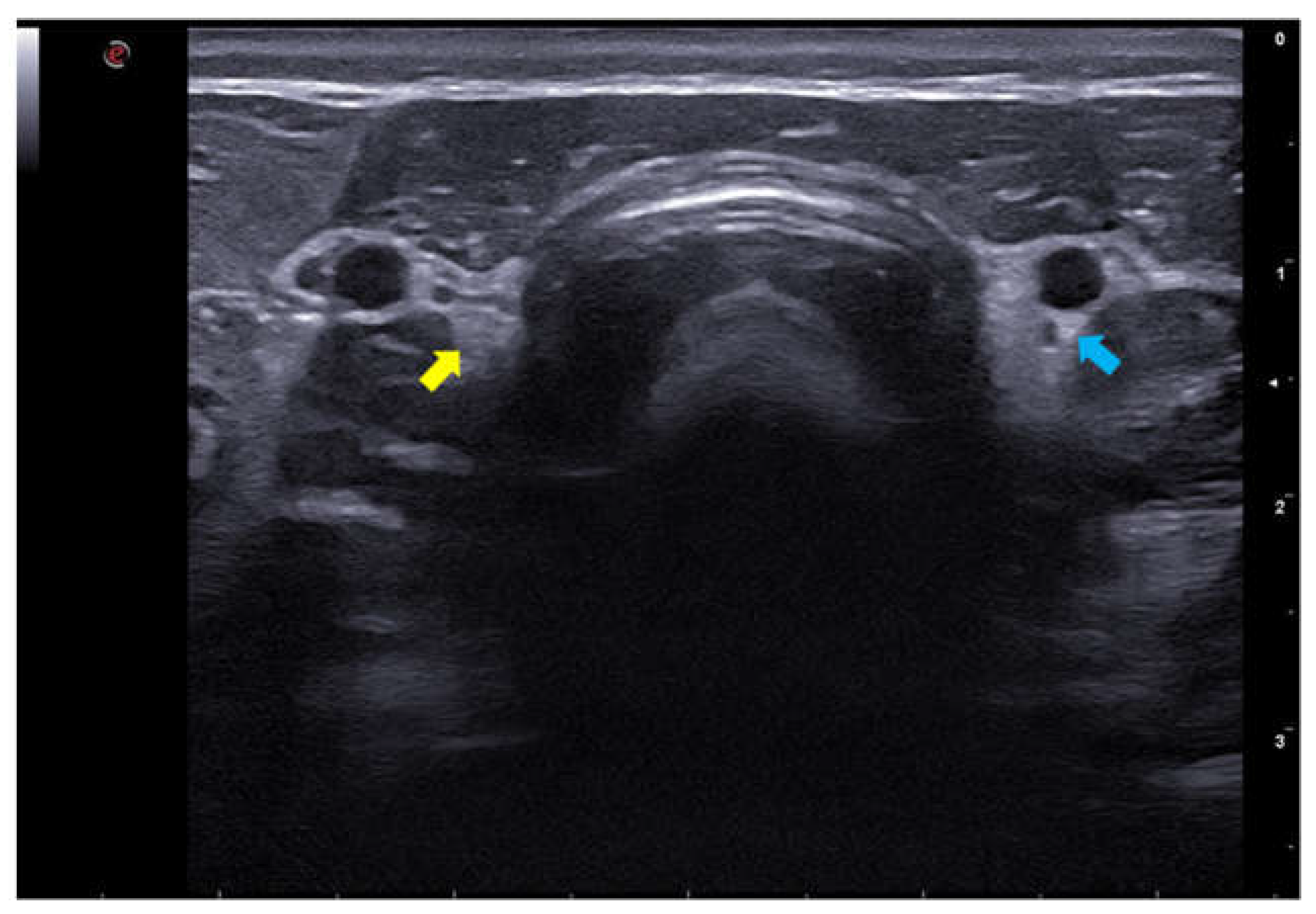
Figure 2.
- B-mode ultrasound image of the right thyroid lobes of female dogs of different ages. (A) Right thyroid lobe (yellow arrow) of a 3-year-old female dog. (B) Right thyroid lobe (blue arrow) of a 7-year-old female dog.
Figure 2.
- B-mode ultrasound image of the right thyroid lobes of female dogs of different ages. (A) Right thyroid lobe (yellow arrow) of a 3-year-old female dog. (B) Right thyroid lobe (blue arrow) of a 7-year-old female dog.
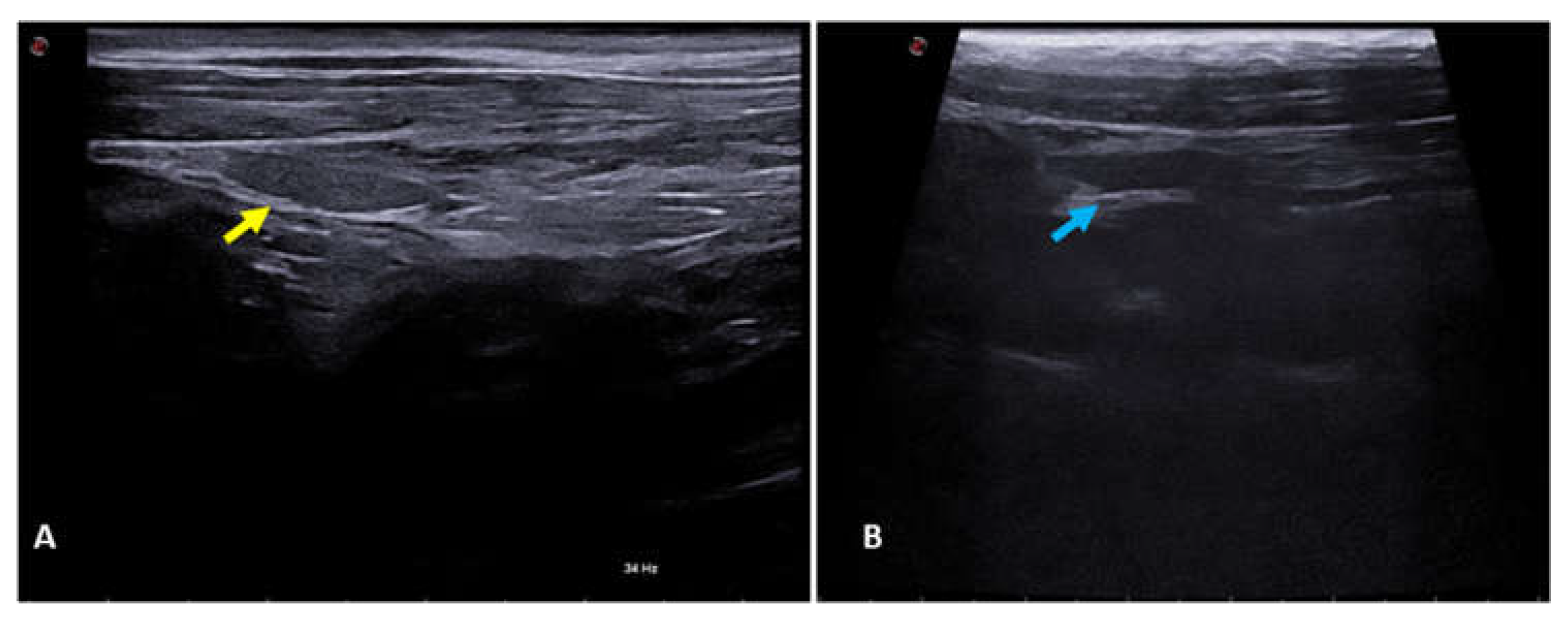
Figure 3.
- 2D shear wave elastography image of the left thyroid lobe of a 10-year-old dog. Elastographic image on the left showing the QelaXtoTM 2D software quality map colored in green, indicating high quality for shear wave velocity measurement (quantitative). Right side of the elastographic image showing the colored elastogram (qualitative) from the QelaXtoTM 2D software in blue, indicating that the thyroid parenchyma had low stiffness (soft).
Figure 3.
- 2D shear wave elastography image of the left thyroid lobe of a 10-year-old dog. Elastographic image on the left showing the QelaXtoTM 2D software quality map colored in green, indicating high quality for shear wave velocity measurement (quantitative). Right side of the elastographic image showing the colored elastogram (qualitative) from the QelaXtoTM 2D software in blue, indicating that the thyroid parenchyma had low stiffness (soft).
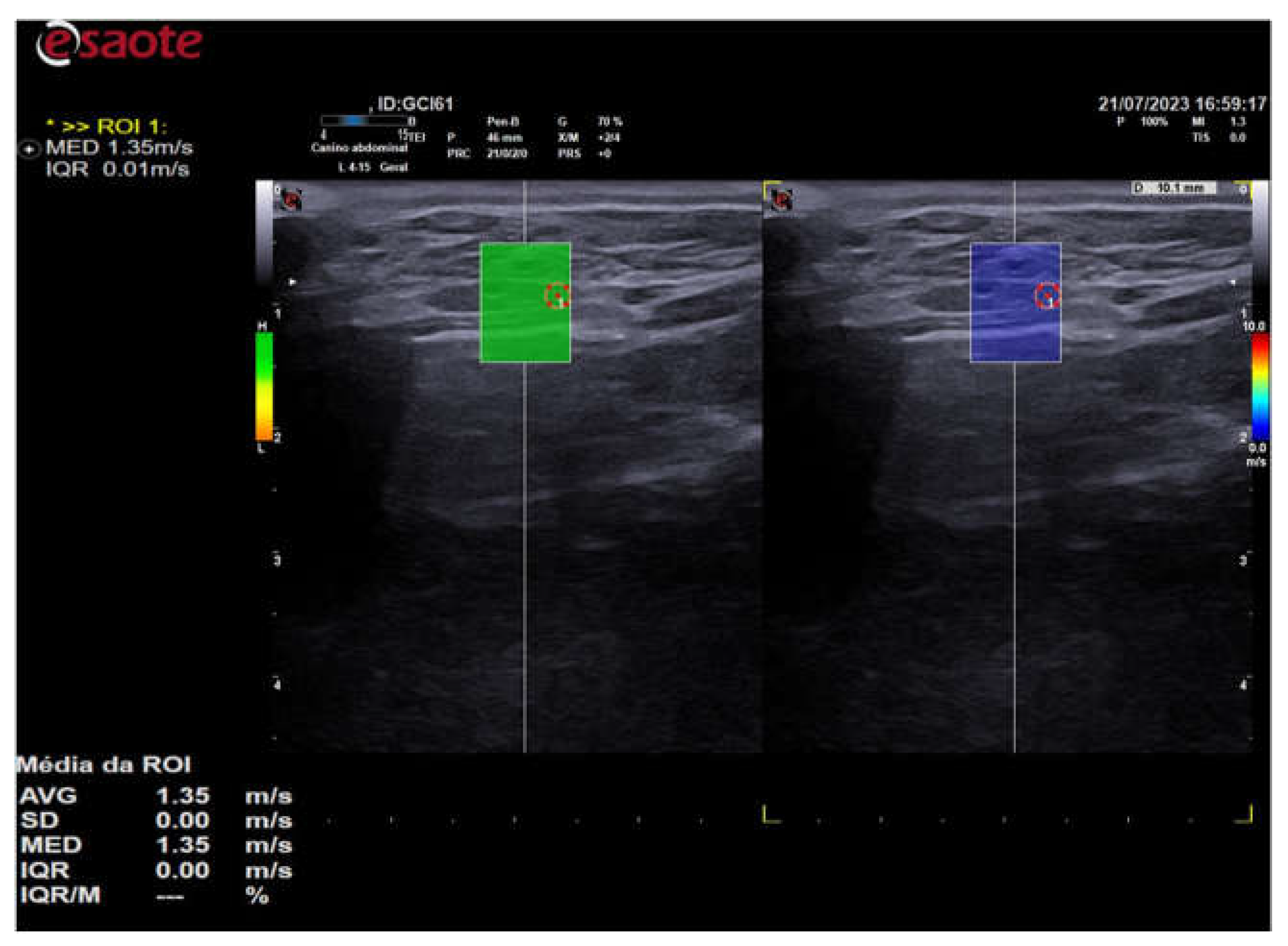
Figure 4.
- 2D shear wave elastography image of the left thyroid lobe of a 3-year-old female dog. Elastographic image on the left showing the QelaXtoTM 2D software quality map colored in orange, indicating low quality for measuring shear wave velocity (quantitative). Right side of the elastographic image showing the color elastogram (qualitative) from the QelaXtoTM 2D software colored in blue, indicating that the thyroid parenchyma had low rigidity (soft/soft).
Figure 4.
- 2D shear wave elastography image of the left thyroid lobe of a 3-year-old female dog. Elastographic image on the left showing the QelaXtoTM 2D software quality map colored in orange, indicating low quality for measuring shear wave velocity (quantitative). Right side of the elastographic image showing the color elastogram (qualitative) from the QelaXtoTM 2D software colored in blue, indicating that the thyroid parenchyma had low rigidity (soft/soft).
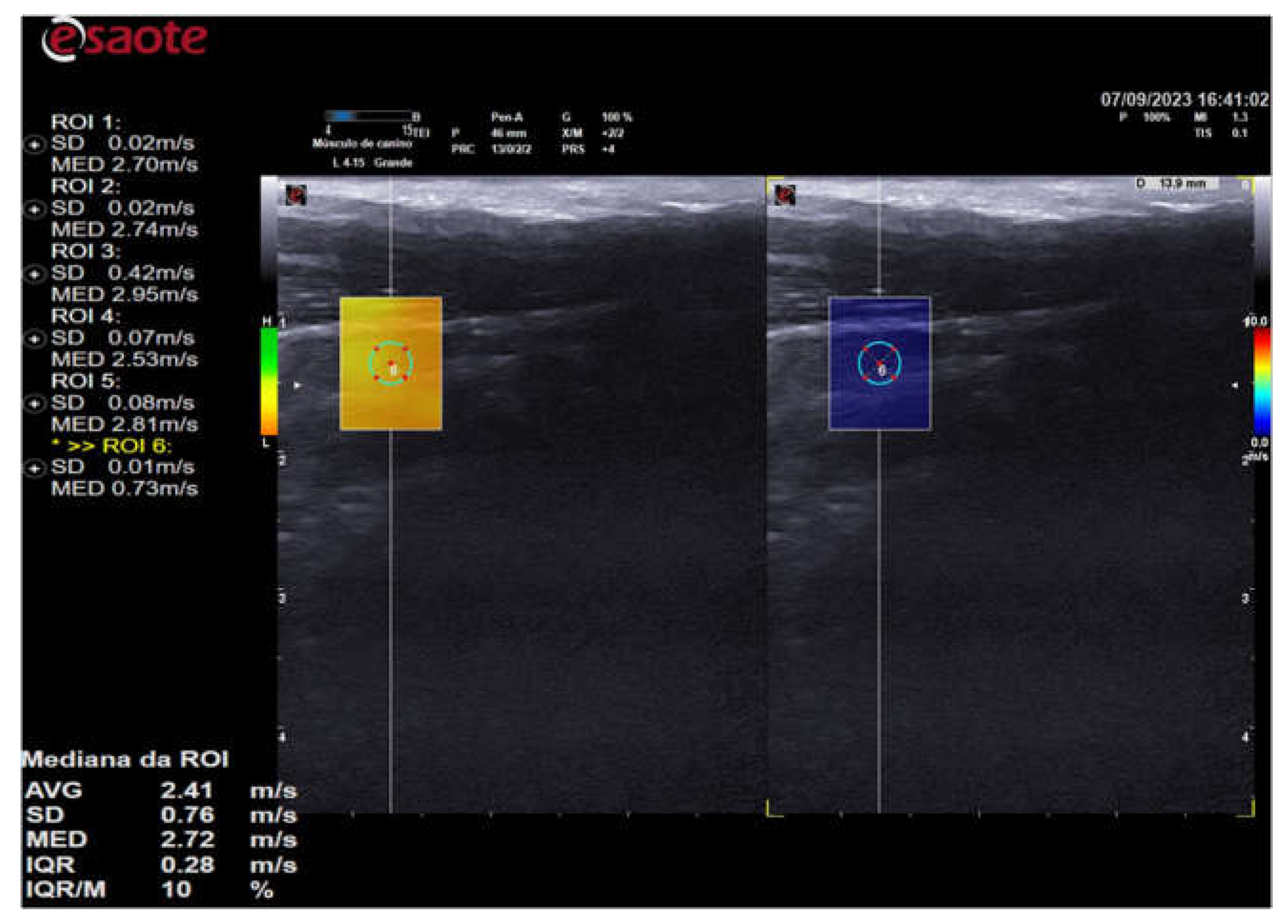
Figure 5.
- 2D shear wave elastography image of the right thyroid lobe of a 10-year-old dog. Elastographic image on the left showing the QelaXtoTM 2D software quality map colored in green, indicating high quality for measuring shear wave velocity (quantitative) and selection of four ROIs for measuring shear wave velocity. Right side of the elastographic image showing the color elastogram (qualitative) from the QelaXtoTM 2D software colored in blue, indicating that the thyroid parenchyma had low rigidity (soft/soft).
Figure 5.
- 2D shear wave elastography image of the right thyroid lobe of a 10-year-old dog. Elastographic image on the left showing the QelaXtoTM 2D software quality map colored in green, indicating high quality for measuring shear wave velocity (quantitative) and selection of four ROIs for measuring shear wave velocity. Right side of the elastographic image showing the color elastogram (qualitative) from the QelaXtoTM 2D software colored in blue, indicating that the thyroid parenchyma had low rigidity (soft/soft).
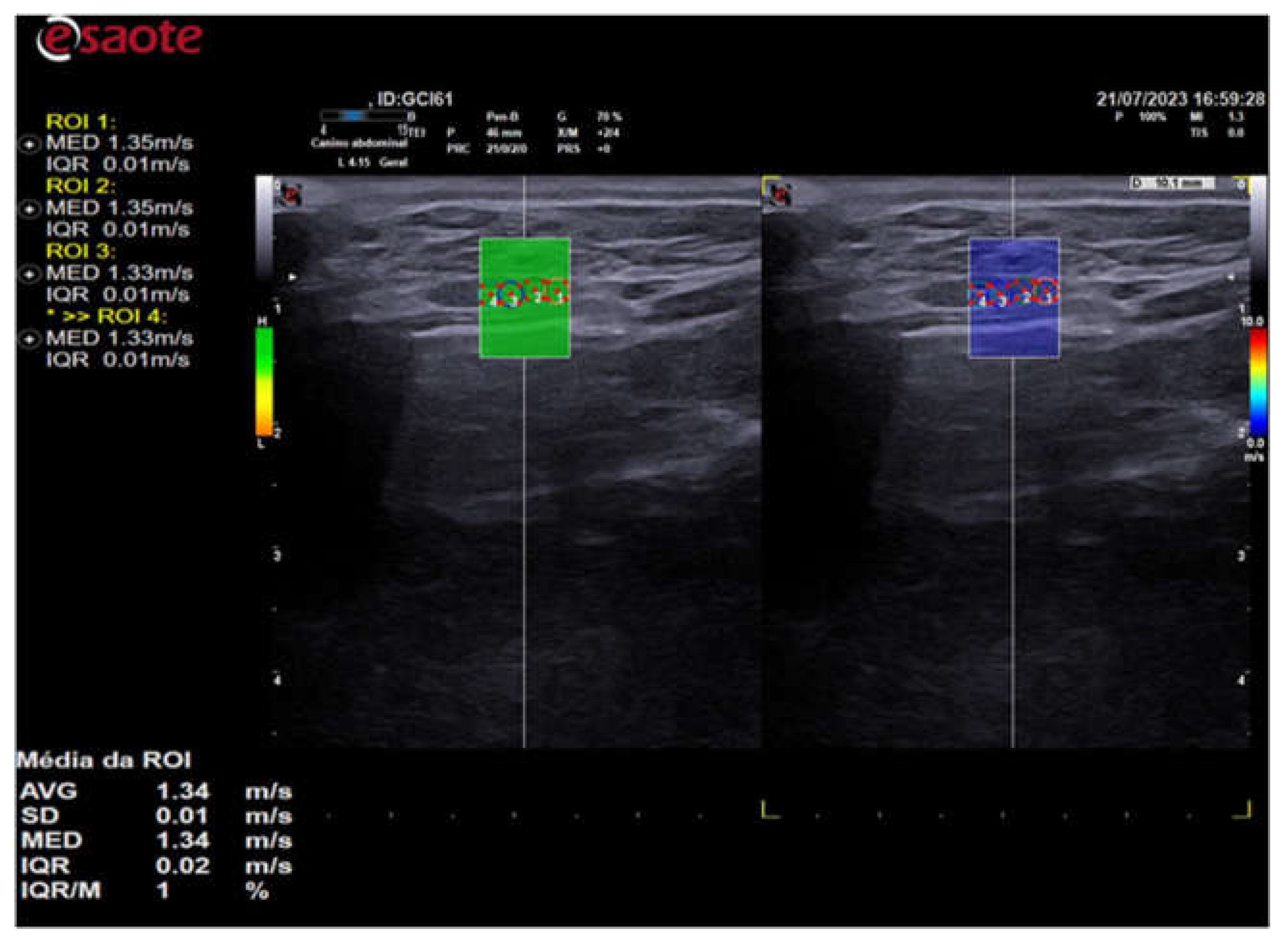
Figure 6.
- B-mode ultrasound image of the thyroid lobe with the calipers positioned over the lobe to measure length (Dist1) and width (Dist2) in different size dogs. (A) Left thyroid lobe of a large dog. (B) Left thyroid lobe of a medium-sized dog. (C) Right thyroid lobe of a small dog.
Figure 6.
- B-mode ultrasound image of the thyroid lobe with the calipers positioned over the lobe to measure length (Dist1) and width (Dist2) in different size dogs. (A) Left thyroid lobe of a large dog. (B) Left thyroid lobe of a medium-sized dog. (C) Right thyroid lobe of a small dog.
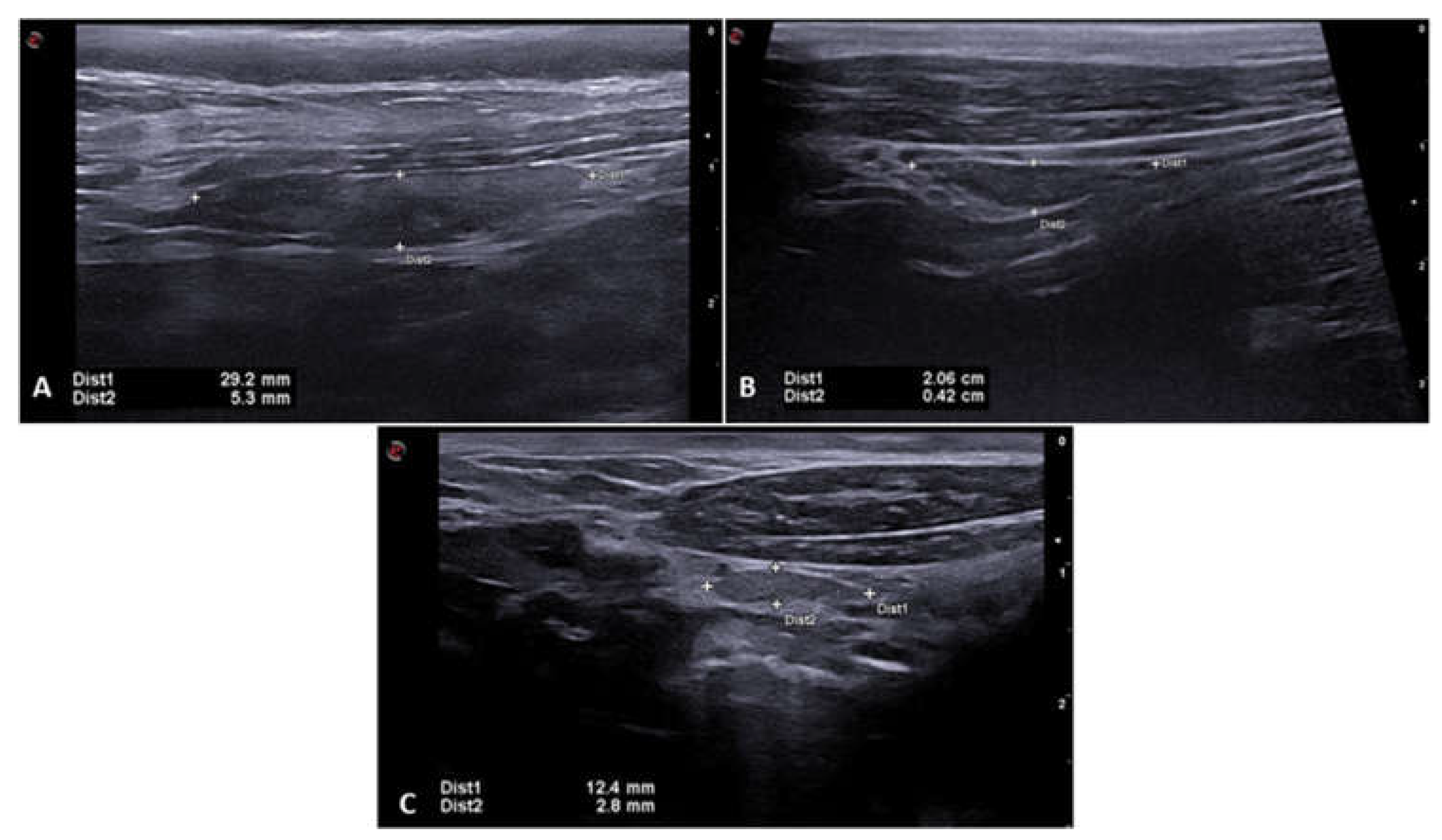

Table 1.
Comparison of values for width, length, echogenicity, shear wave velocity (SWV), and depth between age groups (AG) of adult dogs (AG1) and older dogs (AG2).
Table 1.
Comparison of values for width, length, echogenicity, shear wave velocity (SWV), and depth between age groups (AG) of adult dogs (AG1) and older dogs (AG2).
| Variable | GE | Median ± IQR | p-value | 95%CI |
|---|---|---|---|---|
| Width | GE1 GE2 |
0,335 ± 0,1675 0,345 ± 0,0988 |
0,6779 | 0,32 0,39 |
| Length | GE1 GE2 |
1,95 ± 0,993 1,922 ± 0,553 |
0,6061 | 1,7 2,4 |
| Echogenicity | GE1 GE2 |
3 ± 0 3 ± 2 |
0,1333 | - - |
| SWV | GE1 GE2 |
1,63 ± 0,675 1,877 ± 0,889 |
0,0743 | 1,6 2,0 |
| Depth | GE1 GE2 |
1,37 ± 0,547 1,1575 ± 0,6063 |
0,2345 | 1,1 1,5 |
Where: CI = confidence interval; IQR = interval between quartiles; SWV = shear wave velocity; p<0.005.
Table 2.
Comparison of width, length, shear wave velocity (SWV) and depth values between female and male dogs.
Table 2.
Comparison of width, length, shear wave velocity (SWV) and depth values between female and male dogs.
| Variables | Gender | Median ± IQR | p - value |
|---|---|---|---|
| With | Females Males |
0,3375 ± 0,16 0,34 ± 0,105 |
0,6642 |
| Length | Females Males |
2,023 ± 0,905 1,735 ± 0,475 |
0,1601 |
| SWV | Females Males |
1,797 ± 0,798 1,64 ± 0,415 |
0,8534 |
| Depth | Females Males |
1,3275 ± 0,75 1,33 ± 0,475 |
0,7101 |
Where: IQR = interval between quartiles; SWV = shear wave velocity; p<0.005.
Table 3.
Comparison of width, length, shear wave velocity (SWV) and depth values between large (G), medium (M) and small (P) size dog groups.
Table 3.
Comparison of width, length, shear wave velocity (SWV) and depth values between large (G), medium (M) and small (P) size dog groups.
| Variable | Size | Median ± IQR | p - value | 95% CI |
|---|---|---|---|---|
| Width | G M P |
0,465 ± 0,1425a 0,3375 ± 0,1175b 0,3075 ± 0,08b |
0,0031* | 0,4235; 0,5645 0,3202; 0,4198 0,2759; 0,3547 |
| Length | G M P |
2,858 ± 0,405a 1,99 ± 0,735b 1,72 ± 0,589b |
0,004* | 2,282; 3,064 1,845; 2,407 1,522; 1,960 |
| SWV | G M P |
1,63 ± 0,907a 1,90 ± 0,971a 1,72 ± 0,678a |
0,7222 | 0,853; 2,517 1,563; 2,739 1,469; 2,400 |
| Depth | G M P |
1,905 ± 0,415a 1,3425 ± 0,3513b 1,113 ± 0,482b |
0,0007* | 0,6888; 2,8572 0,5794; 2,1126 0,923; 2,135 |
Where: CI = confidence interval; IQR = interval between quartiles; SWV = shear wave velocity; p<0.005; *significance value.
Disclaimer/Publisher’s Note: The statements, opinions and data contained in all publications are solely those of the individual author(s) and contributor(s) and not of MDPI and/or the editor(s). MDPI and/or the editor(s) disclaim responsibility for any injury to people or property resulting from any ideas, methods, instructions or products referred to in the content. |
© 2024 by the authors. Licensee MDPI, Basel, Switzerland. This article is an open access article distributed under the terms and conditions of the Creative Commons Attribution (CC BY) license (http://creativecommons.org/licenses/by/4.0/).
Copyright: This open access article is published under a Creative Commons CC BY 4.0 license, which permit the free download, distribution, and reuse, provided that the author and preprint are cited in any reuse.


