
Submitted:
05 April 2024
Posted:
08 April 2024
You are already at the latest version
Abstract
Abstract: Canine ehrlichiosis is a zoonotic disease transmitted by ticks, posing a formidable challenge to both veterinary and public health sectors worldwide. Across continents and regions, the prevalence rates of Ehrlichia canis exhibit significant variation, underlining the necessity for a nuanced and globally informed approach to understanding and combating this disease. The review navigates through this complexity, shedding light on the intricate pathogenesis of the illness. Central to this understanding is the bacterium's adept manipulation of the host's immune response, contributing to the diverse clinical manifestations observed in infected animals. Diagnostic methodologies, crucial for timely intervention and management, are subjected to critical assessment. From traditional microscopy to modern molecular techniques and serology, each approach is scrutinized for its strengths and limitations. By acknowledging these nuances, the review aims to equip practitioners with the knowledge necessary to make informed diagnostic decisions. A central tenet of this review is the advocacy for an integrated "One Health" approach. By leveraging advancements in genomics, proteomics, and artificial intelligence, there is a potential to enhance diagnostic accuracy and develop innovative therapeutic and preventive strategies globally. This collaborative approach recognizes the interconnectedness of human, animal, and environmental health, offering a holistic framework for tackling complex zoonotic diseases like canine ehrlichiosis. In conclusion, this review serves as a beacon of knowledge, illuminating the multifaceted landscape of canine ehrlichiosis. Through its synthesis of scientific literature and emphasis on methodological rigor, it provides a foundation upon which future research and interventions can be built. With a unified commitment to "One Health" principles and the integration of cutting-edge technologies, there exists the potential to mitigate the impact of this disease and safeguard both animal and human well-being worldwide
Keywords:
Rhipicephalus sanguineus
; Ehrlichia canis
; Tick
; Ehrlichiosis
; Diagnosis
; Vaccine
1. Introduction
Canibe ehrlichiosis, caused by the obligate intracellular, pleomorphic bacterium of Ehrlichia, primarily Ehrlichia canis, is a significant concern within veterinary and public health [1]. This tick-borne zoonosis mainly affects canines and poses a risk to other animals, including humans [2]. The prevalence of E. canis infection is notable in areas with tropical or temperate climates, where tick proliferation is influenced by environmental factors [3,4] alongside ecological and socioeconomic elements [5]. Upon infection, the disease may manifest acutely, subclinically, or chronically, displaying diverse clinical signs such as fever, anorexia, anemia, thrombocytopenia, hemorrhages, lymphadenopathy, splenomegaly, uveitis, and polyarthritis [6]. Diagnosis of canine ehrlichiosis presents a challenge, necessitating a combination of direct and indirect methods, each with limitations in sensitivity, specificity, availability, and cost [7].
Treatment of the disease typically relies on antibiotics, primarily doxycycline, albeit effectiveness and affordability vary [8,9]. Despite treatment efforts, prevention of canine ehrlichiosis hinges on tick control and immunoprophylaxis, yet a commercially available vaccine remains elusive [10,11]. Consequently, canine ehrlichiosis stands as a zoonotic threat to public and animal health, demanding an integrated "One Health" approach for mitigation. This article aims to review the scientific literature on canine ehrlichiosis, exploring future perspectives and recent advances in genomics, proteomics, and artificial intelligence technologies to enhance diagnostic accuracy, identify therapeutic targets and biomarkers, and develop preventive strategies on a global scale.
2. Ehrlichia and Its Morphology
Ehrlichiosis stems from intracellular bacteria belonging to the genus Ehrlichia, encompassing six species: E. canis, E. chaffeensis, E. ewingii, E. muris, E. ruminantium, and E. mineirensis [1,12]. While E. chaffeensis, E. muris, and E. ewingii have been linked to ehrlichiosis in humans [2], reports indicate that E. ewingii and E. chaffeensis can infect dogs [4,13]. Thus, ehrlichiosis presents in two manifestations in both species: Canine Monocytic Ehrlichiosis (CME), primarily caused by E. canis, and Canine Granulocytic Ehrlichiosis (CGE), induced by E. ewingii [4]. Human Monocytic Ehrlichiosis (HME) involves E. chaffeensis, while E. ewingii contributes to Human Granulocytic Ehrlichiosis (HGE) alongside other agents like A. phagocytophilum and N. sennetsu [14]. Morphologically, Ehrlichia exhibits two distinct forms throughout its life cycle: an infectious, non-replicating form and a non-infectious form that undergoes binary fission for replication. Intracellular bacteria employ various mechanisms via surface-expressed proteins, facilitating functions such as adhesion to host cell membranes, nutrient absorption, cell signaling, and evasion [15,16]. They display morphological variations, encased in a thin, undulating outer membrane, featuring a narrow periplasmic space, and lacking a capsular layer. Moreover, Ehrlichia avoids the expression of lipopolysaccharide (LPS) and peptidoglycan on its surface, evading recognition by human leukocyte receptors and vector hemocytes, thereby thwarting elimination [17,18].
3. Tick Biological Cycle
The R. sanguineus tick serves as the primary biological vector for transmitting E. canis to dogs, although other tick species also possess this capability [3,19]. Consequently, dogs function as the principal host for R. sanguineus, concurrently acting as reservoirs due to prolonged bacteremia [20,21]. However, R. sanguineus infests other hosts, including small mammals and large animals, such as humans [19].
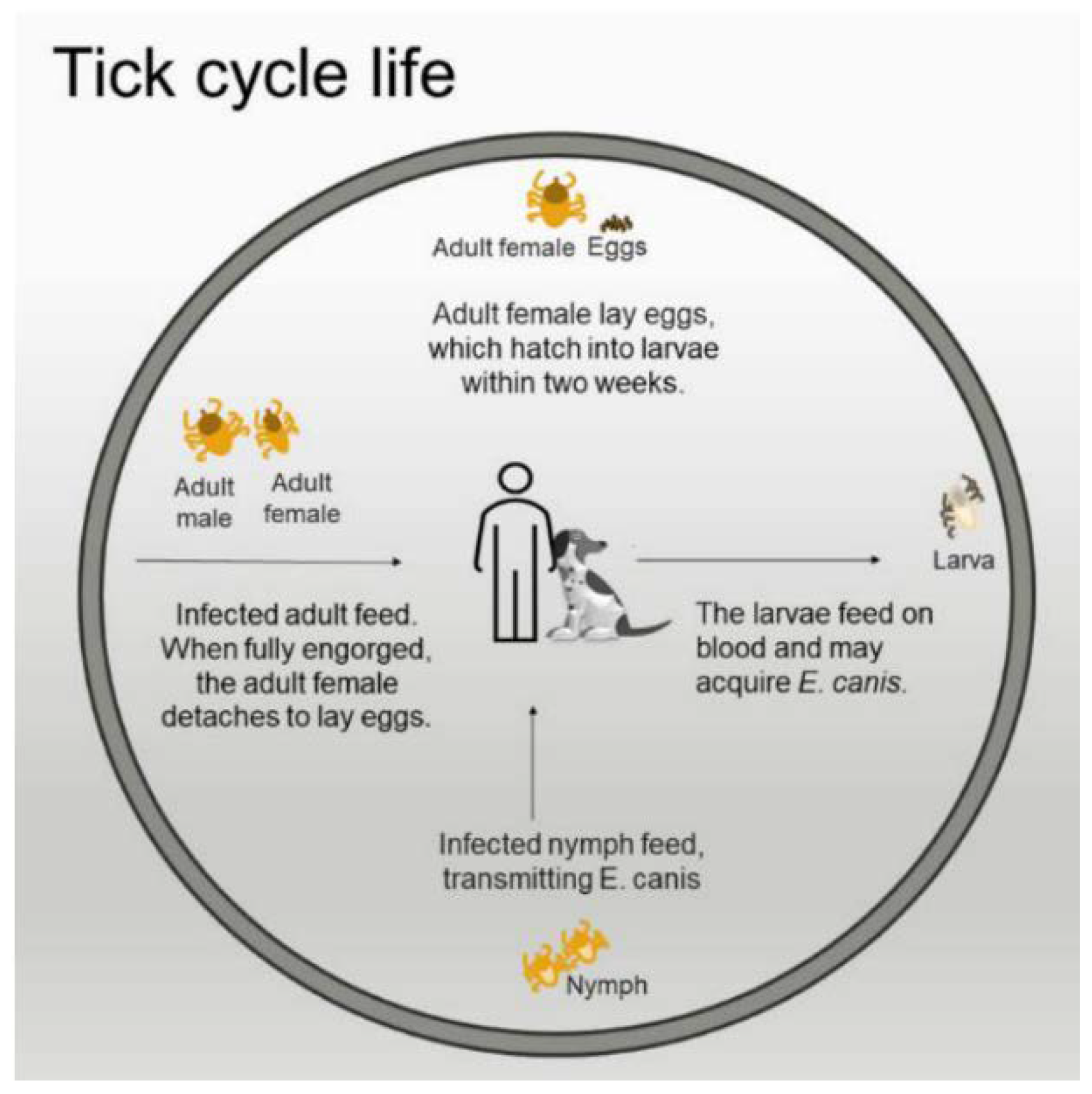
The transmission of canine ehrlichiosis initiates when a tick feeds on a previously infected host, thereby acquiring E. canis [2,22]. Throughout its biological cycle, the tick progresses through four life stages: egg, larva, nymph, and adult. While larvae, nymphs, and adults partake in blood meals and can acquire E. canis, only nymphs and adults can transmit the pathogen. Larvae, having not been previously exposed to the pathogen, cannot transmit it. Consequently, transstadial transmission, where the pathogen persists across the various life stages of the tick, is feasible, but transovarian transmission does not occur [2,22]. Subsequently, during the nymph and adult stages of its life cycle, the tick may transmit the bacteria to other hosts during subsequent blood meals.
The prevalence of E. canis typically peaks during warmer periods of the year in tropical and subtropical regions, correlating with the tick's life cycle (Figure 1).
4. Epidemiology of Canine Ehrlichiosis
Canine ehrlichiosis exhibits a widespread global distribution, particularly prevalent in regions with tropical and subtropical climates [23]. Noteworthy studies shed light on the disease's prevalence across various continents.
Laboso and colleagues conducted a comprehensive assessment spanning 2016 to 2021 in Africa, analyzing 400 samples from Kenya and Tanzania using the IDEXX SNAP 4Dx™ Plus test. Results unveiled a seropositivity of 31% (29/245) in Kenya and a surprising 69% (63/155) in Tanzania, underscoring the significant dissemination of ehrlichiosis in the region [24]. In contrast, a study in Europe by Guadalupe et al. over five years (2016-2020), covering samples from 35 countries, revealed an intriguing trend. Despite an increase in the number of tests conducted, there was a decline in Ehrlichia spp. Positivity, with seropositivity dropping from 4.3% to 3.4% [25].
In Latin America, evidence from Argentina indicates E. canis infection prevalence ranging between 13.5% and 37.5% in symptomatic dogs. In Paraguay, a study in domestic dogs reported a seroprevalence of 23.5%, accompanied by a molecular prevalence of 11.8%. Similarly, in Peru, seroprevalence was recorded at 16.5% for 140 dogs [10].
Previous studies in Brazil highlighted variability in canine ehrlichiosis seroprevalence, which is associated with different regions of the country. Values ranged from 62.8% in asymptomatic dogs to 78% in symptomatic dogs, with the molecular prevalence of E. canis ranging from 15% to 88%. For instance, Aguiar et al. observed a lower prevalence of 24.8% among 161 dogs from rural areas compared to 37.9% among 153 dogs from urban areas in the municipality of Monte Negro. Additionally, studies reported prevalence ranging from 5% to 16% in Rio de Janeiro and 27.6% in São Paulo, suggesting a significantly higher incidence of canine ehrlichiosis in urban areas compared to rural regions [26,27]. Conversely, seroprevalence in Brazil's southern region consistently appears low. Further insights come from Pereira and colleagues, reporting a molecular prevalence of 42.5% in dogs in the state of Mato Grosso Pantanal Norte [29]. Similarly, studies have depicted varying E. canis seropositivity rates, including 12.2% among 327 samples from dogs in the Amazon region, 13.7% among 153 dogs from the coastal area of Ceará state, and 59.1% among 264 dogs from the Midwest [30,31,32].
5. Pathogenesis
Upon tick infection, E. canis dissemination begins within the epithelial cells of the intestine, progressing to the tick's hemocytes and salivary gland cells [33]. Transmission to dogs occurs when infected ticks feed on blood, transferring the infective form of E. canis and their salivary secretions, which contain molecules facilitating pathogen acquisition and transmission, alongside exhibiting anticoagulant and anti-inflammatory properties [6,8].
Within the vertebrate host, the bacteria infiltrate monocytes, forming intracellular aggregates known as "morulas." Multiplication transpires within the phagolysosome and vacuoles of the host cell, affording isolation and protection from the immune system [34]. After 7 to 12 days post-infection, morulae are released into the bloodstream, infecting other cells. This interaction between infected and uninfected cells within blood vessels can incite vasculitis and perivascular migration of macrophages and lymphocytes. Predominant multiplication within macrophages and lymphocytes can lead to splenomegaly, hepatomegaly, and lymphadenopathy [8].
During host-pathogen interaction, various glycoproteins (GP) like GP19, gp36, gp140, and O-glycan, alongside outer membrane proteins such as P28/OMP and TRP 120, are pivotal for growth, accentuating the significance of these immunogenic outer membrane-associated proteins for pathogen replication within macrophages both in vitro and in vivo [12,15,35,36]. Studies conducted by McBride and colleagues underscore the importance of the TRP 120 protein in mediating E. canis and host cell interaction, influencing adhesion and internalization by phagosomes [36,37]. This capability enables bacteria to modulate the host's immune response, diminishing reactive oxygen species production and impeding host cell apoptosis. Additionally, the gp200 gene plays a critical role in immune response modulation, facilitating immune system evasion [38] and the potential to differentiate distinct pathogen strains [39].
The onset of the immune response, from initial exposure to symptom manifestation in canine ehrlichiosis, encompasses an incubation period spanning 8 to 20 days [8]. Subsequently, the disease progresses through acute, chronic, and subclinical phases, with the latter potentially persisting for several years without evident symptoms, underscoring the complexity and variability of E. canis infection within the host.
6. Immune Response
Both cellular and humoral immune responses are pivotal in defense against E. canis. CD4+ and CD8+ T cells play significant roles in the cellular immune response, crucial for resisting infections caused by this bacterium [40]. During E. canis infection, CD4 T lymphocytes secrete cytokines like IFN-γ and TNF-α, which can modulate the inflammatory response or confer protection to the host [2]. The protective immune response involves IFN-γ and Th1 secretion. However, components of tick saliva act as host immunomodulators during blood meals, reducing the production of IL-9, IL-2, and IL-4 in Th1 cells stimulated by IFN-γ.
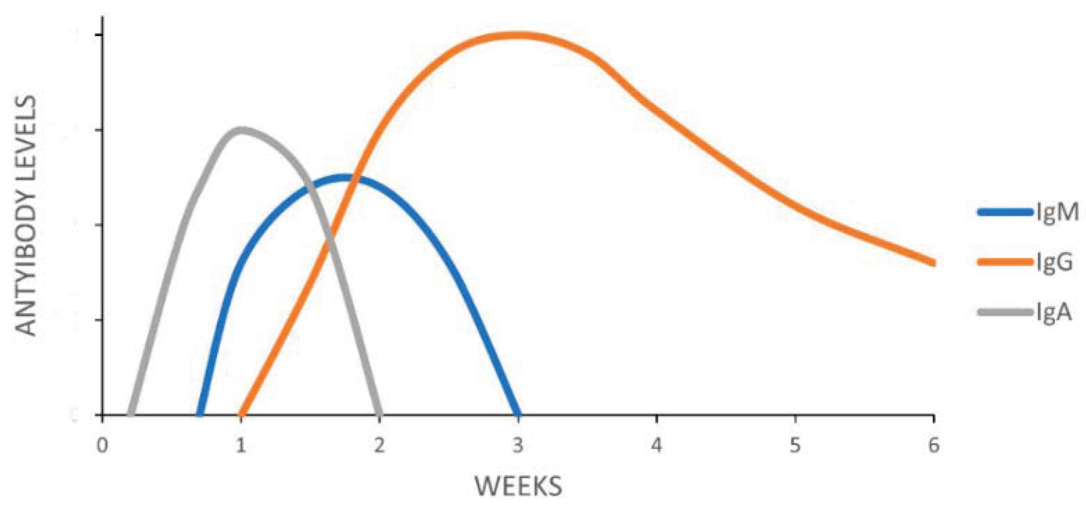
Additionally, Castro and colleagues observed differences in MHCII molecule expression between lymphoid tissues and inflammatory infiltrates in organs of dogs with CME, along with an increase in the IgG2 subclass and a decrease in IgG1 and IgE [9,41]. Following E. canis infection, immunoglobulin release occurs in the blood circulation. IgA appears approximately four to seven days later and IgM around 15 days later, while initial IgG levels are relatively low. IgG titers (Figure 2) notably increase as the infection progresses, predominantly comprising the IgG2 subclass during both acute and convalescent phases [7,42,43].
However, the pathogen also exhibits immune system evasion mechanisms during infection. Two studies noted a decrease in MHC II expression, suggesting an evasion mechanism by E. canis [34,41]. Another observed evasion mechanism is the absence of LPS in E. canis, which, upon infecting leukocytes, aids their circulation through the vascular system, facilitating bacterial spread throughout the host's body and potentially enhancing survival in cellular vacuoles [18].
Figure 2.
Antibody response about E. canis infection and time.
7. Clinical Signs
Canine monocytic ehrlichiosis (CME) presents as a polysystemic disease with clinical or subclinical manifestations [43,44], categorized as (A) Acute Phase: This initial stage, lasting 15 to 30 days, typically exhibits more pronounced symptoms. Bacteria multiply in mononuclear cells during this phase, spreading within the host. Clinical signs include fever, loss of appetite and weight, petechiae and/or cutaneous ecchymosis, lymphadenopathy and/or splenomegaly, muscle weakness, and vomiting [43,44]. (B) Chronic Phase: This phase resembles an autoimmune condition impacting the host's immune system. Symptoms mirror those of the acute phase but are more severe. It represents a prolonged and persistent stage of the infection, with intensified symptoms such as glomerulonephritis, nephrotic syndrome, bone marrow suppression leading to pancytopenia, and susceptibility to secondary infections [44,45]. (C) Subclinical Phase: Characterized by asymptomatic presentation for 6 to 9 weeks or persistent absence of clear clinical signs over several years. Nevertheless, it may present with non-regenerative anemia, leukopenia, and thrombocytopenia [8,46]. Proteins associated with evasion, adherence, DNA repair, and efflux pump influence pathogenesis and bacterial virulence [47]. Variability in E. canis strain virulence can lead to differing disease severities. This broad range of clinical manifestations underscores the complexity of E. canis infection.
8. Diagnosis
Diagnosing canine ehrlichiosis entails amalgamating clinical and epidemiological data with direct or indirect methods to confirm the suspicion, guiding appropriate treatment, and epidemiological control, which is crucial for both veterinary and public health, given the zoonotic nature of ehrlichiosis [8].
Microscopy: Microscopic analysis enables the identification of E. canis bacteria in various clinical samples, including peripheral blood, bone marrow, and biological fluids. Confirmation of canine ehrlichiosis relies on detecting microcolonies or morulae in the cytoplasm of specific cells, like monocytes or leukocytes [23]. This method is most effective during the acute phase, although its sensitivity diminishes in chronic and subclinical phases. Expertise is required to differentiate between morulae and other tissue structures [8].
Molecular: Polymerase chain reaction (PCR) assays are increasingly utilized due to their ability to detect minute amounts of genetic material with high precision, providing evidence of active infection and superior sensitivity to conventional microscopy [11,23]. Despite the wide use of PCR, discrepancies between PCR and microscopic results underscore the need for cautious interpretation [51]. Quantitative real-time PCR (qPCR) and multiplex PCR assays represent advancements in enhancing sensitivity and enabling simultaneous detection of multiple Ehrlichia species [52]. However, some studies report lower sensitivity than conventional PCR [53,54].
Serology: Serological tests, including indirect immunofluorescence reaction test (IFAT) and enzyme-linked immunosorbent assay (ELISA), detect antibodies against E. canis immunoreactive proteins [53,55]. Commercial tests for both methods are available, with varying sensitivities and specificities compared to the "gold standard" IFAT [56,57,58]. Limitations exist in serological tests, particularly in distinguishing acute from chronic infection and defining previous exposure, prompting research into new protein targets for improved diagnosis [44,59].
Recently, an innovative study combined statistics artificial intelligence (AI) and machine learning (ML) techniques to identify biomarkers by analyzing the transcriptome of patients with cardiovascular diseases (CVD). Eighteen transcriptomic biomarkers were identified with 96% accuracy [60]. This approach may contribute to the early diagnosis of tick-borne diseases.
In summary, a multifaceted approach combining different diagnostic methods is essential for accurate diagnosis, treatment, and control of canine ehrlichiosis.
9. Future Outlook and Recent Advances
Advancements in genomics and proteomics have facilitated the identification of E. canis antigens, enhancing our comprehension of host-pathogen interactions in this complex zoonosis. The pathogen's ability to disrupt the host's immune response poses diagnostic challenges, but screening antigenic protein epitopes with automated systems has improved research outcomes.
Genetic variability in bacteria, a consequence of mutation and genetic recombination, presents challenges for diagnosis and vaccination. Studies combining genomics, bioinformatics, and immunological screening have identified novel immunoreactive proteins from E. chaffeensis and E. canis, potentially serving as therapeu tic targets and predictive biomarkers [61,62].
Comparative genomics [63,64] has elucidated intraspecific variability in Ehrlichia bacteria, aiding in understanding the genetic basis of different clinical manifestations. Markers like the "TEDSVSAPA" repeat motif found in Australian trp36 sequences offer insights for phylogenetic and epidemiological studies of E. canis.
Immune response molecules are increasingly utilized as disease biomarkers [65]. CXCL13, for instance, shows promise in diagnosing Lyme neuroborreliosis, with studies comparing assay methods to enhance sensitivity and specificity. Additionally, innovative approaches combining statistics, artificial intelligence (AI), and machine learning (ML) techniques have identified transcriptomic biomarkers for cardiovascular diseases (CVD), potentially enabling early diagnosis of tick-borne diseases.
These advancements underscore the multidisciplinary efforts to improve diagnostics, understand disease mechanisms, and develop targeted interventions for canine ehrlichiosis and related conditions.
Author Contributions
The authors contributed equally to the development of the work. All authors have read and agreed to the published version of the manuscript.
Funding
S.G.S. received support from the Brazilian Council for Scientific Research (CNPq #305.157-2020-50; #3013322015-0) and Carlos Chagas Filho Foundation for Research Support of the State of Rio de Janeiro (FAPERJ #2010.470-2021).
Institutional Review Board Statement
Not applicable.
Informed Consent Statement
Not applicable.
Data Availability Statement
Not applicable.
Acknowledgments
M.C is doctoral fellow from the Post-Graduation Program in Science and Biotechnology from the Federal Fluminense University.
Conflicts of Interest
The authors declare no conflicts of interest.
References
- Dumler, J.S.; Barbet, A.F.; Bekker, C.P.; Dasch, G.A.; Palmer, G.H.; Ray, S.C.; Rikihisa, Y.; Rurangirwa, F.R. Reorganization of genera in the families Rickettsiaceae and Anaplasmataceae in the order Rickettsiales: unification of some species of Ehrlichia with Anaplasma, Cowdria with Ehrlichia and Ehrlichia with Neorickettsia, descriptions of six new species combinations and designation of Ehrlichia equi and 'HGE agent' as subjective synonyms of Ehrlichia phagocytophila. Int J Syst Evol Microbiol 2001, 51, 2145–2165. [Google Scholar] [CrossRef] [PubMed]
- Saito, T.B.; Walker, D.H. Ehrlichioses: An Important One Health Opportunity. Vet Sci 2016, 3, 20. [Google Scholar] [CrossRef] [PubMed]
- Sanches, G.S.; Villar, M.; Couto, J.; Ferrolho, J.; Fernández de Mera, I.G.; André, M.R.; Barros-Battesti, D.M.; Machado, R.Z.; Bechara, G.H.; Mateos-Hernández, L.; et al. Comparative proteomic analysis of Rhipicephalus sanguineus sensu lato (Acari: Ixodidae) tropical and temperate lineages: Uncovering differences during Ehrlichia canis infection. Front Cell Infect Microbiol 2020, 10, 611113. [Google Scholar] [CrossRef] [PubMed]
- Beall, M.J.; Alleman, A.R.; Breitschwerdt, E.B.; Cohn, L.A.; Couto, C.G.; Dryden, M.W.; Guptill, L.C.; Iazbik, C.; Kania, S.A.; Lathan, P.; et al. Seroprevalence of Ehrlichia canis, Ehrlichia chaffeensis, and Ehrlichia ewingii in dogs in North America. Parasit Vectors 2012, 5, 29. [Google Scholar] [CrossRef] [PubMed]
- Dantas-Torres, F. Canine vector-borne diseases in Brazil. Parasit Vectors 2008, 1, 25. [Google Scholar] [CrossRef] [PubMed]
- de Castro, M.B.; Machado, R.Z.; de Aquino, L.P.; Alessi, A.C.; Costa, M.T. Experimental acute canine monocytic ehrlichiosis: clinicopathological and immunopathological findings. Vet Parasitol 2004, 119, 73–86. [Google Scholar] [CrossRef]
- Waner, T.; Harrus, S.; Jongejan, F.; Bark, H.; Keysary, A.; Cornelissen, A.W. Significance of serological testing for ehrlichial diseases in dogs with special emphasis on the diagnosis of canine monocytic ehrlichiosis caused by Ehrlichia canis. Vet Parasitol 2001, 95, 1–15. [Google Scholar] [CrossRef] [PubMed]
- Mylonakis, M.E.; Harrus, S.; Breitschwerdt, E.B. An update on the treatment of canine monocytic ehrlichiosis (Ehrlichia canis). Vet J 2019, 246, 45–53. [Google Scholar] [CrossRef]
- Harrus, S.; Waner, T.; Strauss-Ayali, D.; Bark, H.; Jongejan, F.; Hecht, G.; Baneth, G. Dynamics of IgG1 and IgG2 subclass response in dogs naturally and experimentally infected with Ehrlichia canis. Vet Parasitol 2001, 99, 63–71. [Google Scholar] [CrossRef]
- Maggi, R.G.; Krämer, F. A review on the occurrence of companion vector-borne diseases in pet animals in Latin America. Parasit Vectors 2019, 12, 145. [Google Scholar] [CrossRef]
- Sainz, Á.; Roura, X.; Miró, G.; Estrada-Peña, A.; Kohn, B.; Harrus, S.; Solano-Gallego, L. Guideline for veterinary practitioners on canine ehrlichiosis and anaplasmosis in Europe. Parasit Vectors 2015, 8, 75. [Google Scholar] [CrossRef]
- Cruz, A.C.; Zweygarth, E.; Ribeiro, M.F.; da Silveira, J.A.; de la Fuente, J.; Grubhoffer, L.; Valdés, J.J.; Passos, L.M. New species of Ehrlichia isolated from Rhipicephalus (Boophilus) microplus shows an ortholog of the E. canis major immunogenic glycoprotein gp36 with a new sequence of tandem repeats. Parasit Vectors 2012, 5, 291. [Google Scholar] [CrossRef]
- Goodman, R.A.; Hawkins, E.C.; Olby, N.J.; Grindem, C.B.; Hegarty, B.; Breitschwerdt, E.B. Molecular identification of Ehrlichia ewingii infection in dogs: 15 cases (1997–2001). J Am Vet Med Assoc 2003, 222, 1102–1107. [Google Scholar] [CrossRef]
- Olano, J.P.; Walker, D.H. Human ehrlichioses. Med Clin North Am 2002, 86, 375–392. [Google Scholar] [CrossRef]
- Popov, V.L.; Yu, X.; Walker, D.H. The 120 kDa outer membrane protein of Ehrlichia chaffeensis: preferential expression on dense-core cells and gene expression in Escherichia coli associated with attachment and entry. Microb Pathog 2000, 28, 71–80. [Google Scholar] [CrossRef]
- Ge, Y.; Rikihisa, Y. Surface-exposed proteins of Ehrlichia chaffeensis. Infect Immun 2007, 75, 3833–3841. [Google Scholar] [CrossRef]
- Dunning Hotopp, J.C.; Lin, M.; Madupu, R.; Crabtree, J.; Angiuoli, S.V.; Eisen, J.A.; Seshadri, R.; Ren, Q.; Wu, M.; Utterback, T.R.; et al. Comparative genomics of emerging human ehrlichiosis agents. PLoS Genet 2006, 2, e21. [Google Scholar] [CrossRef]
- Rikihisa, Y. Molecular events involved in cellular invasion by Ehrlichia chaffeensis and Anaplasma phagocytophilum. Vet Parasitol 2010, 167, 155–166. [Google Scholar] [CrossRef]
- Dantas-Torres, F. The brown dog tick, Rhipicephalus sanguineus (Latreille, 1806) (Acari: Ixodidae): from taxonomy to control. Vet Parasitol 2008, 152, 173–185. [Google Scholar] [CrossRef]
- Movilla, R.; García, C.; Siebert, S.; Roura, X. Countrywide serological evaluation of canine prevalence for Anaplasma spp., Borrelia burgdorferi (sensu lato), Dirofilaria immitis and Ehrlichia canis in Mexico. Parasit Vectors 2016, 9, 421. [Google Scholar] [CrossRef]
- Taques, I.; Campos, A.N.S.; Kavasaki, M.L.; de Almeida, S.L.H.; de Aguiar, D.M. Geographic distribution of Ehrlichia canis TRP genotypes in Brazil. Vet Sci 2020, 7, 165. [Google Scholar] [CrossRef] [PubMed]
- Bremer, W.G.; Schaefer, J.J.; Wagner, E.R.; Ewing, S.A.; Rikihisa, Y.; Needham, G.R.; Jittapalapong, S.; Moore, D.L.; Stich, R.W. Transstadial and intrastadial experimental transmission of Ehrlichia canis by male Rhipicephalus sanguineus. Vet Parasitol 2005, 131, 95–105. [Google Scholar] [CrossRef]
- Diniz, P.; Moura de Aguiar, D. Ehrlichiosis and Anaplasmosis: An update. Vet Clin North Am Small Anim Pract 2022, 52, 1225–1266. [Google Scholar] [CrossRef]
- Judy, L.; David, K.; Peter, K.; Dhaval, S. Canine ehrlichiosis seropositivity and associated factors in Kenya and Tanzania: a retrospective study. BMC Vet Res 2023, 19, 175. [Google Scholar] [CrossRef]
- Miró, G.; Wright, I.; Michael, H.; Burton, W.; Hegarty, E.; Rodón, J.; Buch, J.; Pantchev, N.; von Samson-Himmelstjerna, G. Seropositivity of main vector-borne pathogens in dogs across Europe. Parasit Vectors 2022, 15, 189. [Google Scholar] [CrossRef]
- Ferreira, R.; Cerqueira, A.; Castro, T.; Ferreira, E.; Neves, F.; Barbosa, A.; Macieira, D.; Almosny, N. Genetic diversity of Ehrlichia canis strains from naturally infected dogs in Rio de Janeiro, Brazil. Revista brasileira de parasitologia veterinaria. Braz Jn Vet Parasitol 2014, 23, 301–308. [Google Scholar] [CrossRef]
- Nakaghi, A.C.; Machado, R.Z.; Ferro, J.A.; Labruna, M.B.; Chryssafidis, A.L.; André, M.R.; Baldani, C.D. Sensitivity evaluation of a single-step PCR assay using Ehrlichia canis p28 gene as a target and its application in diagnosis of canine ehrlichiosis. Rev Bras Parasitol Vet 2010, 19, 75–79. [Google Scholar] [CrossRef]
- Aguiar, D.M.; Cavalcante, G.T.; Pinter, A.; Gennari, S.M.; Camargo, L.M.; Labruna, M.B. Prevalence of Ehrlichia canis (Rickettsiales: Anaplasmataceae) in dogs and Rhipicephalus sanguineus (Acari: Ixodidae) ticks from Brazil. J Med Entomol 2007, 44, 126–132. [Google Scholar] [CrossRef]
- Pereira, M.E.; Canei, D.H.; Carvalho, M.R.; Dias Á, F.L.R.; de Almeida, A.; Nakazato, L.; Sousa, V.R.F. Molecular prevalence and factors associated with Ehrlichia canis infection in dogs from the North Pantanal wetland, Brazil. Vet World 2023, 16, 1209–1213. [Google Scholar] [CrossRef]
- Minervino, A.H.H.; Marcili, A.; Moraes-Filho, J.; Lima, J.T.R.; Soares, H.S.; Malheiros, A.F.; Dias, S.R.; Gennari, S.M.; Labruna, M.B. Molecular detection of tick-borne pathogens in dogs from indigenous communities, Amazon, Brazil. Vector Borne Zoonotic Dis 2023, 23, 458–464. [Google Scholar] [CrossRef]
- Fonsêca, A.D.V.; Oliveira, L.M.B.; Jorge, F.R.; Cavalcante, R.O.; Bevilaqua, C.M.L.; Pinto, F.J.M.; Santos, J.; Teixeira, B.M.; Rodrigues, A.; Braz, G.F.; et al. Occurrence of tick-borne pathogens in dogs in a coastal region of the state of Ceará, northeastern Brazil. Rev Bras Parasitol Vet 2022, 31, e021321. [Google Scholar] [CrossRef] [PubMed]
- Paula, W.V.d.F.; Taques, Í.I.G.G.; Miranda, V.C.; Barreto, A.L.G.; Paula, L.G.F.d.; Martins, D.B.; Damasceno, A.D.; Muñoz-Leal, S.; Sevá, A.d.P.; Dantas-Torres, F.; et al. Seroprevalence and hematological abnormalities associated with Ehrlichia canis in dogs referred to a veterinary teaching hospital in central-western Brazil. Ciência Rural 2022, 52, e20201131. [Google Scholar] [CrossRef]
- Pereira, L.S.; Oliveira, P.L.; Barja-Fidalgo, C.; Daffre, S. Production of reactive oxygen species by hemocytes from the cattle tick Boophilus microplus. Exp Parasitol 2001, 99, 66–72. [Google Scholar] [CrossRef] [PubMed]
- Harrus, S.; Waner, T.; Friedmann-Morvinski, D.; Fishman, Z.; Bark, H.; Harmelin, A. Down-regulation of MHC class II receptors of DH82 cells, following infection with Ehrlichia canis. Vet Immunol Immunopathol 2003, 96, 239–243. [Google Scholar] [CrossRef]
- Cárdenas, A.M.; Doyle, C.K.; Zhang, X.; Nethery, K.; Corstvet, R.E.; Walker, D.H.; McBride, J.W. Enzyme-linked immunosorbent assay with conserved immunoreactive glycoproteins gp36 and gp19 has enhanced sensitivity and provides species-specific immunodiagnosis of Ehrlichia canis infection. Clin Vaccine Immunol 2007, 14, 123–128. [Google Scholar] [CrossRef]
- McBride, J.W.; Walker, D.H. Molecular and cellular pathobiology of Ehrlichia infection: targets for new therapeutics and immunomodulation strategies. Expert Rev Mol Med 2011, 13, e3. [Google Scholar] [CrossRef]
- Byerly, C.D.; Patterson, L.L.; McBride, J.W. Ehrlichia TRP effectors: moonlighting, mimicry and infection. Pathog Dis 2021, 79, ftab026. [Google Scholar] [CrossRef]
- Nethery, K.A.; Doyle, C.K.; Zhang, X.; McBride, J.W. Ehrlichia canis gp200 contains dominant species-specific antibody epitopes in terminal acidic domains. Infect Immun 2007, 75, 4900–4908. [Google Scholar] [CrossRef] [PubMed]
- Zhang, X.; Luo, T.; Keysary, A.; Baneth, G.; Miyashiro, S.; Strenger, C.; Waner, T.; McBride, J.W. Genetic and antigenic diversities of major immunoreactive proteins in globally distributed Ehrlichia canis strains. Clin Vaccine Immunol 2008, 15, 1080–1088. [Google Scholar] [CrossRef]
- Feng, H.M.; Walker, D.H. Mechanisms of immunity to Ehrlichia muris: a model of monocytotropic ehrlichiosis. Infect Immun 2004, 72, 966–971. [Google Scholar] [CrossRef]
- Castro, M.B.; Szabó, M.P.J.; Aquino, L.; Dagnoni, A.S.; Alessi, A.C.; Costa, M.T.; Nakaghi, A.C.H.; Santi, M.; Calchi, A.C.; André, M.R.; et al. Immunophenotypical and pathological changes in dogs experimentally infected with Ehrlichia canis. Rev Bras Parasitol Vet 2022, 31, e021621. [Google Scholar] [CrossRef] [PubMed]
- McBride, J.W.; Corstvet, R.E.; Gaunt, S.D.; Boudreaux, C.; Guedry, T.; Walker, D.H. Kinetics of antibody response to Ehrlichia canis immunoreactive proteins. Infect Immun 2003, 71, 2516–2524. [Google Scholar] [CrossRef] [PubMed]
- Mylonakis, M.E.; Ceron, J.J.; Leontides, L.; Siarkou, V.I.; Martinez, S.; Tvarijonaviciute, A.; Koutinas, A.F.; Harrus, S. Serum acute phase proteins as clinical phase indicators and outcome predictors in naturally occurring canine monocytic ehrlichiosis. J Vet Intern Med 2011, 25, 811–817. [Google Scholar] [CrossRef]
- Harrus, S.; Waner, T. Diagnosis of canine monocytotropic ehrlichiosis (Ehrlichia canis): an overview. Vet J 2011, 187, 292–296. [Google Scholar] [CrossRef] [PubMed]
- Shipov, A.; Klement, E.; Reuveni-Tager, L.; Waner, T.; Harrus, S. Prognostic indicators for canine monocytic ehrlichiosis. Vet Parasitol 2008, 153, 131–138. [Google Scholar] [CrossRef]
- Rodríguez-Alarcón, C.A.; Beristain-Ruiz, D.M.; Olivares-Muñoz, A.; Quezada-Casasola, A.; Pérez-Casio, F.; Álvarez-Martínez, J.A.; Tapia-Alanís, J.; Lira-Amaya, J.J.; Rivera-Barreno, R.; Cera-Hurtado, O.S.; et al. Demonstrating the presence of Ehrlichia canis DNA from different tissues of dogs with suspected subclinical ehrlichiosis. Parasit Vectors 2020, 13, 518. [Google Scholar] [CrossRef] [PubMed]
- Wang, Y.; Nair, A.D.S.; Alhassan, A.; Jaworski, D.C.; Liu, H.; Trinkl, K.; Hove, P.; Ganta, C.K.; Burkhardt, N.; Munderloh, U.G.; et al. Multiple Ehrlichia chaffeensis genes critical for its persistent infection in a vertebrate host are identified by random mutagenesis coupled with in vivo infection assessment. Infect Immun 2020, 88. [Google Scholar] [CrossRef] [PubMed]
- Franco-Zetina, M.; Adame-Gallegos, J.; Dzul-Rosado, K. Effectivity of diagnostic methods for the detection of human and canine monocytic ehrlichiosis. Rev Chilena Infectol 2019, 36, 650–655. [Google Scholar] [CrossRef]
- Mylonakis, M.E.; Koutinas, A.F.; Billinis, C.; Leontides, L.S.; Kontos, V.; Papadopoulos, O.; Rallis, T.; Fytianou, A. Evaluation of cytology in the diagnosis of acute canine monocytic ehrlichiosis (Ehrlichia canis): a comparison between five methods. Vet Microbiol 2003, 91, 197–204. [Google Scholar] [CrossRef]
- Wichianchot, S.; Hongsrichan, N.; Maneeruttanarungroj, C.; Pinlaor, S.; Iamrod, K.; Purisarn, A.; Donthaisong, P.; Karanis, P.; Nimsuphan, B.; Rucksaken, R. A newly developed droplet digital PCR for Ehrlichia canis detection: comparisons to conventional PCR and blood smear techniques. J Vet Med Sci 2022, 84, 831–840. [Google Scholar] [CrossRef]
- Silva, L.; Oliveira, P.; Campos, A.; Silva, V.; Saturnino, K.; Braga, Í.; Aguiar, D.; Ramos, D.; Moreira, C. Misdiagnosis of canine monocytic ehrlichiosis: why do we still risk animal lives? Braz J Vet Res An Sci 2023, 60, e213508. [Google Scholar] [CrossRef]
- Kledmanee, K.; Suwanpakdee, S.; Krajangwong, S.; Chatsiriwech, J.; Suksai, P.; Suwannachat, P.; Sariya, L.; Buddhirongawatr, R.; Charoonrut, P.; Chaichoun, K. Development of multiplex polymerase chain reaction for detection of Ehrlichia canis, Babesia spp and Hepatozoon canis in canine blood. Southeast Asian J Trop Med Public Health 2009, 40, 35–39. [Google Scholar]
- Azhahianambi, P.; Jyothimol, G.; Baranidharan, G.R.; Aravind, M.; Latha, B.R.; Raman, M. Evaluation of multiplex PCR assay for detection of Babesia spp, Ehrlichia canis and Trypanosoma evansi in dogs. Acta Trop 2018, 188, 58–67. [Google Scholar] [CrossRef] [PubMed]
- Bilgiç, H.B.; Karagenç, T.; Simuunza, M.; Shiels, B.; Tait, A.; Eren, H.; Weir, W. Development of a multiplex PCR assay for simultaneous detection of Theileria annulata, Babesia bovis and Anaplasma marginale in cattle. Exp Parasitol 2013, 133, 222–229. [Google Scholar] [CrossRef] [PubMed]
- Aziz, M.U.; Hussain, S.; Song, B.; Ghauri, H.N.; Zeb, J.; Sparagano, O.A. Ehrlichiosis in Dogs: A Comprehensive Review about the Pathogen and Its Vectors with Emphasis on South and East Asian Countries. Vet Sci 2022, 10, 21. [Google Scholar] [CrossRef]
- Little, S.E.; Beall, M.J.; Bowman, D.D.; Chandrashekar, R.; Stamaris, J. Canine infection with Dirofilaria immitis, Borrelia burgdorferi, Anaplasma spp., and Ehrlichia spp. in the United States, 2010–2012. Parasit Vectors 2014, 7, 257. [Google Scholar] [CrossRef]
- Bélanger, M.; Sorenson, H.L.; France, M.K.; Bowie, M.V.; Barbet, A.F.; Breitschwerdt, E.B.; Alleman, A.R. Comparison of serological detection methods for diagnosis of Ehrlichia canis infections in dogs. J Clin Microbiol 2002, 40, 3506–3508. [Google Scholar] [CrossRef] [PubMed]
- Harrus, S.; Alleman, A.R.; Bark, H.; Mahan, S.M.; Waner, T. Comparison of three enzyme-linked immunosorbant assays with the indirect immunofluorescent antibody test for the diagnosis of canine infection with Ehrlichia canis. Vet Microbiol 2002, 86, 361–368. [Google Scholar] [CrossRef]
- Kaewmongkol, S.; Suwan, E.; Sirinarumitr, T.; Jittapalapong, S.; Fenwick, S.G.; Kaewmongkol, G. Detection of specific IgM and IgG antibodies in acute canine monocytic ehrlichiosis that recognize recombinant gp36 antigens. Heliyon 2020, 6, e04409. [Google Scholar] [CrossRef]
- Haglund, S.; Lager, M.; Gyllemark, P.; Andersson, G.; Ekelund, O.; Sundqvist, M.; Henningsson, A.J. CXCL13 in laboratory diagnosis of Lyme neuroborreliosis-the performance of the recomBead and ReaScan CXCL13 assays in human cerebrospinal fluid samples. Eur J Clin Microbiol Infect Dis 2022, 41, 175–179. [Google Scholar] [CrossRef]
- DeGroat, W.; Abdelhalim, H.; Patel, K.; Mendhe, D.; Zeeshan, S.; Ahmed, Z. Discovering biomarkers associated and predicting cardiovascular disease with high accuracy using a novel nexus of machine learning techniques for precision medicine. Sci Rep 2024, 14, 1. [Google Scholar] [CrossRef] [PubMed]
- Magni, R.; Luchini, A.; Liotta, L.; Molestina, R.E. Proteomic analysis reveals pathogen-derived biomarkers of acute babesiosis in erythrocytes, plasma, and urine of infected hamsters. Parasitol Res 2020, 119, 2227–2235. [Google Scholar] [CrossRef] [PubMed]
- Setubal, J.C.; Almeida, N.F.; Wattam, A.R. Comparative genomics for prokaryotes. Methods Mol Biol 2018, 1704, 55–78. [Google Scholar] [CrossRef]
- Neave, M.J.; Mileto, P.; Joseph, A.; Reid, T.J.; Scott, A.; Williams, D.T.; Keyburn, A.L. Comparative genomic analysis of the first Ehrlichia canis detections in Australia. Ticks Tick Borne Dis 2022, 13, 101909. [Google Scholar] [CrossRef] [PubMed]
- Luo, T.; Patel, J.G.; Zhang, X.; Walker, D.H.; McBride, J.W. Immunoreactive protein repertoires of Ehrlichia chaffeensis and E. canis reveal the dominance of hypothetical proteins and conformation-dependent antibody epitopes. Infect Immun 2021, 89, e0022421. [Google Scholar] [CrossRef]
Figure 1.
Cycle of canine ehrlichiosis. When the tick is fed by a previously infected host, the biological cycle of E. canis in the tick goes through four life stages: egg, larva, nymph, and adult.
Figure 1.
Cycle of canine ehrlichiosis. When the tick is fed by a previously infected host, the biological cycle of E. canis in the tick goes through four life stages: egg, larva, nymph, and adult.
Disclaimer/Publisher’s Note: The statements, opinions and data contained in all publications are solely those of the individual author(s) and contributor(s) and not of MDPI and/or the editor(s). MDPI and/or the editor(s) disclaim responsibility for any injury to people or property resulting from any ideas, methods, instructions or products referred to in the content. |
© 2024 by the authors. Licensee MDPI, Basel, Switzerland. This article is an open access article distributed under the terms and conditions of the Creative Commons Attribution (CC BY) license (http://creativecommons.org/licenses/by/4.0/).
Copyright: This open access article is published under a Creative Commons CC BY 4.0 license, which permit the free download, distribution, and reuse, provided that the author and preprint are cited in any reuse.


