
Submitted:
26 June 2024
Posted:
27 June 2024
You are already at the latest version
Abstract
Human papillomavirus (HPV), a DNA virus, causes one the most common sexually transmitted infections (STI), spreading through direct sexual contact to the anogenital and oropharyngeal regions. Although most infected individuals may be asymptomatic and naturally undergo viral clearance, in some individuals HPV causes carcinogenesis of the infected epithelia. HPV now accounts for over 70% of oropharyngeal carcinomas (OPC). More than 90% of these OPC are associated with HPV 16, one of the 14 high-risk subtypes. In recent years, HPV positive (HPV+) OPC has been one of the most rapidly rising cancers, by new cancer incidence rates, especially among men. The HPV driven OPCs present some unique infection driven cellular and molecular features, constituting distinct preventive, therapeutic and prognostic strategies, in contrast to HPV negative (HPV-) OPC. In this manuscript, we drive through the role of HPV, from a sexually transmitted infection to malignancy transformation landscape, while parsing through the significant immune response environment. In this comprehensive review we collate key literature around transmission and epidemiology of HPV, while addressing the current state of clinical management, diagnosis, prevention and screening strategies, that can help shed light on this critical area.
Keywords:
Human papillomavirus
; Sexually transmitted infection
; Oropharyngeal carcinoma
; Oral cancer
; Head and neck cancers
1. Background
The human papillomavirus (HPV) is estimated to be the most common; prevalent and incident, STI among adults, infecting the squamous epithelial cells and spreading through any intimate skin to skin contact, including vaginal, anal or oral sex [1,2]. Subsequent to a persistent HPV infection, it is the etiological agent for a wide range of benign and malignant conditions in humans. HPV belongs to the Papillomaviridae family containing more than 400 types isolated from humans, birds, non-human mammals and reptiles [3,4]. These are epitheliotropic viruses and have a potential to initiate the process of squamous epithelial neoplasia – benign and malignant. Infectious agents are the cause for 13% of global cancer burden. HPV is one of the key drivers of this global, infection-caused, cancer burden [5].
Orogenital, epithelial warts and their potential infectivity have been described since the Greek and Roman era. The contagious nature of cutaneous warts was first reported by Payne (1891). Orogenital condyloma - a late 19th century terminology for genital wart was first linked with sexual behavior by Heidingsfield (1901). Subsequently, the viral etiology of genital lesions was demonstrated by Ciuffo (1907) [6]. Around 40 years later Strauss et al (1949) first isolated the virus by using electron microscopy [7]. It was during this time that all cutaneous and genital warts were associated with the same virus – the human wart virus. Subsequent molecular studies of the bovine papillomavirus helped analyze different viral subtypes, while the characterization of the viral double-stranded circular DNA were first reported by Crawford (1965) - described as HPV [8], and Klug and Finch (1965) – described as the human wart virus of the papilloma-polyoma family of viruses [9]. By 1983 Dr. zur Hausen and his group were the first to demonstrate that HPV was the causative agent for human cervical cancer, incorporating its genes into the host cell DNA [10,11]. The largely sexually transmitted HPV was successively found to be the cause of multiple other cancers – anogenital, laryngeal, penile, oropharyngeal, vaginal, vulvar [12,13,14,15,16,17,18,19,20,21,22].
1.1. Transmission of HPV
The main mode of transmission for HPV is intimate skin to skin contact including genital and extragenital (oral cavity or anus) sites. HPV is one of the most common STI. In a study by Sonawane et al, determining prevalence of oral HPV infection across NHANES data, the risk of infection was 11.5% in men and 3.2% in women. The transmission rate for high-risk HPV in men was found to be 12.7% when reported having same-sex oral sex partners, and significantly increased to 22.2% if 2 or more partners were reported [23].
The transmission of virus from women to men was found to be higher than from male to female indicating a higher prevalence of virus in men. Transmission between the female anus and scrotum was also demonstrated through non-penetrative sexual contact [24]. It was found that bisexual, gay men and men who have sex with men had 17 times increased risk of developing anal cancer as compared to heterosexual men, especially those with HIV infection [25]. Partners of oral HPV+ individuals were found to have increased prevalence of oral HPV infection indicating oral-oral transmission. There has been an association between deep mouth kissing and the development of oral HPV infection [26]. Oro-genital contact is the most important for HPV leading to oropharyngeal lesions. It was found that there was an increased incidence ratio of OPC (tonsillar and tongue carcinoma) - 2.7:1 in husbands of women who had invasive cervical cancer [27]. Another study showed that the incidence of OPC, especially in the anatomical sites of tonsils and tongue, in husbands of women with cervical cancer was much higher than those women with cervical intraepithelial neoplasia. Other modes may include autoinoculation, vertical transmission or contact with hands [28]. Autoinoculation is also thought to be a mode of transmission of the virus between the genital and oral sites. It has been observed that women who have been diagnosed with invasive cervical cancer, cervical dysplasia or cervical infection with HPV tend to have a higher prevalence of the virus in the oral samples [29]. Nosocomial transmission of the HPV is also possible which may occur during the use of flexible endoscopes used in otolaryngology may also serve as a source of infection in the oral region [30]. 3% of the probes were found to be positive for HPV DNA after examination and 1.9% were found to be positive in pre-examination samples [31]. Early onset of sexual activity in adolescents, multiple sexual partners, cigarette smoking, concomitant STD serve as a risk factor for HPV infection and its persistence [32].
1.2. Epidemiology
Newell et al in 1975 were one of the earliest to report a 5-6-fold increased risk for oral cancer in women with cervical cancer [33]. By 1982-83, Jenson et al and Syrjänen et al reported the first evidence of HPV involvement in benign oral lesions - oral squamous papillomas and oral squamous cell carcinoma (OSCC) [15,34,35]. This association was further confirmed throughout the late 1980s and 1990s, across multiple studies, demonstrating detection of HPV 16, 11, 18, 6, 2 in OPC, verrucous carcinoma, tonsil carcinoma [35].
In a key systematic review by Kreimer et al (2005), across 60 studies and covering 5,046 head and neck squamous cell carcinoma (HNSCC) specimens, HPV prevalence was found to be significantly higher in OPC than OSCC, particularly the HPV 16 subtype, while fewer HPV+ HNSCC were associated with HPV 18 and other high-risk oncogenic subtypes [36]. Further epidemiologic support of the association of HPV in OPC pathogenesis was established in a case-control study by D’Souza et al. The strength of this association was evidenced across a subgroup of OPC patients and underscoring the connection between high-risk sexual behavior and oral HPV infection, particularly HPV 16 [37].
It is now well established that around 52 - 70% of all OPC in the UK and US are attributable to HPV, particularly HPV 16 infection (85-96% of all HPV+ OPC) [38]. In contrast 3.9% OSCC, including significantly lower incidence of HPV+ carcinomas in the larynx [39,40]. Curiously, tonsillar OPC are suggested to have higher susceptibility to HPV infections as a result of the single layered discontinuous epithelial arrangement in the tonsillar crypts, making them more prone to carcinogenesis, compared to OSCCs. HPV- HNSCC can be associated with Epstein-Barr virus (EBV) and polyomaviruses with potential HPV co-infection [41]. It is important to note that there is still some discrepancy in data regarding the association of HPV and different anatomical subsites in HNSCC, possibly attributable to insufficient detection methods by lack of localization distinction [40].
HPV+OPC is one of the most rapidly rising cancers, especially in high income countries [42]. Some of these countries have seen a 3-fold increase in HPV+OPC, over the last 2 decades [43,44,45,46]. Interestingly, the incidence of OPC in men, involving the base of the tongue and tonsils in younger men <50 years without any history of alcoholism and smoking, has surpassed that of cervical cancer in women [47,48]. A recent systematic review and meta-analysis showed that globally almost 1 in 3 men over the age of 15 are infected with at least one genital HPV subtype, while 1 in 5 men are infected with more than one or more high-risk HPV subtypes [49]. In a case study involving 240 cases of OPC, patients who were positive for HPV-16 had a history of oral sex and multiple sexual partners while those who were negative for HPV-16 did not have the history of oral sex but had a strong history of smoking and drinking [50]. Additionally, the risk stratification for oral HPV infection in an individual is associated with frequency and number of oral sexual partners, especially within the previous 3 months. Studies have also shown that oral HPV clearance is significantly lesser in males than female [51]. Although smoking and alcohol have been demonstrated as risk factors for HNSCC and a substantial history of consumption of these agents is associated with worse outcomes even in HPV+ OSCC [52]. However, the association between HPV positivity and HNSCC, especially the OPC, demonstrates a distinct disease entity in comparison to HPV- HNSCC. Clinical studies have demonstrated that HPV+ HNSCC have more favorable prognosis and are more susceptible to radiation therapy and anticancer drugs, in contrast to HPV- HNSCC [53,54]. This may be due to the stronger immune response (tumor T-cell infiltration), tumor biology (eg; cell cycle dysregulation and impaired DNA double stranded break repair), among other mechanisms associated with HPV infection driven oncogenesis. Recently there has also been interest to help establish these factors and develop a risk-stratification strategy for HPV+ HNSCC [55,56,57].
2. HPV Structure
HPV is a small non-enveloped double stranded DNA virus, icosahedral and 50-60 nm in diameter. Papillomaviruses are highly species specific, infecting the epithelium and mucosa across fish to mammals, and have co-evolved with the vertebral host. The Papillomaviridae family is organized into 5 genera – Alphapapillomaviruses, Betapapillomaviruses, Gammapapillomaviruses, Mupapillomaviruses and Nupapillomaviruses. Many of the HPVs cause asymptomatic infections in humans and are considered normal epithelium microflora [58]. Although it is the Alphapapillomaviruses that are tropic for genital and oral epithelium and mucosa, while the Betapapillomaviruses, Gammapapillomaviruses are cutaneous HPV subtypes [59].
The mucosal type or Alphapapillomaviruses can be divided into low risk and high risk. Some of the benign conditions associated with these include plantar warts, periungual warts, anogenital warts, recurrent respiratory papillomatosis, conjunctival papillomatosis [60]. Malignant conditions include cervical, vaginal, vulvar, penile, anogenital cancers, squamous cell carcinoma of the tonsils, pharynx, base of the tongue, larynx, etc . More than 450 subtypes of the HPV have been identified [59], out of which the low risk subtypes include 6 and 11 which cause warts of the genitals, anus, mouth or throat, and larynx or respiratory tract (which may lead to respiratory papillomatosis). There are 12 high risk HPV subtypes - 16, 18, 31, 33, 35, 45, 51, 52, 56, 58 and 59, which have the propensity to cause squamous cell carcinomas [1,61], while HPV 16 and 18 are the most commonly carcinoma causing subtypes. Co-evolution with humans has allowed all these viruses to benefit, persist and replicate across diverse mucosal epithelium, anatomical and biological niches, while exploiting host cellular pathways, immune response, to engineering proliferation and differentiation [59].
The HPV genome contains 8000 base pairs containing episomes which contain 8-9 ORFs. There are 3 distinct regions in the HPV genome – early (E) spread across 50% of the genome encoding the nonstructural proteins, late (L) encoding structural proteins and representing 40% of the genome. The remaining genome is largely represented by the noncoding or long regulatory regions [62,63]. The capsid is composed of 72 pentameric capsomers all around. It is the capsid that contains the two late structural proteins L1 (360 copies) and L2 (12 copies) [64,65]. The L1 and E1 protein encoding regions are the most conserved in HPV genome, hence HPV taxonomy is based on nucleotide sequence coding for the capsid based L1 protein. At least a 10% Differences within the L1 gene sequence is required for distinguishing HPV types and genotypes. While differences of 2-10% between HPV variants are considered subtypes [66,67].
2.1. Host Cell Entry & Infection
Entry of the virus into the host cell occurs when L1 binds to the heparin sulphate proteoglycans which is present in the epithelial basement membrane. There occurs a conformational change in the capsid of the virus which helps in the exposure of L2 which binds to molecules on wound keratinocyte causing conformational change again which causes the L1 to be exposed and be more accessible to attachment. Through micro abrasions, the virus gains access to the basal layer of the epithelium [68,69]. The life cycle of the virus is intra epithelial, it causes no viremia, lysis/ death of the cell. There is no role of inflammatory cytokines and the signals for the recruitment and migration of langerhans cells and dendritic cells is absent. There occurs release of the viral particles from the epithelium away from the immune cells. Once entry of the virus occurs, there occurs replication of the viral DNA as the basal cells progress to the surface epithelium. When the virus is present at the basal layer, the replication occurs at a slower rate while there is amplification of the DNA to a higher number in the superficial layer [70]. This begins the transcription of E6 and E7 which respectively act on tumor protein 53 (p53) and retinoblastoma (Rb). The E5 gene induces mitogen activated protein kinase activity which leads to cellular proliferation. After this E1 and E2 synthesis occurs. The E2 inhibits E6 and E7 transcription which then allows E1 to bind to the origin of replication. This in turn initiates replication as extrachromosomal elements. L1 and L2 are activated by the late promoter. E4 helps in maturation and release of the viral particles from the superficial layer of the epithelium. Although the viral DNA is present in all the layers, it is only in the superficial layer that the virions are present and released from [70].
2.2. HPV & The Oropharynx
Studies have shown that while the discontinuously organized oropharyngeal stratified squamous epithelium, particularly tonsillar crypt epithelial cells, may be ideal for a productive HPV infection, in fact reflect low infection rate and may be non-permissive for productive infection; based on exposure and immune control, yielding high viral titers. While the more stratified squamous epithelium of the oral cavity seems less conducive to HPV infection, in reality, HPV positivity is higher in the oral cavity [71,72]. However, these HPV+ oral cells hardly transform to carcinoma (<3.9%), and while tonsillar HPV infections are rare a significant proportion transform to carcinoma [73]. HPV driven pathogenesis in OPC, like infected epithelium transitioning to dysplastic precursors, still remains little known. HPV+ dysplastic lesions in the oropharynx are rarely observed or found [74].
Observations from the pathologic changes across high-risk HPV infected cells, where at least 20% infections result in dysplastic lesions, can help shed light into the transformation of HPV+OPC. These productive infections display mild to moderate dysplasia, without oncogenic transformation and minimal E6 and E7 expression. While in 3-5% of high-risk HPV+ proliferating cells, E6 and E7 expression is strongly upregulated, leading to moderate to severe dysplasia [75]. Herfs et al suggest that the squamocolumnar junction in non-oropharyngeal HPV infected cells, often positive for cytokeratin7 – CK7 immunostain, are associated with highly susceptibility for infections’ transformation to carcinomas. Generally, productive infections in the oropharyngeal regions may be arising from basal squamous epithelial populations [76]. However, in tonsillar specimens, a strong CK7 staining of the tonsillar crypt epithelia and absence in stratified squamous epithelia, also suggest a strong association with high-risk HPV+OPC [77,78].
Aside from the tonsillar and other oropharyngeal anatomical sites, other potential reservoirs within the oral cavity encompass inflamed gingival pockets, ductal epithelium within salivary glands, cryptal epithelium of the tonsils, the oral cavity's border, and the oropharynx—akin to the border between ectoderm and endoderm, analogous to the transformation zone observed in the uterine cervix [79]. Since gingival pockets have basal cells – the known targets of latent HPV infection it is hypothesized that inflamed gingival pockets could be a possible first site of infection in oral mucosa. A study by Tezal et al also concluded that chronic periodontitis played a significant factor in the natural history of HPV infection in patients with base of tongue cancers [80]. Benign oral lesions such as squamous papilloma is the most common manifestation of oral HPV infection and is associated with HPV 6, 11. It is localized on the nonkeratinized mucosa (lingual belly, soft palate) or keratinized (hard palate) and appears as an exophytic neoformation [81].
3. HPV & Molecular Features of Oncogenesis
Replication of the viral genome and the transcription of proteins is regulated by E1 and E2 proteins [82,83]. E2 protein also helps in the repression of transcription of the oncoproteins E6 and E7. Hence, the loss or deletion of E2 protein results in upregulation of the oncoproteins leading to tumorigenesis [84]. E6 and E7 proteins are key drivers of malignant transformation in high-risk HPV subtypes. They facilitate integration of the viral genome to host DNA, inactivate tumor suppressor proteins, like p53 - associated with 60-80% HNSCC, and Rb which promotes tumor growth [85,86,87]. E6 and E7, also help in viral replication into the keratinocytes, while promoting cell-cycle progression [88,89]. HPV+OPC is hence very different from HPV- carcinoma, relating to the distinct roles of E6 and E7 and the p53, Rb pathways disruption [90].
An early and frequent genetic alteration in HNSCCs is the cell cycle control pathway. Loss of CDKN2A combined with amplification of cyclin D1 causes unscheduled DNA replication, through G1-S checkpoint of cell cycle – leading to DNA damage and p53 activation [91]. The binding of E6 protein to cellular E3 ligase and E6 associating protein leads to the formation of a heterodimer which degrades the p53 via ubiquitin- proteasome pathway [85,86,92]. The E6 protein causes repression of apoptosis and promotes the survival of damaged cells which leads to immortalization of cells. It also upregulates telomerase, maintaining telomere length and preventing senescence in continually proliferating cells [93,94]. Similarly, E7 protein which plays a role in cell growth and replication causes degradation of pRB, likely by inactivating cell cycle regulators like cyclin D1, CDK6 [95,96].
Although E5 protein is weakly oncogenic, it is encoded only by Alphapapillomaviruses, it helps in enhancement of the oncogenic potential of E6 and E7 proteins. E5 also causes downregulation of antigen processing, immunoproteasome function inhibition – downregulating host antiviral responses [97,98]. There is evidence that E6, E7 and E5 oncoproteins also alter multiple other cellular signaling pathways - JAK-STAT activation plays an important role in the carcinogenesis, making the cancer cells more sensitive to the chemotherapeutic drugs [99,100,101]. E6 is shown to enhance the Wnt/ β-Catenin Pathway, possibly as the key mediator of a broad range of cell proliferation genes [102]. PI3K signaling activation by somatic mutation and/or copy number alteration is another key feature of HPV+OPC, early on during carcinogenesis [103,104]. Loss of function mutation of CYLD and PTEN, and gain of function mutation in FGFR3 – all activating PI3K signaling, are significantly enriched in HPV+OPC [50]. Indeed, the PI3K amplifications have been associated with longer overall survival in these OPC [105].
These oncogenic HPV drive phenotypical changes in epithelial cells, causing a polar shift of infected cells towards tissue invasion, metastatic potential, inhibition of apoptosis and senescence, creation of an immunosuppressive microenvironment, potential therapy resistance – identified as oncogenic epithelial-mesenchymal transition (EMT). E6 and E7 induce the development of spindle shaped mesenchymal-like translation of the otherwise cobblestone-shaped epithelium and induce the expression of Slug, Twist and ZEB 1/2 transcription factors associated with upregulated tumor invasion [106]. E7 also causes actin reorganization and cell adhesion changes [107]. E5 upregulates EGFR and allows evasion of programmed cell death. Loss of the typical apico-basal orientation is driven by detachment of cells from the basal membrane through proteolytic degradation and associated angiogenesis - promoting metastasis of HPV+ cancers, by upregulating MET and hepatocyte growth factor receptor [108,109]. This causes chronic stromal inflammation. Although, the virus inhibits the NF-Κb which signals the inflammatory pathway, as the progression of the cancer takes place, there is increased inflammation. There is increased recruitment of Th17 which is mediated by CCL20. It promotes the further growth of the tumor and angiogenesis. Figure 1 summarizes HPV driven oropharyngeal epithelial infection and carcinogenesis processes.
4. HPV & Host Immune Response
The well-orchestrated life cycle of HPV mediates multiple host immune response evasion strategies. The co-evolution of the host and HPV, across multiple anatomical sites, has allowed the virus to strategize defenses not only through cell-mediated immune responses but also physically. Owing to the entire life cycle of the HPV occurring within the host epithelial cells particularly the most well-differentiated, superficial layers of the stratified squamous epithelium, and preventing any cell lysis or viremia immediately post infection, the viral machinery and infected cells are situated far away from innate host defenses while it undergoes high levels of viral gene expression and replication [110]. Its virions eventually do shed alongside fully differentiated superficial cells, during desquamation, but this early and robust viral replication benefits from immune non-detection. This strategy also significantly impedes any antibody-mediated humoral or T cell response in infected individuals, against HPV proteins [111,112]. Indeed, another early response to evade the innate immune surveillance begins immediately at the time viral entry into the host epithelial cell, hitchhiking its genome via the protection of endosomal vesicles into the nucleus [110].
The viral L2 and other early viral proteins, especially in high-risk oncogenic subtypes, further disrupts the innate immune signaling pathway by repressing the antiviral component of the host cell nuclear bodies, during early stages of infection [113]. In most HPV infections, the cell-mediated immune response does eventually recognize the infected cells and responds to HPV viral proteins - L1 capsid protein, E6, and E7 being the primary antigens recognized, subsequently attracting T cells to infiltrate for infection resolution and clearance [114]. Interestingly, post-infection humoral response against specific HPV types is highly limited, in contrast to HPV vaccination where the humoral response to the vaccine achieves high seroconversion at the levels of infection site, mucus and serum [115]. The persistence of the virus occurs when there are not enough effective immune control mechanisms. There is clearance of the virus in immunocompetent patients, while those who have impaired immunity show a higher prevalence of the infection and subsequent carcinogenesis [116]. As an example, Fanconi anemia patients have very high susceptibility to HPV infections leading to carcinoma, proposed to be associated with the high genetic mutations in cells as a result of HPV oncogene promoted genetic instability in Fanconi anemia cells with already defective DNA repair mechanism [117]. Carcinogenesis through oncogenic HPV is not a process that occurs within a few days, it takes years to occur and the immune system evasion of HPV plays a key role in this process.
Studies have shown that oncogenic HPV E5 and E7 antagonize activating stimulator of interferon genes (STING) protein, thus downregulating the viral detection pathway via Type-1 interferon (IFN) response. E5 protein allows evasions of both innate and adaptive immunity. It disrupts the function and synthesis of major histocompatibility complex (MHC) class I and II proteins, in turn promoting immune evasion by preventing infected cell recognition by the CD8+ cytotoxic T cells [97,118]. Antigen presenting cells such as langerhans cells and dendritic cells play a role in clearing the infection. HPV causes a reduced expression of E-cadherin on the epithelium which is why the localization of langerhans cells is reduced. Toll-like receptors generate an immune response against the virus which releases inflammatory cytokines like interferons, tumor necrosis factor and interleukins [119]. E6 and E7 oncogenes also prevent secretion of CXCL14 chemokines and NF-κB-dependent CCL20, interfering with langherhan and dendritic cell migration to infection sites [120,121]. The virus induces a lot of immune evasion mechanisms. Not only does the virus have the ability to hide itself from recognition by downregulation of the antigens, it sheds from the superficial layer of the epithelium where the presence of immune cells is minimal [111].
Immunosuppressive microenvironments are well established across most tumors, including avoiding recruitment of potent cytotoxic natural killer (NK) cells. There is also impaired tumor infiltrating T cell recognition of antigens, cytokine secretion and activation of CD8+ cytotoxic T lymphocytes (CTLs), a T cell subset efficient in anti-tumor responses. It has been observed that HPV-positive HNSCC and OPC had a significantly higher tumor infiltrating CD4+, CD8+, and CD3+ subsets and chemokine response [122,123]. HPV positive OPC are one of the most highly immune-cell infiltrated tumors that includes - CD3+ T cells, CD8+ T cells, Treg cells, B cells, and plasma cells, in comparison to HPV- OPC [124]. Additionally, ~64% of HPV+OPC have been shown to have HPV 16 specific infiltrating T cells as well [125]. Some studies have recently shown that tissue infiltrating macrophages like the M1 (CD68+) macrophage in HPV+OPC also add to anti-tumor activity by supporting CD4+ T cell differentiation, activation of CD8+ T cells. Multiple studies have shown that tumors with high CTL infiltration, also had high chemokine signature and were associated with improved patient prognosis and overall survival [126].
However, despite the high tumor microenvironment infiltration HPV+OPC still exhibit continuous immune evasion and disease relapse by benefitting the immune checkpoint mechanisms. One such checkpoint – programmed cell-death 1 (PD-1) expressed on activated T and B cells limits T cell function and is seen to be highly expressed in some HPV+OPC [127]. These tumors are now being targeted by immune checkpoint inhibitors across some key clinical trials and demonstrating improved response rate [128,129,130,131], paving the way for further assessment of various immune-checkpoint inhibitors for treatment.
5. Clinical Management
5.1. Standard Management of HPV
Individuals who have developed HPV symptomatic infections may sometimes require treatment. Unlike other viral infections that respond to drug therapy, there are no active ingredients currently available to eliminate HPV infections or regress the clinical lesions. Antiviral drugs like acyclovir and ribavirin have shown no success in eliminating oral HPV infections, mirroring the lack of effective treatment for their genital counterparts [81]. The commonly available treatments available for papillomas, condylomas, verrucas, and Heck’s diseases are cryotherapy, electrosurgery, surgical removal, laser therapy, and trichloroacetic acid [79]. Lordyua et al. demonstrated that three applications (each lasting 30–60 seconds) of trichloroacetic acid led to atraumatic resolution of such oral lesions within 45 days. Additionally, Mendoz-Flores et al. successfully treated Heck’s lesions by applying topical 5% imiquimod cream every night for two weeks [132,133].
5.2. Management of OPC
Approximately 30 - 40% of all OPC patients present at an early-stage, and are typically treated with curative intent, employing either single-modality treatments such as radiotherapy with or without chemotherapy, or surgery alone. The decision between the two modalities is generally influenced by an evaluation of functional, cosmetic outcomes and competing morbidities, as both options yield comparable rates of local control and survival [134]. Up to 50% of fatalities in HNSCC are attributed to locoregionally recurrent disease as the exclusive site of failure [135]. In cases where surgery serves as the primary treatment, adjuvant therapy is traditionally considered if positive or close margins, bone involvement, or pathologically positive lymph nodes are present. Risk stratification is key to designate patients into management options to achieve improved outcomes. Studies have shown that primary surgery without adjuvant therapy (chemoradiotherapy with concurrent cisplatin and post-operative radiotherapy) increases risk for recurrence [136]. Ongoing and recently concluded clinical trials exploring the effectiveness of de-escalated adjuvant therapy or reduced-dose radiotherapy are finding favorable outcomes in the target group (eg; PATHOS - NCT02215265, MINT - NCT03621696, NCT04178174, Quarterback trials - NCT01706939, ECOG3311, DELPHI - NCT03396718). Up to 25% patients undergo disease relapse within 2 years of initial diagnosis. Effective follow up and monitoring through examination every 1-3 months in the first year, 2-6 months in the second year and 4-8 months until 5 years is recommended by the National comprehensive Cancer Network [137].
The utilization of immunotherapy in HPV+ cancers has been explored for over 20 years. In 2016, Pembrolizumab and nivolumab, two anti-PD-1 antibodies was approved for use in platinum-refractory metastatic HNSCC, while the former was approved as first-line monotherapy in PD-L1+ unresectable or metastatic HNSCC – both studies including HPV+ carcinomas [128,130,131]. Further studies branching from these three clinical trials have explored the efficacy of PD-L1 blockade, showing improvement in overall survival, objective response rates [138]. These landmark studies have allowed the introduction of the PD-1 inhibitors as frontline and second-line therapy for recurrent/metastatic (R/M) HNSCC, per current ASCO guidelines [139]. Several ongoing clinical trials are also investigating immune checkpoint inhibitors in a curative setting, alternative immune checkpoint inhibitors, immunotherapy vaccines, and chimeric antigen receptor (CAR) T-cell therapy against HPV+OPC with standard of care therapy (some eg; Keynote-689 - NCT03765918, IMSTAR-HN - NCT03700905, RTOG 1216-NCT01810913, NCT04080804, NCT03690986, NCT04847466, NCT05639972, NCT04290546).
Recently, some clinical trials have also examined targeted therapy – anti-EGFR monoclonal antibody (Cetuximab), in combination with other treatment modalities, an alternative to cisplatin (De-ESCALaTE HPV - ISRCTN33522080, NRG Oncology RTOG 1016 - NCT01302834). However, the cetuximab treatment arm was associated with no reduction in treatment associated morbidity or toxicity, lower overall survival and high recurrence rate during the 2-year timeline, in comparison to the cisplatin arm. Genomic studies have shown that the difference in EGFR expression in HPV+ tumors compared to HPV- OPC may demonstrate the lack of efficacy with anti-EGFR monoclonal antibodies [140].
E6/E7 based therapeutic vaccines, in combination with immunomodulatory agents are also currently being explored in trials with HPV+OPC patients; eg: HARE-40 - NCT03418480. In a first, a human trial - NCT04180215, the E6/E7-targeting single vector therapy and two-vector therapy is being examined across metastatic or recurrent HPV+OPC patients, otherwise eligible to receive pembrolizumab as standard care.
6. Prevention
Currently, three prophylactic vaccines have been approved for HPV infections. Cervarix (GlaxoSmithKline), Gardasil-4 (now discontinued in the US), and Gardasil-9 (Merck Sharp and Dohme). These vaccines consist of virus-like particles (VLPs) derived from the major capsid protein (L1) of HPV. While these vaccines exhibit high immunogenicity, it's essential to note that their effectiveness is primarily limited to the HPV types specifically included in the vaccines. These L1 proteins from which these VLPs are derived, lack conservation across various HPV types, resulting in minimal cross-protection against non-vaccine HPV types [141,142,143].
Cervarix, a bivalent vaccine, predominantly protects against HPV16 and HPV18, is approved for females aged 9 to 25, while Gardasil-4 - a tetravalent vaccine, protects against HPV6, HPV11, HPV16, and HPV18. Gardasil-9, a second-generation vaccine, provides protection against the above-mentioned four high-risk HPV types and additionally targets other high-risk HPV types - HPV31, HPV33, HPV45, HPV52, and HPV58. Gardasil-4 and Gardasil-9 are approved for administration to everyone aged 9 to 26, and per clinician’s consideration vaccines can be administered at 27 - 45 years of age as well [144]. Despite these vaccines being available for over a decade, vaccination campaigns have primarily focused on preventing cervical, vaginal, anal, penile cancers, and genital warts. Limited information exists regarding the efficacy in preventing HPV-related HNSCC, especially in men. However, recent studies suggest that these vaccines may confer protection against oral HPV infections [145].
Vaccination with Gardasil-4 or Cervarix has been shown to induce anti-HPV16 and HPV18 IgG antibodies in the oral cavity. Another study observed that individuals immunized with Gardasil-4 produced anti-HPV IgG antibodies in saliva, which, albeit at low titers, neutralized pseudoviruses representing HPV6, HPV16, and HPV18 in vitro [145,146]. The detection of neutralizing anti-HPV antibodies in saliva post intramuscular immunization indicates a potential prevention of oral infections with these HPV types and, consequently, a subset of HPV + HNSCC. However, it is crucial to emphasize that certain findings mentioned earlier [145,147], were solely based on the detection of antibody titers in the saliva of individuals vaccinated with the HPV vaccine and/or in vitro neutralization assays utilizing saliva from these individuals. In a very recent retrospective study, from the TriNetX United States Collaborative Network, males (n = 760,540) vaccinated for HPV were at decreased odds for HPV-related cancers, primarily driven by a significant reduction in HNSCC [148]. These results provide early evidence of the significant efficacy of HPV vaccine in preventing HPV driven cancers. Figure 2 summarizes key milestones in the association of HPV and OPC.
7. Diagnosis
Patients with OPC routinely present with a sore throat or as a neck mass, dysphagia, globus sensation, otalgia, odynophagia or a visual mass and nodal metastases [149]. Initial diagnosis of suspicious lesions requires tumor visualization either endoscopically, a rhinoscope or ultrasonography, followed by fine-needle biopsy sampling of the lesion [150]. The presence of HPV DNA in biopsied tissue is routinely determined by PCR techniques, unrelated to oncogenesis. Testing for HPV+OPC is recommended through detection of E6/E7 oncogenes via RT-PCR, p16 and HPV DNA detection in situ hybridization (ISH) [73,151]. However, these methods are labor, cost and resource intensive. P16 immunohistochemistry in biopsied using formalin-fixed paraffin-embedded tissue specimen has increasingly been used as a recommended stand-alone prognostic, surrogate, marker for detection of high-risk HPV, as a result of the overexpression of p16, a cell-cycle protein, in the carcinogenesis process of HPV+OPC, per the 8th edition of American joint commission on Cancer (AJCC) – to TNM stage OPC [128]. Although recent evidence suggests p16 may not always be consistent with HPV+OPC [152]. Viral detection spanning across various detection methods have been found to either over- or underestimate the number of patients affected with HPV+OPC. Smith et al suggest that testing for a combination of markers – HPV, p16 and p53 may improve the prognostic accuracy of OPC [153]. As a result of moderate specificity associated with p16 testing, diagnosis of HPV+OPC is recommended in combination with HPV DNA PCR, for optimal accuracy. Additionally, both MRI and PET-CT imaging are recommended for primary tumor staging, determination of tumor invasion degree, extent of metastasis or nodal spread [154]. In the United States, [18F] fluoro-2-deoxy-D-glucose (18F-FDG) PET–CT is the primary modality employed to evaluate tumor extent and the presence of metastases, with MRI potentially used to assess local invasion extent [53]. Clinical prognostication generally relies on factors such as tumor diameter, nodal status, positive surgical margins, and grade of differentiation (well, moderate, or poorly differentiated). This includes assessing the invasive front's grade, considering factors like keratinization, pleomorphism, mitotic rate, invasion pattern, and patient response [155].
7.1. Screening
The incidence of OPC in middle-aged and older adult men is projected to double over the next decade, despite current prophylactic vaccination efforts [156]. These shifts in HPV driven oncogenesis of the head and neck highlight the need for parallel cervical cytology screening in the head and neck area. However, these measures have been argued against by the need for further research in the OPC area, low incidence rate in comparison to other HPV driven carcinomas, available therapy and favorable outcomes, and prevention strategies through HPV vaccination. Other deterrents to screening include the need for a robust understanding and strategy for identifying precancerous lesion, efficient biomarker and diagnostic technology to detect lesions, particularly minimally invasively, and the absence of triaging and subsequent clinical management for patients with precancerous lesions or early-stage OPC [156].
Oral cytology, similar to cervical cytology screening methods, has been garnering significant importance in recent times [157,158]. In a study by Broglie et al, brush cytology was able to detect HPV positivity in 66% patients, but was able to identify dysplastic cells in 88% patients [159]. Castillo et al performed liquid -based brush cytology for detection of HPV in patient suspicious of OPC, with 88% sensitivity of the cytology assessment pre-treatment, 71% post-treatment, while 91% accuracy of HPV-DNA assessment, across 75 patients [160]. In the OHMAR study by Benevolo et al, testing of oropharyngeal cytobrush samples and oral rinse and gargle specimens were testing by cytological evaluation and HPV genotyping, respectively, in a group of men who have sex with men at 6-months intervals. The study found that infection of high-risk HPVs, including HPV 16 did not increase the risk for cytologic abnormalities [161]. While these and other studies highlight the growing value of oral cytology, further research is necessary to evaluate the efficacy of this screening methodology in the same light as HPV driven cervical cytology, including the inability to access the target anatomical sites region like the tonsils or base of the tongue [162].
8. Conclusion
HPV have been long associated and co-evolved with humans, allowing adaptation to multiple epithelial anatomical niches, establishing robust infection, and in some cases furthering the infection state into carcinogenesis. HPV driven oncogenesis in the oropharynx presents a complex and unique molecular landscape, tumor microenvironment and immune response, in comparison to HPV- HNSCC. Recent epidemiology data has well documented the rapidly rising rates of HPV+OPC, paralleled with rising infection rates in high income countries. Prophylactic vaccination campaigns have also primarily focused on preventing cervical, vaginal, anal, penile cancers, and genital warts, although recent studies suggest that these vaccines may confer protection against oral HPV infections as well. Despite these successes, control in the rising HPV+ OPC, particularly among men, is not expected immediately, especially unless these campaigns robustly target vaccinations in men. While vaccinations, routine screening and early detection have led to milestone achievements in HPV+ cervical cancer, the lack of precursor lesions in OPC remains perplexing, indicating there remain key differences across local tissue ecology where HPV infection occurs and transition to malignancy. Recent therapeutic strategies from completed and ongoing clinical trials have improved survival outcomes in HPV+OPC, though significantly more trials are clearly needed to significantly reduce disease and therapy associated morbidity, and mortality. Rapid advances in the fields of multi-omics, epigenetics and imaging technologies are continuing to improve our understanding of oral and oropharyngeal HPV infection and associated oncogenesis, towards improving screening, early detection, triaging, risk stratification of patients for better outcome clinical management, and development of new therapeutic opportunities.
Author’s Contribution: Conceptualization, K.S.R.; Methodology, K.S.R.; Software, K.S.R.; Data Curation, K.S.R., V.R., and S.V.; Writing – Original Draft Preparation, K.S.R., V.R. and S.V; Writing – Review & Editing, K.S.R.; Visualization, K.S.R., V.R, and S.V.; Supervision, K.S.R.
Funding
None
Conflicts of Interest
None.
References
- HPV and Cancer - NCI Available online:. Available online: https://www.cancer.gov/about-cancer/causes-prevention/risk/infectious-agents/hpv-and-cancer (accessed on 16 March 2024).
- Kreisel, K.M.; Spicknall, I.H.; Gargano, J.W.; Lewis, F.M.T.; Lewis, R.M.; Markowitz, L.E.; Roberts, H.; Johnson, A.S.; Song, R.; St. Cyr, S.B.; et al. Sexually Transmitted Infections Among US Women and Men: Prevalence and Incidence Estimates, 2018. Sex. Transm. Dis. 2021, 48, 208–214. [Google Scholar] [CrossRef] [PubMed]
- Bzhalava, D.; Eklund, C.; Dillner, J. International Standardization and Classification of Human Papillomavirus Types. Virology 2015, 476, 341–344. [Google Scholar] [CrossRef] [PubMed]
- Psyrri, A.; DiMaio, D. Human Papillomavirus in Cervical and Head-and-Neck Cancer. Nat. Clin. Pract. Oncol. 2008, 5, 24–31. [Google Scholar] [CrossRef] [PubMed]
- De Martel, C.; Georges, D.; Bray, F.; Ferlay, J.; Clifford, G.M. Global Burden of Cancer Attributable to Infections in 2018: A Worldwide Incidence Analysis. Lancet Glob. Health 2020, 8, e180–e190. [Google Scholar] [CrossRef] [PubMed]
- Syrjänen, S.; Syrjänen, K. The History of Papillomavirus Research. Cent. Eur. J. Public Health 2008, 16 Suppl, S7–13. [Google Scholar]
- Strauss, M.J.; Shaw, E.W.; Bunting, H.; Melnick, J.L. “Crystalline” Virus-Like Particles from Skin Papillomas Characterized by Intranuclear Inclusion Bodies. Exp. Biol. Med. 1949, 72, 46–50. [Google Scholar] [CrossRef] [PubMed]
- Crawford, L.V. A Study of Human Papilloma Virus DNA. J. Mol. Biol. 1965, 13, 362–372. [Google Scholar] [CrossRef] [PubMed]
- Klug, A.; Finch, J.T. Structure of Viruses of the Papilloma-Polyoma Type. J. Mol. Biol. 1965, 11, 403–IN44. [Google Scholar] [CrossRef] [PubMed]
- Dürst, M.; Gissmann, L.; Ikenberg, H.; Zur Hausen, H. A Papillomavirus DNA from a Cervical Carcinoma and Its Prevalence in Cancer Biopsy Samples from Different Geographic Regions. Proc. Natl. Acad. Sci. 1983, 80, 3812–3815. [Google Scholar] [CrossRef]
- Boshart, M.; Gissmann, L.; Ikenberg, H.; Kleinheinz, A.; Scheurlen, W.; Zur Hausen, H. A New Type of Papillomavirus DNA, Its Presence in Genital Cancer Biopsies and in Cell Lines Derived from Cervical Cancer. EMBO J. 1984, 3, 1151–1157. [Google Scholar] [CrossRef]
- Orth, G.; Favre, M.; Croissant, O. Characterization of a New Type of Human Papillomavirus That Causes Skin Warts. J. Virol. 1977, 24, 108–120. [Google Scholar] [CrossRef]
- Gissmann, L.; Diehl, V.; Schultz-Coulon, H.J.; Zur Hausen, H. Molecular Cloning and Characterization of Human Papilloma Virus DNA Derived from a Laryngeal Papilloma. J. Virol. 1982, 44, 393–400. [Google Scholar] [CrossRef] [PubMed]
- Zachow, K.R.; Ostrow, R.S.; Bender, M.; Watts, S.; Okagaki, T.; Pass, F.; Faras, A.J. Detection of Human Papillomavirus DNA in Anogenital Neoplasias. Nature 1982, 300, 771–773. [Google Scholar] [CrossRef]
- Syrjänen, K.J.; Pyrhönen, S.; Syrjänen, S.M.; Lamberg, M.A. Immunohistochemical Demonstration of Human Papilloma Virus (HPV) Antigens in Oral Squamous Cell Lesions. Br. J. Oral Surg. 1983, 21, 147–153. [Google Scholar] [CrossRef]
- Löning, T.; Ikenberg, H.; Becker, J.; Gissmann, L.; Hoepfer, I.; Zur Hausen, H. Analysis of Oral Papillomas, Leukoplakias, and Invasive Carcinomas for Human Papillomavirus Type Related DNA. J. Invest. Dermatol. 1985, 84, 417–420. [Google Scholar] [CrossRef]
- De Villiers, E. -M.; Weidauer, H.; Otto, H.; Zur Hausen, H. Papillomavirus DNA in Human Tongue Carcinomas. Int. J. Cancer 1985, 36, 575–578. [Google Scholar] [CrossRef]
- Scheurlen, W.; Stremlau, A.; Gissmann, L.; Höhn, D.; Zenner, H.; Hausen, H.Z. Rearranged HPV 16 Molecules in an Anal and in a Laryngeal Carcinoma. Int. J. Cancer 1986, 38, 671–676. [Google Scholar] [CrossRef]
- Ikenberg, H.; Gissmann, L.; Gross, G.; Grussendorf-Conen, E.; Hausen, H.Z. Human Papillomavirus Type-16-related DNA in Genital Bowen’s Disease and in Bowenoid Papulosis. Int. J. Cancer 1983, 32, 563–565. [Google Scholar] [CrossRef]
- Frisch, M.; Fenger, C.; van den Brule, A.J.; Sørensen, P.; Meijer, C.J.; Walboomers, J.M.; Adami, H.O.; Melbye, M.; Glimelius, B. Variants of Squamous Cell Carcinoma of the Anal Canal and Perianal Skin and Their Relation to Human Papillomaviruses. Cancer Res. 1999, 59, 753–757. [Google Scholar] [PubMed]
- Rubin, M.A.; Kleter, B.; Zhou, M.; Ayala, G.; Cubilla, A.L.; Quint, W.G.V.; Pirog, E.C. Detection and Typing of Human Papillomavirus DNA in Penile Carcinoma. Am. J. Pathol. 2001, 159, 1211–1218. [Google Scholar] [CrossRef]
- Madsen, B.S.; Jensen, H.L.; Van Den Brule, A.J.C.; Wohlfahrt, J.; Frisch, M. Risk Factors for Invasive Squamous Cell Carcinoma of the Vulva and Vagina—Population-based Case–Control Study in Denmark. Int. J. Cancer 2008, 122, 2827–2834. [Google Scholar] [CrossRef] [PubMed]
- Sonawane, K.; Suk, R.; Chiao, E.Y.; Chhatwal, J.; Qiu, P.; Wilkin, T.; Nyitray, A.G.; Sikora, A.G.; Deshmukh, A.A. Oral Human Papillomavirus Infection: Differences in Prevalence Between Sexes and Concordance With Genital Human Papillomavirus Infection, NHANES 2011 to 2014. Ann. Intern. Med. 2017, 167, 714. [Google Scholar] [CrossRef] [PubMed]
- Petca, A.; Borislavschi, A.; Zvanca, M.; Petca, R.-C.; Sandru, F.; Dumitrascu, M. Non-Sexual HPV Transmission and Role of Vaccination for a Better Future (Review). Exp. Ther. Med. 2020, 20, 1–1. [Google Scholar] [CrossRef] [PubMed]
- Sexually Transmitted Diseases Among Gay and Bisexual Men | CDC Available online:. Available online: https://www.cdc.gov/msmhealth/STD.htm (accessed on 16 March 2024).
- Dahlstrom, K.R.; Burchell, A.N.; Ramanakumar, A.V.; Rodrigues, A.; Tellier, P.-P.; Hanley, J.; Coutlée, F.; Franco, E.L. Sexual Transmission of Oral Human Papillomavirus Infection among Men. Cancer Epidemiol. Biomarkers Prev. 2014, 23, 2959–2964. [Google Scholar] [CrossRef] [PubMed]
- Wierzbicka, M.; San Giorgi, M.R.M.; Dikkers, F.G. Transmission and Clearance of Human Papillomavirus Infection in the Oral Cavity and Its Role in Oropharyngeal Carcinoma – A Review. Rev. Med. Virol. 2023, 33, e2337. [Google Scholar] [CrossRef] [PubMed]
- Hemminki, K.; Dong, C.; Frisch, M. Tonsillar and Other Upper Aerodigestive Tract Cancers among Cervical Cancer Patients and Their Husbands: Eur. J. Cancer Prev. 2000, 9, 433–437. [Google Scholar] [CrossRef] [PubMed]
- Termine, N.; Giovannelli, L.; Matranga, D.; Caleca, M.P.; Bellavia, C.; Perino, A.; Campisi, G. Oral Human Papillomavirus Infection in Women with Cervical HPV Infection: New Data from an Italian Cohort and a Metanalysis of the Literature. Oral Oncol. 2011, 47, 244–250. [Google Scholar] [CrossRef]
- Tucker, J.; Milici, J.; Alam, S.; Ferster, A.P.O.; Goldenberg, D.; Meyers, C.; Goyal, N. Assessing Nonsexual Transmission of the Human Papillomavirus (HPV): Do Our Current Cleaning Methods Work? J. Med. Virol. 2022, 94, 3956–3961. [Google Scholar] [CrossRef] [PubMed]
- Casalegno, J.; Le Bail Carval, K.; Eibach, D.; Valdeyron, M.-L.; Lamblin, G.; Jacquemoud, H.; Mellier, G.; Lina, B.; Gaucherand, P.; Mathevet, P.; et al. High Risk HPV Contamination of Endocavity Vaginal Ultrasound Probes: An Underestimated Route of Nosocomial Infection? PLoS ONE 2012, 7, e48137. [Google Scholar] [CrossRef]
- Fakhry, C.; Gillison, M.L.; D’Souza, G. Tobacco Use and Oral HPV-16 Infection. JAMA 2014, 312, 1465. [Google Scholar] [CrossRef]
- Newell, G.R.; Krementz, E.T.; Roberts, J.D. Excess Occurrence of Cancer of the Oral Cavity, Lung, and Bladder Following Cancer of the Cervix. Cancer 1975, 36, 2155–2158. [Google Scholar] [CrossRef] [PubMed]
- Jenson, A.B.; Lancaster, W.D.; Hartmann, D.P.; Shaffer, E.L. Frequency and Distribution of Papillomavirus Structural Antigens in Verrucae, Multiple Papillomas, and Condylomata of the Oral Cavity. Am. J. Pathol. 1982, 107, 212–218. [Google Scholar] [PubMed]
- Syrjänen, S.; Rautava, J.; Syrjänen, K. HPV in Head and Neck Cancer—30 Years of History. In HPV Infection in Head and Neck Cancer; Golusiński, W., Leemans, C.R., Dietz, A., Eds.; Springer International Publishing: Cham, 2017; pp. 3–25. ISBN 978-3-319-43580-0. [Google Scholar]
- Kreimer, A.R.; Clifford, G.M.; Boyle, P.; Franceschi, S. Human Papillomavirus Types in Head and Neck Squamous Cell Carcinomas Worldwide: A Systematic Review. Cancer Epidemiol. Biomarkers Prev. 2005, 14, 467–475. [Google Scholar] [CrossRef] [PubMed]
- D’Souza, G.; Kreimer, A.R.; Viscidi, R.; Pawlita, M.; Fakhry, C.; Koch, W.M.; Westra, W.H.; Gillison, M.L. Case–Control Study of Human Papillomavirus and Oropharyngeal Cancer. N. Engl. J. Med. 2007, 356, 1944–1956. [Google Scholar] [CrossRef] [PubMed]
- Schache, A.G.; Powell, N.G.; Cuschieri, K.S.; Robinson, M.; Leary, S.; Mehanna, H.; Rapozo, D.; Long, A.; Cubie, H.; Junor, E.; et al. HPV-Related Oropharynx Cancer in the United Kingdom: An Evolution in the Understanding of Disease Etiology. Cancer Res. 2016, 76, 6598–6606. [Google Scholar] [CrossRef] [PubMed]
- Leemans, C.R.; Snijders, P.J.F.; Brakenhoff, R.H. The Molecular Landscape of Head and Neck Cancer. Nat. Rev. Cancer 2018, 18, 269–282. [Google Scholar] [CrossRef] [PubMed]
- G�tz, C.; Bischof, C.; Wolff, K.-D.; Kolk, A. Detection of HPV Infection in Head and Neck Cancers: Promise and Pitfalls in the Last Ten Years: A Meta-Analysis. Mol. Clin. Oncol. 2018. [Google Scholar] [CrossRef] [PubMed]
- Sabatini, M.E.; Chiocca, S. Human Papillomavirus as a Driver of Head and Neck Cancers. Br. J. Cancer 2020, 122, 306–314. [Google Scholar] [CrossRef] [PubMed]
- Faraji, F.; Rettig, E.M.; Tsai, H.; El Asmar, M.; Fung, N.; Eisele, D.W.; Fakhry, C. The Prevalence of Human Papillomavirus in Oropharyngeal Cancer Is Increasing Regardless of Sex or Race, and the Influence of Sex and Race on Survival Is Modified by Human Papillomavirus Tumor Status. Cancer 2019, 125, 761–769. [Google Scholar] [CrossRef] [PubMed]
- Zamani, M.; Grønhøj, C.; Jensen, D.H.; Carlander, A.F.; Agander, T.; Kiss, K.; Olsen, C.; Baandrup, L.; Nielsen, F.C.; Andersen, E.; et al. The Current Epidemic of HPV-Associated Oropharyngeal Cancer: An 18-Year Danish Population-Based Study with 2,169 Patients. Eur. J. Cancer 2020, 134, 52–59. [Google Scholar] [CrossRef]
- Del Mistro, A.; Frayle, H.; Menegaldo, A.; Favaretto, N.; Gori, S.; Nicolai, P.; Spinato, G.; Romeo, S.; Tirelli, G.; Da Mosto, M.C.; et al. Age-Independent Increasing Prevalence of Human Papillomavirus-Driven Oropharyngeal Carcinomas in North-East Italy. Sci. Rep. 2020, 10, 9320. [Google Scholar] [CrossRef]
- Wittekindt, C.; Wagner, S.; Bushnak, A.; Prigge, E.-S.; Von Knebel Doeberitz, M.; Würdemann, N.; Bernhardt, K.; Pons-Kühnemann, J.; Maulbecker-Armstrong, C.; Klussmann, J.P. Increasing Incidence Rates of Oropharyngeal Squamous Cell Carcinoma in Germany and Significance of Disease Burden Attributed to Human Papillomavirus. Cancer Prev. Res. (Phila. Pa.) 2019, 12, 375–382. [Google Scholar] [CrossRef]
- Haeggblom, L.; Attoff, T.; Yu, J.; Holzhauser, S.; Vlastos, A.; Mirzae, L.; Ährlund-Richter, A.; Munck-Wikland, E.; Marklund, L.; Hammarstedt-Nordenvall, L.; et al. Changes in Incidence and Prevalence of Human Papillomavirus in Tonsillar and Base of Tongue Cancer during 2000-2016 in the Stockholm Region and Sweden. Head Neck 2019, 41, 1583–1590. [Google Scholar] [CrossRef]
- Lechner, M.; Jones, O.S.; Breeze, C.E.; Gilson, R. Gender-Neutral HPV Vaccination in the UK, Rising Male Oropharyngeal Cancer Rates, and Lack of HPV Awareness. Lancet Infect. Dis. 2019, 19, 131–132. [Google Scholar] [CrossRef] [PubMed]
- Sathish, N.; Wang, X.; Yuan, Y. Human Papillomavirus (HPV)-Associated Oral Cancers and Treatment Strategies. J. Dent. Res. 2014, 93, 29S–36S. [Google Scholar] [CrossRef]
- Bruni, L.; Albero, G.; Rowley, J.; Alemany, L.; Arbyn, M.; Giuliano, A.R.; Markowitz, L.E.; Broutet, N.; Taylor, M. Global and Regional Estimates of Genital Human Papillomavirus Prevalence among Men: A Systematic Review and Meta-Analysis. Lancet Glob. Health 2023, 11, e1345–e1362. [Google Scholar] [CrossRef] [PubMed]
- Gillison, M.L.; D’Souza, G.; Westra, W.; Sugar, E.; Xiao, W.; Begum, S.; Viscidi, R. Distinct Risk Factor Profiles for Human Papillomavirus Type 16–Positive and Human Papillomavirus Type 16–Negative Head and Neck Cancers. JNCI J. Natl. Cancer Inst. 2008, 100, 407–420. [Google Scholar] [CrossRef]
- D’Souza, G.; Wentz, A.; Kluz, N.; Zhang, Y.; Sugar, E.; Youngfellow, R.M.; Guo, Y.; Xiao, W.; Gillison, M.L. Sex Differences in Risk Factors and Natural History of Oral Human Papillomavirus Infection. J. Infect. Dis. 2016, 213, 1893–1896. [Google Scholar] [CrossRef]
- Chen, S.Y.; Massa, S.; Mazul, A.L.; Kallogjeri, D.; Yaeger, L.; Jackson, R.S.; Zevallos, J.; Pipkorn, P. The Association of Smoking and Outcomes in HPV-Positive Oropharyngeal Cancer: A Systematic Review. Am. J. Otolaryngol. 2020, 41, 102592. [Google Scholar] [CrossRef]
- Lechner, M.; Liu, J.; Masterson, L.; Fenton, T.R. HPV-Associated Oropharyngeal Cancer: Epidemiology, Molecular Biology and Clinical Management. Nat. Rev. Clin. Oncol. 2022, 19, 306–327. [Google Scholar] [CrossRef]
- Zhu, G.; Amin, N.; Herberg, M.E.; Maroun, C.A.; Wang, H.; Guller, M.; Gourin, C.G.; Rooper, L.M.; Vosler, P.S.; Tan, M.; et al. Association of Tumor Site With the Prognosis and Immunogenomic Landscape of Human Papillomavirus–Related Head and Neck and Cervical Cancers. JAMA Otolaryngol. Neck Surg. 2022, 148, 70–79. [Google Scholar] [CrossRef] [PubMed]
- Liu, C.; Mann, D.; Sinha, U.K.; Kokot, N.C. The Molecular Mechanisms of Increased Radiosensitivity of HPV-Positive Oropharyngeal Squamous Cell Carcinoma (OPSCC): An Extensive Review. J. Otolaryngol. - Head Neck Surg. 2018, 47, 59. [Google Scholar] [CrossRef]
- Zhang, M.; Hong, A.M. The Human Papillomavirus Confers Radiosensitivity in Oropharyngeal Cancer Cells by Enhancing DNA Double Strand Break. Oncotarget 2020, 11, 1417–1426. [Google Scholar] [CrossRef] [PubMed]
- Zeng, P.Y.F.; Cecchini, M.J.; Barrett, J.W.; Shammas-Toma, M.; De Cecco, L.; Serafini, M.S.; Cavalieri, S.; Licitra, L.; Hoebers, F.; Brakenhoff, R.H.; et al. Immune-Based Classification of HPV-Associated Oropharyngeal Cancer with Implications for Biomarker-Driven Treatment de-Intensification. eBioMedicine 2022, 86, 104373. [Google Scholar] [CrossRef] [PubMed]
- McBride, A.A. Human Papillomaviruses: Diversity, Infection and Host Interactions. Nat. Rev. Microbiol. 2022, 20, 95–108. [Google Scholar] [CrossRef] [PubMed]
- Antonsson, A.; Karanfilovska, S.; Lindqvist, P.G.; Hansson, B.G. General Acquisition of Human Papillomavirus Infections of Skin Occurs in Early Infancy. J. Clin. Microbiol. 2003, 41, 2509–2514. [Google Scholar] [CrossRef] [PubMed]
- Mergner, T.; Pompeiano, O. Single Unit Firing Patterns in the Vestibular Nuclei Related to Saccadic Eye Movement in the Decerebrate Cat. Arch. Ital. Biol. 1978, 116, 91–119. [Google Scholar] [PubMed]
- Luria, L.; Cardoza-Favarato, G. Human Papillomavirus. In StatPearls; StatPearls Publishing: Treasure Island (FL), 2024. [Google Scholar]
- De Villiers, E.-M.; Fauquet, C.; Broker, T.R.; Bernard, H.-U.; Zur Hausen, H. Classification of Papillomaviruses. Virology 2004, 324, 17–27. [Google Scholar] [CrossRef]
- PaVE Available online:. Available online: https://pave.niaid.nih.gov/#home (accessed on 16 March 2024).
- Pereira, R.; Hitzeroth, I.I.; Rybicki, E.P. Insights into the Role and Function of L2, the Minor Capsid Protein of Papillomaviruses. Arch. Virol. 2009, 154, 187–197. [Google Scholar] [CrossRef]
- Buck, C.B.; Cheng, N.; Thompson, C.D.; Lowy, D.R.; Steven, A.C.; Schiller, J.T.; Trus, B.L. Arrangement of L2 within the Papillomavirus Capsid. J. Virol. 2008, 82, 5190–5197. [Google Scholar] [CrossRef]
- Bernard, H.; Calleja-Macias, I.E.; Dunn, S.T. Genome Variation of Human Papillomavirus Types: Phylogenetic and Medical Implications. Int. J. Cancer 2006, 118, 1071–1076. [Google Scholar] [CrossRef] [PubMed]
- Bernard, H.-U.; Chan, S.-Y.; Manos, M.M.; Ong, C.-K.; Villa, L.L.; Delius, H.; Peyton, C.L.; Bauer, H.M.; Wheeler, C.M. Identification and Assessment Of Known And Novel Human Papillomaviruses by Polymerase Chain Reaction Amplification, Restriction Fragment Length Polymorphisms, Nucleotide Sequence, and Phylogenetic Algorithms. J. Infect. Dis. 1994, 170, 1077–1085. [Google Scholar] [CrossRef] [PubMed]
- Doorbar, J. The Papillomavirus Life Cycle. J. Clin. Virol. 2005, 32, 7–15. [Google Scholar] [CrossRef] [PubMed]
- Fehrmann, F.; Laimins, L.A. Human Papillomaviruses: Targeting Differentiating Epithelial Cells for Malignant Transformation. Oncogene 2003, 22, 5201–5207. [Google Scholar] [CrossRef] [PubMed]
- Doorbar, J. Molecular Biology of Human Papillomavirus Infection and Cervical Cancer. Clin. Sci. 2006, 110, 525–541. [Google Scholar] [CrossRef] [PubMed]
- Combes, J.-D.; Dalstein, V.; Gheit, T.; Clifford, G.M.; Tommasino, M.; Clavel, C.; Lacau St Guily, J.; Franceschi, S. Prevalence of Human Papillomavirus in Tonsil Brushings and Gargles in Cancer-Free Patients: The SPLIT Study. Oral Oncol. 2017, 66, 52–57. [Google Scholar] [CrossRef] [PubMed]
- Kreimer, A.R.; Bhatia, R.K.; Messeguer, A.L.; González, P.; Herrero, R.; Giuliano, A.R. Oral Human Papillomavirus in Healthy Individuals: A Systematic Review of the Literature. Sex. Transm. Dis. 2010, 37, 386–391. [Google Scholar] [CrossRef] [PubMed]
- Castellsagué, X.; Alemany, L.; Quer, M.; Halec, G.; Quirós, B.; Tous, S.; Clavero, O.; Alòs, L.; Biegner, T.; Szafarowski, T.; et al. HPV Involvement in Head and Neck Cancers: Comprehensive Assessment of Biomarkers in 3680 Patients. J. Natl. Cancer Inst. 2016, 108, djv403. [Google Scholar] [CrossRef] [PubMed]
- Rietbergen, M.M.; Brakenhoff, R.H.; Bloemena, E.; Witte, B.I.; Snijders, P.J.F.; Heideman, D.A.M.; Boon, D.; Koljenovic, S.; Baatenburg-de Jong, R.J.; Leemans, C.R. Human Papillomavirus Detection and Comorbidity: Critical Issues in Selection of Patients with Oropharyngeal Cancer for Treatment De-Escalation Trials. Ann. Oncol. 2013, 24, 2740–2745. [Google Scholar] [CrossRef]
- Steenbergen, R.D.M.; Snijders, P.J.F.; Heideman, D.A.M.; Meijer, C.J.L.M. Clinical Implications of (Epi)Genetic Changes in HPV-Induced Cervical Precancerous Lesions. Nat. Rev. Cancer 2014, 14, 395–405. [Google Scholar] [CrossRef]
- Herfs, M.; Yamamoto, Y.; Laury, A.; Wang, X.; Nucci, M.R.; McLaughlin-Drubin, M.E.; Münger, K.; Feldman, S.; McKeon, F.D.; Xian, W.; et al. A Discrete Population of Squamocolumnar Junction Cells Implicated in the Pathogenesis of Cervical Cancer. Proc. Natl. Acad. Sci. 2012, 109, 10516–10521. [Google Scholar] [CrossRef] [PubMed]
- Woods, R.S.R.; Keegan, H.; White, C.; Tewari, P.; Toner, M.; Kennedy, S.; O’Regan, E.M.; Martin, C.M.; Timon, C.V.I.; O’Leary, J.J. Cytokeratin 7 in Oropharyngeal Squamous Cell Carcinoma: A Junctional Biomarker for Human Papillomavirus–Related Tumors. Cancer Epidemiol. Biomarkers Prev. 2017, 26, 702–710. [Google Scholar] [CrossRef]
- Kang, S.Y.C.; Kannan, N.; Zhang, L.; Martinez, V.; Rosin, M.P.; Eaves, C.J. Characterization of Epithelial Progenitors in Normal Human Palatine Tonsils and Their HPV16 E6/E7-Induced Perturbation. Stem Cell Rep. 2015, 5, 1210–1225. [Google Scholar] [CrossRef]
- Syrjänen, S. Oral Manifestations of Human Papillomavirus Infections. Eur. J. Oral Sci. 2018, 126, 49–66. [Google Scholar] [CrossRef] [PubMed]
- Tezal, M.; Sullivan Nasca, M.; Stoler, D.L.; Melendy, T.; Hyland, A.; Smaldino, P.J.; Rigual, N.R.; Loree, T.R. Chronic Periodontitis−Human Papillomavirus Synergy in Base of Tongue Cancers. Arch. Otolaryngol. Neck Surg. 2009, 135, 391. [Google Scholar] [CrossRef]
- Fiorillo, L.; Cervino, G.; Surace, G.; De Stefano, R.; Laino, L.; D’Amico, C.; Fiorillo, M.T.; Meto, A.; Herford, A.S.; Arzukanyan, A.V.; et al. Human Papilloma Virus: Current Knowledge and Focus on Oral Health. BioMed Res. Int. 2021, 2021, 1–10. [Google Scholar] [CrossRef]
- Hughes, F.J.; Romanos, M.A. E1 Protein of Human Papillomavirus Is a DNA Helicase/ATPase. Nucleic Acids Res. 1993, 21, 5817–5823. [Google Scholar] [CrossRef]
- Mohr, I.J.; Clark, R.; Sun, S.; Androphy, E.J.; MacPherson, P.; Botchan, M.R. Targeting the E1 Replication Protein to the Papillomavirus Origin of Replication by Complex Formation with the E2 Transactivator. Science 1990, 250, 1694–1699. [Google Scholar] [CrossRef]
- Thierry, F.; Yaniv, M. The BPV1-E2 Trans-Acting Protein Can Be Either an Activator or a Repressor of the HPV18 Regulatory Region. EMBO J. 1987, 6, 3391–3397. [Google Scholar] [CrossRef]
- Werness, B.A.; Levine, A.J.; Howley, P.M. Association of Human Papillomavirus Types 16 and 18 E6 Proteins with P53. Science 1990, 248, 76–79. [Google Scholar] [CrossRef]
- Scheffner, M.; Werness, B.A.; Huibregtse, J.M.; Levine, A.J.; Howley, P.M. The E6 Oncoprotein Encoded by Human Papillomavirus Types 16 and 18 Promotes the Degradation of P53. Cell 1990, 63, 1129–1136. [Google Scholar] [CrossRef] [PubMed]
- Boyer, S.N.; Wazer, D.E.; Band, V. E7 Protein of Human Papilloma Virus-16 Induces Degradation of Retinoblastoma Protein through the Ubiquitin-Proteasome Pathway. Cancer Res. 1996, 56, 4620–4624. [Google Scholar] [PubMed]
- Flores, E.R.; Allen-Hoffmann, B.L.; Lee, D.; Lambert, P.F. The Human Papillomavirus Type 16 E7 Oncogene Is Required for the Productive Stage of the Viral Life Cycle. J. Virol. 2000, 74, 6622–6631. [Google Scholar] [CrossRef] [PubMed]
- McLaughlin-Drubin, M.E.; Bromberg-White, J.L.; Meyers, C. The Role of the Human Papillomavirus Type 18 E7 Oncoprotein during the Complete Viral Life Cycle. Virology 2005, 338, 61–68. [Google Scholar] [CrossRef] [PubMed]
- Ferris, R.L.; Westra, W. Oropharyngeal Carcinoma with a Special Focus on HPV-Related Squamous Cell Carcinoma. Annu. Rev. Pathol. Mech. Dis. 2023, 18, 515–535. [Google Scholar] [CrossRef] [PubMed]
- Toledo, L.; Neelsen, K.J.; Lukas, J. Replication Catastrophe: When a Checkpoint Fails Because of Exhaustion. Mol. Cell 2017, 66, 735–749. [Google Scholar] [CrossRef]
- Hebner, C.M.; Laimins, L.A. Human Papillomaviruses: Basic Mechanisms of Pathogenesis and Oncogenicity. Rev. Med. Virol. 2006, 16, 83–97. [Google Scholar] [CrossRef]
- Zanier, K.; ould M’hamed ould Sidi, A.; Boulade-Ladame, C.; Rybin, V.; Chappelle, A.; Atkinson, A.; Kieffer, B.; Travé, G. Solution Structure Analysis of the HPV16 E6 Oncoprotein Reveals a Self-Association Mechanism Required for E6-Mediated Degradation of P53. Structure 2012, 20, 604–617. [Google Scholar] [CrossRef]
- Katzenellenbogen, R. Telomerase Induction in HPV Infection and Oncogenesis. Viruses 2017, 9, 180. [Google Scholar] [CrossRef]
- Boulet, G.; Horvath, C.; Broeck, D.V.; Sahebali, S.; Bogers, J. Human Papillomavirus: E6 and E7 Oncogenes. Int. J. Biochem. Cell Biol. 2007, 39, 2006–2011. [Google Scholar] [CrossRef]
- Rashid, N.N.; Rothan, H.A.; Yusoff, M.S.M. The Association of Mammalian DREAM Complex and HPV16 E7 Proteins. Am. J. Cancer Res. 2015, 5, 3525–3533. [Google Scholar] [PubMed]
- DiMaio, D.; Petti, L.M. The E5 Proteins. Virology 2013, 445, 99–114. [Google Scholar] [CrossRef] [PubMed]
- Valle, G.F.; Banks, L. The Human Papillomavirus (HPV)-6 and HPV-16 E5 Proteins Co-Operate with HPV-16 E7 in the Transformation of Primary Rodent Cells. J. Gen. Virol. 1995, 76, 1239–1245. [Google Scholar] [CrossRef] [PubMed]
- Owen, K.L.; Brockwell, N.K.; Parker, B.S. JAK-STAT Signaling: A Double-Edged Sword of Immune Regulation and Cancer Progression. Cancers 2019, 11, 2002. [Google Scholar] [CrossRef] [PubMed]
- Morgan, E.L.; Macdonald, A. Autocrine STAT3 Activation in HPV Positive Cervical Cancer through a Virus-Driven Rac1—NFκB—IL-6 Signalling Axis. PLOS Pathog. 2019, 15, e1007835. [Google Scholar] [CrossRef] [PubMed]
- Schröer, N.; Pahne, J.; Walch, B.; Wickenhauser, C.; Smola, S. Molecular Pathobiology of Human Cervical High-Grade Lesions: Paracrine STAT3 Activation in Tumor-Instructed Myeloid Cells Drives Local MMP-9 Expression. Cancer Res. 2011, 71, 87–97. [Google Scholar] [CrossRef] [PubMed]
- Bonilla-Delgado, J.; Bulut, G.; Liu, X.; Cortés-Malagón, E.M.; Schlegel, R.; Flores-Maldonado, C.; Contreras, R.G.; Chung, S.-H.; Lambert, P.F.; Üren, A.; et al. The E6 Oncoprotein from HPV16 Enhances the Canonical Wnt/β-Catenin Pathway in Skin Epidermis In Vivo. Mol. Cancer Res. 2012, 10, 250–258. [Google Scholar] [CrossRef]
- Nichols, A.C. High Frequency of Activating PIK3CA Mutations in Human Papillomavirus–Positive Oropharyngeal CancerPIK3CA in HPV+ Oropharyngeal Squamous Cell Carcinoma. JAMA Otolaryngol. Neck Surg. 2013, 139, 617. [Google Scholar] [CrossRef] [PubMed]
- Lui, V.W.Y.; Hedberg, M.L.; Li, H.; Vangara, B.S.; Pendleton, K.; Zeng, Y.; Lu, Y.; Zhang, Q.; Du, Y.; Gilbert, B.R.; et al. Frequent Mutation of the PI3K Pathway in Head and Neck Cancer Defines Predictive Biomarkers. Cancer Discov. 2013, 3, 761–769. [Google Scholar] [CrossRef]
- Hanna, G.J.; Kacew, A.; Chau, N.G.; Shivdasani, P.; Lorch, J.H.; Uppaluri, R.; Haddad, R.I.; MacConaill, L.E. Improved Outcomes in PI3K-Pathway-Altered Metastatic HPV Oropharyngeal Cancer. JCI Insight 2018, 3, e122799. [Google Scholar] [CrossRef]
- Jung, Y.-S.; Kato, I.; Kim, H.-R.C. A Novel Function of HPV16-E6/E7 in Epithelial–Mesenchymal Transition. Biochem. Biophys. Res. Commun. 2013, 435, 339–344. [Google Scholar] [CrossRef] [PubMed]
- Rodrigues, I.S.; Lavorato-Rocha, A.M.; De M Maia, B.; Stiepcich, M.M.A.; De Carvalho, F.M.; Baiocchi, G.; Soares, F.A.; Rocha, R.M. Epithelial-Mesenchymal Transition-like Events in Vulvar Cancer and Its Relation with HPV. Br. J. Cancer 2013, 109, 184–194. [Google Scholar] [CrossRef] [PubMed]
- Maufort, J.P.; Genther Williams, S.M.; Pitot, H.C.; Lambert, P.F. Human Papillomavirus 16 E5 Oncogene Contributes to Two Stages of Skin Carcinogenesis. Cancer Res. 2007, 67, 6106–6112. [Google Scholar] [CrossRef] [PubMed]
- Scott, M.L.; Coleman, D.T.; Kelly, K.C.; Carroll, J.L.; Woodby, B.; Songock, W.K.; Cardelli, J.A.; Bodily, J.M. Human Papillomavirus Type 16 E5-Mediated Upregulation of Met in Human Keratinocytes. Virology 2018, 519, 1–11. [Google Scholar] [CrossRef] [PubMed]
- Coursey, T.L.; McBride, A.A. Hitchhiking of Viral Genomes on Cellular Chromosomes. Annu. Rev. Virol. 2019, 6, 275–296. [Google Scholar] [CrossRef] [PubMed]
- Steinbach, A.; Riemer, A.B. Immune Evasion Mechanisms of Human Papillomavirus: An Update. Int. J. Cancer 2018, 142, 224–229. [Google Scholar] [CrossRef]
- Stanley, M.A. Epithelial Cell Responses to Infection with Human Papillomavirus. Clin. Microbiol. Rev. 2012, 25, 215–222. [Google Scholar] [CrossRef] [PubMed]
- Ferreira, A.R.; Ramalho, A.C.; Marques, M.; Ribeiro, D. The Interplay between Antiviral Signalling and Carcinogenesis in Human Papillomavirus Infections. Cancers 2020, 12, 646. [Google Scholar] [CrossRef]
- Stern, P.L. Harnessing Immunity for Therapy in Human Papillomavirus Driven Cancers. Tumour Virus Res. 2021, 11, 200212. [Google Scholar] [CrossRef] [PubMed]
- Schiller, J.; Lowy, D. Explanations for the High Potency of HPV Prophylactic Vaccines. Vaccine 2018, 36, 4768–4773. [Google Scholar] [CrossRef]
- Leiding, J.W.; Holland, S.M. Warts and All: Human Papillomavirus in Primary Immunodeficiencies. J. Allergy Clin. Immunol. 2012, 130, 1030–1048. [Google Scholar] [CrossRef] [PubMed]
- Park, S.; Park, J.W.; Pitot, H.C.; Lambert, P.F. Loss of Dependence on Continued Expression of the Human Papillomavirus 16 E7 Oncogene in Cervical Cancers and Precancerous Lesions Arising in Fanconi Anemia Pathway-Deficient Mice. mBio 2016, 7, e00628–16. [Google Scholar] [CrossRef]
- Cortese, M.S.; Ashrafi, G.H.; Campo, M.S. All 4 Di-leucine Motifs in the First Hydrophobic Domain of the E5 Oncoprotein of Human Papillomavirus Type 16 Are Essential for Surface MHC Class I Downregulation Activity and E5 Endomembrane Localization. Int. J. Cancer 2010, 126, 1675–1682. [Google Scholar] [CrossRef]
- DeCarlo, C.A.; Rosa, B.; Jackson, R.; Niccoli, S.; Escott, N.G.; Zehbe, I. Toll-Like Receptor Transcriptome in the HPV-Positive Cervical Cancer Microenvironment. Clin. Dev. Immunol. 2012, 2012, 1–9. [Google Scholar] [CrossRef] [PubMed]
- Karim, R.; Meyers, C.; Backendorf, C.; Ludigs, K.; Offringa, R.; Van Ommen, G.-J.B.; Melief, C.J.M.; Van Der Burg, S.H.; Boer, J.M. Human Papillomavirus Deregulates the Response of a Cellular Network Comprising of Chemotactic and Proinflammatory Genes. PLoS ONE 2011, 6, e17848. [Google Scholar] [CrossRef]
- Caberg, J.-H.; Hubert, P.; Herman, L.; Herfs, M.; Roncarati, P.; Boniver, J.; Delvenne, P. Increased Migration of Langerhans Cells in Response to HPV16 E6 and E7 Oncogene Silencing: Role of CCL20. Cancer Immunol. Immunother. 2009, 58, 39–47. [Google Scholar] [CrossRef] [PubMed]
- Baudouin, R.; Hans, S.; Lisan, Q.; Morin, B.; Adimi, Y.; Martin, J.; Lechien, J.R.; Tartour, E.; Badoual, C. Prognostic Significance of the Microenvironment in Human Papillomavirus Oropharyngeal Carcinoma: A Systematic Review. The Laryngoscope 2024, 134, 1507–1516. [Google Scholar] [CrossRef]
- Saloura, V.; Izumchenko, E.; Zuo, Z.; Bao, R.; Korzinkin, M.; Ozerov, I.; Zhavoronkov, A.; Sidransky, D.; Bedi, A.; Hoque, M.O.; et al. Immune Profiles in Primary Squamous Cell Carcinoma of the Head and Neck. Oral Oncol. 2019, 96, 77–88. [Google Scholar] [CrossRef]
- Mandal, R.; Şenbabaoğlu, Y.; Desrichard, A.; Havel, J.J.; Dalin, M.G.; Riaz, N.; Lee, K.-W.; Ganly, I.; Hakimi, A.A.; Chan, T.A.; et al. The Head and Neck Cancer Immune Landscape and Its Immunotherapeutic Implications. JCI Insight 2016, 1. [Google Scholar] [CrossRef]
- Welters, M.J.P.; Ma, W.; Santegoets, S.J.A.M.; Goedemans, R.; Ehsan, I.; Jordanova, E.S.; Van Ham, V.J.; Van Unen, V.; Koning, F.; Van Egmond, S.I.; et al. Intratumoral HPV16-Specific T Cells Constitute a Type I–Oriented Tumor Microenvironment to Improve Survival in HPV16-Driven Oropharyngeal Cancer. Clin. Cancer Res. 2018, 24, 634–647. [Google Scholar] [CrossRef]
- Whitmarsh, A.; Pring, M.; Thomas, S.J.; Waylen, A.; Ness, A.R.; Dudding, T.; Pawlita, M.; Brenner, N.; Waterboer, T.; Schroeder, L. Survival Advantage in Patients with Human Papillomavirus-driven Oropharyngeal Cancer and Variation by Demographic Characteristics and Serologic Response: Findings from Head and Neck 5000. Cancer 2021, 127, 2442–2452. [Google Scholar] [CrossRef]
- Tosi, A.; Parisatto, B.; Menegaldo, A.; Spinato, G.; Guido, M.; Del Mistro, A.; Bussani, R.; Zanconati, F.; Tofanelli, M.; Tirelli, G.; et al. The Immune Microenvironment of HPV-Positive and HPV-Negative Oropharyngeal Squamous Cell Carcinoma: A Multiparametric Quantitative and Spatial Analysis Unveils a Rationale to Target Treatment-Naïve Tumors with Immune Checkpoint Inhibitors. J. Exp. Clin. Cancer Res. 2022, 41, 279. [Google Scholar] [CrossRef]
- Cohen, E.E.W.; Soulières, D.; Le Tourneau, C.; Dinis, J.; Licitra, L.; Ahn, M.-J.; Soria, A.; Machiels, J.-P.; Mach, N.; Mehra, R.; et al. Pembrolizumab versus Methotrexate, Docetaxel, or Cetuximab for Recurrent or Metastatic Head-and-Neck Squamous Cell Carcinoma (KEYNOTE-040): A Randomised, Open-Label, Phase 3 Study. The Lancet 2019, 393, 156–167. [Google Scholar] [CrossRef]
- Seiwert, T.Y.; Burtness, B.; Mehra, R.; Weiss, J.; Berger, R.; Eder, J.P.; Heath, K.; McClanahan, T.; Lunceford, J.; Gause, C.; et al. Safety and Clinical Activity of Pembrolizumab for Treatment of Recurrent or Metastatic Squamous Cell Carcinoma of the Head and Neck (KEYNOTE-012): An Open-Label, Multicentre, Phase 1b Trial. Lancet Oncol. 2016, 17, 956–965. [Google Scholar] [CrossRef] [PubMed]
- Ferris, R.L.; Blumenschein, G.; Fayette, J.; Guigay, J.; Colevas, A.D.; Licitra, L.; Harrington, K.; Kasper, S.; Vokes, E.E.; Even, C.; et al. Nivolumab for Recurrent Squamous-Cell Carcinoma of the Head and Neck. N. Engl. J. Med. 2016, 375, 1856–1867. [Google Scholar] [CrossRef]
- Burtness, B.; Harrington, K.J.; Greil, R.; Soulières, D.; Tahara, M.; De Castro, G.; Psyrri, A.; Basté, N.; Neupane, P.; Bratland, Å.; et al. Pembrolizumab Alone or with Chemotherapy versus Cetuximab with Chemotherapy for Recurrent or Metastatic Squamous Cell Carcinoma of the Head and Neck (KEYNOTE-048): A Randomised, Open-Label, Phase 3 Study. The Lancet 2019, 394, 1915–1928. [Google Scholar] [CrossRef]
- Carmona Lorduy, M.; Harris Ricardo, J.; Hernández Arenas, Y.; Medina Carmona, W. Use of Trichloroacetic Acid for Management of Oral Lesions Caused by Human Papillomavirus. Gen. Dent. 2018, 66, 47–49. [Google Scholar]
- Méndez-Flores, S.; Esquivel-Pedraza, L.; Hernández-Salazar, A.; Charli-Joseph, Y.; Saeb-Lima, M. Focal Epithelial Hyperplasia in Adult Patients With HIV Infection: Clearance With Topical Imiquimod. Skinmed 2016, 14, 395–397. [Google Scholar] [PubMed]
- Cooper, J.S.; Pajak, T.F.; Forastiere, A.A.; Jacobs, J.; Campbell, B.H.; Saxman, S.B.; Kish, J.A.; Kim, H.E.; Cmelak, A.J.; Rotman, M.; et al. Postoperative Concurrent Radiotherapy and Chemotherapy for High-Risk Squamous-Cell Carcinoma of the Head and Neck. N. Engl. J. Med. 2004, 350, 1937–1944. [Google Scholar] [CrossRef]
- Goon, P.K.; Stanley, M.A.; Ebmeyer, J.; Steinsträsser, L.; Upile, T.; Jerjes, W.; Bernal-Sprekelsen, M.; Görner, M.; Sudhoff, H.H. HPV & Head and Neck Cancer: A Descriptive Update. Head Neck Oncol. 2009, 1, 36. [Google Scholar] [CrossRef]
- Carey, R.M.; Shimunov, D.; Weinstein, G.S.; Cannady, S.B.; Lukens, J.N.; Lin, A.; Swisher-McClure, S.; Bauml, J.M.; Aggarwal, C.; Cohen, R.B.; et al. Increased Rate of Recurrence and High Rate of Salvage in Patients with Human Papillomavirus–Associated Oropharyngeal Squamous Cell Carcinoma with Adverse Features Treated with Primary Surgery without Recommended Adjuvant Therapy. Head Neck 2021, 43, 1128–1141. [Google Scholar] [CrossRef] [PubMed]
- INTEGRATE (The UK ENT Trainee Research Network) Post-Treatment Head and Neck Cancer Care: National Audit and Analysis of Current Practice in the United Kingdom. Clin. Otolaryngol. 2021, 46, 284–294. [CrossRef] [PubMed]
- Galvis, M.M.; Borges, G.A.; Oliveira, T.B.D.; Toledo, I.P.D.; Castilho, R.M.; Guerra, E.N.S.; Kowalski, L.P.; Squarize, C.H. Immunotherapy Improves Efficacy and Safety of Patients with HPV Positive and Negative Head and Neck Cancer: A Systematic Review and Meta-Analysis. Crit. Rev. Oncol. Hematol. 2020, 150, 102966. [Google Scholar] [CrossRef]
- Yilmaz, E.; Ismaila, N.; Bauman, J.E.; Dabney, R.; Gan, G.; Jordan, R.; Kaufman, M.; Kirtane, K.; McBride, S.M.; Old, M.O.; et al. Immunotherapy and Biomarker Testing in Recurrent and Metastatic Head and Neck Cancers: ASCO Guideline. J. Clin. Oncol. 2023, 41, 1132–1146. [Google Scholar] [CrossRef] [PubMed]
- Seiwert, T.Y.; Zuo, Z.; Keck, M.K.; Khattri, A.; Pedamallu, C.S.; Stricker, T.; Brown, C.; Pugh, T.J.; Stojanov, P.; Cho, J.; et al. Integrative and Comparative Genomic Analysis of HPV-Positive and HPV-Negative Head and Neck Squamous Cell Carcinomas. Clin. Cancer Res. 2015, 21, 632–641. [Google Scholar] [CrossRef] [PubMed]
- Quadrivalent Human Papillomavirus Vaccine Recommendations of the Advisory Committee on Immunization Practices (ACIP) Available online:. Available online: https://www.cdc.gov/mmwr/preview/mmwrhtml/rr5602a1.htm (accessed on 17 March 2024).
- Joura, E.A.; Giuliano, A.R.; Iversen, O.-E.; Bouchard, C.; Mao, C.; Mehlsen, J.; Moreira, E.D.; Ngan, Y.; Petersen, L.K.; Lazcano-Ponce, E.; et al. A 9-Valent HPV Vaccine against Infection and Intraepithelial Neoplasia in Women. N. Engl. J. Med. 2015, 372, 711–723. [Google Scholar] [CrossRef] [PubMed]
- Research, C. for B.E. and Cervarix. FDA 2022. [Google Scholar]
- HPV Vaccination Recommendations | CDC Available online:. Available online: https://www.cdc.gov/vaccines/vpd/hpv/hcp/recommendations.html (accessed on 17 March 2024).
- Pinto, L.A.; Kemp, T.J.; Torres, B.N.; Isaacs-Soriano, K.; Ingles, D.; Abrahamsen, M.; Pan, Y.; Lazcano-Ponce, E.; Salmeron, J.; Giuliano, A.R. Quadrivalent Human Papillomavirus (HPV) Vaccine Induces HPV-Specific Antibodies in the Oral Cavity: Results From the Mid-Adult Male Vaccine Trial. J. Infect. Dis. 2016, 214, 1276–1283. [Google Scholar] [CrossRef]
- Hirth, J.M.; Chang, M.; Resto, V.A.; Guo, F.; Berenson, A.B. Prevalence of Oral Human Papillomavirus by Vaccination Status among Young Adults (18–30 Years Old). Vaccine 2017, 35, 3446–3451. [Google Scholar] [CrossRef]
- Parker, K.H.; Kemp, T.J.; Pan, Y.; Yang, Z.; Giuliano, A.R.; Pinto, L.A. Evaluation of HPV-16 and HPV-18 Specific Antibody Measurements in Saliva Collected in Oral Rinses and Merocel® Sponges. Vaccine 2018, 36, 2705–2711. [Google Scholar] [CrossRef]
- DeKloe, J.; Urdang, Z.D.; Martinez Outschoorn, U.E.; Curry, J.M. Effects of HPV Vaccination on the Development of HPV-Related Cancers: A Retrospective Analysis of a United States-Based Cohort. J. Clin. Oncol. 2024, 42, 10507–10507. [Google Scholar] [CrossRef]
- McIlwain, W.R.; Sood, A.J.; Nguyen, S.A.; Day, T.A. Initial Symptoms in Patients With HPV-Positive and HPV-Negative Oropharyngeal Cancer. JAMA Otolaryngol. Neck Surg. 2014, 140, 441. [Google Scholar] [CrossRef] [PubMed]
- Khalid, M.B.; Ting, P.; Pai, A.; Russo, J.L.; Bakst, R.; Chai, R.L.; Teng, M.S.; Genden, E.M.; Miles, B.A. Initial Presentation of Human Papillomavirus-related Head and Neck Cancer: A Retrospective Review. The Laryngoscope 2019, 129, 877–882. [Google Scholar] [CrossRef]
- Lewis, J.S. P16 Immunohistochemistry As a Standalone Test for Risk Stratification in Oropharyngeal Squamous Cell Carcinoma. Head Neck Pathol. 2012, 6, 75–82. [Google Scholar] [CrossRef]
- Fakhry, C.; Ferris, R.L. P16 as a Prognostic Biomarker for Nonoropharyngeal Squamous Cell Cancers: Avatar or Mirage? JNCI J. Natl. Cancer Inst. 2018. [Google Scholar] [CrossRef] [PubMed]
- Smith, E.M.; Rubenstein, L.M.; Hoffman, H.; Haugen, T.H.; Turek, L.P. Human Papillomavirus, P16 and P53 Expression Associated with Survival of Head and Neck Cancer. Infect. Agent. Cancer 2010, 5, 4. [Google Scholar] [CrossRef] [PubMed]
- Gage, K.L.; Thomas, K.; Jeong, D.; Stallworth, D.G.; Arrington, J.A. Multimodal Imaging of Head and Neck Squamous Cell Carcinoma. Cancer Control 2017, 24, 172–179. [Google Scholar] [CrossRef] [PubMed]
- Bryne, M.; Koppang, H.S.; Lilleng, R.; Kjærheim, Å. Malignancy Grading of the Deep Invasive Margins of Oral Squamous Cell Carcinomas Has High Prognostic Value. J. Pathol. 1992, 166, 375–381. [Google Scholar] [CrossRef]
- Kreimer, A.R.; Shiels, M.S.; Fakhry, C.; Johansson, M.; Pawlita, M.; Brennan, P.; Hildesheim, A.; Waterboer, T. Screening for Human Papillomavirus-driven Oropharyngeal Cancer: Considerations for Feasibility and Strategies for Research. Cancer 2018, 124, 1859–1866. [Google Scholar] [CrossRef]
- Srinivasan Rajsri, K.; K. Durab, S.; A. Varghese, I.; Vigneswaran, N.; T. McDevitt, J.; Kerr, A.R. A Brief Review of Cytology in Dentistry. Br. Dent. J. 2024, 236, 329–336. [Google Scholar] [CrossRef]
- McRae, M.P.; Srinivasan Rajsri, K.; Ross Kerr, A.; Vigneswaran, N.; Redding, S.W.; Janal, M.; Kang, S.K.; Palomo, L.; Christodoulides, N.J.; Singh, M.; et al. A Cytomics-on-a-Chip Platform and Diagnostic Model Stratifies Risk for Oral Lichenoid Conditions. Oral Surg. Oral Med. Oral Pathol. Oral Radiol. 2024, S221244032400155X. [Google Scholar] [CrossRef] [PubMed]
- Broglie, M.A.; Jochum, W.; Förbs, D.; Schönegg, R.; Stoeckli, S.J. Brush Cytology for the Detection of High-risk HPV Infection in Oropharyngeal Squamous Cell Carcinoma. Cancer Cytopathol. 2015, 123, 732–738. [Google Scholar] [CrossRef] [PubMed]
- Castillo, P.; De La Oliva, J.; Alos, S.; Perez, F.; Vega, N.; Vilaseca, I.; Marti, C.; Ferrer, A.; Alos, L. Accuracy of Liquid-Based Brush Cytology and HPV Detection for the Diagnosis and Management of Patients with Oropharyngeal and Oral Cancer. Clin. Oral Investig. 2022, 26, 2587–2595. [Google Scholar] [CrossRef] [PubMed]
- Benevolo, M.; Rollo, F.; Giuliani, M.; Pichi, B.; Latini, A.; Pellini, R.; Vescio, M.F.; Morrone, A.; Cristaudo, A.; Donà, M.G. Abnormal Cytology in Oropharyngeal Brushings and in Oral Rinses Is Not Associated with HPV Infection: The OHMAR Study. Cancer Cytopathol. 2020, 128, 648–655. [Google Scholar] [CrossRef]
- Lingen, M.W. Brush-Based Cytology Screening in the Tonsils and Cervix: There Is a Difference! Cancer Prev. Res. (Phila. Pa.) 2011, 4, 1350–1352. [Google Scholar] [CrossRef]
Figure 1.
A schematic of the HPV driven oropharyngeal epithelial infection and carcinogenesis. The oropharyngeal anatomy involving the soft palate, palatine tonsil, posterior pharyngeal wall and lingual tonsil may be sites for HPV infection, largely sexual transmission (A). HPV infection of the oropharyngeal epithelium in B describes the life cycle and key viral proteins involved in its genome maintenance, replication, maturation, virion assembly and shedding across the various host squamous stratified epithelial cells layers (viral copy number is higher in the superficial layer and reduces towards the basal epithelial layer). In most individuals, HPV infection clearance occurs naturally without any further consequence (C). In some individuals HPV infection may persist for years without further changes (D). Further, in some individuals persistent E6/E7 driven clonal selection and expansion (among other viral proteins) can drive malignant transformation (E). Figure created using Biorender.com.
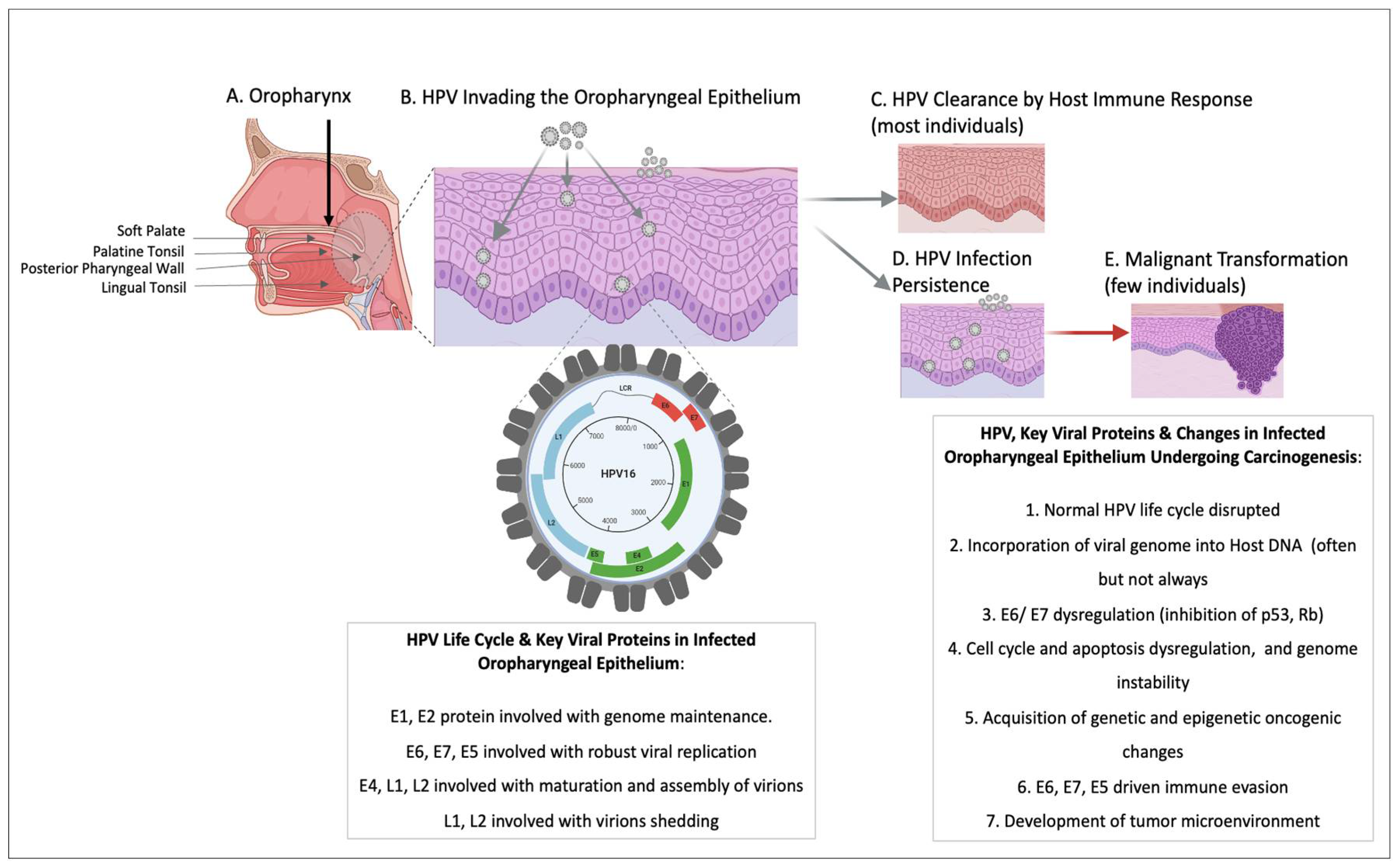
Figure 2.
A timeline of key milestones in the journey of HPV and OPC.
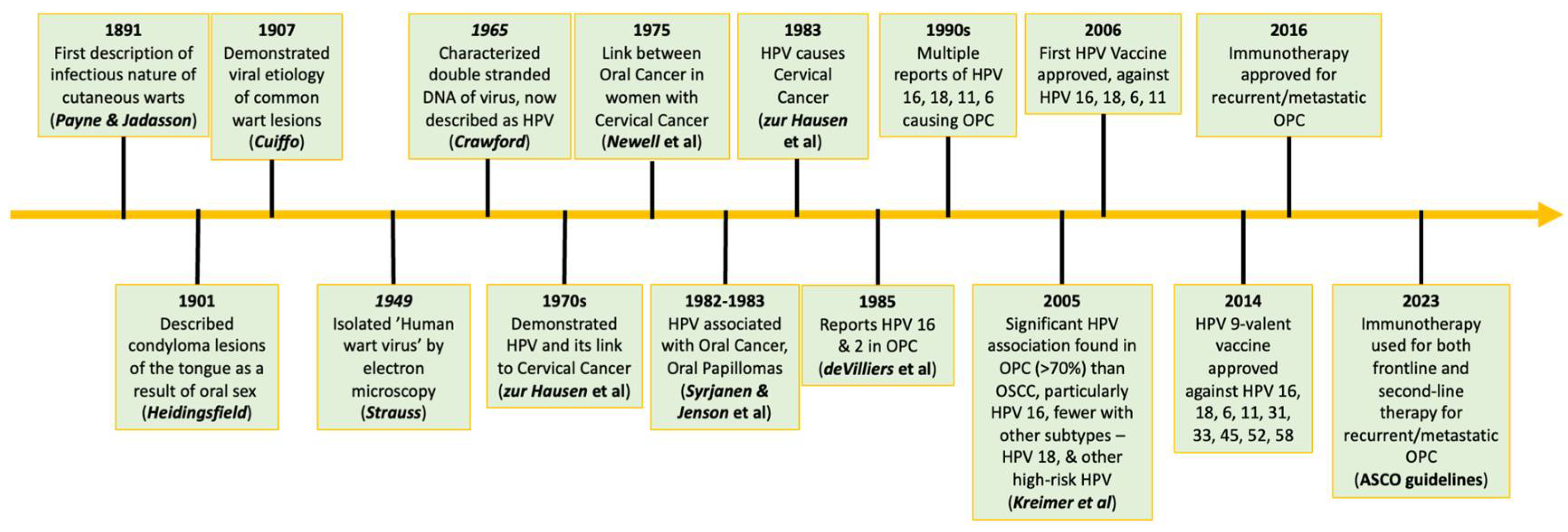
Disclaimer/Publisher’s Note: The statements, opinions and data contained in all publications are solely those of the individual author(s) and contributor(s) and not of MDPI and/or the editor(s). MDPI and/or the editor(s) disclaim responsibility for any injury to people or property resulting from any ideas, methods, instructions or products referred to in the content. |
© 2024 by the authors. Licensee MDPI, Basel, Switzerland. This article is an open access article distributed under the terms and conditions of the Creative Commons Attribution (CC BY) license (http://creativecommons.org/licenses/by/4.0/).
Copyright: This open access article is published under a Creative Commons CC BY 4.0 license, which permit the free download, distribution, and reuse, provided that the author and preprint are cited in any reuse.


