
Submitted:
15 March 2024
Posted:
18 March 2024
You are already at the latest version
Abstract
The objective of this study was to carry out the extraction and characterization of chitin and chitosan from the exoskeletons of the Farfantepenaeus californiensis species of shrimp, as well as its application in the formulation of polymeric films plasticized with glycerol used in the process of food packaging. The characterization of the extracted polymer was made by Fourier transform infrared spectroscopy (FTIR), Scanning electron microscopy (SEM), and physicochemical analysis of chitin and chitosan, resulting in a theoretical performance of 29.33% and 16.46%, respectively. In terms of quality, it was determined that the extracted chitosan had a molecular weight of 1.10 x10⁵ g/mol and a deacetylation degree of 80.23%, which classifies it as a low-molecular weight chitosan, suitable for food packaging. It was shown that the extracted chitosan is fit for the production of the films by using the casting method, presenting a tension resistance of 1 MPa. The Antibacterial analysis was carried out using Staphylococcus aureus (S. aureus) and Escherichia coli (E. coli), bacterial models commonly found in food. The results showed an effective inhibition capability of the polymeric materials. Overall, the results for both chitosan and the films indicate a high potential for application in the food packaging sector.
Keywords:
Chitin
; Chitosan
; packaging
; biopolymers
; biomaterials
1. Introduction
The incorporation of bioplastics of plant and animal origin has attracted enormous attention in the materials industry of the food packaging sector [1,2,3]. The advantages of biopolymers against traditional counterparts can be appreciated due to their biocompatibility, biodegradability, and occurring abundance [4]. Several examples of biopolymers that have been frequently used in the food industry are cellulose, hyaluronic acid, collagen, chitosan, and chitin, this last one standing out the most due to its availability (the most abundant after cellulose) and its intrinsic physicochemical properties [5].
Chitin is a natural polysaccharide characterized by its structural function. Its polymer is N-acetylglucosamine repeating units, which is abundant in the exoskeletons of crustaceans, insects, and some kind of fungi [6]. The chitin percentage found in exoskeletons of crustaceans and insects is around 30-40%, while in fungi, it ranges from 13-37% approximately [7]. Despite the potential of chitin and chitosan, the source of these biopolymers is still being wasted nowadays; this is because around 6 to 8 tons of crustacean waste are discarded, which causes contamination to both soil and water as a consequence [8]. In Mexico, about 60 species of shrimp [9] of which only 8 have cultivation potential and are economically important to the country [10]. Due to its geographic position, Mexico’s fishing regions are divided into the Pacific and the Gulf-Caribbean regions. Within these regions, the most commonly caught species are Litopenaeus stylirostris, Litopenaeus vannamei, Litopenaeus occidantalis, Farfantepenaeus brevirostris, and Farfantepenaeus californiensis, the latter being the most important [11]. Farfatepenaeus californiensis is the only species that is the most abundant in terms of weight of national fishery production value [9,12], and its cultivation grew by more than 100 thousand tons in 2014 [13]. As previously mentioned, Farfatepenaeus californiensis is positioned as a source of high commercial and productive interest, almost unexplored in the topic of extraction and application of chitin and chitosan [9]. Meanwhile, other species, such as Litopenaeus vannamei [14,15], Penaeus vannamei [16,17], and Penaeus monodon [18,19,20,21,22], are commonly studied for the extraction of chitin and chitosan.
The application of environmentally friendly materials in food packaging has recently been studied, considering biodegradable polymers derived from biomass as a viable and widely used option [23]. Biopolymers derived from polysaccharides which are commonly used, such as starch, pectin, alginate, and chitosan, share important physicochemical properties for film formation ability, non-toxicity, biocompatibility, biodegradability, and chemical stability [24]. However, chitosan draws attention for its antioxidant and antimicrobial properties since it has the presence of amino groups in the polymeric structure, follows electronegative behavior, takes protons, and develops a positive charge (polycationic). Thus, it can interact and interfere with negative electrostatic surfaces in some types of cells (such as bacteria) [25,26,27].
In this work, we valorize the waste shells of Farfatepenaeus californiensis as a source of chitin and chitosan with characterization techniques such as degree of acetylation, molecular weight, and solubility. In addition to its potential use in food packaging through the production of films using the casting method. The results obtained in this work will provide visibility, social attention, and industrial consideration to a scarcely explored source of chitin and chitosan.
2. Materials and Methods
2.1. Obtaining and Processing Farfatepenaeus Californiensis Shells
Before the visual evaluation of shrimp species (Farfatepenaeus Californiensis), the shrimp exoskeletons were collected from the waste of seafood restaurants in Mexicali, Baja California, Mexico. Subsequent to its collection, the exoskeletons were washed three times using tap water, and the last step was liquid soap (1 wt. %). The clean shells were disinfected in sodium hypochlorite (1 wt. %) for 1 h. Lastly, the exoskeletons were cleaned two times with distilled water to eliminate any sodium hypochlorite residue. After disinfecting, the exoskeletons were sun-dried on a metal plate for 8 h at 46 ºC. Once dried, the shells were shredded in a conventional blender until a thick powder was obtained (10 µm-particle size).
2.2. Extraction of Chitin and Chitosan
For the extraction of chitin and chitosan, demineralization, deproteinization, bleaching, and deacetylation of the shrimp shell powder was carried out following the sequence shown in Figure 1b and Figure 2c, with some modifications [7,28,29].
The shrimp powder was initially demineralized in 1N HCl (1:10, v/v%) at 60 ºC for 35 min with constant stirring. The demineralized shells were washed 5 times with abundant distilled water until a neutral pH was reached. For the deproteinization process, NaOH (15 wt. %, ratio of 1:10) was added at 65 ºC ± 3 ºC for 2 h under constant stirring, resulting in chitin. Next, the bleaching was performed using NaOCl (1 wt. %) for 30 min; then, the bleached shells were dried in a dry air oven (Thermo Scientific, USA) at 60 ºC for 2 h. Finally, the obtained chitin was deacetylated through a NaOH (50 wt. %, ratio of 1:10) and boiled for 2 h under constant stirring. The resulting product is then rinsed with abundant distilled water until neutralized and dried in an air oven at 60 ºC for 2 h, resulting in chitosan.
2.3. Physicochemical Characterization of Chitin and Chitosan
2.3.1. Morphological and Elemental Analysis
The surface morphology of the chitosan powders was analyzed by means of SEM (JSM-6010LA, JEOL, USA) at 10 kV accelerating voltage. A thin gold layer was sputter-coated. For the chitin sample, a Field-emission SEM (FE-SEM; Lyra 3 Tescan) was used at 15kV without any coating. The elemental compositions were analyzed using energy dispersive X-ray spectroscopy (EDX, Brucker XFlash) coupled to the FE-SEM.
2.3.2. Fourier Transform Infrared Spectroscopy (FTIR)
To evaluate the extraction quality of polymers, samples were analyzed by attenuated total reflection Fourier transform infrared spectrometer (ATR-FTIR, Frontier, Perkin Elmer, USA) was used [30]. The spectra were acquired by applying a scanning range of 4000-400cm-1 and a sweep speed of 0.5 cm-1/s at room temperature. The resulting spectra were compared to commercial chitosan (Sigma-Aldrich, St. Louis, USA).
2.3.3. Ash Content
To measure the ash content, 1g of the extracted chitosan was placed in a refractory crucible for a pyrolysis heating to 600 ºC with a heating ramp of 10 °C/min for 5 h using a muffle (Vulcan). The resulting product was then weighted in an analytical balance. The following formula was used to calculate the ash level [31]:
Where “MS” stands for the weight of the initial sample in grams and “MR” is the weight of the residue in grams.
2.3.4. Total Yield
To calculate the extraction yield of both chitin and chitosan, the following formula was used [32]:
2.3.5. Deacetylation Degree
The chitosan powder was dissolved in HCl excess to protonate the free amino group of the chitosan chains. Subsequently, a valuation is performed using NaOH until pH stabilization. Therefore, we generated a titration curve presenting two inflection points. The difference between the NaOH and the acid required to protonate the chitosan amino groups can be determined by the expression [33]:
Where “y” is the major inflection point, “x” is the minor inflection point (expressed in volumes), “f” represents the molarity of the NaOH solution, “w” is the mass in grams of the samples, and “16.1” is a factor associated with the type of protein under study. The samples were analyzed using a HATCH potentiometer, to determine the content amino groups. Chitosan (0.5 g) was diluted into 20 mL of HCl (0.3 M), and then a NaOH solution (0.1 M) was titrated, measuring pH changes every 2mL of titrant, under stirring. Finally, a titration curve of pH vs. mL of NaOH added was constructed to calculate the degree of N-deacetylation of chitosan.
2.3.6. Degree of N-acetylation
The N-acetylation of chitin was calculated according to equation 4, where the ratio of the FTIR characteristic bands of Amide I (1630 cm-1) and the hydroxyl (3437 cm-1) groups are measured. On the other hand, for the N-acetylation of chitosan, we followed equation 5, expressing the ratio of the characteristic bands of amide III (1320 cm-1) and the methyl groups (1420 cm-1) [34]:
Subsequently, the degree of N-acetylation of chitin and chitosan are calculated by applying Equation 6 [35]:
2.3.7. Molecular Weight of the Biopolymers
The average molecular weight of each sample was determined using capillary viscosity through a Cannon-Fenske type viscometer at 30 °C (±1 ºC) [36]. The chitosan samples were prepared in acetic acid (0.25 M) and sodium acetate (0.25 M). From this mixture, a standard solution of chitosan was prepared with a concentration of 2 x 10⁻³g/mL. Next, the dilution aliquots were prepared at 1.82 x 10⁻⁴g/mL, 3.3 10-4g/mL, 4.62 10-4g/mL, 5.71 10-4g/mL, 6.67 10-4g/mL and 7.50 10-4g/mL. The calculations were based on the measurement of the viscosity of the solution compared to the time it takes for a specific volume to fall through a capillary tube versus the time of the solvent used [37].
2.3.8. Thermal Analysis (DSC)
The Differential scanning calorimetry (DSC; TA Instruments DSC Q2000) was applied by placing a chitosan prepared sample (1mg) in a stainless-steel crucible, hermetically sealed, and mounted in the DSC. Then, the samples were heated from 30 °C to 900 °C in ramps of 20 °C per minute in nitrogen atmosphere.
2.3.9. Thermal Gravimetric Analysis (TGA)
For the TGA test, 1 mg of chitosan, was placed in a thermal analyzer (TA Instruments Q500) under a constant nitrogen flow, and heated at a ramp rate of 20 °C/min between 25 - 900 °C.
2.4. Preparation for Antibacterial Packaging
Initially, 2 g of the prepared chitosan powder was added to 100 mL of a 2% wt. citric acid solution at 60 °C under constant stirring for 2 h or until a clear solution was obtained. Subsequently, the mixture was filtered using a Whatman #1 filter to eliminate any residue present in the solution. Finally, this working solution was stored at 4 °C until use.
For the casting method, 20 mL of the prepared chitosan was mixed with 0.05 mL of glycerol solution for 1 h to assess an utterly homogeneous solution. Then, the polymeric solution was drop cast into a 100 mm diameter polystyrene Petri dish (VWR, USA) without generating any air bubbles. Finally, polymers were dried using a convection oven at 70 °C for 2 h, and placed in a desiccator for 1 h to avoid moisture residue.
2.5. Active Packaging Characterization
2.5.1. Mechanical Testing
The mechanical properties such as tensile stress (TS), elongation at break (EB), and Young’s Modulus (YM) were determined through tensile testing, following the ASTM D882 [38] norm. The INSTRON (Bluehill) was operated at 10 mm/min using test samples (25mm in width and 140mm in length), a distance between gaps of 100mm, and a grip between gaps of 20mm on each side. For each test, 5 samples were used. The TS and the EB were calculated with the TS vs. EB curve and Young’s Modulus with the angular coefficient of these curves.
2.5.2. Percentage of Moisture Content, Degree of Swelling and Solubility
The calculation of moisture content (MC) swelling capacity (SP) and solubility (S) of the antimicrobial packings was performed. 5 pieces of chitosan film (2 x 2 cm) were taken and weighed (W0) on an analytical balance. Then, the pieces were dried in an oven at 100 °C for 24 h, at the end of which they were weighed once again (W1). The dry samples were immersed in beakers with 50 mL of distilled water, were they remained under occasional agitation for 24 h at room temperature. Once the time was over, the samples were filtered using a Whatman #1 filter and weighted once again (W2). Finally, each piece was dried at 100 °C for 24 h, and the final weight was obtained (W3). The MC%, SP% and S% were calculated by using equations 7, 8 and 9 respectively [39]:
2.5.3. Antimicrobial Activity
For the analysis of the antimicrobial properties, individual inocula were prepared in trypticasein soy broth (Becton Dickinson, Sparks, MD, USA) from fresh colonies of Staphylococcus aureus (S. aureus, ATCC 25923) and Escherichia coli (E. coli, ATCC 25922) strains. The inoculum was incubated for 24h at 37 °C under aerobic conditions [40]. Subsequently, each inoculum was adjusted to a concentration of approximately 1 × 10⁷CFU/mL in trypticasein soy broth. Then, 0.1mL of each inoculum was cultured on trypticasein soy agar (15 mL of liquid per 100 mm Petri dish). After 30 min, 4 circular polymeric samples (10 mm in diameter) were placed on the inoculated trypticasein soy agar plates and incubated for 24 h at 37 °C. Finally, each agar plate was mounted on a darkfield colony counter (Reichert, NY, NY, USA), the inhibition zones were measured with an electronic digital caliper, and digitalized [41].
2.6. Statistical Analysis
The numerical information of the inhibition halos was expressed as the mean ± standard deviation (SD) and analyzed using GraphPad Prism 7 (GraphPad Software Inc., San Diego, CA, USA). The significance of differences was evaluated using one way analysis of variance (ANOVA) followed by the Tukey’s multiple comparison test. A P < 0.05 was considered statistically significant.
3. Results and Discussion
3.1. Chitin and Chitosan Extraction
It has been widely described as many naturally occurring chitin and chitosan obtaining sources; however, the exoskeletons of crabs and shrimps have proved to be the principal industrial and scalable fountain of importance [35]. Moreover, the exoskeletons commonly used for chitin and chitosan obtention are composed of organic and inorganic molecules, such as proteins, carbohydrates, and minerals. Interestingly, the molecular composition of this complex network has a circumstantial impact on the economics and physiochemistry of chitosan [27,42]. From the economic point of view, the increase in the percentage of by-products elevates the energy demanding period in the demineralization and deproteinization processes, thus far, demanding novel and efficient extraction and processing alternatives. On the other hand, the physicochemical properties such as hydrophobicity/hydrophilicity, solubility, cellular responsiveness, and crystallinity degree are strongly altered parameters that impact the food packaging response of chitosan and its derived polymers [43].
The Chitin and chitosan biopolymers represent only a fraction of the entire complex network forming the exoskeleton structure of F. californiensis. Previous research focused on other crustacean exoskeletons delimit the percentage of chitin between 4 - 36 % and chitosan between 12 – 35 % [44]. For example, Junior et al. obtained a 36% efficiency of chitin (based on exoskeletons) from the shrimp Litopenaeus vannamei [17]. On the other hand, Rakshit et al. obtained a chitin yield of 23.23% coming from Litopenaeus vannamei [45]. Moreover, it was reported that Metapeneus monodon could generate up to 24.33 % chitin and 15.25 % in chitosan of the total weight of the shells [46], and the species Fenneropenaeus indicus showed 45.14% chitosan throwput [47]. Therefore, our current work suggests that our extraction protocol yields an efficient obtention performance using F. Californiensis shrimp (29.33% for chitin and 16.46% for chitosan) being within the required range for these crustacean exoskeletons [48].
It is crucial to consider the extraction method as a mandatory factor controlling the yielding efficiency of chitin and chitosan biopolymers from crustacean sources. The chemical method is the gold standard option for industrial scale level operations, considering the application of strong acids (e.g., HCl, H2SO4) and alkaline solutions (NaOH, KOH) for obtaining minerals and proteins contained in the shrimp exoskeletons. Moreover, the chemical method allows precise control of the extraction process, permitting the establishment of a regulable scaling up of the extraction process [49]. On the other hand, a biological method can be applied to perform the extraction with non-aggressive and green chemical processes, using microorganisms that produce enzymes, bacteria, and organic acids (weak acids) [50]. However, this method is time-demanding (taking several days to accomplish each extraction step) [51] and can only be applied on a laboratory scale [52]. For example, Cira et al. reported an average period of 6 days for the demineralization and deproteinization of Penaeus monodon exoskeleton species using Lactobacillus bacteria [53]. On the other hand, Neves et al. extracted chitin using the Brazilian farmed shrimp Stenopus hispidus by fermenting it with Lactobacillus plantarum; the process demanded 72 h [54]. Finally, Xie et al. showed that Penaeus vannamei (from Chinese coasts) required 96 h fermentation using L. acidophilus and E. profundum [55] to ensure the extraction and deacetylation procedure. Therefore, considering the above-stated information for assessing an efficient and high-yielding process, we selected the chemical method for the extraction of chitin and chitosan from F. Californiensis, shorting the required extraction process and providing evidence for selecting this crustacean model [9].
The surface morphology analyzed by means of SEM illustrated noticeable differences between the extracted chitin and chitosan polymers (Figure 3). Moreover, in Figure 3b, we observed that chitosan presents a homogeneous, smooth, and non-fibrous structure, suggesting a well-orientated crystalline structure (Figure 3a). Meanwhile, chitin presented regular cracks that could be the result of the agitation subjected during the deacetylation process (Figure 3b) [38]. In turn, it was found that the chitosan has little porosity and a regular surface, proposing that the n-acetyl and amino groups form weak connections among each other, resulting in the chitosan deacetylation [56]. On the other hand, chitin displayed a laminated and homogeneous surface morphology (Figure 3a), reflecting that chitin may form a uniform structure. Unlike chitosan, which has some irregularities in its interlaminar and intralaminar structure, indicating semi-crystalline regions caused by the anisotropic organization of the intermolecular hydrogen bonds [32]. For example, the occurring chitin and chitosan structural morphology from Penaeus monodon [18], Parapenaeus longirostris [57], and Farfantepenaeus aztecus [58], were characterized due to slighter porosity and microfibrillar, compact and layered arrangements than those observed here. Furthermore, the chemical extraction method directly influences the modulating chitin and chitosan morphological conditions [22]. Thus, these structure forming properties are classified as microfibrillar and porosity textures, neither porosity nor microfibrillar configurations, and lastly, with only microfibrillar architecture [22]. Interestingly, it has been shown that the surface structure can modulate the cellular and bacterial behavior [59], thus far, proposing that the chitin and chitosan smooth and regularly generated surfaces can provide an antimicrobial response for film applications [48,60].
The quality of chitosan is one of the most important characteristics that defines its performance for qualifying in the food-packing industry. Therefore, special attention must be paid to the degree of N-acetylation/deacetylation, the molecular weight, and the polymer solubility [61,62]. In this work, we evaluated the purity of the extracted chitin and chitosan by FTIR and compared it to a high-purity commercially available chitosan standard. In Figure 4, spectra of chitin, extracted chitosan, and commercial chitosan are presented, showing bands at 3257cm-1 and 2881cm-1, which are assigned to N-H and O-H stretching vibrations for chitin [63]. Moreover, bands at 1656cm-1 and 1626cm-1 are appointed to stretching vibrations to C=O (amide I) and C-N (amide II), respectively. Likewise, the bands at 1552cm-1 and 1375cm-1 are assigned to C=O stretching vibrations of amide II bonds and buckling vibrations of amide-NH2 bonds, indicating the N-acetylation resulting in modifications. Finally, a band at 897cm-1 corresponding to the CH group of the pyranose and the glycosidic ring [64] of the polysaccharides was detected [45]. It is noticeable that the bands in the region of 3500-3350cm-1 are absent for the extracted and commercial chitosan, and the signals at 1656cm-1 and 1626cm-1 considerably reduced, which demonstrates the correct extraction of chitosan by the method employed [48]. The experimental and commercial chitosan polymers presented similar spectra, displaying bands at 3372cm-1 and 287cm-1, which are assigned to N-H and O-H stretching vibrations [19]. Further bands at 1586 cm-1 and 1374 cm-1 corresponding to C=O stretching vibrations of amide bonds and buckling vibrations of amide-NH2 bonds were also present. Finally, the typical C-O-C stretching vibrations at 881cm-1 for these carbohydrates were detected. This information suggests that our extraction method generates a purity level similar to that of commercial reference control, supporting its application as a packaging coating for the food industry.
In addition to the differentiation between chitin and chitosan purity, the degree of N-acetylation was analyzed by FTIR [34]. For chitosan, the characteristic bands of the amide III groups (1630cm-1) and, as a reference, the methyl groups (3437cm-1) indicate a deacetylation percentage of 80.33%. This result corroborates that the polymer follows the purity level required since it is within the parameters established for an application in food packaging (70 - 85%) [49]. Moreover, the molecular weight represents the average length of the chitosan polymeric chains [57], thus, the sum of the acetylated and deacetylated units [65], defining the physicochemical, biological, and mechanical properties of the biopolymer [36]. Although it is not possible to obtain two polymers with the same molecular weight, the commercial chitosan has been divided as low (1000 g/mol ≤ Mw ≤ 10000 g/mol), medium (10000 g/mol < Mw ≤ 250000 g/mol) and high (Mw > 250000 g/mol) molecular weight [66]. In this work, we calculated the molecular weight by intrinsic viscosity, showing 239.23dL/g of viscosity and a molecular weight of 1.10 x10⁵ g/mol (Figure 5, Table 2). This evidence suggested that the extracted chitosan falls into a low molecular weight classification, indicating that it is suitable for food packaging applications due to its biodegradability, biocompatibility, bioactivity, and low toxicity compared to high molecular weight chitosan [65,67].
Figure 5.
Intrinsic viscosity of chitosan.
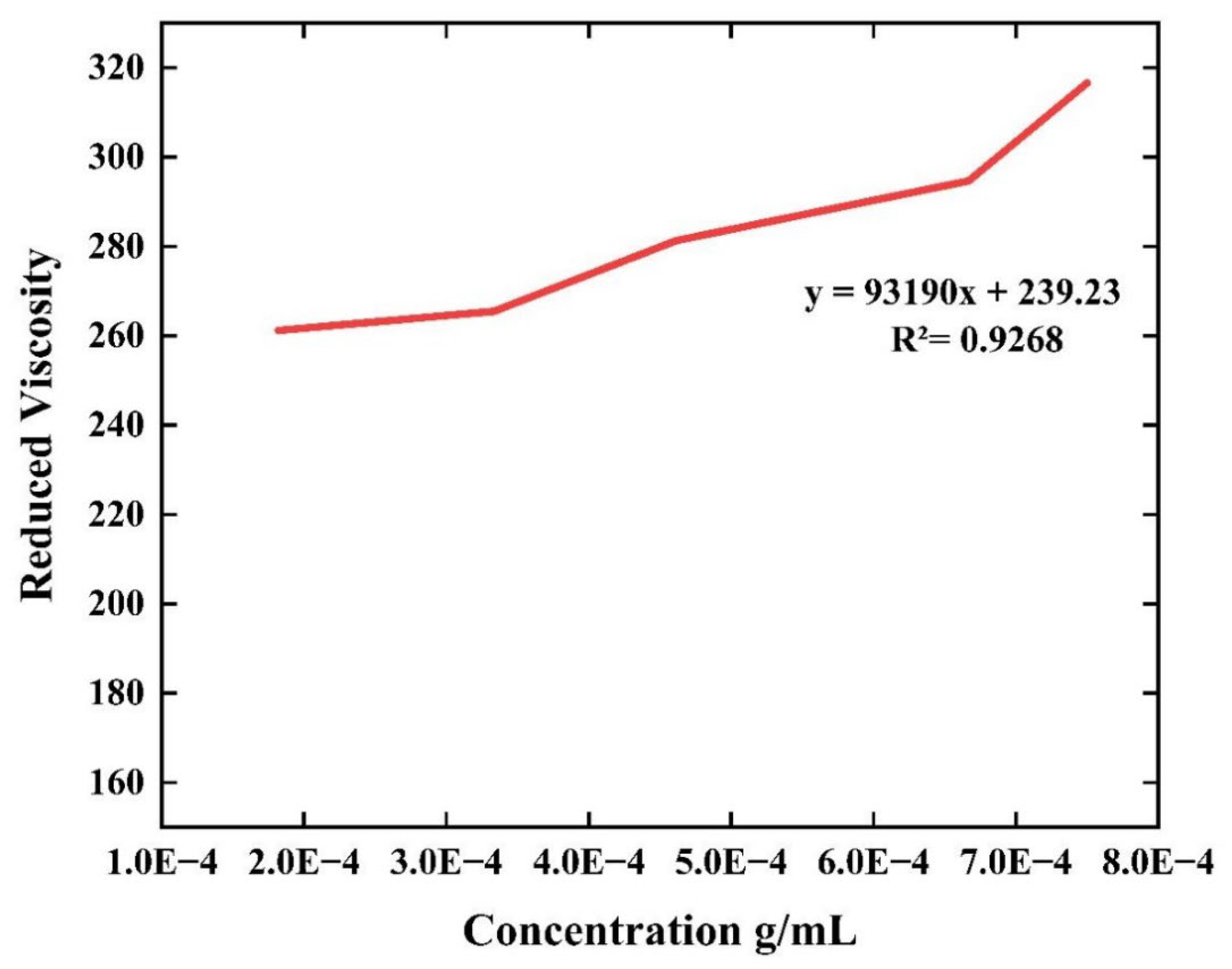

Table 1.
Comparative analysis of intrinsic viscosity and molecular weight of extracted chitosan and commercial chitosan.
Table 1.
Comparative analysis of intrinsic viscosity and molecular weight of extracted chitosan and commercial chitosan.
| Material | Intrinsic viscosity [η] (dL/g) |
Molecular Weight (g/mol) |
|---|---|---|
| Farfantepenaeus californiensis extracted chitosan | 239.23 | 1.26 x 10⁵ |
| Commercial chitosan | 242.54 | 1.28 x 10⁵ |
Table 2.
Mechanical properties of chitosan film.
| Film | TS (Mpa) | EAB (%) | Young’s Modulus (Mpa) |
|---|---|---|---|
| Chitosan Film | 1.10±0.32 | 186.31±90.33 | 1.565±0.45 |
The molecular weight, the degree of deacetylation (DDA) of chitosan, and the extraction conditions are fundamental parameters that directly influence the biological, physicochemical, and mechanical polymeric properties [7]. Thus, we analyzed the DDA showing 70.85 % achieved by our protocol, describing the directly proportional relationship between the amino groups and the degree of deacetylation, so we can determine that the higher the concentration of amino groups, the higher the degree of deacetylation [61]. In addition, it has been reported that a DDA between 60 - 90% chitosan could solubilize in organic acids due to the formation of a polycation of the N-amino-D-glucosamine units [68].
In order to evaluate the purity of chitin and chitosan biopolymers, we tested the thermal stability, degradation stages, and conditions in the phase transformation process [69]. Thus, the DTG results (Figure 6) of chitin samples showed an endothermic peak at 47°C (corresponding to water loss) and at 298°C, corresponding to protein and pigment residues [70]. Moreover, we detected that at 400.6°C can take place the acetylation conversion of chitin to an amino group characterized for chitosan [70]. The prominent endothermic peaks are shown at different temperatures, which demonstrates the crystallization of chitin, as mentioned above. Interestingly, we can hypothesize that the hydrogen bonds formed between the fibers bring good thermal stability (high-temperature transformation phase) to the chitin molecular structure [71]. On the other hand, chitosan (Figure 6c) manifestoed two degradation stages highlighted by a first endothermic peak at 48°C corresponding to moisture loss and pressure exerted due to water evaporation [70]. In this regard, polysaccharides such as chitosan in a solid state possess amorphous structural regions promoting it with a strong affinity to water absorption and easy hydration [72]. The second endothermic peak was detected at 298°C (Figure 6d), proposing the degradation of the saccharide rings, chain structures, and deacetylated monomers (amino groups)[64]. It is particularly interesting to focus on the fact that carbohydrate derived biopolymers, such as those of chitin and chitosan, do not follow a clear glass transition (Tg). Therefore, we can point out the overcoming parameter of the crosslinking degree, poor water removal, the sample preparation procedure, the sensitivity of the equipment, and the measurement technique in the impact of the Tg behavior [73].
In Figure 7, we presented the DSC behavior of the chitin and chitosan packing polymers between 0 - 700 °C. Moreover, chitin presents two marked stages of degradation (Figure 7a); the first is given at 248 °C due to weight loss evaporation of water (6 %) and the hydrophobic groups present within the chitin chains. The second stage (385 °C) featured the degradation of the polymeric structure, characterizing the dehydration of the saccharide ring and deterioration of the acetylated units [74]. On the other hand, the chitosan samples showed two phases of weight loss (Figure 7b); initially at 70 °C we detected a first degradation steep due to the loss of water moisture and the subsequent chitosan degradation when reaching 298 °C (weight loss of 44%). This is related to the depolymerization and decomposition of the acetyl and amine groups of chitosan [56], which followed a complete degradation at approximately 425 °C, resulting in a residue of 33.22%.
Finally, the purity of the extracted chitosan was evaluated by incineration and ash content. Ash is the inorganic residue remaining after the total degradation of chitosan by calcination. The measurement of ash content is an important indicator of the content of minerals such as calcium carbonate, as it shows the effectiveness of the demineralization process. In the extracted chitosan sample, 0.22% ash was obtained. Regarding the solubility of chitosan and its viscosity, this is affected by the presence of remaining mineral residues present in the ashes. To have high quality chitosan, it must have less than 1% of ash content [75].
3.2. Food Active Packaging
Chitosan is a characteristic polymer that stands out from other biopolymers (e.g., collagen, elastin, and more) thanks to its abundance as raw material, the high solubility in acidic aqueous mediums (pH < 4), and its antimicrobial properties conducted by the protonation of the amino group [76]. Thus, we can postulate chitosan and chitosan-derived co-polymers as an excellent precursor option for industrial applications, especially in smart food packaging [77].
In the alimentary processing sector, food packaging must comply with particular mechanical, aesthetic, and impermeability characteristics, achieving strict quality controls and legal regulations [25]. From the mechanical point of view, food packaging must fulfill controllable malleability, flexibility, and impact resistance. Regarding aesthetics, transparent food packaging is preferred to facilitate technical and management inspection. Moreover, the packaging should act as an impermeable barrier between the environment and the food to prevent subsequent moisture-associated food damage [78]. To meet the traditional requirements for food packaging, our work evaluated the mechanical (tensile strength, elongation at break, and Young’s modulus), aesthetic (color), and impermeability (moisture content, degree of swelling, and water solubility) properties of chitosan films fabricated by the casting method.
The mechanical properties of food packaging materials are crucial to ensure their integrity and ability to withstand mechanical stress during shipping, handling, and storage. Figure 8 presents the stress-strain graph of the films made by the casting method, along with the numerical parameters assessed from the mechanical test (Table 3). The graph shows that the polymer has a wide area in the elastic zone, split into two parts, with a maximum stress of 0.25MPa in area 1 and 1MPa in area 2. After reaching the maximum stress, the tensile stress decreases drastically to zero for a strain of 1.5 mm/mm.
Based on the stress-strain graph obtained for the chitosan film, it is observed that its tensile capacity is lower compared to other polymers used in the food packaging industry [79].
Chitosan films have low mechanical properties; they are rigid and brittle, because the hydrogen bonds have strong intermolecular interaction between the polymer chains. The use of plasticizers helps to improve those mechanical properties by reducing the rigidity [80]. However, the variation of the films mechanical properties are in function of the molecular weight, deacetylation degree, source, extraction, manufacturing process, and the plasticizer [81]. Thus, paying special attention of the applicating polyol glycerol, that have small molecules facilitating the incorporation into low molecular weight chitosan [82]. Chitosan and glycerol have high affinity, because hydrogen bonds are formed and it decreases the intermolecular forces in the polymer chain, resulting in permeability between the molecules, facilitating the intermolecular space and sliding behavior of the chains, decreasing the cohesion and increasing the force elongation at break [83]. The crystalline domain of the polymeric chain of chitosan is disrupted by glycerol [80], and the increasing cohesion prevents the separation structural, so the elongation at break it is indeed increased [81]. In addition, it is known that glycerol does not have migratory process for food and do not have real problem with human health, so it is a good choice for food packaging application [84]. Moreover, it was reported by Mujtaba et al. that chitosan dissolution in citric acid decreases the tensile strength compared to chitosan dissolved in acetic acid [85]. Therefore, highlighting the importance of food grade accepted plasticizer for the synthesis of transparent and flexible polymeric films for food packaging protection.
However, when comparing to other chitosan films previously reported, we appreciated that the film elongation has comparable values. In a study conducted by Park et al., the tensile stress for films synthesized with a 2% chitosan solution extracted from red crab shell was 6.7 MPa, featuring an elongation at break of 117.8 MPa [86]. In turn, Smirnov et al. detected a strain stress of 8 MPa and an elongation at break of 40 MPa for commercially available low molecular weight chitosan films (Sigma Aldrich), synthesized with citric acid/choline chloride solution [87].
Therefore, the chitosan extracted from F. californiensis is competitive with current products achievable in the market. Aesthetic properties, such as film transparency, are critical in the food packing industry because the user must be capable of detecting any alteration at all times. For this reason, the film transparency was assessed visually. In Figure 15, the synthesized film has favorable optical characteristics, as it is completely transparent with a yellowish color, which is distinctive of chitosan.
Figure 9.
Film transparency.
Interestingly, the percentage of moisture content is related to the physical properties of biopolymers and their barrier capacity, playing a crucial parameter in the food industry. Unlike the chitosan content, the moisture content of the biopolymer solution is unaffected. In the case of the chitosan film evaluated here, the moisture content level obtained was 21.74%. This effect can be explained because the drying time and heat temperature are thermodynamic factors that inversely affect the moisture content of the films. Therefore, by applying an extended drying period at 40 °C, we can control the structure of the polymer solution, which is reorganized by the interactions between the hydrogen bonds and the Van der Waals forces of the water molecules, as well as the hydrophilic behavior of the chitosan. In this regard, the solution acquires a gelatinous consistency that subsequently turns into a solid film. On the contrary, if we applied a shortened drying time at higher temperatures, as occurred in this high heat process (70 °C), the gel formation stage is reduced, and the solution immediately solidifies as a film, due to the accelerated evaporation of water. Consequently, a disordered and fragile biopolymer structure and a low moisture percentage are obtained [39].
We discovered that the film had limited water absorption, showing swelling of 8.7%, which indicates that the film had low water-holding capacity in its matrix and was almost impermeable. This important behavior can be explained by the small amount of glycerol added during the film preparation [1]. Glycerol is used as a plasticizer agent that has carboxyl and hydroxyl groups, providing it a hydrophilic nature, so its scarcity in the polymeric material matrix reduced the interaction with water molecules [39]. Moreover, solubility (S) is an inverse measure of the resistance of a material to the presence of water and is closely related to the hydrophilic properties of the biopolymer [88]. Nonetheless, our study indicated that the films have a solubility capability of 64.63 %, which can be attributed to the hydroxyl groups provided by the added glycerol. These groups are inserted into the polymeric chain of the material and promote the migration of water molecules among the internal matrix, facilitating the interactions between the sample components. As a result, swelling of the material is reduced, and dissolution is promoted [89].
In addition to mechanical, aesthetic, and impermeable features, packaging can add specific functionalities such as preventing product contamination, indicating the product status [90], determining its pH, or being antimicrobial; those films are considered active or intelligent packages [91]. In our research, we propose classifying chitosan films as active packaging due to their added antimicrobial activity.
The antimicrobial effects of the chitosan biopolymer were evaluated against S. aureus and E. coli, considered critical pathogens commonly generating food contamination and causing gastrointestinal diseases [92]. The generated inhibition zones are presented in Figure 10, showing an active antimicrobial action against S. aureus and E. coli. The inhibition capability of our films, in part, can be explained by the molecular weight and the surface structure. In this regard, the extracted chitosan showed a low molecular weight, which brings extended increased extension and relaxation of the polymeric chains when compared to those of high molecular weight [93]. Far more interesting is the antimicrobial mechanism of chitosan, prominently tailored due to the protonation of the amino group under acidic conditions, harboring ionic interactions among the bacterial cell membrane. Thus, orchestrating the hydrolysis of the peptidoglycan layers (at the membrane or cell wall level), resulting in the rupture and subsequent leakage of cytoplasmatic electrolytes and DNA/RNA components, leading to bacteria lysis [77,94]. Therefore, the molecular weight of chitosan directly influences on the antimicrobial action, which could be improved by adding glycerol to increasing the polymeric response [89].
3.3. Chemical and Thermal Properties of the Film
In order to evaluate the intermolecular interactions of the polymeric functional groups with the glycerol (plasticizer) in the films we need to pay special attention into the functional bonding groups [74]. In Figure 11, the chitosan film spectrum reveals the presence of bands at 3247 cm-1 and 2878 cm-1, which corresponds to N-H and C-H stretching vibrations [95], respectively. It could explain that, the hydrogen bonding of the chitosan chains was disrupted and weakened, due to the polyols of glycerol interacting by the hydroxyl groups, rather than the amino group of chitosan and conserving the electrostatic interactions [96].
Moreover, the peaks at 1549 cm-1 and 1406 cm-1 are attributed to C=O stretching vibrations of amide II and buckling vibrations in C-OH [25,78]. Additionally, the 1024 cm-1 band also displays stretching vibrations at C-O [82]. The addition of glycerol changes the internal hydrogen bond interactions, involving the amino group (-NH2) and amido group (NH-) of chitosan, turning the polymeric chains further mobile, resulting in a more a flexible film [97].
The authors consider that those results significantly explain the physicochemical behavior achieved for the design of food packaging materials, with advanced and efficient film functional properties.
The thermal stability and film properties under storage conditions, the temperature ranges at which the material degrades, and decomposes were determined by means of DSC, TGA, and DTG. The DSC thermograms of the chitosan biopolymer (Figure 12a) show the formation of endothermic peaks (62 °C and 194 °C) and an exothermic transition at 262 °C. The first endothermic peak is due to the loss of water molecules in the chitosan, which releases the bonds of the hydrophilic groups of the chitosan molecules, leading to dehydration of the polymer [74,98]. The second peak (194 °C) corresponds to the Tg of the polycarbohydrate, which indicates the change from a glassy to a rubbery state. This event is associated with an increase in the viscoelastic nature of the material. Thus, a subsequent peak before this phase is expected be observed indicating, the melting points. However, the polysaccharide nature of chitosan does not allow melting transformation, instead, it undergoes thermal degradation at relatively low temperatures. Consequently, the exothermic peak is observed in the material, corresponding to the thermal degradation of chitosan. At this point, the molecular structure of the chitosan is affected by the onset of degradation of the glucosamine units present in polymeric composition [73].
The TGA and DTG curves of the analyzed materials (Figure 12 b, c) show the weight loss stages located at approximately 50 °C, displaying a weight loss of 0.41%, attributable to the removal of the water in the sample. During this process, the water molecules release the amino and hydroxyl functional groups of the chitosan [72]. Subsequently, at 175 °C, a second stage of weight loss is observed, corresponding to glycerol degradation with a 6.15% decrease in weight. It should be noted that glycerol has a lower decomposition temperature than chitosan, resulting in a reduction of the thermal stability of the polymer. This decomposition weakens the hydrogen bonds in the chitosan network, promoting the mobility of the molecular chains and, consequently, reducing the crystallinity [99]. The third stage starts at 209 °C (Td) with a weight loss of 23.87%, indicating the thermal degradation zone of the polymer. This process continues until total degradation was reached at 357 °C. Next, a residue of 20.32% was detected, which corresponds to the minerals and inorganic compounds present in the chitosan solution used to manufacture the polymer [73].
Finally, the surface materials were characterized using SEM (Figure 13). The micrographs revealed that the film exhibits a smooth and homogeneous surface with small wrinkles characteristic of the marks left by the mold used in the casting method, suggesting efficient compatibility between chitosan and glycerol. Some white spots were observed in the images, which correspond to residues remaining from the filtration process of the chitosan solution prior to casting [73]. These results propose that the chitosan content and the amount of added glycerol do not alter the structure and morphology of the films [89].
4. Conclusions
This study assesses the physicochemical properties of chitin and chitosan extracted from exoskeletons of F. californiensis, as well as the potential application in food packaging as an active film. The main findings of the work are as follows:
- The extraction purity demonstrated that F. californiensis is a potential source for obtaining high-quality chitin and chitosan. Our method yields an ideal low molecular weight natural polymer for the packaging industry.
- The application of the extracted chitosan by the casting method proved to be an effective strategy for creating active packaging films, showing antibacterial action against common enteric infection agents.
- The mechanical properties of the film are not as efficient as synthetic polymeric films, but it can be increased adding other additives o making blends with other biopolymers.
The results highlight the potential economic and environmental benefits of using natural polymers as an innovative approach for active packaging. Furthermore, the use of exoskeletons of F. californiensis, a by-product of the fishing industry, demonstrated to promotes sustainability by reducing waste generation.
Author Contributions
Conceptualization, I.M.W.-M, B.V.-S., and E.B.-P.; methodology, I.M.W.-M, K.G.-C., J.S.-C., P.M., and J.C.-S.; investigation, I.M.W.-M, J.S.-C., P.M., and J.C.-S.; resources, B.V.-S., N.C., and E.B.-P.; writing—original draft preparation, I.M.W.-M, B.V.-S., J.S.-C., and E.B.-P.; writing—review and editing, B.V.-S. and E.B.-P.; supervision, B.V.-S. and E.B.-P.; project administration, B.V.-S. and E.B.-P.; funding acquisition, B.V.-S. N.C., and E.B.-P. All authors have read and agreed to the published version of the manuscript.
Funding
This research was funded by SEP-CONACYT “Proyecto Apoyado por el Fondo Sectorial de Investigación para la Educación” CB2017–2018, grant number A1-S-38368, and Program No. 317330 FOP02 CONACYT, for financial support.
Institutional Review Board Statement
Not applicable.
Data Availability Statement
The data presented in this study are available on request from the corresponding author.
Acknowledgments
We thank to Instituto de Ingeniería of Universidad Autónoma de Baja California.
Conflicts of Interest
Authors Patrick Moe and Nelson Cheng were employed by the company Magna International Pte Ltd. The remaining authors declare that the research was conducted in the absence of any commercial or financial relationships that could be construed as a potential conflict of interest.
References
- Castelló, M. E., Anbinder, P. S., Amalvy, J. I., & Peruzzo, P. J. Production and characterization of chitosan and glycerol-chitosan films. MRS Advances, 2018; 3(61), 3601–3610. [CrossRef]
- Luo, A., Hu, B., Feng, J., Lv, J., & Xie, S. Preparation, and physicochemical and biological evaluation of chitosan- Arthrospira platensis polysaccharide active films for food packaging. Journal of Food Science, 2021; 86(3), 987–995. [CrossRef]
- Zheng, K., Xiao, S., Li, W., Wang, W., Chen, H., Yang, F., & Qin, C. Chitosan-acorn starch-eugenol edible film: Physico-chemical, barrier, antimicrobial, antioxidant and structural properties. International Journal of Biological Macromolecules, 2019; 135, 344–352. [CrossRef]
- Wang, H., Qian, J., & Ding, F. Emerging Chitosan-Based Films for Food Packaging Applications. Journal of Agricultural and Food Chemistry, 2018; 66(2), 395–413. [CrossRef]
- Pakizeh, M., Moradi, A., & Ghassemi, T. (2021). Chemical extraction and modification of chitin and chitosan from shrimp shells. European Polymer Journal, 2021 ; 159, 110709. [CrossRef]
- Muzzarelli, R. A. A. Chitin. 1st ed.; Pergamon Press: United Kingdom. 1977; pp. 1-2.
- Ahmed, Shakeel, and Saiqa Ikram, eds. Chitosan: derivatives, composites and applications. John Wiley & Sons, 2017.
- Vidal, J. L., Jin, T., Lam, E., Kerton, F., & Moores, A. Blue is the new green: Valorization of crustacean waste. Current Research in Green and Sustainable Chemistry, 2022; 5, 100330. [CrossRef]
- Martinez-Robinson, K.G. Martinez-Inzunza, A, Rochin-Wong, S. Rodríguez Córdova, R. J., Vasquez-Garcia, S. R., & Fernández-Quiroz. Physicochemical study of chitin and chitosan obtained from California brown shrimp (Farfantepenaeus californiensis) exoskeleton. Biotecnia, 24(2), 28–35. [CrossRef]
- Arredondo-Figueroa, J. L. El cultivo de camarón en México, actualidades y perspectivas. Contactos. 2002. 43, 41-54 https://cesasin.mx/wp-content/uploads/2017/12/Cam-Actualidades-en-el-cultivo-de-camaro%CC%81n.pdf.
- Lopez y Galvez, M. D., Zazueta-Solano, R., Guillen-Pompa M., Plan maestro de camaron de altamar del estado de sinaloa. 1st ed.; SAGARPA-CONAPESCA: Mexico, 2009; pp. 7.
- Ruiz-Luna, A., Meraz-Sánchez, R., & Madrid-Vera, J. Abundance distribution patterns of commercial shrimp off northwestern Mexico modeled with geographic information systems. Ciencias Marinas, 2010, 36(2). [CrossRef]
- INAPESCA. (n.d.). Diario Oficial de la Federacion, Carta Nacional Acuicola. Gobierno de Mexico. https://www.gob.mx/inapesca/acciones-y-programas/carta-nacional-acuicola.
- Darzi Arbabi, H., Motamedzadegan, A., Pirdashti, Mohsen, Shahrokhi, B., & Arzideh, S. M. Extraction and Physicochemical Characterization of Chitosan from Litopenaeus vannamei Shells. Iranian Journal of Chemistry and Chemical Engineering (IJCCE), 2022, Online First. [CrossRef]
- Mittal, A., Singh, A., Aluko, R. E., & Benjakul, S. Pacific white shrimp (Litopenaeus vannamei) shell chitosan and the conjugate with epigallocatechin gallate: Antioxidative and antimicrobial activities. Journal of Food Biochemistry, 2021 45(1). [CrossRef]
- Dong, Q., Qiu, W., Feng, Y., Jin, Y., Deng, S., Tao, N., & Jin, Y. Proteases and microwave treatment on the quality of chitin and chitosan produced from white shrimp (Penaeus vannamei). eFood, 2023, 4(2), e73. [CrossRef]
- Vilar Junior, J. C., Ribeaux, D. R., Alves Da Silva, C. A., & De Campos-Takaki, G. M. Physicochemical and Antibacterial Properties of Chitosan Extracted from Waste Shrimp Shells. International Journal of Microbiology, 2016, 1–7. [CrossRef]
- Srinivasan, H., Kanayairam, V., & Ravichandran, R. Chitin and chitosan preparation from shrimp shells Penaeus monodon and its human ovarian cancer cell line, PA-1. International Journal of Biological Macromolecules, 2018, 107, 662–667. [CrossRef]
- Leo Edward, M., Dharanibalaji, K. C., Kumar, K. T., Chandrabose, A. R. S., Shanmugharaj, A. M., & Jaisankar, V. Preparation and characterisation of chitosan extracted from shrimp shell (Penaeus monodon) and chitosan-based blended solid polymer electrolyte for lithium-ion batteries. Polymer Bulletin, 2022, 79(1), 587–604. [CrossRef]
- Charoenvuttitham, P., Shi, J., & Mittal, G. S. Chitin Extraction from Black Tiger Shrimp (Penaeus monodon) Waste using Organic Acids. Separation Science and Technology, 2006, 41(6), 1135–1153. [CrossRef]
- Fatika, F. A. W., Anwar, M., Prasetyo, D. J., Rizal, W. A., Suryani, R., Yuliyanto, P., Hariyadi, S., Suwanto, A., Bahmid, N. A., Wahono, S. K., Sriherfyna, F. H., Poeloengasih, C. D., Purwono, B., Agustian, E., Maryana, R., & Hernawan, H. Facile fabrication of chitosan Schiff bases from giant tiger prawn shells (Penaeus monodon) via solvent-free mechanochemical grafting. International Journal of Biological Macromolecules, 2023, 247, 125759. [CrossRef]
- Iber, B. T., Torsabo, D., Chik, C. E. N. C. E., Wahab, F., Abdullah, S. R. S., Hassan, H. A., & Kasan, N. A. The impact of re-ordering the conventional chemical steps on the production and characterization of natural chitosan from biowaste of Black Tiger Shrimp, Penaeus monodon. Journal of Sea Research, 2022, 190, 102306. [CrossRef]
- Haghighi, H., Licciardello, F., Fava, P., Siesler, H. W., & Pulvirenti, A. Recent advances on chitosan-based films for sustainable food packaging applications. Food Packaging and Shelf Life, 2020,26, 100551. [CrossRef]
- Perera, K. Y., Jaiswal, A. K., & Jaiswal, S. Biopolymer-Based Sustainable Food Packaging Materials: Challenges, Solutions, and Applications. Foods, 2023, 12(12), 2422. [CrossRef]
- Priyadarshi, R., & Rhim, J.-W. Chitosan-based biodegradable functional films for food packaging applications. Innovative Food Science & Emerging Technologies, 2020, 62, 102346. [CrossRef]
- Van Den Broek, L. A. M., Knoop, R. J. I., Kappen, F. H. J., & Boeriu, C. G. Chitosan films and blends for packaging material. Carbohydrate Polymers, 2015, 116, 237–242. [CrossRef]
- Guillén-Carvajal, K., Valdez-Salas, B., Beltrán-Partida, E., Salomón-Carlos, J., & Cheng, N. Chitosan, Gelatin, and Collagen Hydrogels for Bone Regeneration. Polymers, 2023, 15(13), 2762. [CrossRef]
- Abdel-Rahman, R. M., Hrdina, R., Abdel-Mohsen, A. M., Fouda, M. M. G., Soliman, A. Y., Mohamed, F. K., Mohsin, K., & Pinto, T. D. Chitin and chitosan from Brazilian Atlantic Coast: Isolation, characterization and antibacterial activity. International Journal of Biological Macromolecules, 2015, 80, 107–120. [CrossRef]
- Arrouze, F., Desbrieres, J., Rhazi, M., Essahli, M., & Tolaimate, A. Valorization of chitins extracted from North Morocco shrimps: Comparison of chitin reactivity and characteristics. Journal of Applied Polymer Science, 2019, 136(30), 47804. [CrossRef]
- Valdez-Salas, B., Beltran-Partida, E., Cheng, N., Salvador-Carlos, J., Valdez-Salas, E. A., Curiel-Alvarez, M., & Ibarra-Wiley, R. Promotion of Surgical Masks Antimicrobial Activity by Disinfection and Impregnation with Disinfectant Silver Nanoparticles. International Journal of Nanomedicine, 2021, Volume 16, 2689–2702. [CrossRef]
- AOAC Official Methods of analysis. 2000. USA: Maryland.
- Rasweefali, M. K., Sabu, S., Sunooj, K. V., Sasidharan, A., & Xavier, K. A. M. Consequences of chemical deacetylation on physicochemical, structural and functional characteristics of chitosan extracted from deep-sea mud shrimp. Carbohydrate Polymer Technologies and Applications, 2021, 2, 100032. [CrossRef]
- Broussignac, P. A natural polymer not well known by the industry. Chem. Ind. Genie Chim., 1968, 1241–1247.
- Brugnerotto, J., Lizardi, J., Goycoolea, F. M., Argüelles-Monal, W., Desbrières, J., & Rinaudo, M. An infrared investigation in relation with chitin and chitosan characterization. Polymer, 2001 42(8), 3569–3580. [CrossRef]
- Yen, M.-T., Yang, J.-H., & Mau, J.-L. Physicochemical characterization of chitin and chitosan from crab shells. Carbohydrate Polymers, 2009, 75(1), 15–21. [CrossRef]
- Kasaai, M. R. Calculation of Mark–Houwink–Sakurada (MHS) equation viscometric constants for chitosan in any solvent–temperature system using experimental reported viscometric constants data. Carbohydrate Polymers, 2007, 68(3), 477–488. [CrossRef]
- Rinaudo, M., Milas, M., & Dung, P. L. Characterization of chitosan. Influence of ionic strength and degree of acetylation on chain expansion. International Journal of Biological Macromolecules, 1993, 15(5), 281–285. [CrossRef]
- ASTM D882-18 Standard Test Method for Tensile Properties of Thin Plastic Sheeting (18). 2018. https://www.astm.org/d0882-18.html.
- Homez-Jara, A., Daza, L. D., Aguirre, D. M., Muñoz, J. A., Solanilla, J. F., & Váquiro, H. A. Characterization of chitosan edible films obtained with various polymer concentrations and drying temperatures. International Journal of Biological Macromolecules, 2018, 113, 1233–1240. [CrossRef]
- Beltrán-Partida, E., Valdez-Salas, B., Escamilla, A., Moreno-Ulloa, A., Burtseva, L., Valdez-Salas, E., Curiel Alvarez, M., & Nedev, N. (2015). The Promotion of Antibacterial Effects of Ti6Al4V Alloy Modified with TiO 2 Nanotubes Using a Superoxidized Solution. Journal of Nanomaterials, 2015, 1–9. [CrossRef]
- Rivera-Martinez, T., Valdez-Salas, B., Salvador-Carlos, J., Stoytcheva, M., Zlatev, R., & Beltrán-Partida, E. (2023). Improvement of the antibacterial and skin-protective performance of alcohol-based sanitizers using hydroglycolic phytocompounds. Biotechnology & Biotechnological Equipment, 2023, 37(1), 2253927. [CrossRef]
- Joseph, S. M., Krishnamoorthy, S., Paranthaman, R., Moses, J. A., & Anandharamakrishnan, C. A review on source-specific chemistry, functionality, and applications of chitin and chitosan. Carbohydrate Polymer Technologies and Applications, 2021, 2, 100036. [CrossRef]
- Jollès, P., & Muzzarelli, R. A. A. Chitin and Chitinases, 1st ed.; Birkhäuser Verlag: Switerland, 1999; pp. 100.
- Hisham, F., Maziati Akmal, M. H., Ahmad, F., Ahmad, K., & Samat, N. Biopolymer chitosan: Potential sources, extraction methods, and emerging applications. Ain Shams Engineering Journal, 2024, 15(2), 102424. [CrossRef]
- Rakshit, S., Mondal, S., Pal, K., Jana, A., Soren, J. P., Barman, P., Mondal, K. C., & Halder, S. K. Extraction of chitin from Litopenaeus vannamei shell and its subsequent characterization: An approach of waste valorization through microbial bioprocessing. Bioprocess and Biosystems Engineering, 2021, 44(9), 1943–1956. [CrossRef]
- Alishahi, A., Mirvaghefi, A., Tehrani, M. R., Farahmand, H., Shojaosadati, S. A., Dorkoosh, F. A., & Elsabee, M. Z. Enhancement and Characterization of Chitosan Extraction from the Wastes of Shrimp Packaging Plants. Journal of Polymers and the Environment, 2011, 19(3), 776–783. [CrossRef]
- Parthiban, F., Balasundari, S., Gopalakannan, A., Rathnakumar, K., & Felix, S. Comparison of the Quality of Chitin and Chitosan from Shrimp, Crab and Squilla Waste. Current World Environment, 2017 12(3), 670–677. [CrossRef]
- Hosney, A., Ullah, S., & Barčauskaitė, K. A Review of the Chemical Extraction of Chitosan from Shrimp Wastes and Prediction of Factors Affecting Chitosan Yield by Using an Artificial Neural Network. Marine Drugs, 2022, 20(11), 675. [CrossRef]
- Davis, S. P. Chitosan: Manufacture, properties, and usage. 1st ed.; Nova Science Publishers: USA, 2011.
- Santos, V. P., Marques, N. S. S., Maia, P. C. S. V., Lima, M. A. B. de, Franco, L. de O., & Campos-Takaki, G. M. de. Seafood Waste as Attractive Source of Chitin and Chitosan Production and Their Applications. International Journal of Molecular Sciences, 2020. 21(12), E4290. [CrossRef]
- El Knidri, H., Belaabed, R., Addaou, A., Laajeb, A., & Lahsini, A. Extraction, chemical modification and characterization of chitin and chitosan. International Journal of Biological Macromolecules, 2018, 120, 1181–1189. [CrossRef]
- Younes, I., & Rinaudo, M. Chitin and Chitosan Preparation from Marine Sources. Structure, Properties and Applications. Marine Drugs, 2015, 13(3), 1133–1174. [CrossRef]
- Cira, L. A., Huerta, S., Hall, G. M., & Shirai, K. Pilot scale lactic acid fermentation of shrimp wastes for chitin recovery. Process Biochemistry, 2002, 37(12), 1359–1366. [CrossRef]
- Neves, A. C., Zanette, C., Grade, S. T., Schaffer, J. V., Alves, H. J., & Arantes, M. K. Optimization of lactic fermentation for extraction of chitin from freshwater shrimp waste. Acta Scientiarum. Technology, 2017, 39(2), 125. [CrossRef]
- Xie, J., Xie, W., Yu, J., Xin, R., Shi, Z., Song, L., & Yang, X. Extraction of Chitin from Shrimp Shell by Successive Two-Step Fermentation of Exiguobacterium profundum and Lactobacillus acidophilus. Frontiers in Microbiology, 2021, 12, 677126. [CrossRef]
- Eddya, M., Tbib, B., & EL-Hami, K. A comparison of chitosan properties after extraction from shrimp shells by diluted and concentrated acids. Heliyon, 2020, 6(2), e03486. [CrossRef]
- Öğretmen, Ö. Y., Karsli, B., & Çağlak, E. Extraction and Physicochemical Characterization of Chitosan from Pink Shrimp (Parapenaeus longirostris) Shell Wastes. Tarım Bilimleri Dergisi. 2021. [CrossRef]
- Borja-Urzola, A. D. C., García-Gómez, R. S., Flores, R., & Durán-Domínguez-de-Bazúa, M. D. C. Chitosan from shrimp residues with a saturated solution of calcium chloride in methanol and water. Carbohydrate Research, 2020, 497, 108116. [CrossRef]
- Mohan, K., Muralisankar, T., Jayakumar, R., & Rajeevgandhi, C. A study on structural comparisons of α-chitin extracted from marine crustacean shell waste. Carbohydrate Polymer Technologies and Applications, 2021, 2, 100037. [CrossRef]
- El-araby, A., El Ghadraoui, L., & Errachidi, F. Usage of biological chitosan against the contamination of post-harvest treatment of strawberries by Aspergillus niger. Frontiers in Sustainable Food Systems, 2022, 6, 881434. [CrossRef]
- Lizardi-Mendoza, J., Argüelles Monal, W. M., & Goycoolea Valencia, F. M. Chemical Characteristics and Functional Properties of Chitosan. In Chitosan in the Preservation of Agricultural Commodities. Elsevier. 2016, (pp. 3–31). [CrossRef]
- Peter, S., Lyczko, N., Gopakumar, D., Maria, H. J., Nzihou, A., & Thomas, S. Chitin and Chitosan Based Composites for Energy and Environmental Applications: A Review. Waste and Biomass Valorization, 2021, 12(9), 4777–4804. [CrossRef]
- Boudouaia, N., Bengharez, Z., & Jellali, S. Preparation and characterization of chitosan extracted from shrimp shells waste and chitosan film: Application for Eriochrome black T removal from aqueous solutions. Applied Water Science, 2019, (4), 91. [CrossRef]
- Acosta-Ferreira, S., Castillo, O. S., Madera-Santana, J. T., Mendoza-García, D. A., Núñez-Colín, C. A., Grijalva-Verdugo, C., Villa-Lerma, A. G., Morales-Vargas, A. T., & Rodríguez-Núñez, J. R. Production and physicochemical characterization of chitosan for the harvesting of wild microalgae consortia. Biotechnology Reports, 2020, 28, e00554. [CrossRef]
- Tian, M., Tan, H., Li, H., & You, C. Molecular weight dependence of structure and properties of chitosan oligomers. RSC Advances, 2015, 5(85), 69445–69452. [CrossRef]
- Pavinatto, A., Pavinatto, F. J., Delezuk, J. A. D. M., Nobre, T. M., Souza, A. L., Campana-Filho, S. P., & Oliveira, O. N. Low molecular-weight chitosans are stronger biomembrane model perturbants. Colloids and Surfaces B: Biointerfaces, 2013, 104, 48–53. [CrossRef]
- Zheng, T., Tang, P., & Li, G. Effects of chitosan molecular weight and deacetylation degree on the properties of collagen-chitosan composite films for food packaging. Journal of Applied Polymer Science, 2022, 139(41). [CrossRef]
- Roberts, G. A. F. (1992). Chitin Chemistry. Macmillan Education UK. [CrossRef]
- Ioelovich, M. Nitrogenated Polysaccharides – Chitin and Chitosan, Characterization and Application ,1st ed.; Wiley, USA; 2017.
- Ben Seghir, B., & Benhamza, M. H. Preparation, optimization and characterization of chitosan polymer from shrimp shells. Journal of Food Measurement and Characterization, 2017, 11(3), 1137–1147. [CrossRef]
- Ilyas, H. N., Zia, K. M., Rehman, S., Ilyas, R., & Sultana, S. Utilization of Shellfish Industrial Waste for Isolation, Purification, and Characterizations of Chitin from Crustacean’s Sources in Pakistan. Journal of Polymers and the Environment, 2021, 29(7), 2337–2348. [CrossRef]
- Muley, A. B., Chaudhari, S. A., Mulchandani, K. H., & Singhal, R. S. Extraction and characterization of chitosan from prawn shell waste and its conjugation with cutinase for enhanced thermo-stability. International Journal of Biological Macromolecules, 2018, 111, 1047–1058. [CrossRef]
- Rodrigues, C., De Mello, J. M. M., Dalcanton, F., Macuvele, D. L. P., Padoin, N., Fiori, M. A., Soares, C., & Riella, H. G. Mechanical, Thermal and Antimicrobial Properties of Chitosan-Based-Nanocomposite with Potential Applications for Food Packaging. Journal of Polymers and the Environment, 2020, 28(4), 1216–1236. [CrossRef]
- Kaya, M., Khadem, S., Cakmak, Y. S., Mujtaba, M., Ilk, S., Akyuz, L., Salaberria, A. M., Labidi, J., Abdulqadir, A. H., & Deligöz, E. Antioxidative and antimicrobial edible chitosan films blended with stem, leaf and seed extracts of Pistacia terebinthus for active food packaging. RSC Advances, 2018, 8(8), 3941–3950. [CrossRef]
- Sarbon, N. M., Sandanamsamy, S., Kamaruzaman, S. F. S., & Ahmad, F. Chitosan extracted from mud crab (Scylla olivicea) shells: Physicochemical and antioxidant properties. Journal of Food Science and Technology, 2015, 52(7), 4266–4275. [CrossRef]
- Oyatogun, G. M., Esan, T. A., Akpan, E. I., Adeosun, S. O., Popoola, A. P. I., Imasogie, B. I., Soboyejo, W. O., Afonja, A. A., Ibitoye, S. A., Abere, V. D., Oyatogun, A. O., Oluwasegun, K. M., Akinwole, I. E., & Akinluwade, K. J. Chitin, chitosan, marine to market. In Handbook of Chitin and Chitosan, Elsevier, 2020, (pp. 335–376). [CrossRef]
- Liu, T., Li, J., Tang, Q., Qiu, P., Gou, D., & Zhao, J. Chitosan-Based Materials: An Overview of Potential Applications in Food Packaging. Foods, 2022, 11(10), 1490. [CrossRef]
- Marsh, K., & Bugusu, B. Food Packaging Roles, Materials, and Environmental Issues. Journal of Food Science, 2007, 72(3), R39–R55. [CrossRef]
- Khouri, J., Penlidis, A., & Moresoli, C. Viscoelastic Properties of Crosslinked Chitosan Films. Processes, 2019, 7(3), 157. [CrossRef]
- Smith, D. R., Escobar, A. P., Andris, M. N., Boardman, B. M., & Peters, G. M. Understanding the Molecular-Level Interactions of Glucosamine-Glycerol Assemblies: A Model System for Chitosan Plasticization. ACS Omega, 2021, 6(39), 25227–25234. [CrossRef]
- Escárcega-Galaz, A. A., Sánchez-Machado, D. I., López-Cervantes, J., Sanches-Silva, A., Madera-Santana, T. J., & Paseiro-Losada, P. Mechanical, structural and physical aspects of chitosan-based films as antimicrobial dressings. International Journal of Biological Macromolecules, 2018, 116, 472–481. [CrossRef]
- Leceta, I., Guerrero, P., Ibarburu, I., Dueñas, M. T., & De La Caba, K. Characterization and antimicrobial analysis of chitosan-based films. Journal of Food Engineering, 2013, 116(4), 889–899. [CrossRef]
- Pavinatto, A., De Almeida Mattos, A. V., Malpass, A. C. G., Okura, M. H., Balogh, D. T., & Sanfelice, R. C. Coating with chitosan-based edible films for mechanical/biological protection of strawberries. International Journal of Biological Macromolecules, 2020, 151, 1004–1011. [CrossRef]
- Basiak, E., Lenart, A., & Debeaufort, F. How Glycerol and Water Contents Affect the Structural and Functional Properties of Starch-Based Edible Films. Polymers, 2018, 10(4), 412. [CrossRef]
- Mujtaba, M., Morsi, R. E., Kerch, G., Elsabee, M. Z., Kaya, M., Labidi, J., & Khawar, K. M. Current advancements in chitosan-based film production for food technology; A review. International Journal of Biological Macromolecules, 2019, 121, 889–904. [CrossRef]
- Park, S. Y., Marsh, K. S., & Rhim, J. W. Characteristics of Different Molecular Weight Chitosan Films Affected by the Type of Organic Solvents. Journal of Food Science, 67(1), 2002, 194–197. [CrossRef]
- Smirnov, M. A., Nikolaeva, A. L., Bobrova, N. V., Vorobiov, V. K., Smirnov, A. V., Lahderanta, E., & Sokolova, M. P. Self-healing films based on chitosan containing citric acid/choline chloride deep eutectic solvent. Polymer Testing, 2021, 97, 107156. [CrossRef]
- Hamdi, M., Hammami, A., Hajji, S., Jridi, M., Nasri, M., & Nasri, R. Chitin extraction from blue crab (Portunus segnis) and shrimp (Penaeus kerathurus) shells using digestive alkaline proteases from P. segnis viscera. International Journal of Biological Macromolecules, 2017, 101, 455–463. [CrossRef]
- Liu, Y., Yuan, Y., Duan, S., Li, C., Hu, B., Liu, A., Wu, D., Cui, H., Lin, L., He, J., & Wu, W. Preparation and characterization of chitosan films with three kinds of molecular weight for food packaging. International Journal of Biological Macromolecules, 2020, 155, 249–259. [CrossRef]
- Cazón, P., & Vázquez, M.. Mechanical and barrier properties of chitosan combined with other components as food packaging film. Environmental Chemistry Letters, 2020, 18(2), 257–267. [CrossRef]
- Balbinot-Alfaro, E., Craveiro, D. V., Lima, K. O., Costa, H. L. G., Lopes, D. R., & Prentice, C. Intelligent Packaging with pH Indicator Potential. Food Engineering Reviews, 2019, 11(4), 235–244. [CrossRef]
- Tripathi, S., Mehrotra, G. K., & Dutta, P. K. Physicochemical and bioactivity of cross-linked chitosan–PVA film for food packaging applications. International Journal of Biological Macromolecules, 2009, 45(4), 372–376. [CrossRef]
- Kou, S. (Gabriel), Peters, L. M., & Mucalo, M. R. Chitosan: A review of sources and preparation methods. International Journal of Biological Macromolecules, 2021, 169, 85–94. [CrossRef]
- Goy, R. C., Morais, S. T. B., & Assis, O. B. G. Evaluation of the antimicrobial activity of chitosan and its quaternized derivative on E. coli and S. aureus growth. Revista Brasileira de Farmacognosia, 2016, 26(1), 122–127. [CrossRef]
- Rachtanapun, P., Klunklin, W., Jantrawut, P., Jantanasakulwong, K., Phimolsiripol, Y., Seesuriyachan, P., Leksawasdi, N., Chaiyaso, T., Ruksiriwanich, W., Phongthai, S., Sommano, S. R., Punyodom, W., Reungsang, A., & Ngo, T. M. P. Characterization of Chitosan Film Incorporated with Curcumin Extract. Polymers, 2021, 13(6), 963. [CrossRef]
- Ma, X., Qiao, C., Wang, X., Yao, J., & Xu, J. (2019). Structural characterization and properties of polyols plasticized chitosan films. International Journal of Biological Macromolecules, 135, 240–245. [CrossRef]
- Chen, P., Xie, F., Tang, F., & McNally, T. Glycerol plasticisation of chitosan/carboxymethyl cellulose composites: Role of interactions in determining structure and properties. International Journal of Biological Macromolecules, 2020, 163, 683–693. [CrossRef]
- Blanco, I., & Siracusa, V. The Use of Thermal Techniques in the Characterization of Bio-Sourced Polymers. Materials, 2021, 14(7), 1686. [CrossRef]
- Maria V, D., Bernal, C., & Francois, N. J. Development of Biodegradable Films Based on Chitosan/Glycerol Blends Suitable for Biomedical Applications. Journal of Tissue Science & Engineering, 2016, 07(03). [CrossRef]
Figure 1.
Procedure for a) cleaning and treatment of F. californiensis exoskeletons; b) chitin extraction; and c) chitosan extraction.
Figure 1.
Procedure for a) cleaning and treatment of F. californiensis exoskeletons; b) chitin extraction; and c) chitosan extraction.
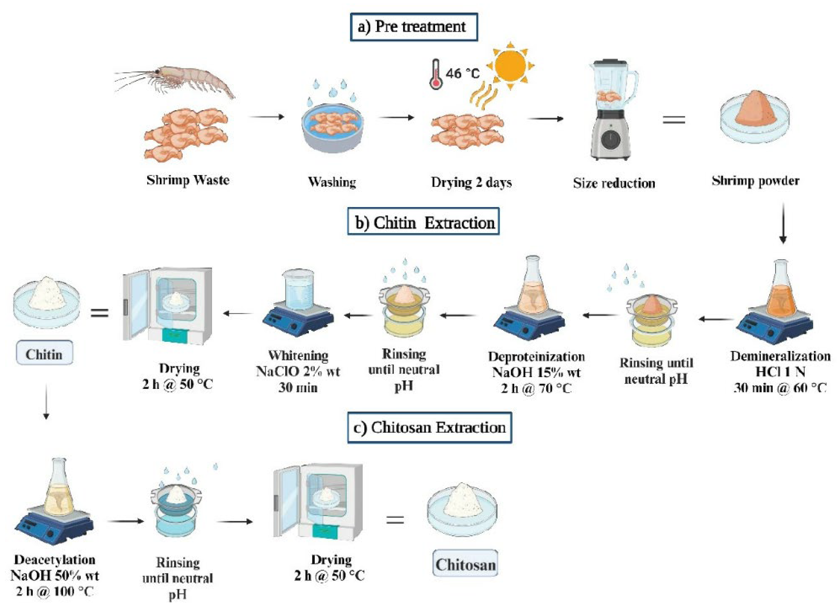
Figure 2.
Procedure for a) preparation of chitosan solution and b) chitosan film.
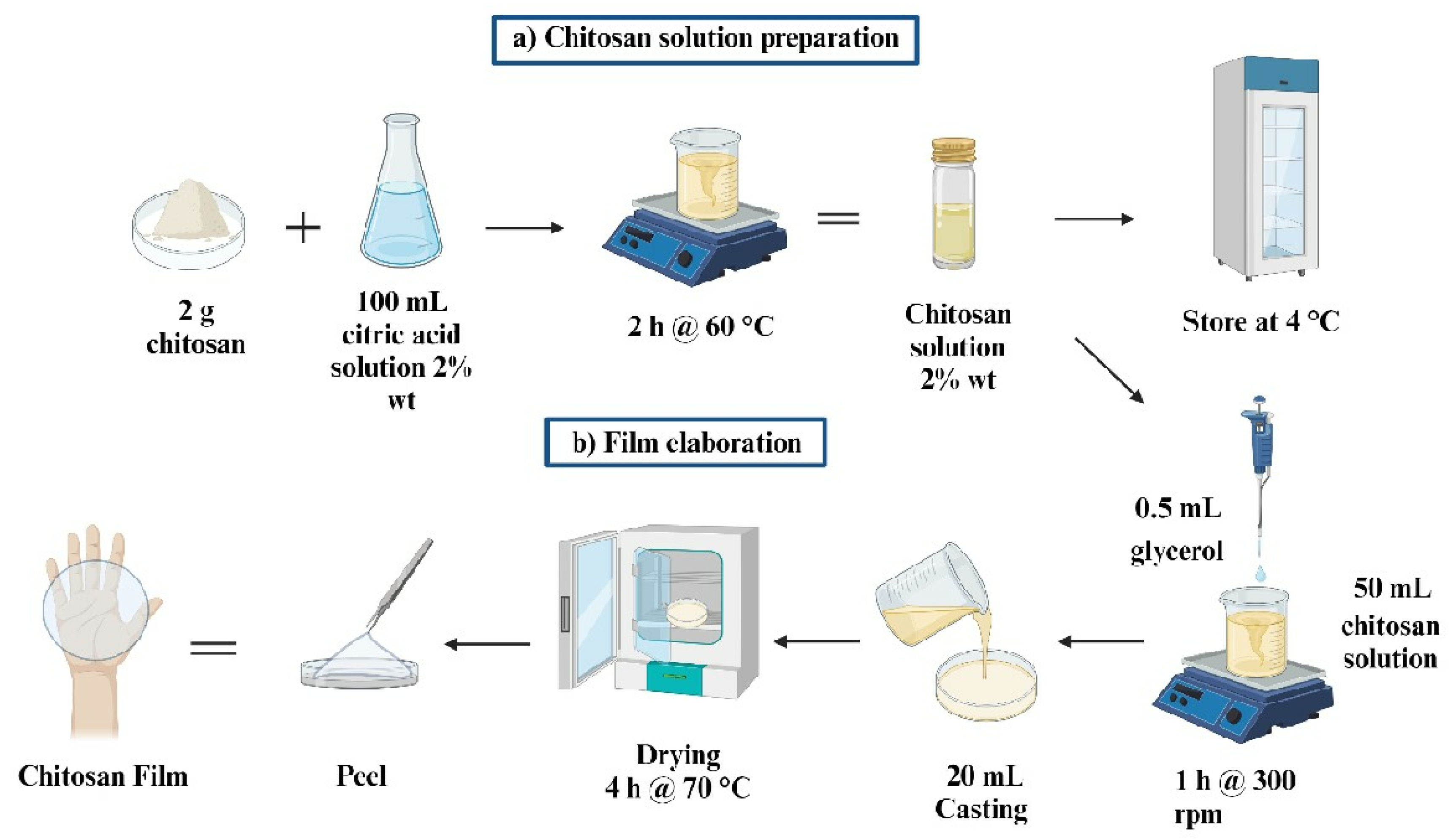
Figure 3.
SEM micrographs of a) chitin and b) chitosan.
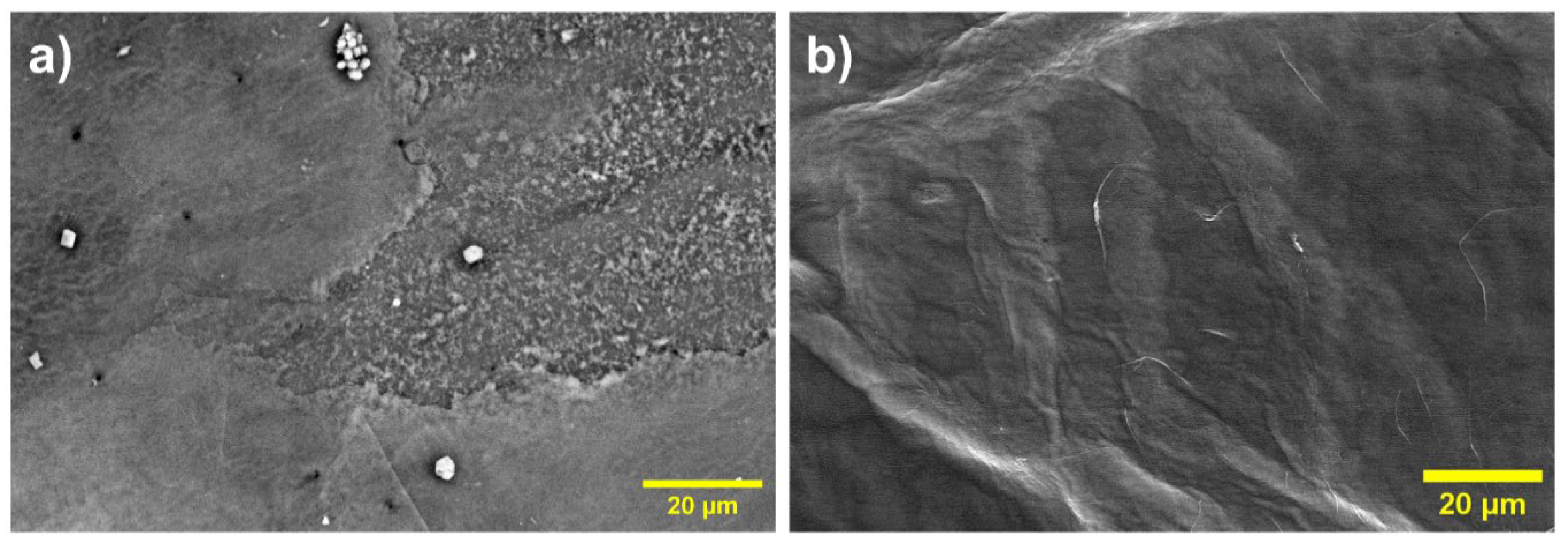
Figure 4.
FTIR comparative spectra between chitin, chitosan, and commercial chitosan.
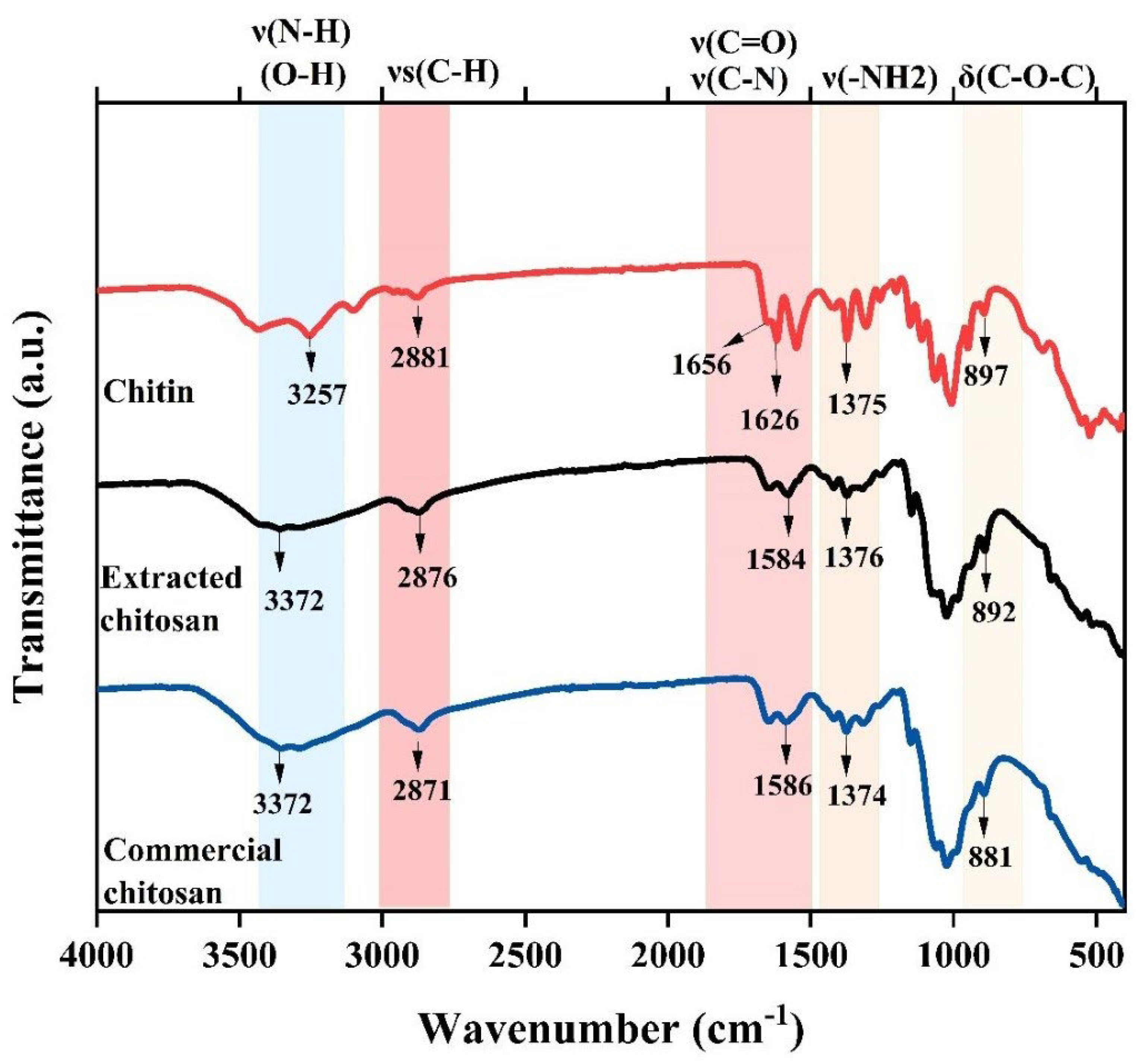
Figure 6.
TGA and DTG of a) chitin and b) chitosan.
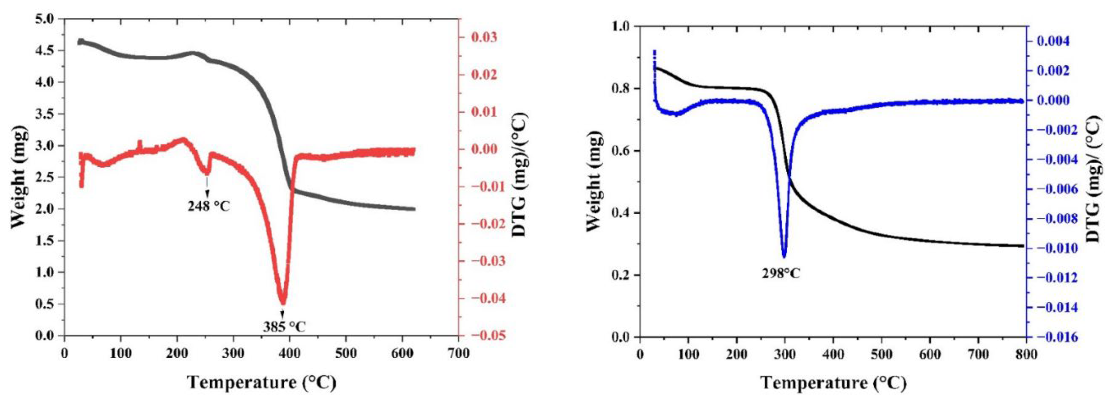
Figure 7.
DSC analysis of a) chitin and b) chitosan.
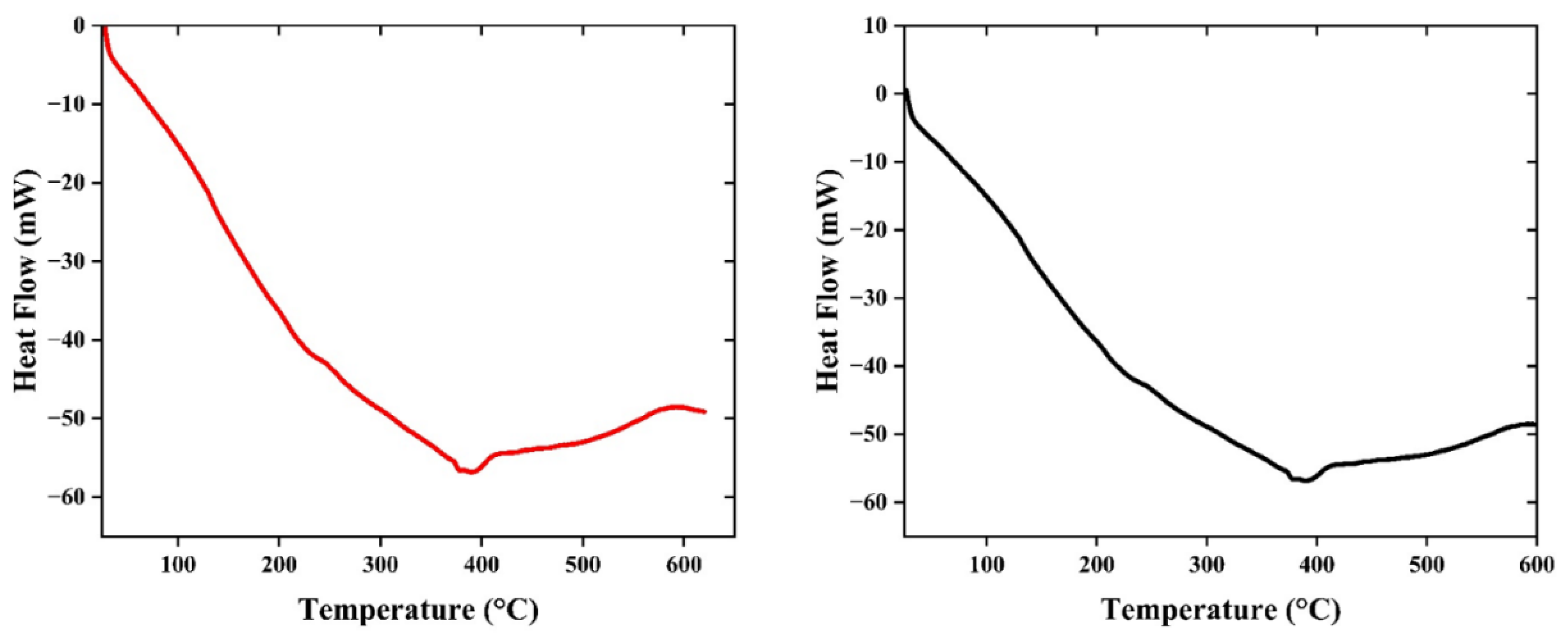
Figure 8.
Tensile strength of chitosan film.
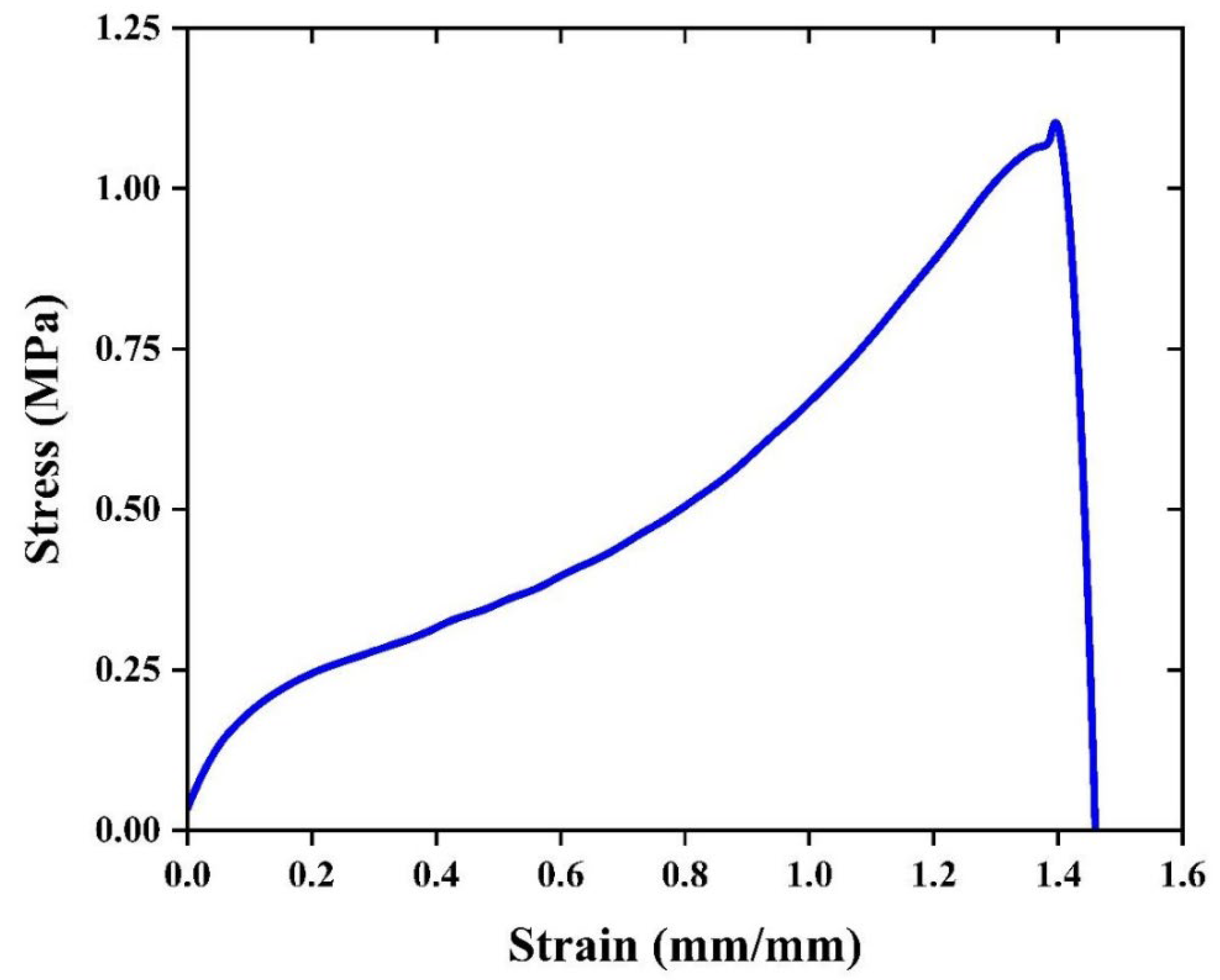
Figure 10.
Antibacterial activity of the chitosan film.
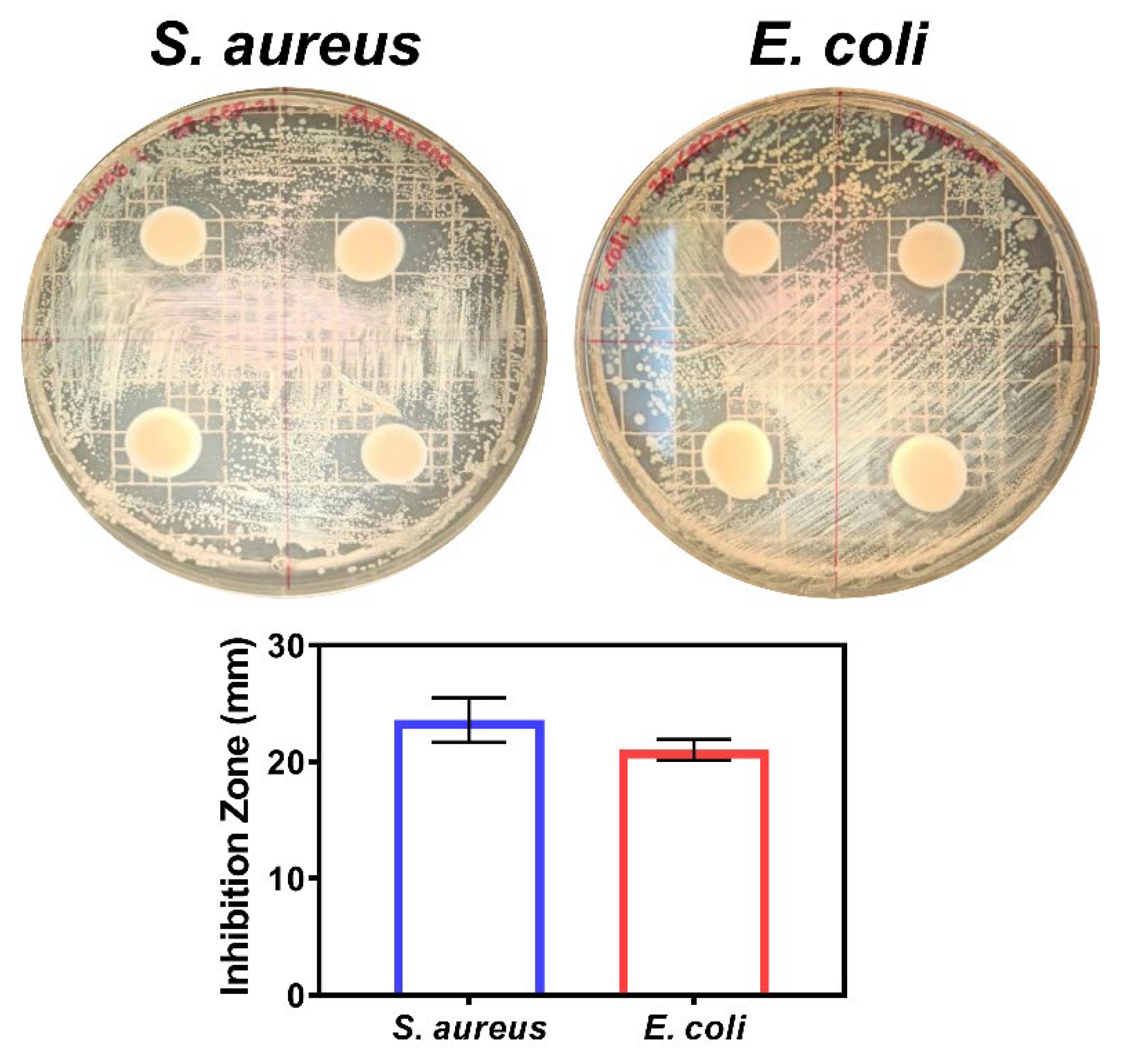
Figure 11.
FTIR spectra of chitosan film.
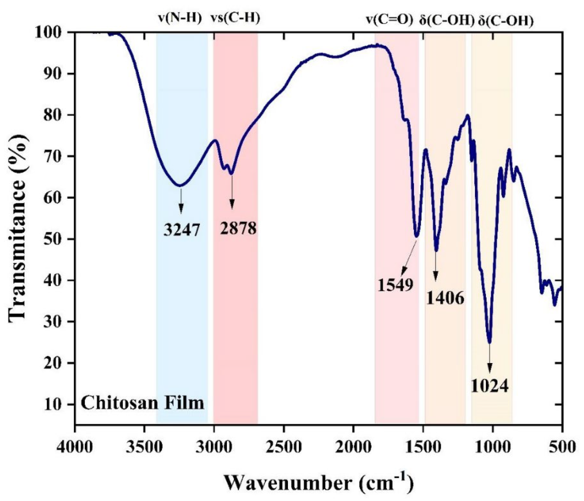
Figure 12.
Thermogravimetric analysis of chitosan film: a) DSC, b) TG and DTG.

Figure 13.
SEM micrograph of chitosan film.
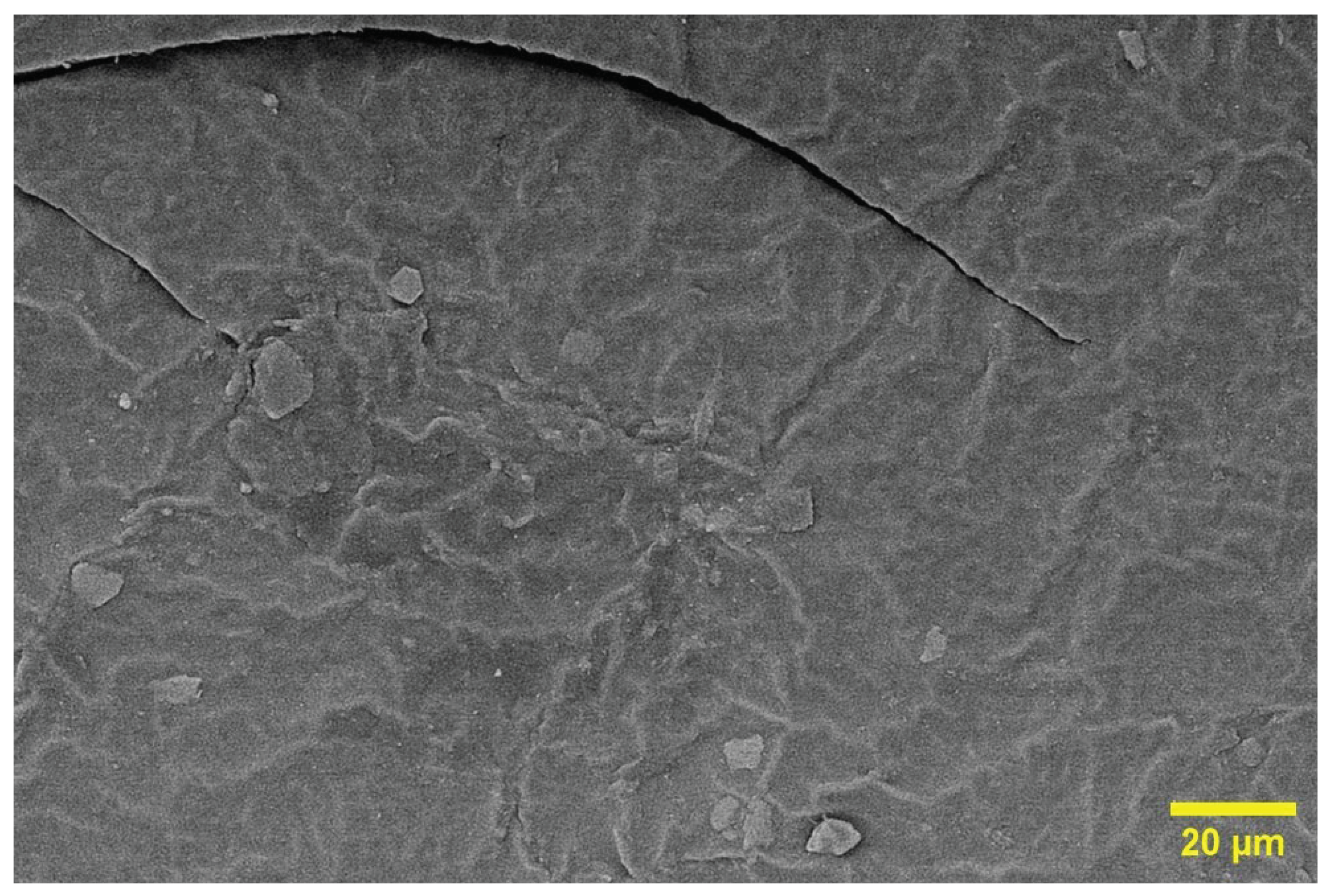
Table 3.
Moisture content (MC), swelling power (SP) and water solubility (S) of chitosan film.
| Film | MC (%) | SP (%) | S (%) |
|---|---|---|---|
| Chitosan Film | 21.74 | 8.27 | 64.63 |
Disclaimer/Publisher’s Note: The statements, opinions and data contained in all publications are solely those of the individual author(s) and contributor(s) and not of MDPI and/or the editor(s). MDPI and/or the editor(s) disclaim responsibility for any injury to people or property resulting from any ideas, methods, instructions or products referred to in the content. |
© 2024 by the authors. Licensee MDPI, Basel, Switzerland. This article is an open access article distributed under the terms and conditions of the Creative Commons Attribution (CC BY) license (http://creativecommons.org/licenses/by/4.0/).
Copyright: This open access article is published under a Creative Commons CC BY 4.0 license, which permit the free download, distribution, and reuse, provided that the author and preprint are cited in any reuse.


