
Submitted:
12 March 2024
Posted:
18 March 2024
You are already at the latest version
Abstract
Introduction: Contrast-enhanced spectral mammography (CESM) is a novel technique employed in breast cancer screening and detection. In this systematic review, we aim to compare the diagnostic performance of CESM with magnetic resonance imaging (MRI) in the context of breast cancer diagnosis.
Material and Method: We have searched for paper in Pubmed and Scopus from July 2023 to February 2024, finding 256 suitable papers according to our selection criteria (perspective or retrospective studies, women diagnosed with breast cancer, comparison of Contrast-enhanced Spectral Mammography and Magnetic Resonance Imaging, sensitivity and specificity metrics provided). Every paper was then checked for quality using the QUADAS 2 assesement score. Sensibility, specificity, Positive Predictive Value (PPV), Negative Predictive Value (NPV) and accuracy were recorded.
Results: 15 studies were selected for analysis. CESM demonstrated comparable sensitivity to MRI, with values ranging from 36.4% to 100% for CESM and 0% to 100% for MRI. Specificity varied between the modalities, with CESM showing specificity ranges of 17% to 100% compared to 23% to 92.31% for MRI.
Discussion and Conclusion: CESM has comparable diagnostic validity to MRI, and is a valuable tool in detecting breast cancer.
Keywords:
CESM
; MRI
; breast cancer
; mammography screening
Introduction
Breast cancer stands as the most prevalent malignancy among women [1]. Given its substantial impact and mortality rates, there has been a progressive development of sophisticated screening programs aimed at early lesion detection, contributing significantly to reduced mortality by identifying lesions before clinical manifestation [2]. Radiodiagnostic techniques play a pivotal role in achieving this goal [2]. Indeed, mammography stands as the most extensively utilized method in screening programs across numerous countries [3]. Other radiological imaging techniques can also be used, such as ultrasound (a very cost-effective and reproducible technique, but with low specificity [4]) or magnetic resonance imaging (MRI, especially effective in identifying new lesions with very high sensitivity and used in screening women with risk factors [5]). Over time, techniques utilizing contrast medium have been developed, such as Contrast-Enhanced Spectral Mammography (CESM). CESM employs an iodinated contrast medium to enhance the visualization of vascular patterns in lesions, thereby improving diagnostic accuracy while maintaining the ease of use associated with conventional mammography [6], with some authors even speculated it might be a better diagnostic tool than MRI [7]. In this systematic review (conducted in accordance with the PRISMA statement [8]) we evaluate the sensitivity and specificity performance of Contrast-Enhanced Spectral Mammography (CESM) compared to Magnetic Resonance Imaging (MRI).
Material and Method
The search for papers was conducted between July 2023 and February 2024 on PubMed and Scopus. Various search terms were utilized, including “Contrast-enhanced mammography AND magnetic resonance,” “CESM AND MRI,” “Contrast-enhanced spectral mammography AND magnetic resonance,” “Contrast-enhanced spectral mammography AND magnetic resonance AND Breast,” and “Contrast-enhanced spectral mammography OR magnetic resonance AND Breast” on Pubmed and Scopus. Initially, 19,589 results were identified, out of which 19,603 were excluded based on title and abstract screening and duplicates. Subsequently, 256 papers were analyzed, with 212 being excluded after full-text reading. Of the remaining 44 eligible papers, 29 were excluded based on study quality assessment. Finally, 15 papers were deemed eligible for review [9,10,11,12,13,14,15,16,17,18,19,20,21,22,23] (Figure 1). The selection criteria for the papers included: perspective or retrospective studies, women diagnosed with breast cancer, comparison of Contrast-enhanced Spectral Mammography (CESM) and Magnetic Resonance Imaging (MRI), sensitivity and specificity metrics provided,focus on initial diagnosis.
Results
A total of 15 studies were included in the systematic review, examining the diagnostic performance of Contrast-Enhanced Spectral Mammography (CESM) compared to Magnetic Resonance Imaging (MRI) in detecting breast lesions. Across the studies, CESM demonstrated comparable sensitivity to MRI, with values ranging from 36.4% to 100% for CESM and 0% to 100% for MRI. Specificity varied between the modalities, with CESM showing specificity ranges of 17% to 100% compared to 23% to 92.31% for MRI. In terms of positive predictive values (PPV), CESM showed a range from 55.56% to 100%, while MRI exhibited PPV ranges of 0% to 96.9%. Negative predictive values (NPV) were generally high for both CESM and MRI, with CESM ranging from 0% to 96.9% and MRI from 32% to 96.15%.
Discussion and Conclusions
The diagnostic performance between Contrast-Enhanced Spectral Mammography (CESM) and Magnetic Resonance Imaging (MRI) emerges as comparable, particularly regarding sensitivity. However, CESM exhibits a more favorable specificity profile, potentially leading to a reduced incidence of false positives in lesion detection. This advantage could translate into fewer unnecessary invasive interventions in individuals without the disease, enhancing patient care and reducing healthcare costs.
One potential critical issue in our findings is the wide variability observed in the results across the selected studies. However, it’s worth noting that similar considerations and results have been reported by other authors in the literature. This variability could stem from differences in imaging protocols, patient populations, and study methodologies, highlighting the need for further standardization and validation of diagnostic criteria in future research. Despite this variability, the overall trends and conclusions drawn from our systematic review align with existing evidence in the field [25,26]. Despite demonstrating equivalent diagnostic performance, CESM also appears to offer economic advantages over MRI. Some authors have estimated that the cost to health systems for CESM is nearly half that of MRI [27]. This economic consideration underscores the potential value of CESM as a cost-effective alternative for breast lesion detection, particularly in resource-constrained settings where cost-effectiveness is a critical factor in decision-making processes regarding healthcare resource allocation. In addition to the economic advantages, there are also advantages in patient compliance: in fact, CESM is found to be tolerated more to MRI in cancer patients, both in the context of screening and follow-up [28,29]. This improved tolerance may be attributed to factors such as shorter examination times and reduced claustrophobia associated with CESM compared to MRI.
In conclusion, CESM turns out to be an effective radiodiagnostic method in the prevention and monitoring of breast cancer, flanking performance comparable to the gold standard with better economic manageability and greater tolerability by patients.
Funding
none.
Conflicts of Interest
We have no Conflict of Interest to declare.
References
- Sung H, Ferlay J, Siegel RL, Laversanne M, Soerjomataram I, Jemal A, Bray F. Global Cancer Statistics 2020: GLOBOCAN Estimates of Incidence and Mortality Worldwide for 36 Cancers in 185 Countries. CA Cancer J Clin. 2021 May;71(3):209-249. [CrossRef]
- Tabar L, Yen MF, Vitak B, Chen HH, Smith RA, Duffy SW. Mammography service screening and mortality in breast cancer patients: 20-year follow-up before and after introduction of screening. Lancet. 2003 Apr 26;361(9367):1405-10. [CrossRef]
- Coleman, C. Early Detection and Screening for Breast Cancer. Semin Oncol Nurs. 2017 May;33(2):141-155. [CrossRef]
- Yuan, W.H.; Hsu, H.C.; Chen, Y.Y.; Wu, C.H. Supplemental Breast Cancer-Screening Ultrasonography in Women with Dense Breasts: A Systematic Review and Meta-Analysis. Br. J. Cancer 2020, 123, 673–688. [Google Scholar] [CrossRef] [PubMed]
- Sung JS, Lebron L, Keating D, D’Alessio D, Comstock CE, Lee CH, Pike MC, Ayhan M, Moskowitz CS, Morris EA, Jochelson MS. Performance of Dual-Energy Contrast-enhanced Digital Mammography for Screening Women at Increased Risk of Breast Cancer. Radiology. 2019 Oct;293(1):81-88. [CrossRef]
- Sogani, J.; Mango, V.L.; Keating, D.; Sung, J.S.; Jochelson, M.S. Contrast-Enhanced Mammography: Past, Present, and Future. Clin. Imaging 2021, 69, 269–279. [Google Scholar] [CrossRef] [PubMed]
- Thibault F, Balleyguier C, Tardivon A, Dromain C. Contrast enhanced spectral mammography: better than MRI? Eur J Radiol. 2012 Sep;81 Suppl 1:S162-4. [CrossRef]
- Page, M.J., McKenzie, J.E., Bossuyt, P.M. et al. The PRISMA 2020 statement: an updated guideline for reporting systematic reviews. Syst Rev 10, 89 (2021). [CrossRef]
- Łuczyńska E, Heinze-Paluchowska S, Hendrick E, Dyczek S, Ryś J, Herman K, Blecharz P, Jakubowicz J. Comparison between breast MRI and contrast-enhanced spectral mammography. Med Sci Monit. 2015 ;21:1358-67. 12 May. [CrossRef]
- Wang Q, Li K, Wang L, Zhang J, Zhou Z, Feng Y. Preclinical study of diagnostic performances of contrast-enhanced spectral mammography versus MRI for breast diseases in China. Springerplus. 2016 Jun 17;5(1):763. [CrossRef]
- Li L, Roth R, Germaine P, Ren S, Lee M, Hunter K, Tinney E, Liao L. Contrast-enhanced spectral mammography (CESM) versus breast magnetic resonance imaging (MRI): A retrospective comparison in 66 breast lesions. Diagn Interv Imaging. 2017 Feb;98(2):113-123. [CrossRef]
- Iotti V, Ravaioli S, Vacondio R, Coriani C, Caffarri S, Sghedoni R, Nitrosi A, Ragazzi M, Gasparini E, Masini C, Bisagni G, Falco G, Ferrari G, Braglia L, Del Prato A, Malavolti I, Ginocchi V, Pattacini P. Contrast-enhanced spectral mammography in neoadjuvant chemotherapy monitoring: a comparison with breast magnetic resonance imaging. Breast Cancer Res. 2017 Sep 11;19(1):106. [CrossRef]
- Lee-Felker SA, Tekchandani L, Thomas M, Gupta E, Andrews-Tang D, Roth A, Sayre J, Rahbar G. Newly Diagnosed Breast Cancer: Comparison of Contrast-enhanced Spectral Mammography and Breast MR Imaging in the Evaluation of Extent of Disease. Radiology. 2017 Nov;285(2):389-400. [CrossRef]
- Kim EY, Youn I, Lee KH, Yun JS, Park YL, Park CH, Moon J, Choi SH, Choi YJ, Ham SY, Kook SH. Diagnostic Value of Contrast-Enhanced Digital Mammography versus Contrast-Enhanced Magnetic Resonance Imaging for the Preoperative Evaluation of Breast Cancer. J Breast Cancer. 2018 Dec;21(4):453-462. [CrossRef]
- Patel BK, Hilal T, Covington M, Zhang N, Kosiorek HE, Lobbes M, Northfelt DW, Pockaj BA. Contrast-Enhanced Spectral Mammography is Comparable to MRI in the Assessment of Residual Breast Cancer Following Neoadjuvant Systemic Therapy. Ann Surg Oncol. 2018 May;25(5):1350-1356. [CrossRef]
- Barra FR, Sobrinho AB, Barra RR, et al. Contrast-Enhanced Mammography (CEM) for Detecting Residual Disease after Neoadjuvant Chemotherapy: A Comparison with Breast Magnetic Resonance Imaging (MRI). Biomed Res Int. 2018;2018:8531916. Published 2018 Nov 8. [CrossRef]
- Xing D, Lv Y, Sun B, Xie H, Dong J, Hao C, Chen Q, Chi X. Diagnostic Value of Contrast-Enhanced Spectral Mammography in Comparison to Magnetic Resonance Imaging in Breast Lesions. J Comput Assist Tomogr. 2019 Mar/Apr;43(2):245-251. [CrossRef]
- Clauser P, Baltzer PAT, Kapetas P, Hoernig M, Weber M, Leone F, Bernathova M, Helbich TH. Low-Dose, Contrast-Enhanced Mammography Compared to Contrast-Enhanced Breast MRI: A Feasibility Study. J Magn Reson Imaging. 2020 Aug;52(2):589-595. [CrossRef]
- Rudnicki W, Piegza T, Rozum-Liszewska N, Górski M, Popiela TJ, Basta P, Heinze S, Luczynska E. The effectiveness of contrast-enhanced spectral mammography and magnetic resonance imaging in dense breasts. Pol J Radiol. 2021 Mar 15;86:e159-e164. [CrossRef]
- Steinhof-Radwańska K, Lorek A, Holecki M, Barczyk-Gutkowska A, Grażyńska A, Szczudło-Chraścina J, Bożek O, Habas J, Szyluk K, Niemiec P, Gisterek I. Multifocality and Multicentrality in Breast Cancer: Comparison of the Efficiency of Mammography, Contrast-Enhanced Spectral Mammography, and Magnetic Resonance Imaging in a Group of Patients with Primarily Operable Breast Cancer. Curr Oncol. 2021 Oct 8;28(5):4016-4030. [CrossRef]
- Lee SC, Hovanessian-Larsen L, Stahl D, Cen S, Lei X, Desai B, Yamashita M. Accuracy of contrast-enhanced spectral mammography compared with MRI for invasive breast cancers: Prospective study in population of predominantly underrepresented minorities. Clin Imaging. 2021 Dec;80:364-370. [CrossRef]
- Ferranti FR, Vasselli F, Barba M, Sperati F, Terrenato I, Graziano F, Vici P, Botti C, Vidiri A. Diagnostic Accuracy of Contrast-Enhanced, Spectral Mammography (CESM) and 3T Magnetic Resonance Compared to Full-Field Digital Mammography plus Ultrasound in Breast Lesions: Results of a (Pilot) Open-Label, Single-Centre Prospective Study. Cancers (Basel). 2022 Mar 7;14(5):1351. [CrossRef]
- Feng L, Sheng L, Zhang L, Li N, Xie Y. Comparison of Contrast-Enhanced Spectral Mammography and Contrast-Enhanced MRI in Screening Multifocal and Multicentric Lesions in Breast Cancer Patients. Contrast Media Mol Imaging. 2022 Apr 6;2022:4224701. [CrossRef]
- Whiting PF, Rutjes AW, Westwood ME, Mallett S, Deeks JJ, Reitsma JB, Leeflang MM, Sterne JA, Bossuyt PM; QUADAS-2 Group. QUADAS-2: a revised tool for the quality assessment of diagnostic accuracy studies. Ann Intern Med. 2011 Oct 18;155(8):529-36. [CrossRef]
- Gelardi F, Ragaini EM, Sollini M, Bernardi D, Chiti A. Contrast-Enhanced Mammography versus Breast Magnetic Resonance Imaging: A Systematic Review and Meta-Analysis. Diagnostics (Basel). 2022 Aug 4;12(8):1890. [CrossRef]
- Kornecki, A. Current Status of Contrast Enhanced Mammography: A Comprehensive Review. Can Assoc Radiol J. 2022 Feb;73(1):141-156. [CrossRef]
- Patel BK, Gray RJ, Pockaj BA. Potential Cost Savings of Contrast-Enhanced Digital Mammography. AJR Am J Roentgenol. 2017 Jun;208(6):W231-W237. [CrossRef]
- Phillips J, Miller MM, Mehta TS, Fein-Zachary V, Nathanson A, Hori W, Monahan-Earley R, Slanetz PJ. Contrast-enhanced spectral mammography (CESM) versus MRI in the high-risk screening setting: patient preferences and attitudes. Clin Imaging. 2017 Mar-Apr;42:193-197. [CrossRef]
- Savaridas SL, Whelehan P, Warwick VR, Vinnicombe SJ, Evans AJ. Contrast-enhanced digital breast tomosythesis and breast MRI to monitor response to neoadjuvant chemotherapy: patient tolerance and preference. Br J Radiol. 2022 Jun 1;95(1134):20210779. [CrossRef]
Figure 1.
PRISMA Flow diagram.
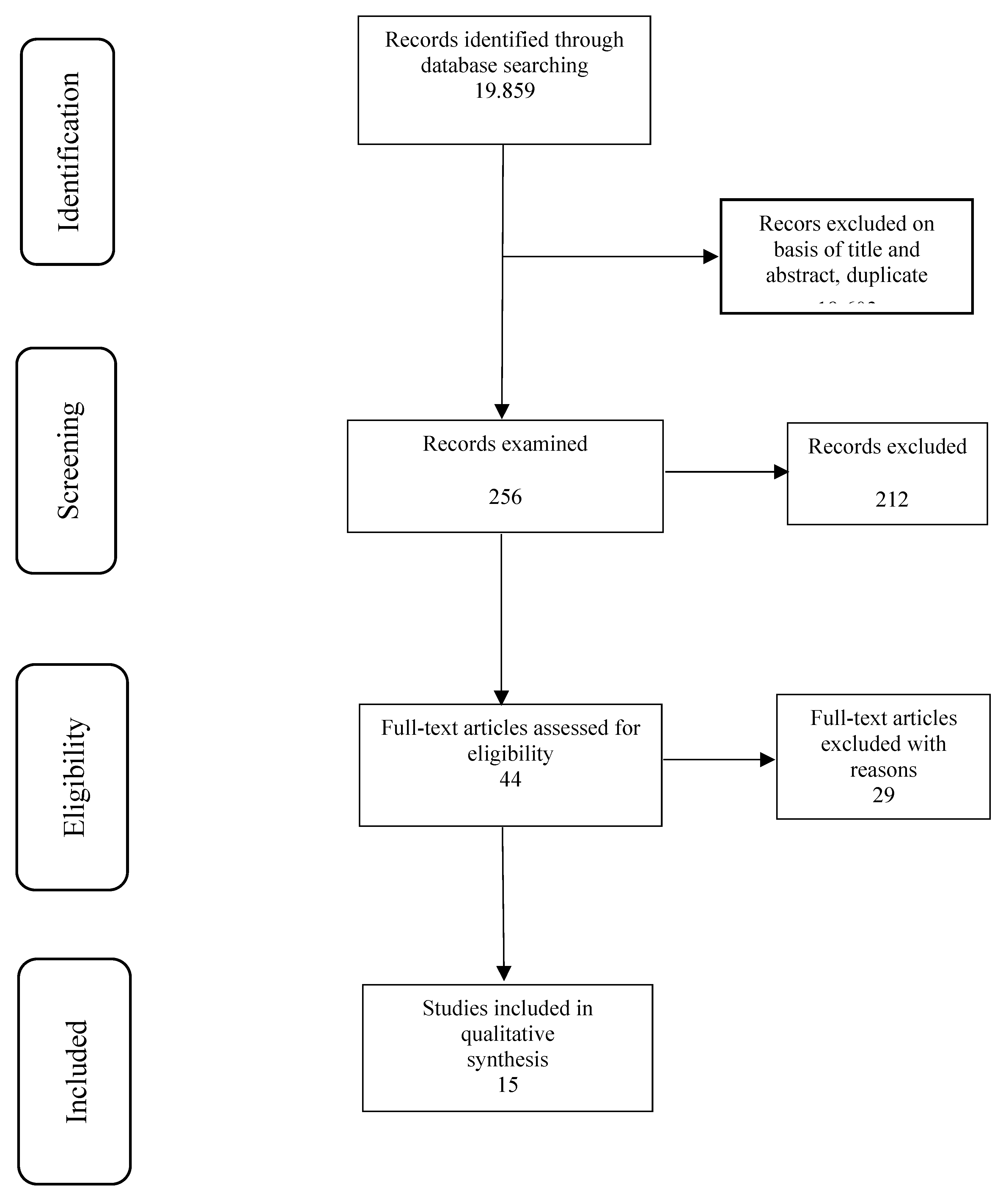
Figure 2.
Forrest plot of CESM and MRI sensitivity and specificity.
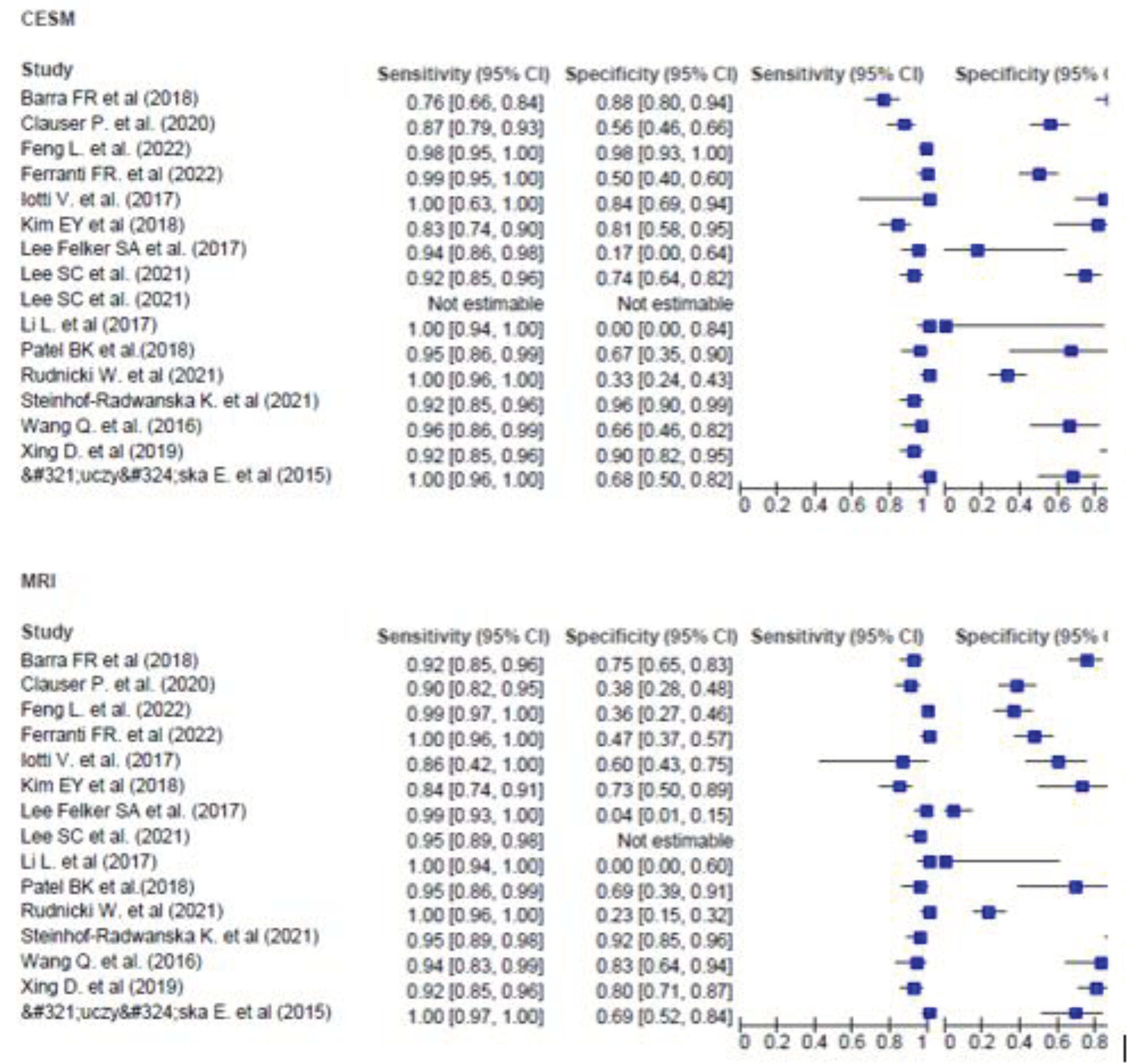

Table 1.
QUADAS 2 quality assessment.
| Study | Risk of Bias | Applicability Concerns | |||||
|
Patient Selection |
Index Test |
Reference Standard |
Flow and Timing |
Patient Selection |
Index Test |
Reference Standard |
|
| Łuczyńska E. et al (2015) [9] | Low | Low | Low | Low | Low | Low | Low |
| Wang Q. et al. (2016) [10] | Unclear | Low | Low | Unclear | Unclear | Low | Low |
| Li L. et al (2017) [11] | Low | Low | Low | Low | Low | Low | Low |
| Iotti V. et al. (2017) [12] | Low | Low | Low | Low | Low | Low | Low |
| Lee Felker SA et al. (2017) [13] | Low | Low | Low | Unclear | Low | Low | Low |
| Kim EY et al (2018) [14] | Unclear | Low | Low | Unclear | Unclear | Low | Low |
|
Patel BK et al. (2018) [15] |
Low | Low | Unclear | Unclear | Low | Low | Low |
| Barra FR et al (2018) [16] | Low | Low | Low | Unclear | Low | Low | Low |
| Xing D. et al (2019) [17] | Low | Low | Low | Unclear | Low | Low | Low |
| Clauser P. et al. (2020) [18] | Low | Low | Low | Unclear | Low | Low | Low |
| Rudnicki W. et al (2021) [19] | Unclear | Low | Low | Unclear | Unclear | Low | Low |
| Steinhof-Radwanska K. et al (2021) [20] | Low | Low | Low | Low | Low | Low | Low |
| Lee SC et al. (2021) [21] | Low | Low | Low | Unclear | Low | Low | Low |
| Ferranti FR. et al (2022) [22] | Low | Low | Low | Low | Low | Low | Low |
| Feng L. et al. (2022) [23] | Low | Low | Low | Unclear | Low | Low | Low |
Table 2.
Characteristics and diagnostic accuracy of CESM and MRI.
| Study | Studytype | CESM Contrast | Patients(n) | Detected Lesions | Sensitivity | Specificity | Positive Predictive Value |
Negative Predictive Value |
Accuracy |
| Łuczyńska E. et al (2015) [9] | Prospective | 1.5 ml/kg of body mass of non-ionic contrast agent (Iopromide 370) |
102 | 118 total (81 malignant, 37 benign) MRI: 107 (75 malignant, 32 bening) CESM: 106 (81 malignant, 25 benign) |
MRI: 93% CESM: 100% |
MRI:69% CESM:68% |
MRI: 74% CESM:77% |
MRI:65% CESM; 100% |
MRI: 73% CESM:79% |
| Wang Q. et al. (2016) [10] | Prospective | Omnipaque, 350 mgI/mL; GE Healthcare, Dublin, Ireland) at a 1.5 mL/kg |
68 | 77 (48 malignant, 29 benign) | MRI: 93.8% CESM:95.8% |
MRI:82.8% CESM:65.5% |
MRI: 88.2% CESM:82.1% |
MRI:92.3% CESM: 90.5% |
MRI: 89.6% CESM: 84.4% |
| Li L. et al (2017) [11] | Retrospective | Sieve®370 (Iopamidol injection 76%) | 48 | MRI:66 (62 malignant 4 benign) CESM:64 (62 malignant 2 benign) |
MRI: 100% CESM: 100% |
Not estimable | MRI: 93,9% CESM: 96.9% |
Not estimable | MRI: 93.9% CESM: 96.9% |
| Iotti V. et al. (2017) [12] | Prospective | ioversolo 350 mg/ ml at 1.5 ml/kg |
46 | 46 | MRI: 87% CESM: 100% |
MRI: 60% CESM: 84% |
MRI: 32% CESM: 57% |
MRI:96% CESM: 100% |
MRI:65% CESM:87% |
| Lee Felker SA et al. (2017) [13] | Retrospective | 90 mL of iodinated contrast material (Omnipaque 350, GE Healthcare |
52 | 120 | MRI: 99% CESM: 94% |
MRI: 4% CESM: 17% |
MRI: 60% CESM: 93% |
MRI:67% CESM: 20% |
MRI: 60.17% CESM:87.1% |
| Kim EY et al (2018) [14] | Prospective | Omnipaque 350 (GE Healthcare, Shanghai, China; 1.5 mL/kg of body weight |
84 | 121 | MRI: 83.9% CESM:83.9% |
MRI: 73.6% CESM:81.1% |
MRI:65% CESM:72.2% |
MRI:88.6% CESM:89.6% |
MRI:77.4% CESM:82.1% |
|
Patel BK et al. (2018) [15] |
Propsective | 1.5 mL/kg of iohexol (Omnipaque 350; GE Healthcare) |
65 | 65 | MRI: 95% CESM:95% |
MRI:68.9% CESM: 66.7% |
MRI:57.6% CESM:55.9% |
MRI:96.9% CESM:96.7& |
MRI:85.9% CESM:85.1% |
| Barra FR et al (2018) [16] | Porspective | 1.5 ml/kg of non-ionic contrast medium (Iohexol, 300 mg/ml) |
33 | 33 | MRI: 92% CESM: 76% |
MRI:75% CESM:87.5% |
MRI: 92% CESM:95% |
MRI:75% CESM:86.4% |
MRI: 80.61% CESM: 83.7% |
| Xing D. et al (2019) [17] | Prospective | iohexol at 1.5 mL/kg body | 235 | MRI:258 CESM: 259 |
MRI:91.5% CESM: 91.5% |
MRI: 80.2% CESM: 89.5% |
MRI: 90.5% CESM: 94.7% |
MRI:82.1% CESM:83.7% |
MRI:71.7% CESM:81% |
| Clauser P. et al. (2020) [18] | Prospective | 2 mL/kg body weight of nonionic iodine contrast agent (Iobitridol/Xenetix 350, Guerbet, Villepinte, France |
80 | 93 | MRI: 83.6%-93.4% CESM: 65.6%-90.2% |
MRI: 37.5%-53.1% CESM: 46.9%-96.9% |
MRI: 73.3%-77% CESM: 76.4%-97.6% |
MRI:63%-76.5% CESM:59.6%-71.4% |
MRI:72%-75.3% CESM:75.3%-76.3% |
| Rudnicki W. et al (2021) [19] | Retrospective | Iopromide (1.5 ml/kg of body weight |
121 | MRI: 121 CESM:108 |
MRI 100% CESM: 100% |
MRI:23% CESM: 33% |
MRI:72% CESM: 75% |
MRI: 100% CESM:100% |
MRI:78% CESM:68% |
| Steinhof-Radwa ´ nska K. et al (2021) [20] | Retrospective | 1.5 mL/kg of body mass of non-ionic contrast agent |
60 | MRI:33 CESM:30 |
MRI: 91.18% CESM: 85.29% |
MRI:92.31% CESM:96.15% |
MRI:94% CESM:97% |
MRI:89% CESM:84% |
MRI: 91% CESM: 90% |
| Lee SC et al. (2021) [21] | Prospective | 1.5 ml/kg of iodine contrast |
41 CESM 32 MRI |
41 malignant | MRI:94.74% CESM:92% |
MRI: 0 CESM:74.43% |
MRI:85.71% CESM:95-83% |
MRI:0 CESM: 55.56% |
Not reported |
|
Ferranti FR. et al (2022) [22] |
Prospective Cohort Study | Visipaque 320 | 118 | MRI: 108 CESM:110 |
MRI 99% CESM: 100% |
MRI:47% CESM:50% |
MRI:88% CESM: 92% |
MRI: 90% CEMS.100% |
MRI:88% CESM:93% |
| Feng L. et al. (2022) [23] | Prospective | ioversol injection (1.5 ml/kg) | 54 | 188 177 malignant 11 benign |
MRI: 99.4% CESM:98.3% |
MRI:36.4% CESM:98.3% |
MRI: 96.17% CESM: 97.75% |
MRI: 80% CESM: 70% |
MRI:95.7% CESM:96.3% |
Disclaimer/Publisher’s Note: The statements, opinions and data contained in all publications are solely those of the individual author(s) and contributor(s) and not of MDPI and/or the editor(s). MDPI and/or the editor(s) disclaim responsibility for any injury to people or property resulting from any ideas, methods, instructions or products referred to in the content. |
© 2024 by the authors. Licensee MDPI, Basel, Switzerland. This article is an open access article distributed under the terms and conditions of the Creative Commons Attribution (CC BY) license (http://creativecommons.org/licenses/by/4.0/).
Copyright: This open access article is published under a Creative Commons CC BY 4.0 license, which permit the free download, distribution, and reuse, provided that the author and preprint are cited in any reuse.


