Submitted:
04 March 2024
Posted:
05 March 2024
You are already at the latest version
Abstract
The mechanisms of formation of induced intrinsic and impurity radiative states, which consist of intrinsic and impurity electron-hole trapping centers in irradiated Ca2P2O7-Mn and Ca2P2O7 phosphates, were investigated using thermoactivation and vacuum-ultraviolet spectroscopy methods. These centers are excited at photon energies of 4.0 eV and 4.5 eV, which are within the matrix's transparency region. New radiative induced states at 3.06 eV and 2.92 eV are demonstrated to be generated upon the excitation of anions by photons with energies of 5.0 and 5.64 eV. This process is due to charge transfer from the ion to the impurities, specifically Mn^2+ O^2- Mn^2+ and the neighboring ion O^2- (P2O7)^4-. Furthermore, upon the excitation of matrix anions with photon energies exceeding the band gap (8.0 - 8.25 eV), electron trapping by impurities such as Mn^2+ and (P2O7)^4- ions results.
Keywords:
electrons and holes
; trapping center
; luminescence
; XRD
; SEM
; Emission spectra
; Excitation spectra
1. Introduction
Calcium phosphates constitute a diverse family of materials that find widespread use in a variety of fields, including medicine for bone regeneration, catalysis, sensor technology, and as foundational matrices for the development of optical materials. In the study of these materials' optical properties, special emphasis is placed on examining the luminescent characteristics of doped phosphates. These luminescent properties are pivotal in the creation of white light sources, which are achieved by coating blue near-ultraviolet emitting LED chips with phosphors [1]. Furthermore, the enduring phosphorescence and thermoluminescence exhibited by phosphates hold promise for a range of applications across the energy sector, lighting, and optical visualization in the red and infrared spectral ranges [2].
To advance the applications mentioned above, numerous studies have been conducted on the luminescent properties of phosphates. For instance, the research conducted by authors [3] demonstrated the luminescent characteristics of activated by following irradiation with synchrotron radiation. It was revealed that ions are formed in the excited state as a result of charge transfer from the matrix to the impurities . The charge transfer from to requires an energy of 4.6 eV. During the relaxation process from the excited state back to the ground state, recombination emission occurs at 2.9 eV, corresponding to the transition. Additionally, the energy gap from the excited electronic state of to the bottom of the conduction band is estimated to be 0.4 eV.
The nature of impurity emission centers in phosphates activated by ions was the focus of investigations by authors [3,4]. In , it was discovered that broad emission bands at 2.17 eV, 2.08 eV, and 2.14 eV are excited by several bands at 2.48 eV, 3.1 eV, 2.95 eV, and 3.44–3.65 eV. According to the hypothesis presented by the authors in [4], these excitation spectra are indicative of forbidden intra-center transitions of ions situated at the cationic sites of the crystal lattice. Further, authors in [3] identified that emission at 1.93 eV, which is not only excited by intracenter bands but also efficiently by UV emission at 5.9 eV, can be attributed to charge transfer from the matrix, , to the impurities . Additionally, the observation of thermoluminescence at 240 K and between 400–480 K, along with phosphorescence, is linked to the creation of trap centers during x-ray excitation.
The formation of electron-hole trapping centers in phosphates was observed during photon irradiation with energies surpassing the band gap, leading to the creation of free electron-hole pairs. In the research conducted by authors [5], thermoluminescence (TL) was studied in doped with and (where represents ). During the irradiation of , electron trapping centers are formed by the trapping of free electrons. Simultaneously, the trapping of holes by the ground state leads to the formation of hole trapping centers. TL is observed as a result of the recombination decay of these electron-hole trapping centers.
Phosphors doped with impurities are utilized as sources of green radiation, which is instrumental in exciting hole trapping centers, thereby generating stable red and infrared radiation. The photoluminescence, radioluminescence, and phosphorescence properties of these phosphors, when doped with rare earth ions, have been extensively studied by the authors in references [6,7,8,9,10,11,12,13,14,15,16,17].
In the present work, the mechanisms of formation of electron and hole trapping centers upon excitation of the phosphor in a wide spectral (from 4 eV to 12 eV) and temperature (15-300 K) range, will be discussed. Based on measurements of excitation spectra of impurity emission and newly created recombination or tunneling emissions consisting of electron-hole trapping centers, mechanisms of relaxation processes leading to energy transfer to impurities will be investigated.
"In the studies by authors [18,19,20,21], the mechanisms for the accumulation of intrinsic and impurity electron-hole trapping centers were explored in both pure and -activated . It was demonstrated that intrinsic and impurity trapping centers form as a result of free electrons localizing on the anion and impurities, through the reactions and
From a brief review of the literature, several conclusions can be drawn: during the irradiation of excited anion complexes, impurity electron trapping centers form below the conduction band; when free electrons and holes localize on rare-earth ions, electron and hole trapping centers are generated, whose recombination leads to thermoluminescence (TL) and phosphorescence.
This work will discuss the mechanisms of formation of electron and hole trapping centers upon the excitation of the phosphor across a broad spectral (from 4 eV to 12 eV) and temperature range (15-300 K). By analyzing the excitation spectra of impurity emission from and the newly created recombination or tunneling emissions comprising electron-hole trapping centers, the study investigates the relaxation processes that facilitate energy transfer to impurities.
2. Objects and Research Methods
Undoped and Mn-doped powders were synthesized using the wet co-precipitation method, as described in [4,7]. The Mn substitution rate was set at 1 mol.% relative to . The obtained precipitates were annealed at 1200°C, with a heating rate of 5°C/min and a holding time of 10 minutes.
2.1. Characterization
X-ray diffraction analysis (XRD) of the powders was carried out using Ni-filtered Cu-Kα radiation on a Rigaku MiniFlex II diffractometer, operating in the Bragg-Brentano (θ/2θ) geometry. Data were collected over a 2θ range from 10 to 60°, with a step size of 0.02° and a scanning speed of 1°/min.
The morphology of the synthesized products was examined using a Hitachi SU-70 field emission scanning electron microscope (FE-SEM).
Emission and excitation spectra measurements across a wide spectral range were performed using a vacuum monochromator equipped with Solar M-266 detection.
The samples were irradiated using a BSV-23 x-ray tube with a copper anode, operating at a tube current of 10 mA and a voltage of 40 kV. The energy range of the x-ray photons was between 10-15 keV. For excitation in the vacuum ultraviolet spectral range, a hydrogen lamp with a photon energy of 6.2−11.5 eV and a 150 W xenon lamp XBO (OSRAM, Germany) with a photon energy of 1.5−6.2 eV were utilized.
A Solar CM 2203 spectrofluorimeter was employed for measuring emission spectra in the 1.5-6.2 eV range. Low-temperature investigations at 80 K were conducted using liquid nitrogen in a cryostat.
3. Experimental Results
In order to determine the nature of intrinsic, impurity, and recombination emission at trapping centers in and phosphors, excitation and emission spectra were studied upon excitation by ultraviolet and x-ray radiation over a wide temperature range from 15 K to 300 K.
The crystallinity and purity of the synthesized powders were assessed using X-ray diffraction analysis. The X-ray diffraction pattern of the sample doped with , after annealing at 1200°C, is depicted in Figure 1. The diffraction peaks closely match the standard pattern for α- (PDF #073-0440), confirming the successful formation of the target material. A minor presence of the adjacent phase, α-, was also observed, likely resulting from the evaporation of phosphate particles during the process.
The morphological characteristics of the synthesized powders were examined through scanning electron microscopy (SEM). Representative SEM images of the Mn-doped sample, displayed in Figure 2, reveal that the powders are composed of a mix of plate-like and irregularly shaped particles. Notably, the plate-like particles exhibit grains ranging in size from micrometers to sub-micrometers.
In Figure 3, the emission spectra of the phosphor, upon irradiation with 6.2 eV photons at 300 K, is depicted as curve 1. Additionally, the emission spectra measured at 80 K in a cryostat, following excitation with 6.2 eV photons, is shown as curve 2. The analysis of Figure 3 reveals that at 300 K, there is an intracenter emission band at 2.06 eV, attributed to the ion within the phosphor (curve 1), alongside the emergence of new emission bands at 3.06 eV and 2.92 eV (curve 1). Notably, at 80 K, the emission band markedly diminishes, whereas the intensity of the newly observed emission bands at 3.06 eV and 2.92 eV experiences a 1.5-fold increase.
In Figure 4, the excitation spectrum for the main intracenter emission of at 2.06 eV at 300 K (curve 1) and 80 K (curve 2) is presented. The analysis of the graph indicates that at 80 K, the emission band is excited at discrete energies of approximately 2.5 eV, 3.0 eV, 3.5 eV, 4.0 eV, 4.5 eV, and within the range of 6.2 to 5.0 eV. Conversely, at 300 K, these emission bands also occur but exhibit lower intensity, particularly within the narrower range of 4.0 to 4.5 eV.
In Figure 5, the excitation spectra for the newly identified bands at 3.06 eV and 2.92 eV, following pre-irradiation of the phosphor at 6.2 eV, are displayed at both 300 K and 80 K. Excitation bands are identified within the ranges of 3.94 - 4.0 eV, 4.5 – 4.6 eV, and broadly from 6.2 to 5.5 eV. Notably, these bands are prominently observed at 80 K, indicating a stronger response to excitation at this lower temperature. Conversely, at 300 K, these same bands manifest as weak excitation bands, suggesting a diminished excitation efficiency at higher temperatures.
When measuring the excitation spectra for the intracenter emission of the ion at 2.06 eV, emission bands similar to those identified by authors in [2] were observed at 2.5 eV, 3.1 eV, 3.0 eV, and between 3.5 and 3.6 eV. Additionally, alongside the main intracenter emission at 2.06 eV for , the excitation spectrum revealed bands for the newly identified emissions at 3.06 eV and 2.92 eV, specifically within the ranges of 3.94 - 4.0 eV and 4.5 – 4.6 eV. Furthermore, excitation bands at 5.0 eV and 5.64 eV were also detected. Consequently, these newly emerged emission bands are excited by photon energies of 3.94 - 4.0 eV, 4.5 – 4.6 eV, 5.0 eV, and 5.64 eV.
Similar experiments were carried out using pure phosphor. The emission spectrum of this phosphor, when irradiated with photons at energies of 6.2 eV (curves 1 and 2) and 5.64 eV (curves 3 and 4) at temperatures of 80 K and 300 K, is depicted in Figure 6. It is observed that emission bands at 2.92 eV and 3.06 eV appear at both 80 K and 300 K, mirroring those in the phosphor.
Figure 7 presents the excitation spectra of the pre-irradiated phosphor with UV photons at an energy of 6.2 eV for the recombination emission bands at 2.92 eV (curves 1 and 2) and 3.06 eV (curves 3 and 4), at 300 K and 80 K respectively. In alignment with observations from the phosphor, excitation bands are identified at 3.94 – 4.0 eV and 4.5 – 4.6 eV, along with a band at 5.64 eV. Additionally, in pure phosphates, emissions at 2.92 eV are also excited by photon energies of 4.0 eV and within the range of 4.45 – 4.6 eV, occurring within the matrix's transparency region.
Figure 8 illustrates the temperature-dependent behavior of emission intensity for bands at 2.95 eV (curve 1), 3.1 eV (curve 2), and 2.06 eV (curve 3). The data reveal that the intensity of the recombination emissions at 2.95 eV and 3.1 eV shows a gradual decline starting from 80 K. Moreover, it is observed that the intensity of the intracenter emission associated with the impurity at 2.06 eV decreases in a monotonic fashion until reaching 350 K, beyond which it experiences a sharp increase up to 450 K.
"In our forthcoming research, we aim to delve into the formation of electron and hole trapping centers upon excitation within the vacuum-ultraviolet region, specifically at photon energies ranging from 6 to 12 eV. This range is critical as it encompasses the energies at which free electron-hole pairs are predominantly formed, particularly above 8 eV, as highlighted in the studies by authors [22].
The findings reported by [22] reveal that long-lasting phosphorescence and thermally stimulated luminescence are primarily linked to the creation of electron-hole trapping centers. These centers emerge through charge transfer from the oxygen anion to the valence band during the excitation of the state of and the states of rare-earth ions, specifically in transitions involving and complexes. Furthermore, [22] correlates the fundamental absorption band observed in matrices such as , and with electron transitions from the anionic groups and to the conduction band.
In Figure 9, the emission spectrum of the α- phosphor, excited by photons at energies of 8.26 eV and 7.75 eV, is presented for temperatures of 300 K and 15 K. At 300 K, emissions are observed at 2.06 eV, corresponding to the intracenter emission of the ion (curves 1 and 3). When the temperature is lowered to 15 K, the intensity of the 2.06 eV emission from decreases by a factor of 2 to 2.5, and new emission bands emerge at 2.92 eV and 3.06 eV (curves 2 and 4 in Figure 9). This pattern of emissions is consistent with the observations made under UV irradiation at 6.2 eV photon energy at 80 K. The reduction in the emission band leads to the formation of new electronic trapping centers as a result of the reaction , in conjunction with localized holes in , forming complexes.
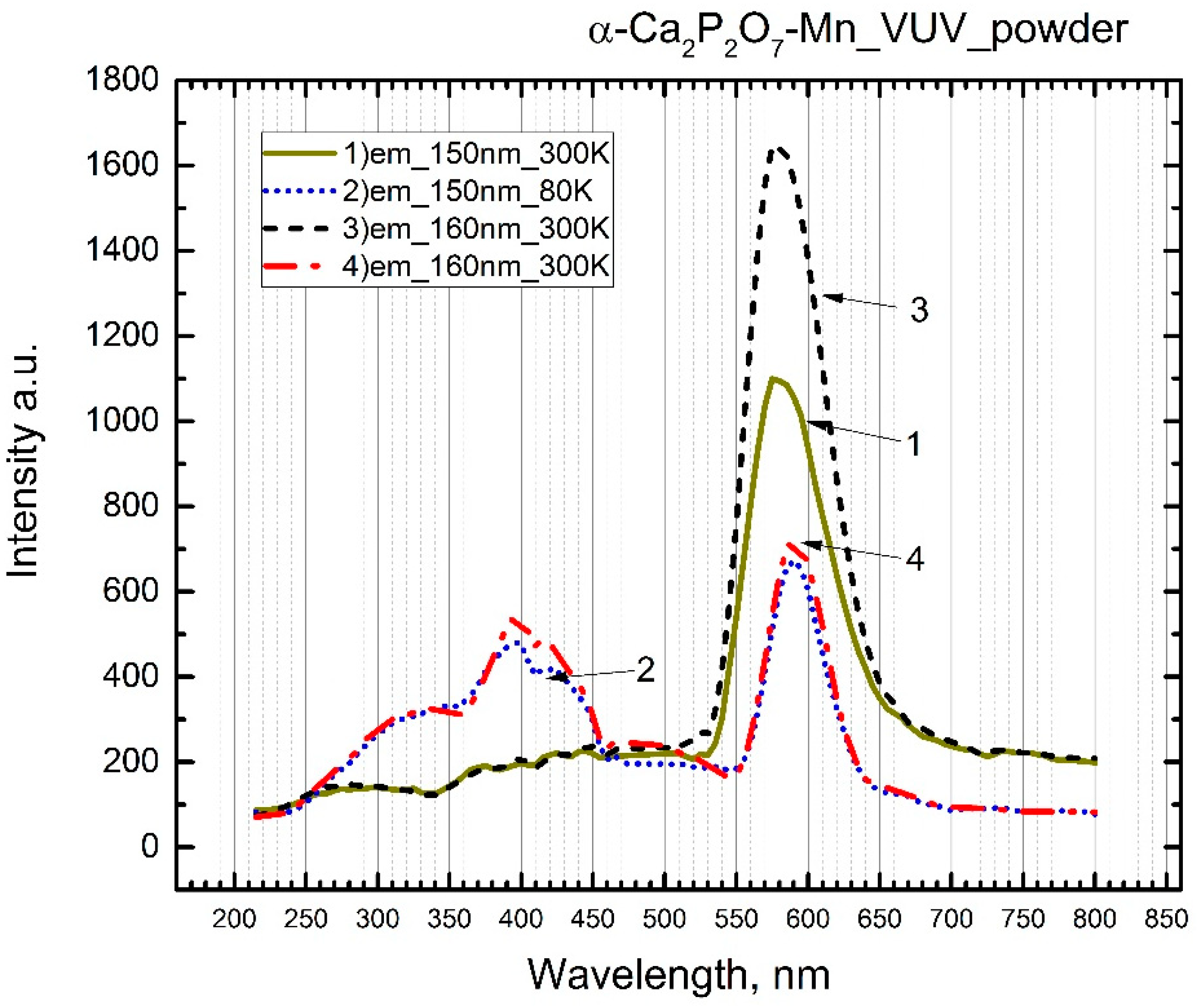
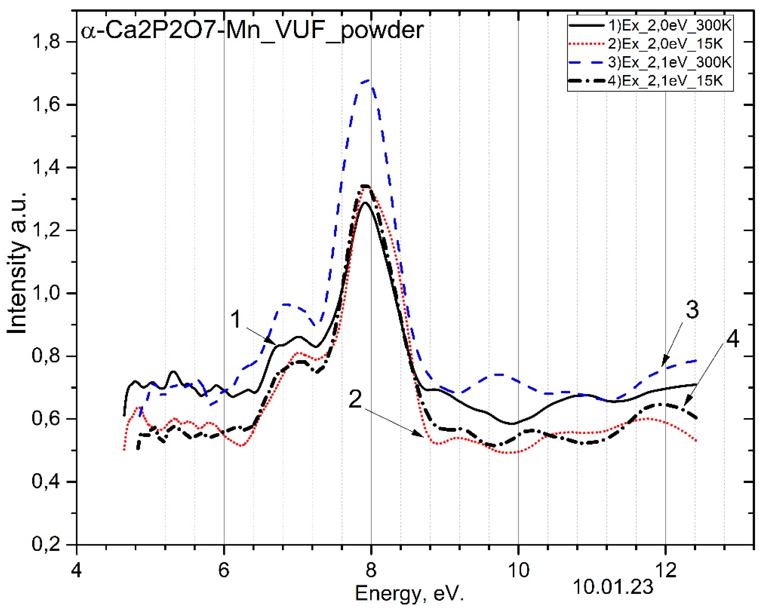
Figure 9.
Emission spectra of α- phosphor under excitation by photons at 8.26 eV (1 - at 300 K, 2 - at 15 K) and under excitation by photons at 7.75 eV (3 - at 300 K, 4 - at 15 K).
Figure 9.
Emission spectra of α- phosphor under excitation by photons at 8.26 eV (1 - at 300 K, 2 - at 15 K) and under excitation by photons at 7.75 eV (3 - at 300 K, 4 - at 15 K).
Figure 10.
Excitation spectra of for the emission band at 2.0 eV: 1 - at 300 K; 2 - at 15 K; and for the emission band at 2.1 eV: 3 - at 300 K; 4 - at 15 K.
Figure 10.
Excitation spectra of for the emission band at 2.0 eV: 1 - at 300 K; 2 - at 15 K; and for the emission band at 2.1 eV: 3 - at 300 K; 4 - at 15 K.
4. Discussion
The experimental results highlight the following processes occurring in the phosphor upon excitation with 6.2 eV ultraviolet photons:
- The occurrence of impurity intracenter emissions at 2.06 eV from the ion, observable at both 300 K and 80 K.
- The generation of new induced recombination emissions at 2.95 eV and 3.1 eV, resulting from charge transfer between excited matrix anions and impurities, specifically between and.
To further elucidate the characteristics of the intracenter emission at 600 nm and the newly induced emissions at 2.95 eV and 3.1 eV, the excitation spectra of these emissions were meticulously measured.
Intracenter emissions at 2.06 eV are excited:
- at 5.64 eV and 4.96 eV as a result of charge transfer from the excited anion to impurities and neighboring anions and.
- ~2.5 eV excitation of trap centers near the ion.
- 4.0 eV and 4.5 eV emissions arising in the transparency region of the matrix.
- 3.
- The newly created induced emissions at 2.95 eV and 3.1 eV are excited at photon energies of 4.0 eV and 4.5 eV, as well as in the spectral range of 4.96 eV and 5.64 eV, where these new induced emissions are mainly created at low temperatures of 80 K.
Next, to clarify the correlation between the above-mentioned regularities with the processes in the pure matrix , similar experimental studies on the phosphor were conducted.
- When the pure phosphor was excited with photon energies ranging from 5.9 to 6.2 eV, emissions at 2.95 eV and 3.1 eV were detected at both 80 K and 300 K.
- These induced emissions are excited at photon energies of 4.0 eV and 4.5 eV in the transparency region of the pure matrix, more intensively at 80 K.
The main question of these studies is the formation of induced or combined radiative states in pure and doped phosphors in the same manner, as a result of charge transfer from the matrix to impurities and neighboring anions and . As a result, intrinsic and impurity electron trapping centers and complementary to hole trapping centers near impurities are formed. During the excitation of the anion , due to the charge transfer of impurities should be created, and the hole component is mainly localized near impurities. It is assumed that similar induced and combined radiative states at 2.95 eV and 3.1 eV should be created upon trapping free electrons and holes by impurities and anions. We conducted experimental research in the vacuum ultraviolet spectral range from 12.4 eV to 4.96 eV, where free electron-hole pairs are created. When irradiated with photons with energies of 8.26 eV and 7.75 eV at temperatures of 300 K and 15 K, intracenter emissions of at 2.15 eV and new induced or combined radiative states at 2.95 eV and 3.1 eV are created. Impurity emissions of at photon energies of 8.0 eV are excited by free electrons. It is known that the band gap of and phosphors are 8.26 eV and 7.65 eV, respectively [22].
The primary focus of these studies is to understand the formation of induced or combined radiative states in both pure and doped phosphors, facilitated by charge transfer from the matrix to impurities and neighboring anions, specifically and . This process results in the formation of intrinsic and impurity electron trapping centers (and () that complement hole trapping centers located near impurities. It is proposed that during the excitation of the anion , charge transfer from impurities should lead to the creation of , with the hole component primarily localized near impurities. Consequently, it is hypothesized that similar induced and combined radiative states at 2.95 eV and 3.1 eV are formed by the trapping of free electrons and holes by impurities and anions.
The estimated band gap of is approximately 8.0 eV.
Thus, the new combined radiative states are created as a result of charge transfer from the excited ion to impurities and ions .
These radiative states are generated when electrons are captured by impurities and intrinsic centers, specifically through the reactions and . This results in the formation of impurity and intrinsic - electron-hole trapping centers. The trapping of electrons by ions and anions leads to the creation of these specific centers, facilitating the observed radiative states
The temperature-dependent measurements of the new radiative states at 2.95 eV and 3.1 eV reveal a distinct pattern: their intensity steadily decreases from 80 K to 250 K, experiences a surge between 250 K and 300 K, and then undergoes a sharp decline from 300 K to 400 K. Concurrently, the intensity of the intracenter emission at 2.06 eV shows a slight decrease over the same temperature range, followed by a significant increase between 370 K and 450 K.
The experimental results, as illustrated in Figure 3, Figure 6 and Figure 8, confirm the temperature dependence of the intensity for the newly induced emissions at 2.95 eV and 3.1 eV. Notably, the efficiency of generating these induced emissions at lower temperatures, specifically at 15 K and 80 K, is found to be twice as high compared to that at higher temperatures ranging from 300 K to 350 K.
The observed temperature-dependent behavior of the radiative states at 2.95 eV and 3.1 eV, particularly the flaring up and subsequent rapid decrease in intensity between 250 K and 300 K, can be attributed to ionization processes: . This ionization leads to the generation of free electrons in the conduction band, which then recombine with localized holes near the impurity, specifically . The energy released during this recombination is transferred to the impurities, resulting in an observed increase in the intensity of intracenter emission at 2.06 eV for . Consequently, this process facilitates energy transfer from the matrix to the impurities. In analogous research conducted by authors [3] on , electron ionization from the ion into the conduction band was observed with an activation energy of 0.4 eV, illustrating a similar charge transfer mechanism.
The formation of induced or combined radiative states, which include the electronic configurations of electron-hole trapping centers and , is supported by experimental evidence. Specifically, new induced emissions at 2.95 eV and 3.1 eV, stimulated by the irradiation of trapping centers, are excited at photon energies of 4.0 eV and 4.5 eV within the transparency region of the matrix, as shown in Figure 5 and Figure 6. Similarly, intracenter emissions from the ion are excited at these same photon energies, as demonstrated in Figure 4, Figure 5 and Figure 7.
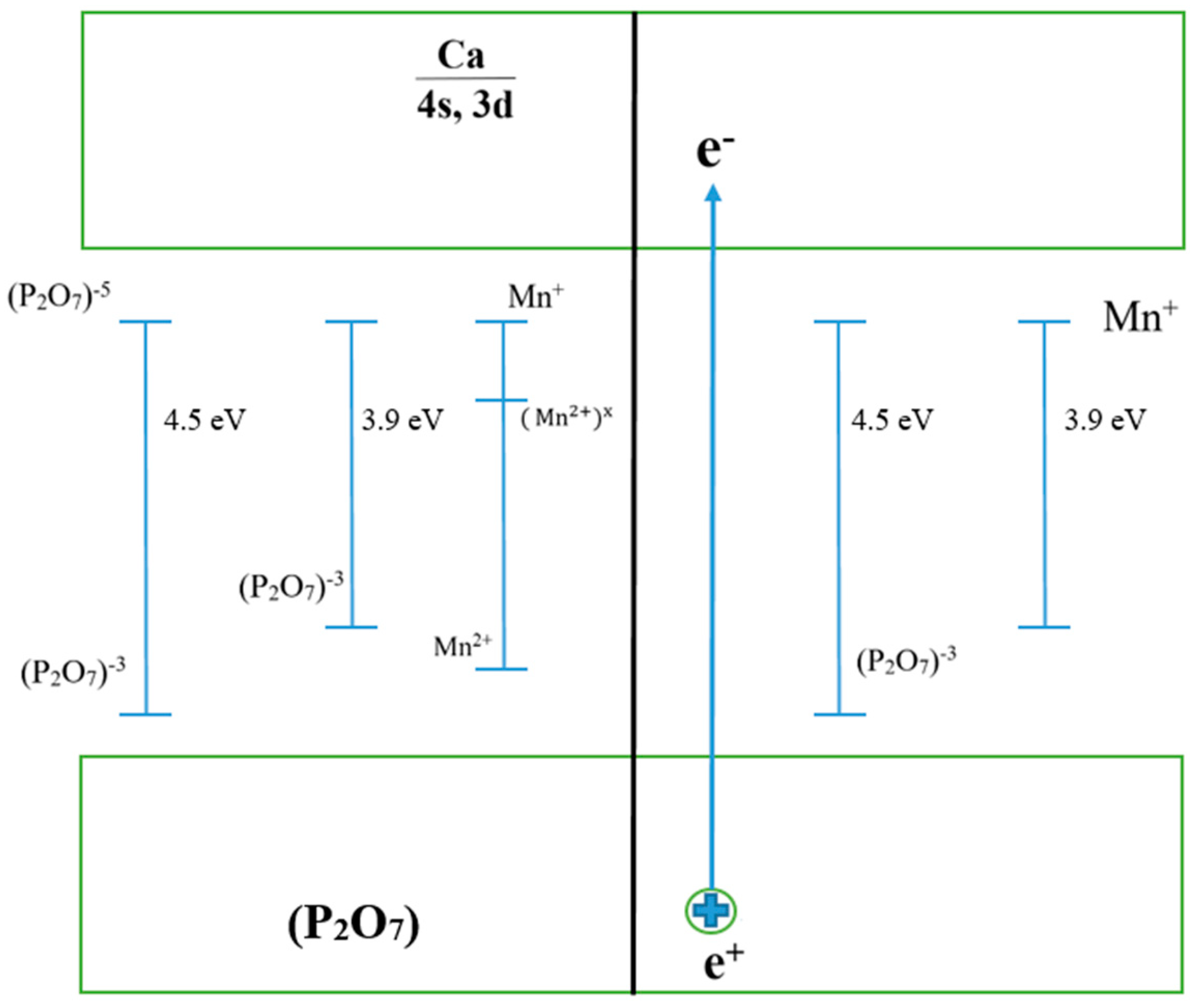
The experimental evidence solidly supports that induced states arise from the electronic configurations of both intrinsic and impurity trapping centers. Specifically, the radiative decay and relaxation processes involving the excited state commence from a singular energy level as the temperature increases. This behavior underscores the dynamic interaction between these trapping centers under thermal variation. Figure 11 presents a model illustrating the presumed arrangement of electronic and hole trapping centers, providing a visual representation of these complex interactions.
The experimental data, obtained by analyzing the photoluminescence decay curves of the samples, reveal distinct decay times for different emission bands. Specifically, the decay time for the intracenter emission at 2.06 eV (corresponding to 600 nm, or 2 bands) is measured at 55 μs. In contrast, the decay time for the induced emission at 3.1 eV (corresponding to 400 nm, or 1 band) is significantly shorter, at 22 μs. The induced photoluminescence from emissions is notably observed following cascade recombination processes, highlighting the complex dynamics of electron-hole recombination in these materials."
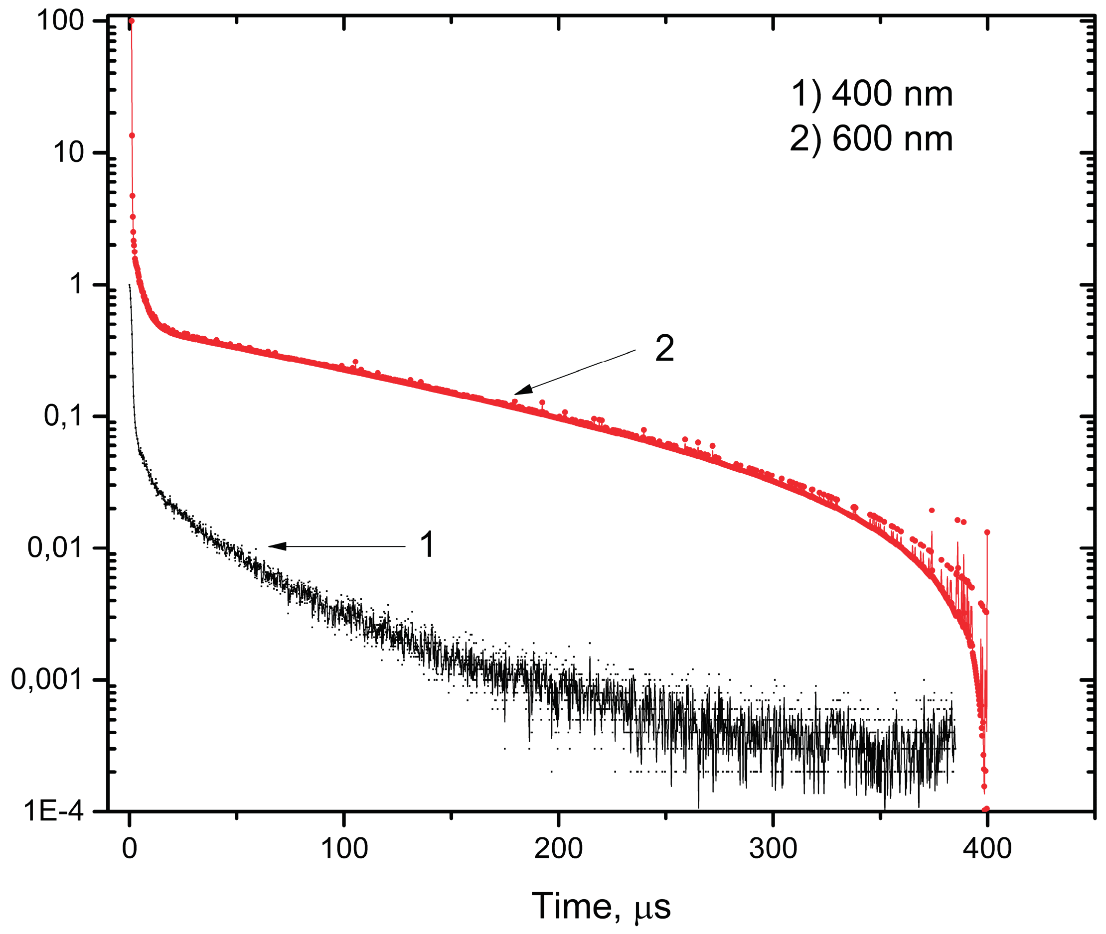
Figure 12.
Decay curves for emission bands at 1) 3.1 eV; 2) 2.06 eV.
The photon energies of 4.0 eV and 4.5 eV, responsible for exciting the intrinsic and impurity radiative states at 2.95 eV and 3.1 eV, correspond to the absorption and excitation energies bridging the ground and excited states of both intrinsic and impurity electron-hole trapping centers. This relationship is detailed in the band model illustrated in Figure 11. The model elucidates how these specific photon energies facilitate transitions that result in the observed radiative states, providing a comprehensive understanding of the energy dynamics within the phosphor.
5. Conclusion
In irradiated , new induced emissions at 2.95 eV and 3.1 eV have been detected, which are excited at photon energies of 4.0 and 4.5 eV in the transparency range of the matrix.
- 4.
- The new radiative states at 2.95 eV and 3.1 eV are created upon excitation of anions by photons with energies of 5.0 eV and 5.64 eV, resulting from charge transfer from the ion to the impurities and neighboring ions according to the reaction and .
- 5.
- It is shown that the radiative states at 2.95 eV and 3.1 eV are also created upon excitation of anions with photon energies of 8.0 eV and 8.25 eV exceeding the band gap, due to electron trapping by impurities and neighboring ions according to the reactions and .
- 6.
- Based on the measurement of excitation spectra of recombination emission at 2.95 eV and 3.1 eV and impurity emission at 2.06 eV, it has been shown that they are simultaneously excited at photon energies of 4.0 eV and 4.5 eV. These values represent the excitation spectrum of the new induced electron radiative states at 2.95 eV and 3.1 eV consisting of intrinsic and impurity electronic trapping center states.
References
- Cao R. et al. Luminescence properties of Sr2Mg3P4O15: Mn2+ phosphor and the improvement by co-doping Bi3+ //Optical Materials. – 2018. – V. 79. – P. 223-226. [CrossRef]
- Lecointre A. et al. Red long-lasting phosphorescence (LLP) in β-TCP type Ca9.5Mn(PO4)7 compounds //Optical Materials. – 2011. – V. 34. – №. 2. – P. 376-380. [CrossRef]
- Zhou R. et al. Insight into Eu redox and Pr3+ 5d emission in KSrPO4 by VRBE scheme construction //Dalton Transactions. – 2018. – V. 47. – №. 2. – P. 306-313. [CrossRef]
- Griesiute D. et al. Synthesis, structural and luminescent properties of Mn-doped calcium pyrophosphate (Ca2P2O7) polymorphs //Scientific reports. – 2022. –V. 12. – №. 1. –P. 7116. [CrossRef]
- (12) Lecointre A. et al. Designing a Red Persistent Luminescence Phosphor: The Example of YPO4:Pr3+,Ln3+ (Ln = Nd, Er, Ho, Dy)// The Journal of Physical Chemistry C 2011, 115, 10, 4217-4227. [CrossRef]
- Zhou R. et al. Site Occupancies, VUV-UV–vis photoluminescence, and X-ray radioluminescence of Eu2+-doped RbBaPO4 //Inorganic Chemistry. – 2020. –V. 59. – №. 23. –P. 17421-17429. [CrossRef]
- Wang Y. et al. Enhanced green emission of Eu2+ by energy transfer from the 5D3 level of Tb3+ in NaCaPO4 //The Journal of Physical Chemistry C. – 2014. –V. 118. – №. 13. –P. 7002-7009. [CrossRef]
- Lecointre A. et. al. Red long-lasting luminescence in clinoenstatite// Journal of Luminescence, -V. 129, Issue 12, December 2009, -P. 1527-1530. [CrossRef]
- Tang O. et. al. Thermoluminescence spectra and dose responses of SrSO4 phosphors doped with rare earths (Eu, Dy, Tm) and phosphorus// Radiation Protection Dosimetry, -V.187, Issue 2, December 2019, -P.164–173,. [CrossRef]
- Zhou R. et al. Site Occupancies, VUV-UV–vis photoluminescence, and X-ray radioluminescence of Eu2+-doped RbBaPO4 //Inorganic Chemistry. – 2020. – Т. 59. – №. 23. – С. 17421-17429. [CrossRef]
- Hee Jo Song et. al. RbBaPO4:Eu2+: a new alternative blue-emitting phosphor for UV-based white light-emitting diodes// Journal of Materials Chemistry C, 2013, 1, 500-505. [CrossRef]
- Chenglong Zhao et. al. Thermally stable luminescence and structure evolution of (K, Rb)BaPO4:Eu2+ solid-solution phosphors// J. Mater. Chem. C, 2014,2, 6032-6039. [CrossRef]
- Hussain T. et. al. Thermoluminescence properties of nanocrystalline BaSO4 doped with Eu2+ produced by solid state combustion synthesis// Radiation Physics and Chemistry, -V. 186, September 2021, 109531. [CrossRef]
- Bao-gai Zhai et. al. Tuning the photoluminescence of Eu2+ and Eu3+ co-doped SrSO4 through post annealing technique// Journal of Luminescence, -V. 194, February 2018, -P. 485-493. [CrossRef]
- Veysi Guckan et. al. Studying CaSO4:Eu as an OSL phosphor// Nuclear Instruments and Methods in Physics Research Section B: Beam Interactions with Materials and Atoms, -V. 407, 15 September 2017, -P. 145-154. [CrossRef]
- Annalakshmi et. al. Synthesis and characterisation of BaSo4:Eu thermoluminescence phosphor// Radiation Protection Dosimetry, -V. 150, Issue 2, June 2012, -P. 127–133. [CrossRef]
- Sun L. Photoluminescence investigation of Eu2+-doped BaSO4 //Experimental and Theoretical NANOTECHNOLOGY. – 2022. – С. 387-394. [CrossRef]
- Nurakhmetov T. N et al. Specific Features of Formation of Electron and Hole Trapping Centers in Irradiated CaSO4-Mn and BaSO4-Mn// Crystals 2023, 13(7), 1054. [CrossRef]
- Yussupbekova B. N. et al. Intrinsic and impurity emission and formation mechanism of trapping centers in LiKSO4-Cu crystals //Nuclear Instruments and Methods in Physics Research Section B: Beam Interactions with Materials and Atoms. – 2020. – V. 481. – P. 19-23. [CrossRef]
- Nurakhmetov T. N. et al. Influence of Cu+ impurity on the efficiency of creation of electron hole trapping centers in irradiated Na2SO4-Cu crystals //Eurasian Journal of Physics and Functional Materials. – 2021. – V. 5. – №. 3. – P. 200-208. //. [CrossRef]
- Nurakhmetov T. N. et al. Specific Features of Formation of Electron and Hole Trapping Centers in Irradiated CaSO4-Mn and BaSO4-Mn //Crystals. – 2023. – V. 13. – №. 7. – P. 1054. [CrossRef]
- Dorenbos P. et al. 4f–5d spectroscopy of Ce3+ in CaBPO5, LiCaPO4 and Li2CaSiO4 //Journal of Physics: Condensed Matter. – 2003. – V. 15. – №. 3. – P. 511. [CrossRef]
- Nurakhmetov T. N. et al. Energy transfer of intrinsic electronic excitation to impurities in the CaSO4-Mn //Eurasian Journal of Physics and Functional Materials. – 2021. – V. 5. – №. 1. – P. 31-38. [CrossRef]
Figure 1.
X-ray diffraction pattern of α- powder alongside the standard sample.
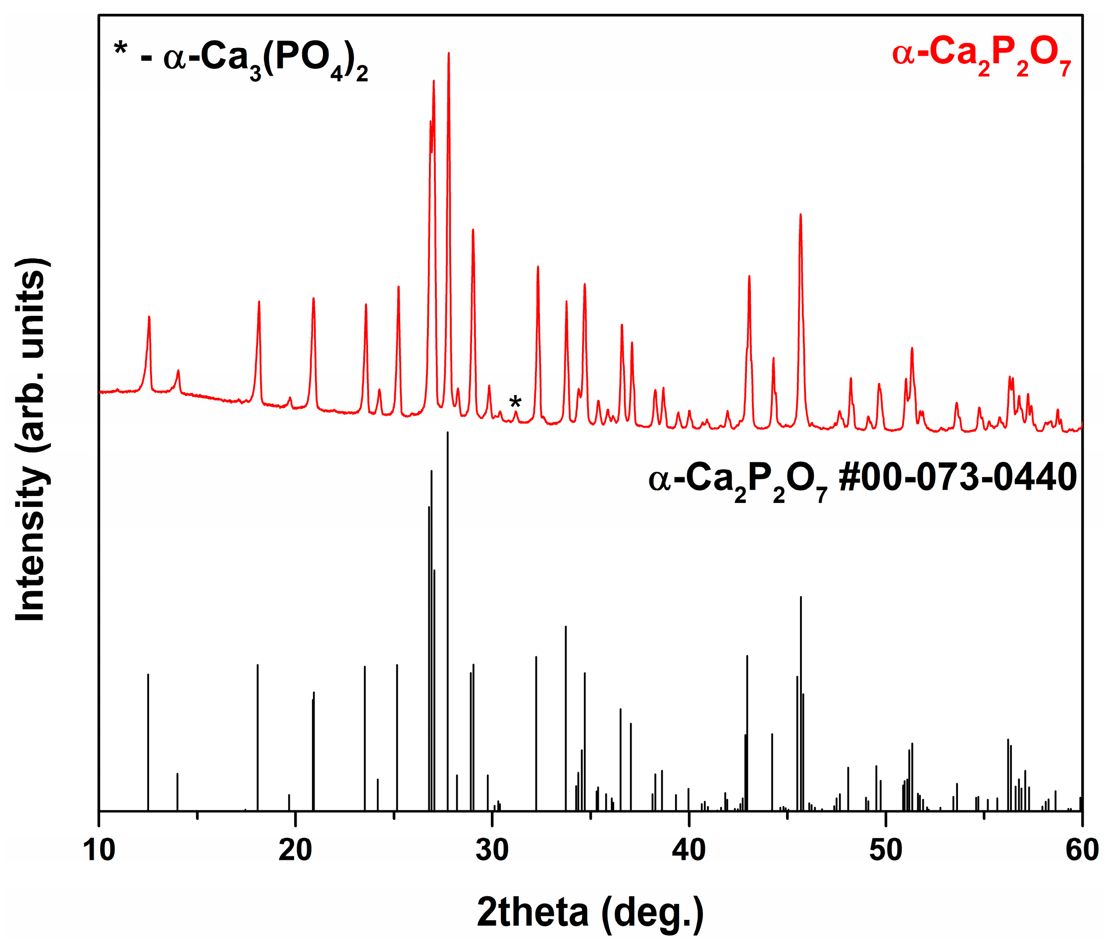
Figure 2.
SEM micrographs of powder.
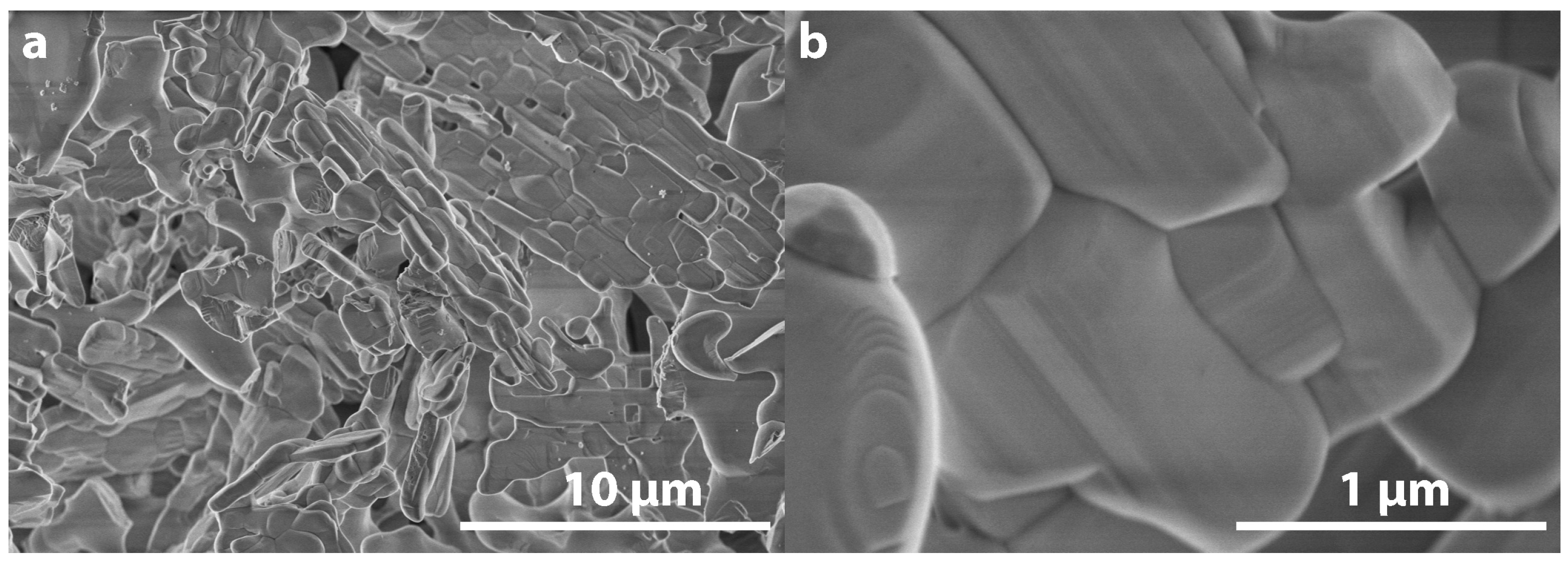
Figure 3.
Emission spectra of the phosphor upon excitation with 6.2 eV photons (1 - at 300 K, 2 - at 80 K).
Figure 3.
Emission spectra of the phosphor upon excitation with 6.2 eV photons (1 - at 300 K, 2 - at 80 K).
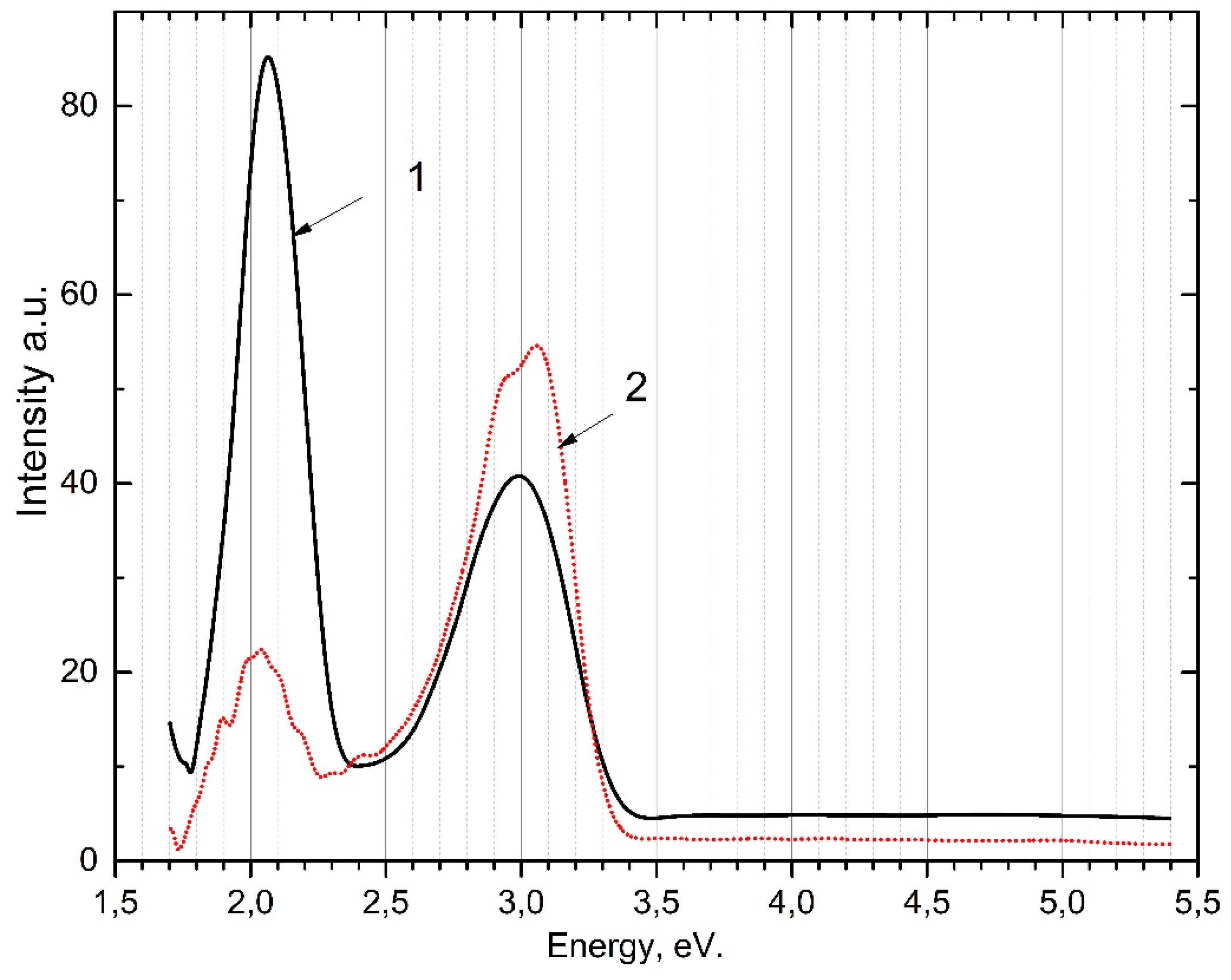
Figure 4.
Excitation spectra of the intra-center emission for the 2.06 eV band, irradiated (1 - at 300 K, 2 - at 80 K).
Figure 4.
Excitation spectra of the intra-center emission for the 2.06 eV band, irradiated (1 - at 300 K, 2 - at 80 K).
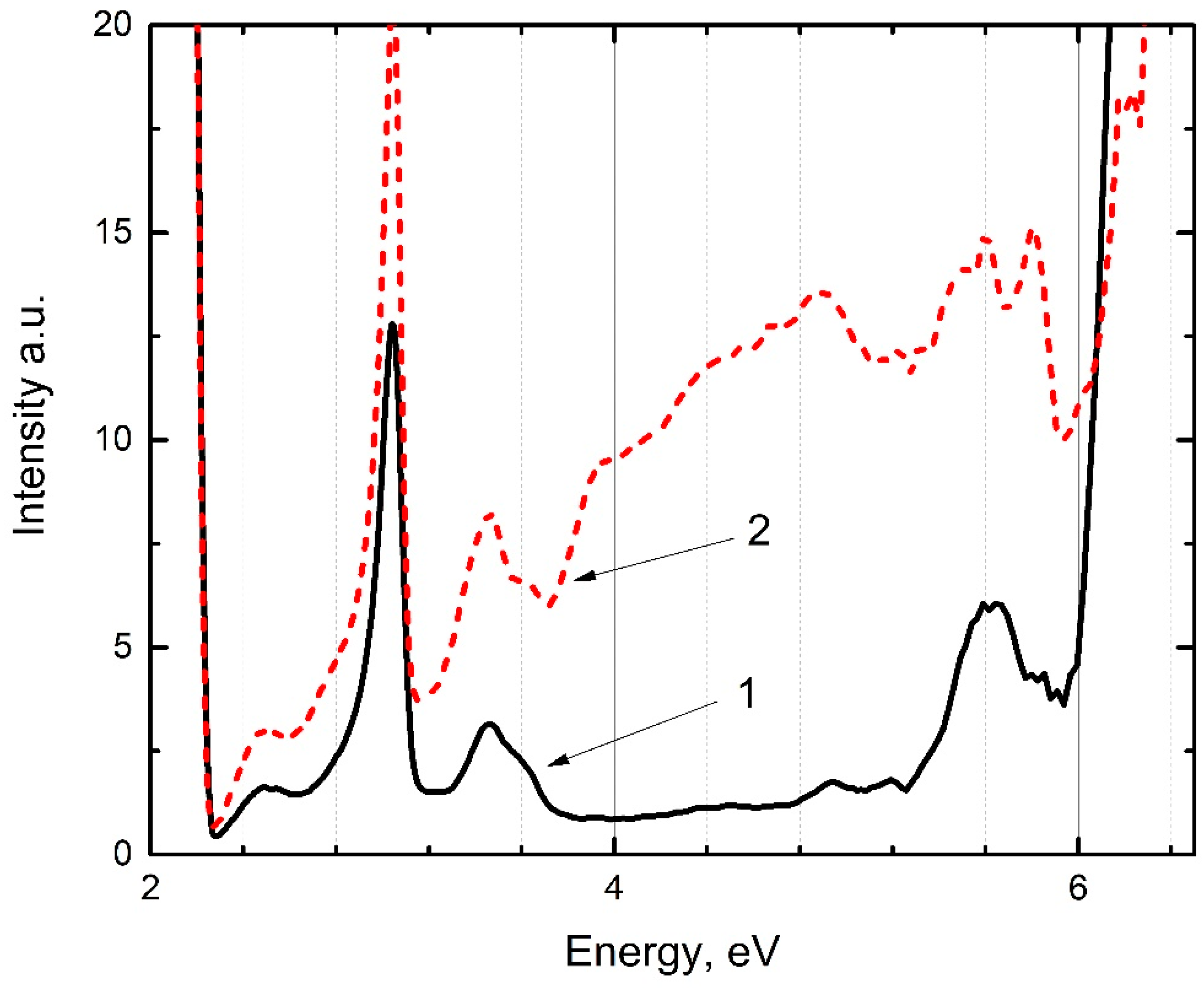
Figure 5.
Excitation spectra of the pre-irradiated phosphor: 1 - for the emission band at 2.92 eV at 300 K; 2 - for the emission band at 2.92 eV at 80 K; 3 - for the emission band at 3.06 eV at 300 K; 4 - for the emission band at 3.06 eV at 80 K.
Figure 5.
Excitation spectra of the pre-irradiated phosphor: 1 - for the emission band at 2.92 eV at 300 K; 2 - for the emission band at 2.92 eV at 80 K; 3 - for the emission band at 3.06 eV at 300 K; 4 - for the emission band at 3.06 eV at 80 K.

Figure 6.
Emission spectrum of the phosphor when excited by photons at 6.2 eV (1 - at 300 K, 2 - at 80 K) and when excited by photons at 5.64 eV (3 - at 300 K, 4 - at 80 K).
Figure 6.
Emission spectrum of the phosphor when excited by photons at 6.2 eV (1 - at 300 K, 2 - at 80 K) and when excited by photons at 5.64 eV (3 - at 300 K, 4 - at 80 K).
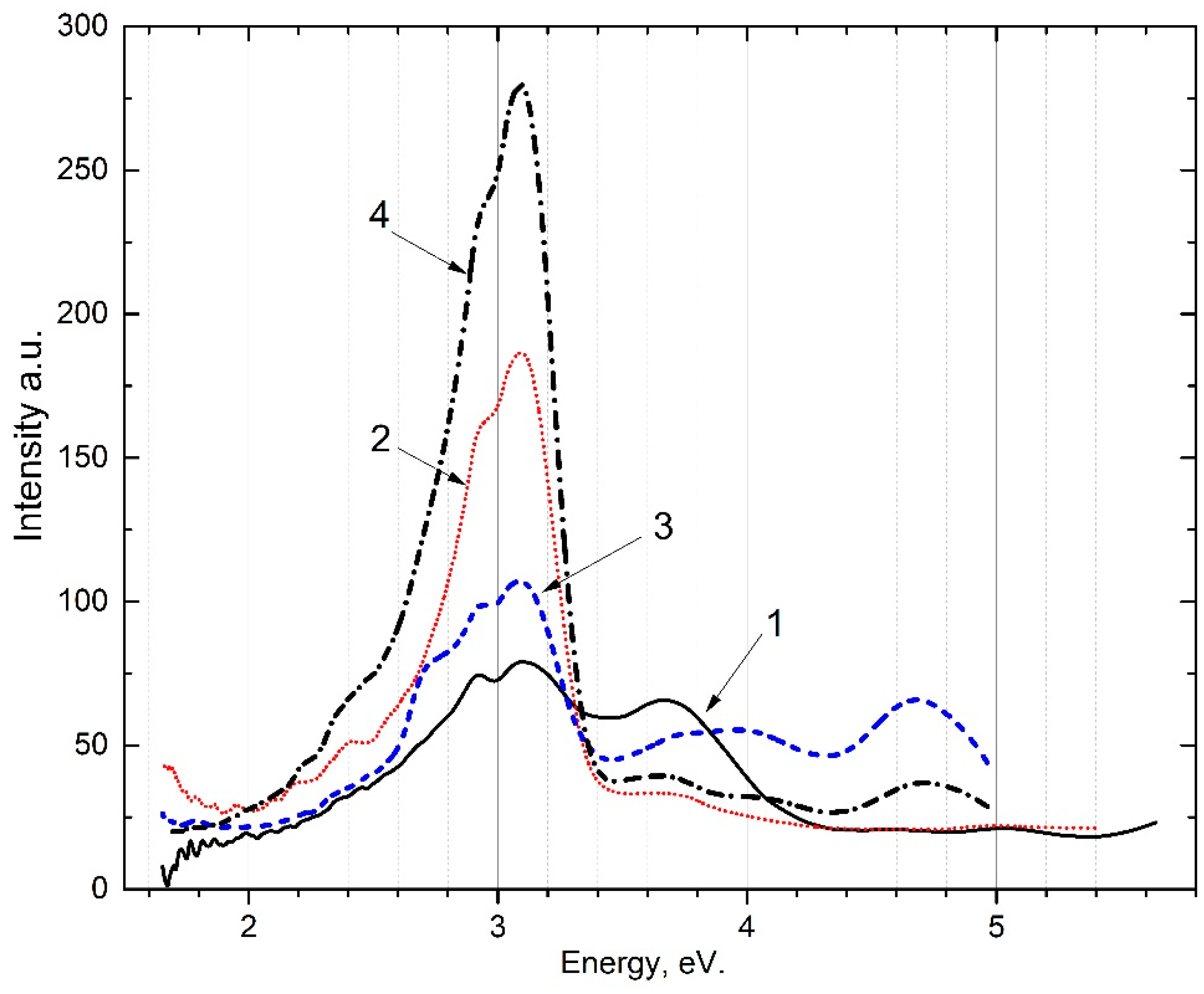
Figure 7.
Excitation spectra of pre-irradiated phosphor: for the emission band at 2.95 eV: 1 - at 300 K; 2 - at 80 K; for the emission band at 3.1 eV: 3 - at 300 K; 4 - at 80 K.
Figure 7.
Excitation spectra of pre-irradiated phosphor: for the emission band at 2.95 eV: 1 - at 300 K; 2 - at 80 K; for the emission band at 3.1 eV: 3 - at 300 K; 4 - at 80 K.
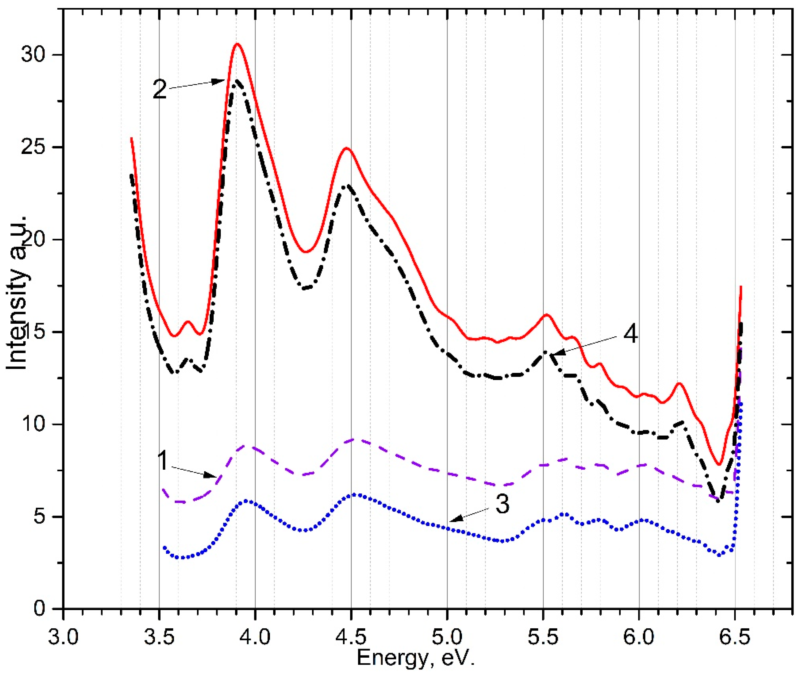
Figure 8.
Temperature dependence of emission intensity at 2.95 eV (1), 3.1 eV (2), and 2.06 eV (3).
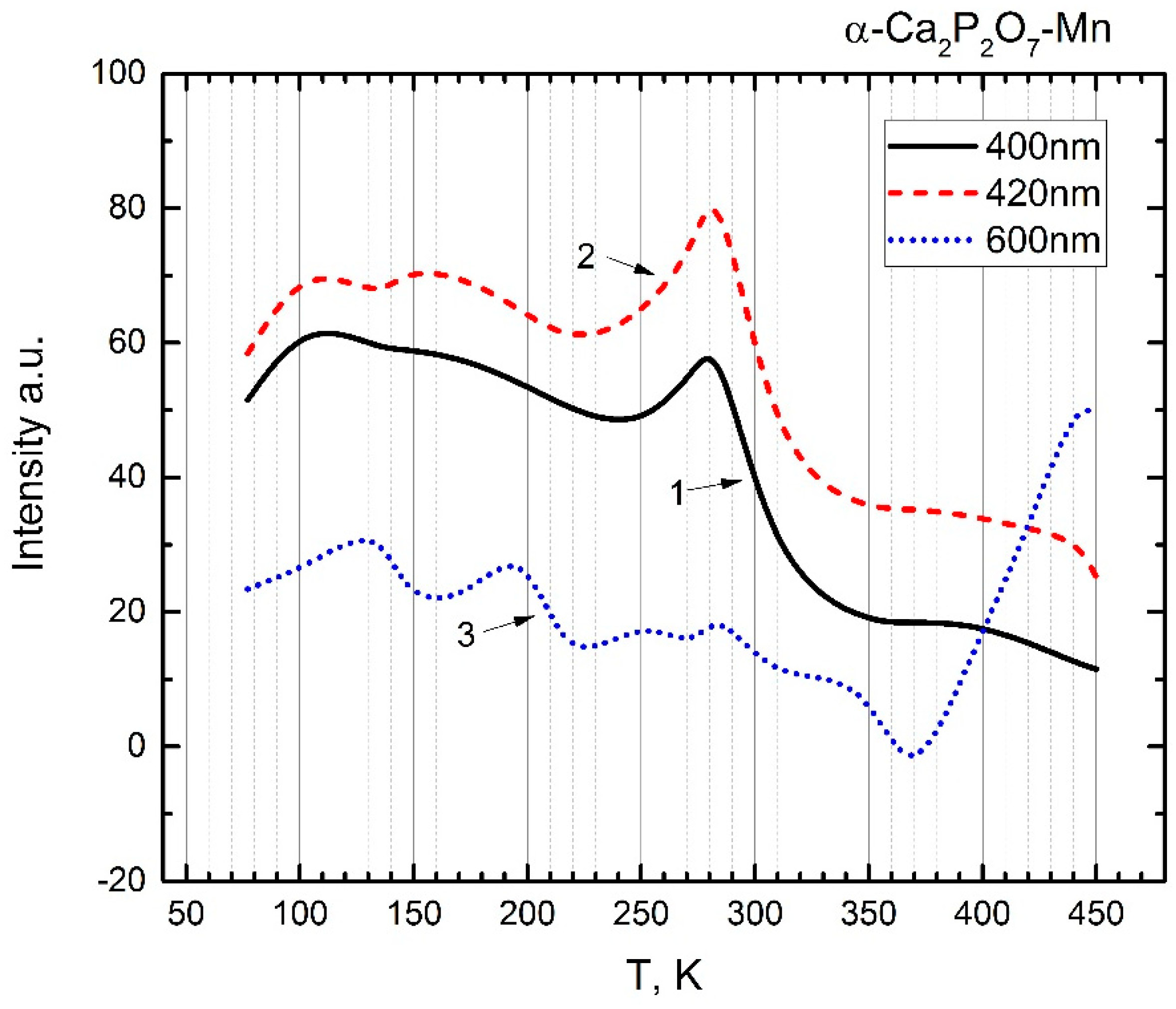
Figure 11.
Model depicting the arrangement of electronic and hole trapping centers.
Disclaimer/Publisher’s Note: The statements, opinions and data contained in all publications are solely those of the individual author(s) and contributor(s) and not of MDPI and/or the editor(s). MDPI and/or the editor(s) disclaim responsibility for any injury to people or property resulting from any ideas, methods, instructions or products referred to in the content. |
© 2024 by the authors. Licensee MDPI, Basel, Switzerland. This article is an open access article distributed under the terms and conditions of the Creative Commons Attribution (CC BY) license (http://creativecommons.org/licenses/by/4.0/).
Copyright: This open access article is published under a Creative Commons CC BY 4.0 license, which permit the free download, distribution, and reuse, provided that the author and preprint are cited in any reuse.


