Submitted:
27 February 2024
Posted:
27 February 2024
You are already at the latest version
Abstract
The Serra da Estrela Natural Park (NPSEs) in Portugal stands out as a well-preserved region abundant in medicinal plants, particularly known for their pharmaceutical applications in diabetes prevention and treatment. This comprehensive review explores these plants' botanical diversity, traditional uses, pharmacological applications, and chemical composition. The NPSEs boast a rich diversity with 138 medicinal plants across 55 families identified as traditionally and pharmacologically used against diabetes globally. Notably, the Asteraceae and Lamiaceae families are prevalent in anti-diabetic applications. In vitro studies reveal their significant inhibition of carbohydrate-metabolizing enzymes, and certain plant co-products regulate genes involved in carbohydrate metabolism and insulin secretion. In vivo trials demonstrate anti-diabetic effects, including glycaemia regulation, insulin secretion, antioxidant activity, and lipid profile modulation. Medicinal plants in NPSE exhibit various activities beyond anti-diabetic, such as antioxidant, anti-inflammatory, antibacterial, anticancer, and more. Chemical analysis identifies over fifty compounds like phenolic acids, flavonoids, terpenoids, and polysaccharides responsible for their efficacy against diabetes. The findings underscore the potential of NPSE medicinal plants as anti-diabetic candidates, urging further research to develop effective plant-based anti-diabetic drugs, beverages, and supplements.
Keywords:
Natural Park of Serra da Estrela
; Botanical diversity
; Medicinal plants
; Diabetes prevention and treatment
; Pharmacological applications
; chemical composition.
1. Introduction
Type 2 diabetes mellitus (T2DM) is a chronic metabolic disease associated with multiple dysfunctions in the endocrine system [1], affecting the metabolism of carbohydrates, lipids and proteins [2]. This disorder is related to a defect in insulin secretion and/or a progressive alteration in its function in the body [3,4]. The onset of hyperglycemia and insulin resistance leads to the accumulation of free fatty acids, lipid peroxidation, disruption of cellular antioxidant defences and inflammatory reactions [5,6,7,8]. Although some of the underlying mechanisms are uncertain, all these factors contribute to the disturbance of the integrity of physiological barriers [9], mainly by altering the vascular integrity of tissues, and may contribute to the clinically recognised complications of diabetes (hypertension, diabetic peripheral neuropathy, chronic kidney disease, retinopathy, cardiovascular disease and others) [10,11,12,13,14,15]. T2DM is often associated with fatty liver, sleep apnea syndrome, depression, cognitive decline, and dementia [16].
According to current diabetes statistics, more than 90% of cases worldwide are T2DM; the older generation is the most affected of the 500 million people suffering from the disease[17]. About 422 million people worldwide have T2DM, most living in low- middle-income countries, and 1.5 million deaths are directly attributed to diabetes yearly [18]. In Portugal, the incidence of T2DM is much higher than in other types of diabetes [19,20]. Epidemiological studies show that Portugal (9.1%) is among the countries with the highest rates in Europe, alongside Turkey (14.5%), Spain (10.3%), Andorra (9.7%) and Serbia (9.1%) [17]. It is estimated that T2DM affects 13.6% of the Portuguese population aged between 20 and 79; An equivalent rate of 5.9% of people are unaware that they have the disease [21]. The data for 2021 already show an increase in the number of new cases identified [19]. Around 857,272 people with T2DM have been registered with the Portuguese National Health Service, including 74,396 new diagnoses [19]. Approximately 200 new patients are diagnosed with diabetes daily in Portugal [19].
T2DM can occur for a variety of reasons. Hyperglycaemia, obesity, hypertriglyceridaemia, poor eating habits, lack of exercise, ageing, family history, alcohol consumption, smoking, stress, anxiety and depression are the main risk factors for the onset of the disease in adults [22,23,24,25,26]. Statistics show it is present in young people due to poor diet and lifestyle [17,27,28]. Multiple studies have shown that people from different ethnic backgrounds may have specific phenotypes that increase susceptibility to hypertension, insulin resistance and dyslipidaemia [29]. People of Latin American, East, Southeast and South Asian, sub-Saharan African, Middle Eastern and North African origin living in Europe were 1.3 to 3.7 times more likely to have T2DM than white European populations [30]. In the United States, 14.7% of American Indians and Alaska Natives are diagnosed with diabetes, compared with 7.5% of non-Hispanic white Americans [31]. Regarding the gender factor, both men and women are affected, but worldwide, an estimated 17.7 million more men than women have T2DM [17,32,33]. Insulin sensitivity and response capacity are significantly higher in women than men [34]. Genetic polymorphisms between the two sexes, differences in the mechanism of action of sex hormones, visceral and hepatic adiposity, hypoadiponectinemia, adiponectin, insulin-sensitive hormone, resting energy expenditure and lipid metabolism may contribute to higher insulin sensitivity in men than in women [35,36,37,38,39].
The accumulation of excessive body fat (obesity) triggers a broad spectrum of metabolic issues and diseases, comprising insulin resistance, atherogenic dyslipidaemia [high plasma triglyceride and low HDL (high-density lipoprotein) cholesterol content], non-alcoholic fatty liver disease (NAFLD), -cell dysfunction, pre-diabetes, and T2DM [40,41]. Obesity strongly influences T2DM in adults, children, and adolescents [42,43]. It's a serious concern associated with poorer mental outcomes, reduced quality of life, and the leading cause of death worldwide [44]. Obesity increases the risk of developing T2DM by at least a factor of six [17,45]. The prevalence of obesity in adults (age ≥20 years) was 38.8% between 2013 and 2016 [46]. If the obesity trends continue, projections are that one in three adults will have type 2 by 2050 [47,48]. The prevalence of T2DM is also positively correlated with the duration of obesity and body mass index (BMI) in childhood [43,49,50]. The proportion of T2DM is higher in people who were obese in childhood and of normal weight in adulthood than in people of normal weight in childhood [43,51]. Obesity and type T2DM represent the greatest threat to the development of atherosclerosis and CAD (coronary artery disease) [52]. These two health conditions are oxidative damage, inflammation, and insulin resistance [5]. Indeed, under diabetic or hyperglycaemic conditions, excess reactive oxygen species (ROS) are released in various tissues and may play a role in developing many complications [53]. This state can persist even when hyperglycaemia is controlled. A disequilibrium occurs with the antioxidant defence systems. This modification scenario is known as oxidative stress [54]. It mainly causes endothelial dysfunction, leading to vascular lesions, abnormal production of plasma lipids, activation of platelets and increased coagulation, and activation of inflammatory processes [54,55]. This damage can be prevented when adequate glycaemic control is established early but is not easily reversed if poor control is maintained over a prolonged period [56]. Oxidative stress causes potential damage to lipids, DNA and proteins and is responsible for altering intracellular signalling pathways, leading to insulin resistance [57]. The hyperglycemic environment and free fatty acids lead to the appearance of metabolic stress because of an increase in ROS and a change in the mitochondrial electron transport chain [58,59,60]. ROS are considered key signalling molecules that play an important role in the progression of inflammatory disorders, contributing to the development of insulin resistance and the predicted long-term complications of T2DM [57,61]. Activation of the immune system and increased circulating acute-phase inflammatory markers can significantly and directly impact insulin resistance or blood glucose levels [62].
Postprandial hyperglycaemia in people with T2DM can be managed by several approaches, including lifestyle modification, i.e. regular physical activity and a healthy diet [63]. The administration of pharmacological drugs accompanies these measures. Some of these can delay carbohydrate absorption by reducing the digestion of polysaccharides and their bioavailability (e.g. α-glucosidase inhibitors) [64,65]. Others are mainly used to increase the availability of endogenous insulin. These include sulphonylureas, such as Glibenclamide, and glinides, insulin analogues that act on the sulphonylurea receptor in the pancreas to promote insulin secretion. Glucagon-like peptide-1 (GLP-1) agonists and dipeptidyl peptidase-IV (DPP-IV) inhibitors can also increase endogenous insulin by acting on ileal cells in the small intestine. Other drugs used to improve insulin sensitivity include thiazolidinediones, peroxisome proliferator-activated receptor gamma (PPARγ) agonists, and metformin, a biguanide [66,67]. All these drug treatments are prescribed either as monotherapy or with other hypoglycaemic agents [68]. Administration of exogenous insulin remains the primary treatment for some patients with T2DM who are unable to control their blood glucose with oral hypoglycaemic agents [68,69]. If all types of oral hypoglycaemic agents and insulin are administered correctly, and with a healthy lifestyle, people with T2DM can manage and reduce the side effects of the disease. However, certain iterations linked to the risk of hypoglycaemia or co-morbidities have been observed [70]. These occur following a progressive decline in β-cell function and a reduction in therapeutic efficacy due to inappropriate or ineffective dosing regimens, altered drug metabolism, lack of target specificity, and solubility and permeability problems [68]. In treated patients, weight gain, weakness, fatigue, lactic acidosis, nausea or diarrhoea, abdominal discomfort and a metallic taste have been observed [71,72].
In this context, medicinal plants have a well-established record of circumventing the problems mentioned about the conventional use of drugs [73]. Medicinal compounds derived from plants could provide new, straightforward approaches to preventing and treating T2DM and its complications [74,75,76]. Traditional knowledge and practices have enabled the development of most modern medicines [73,77]. Many natural resources have been used to develop almost 25% of the major pharmaceutical compounds and their derivatives currently on the market [78,79]. These plant resources have great potential as alternative hypoglycaemic medicines because of their safety, efficacy, affordability and availability. They constitute an almost unlimited source of bioactive compounds, and their use as antidiabetic agents has been exploited in various ways [74,75]. Secondary metabolites, such as flavonoids, phenolic acids, alkaloids, tannins, terpenoids, saponins, triterpenoids, steroidal glycosides, etc., have shown innumerable promising results against T2DM [80,81,82,83]. They are effective in different stages of diabetes. They can control insulin resistance, impact glucose absorption, regulate multiple glucose and lipid metabolism stages and inhibition and/or activation of the expression of genes involved in glucose homeostasis [81]. These natural antidiabetic agents can act alone or with conventional treatments to strengthen the body's ability to cope with the disease [84]. However, some of these compounds have not yet been studied in depth. As some of the antidiabetic actions of many medicinal plants are still unexplored, researchers are focusing more and more on finding new treatments that work quickly and at lower costs [85].
The information needed to assess the efficacy of potentially important medicinal plants and to prove their antidiabetic value must be effective and well-validated [74,86]. One method of sourcing information on medicinal plants is ethnopharmacological studies [87]. They provide rich information from the local community and contribute to discovering and developing natural medicines [86]. Analysis of the medicinal literature concerning the NPSEs (The Serra da Estrela Natural Park) shows that documentation on local medicinal plants is weak and almost non-existent, hence the importance of an in-depth study [88,89,90,91,92,93,94,95,96,97,98]. Therefore, to obtain a complete perspective on the potential use of medicinal plants from the NPSEs as alternative solutions for combating diabetes, the most relevant studies concerning the botanical diversity, known traditional uses of local plants, the validation of their antidiabetic activities (in vitro and in vivo studies), the underlying mechanisms of action, their pharmacological activities, the plant-derived chemical compounds that may be responsible for these activities, the challenges and prospects for the antidiabetic activity of medicinal plants from the NPSEs have been critically analysed in this review.
2. Materials and Methods
2.1. Geographical and Climate Features of the Serra da Estrela Natural Park
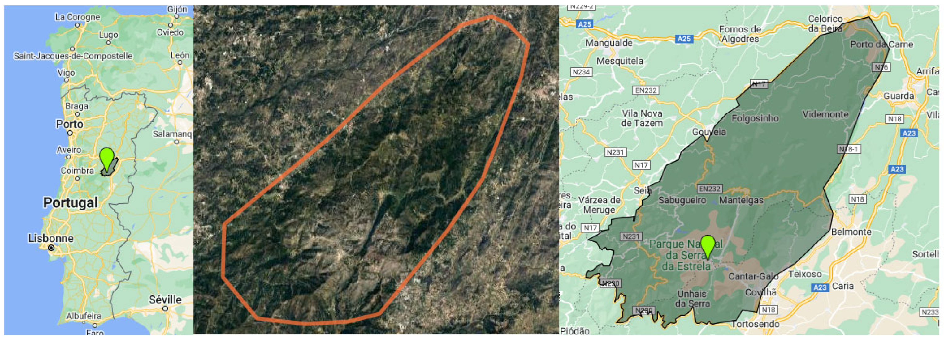
Continental Portugal has several mountain ranges. The highest is in the centre-east, the "Serra da Estrela" (40º20'N, 7º35'W) (Figure 1) [99]. Its massif forms the western part of the Cordillera Central, with its highest point called "La Torre" at an altitude of 1993 m [88,89,90,91]. Part of the Iberian Peninsula is traversed by this mountain range, over 500km long, stretching from almost the Atlantic coast to just north of Madrid [88,89,90,91]. Most of these mountains lie within the boundaries of the NPSEs, created in 1976 [100,101,102] and covering around 100,000 hectares [88,89,90,91]. This area is a biological and community interest site integral to the Natura 2000 network [100,101,103]. Six municipalities (Seia, Gouveia, Celorico da Beira, Guarda, Manteigas and Covilha) (Figure 1) and two districts (in the north, the district of Guarda and the south, the community of Castelo Branco) have joined forces to draw up this project [88,89,90,91,100,101,102,103]. The mountains are mainly composed of granite in the central part and schist in the periphery, dominating the Mondego and the Zezere plains (a tributary of the Tagus) [88,89,90,91]. In the north-east, the landscape is characterised by the watersheds of three major basins: the Douro (the largest river on the Iberian Peninsula), the Tagus (the longest river on the Iberian Peninsula) and the Mondego (the largest river in Portugal) [88,89,90,91].
The climate of Serra da Estrela is influenced by several factors: temperature, atmospheric pressure, wind, humidity and precipitation, as well as geographical factors [104,105]. Its high altitude among the surrounding land, the general organisation of the relief and the relative proximity of the Atlantic Ocean, some one hundred kilometres away, play a decisive role in the complex mosaic of local climates that characterise the region [88,89,90,91]. Thus, all the climate factors are controlled by the overall latitudinal position of the mountains and influenced by the north-south temperate climate and the southeast-northwest Mediterranean macroclimate [99,104,105]. They are also controlled by the Atlantic's longitudinal position and the Iberian Peninsula's interior (maritime influences mainly to the west and continental influences to the east and west) [88,89,90,91]. Average annual rainfall is around 2,500 mm at the summit, while the plateaux record more than 2,000 mm [88,89,99,106]. The highest number of precipitation days is observed in the western part of the mountains, while the lowest values are in the basal areas, in the north-western and south-eastern sectors, with around 1,000 to 1,200 mm [88,89,90,99,106]. The region is characterised by hot, dry summers and wet winters, with snowfall more frequent between December and March [99,104,105]. The most striking aspects of the relief are the glacial forms and deposits. The snowfall is heaviest in the higher mountain areas but lightest and most irregular in the lower regions [88,89,90,91].
Figure 1.
Geolocalization of Serra da Estrela Natural Park [107].
Figure 1.
Geolocalization of Serra da Estrela Natural Park [107].
2.2. Ethnobotanical Data Collection and Selection Criteria
The information on the plant species of the Serra da Estrela region has been collected from the databases of the World Checklist of Vascular Plants [108]. It's an international collaborative programme with editors, compilers and reviewers from all over the world. The main objective of the database is to provide high-quality, expertly reviewed taxonomic data on all vascular plants based on the nomenclatural data provided by the International Plant Name Index (IPNI).
The Flora-On database is also used; it is a portal coordinated by the Portuguese Botanical Society containing photographic, geographical, morphological and ecological information for all vascular plant species in Portugal [109]. The search was supported by the INaturalist database [110]. It is a joint initiative of the California Academy of Sciences and the National Geographic Society. It is also a species identification system and a tool for recording the occurrence of organisms. It can be used to record sightings, get help with identifications, collaborate with others to collect information for a common purpose, or access sighting data collected by iNaturalist users [110]. All databases were screened using a combination of the keywords "Family", "Species", "Species Synonyms", and "Subspecies". This approach enabled us to find 97 different families. The total number of species is 888 (a complete list of the taxa is given in Table S1). Based on this list of plants, a systematic literature search on their traditional and diabetic uses was conducted. Data were obtained from scientific databases, including NCBI, Scopus, Web of Science and Google Scholar (Figure 2). The preliminary selection was initially performed using the search terms "Serra da Estrela" and "medicinal plants" to cover the maximum range of medicinal plants used against diabetes. As the number of studies was small, we carried out another selection, but universal, by searching by keyword for all medicinal plants from the Serra da Estrela region and their possible worldwide uses. The Boolean operator "AND" followed by the keyword "Diabetes" or "Hypoglycaemic" was used for this search to cover literature reports dealing exclusively with T2DM and anywhere in the world. This search was carried out specifically for each plant on the databases, using the leading taxonomic designation of the species and other botanical synonym names, followed by the keywords mentioned. The names of each plant and combinations of the terms "Traditional", "Ethnobotanical", "Ethnobotany", "Folk remedies", and "Ethnomedicinal" were used to search the above databases (Figure 2).
3. Results
3.1. Botanical Diversity of NPSE and Ethnopharmacological Uses of Medicinal Plants with Antidiabetic Potential
Mountains have always been an excellent challenge for humankind, who has never ceased climbing, cultivating, and domesticating. They are open-air laboratories of knowledge, home to species and communities that have adapted to their environment in various ways. They provide fertile ground for observing and understanding the evolution of species and the distribution of organisms in similar contexts, from one mountain to another thousands of kilometres away. They are, therefore, important ecosystems because they harbour high levels of biodiversity and endemicity [111]. They provide essential services such as climate regulation, freshwater supply and purification, and nutrient cycling [112,113,114,115,116].
The inaccessible NPSE vegetation is the best preserved in the region [88,89]. The isolation of the summits and the extreme conditions that prevail there have encouraged the appearance of new species and facilitated species isolation, speciation, extinction, and migration [115]. According to Jansen et al. [88], the flora of this mountainous region shows significant contrasts as you go up in altitude. The vegetation is divided into several levels, the boundaries of which vary according to exposure. Within this tiering, the transitions in vegetation are distinguishable, and each level corresponds to a well-defined ecosystem. In addition, the isolation of the summits and the extreme prevailing conditions have encouraged the appearance of new species. Many species are endemic to these areas, making the NPSE one of the wealthiest regions in Portugal for certain groups of plants [88,89].
The climatic heterogeneity contributing to the region's high biodiversity has attracted botanists' interest [99]. Floristic expeditions from the 18th century to the present day have enabled a rigorous characterisation of the ecosystems' flora, which is essential for their in-depth knowledge and conservation [93]. According to the three bioclimatic levels (Meso-Mediterranean, Supra-Mediterranean, Oromediterranean) defined in the Serra da Estrela region, three vegetation ranges (basal, intermediate and upper) have been characterised. The three combined levels have identified approximately 900 vascular species and subspecies [88]. Endemic Iberian species belonging to the Mediterranean and Atlantic flora are particularly well represented and distributed in an area more or less delimited by the altitude of the massif and the climatic, edaphic and sun exposure conditions [88,89,91,102]. Some relict plant populations from northern and central Europe have also invaded the area [88].
The botanical census of NPSE diversity enabled us to identify 97 families, 112 genera, and 888 vascular species (after eliminating synonyms) (Table S1). The number of native species on the Iberian Peninsula is 133, while there are only 9 endemic species in Portugal. By contrast, the number of introduced species is 36 [108,109,117].
The Asteraceae is the family with the most species/subspecies (sp) in the region (108 sp). It is followed by Poaceae (81 sp), Fabaceae (74 sp), Caryophyllaceae (60 sp), Brassicaceae (33 sp), Apiaceae (29 sp), Lamiaceae (29 sp), Rosaceae (28 sp), Plantaginaceae (27 sp), Polygalaceae (22 sp), Ranunculaceae (21 sp), Cyperaceae (19 sp), Juncaceae (18 sp), Rubiaceae (17 sp), Cistaceae (16 sp), Amaryllidaceae (15 sp) and Crassulaceae (14 sp).
Several plants found in the NPSE have been used to treat diabetes. Despite the relatively large number of studies worldwide reporting their biological potential, NPSE species have been little investigated, and species with antidiabetic potential will be the subject of particular attention in the following section. Of the 888 species listed, only 138 plants (15.54 %) from different geographical regions have been selected based on traditional uses and studies into their antidiabetic potential (Table S2). The families with the highest number of species are Asteraceae (21 sp), Lamiaceae (12 sp), Fabaceae (9 sp), Rosaceae (8 sp), Caryophyllaceae (6 sp), Polygalaceae (5 sp) (Figure 3). The Apiaceae, Asparagaceae and Ericaceae contain four species/subspecies, and Boraginaceae, Geraniaceae, Hypericaceae and Fagaceae comprise three (Figure 3). However, the families Brassicaceae, Cistaceae, Amaryllidaceae, Scrophulariaceae, Papaveraceae, Pteridaceae, Caprifoliaceae, Gentianaceae, Urticaceae, Malvaceae, Cupressaceae, Cytinaceae and Pinaceae present only two species with antidiabetic potential. As for the rest, 30 families have only one species, and 42 families have never been traditionally used or studied for their effects on diabetes (Table S2).
The antidiabetic plants belong to fifty-five families (6.20% of the total families of NPSE) and have been reported in the literature for various traditional uses (Supplementary Table S3). The number of species for which evidence of traditional use against diabetes had been found was 83 (Supplementary Tables S3 and S4). The parts used and the preparation method vary from one plant to another. In most cases, the plant parts were used singularly and sometimes as a combination of two or more parts. However, there are 55 other species whose traditional use has not been revealed. They have, however, been studied for their antidiabetic efficacy (Supplementary Table S4).
3.1.1. Asteraceae
The Asteraceae family has the most significant number of plants with antidiabetic potential. Twenty-one species were selected, or 19.44 % of all Asteraceae species and 2.17 % of all species identified in the NPSE (Tables S1 and S2). The Asteraceae family includes many flowering plants in nearly 1,600 genera, comprising more than 23,000 species [118,119]. The Asteraceae are herbs, shrubs, trees or lianas, with laticifers or resin ducts in some taxa [120,121]. Leaves are simple or compound, spiral or opposite [rarely whorled], and exstipulate. The most distinctive feature of the Asteraceae is their inflorescence structure: the highly compressed inflorescence branch system called a capitulum or flower head, in which all the flowers are attached to a receptacle surrounded by involucral bracts [120,121]. The capitulum forms a pseudanthium, a structure resembling a single large flower. The anthers, which include a tube, and the lower position of the ovary are other features that help to diagnose the family [122]. Modifying the outer floral whorl into pappus bristles, which help disperse the seeds, is also widespread in the family. The fruit is an achene (or "cypsela", an achene derived from an inferior ovary), typically multiple fruits of achenes, with an elongated beak forming between the fruit and the pappus in some taxa. The seeds are exalbuminate [120,121].
Members of the Asteraceae family are distributed worldwide; some of these species are highly aromatic and have already been reported to have medicinal and therapeutic applications. For centuries, they have been used worldwide as traditional medicine against various human ailments, including T2DM, kidney, heart, lung, liver, and skin toothache inflammation, pain, constipation, toothache, throat pain, snake bite, headache, gastrointestinal disorders, diarrhoea, dysentery, tuberculosis, hepatitis, asthma, menopausal and menstrual disorders, stomach ulcers, sores, scabies, filariasis, elephantiasis, night-blindness, impotence, hair fall, jaundice, nose bleeding, allergies, viral infections, cough, bronchitis, different types of cancers, wounds and cuts, and malaria [123,124,125,126,127,128,129].
In the NPSE, only 19 of the 55 genera listed have been studied for their antidiabetic potential (Tables S1 and S2). Various biological activities have been reported for these Asteraceae species worldwide [119,124,126,127,128,129,130,131]. The species Arctium minus (Hill) Bernh, Achillea millefolium, Anthemis canescens (syn. Matricaria aurea), Arnica montana subsp. Atlantica, Bellis perennis, Bidens frondosa, Calendula arvensis, Chamaemelum nobile (syn. Matricaria chamomilla or Matricaria recutita), Cichorium intybus, Dittrichia viscosa subsp. Viscosa (Syn. Inula viscosa), Galinsoga parviflora (Syn. Galinsoga quadriradiata), Helichrysum stoechas subsp. Stoechas and Hypochaeris radicata have been used extensively in traditional medicine to treat diabetes [132,133,134,135,136,137,138,139,140,141,142,143,144]. Different parts include flowers, leaves, seeds, stems, and roots. However, no evidence exists of using other species, Anthemis canescens, Bellis perennis, Bidens frondose, Helichrysum stoechas subsp. Stoechas, Lactuca serriola, Onopordum acanthium subsp. Acanthium, Senecio vulgaris, Tanacetum parthenium and Tanacetum vulgare (Supplementary Tables S3 and S4).
3.1.2. Lamiaceae
The Lamiaceae or Labiatae are a family of flowering plants with a cosmopolitan distribution, comprising around 236 genera and an estimated 6,900 to 7,200 species [145]. In continental Portugal, it is represented by 29 genera with 95 different species [109]. They are herbs or shrubs, often aromatic with ethereal oils, with generally 4-sided stems, opposite (or verticillate) leaves, a verticillate or thyrse inflorescence (solitary and axillary flowers in some cases), and zygomorphic (rarely actinomorphic) flowers, usually bilabiate, with a superior ovary, often deeply four-lobed (by the formation of "false septa") with a gynobasic style, the fruit being a schizocarp of usually four nuts or a berry or a drupe [146]. Since antiquity, the family has contained many culinary or flavouring herbs widely used as spices, teas, or traditional medicines. Several of its members are also used as sources of essential oils (EO) [147]. They have been reported as a rich source of antidiabetic plants [148].
Based on database analysis, twelve species were selected with antidiabetic potential, representing 37.93% of all Lamiaceae species and 1.13% of all species identified in the NPSE (Tables S1 and S2). Among these species are those traditionally used to treat diabetes, including Clinopodium nepeta subsp. Spruneri (Syn. Calamintha officinalis Moench), Lavandula stoechas, Mentha aquatica, Mentha pulegium, Mentha suaveolens, Origanum vulgare, Prunella vulgaris and Salvia verbenaca [149,150,151,152,153,154,155,156,157,158,159,160,161]. To our knowledge, there is no record of Lavandula pedunculata subsp. Pedunculata, Melissa officinalis, Origanum vulgare subsp. Virens and Thymus mastichina are being used to treat diabetes in folk medicine (Supplementary Tables S3 and S4).
3.1.3. Fabaceae
The Fabaceae (or Leguminosae) are one of the world's twelve flowering plants after the Orchidaceae and Asteraceae, with no fewer than 19,400 species grouped into 740 genera [162]. Thanks to its ability to form root nodules with nitrogen-fixing bacteria [163], this family covers the entire globe in various habitats, with representatives in almost every biome, from deserts to tropical forests [162]. They grow as shrubs, trees and even aquatic plants, have a diverse floral morphology and are adapted to various ecological and climatic conditions. Most species in this family are of significant economic value [164]. Thanks to their nitrogen-fixing behaviour, these plants can produce large quantities of protein, a nutritional source for animal and human consumption [165,166]. They are also considered a good source of fibre, carbohydrates, minerals and vitamins. The Fabaceae members are superior to other dietary supplements due to their low-fat content compared to most cereals [165]—the resistant starch and fibre act as prebiotics for probiotics or beneficial bacteria [166]. Micronutrients are also essential for reducing anaemia risk[166]. Consumption of most Fabaceae species helps moderate blood sugar levels after meals and improves insulin sensitivity. It also positively impacts sight reduction by inducing satiety [167].
In the Serra da Estrela region, nine species had antidiabetic benefits out of 79 species of Fabaceae (12.16%) listed in the area (Table S1, S2), representing 0.93 % of all the species found in the NPSEs. These species comprise Acacia dealbata, Lupinus angustifolius, Lupinus luteus, Pisum sativum, Pterospartum tridentatum, Retama sphaerocarpa, Robinia pseudoacacia, Trifolium pratense subsp. Pratense and Trifolium repens. Only the four species (Pisum sativum, Pterospartum tridentatum, Retama sphaerocarpa and Trifolium pratense subsp. Pratense) have been registered as being used in traditional medicine in ancient times (Supplementary Table S3). All parts of these plants (leaves, stems, roots and flowers) are traditionally used to combat various ailments. Pollen, bark, gum, seeds, fruit and even cladodes are also used. As far as diabetes is concerned, only the species Pisum sativum, Pterospartum tridentatum, Retama sphaerocarpa and Trifolium pratense are recorded as being traditionally used to treat it (Supplementary Tables S3 and S4).
3.1.4. Rosaceae
Rosaceae family include species of herbs, shrubs or trees. They are sometimes rhizomatous, climbing or thorny and are cosmopolitan or sub-cosmopolitan [120,168]. They are very diverse, particularly in the northern hemisphere, and are very important from an economic point of view, as they are the source of many cultivated fruits. These species are economically and ecologically beneficial, providing habitat anchorage [169] and timber [170]. Herbaceous species of the Rosaceae grow in temperate forests as understorey plants, in salt or freshwater marshes, in arctic tundra, in old fields, and along roadsides [120,168]. Woody species are pioneer species that play an essential role in the early stages of forest succession. Rosaceae can also be a minor component of mature mixed deciduous forests [120,168]. Their leaves are spiral (rarely opposite), simple or compound, undivided or divided, generally stipulate (lost in some taxa), and the stipules often adnate at the base of the petiole. The inflorescence is variable. The flowers are bisexual (generally), actinomorphic, perigynous or epi perigynous; the receptacle is sometimes enlarged or sunken [120,168]. The fruit is a drupe, pome, hip, follicetum, achenecetum or capsule. The seeds typically have no endosperm [120,168]. Eight species of Rosaceae are identified in the Serra da Estrela region (Tables S1 and S2), including Agrimonia eupatoria, Crataegus monogyna, Geum urbanum, Potentilla erecta, Prunus avium, Prunus lusitanica subsp. lusitanica, Rosa canina and Sorbus aucuparia. These species have traditionally been used to treat diabetes, except for Prunus lusitanica subsp. Lusitanica.
3.1.5. Caryophyllaceae
The Caryophyllaceae family, commonly known as the rose or carnation family, comprises 104 genera and over 2,000 species. They are annual or perennial herbs or small erect or prostrate shrubs; some species are more prominent or small trees. The species are distributed over almost the entire globe, with the centres of biodiversity being in Europe and Asia's moderate to warm regions [120,171,172]. They are also concentrated in the Mediterranean region, with various habitats and growth forms [171]. The Caryophyllaceae are distinguished by their often-swollen nodes, simple, opposite leaves, an inflorescence of solitary flowers or dichasial cymes, actinomorphic, biseriate flowers, usually pentamerous with distinct, clawed petals, an upper ovary with free or basal distal placentation, and a capsular fruit in which only anthocyanin pigments are present [120,171]. An unusual feature of these families is the stable, long-lasting foam that appears when plant parts are placed in water and shaken [172]. This behaviour is due to saponins, which can be as high as 20% (dry weight) in some species. The most significant quantity of saponins is generally found in the roots or seeds and can vary depending on the growing period, the part of the plant and the season [172].
Corrigiola litoralis, Corrigiola telephiifolia, Paronychia argentea, Saponaria officinalis, and Stellaria media are all plants belonging to the Caryophyllaceae family found in the NPSE that have traditionally been used to treat diabetes (except Spergularia rubra). The leaves and roots are the most widely used parts of the plants identified (Supplementary Tables S3 and S4).
3.1.6. Polygalaceae
The word Polygalaceae, or Milkwort family, comes from a Greek name meaning "much milk", as certain species eaten by cows are thought to increase milk production [120]. This family is almost cosmopolitan (absent only from New Zealand, many islands in the South Pacific, Antarctica and the Arctic), with many genera having a wide distribution [173]. The family has many habits, from rainforest trees to small achlorophyllous grasses, including annual and perennial herbs, lianas and shrubs of various sizes [120,173]. The family comprises 22 genera and between 800 and 1000 species (Forest et al., 2007), characterised by simple, spiral-shaped leaves that are generally exstipulate (modified by a pair of glands or spines in some cases). Their inflorescence is a spike, raceme or panicle. The flowers are bisexual, zygomorphic [rarely almost actinomorphic], hypogynous to perigynous, and subtended by a pair of bracteoles. The fruit is a loculicidal capsule, nut, samara or drupe. The seeds are arillate (with a wattle) and endospermic (proteinaceous) [120].
The species in the Polygalaceae family with antidiabetic potential identified in the NPSE are Polygonum aviculare, Polygonum hydropiper, Rumex acetosa subsp. acetosa, Rumex crispus and Rumex obtusifolius (Supplementary Table S3). Polygonum hydropiper and Rumex obtusifolius have never traditionally been used to treat diabetes, but scientific evidence shows they are effective against the disease (Supplementary Table S4).
3.1.7. Other Families
The families Apiaceae (Daucus carota, Eryngium campestre, Foeniculum vulgare, Heracleum sphondylium), Asparagaceae (Muscari comosum, Polygonatum odoratum, Ruscus aculeatus, Urginea maritima) and Ericaceae (Arbutus unedo, Erica scoparia subsp. Scoparia, Vaccinium myrtillus, Vaccinium uliginosum) include four species whose antidiabetic potential has been studied (Supplementary Tables S3 and S4). However, only three species have been identified for the families of Boraginaceae, Geraniaceae, Hypericaceae and Fagaceae. The species are Anchusa undulata, Echium plantagineum, Lithodora prostrata, Geranium purpureum, Geranium pyrenaicum subsp. Lusitanicum, Geranium robertianum, Castanea sativa, Quercus pyrenaica and Quercus suber (Supplementary Table S3).
The families Amaryllidaceae, Brassicaceae, Caprifoliaceae, Cistaceae, Cupressaceae, Cytinaceae, Gentianaceae, Malvaceae, Papaveraceae, Pinaceae, Scrophulariacea and Urticaceae, each of which is represented by just two species with antidiabetic potential include Allium victorialis, Narcissus pseudonarcissus, Capsella bursa-pastoris, Raphanus raphanistrum subsp. raphanistrum, Lonicera periclymenum, Sambucus nigra, Cistus ladanifer, Cistus salviifolius, Juniperus communis, Juniperus communis subsp. alpina, Cytinus hypocistis, Cytinus hypocistis subsp. hypocistis, Centaurium erythraea, Gentiana lutea subsp. lutea, Malva neglecta, Malva sylvestris, Chelidonium majus, Papaver dubium, Pinus pinaster, Pinus sylvestris, Verbascum sinuatum, Verbascum thapsus, Urtica dioica and Urtica membranacea (Supplementary Table S3).
A single species has been identified in the following families Amaranthaceae (Chenopodium ambrosioides), Betulaceae (Corylus avellana), Buxaceae (Buxus sempervirens), Campanulaceae (Jasione montana var. gracilis), Cannabaceae (Humulus lupulus), Convolvulaceae (Convolvulus arvensis), Cucurbitaceae (Bryonia dioica), Dioscoreaceae (Tamus communis), Dryopteridaceae (Dryopteris dilatata), Juncaceae (Juncus acutus), Lauraceae (Laurus nobilis), Lycopodiaceae (Lycopodium clavatum), Lythraceae (Lythrum salicaria), Moraceae (Ficus carica), Myrtaceae (Eucalyptus globulus), Oleaceae (Olea europaea var. europaea), Oxalidaceae (Oxalis pes-caprae), Phytolaccaceae (Phytolacca americana), Poaceae (Avena sativa), Portulacaceae (Portulaca oleracea), Pteridaceae (Adiantum capillus-veneris L.), Primulaceae (Anagallis arvensis), Rubiaceae (Galium aparine), Simaroubaceae (Ailanthus altissima), Solanaceae (Solanum nigrum), Taxaceae (Taxus baccata), Thymelaeaceae (Daphne gnidium), Ulmaceae (Ulmus glabra), Verbenaceae (Verbena officinalis) and Vitaceae (Vitis vinifera subsp. sylvestris) (Supplementary Tables S3 and S4). Finally, no species has been traditionally used or studied for its anti-diabetic potential in the rest of the families listed (42) in the NPSE (Table S2).
3.2. Medicinal Plants with Antidiabetic Potential in NPSE
3.2.1. Asteraceae Family
- Arctium minus (Hill) Bernh.
Arctium species are known for their pharmacological effects and chemical diversity (Wang et al., 2019). These plants, also known as "burdock", are biennial herbs found in waste ground, streams and roadsides, more rarely in woods and forests, in temperate regions of Europe and Asia, and sporadically in subtropical and tropical regions (Wang et al., 2019). Several Arctium plants have also been reported in folk medicines for T2DM. Among its most investigated members is the species Arctium minus (Hill) Bernh (Table 1). Its extracts exert anti-hyperglycaemic properties through various mechanisms. According to İlgün et al. [175], only the leaf extracts (excluding leaf ethyl acetate extract) showed α-amylase inhibition activity at a 1 mg/mL concentration (Table 2). In the α- glucosidase inhibition assay, the dichloromethane extract of the A. minus leaf had the highest enzyme inhibition activity, with 87.12% inhibition, compared with the other extracts and with acarbose at a concentration of 1 mg/mL [175]. The hypoglycaemic activity of the crude aqueous extract of the leaves and roots of A. minus was also tested in alloxan (ALO)-induced diabetic rats [176].
In this study, the aqueous extract of the leaves caused a 6.2% reduction in blood sugar levels in the rats. The same result was observed with the positive control Glibenclamide. These results are still better than those of the aqueous root extract (5.8%). In any case, these results prove the hypoglycaemic activity of this species [176]. Arctium roots contain inulin, the common name for all linear fructans (insulin-like fructans, ITF), a type of indigestible carbohydrate that is more or less polymerised [177]. It comprises fructose units (2 to 60 units) and a terminal glucose unit. Because of its complex structure, inulin resists breakdown by the digestive enzymes of the small intestine, which are specific to α-glycosidic bonds; the compound is therefore classified as a "non-digestible" oligosaccharide [177]. When inulin remains in the upper gastrointestinal tract, it is fermented by the microbial flora of the colon (or large intestine) to produce short-chain fatty acids (SCFAs), which serve as a source of energy for the resident bacteria while exerting numerous other effects on the health of the host. [177]. Inulin promotes the growth (i.e. an increase in the number) of specific health-promoting intestinal micro-organisms, thereby positively modifying the intestinal ecosystem, in addition to inulin-host interaction or immunomodulatory effects [178,179]. In this way, dietary inulin-induced changes to the microbiota could improve type 2 diabetes mellitus [177,180–182]. The intestinal symbiosis supported by supplementation with inulin, among other dietary fibres, provides preventive and/or therapeutic options for many metabolic disorders, including obesity, type 2 diabetes mellitus, cardiometabolic diseases, kidney disease and hyperuricaemia [177].
As a result, A. minus roots used by diabetic patients can slow the digestion of carbohydrates, reduce absorption and control glucose intolerance [183]. However, controversial results have been obtained by Fereira et al. [184]. In their study, the plant did not control hyperglycaemia in a Goto-Kakizaki (GK) rats model. The plant extract was prepared from a plant sample from a Portuguese herbalist. However, analysis of the plant extract revealed the presence of heavy metals, nickel (Ni) and cadmium (Cd), which could inhibit insulin release and have toxic effects on rats [184]. According to the authors (Table 2), all medicinal plants may contain them, as they can bioaccumulate several heavy metals. These results could be attributed to the different animal models of diabetes, the conditions of experimentation and the different chemotypes investigated [176,184]. Several studies have demonstrated the richness of this plant in bioactive compounds. Arctium minus is rich in polysaccharide compounds, flavonoids, phenolic acids and the lignan Arctiin. These chemical compounds are associated with the diverse biological activities observed by the plant (Erdemoglu et al., 2009; Fischer et al., 2018; Guettaf et al., 2022), which are helpful to diabetic patients in reducing oxidative stress and the common low-grade inflammation related to the disease [188,189].

Table 1.
NPSE medicinal plants reported constituents to pharmacological use.
| Pharmacological Uses | Chemical Constituents | References |
|---|---|---|
| Asteraceae | ||
| Arctium minus (Hill) Bernh | ||
| Antimicrobial, antioxidant, anti-inflammatory, antinociceptive, acetylcholinesterase inhibitory activities, anti-cancer. |
Phenolic acids: Rosmarinic acid, quinic acid, caffeic acid, chlorogenic acid, cynarin, hydroxy cinnamoyl quinic acid. Flavonoids: Rutin, isoquercetin, luteolin kaempferol-3-O-rhamnoglucoside, quercimeritrin, astragalin, arabinose, rhamnose, mannose, cellulose, Inulin. Polysaccharides: Pectic substance, rhamnogalacturonan, hemicellulose (arabinan, arabinogalactan, galactan, xylan, xyloglucan, galacturonic acid, glucose, galactose. |
[175,185,187,190–193] |
| Achillea millefolium | ||
| Anxiolytic, antimicrobial, antioxidant, vasoprotective, vasorelaxant, anti-appetite (orexigenic), anti-tumor, antiulcerogenic, hypotensive, analgesic, modulation of mitochondria respiration, anti-inflammatory, anti-neuroinflammatory, anti-proliferative, antiplatelet, skin-rejuvenating, antinociceptive, hepatoprotective, antiplasmodial, anthelmintic, antispasmodic, anti-cancer, antispermatogenic, for haemorrhoids and dysmenorrhea. |
Phenolic acids: Cis and trans-3,5-O-dicaffeoylquinic acids, chlorogenic acid, p-coumaric acid, neochlorogenic acid, ferulic acid, stachydrine. Flavonoids: Resveratrol, morin, myricetin, naringin, naringenin, apigenin, quercetin, luteolin O-acetylhexoside, apigenin O-acetylhexoside, centaureidin, casticin, artemetin, luteolin 7-glucoside, luteolin 4′-O-glucosid, apigenin 7-glucoside, apigenin 4′-O-α-glucopyranoside, 5-Hydroxy-3,6,7,4’-tetramethoxyflavone, kaempferol, isorhamnetin glycosides, rutin, cynaroside, cosmosiin, vicenin-2. Sesquiterpenoids: paulitin, isopaulitin, psilostachyin C, desacetylmatricarin, sintenin, achillicin, 8a-(Angeloyloxy), artabsin 1,4-endoperoxide, 8a-(Tigloyloxy)artabsin 1,4-endoperoxide, 7b-Hydroxy-a-longipin-2-en-1-one, a-Longipin-2-en-1-one (longipinanes), Millefoliumins F and G, leucodin, 8α-angeloxy-leucodin, achillin, 8α-angeloxy-achillin, desacetylmatricarin. Organic acids and phenols: oxalic, quinic, citric acids, fatty acids (with linoleic and palmitic acids), tocopherols (γ-tocopherol), ascorbic acid, carboxylic acid, salicylic acid, thymol, carvacrol, pyrocatechol, adenine, mandelic acid, methyl esters of caprylic-linolenic-undecylenic acid. |
[174,194–221] |
| Anthemis canescens (syn. Matricaria aurea) | ||
| Antioxidant, anti-inflammatory, anti-ulcer, analgesic, antibacterial and anti-cancer. |
Phenolic acids: p-coumaric acid, ferulic acid, shikimic acid, protocatechuic acid, p-aminobenzoic acid, digalloyl-shikimic acid, epicatechin, p-hydroxybenzoic acid, rosmarinic acid, 7,8-dihydroxycoumarin, chlorogenic acid, 1-O-b-D-glucopyranosyl sinapate, 5-methoxysalicylic acid. Flavonoids: Apigenin, apigenin-7-O-rhamnoglucoside (Rhoifolin), apigenin 8-C-glucoside, apigenin-7-O-glucoside, 4′-Methoxyapigenin (Acacetin), luteolin, luteolin-6-C-glucoside, quercetin, quercetin-3-D-xyloside, quercetin-7-O-rhamnoside, quercetin-3-arabinoside, quercitrin, kaempferol-3-glucuronide, kaempferol-3-O-alpha-L-rhamnoside, kaempferol-3-O-alpha-L-arabinoside, Kaempferide, eriodictyol-7-O-glucoside, baicalin, isovitexin 7-O-glucoside (saponarin), syringetin-3-O-galactoside, rhamnetin, isorhamnetin, isorhamnetin-3-O-rutinoside, isorhamnetin-3-O-glucoside, myricitrin, daidzein-8-C-glucoside, cyanidin-3-glucoside, myricetin, diosmetin 7-O-rutinoside, hesperetin-7-O-neohesperidoside, maritimetin-6-O-glucoside, acacetin-7-O-neohesperidoside, acacetin-7-O-rutinoside, naringenin, esculetin, formononetin, resveratrol, eriodictyol. Others: Anthocyanins (delphinidin-3-rutinoside), terpenes alkaloids (gibberellin A4), chalcones (Okanin-4′-O-glucoside), coumarins (Scopoletin, 4-methylumbelliferone). |
[222–227] |
| Arnica montana | ||
| Antiphlogistic, inotropic, antibiotic, anti-inflammatory, immunomodulatory, antiplatelet, uterotonic, anti-rheumatic, anti-osteoarthritic, antimicrobial, improve circulation, increase respiration, ureotonic, antioxidant, hepatoprotective, insecticidal, hypopigmentation, antihair loss, anticough, antihaemorrhagic and analgesic in febrile conditions. |
Phenolic acids: Chlorogenic acid, 3,5-dicaffeoylquinic acid, 4,5-dicaffeoylquinic acid Flavonoids: Kaempferol 3-O-glucoside, 6-methoxy-kaempferol 3-O-glucoside, hispidulin, quercetin 3-O-glucoside, quercetin 3-O-glucuronic acid, patuletin 3-O-glucoside, luteolin, apigenin. Sesquiterpene lactones: Helenalin,11a,13-dihydohelenalin. Others: Carotenoids, diterpenes, arnidiol, 2-pyrrolidineacetic acid, pyrrolizidine alkaloids (tussilagine and isotussilagine), polyacetylenes, coumarins (umbelliferone and scopoletin), lignans, dicaffeoyl quinic derivatives (1,3- 3,5 and 4,5 dicaffeoyl quinic acids), umbelliferone, scopoletin, oligosaccharides, sesquiterpene lactones (2,3-dihydroaromaticin, chamissonoid, mexicanin 1). |
[228–230] |
| Bellis perennis | ||
| Wound healing, anxiolytic, anti-tumour, antibacterial, anti-fungal, anti-hyperlipidemic, antioxidant, postpartum anti-hemorrhagic, pancreatic lipase inhibitor, and cytotoxic activities. |
Phenolic acids: Chlorogenic acid, neochlorogenic acid, rosmarinic acid, caffeoylquinic acids. Flavonoids: Isorhamnetin 3-O-β-d-galactopyranoside, isorhamnetin 3-O-β-d-(6 ″-acetyl)-galactopyranoside and kaempferol 3-O-β-d-glucopyranoside. Triterpene saponins: Perennisosides I-VII, bellidioside A, asterbatanoside D, bernardioside A/F/B2, bellissaponin BS6/BA1/BA2, Anthocyanins: Cyanidin 3-O-(4 ″-O-(malonyl)-2 ″O-(β d-glucuronyl)-β-d-glucopyranoside), cyanidin 3-O-(2 ″-O-(β-d-glucuronyl)-β-d-glucopyranoside), cyanidin 3-O-(6 ″-O-(malonyl)-2 ″-O-(β-d-glucuronyl)-β-d-glucopyranoside). |
[231–243] |
| Bidens frondosa | ||
| Antibacterial, antioxidant, antidiarrheal, anti-malarial, anti-inflammatory, allelopathic. |
Phenolic acids and their ethers: Caffeic acid, 4,5-di-O caffeoylquinic acid 1-methyl ether, isoferuloyl ethyl ester, protocatechuic acid. Flavonoids: Okanin-4’-O-(6”-O-acetyl-2”-O-caffeoyl-6”-O-glucopyranoside), okanin-4’-O-(2”-O-caffeoyl-6”-O-p-coumaroyl-β-D-glucopyranoside), 4-O-methylokanin-4’-O-(6”-O-p-coumaroyl-β-D-glucopyranoside), 4-O-methylokanin-4’-O-(6”-O-acetyl-β-D-glucopyranoside), 4-O-methylokanin-4’-O-(6”-O-acetyl-2”-O-caffeoyl-β-D-glucopyranoside), okanin-4’-O-(6”-O-p-coumaroyl β-D-glucopyranoside), okanin-4’-O-(6”-O-acetyl-β-D-glucopyranoside), (Z)-6”-O-p-coumaroyl-maritimein, (Z)-6”-O-acetylmaritimein, apigenin, luteolin, luteolin-7-O- β-D-glucopyranoside, luteolin-7-O-(β-dglucopyranosyl)-2-glucopyranoside, kaempferol-3-O-β-D-glucopyranoside, quercetin-3-O-β-D-glucopyranoside, 8,3′,4′-trihydroxyflavone-7-O-(6′′-O-p-coumaroyl)-β-D-glucopyranoside, 6-hydroxyluteolin-7-O-glucoside, 3′′-(3-hydroxy-3-methylglutaroyl)-ester of 6-hydroxyluteolin-7-O-β-D-glucopyranoside, 8,3′,4′-trihydroxyflavone-7-O-β-D-glucopyranoside, 3′-hydroxyscutellarein-7-O-(6′′-Oprotocatechuoyl)-β-glucopyranoside, (−)-4′-methoxy-7-Oβ-dglucopyranosyl-8,3′-dihydroxyflavanone, (−)-4′-methoxy-7-O-(6′′-acetyl)-βdglucopyranosyl-8,3′-dihydroxyflavanone, hesperetin-7-O-β-D-glucopyranoside. Others: 2′-butoxyethylconiferin, butylconiferin, 2-methoxy-4-(2′-hydroxyethyl)-phenol-1-O-β-D-glucopyranoside, (1′R,2′R)-guaiacyl glycerol 3′-O-β-dglucopyranoside, threo-5-hydroxy3,7-dimethoxyphenylpropane-8,9-diol, 3-(4-hydroxy-3-methoxyphenyl)-3-methoxypropane1,2-diol, 3-(4-Hydroxy-3-methoxyphenyl)propane-1,2-diol, guaiacylglycerol, wilfordiol B, caffeoylcalleryanin, 1-O-(E)-caffeoyl-β-dgentiobiose, dihydrophaseic acid, 1,3,5-trimethoxybenzene, vanillin, galacturonic acid, galactose, glucose, arabinose, xylose, rhamnose. |
[244–248] |
| Calendula arvensis | ||
| Antibacterial, anti-fungal, antiparasitic, anti-inflammatory, antioxidant, wound healing, antimutagenic, immunomodulatory, and anti-cancer. |
Phenolic acids: Isomeric form hydroxy ferulic acid hexoside, 5-O-caffeoylquinic acid, 4-O-caffeoylquinic acid, caffeic acid, sinapic acid, sinapic acid hexoside, hexoside derivative, caffeoylshikimic acid, 3,4-O-dicaffeoylquinic acid, 5-O-feruloyl quinic acid, protocatechuic acid pentoside, quinic acid with an aldonic residue. Flavonoids: Quercetin hydrate, quercetin dihexoside, quercetin-3-O-rutinoside, quercetin-3-O-neohesperidoside, quercetin-3-omalonylhexoside, quercetin acetyl hexoside, quercetin hexoside I, quercetin 3-O-β-D-glucopyranoside, quercetin 3-O-β-D-galactopyranoside, apigenin-8-C-pentose-6-chexose or apigenin-8-chexose-6-C- pentose, apigenin-O-hexosylpentosyl, isorhamnetin-3-O-hexoside. Saponins: 3-O-(β-D-galactopyranosyl-(1⟶3)-β-D-glucopyranosyl) oleanolic acid-28-O- β-D-glucopyranoside, 3β-O-(β-D-galactopyranosyl-(1⟶3)-β-D-glucopyranosyl) oleanolic acid, 3β-O-(β-D-galactopyranosyl-(1⟶3)-β-D-glucopyranosyluronic acid) oleanolic acid-28-O- β-D-glucopyranoside, 3β-O-(β-D-galactopyranosyl-(1⟶3)-β-D-glucopyranosyluronic acid) oleanolic acid, 4-O-(β-D-fucopyranosyl)-4-alloaromadendrole, arvensoside A, arvensoside B, derivatives of arvensoside B, calenduloside D, calenduloside C, 4-O-(β-D-fucopyranosyl)-4-alloaromadendrole, 4-O-(β -D-fucopyranosyl)-4-alloaromadendrol-2″-methylpropanoyl esters, 4-O-(β -D-fucopyranosyl)-4-alloaromadendrol -2″-methyl-2″-butenoyl esters, Sesquiterpeneglycosides: 3α,7β-dihydroxy-5β,6β-epoxyeudesm-4(15)-ene-11-(O-β-D-fucopyranoside-2′,4′ -diangelate-3′-acetate), 7β-Hydroxy-3β-acetoxy-5β,6β-epoxyeudesm-5(15)-ene-11-(O-β-D-ficopyranoside-2′,4′-diangelate-3′-acetate), 3α,7β-Dihydroxy-5β,6β-epoxyeudesm-4(15)-ene-11-(O-β-D-fucopyranoside-2′,4′-diangelate-3′-isobutyrate), 3α,7β-dihydroxy -5β, 6β-epoxyeudesm-4(15)-ene-11-(O-β-D-fucopyranoside-2′, 4′-diangelate-3′-methylbutyrate), and 3α,7β-dihydroxy-15-acetoxyeudesm-4(5)-ene-11-(O-β-D-fucopyranoside-2′,4′-diangelate-3′-acetate). Carboxylic acids/Fatty acids: Stearic acid, oleic acid, linoleic acid, linolenic acid, palmitic acid, palmitoleic acid, α-linolenic acid, quinic acid, citric acid, and tetracosanoic acid. Polysaccharides: L-threonic acid, D-(−)-tagatofuranose, D-(−)-fructofuranose, D-(−)-fructopyranose, D-(−)-psicopyranose, D-(+)-mannopyranose, D-(+)-galactopyranose, β-D-glucopyranose, D-gluconic acid, galactaric acid, sucrose, cellobiose. Others: Ethyl butyrate, 2-methyl-3-furanthiol, methional, 1-octen-3-one, ethyl hexanoate, 2-6-Dimethyl-3 ethyl pyrazine, (E)-2-nonenal, (E,E)-2,4-octadienal, 5-methyl-2-furanaldehyde, citronellol, phenethylacetate, α-terpineol, lactone-like, and δ-decalactone, Neophytadiene, phytol, α-bisabolol, 8,14-cedranoxide, stigmasterol, stigmast-5-ene, amyrin, lup-20(29)-en-28-al, 3-oxo-ursan-28-oic acid, myo-inositol, 1H-benzocyclohepten-9-ol, 1-hexacosanol, untriacontane, 4-aminobutanoic acid, isomer of platynecine derivative, ligstroside hexoside, calendasaponin A, calenduloside G isomer, β-sitosterol. |
[249] |
| Chamaemelum nobile (syn. Anthemis nobilis L. or Chamomilla nobilis) | ||
| Anti-inflammatory, antioxidant, antinociceptive, antimutagenic, sedative, anxiolytic, antispasmodic, anxiety, depression, sleep quality and insomnia, postoperative gastrointestinal dysfunction, diarrhoea, colic, nausea, vomiting, acute, diuretic, chronic pain, antibacterial, anti-fungal, insecticidal, hypotensive, antiplatelet aggregation, antioxidant, effect in asthma and polycystic ovary, nervous endocrine, cytotoxic, bronchodilator, antispasmodic, carminative, anti-emetic, antispasmodic, cytostatic, anti-oedema sedative properties |
Phenolic acids: The glucose esters caffeic acid, ferulic acid, anthenobilic acid, trans-caffeic acid-glucose ester, trans- and cis- forms of the caffeic acid, 3-O-caffeoylquinic acid, 5-O-caffeoylquinic acid-hexoside, 3,4-O-dicaffeoylquinic acid, protocatechuic acid, caffeoyl-hexoside-methylglutarate, 5-O-caffeoylquinic acid, p-coumaroyl-hexoside-methylglutarate 1,3,5-O-Tricaffeoylquinic acid, Flavonoids: Apigenin, apigenin 6-C-glucose-8-C-glucose, apigenin O-glucuronide, apigenin O-glucuronylhexoside, luteolin, luteolin O-hexoside, luteolin O-rutinoside, luteolin O-acetylhexoside, luteolin-7-glucoside, luteolin O-pentosylhexoside, luteolin O-glucuronide, luteolin O-rhamnosylhexoside, quercetin, quercetin 3-O-glucuronide, quercetin 7-O-malonylhexoside, quercetin O-acetylhexoside, isorhamnetin O-acetylhexoside, myricetin 3-O-glucoside, rutin, anthemoside (apigenin2,3-dihydorycinnamoyl acid 7-O-β-D-glucose), cosmosioside (apigenin 7-O-β-D-glucose), apiin (apigenin 7-O-β-D-apiosylglucoside), chamaemeloside [apigenin 7-O-β-D-glucose-6˝-(3˝´-hydroxy-3˝´-methyl-glutarate)], luteolin 7-O-β-D-glucose, quercetin 3-O-α-L-rhamnoside, kaempferol, kaempferol O-pentosylhexoside, catechins. Terpenoids and steroids: α-bisabolol, chamazulene, anthesterols, β-amyrin, taraxasterol, pseudotaraxasterol, β-sitosterol. Coumarins: Herniarin, umbelliferone, scopoletin-7-glucoside. Others: Angelic and tiglic acid esters, anthemic acid, choline, phenolic, phytosterols, inositol, oxalic acid, quinic acid, malic acid, citric acid, fumaric acids, octulosonic acid, betahydroperoxyisonobilin, hydroxyisonobiline, germacranolide-type sesquiterpene lactones (nobilin, 3-epinobilin, 1,10-epoxynobilin, 3-dehydronobilin), amyl and isobutyl alcohols, 1β-Hydroperoxyisonobilin, alkyl hydroperoxides, Cis- and trans-spiroether derivatives, cis- and trans-dehydromatricariaester and tiophenesetrs, polyacetylenes. |
[250–252] |
| Cichorium intybus | ||
| The hepatoprotective, anti-inflammatory, antioxidant, sedative, immunomodulatory effect, cardiovascular, hypolipidemic, gastro-protective, anti-tumour, anti-leukaemic, cytotoxic, antimicrobial, allergenic, antibiotic, anti-cancer, anti hyperuricemia, antiprotozoal, anthelmintic, anti-malarial, sedative. |
Phenolic acids: Chlorogenic acid, chicoric acid, p-coumaric acids, protocatechuic acid, p-hydroxybenzoic, iso vanillic, gallic acid, 4-amino-benzoic, p-OH-benzoic, caffeine, ferulic acid, isoferulic acid, vanillic acid, benzoic acid, ellagic acid, alpha-cumaric, 3,4,5-methoxy-cinnamic, salycilic acid, cinnamic acid, 3-O-p-coumaroyl quinic acid. Flavonoids: Quercetin, quercetin glucuronide, luteolin glucuronide, catechin, catechol, epicatechin, cyanidin-3-O-(6″-malonyl-β-glucopyranoside), delphinidin 3,5-di-O-(6-O-malonyl-β-d-glucoside), delphinidin 3-O-(6-O-malonyl-β-d-glucoside)-5-O-β-d-glucoside, delphinidin 3-O-β-d-glucoside-5-O-(6-O-malonyl-β-d-glucoside), delphinidin 3,5-di-O-β-d-glucoside. Fatty acids and derivatives: Lauric acid methyl ester, myristic acid methyl ester, palmitoleic methyl ester, palmitic acid methyl ester, methyl dihydromalvalate, 9,12- linoleic methyl ester, stearic acid methyl ester, methyl linolelaidate, linolenic acid methyl ester, 11-eicosenoic acid methyl ester, eicosanoic acid methyl ester, n-hexadecanyl hexadecanoate, n-pentadecanyl octadec-9-enoate, n-hexadecanyl octadec-9-enoate, n-hexadecanyl octadecenoate, n-octadecanyl octadecenoate, α-linolenic acid, oleic acid, linoleic acid, palmitic acid. Sesquiterpene lactones: Lactucin, 8-deoxylactucin, 11(S),13-dihydro-8-deoxylactucin, lactucopicrin, 11(S),13- dihydrolactucopicrin, jacquinelin, crepidiaside B, lactuside A, 11(S), 13-dihydrolactucin, lactucin, 8-deoxylactucin, 11(S), 13-dihydro-8-deoxylactucin, 11(S),13-dihydrolactucopicrin, lactucopicrin Others: Inulin, coumarin, epigallocatechin gallate. |
[253,254] |
| Dittrichia viscosa subsp. Viscosa (Syn. Inula viscosa) | ||
| Antiphlogistic, antiviral, anti-fungal, antibacterial, antiseptic, anti-inflammatory, allelopathic potential, fungicidal, nematicidal, antiulcerogenic, antihelmintic, anti-cancer, neuroprotective effects |
Phenolic acids and derivatives: Caffeic acid, di-o-caffeoylquinic acid, rosmarinic acid, protocatechuic acid hexoside, caffeoyl hexose, p-coumaroyl hexose, 1-O-caffeoylquinic acid, 3-O-caffeoylquinic acid, 4-O-caffeoylquinic acid, di-O-Caffeoylquinic acid, caffeic acid phenethyl ester, (Epi)-rosmanol methyl ether, rosmanol, epirosmanol, dicaffeoylshikimic acid, N-caffeoyl-tryptophan, dihydroxybenzoic acid. Flavonoids: Dihydroquercetin, 3-O-methylquercetin, quercetin-O-(caffeoyl)-hexoside, quercetin dihexoside, quercetin-3-O-(6″-acetyl) hexoside, quercetin rhamnoside, cirsiliol, 3-O-acetylpadmatin, padmatin, nepetin, spinacetin, diosmetin, rhamnetin, hesperetin, hispidulin, catechin, medioresinol, γ-mangostin, banaxanthone E, epi- granilin, naringenin, isorhamnetin, diosmetin, cirsimaritin derivative, genkwanin, rutin, kaempferol-O-deoxyhexoside, kaempferol-3-O-(6″-acetyl) hexoside, kaempferol-3-O-(caffeoyl)-hexoside, aromadendrin, naringenin-7-O-hexoside, isorhamnetin glycoside, isorhamnetin-O-pentosylhexoside, kaempferol-O-(p-coumaroyl)-hexoside, kaempferol-O-(feruloyl)-hexoside, 3,7-dihydroxycoumarin, nepetin, spinacetin, dihydroxycoumarin, padmatin isomer 1/2, cinchonain. Sesquiterpenes: α- and γ- costic acid isomers, ilicic acid, hydroxyalantolactone, tomentosine/inulviscolide, alantolactone, inulanolide, Others: Galloylquinic acid, (Epi)-gallocatechin-3-gallate, paxanthone, proanthocyanidin dimer, prodelphinidin B3, malic acid I and II, caffeoyl-malic acid, shikimoyl blechnic acid. |
[255–265] |
| Galinsoga parviflora | ||
| Antibacterial, antioxidant, anti-arthritic, antiplatelet, anti-inflammatory, anti-fungal. | Kaempferol, gallic acid, 2,4,5-tricaffeolylglucaric acid, 2,3,4,5-tetracaffeolylglucaric acid, 2,3,4-tricaffeolylaltraric acid, 3,4,5-tricaffeolylaltraric acid, beta-sitosterol-3-O-beta-glucoside, quercetine, beta-sitosterol, 3,5,7,3’,4’pentahydroxyflavanone, 4-hydroxybenzoic acid. | [266] |
| Helichrysum stoechas | ||
| Antibacterial, anti-proliferative, neuroprotective, anti-inflammatory, antioxidant treatment for urolithiasis. | Neo-chlorogenic acid, chlorogenic acid and crypto-chlorogenic acid, isomeric dicaffeoyl quinic acids, isomeric naringenin glucosides, quercetin, isoquercitrin, kaempferol, apigenin glucosides, tetrahydroxychalcone-glucoside, Helipyrone A/B/C, Italipyrone, 20-prenylitalipyrone, Bitalin A (R)-form, 6-methyleuparin, helipyrone, 5,7-dihydroxy-3,6,8-trimethoxyflavone, quercetagetin-7-O-glucopyranoside, santinol B. | [267–270] |
| Hypochaeris radicata | ||
| Treatment of jaundice, rheumatism and antibacterial, anti-fungal properties with antioxidant and anti-inflammatory, antihemolytic. | Chicoric acid, hypochoeroside C, hypochoeroside D, and 5-O-caffeoylshikimic acid, 4-(3,4-dihydroxybenzyl)-2-(3,4-dihydroxyphenyl)tetrahydrofuran-3-carboxy-O-β-D-glucopyranoside, 4-(3,4-dihydroxybenzyl)-2-(3,4-dihydroxyphenyl)tetrahydrofuran-3-carboxy-O-β-D-glucopyranosyl-2′-O-methacrylate, (7S,8R,8′R)-7-(3,4-dihydroxyphenyl)-3′,4′-dihydroxy-7,8,7′,8′-tetrahydronaphtho [8,8′-c]furan-1(3H)-one, and (7S,8R,8′R)-7-(3,4-dihydroxyphenyl)-3′,4′-dihydroxy-8'-(hydroxymethyl)-7,8,7′,8′-tetrahydronaphthalen-8-carboxylic acid, confertin, scopoletin. | [271–274] |
| Lactuca serriola | ||
| Hepatoprotective, antioxidant, antivenom, allelopathic, sedative, anticonvulsant, antiepileptic, anti-inflammatory, anti-carcinogenic activities | Chlorogenic acid and caffeic acid, quercetin, lactutin, 8-deoxylactucin and jacquilenin, 11-β-13-dihydrolactucin, deacetoxymatricarin (=leucodin, leucomisin), loliolide, guaiane ester, the melampolide glucoside, luteolin-7-O-β-D glucoside, protocatechuic acid, 4-hydroxybenzoic acid, lactuside A, kaempferol, lactucone, lactucic acids, lactucopicrin, sesquiterpene esters, vitamins, oxalic acid, β-carotene, iron, lupeol, lupeol acetate, oleanans, α-amyrin, β-amyrin. | [275–282] |
| Onopordum acanthium | ||
| Antihypertensive, bactericide, cardiotonic, hemostatic agent, used against hypotonicity, anti-inflammatory, anti-malarial, anti-inflammatory, anti-tumor, cytotoxicity, antipyretic, analgesic, anti-tumor, regeneration, phytotoxic. |
Phenolic acids and derivatives: Isochlorogenic acid, caffeic acid, Flavonoids: Apigenin, luteolin, scutellarein, nepetin, chrysoeriol, hispidulin, pectolinarigenin, scutellarein 4’-methyl ether, quercetin, apigenin-7-O-glucoside, apigenin-7-O-rutinoside, apigenin-7-O-β-D-glucuronide, luteolin-7-O-glucoside, quercetin-3-O-glucoside, isorhamnetin-3-O-glucoside, riodictyol; cyanin, aconiside. Others: pinoresinol, syringaresinol, medioresinol, nitidanin diisovalerianate; arctiin, aesculin; aesculetin, 4β,15-dihydro-3-dehydrozaluzanin C, zaluzanin C, 4β,15,11β,13-tetrahydrozaluzanin C, onopordopicrin; arctiopicrin, Elemanolide 11(13)-dehydromelitensin β-hydroxyisobutyrate; acanthiolide, α-amyrin; β-amyrin, lupeol; taraxasterol, steroids, heptadecatetraen-(2, 8, 10, 16)-diin-(4, 6)-al-(1), tridecadien-(1, 11)-tetrain-(3, 5, 7, 9), heptadecatetraen-(1, 7, 9, 15)-diin-(11, 13), heptadecatetraen-(2, 8, 10, 16)-diin-(4, 6)-ol-(1), ), linoleic, oleic, palmitic, stearic, pentadecanoic acids, hentriacontanoic acid, nonacosanoic acid, palmitic acid, arachidic, pentadecanoic acid, margaric acid, myristic acid, behenic acid, palmitoleic acid, oleic acid, gadoleic acid, erucic acid, vaccenic acids, α-tocopherol, α-tocotrienol, β-tocopherol, γ-tocopherol, α-tocopherol, 1-amino-2-propanol, stachydrine, choline, phytin. |
[283–290] |
| Senecio vulgaris | ||
| Antioxidant, cytotoxic, antibacterial and anti-fungal |
Phenolic acids and derivatives: Caffeic acid, protocatechuic acid, 3-O-caffeoylquinic acid (chlorogenic acid), dicaffeoylquinic acid, p-hydroxy benzene-acetic acid, vanillic acid, syringic acid, p-hydroxy benzene-acetic acid derivative, p-hydroxycinnamic acid. Flavonoids: Quercitin-3-glucoside (Isoquercitrin), quercetin 3-O-rhamnoside (quercitrin), kaempferol-3-O-di-deoxyhexoside. Pyrrolizidine alkaloid: Retrorsine N-oxide, spartioidine N-oxide, seneciophylline N-oxide, integerrimine N-oxide, senecionine N-oxide, usaramine, neosenkirkine, riddelline, neoplatyphylline, retrorsine, spartioidine, platyphylline, integerrimine, senecionine. |
[291–294] |
| Solidago virgaurea | ||
| Antioxidant, anti-inflammatory, analgesic, spasmolytic, antihypertensive, diuretic effects and benefits in other urinary tract conditions, antibacterial, anti-fungal, antiparasitic, cytotoxic and anti-tumor, antimutagenic, cardioprotective, antisenescence effects. |
Phenolic acids and derivatives: Caffeic acid, chlorogenic acid, 5-O-caffeoylquinic (neo chlorogenic) acid, 3,5-di-O-caffeoylquinic acid, 3,4-di-O-caffeoylquinic acid, 4,5-di-O-caffeoylquinic acid, 3,4,5-tri-O-caffeoylquinic acid, methyl 3,5-di-O-caffeoylquinate, 3-hydroxyphenyl acetic acid, 3,4-dihydroxyphenylacetic acid, 5-p-coumaroylquinic acid, homovanilic acid, p-coumaric acid, ferulic acid, sinapic acid, rosmarinic acid benzoic acid, 3-hydroxybenzoic acid, 4-hydroxybenzoic acid, 3,4-dihydroxybenzoic (protocatechuic) acid, salicylic acid, gentisic acid, vanillic acid, gallic acid, leiocarposide, 2-methoxybenzyl-2,6-dimethoxy benzoate, Flavonoids: Quercetin, quercetin-3-O-glucoside (isoquercitrin), quercetin-3-O-galactoside (hyperoside), quercetin-3-O-rhamnoside (quercitrin), quercetin-3-O-rutinoside (rutin), quercetin-3-O-arabinopyranoside (avicularin), kaempferol-3-O-glucoside (astragalin), kaempferol-3-O-rhamnoside (afzelin), kaempferol-3-O-rutinoside (nicotiflorin), kaempferol-3-O-robinobioside (biorobin), myricetin 3-rhamnoside (myricitrin), Isorhamnetin-3-O-rutinoside (narcissin), cyanidin-3-gentiobioside mono-C-glycosylflavones, di-C-glycosyl flavones. Others: Virgaureasaponins 1–6, solidagosaponins X-XXIX, bellisaponin BA2, erythrodiol-3-acetate, α-tocopherol quinone, 2-phyten-1-ol. |
[295] |
| Sonchus asper | ||
| Antioxidant, anti-inflammatory, antibacterial, insecticidal, and hepatorenal protective, used for treating bronchitis, gastrointestinal infection and cardiac dysfunction, kidney diseases and cancer. |
Phenolic acids and derivatives: Caffeic acid, 3-coumaric acid, chlorogenic acid, gallic acid, luteolin, luteolin-7-o, protocatechuic acid, rosemarinic acid, quinic acid, vanillic acid. Flavonoids: Apigenin, apigenin-7-o, luteolin, pyrocatechol, quercetin, rutin. Others: 11 beta,13-dihydrourospermal A, 15-O-beta-D-glucopyranosyl-11 beta,13-dihydrourospermal A, 15-O-beta-D-glucopyranosylurospermal A, 15-O-[6'-(p-hydroxyphenylacetyl)]-beta-D-glucopyranosylurospermal A and 14-O-methylacetal-15-O-[6'-( p-hydroxyphenylacetyl)]-beta-D-glucopyranosylurospermal A, asperal, emodin, methyl-(3,8-di-hydroxy-6-methyl-9-oxo-9H-xanthene)-1-carboxylate, |
[119,296–306] |
| Sonchus oleraceus | ||
| Antioxidant, anti-inflammatory, anti-tumour, antibacterial, anti-fungal, antidepressant, anxiolytic, and antinociceptive effects, used for treating cancer, diarrhoea and enteritis. |
Phenolic acids and derivatives: Chicorin, caffeic acid glycoside, 4-cafffeoylquinic acid, 5-caffeoylquinic acid, cis-3’ caffeoylquinic acid, 5-coumaroylquinic acid, caftaric acid, chicoric acid, 3,4 dicaffeoylquinic acid, 3,5 dicaffeoylquinic acid, dicaffeoylquinic acid (isomer), cis-3,5 dicaffeoylquinic acid (isomer), tri-O-caffeyolyquinic acid, cis-3,4 dicaffeoylquinic acid, 4,5 dicaffeoylquinic acid. Flavonoids: Quercetin-glucoronide-glycosyl, quercetin-hexose-hexoside, quercetin glucoside glucoronide, luteolin-glycosyl-glucuronide, luteolin-diglucoside, isorhamnetin diglucoside, luteolin, luteolin glucuronide, luteolin glycoside, quercetin-rutinoside, isorhamnetin rutinoside, luteolin, quercetin acetylglycoside, apigenin glucuronide, apigenin rutinoside, kaempferol acetylglycoside sesquiterpenes, crepidiaside A. Others: 7S,10S- 3,9-dioxo-di-nor-eudesma-4-en-11-oic acid, 6 R,7S,10S-3,9-dioxo-7-hydroxyldi-nor-eudesma-4-en-11-oic acid. |
[307–312] |
| Tanacetum parthenium | ||
| Antioxidant, anxiolytic, antidepressant, anti-migraine agent, anticoagulant, anti-inflammatory, neuroprotective, antiviral, anti-apoptotic, anti-cancer, antiparasitic, pain reliever. |
Phenolic acids and derivatives: 4-o-caffeoyl-quinic acid, 3,4-dicaffeoyl-quinic acid, 3,5-dicaffeoyl-quinic acid, 4,5-dicaffeoyl-quinic acid, neochlorogenic acid, ellagic acid, chlorogenic acid. Flavonoids: Kaempferol-3-rutinoside, 6-hydroxykaempferol-3,6,4′-trimethylether (santin), 6-hydroxykaempferol-3,6-dimethylether, quercetagenin-3,6-dimethylether (axillarin), quercetagenin-3,6,3′-trimethylether (jaceidin), quercetagenin-3,6,4′-trimethylether (centaureidin), apigenin, luteolin, santin, chrysoeriol, luteolin-7-glucoronides, methylquercetin, trihydroxy-methoxyflavone, costunolide, dihydro-β-cyclopyrethrosin, sudachitin, aceronin, tanacetol a isomer, nevadensin, parthenolide, casticin, nevadensin, tanaphillin, 3-β-hydroxyanhydroverlotorin, seco-tanapartholide A/B, hispidulin. |
[313–323] |
| Tanacetum vulgare | ||
| Antioxidant, anti-inflammatory, anti-tumour, antibacterial, antiparasitic, anthelmintic, repellent, insecticidal, antiviral, and anti-fungal. |
Phenolic acids and derivatives: Caffeoylgluconic acid, 1-caffeoylquinic acid, protocatechuic acid, p-hydroxyphenylacetic acid 1-O-hexoside, protocatechuic acid-O-hexoside isomer, syringic acid 4-O-hexoside, neochlorogenic (3-caffeoylquinic) acid, O-caffeoyl hexose, vanillic acid 4-O-hexoside, vanillic acid, caffeoylgluconic acid isomer, O-caffeoyl hexose isómer, 4-hydroxybenzoic acid, 4-hydroxybenzoic acid-hexoside, 3-p-coumaroylquinic acid, caffeoylgluconic acid isomer, O-caffeoyl hexose isomer, quinic acid, chlorogenic (5-caffeoylquinic) acid, p-coumaric acid, 3-feruloylquinic acid, caffeic acid-O-hexoside, caffeic acid, gentisic acid, 5-p-coumaroylquinic acid, 3-caffeoyl-5-hydroxy-dihydrocaffeoylquinic acid, p-hydroxyphenylacetic acid, 5-feruloylquinic acid, 1-caffeoyl-3-hydroxy-dihydrocaffeoylquinic acid, vanillic acid-4-O-(6-O-caffeoyl)-hexoside, 3,4-dicaffeoylquinic acid, 3,5-dicaffeoylquinic acid, 3-dehydrocaffeoyl-5-caffeoylquinic acid, 4,5-dicaffeoylquinic acid, shikimic acid, 4-dehydrocaffeoyl-5-caffeoylquinic acid, salicylic acid, 3-feruloyl-4-caffeoylquinic acid, 3-p-coumaroyl-5-caffeoylquinic acid, caffeic acid-O-(salicyl)-hexoside, 3-caffeoyl-5-p-coumaroylquinic acid, 3-feruloyl-5-caffeoylquinic acid, 4-caffeoyl-5-p-coumaroylquinic acid, 4-caffeoyl-5-feruloylquinic acid, 3,4,5-tricaffeoylquinic acid. Flavonoids: Apigenin, apigenin-6,8-diC-hexoside, apigenin 7-O-glucoside, methoxyeriodictyol-O-hexuronide, apigenin-O-hexuronide, luteolin, luteolin-O-hexuronide, luteolin 7-O-glucoside, 6-hydroxyluteolin-O-hexoside, luteolin 7-O-gentiobioside dihexoside (gentiobioside) 6-glucopyranosyl-glucopyranoside, luteolin-7-O-neohesperidoside, luteolin-O-caffeoylhexoside, luteolin-O-acetylhexosidekaempferol 3-O-glucuronide, rutin, quercetin, quercetin 3-O-acetylhexoside, quercetin 7-O-hexuronide, kaempferol, kaempferol 3-O-glucoside, eriodictyol-O-hexuronide, patuletin-O-hexoside, nepetin-O-hexoside, isorhamnetin 3-O-glucoside, naringenin-O-hexuronide, hesperetin 7-O-rutinoside (hesperidin), nepetin-O-hexuronide, hispidulin-O-hexuronide, isorhamnetin-O-hexuronide, chrysoeriol-O-hexuronide, hesperetin-O-hexuronide, jaceosidin -O-hexuronide, patuletin (6-methoxyquercetin), nepetin (6-methoxyluteolin) 6-methoxykaempferol, naringenin, hispidulin (scutellarein-6-methyl ether), chrysoeriol, hesperetin, Isorhamnetin, jaceosidin (6-hydroxyluteolin-6,3′-dimethyl ether), quercetagetin-3,6,3′(4′)-trimethyl ether, cirsimaritin (6-hydroxyapigenin-6,7-dimethyl ether), eupatilin, casticin, acacetin. Sesquiterpene lactones and derivatives: α/β thujone, hydroxyarbusculin, ludovicin С, tanacetin/hydroxyraynosin/armefolin, parthenolide, camphor, caryophyllene oxide, dehydrosantamarin, caryophyllene/bisabolene, linoleamide, palmitamide, oleamide. |
[321,324–333] |
| Lamiaceae | ||
| Calamintha nepeta subsp. nepeta (Syn. Clinopodium nepeta) | ||
| Stimulant, tonic, antiseptic, antispasmodic, antioxidant, antimicrobial, anti-inflammatory, anti-ulcer, phytotoxic. |
Phenolic acids and derivatives: 3-O-Caffeoylquinic acid, 4-O-caffeoylquinic acid, 5-O-caffeoylquinic acid, rosmarinic acid, quercetin-3-O-rutinoside, gallic acid, protocatechuic acid, chlorogenic acid, p-hydroxybenzoic acid, vanillic acid, syringic acid, vanillin, trans-cinnamic acid, coumarin, quinic acid, 12-O-hexosyljasmonate, caffeic acid hexamer, caffeic acid pentamer, rosmarinic acid, 12-O-(6′-caffeoylhexosyl)jasmonate, acacetin 7-O-[hexosyl(1iv → 2″)]deoxyhexosyl(1‴ → 6″) hexoside. Flavonoids: Myricetin, quercetin, luteolin, hesperidin, kaempferol, kaempferol-di-O-hexoside, apigenin, luteolin-8-C-(3-hydroxy-3-methyl-glutaroyl) hexosyl hexoside, 6,8-C-dihexosylapigenin, caffeic acid dimer, quercetin-3-O-hexoside, quercetin-3-O-[6″-O-(3-hydroxy-3-methyl-glutaroyl)]hexoside, kaempferol-3-O-hexoside, salvianolic acid B, acacetin, acacetin 7-O-[6iv-O-acetyl-hexosyl(1iv → 2″)]deoxyhexosyl(1‴ → 6″)hexoside, acacetin 7-O-deoxyhexosyl(1‴ → 6″)hexoside, |
[334–342] |
| Lavandula pedunculata | ||
| Anti-inflammatory, antioxidant, antimicrobial. |
Phenolic acids and derivatives: Salvianolic acid B, rosmarinic acid, caffeic acid, caffeic acid hexoside, p-coumaroyl hexoside, rosmarinic acid, rosmarinic acid hexoside, sangerinic acid, lithospermic acid A, chlorogenic acid, 3-hydroxy-4-methoxybenzaldehyde thiosemicarbazone, ferulic acid, syringic acid, vanilic acid, p-hydroxybenzoic acid, protocatechuic acid, gallic acid. Flavonoids: Luteolin-O-hexosyl-O-glucuronide, eriodictyol-O-glucuronide, luteolin-7-O-glucuronide, methylluteolin-O-glucuronide, eriodictyol-O-glucuronide, herniarin, myricetin. |
[343–346] |
| Lavandula stoechas | ||
| Anti-inflammatory, antioxidant, antispasmodic, sedative, antibacterial, anti-fungal, insecticidal, larvicidal, hepatoprotective, renoprotective, and anti-leishmaniasis. |
Phenolic acids and derivatives: Protocatechuic acid, chlorogenic acid, caffeic acid, rosmarinic acid, ferulic acid, 7-methoxy coumarin. Flavonoids: Flavone di-O-glycosides, flavone 7-O-monoglycosides, pinobanksin, quercetin, pinocembrin, luteolin, vitexin, acacetin, erythrodiol. Others: Ursolic acid, vergatic acid, oleanolic acid, α-amyrin, α-amyrin acetate, β-sitosterol, lupeol, two longipinane derivatives (longipin-2-ene-7β,9α-diol-1-one and longipin-2-ene-7β,9α-diol-1-one-9-monoacetate), lavanol. |
[347–353] |
| Melissa officinalis | ||
| Anti-proliferative, anti-tumor, antioxidant, antiangiogenic, cardioprotective, anxiolytic antidepressant, antinociceptive, neuroprotective, GABA-T inhibitor, anti-kinetoplastidae, analgesic, hypnotic, anti-Alzheimer, antispasmodic, antiviral, anti-fungal, antibacterial, for premenstrual syndromes. |
Phenolic acids: Caffeic acid, caftaric acid, chlorogenic acid, ferulic acid, gentisic acid, p-Coumaric acid, rosmarinic acid. Flavonoids: Apigenin, cynaroside, daidzein, hyperoside, isoquercetin, kaempherol, luteolin, myricetin, quercetin, quercetrol, rutin. Triterpenes: Betulinic acid, oleanolic acid, ursolic acid, 23-sulfate ester of niga-ichigoside F1, 3β,16β,23-trihydroxy-13,28-epoxyurs-11-ene-3-O-β-D-glucopyranoside, 3,23-Disulfate ester of 2α,3β,19α,23-tetrahydroxyurs-12-en-28-oicacid, 3,23-Disulfate ester of 2α,3β,19α,23-tetrahydroxyurs-12-en-28-oicacid 28-O-β-D-glucopyranoside, 3,23-Disulfate ester of2α,3β,23,29-tetrahydroxyolean-12-en-28-oicacid, 3,23-disulfate ester of 3β-23,29-trihydroxyolean-12-en-28-oic acid, 3,23-disulfate ester of 2α,3β-23,29-tetrahydroxyolean-12-ene-28-oicacid, 23-sulfate ester of 2α,3β,19 α,23-tetrahydroxyurs-12-en-28-oic acid, melissioside A, melissioside B, melissioside C. |
[354–357] |
| Mentha aquatica | ||
| Antioxidant, anxiolytic, anti-inflammatory, hepatoprotective, antimicrobial, anti-cancer. |
Phenolic acids: Rosmarinic acid, caffeic acid. Flavonoids: Luteolin-7-O-rutinoid, Eriodictyol-O-rutinoside, naringenin-7-O- rutinoside, hesperetin-7-O- rutinoside, luteolin glucoside, luteolin-7-O-β-D-diglucuronide, eriocitrin, apigenin-7-O-β-D-diglucuronide, luteolin-7-O-glucuronide, narirutin, apigenin-7-O-rutinoside, apigenin-7-O-glucuronide, hesperidin, catechin. Others: methyl ester palmitic acid, methyl ester linolenic acid, ethyl ester linolenic acid, neophytadiene, phytol, viridiflorol, rotundifolone, 2,3-seco-triterpene, 3-O-benzoyltormentic acid, tormentic acid, 1-O-benzoylhyptad, ienic acid, 3-epiursolic acid, hyptadienic acid, 3-epi-maslinic acid, 3-epi-tormentic acid, ursolic acid, β-sitosterol, oleanolic acid, pomolic acid, micromeric acid, 21α-hydroxyursolic acid, pomolic acid, hyptadienic acid, 1-O-linoleoyl-2-O-enadecanoyl-3-O-palmitoleoyl-sn-glycerol, 1-O-oleoyl-2-O-enadecanoyl-3-O-palmitoleoyl-sn-glycerol, 1, 3-O-dioleoyl-2-O-eicosanoyl-sn-glycerol, 1-O-linoleoyl-2-O-palmitoleoyl-sn-glycerol, corosolic acid, asiatic acid, choline, acetic acid, formic acid, lactic acid, quinic acid, salicylic acid, succinic acid, fructose, glucose, sucrose, alanine, aspartic acid, glycine, isoleucine, leucine, phenylalanine, threonine, valine. |
[358–377] |
| Mentha pulegium | ||
| Insecticidal, nematicidal, allelopathic, antioxidant, antimicrobial, antiviral, antileishmanial, anti-tumour, anti-cancer, anti-hemolytic, antihypertensive, anti-inflammatory, burn wound healing, cardioprotective, stomachic, astringent, emmenagogue, decongestant, antispasmodic, antiseptic, depurative, digestive, anti-rheumatic, anti-arthritic, hepatotoxicity. |
Phenolic acids: Gallic acid, chlorogenic acid, caffeic acid, ellagic acid, fumaric acid, protocatechuic acid, p-Hydroxybenzoic acid, syringic acid, cinnamic acid, vanillic acid, ferulic acids, p-coumaric acid, chlorogenic acid, rosmarinic acid. Flavonoids: Epicatechin, catechin, apigenin, salvigenin, salvigenin, luteolin, isorhamnetin, quercetagetin-3,6-dimethylether, kaempferol, kaempferol-3-O-rutinoside, hesperidin, thymonin, jaceosidin, pectolinaringenin, ladanein, sorbifolin, pedalitin, diosmin, luteolin, apigenin, naringenin, chrysin, chrysoeriol, vicenin-2, gallocatechin isomer 1. Others: Alterporriol, atropisomer, altersolanol, stemphypyrone, 6-O-methylalater-nin, macrosporin, salvianolic acid, Lithospermic acid, jaceidinisomer 1, Jaceosidin. |
[377–385] |
| Mentha suaveolens | ||
| Antioxidant, antimicrobial, antimutagenic, analgesic, anti-inflammatory, insecticidal, anti-cancer, antithermal skin-aging effect. |
Phenolic acids: Cinnamic acid, chlorogenic acid, rosmarinic acid, caffeic acid, p-methyl coumarate, ferulic acid, p-coumaric acid, gallic acid, hydroxybenzoic acid, hydroxybenzoic acid, 3-dihydroxybenzoic acid, vanillic acid, salicylic acid, salicylic acid 2-O-β-glucoside, trans-cinnamic acid, p-methyl coumarate, p-anisic acid. Flavonoids: Hesperidin, rutin, quercetin, naringenin, luteolin, kaempferol, apigenin. |
[358,382,386–393] |
| Origanum vulgare L. | ||
| Antibacterial, anti-fungal, antiviral, antiparasitic, antioxidant, anti-inflammatory, antitumoral, beneficial activity on skin disorders, effects on melanin production, on human sperm mobility, anti-Alzheimer, energy producer, stomach booster, nervous system reliever, laxative, reducing the general weakness of the body, anti-cancer, relief of migraine pain, for external use by rubbing in place of fractures and numbness of body parts, toothache, disinfection, antidiarrhoea, anticonvulsant, expectorant, nourishing, menstrual regulator, anti-urinary tract infection, treatment of sexual dysfunction, colic, sinusitis, relaxing, cardiorespiratory booster, nervous system booster, treatment of blockages, hepatoprotective. |
Phenolic acids: Rosmarinic acid, chlorogenic acid, cinnamic acid, caffeic acid, syringic acid, benzoic acid, vanillic acid, galo-coumaric acid, gallic acid, protocatechuic acid, 4-hydroxybenzoic acid, p-coumaric acid, ferulic acid, sinapic acid, trans-cinnamic acid, 2,4-dihydroxybenzoic acid, phenyllactic acid. Flavonoids: Quercetin, apigenin, kaempferol, naringenin, eriodictyol, salvianolic acid B, llithospermic acid B, amburosides A, luteolin, luteolin 7-O-glucuronide, apigenin 7-O-glucuronide, (−)-epigallocatechin, (+)-catechin, rutin, Others: Thymoquinone, thymol, carvacrol, demethylbenzolignanoid, chicoric acid, calleryanin 3,4-dihydroxybenzoate, calleryanin 3-hydroxy,4-methoxybenzoate, gastrodin 3,4-dihydroxybenzoate. |
[394–402] |
| Prunella vulgaris | ||
| Anti-tumour, anti-inflammation, immunoregulation, antiviral, antioxidant, anti-osteoporosis, anti-depression, hypotensive, hypolipemic, cardioprotective, anti-dementia, anti-amnesia. |
Phenolic acids: p-coumaric acid, caffeic acid, rosmarinic acid. Flavonoids: Kaempferol, luteolin, delphinidin, quercetin, quercetin-3-O-β-D-galactoside, homoorinet, cinaroside, quercetin-3-O-β-D-glucoside, kaempferol-3-O-β-D-glucoside. Steroids and derivatives: Beta-sitosterol, spinasterol, stigmasterol, vulgaxanthin-I, poriferasterol monoglucoside, morin, ducosterol, (22E20S24S)-stigmast-7,22-diene-3-e, Stigmast-7-en-3β-ol. Triterpenes: Oleanolic acid, ursolic acid, vulgarsaponin A/B, methyl oleanolate, methyl ursolate, methyl, maslinate, pravuloside A/B, palmitic acid, ethyl palmitate, tetracosanoic acid, stearic acid, 6,9-octodecadienoic acid, 3,6,7-eicosatrienoic acid, oleic acid, peanut oleic acid, moringoic acid, lauric acid, myristic acid, linolenic acid, palmitic acid, myristic acid, and linoleic acid. Coumarins: Umbelliferon, scopoletin, esculetin. |
[403] |
| Salvia verbenaca | ||
| Antibacterial, antioxidant, anti-cancer, antiparasitic, insecticidal, antihemolytic. |
Phenolic acids: p-Hydroxybenzoic acid, p-coumaric acid, rosmarinic acid, vanillic acid, caffeic acid, ferulic acid, 3-O- and 4-O-caffeoylquinic acids. Flavonoids: Naringenin, cirsiliol, luteolin, apigenin, naringin, hesperidin, genkwanin. Others: Palmitic acid, stearic acid, linolenic acid, arachidic acid, oleic acid, linoleic acid, palmitoleic acid, arachidic acid, verbenacines, salvinines, 6,7-dehydroroyleanone, cryptanol, sitosterol, campesterol, 6-hydroxysalvonolone, microstegiol, stigmasterol, carnosic acids, methyl carnosate contents, carnosol. |
[393,404–406] |
| Thymus mastichina | ||
| Antibacterial, anti-fungal, antioxidant, anti-cancer, antiviral, insecticidal, insect repellent, anti-Alzheimer, anti-inflammatory. |
Phenolic acids: Rosmarinic acid, hydroxycinnamoylquinic acid, 3-methoxysalicylic acid, caffeic acid, chlorogenic acid, salvianolic acid B/E, salvianolic acid A/K isomer. Flavonoids: Quercetin glucoside, 6-hydroxyluteolin-7-O-glucopyranoside, luteolin glucoside, 6- hydroxyapigenin7-Oglucopyranoside, apigenin-7-Oglucoside, naringenin, luteolin, carnosol, apigenin, kaempferol, chrysoeriol-O-hexuronide, sakuranetin, sterubin. Others: Oleanolic acid, ursolic acid, xanthophyll lutein, β-sitosterol. |
[407] |
- Achillea millefolium L.
The Achillea genus is well known for its use in preventing diabetes (Table 2). Most of the research has been carried out on Achillea millefolium L. Yarrow, a perennial plant native to the temperate regions of Europe and Asia. Humans have used it for over 3,000 years [218]. It's commonly known as yarrow (milefólio or erva-carpinteira) and is widespread in mountain meadows, pathways, crop fields and home gardens [128]. In the study by Rezaei et al. [408], the effect of a hydroalcoholic extract of this plant at 25 mg/kg/day and 100 mg/kg/day was evaluated on streptozotocin (STZ)-induced diabetic rats. The results showed that this extract had a beneficial effect on serum glucose, lipids and liver enzymes compared with the metformin-treated groups and controls. These effects were also more pronounced at 100 mg/kg/day than at 25 mg/kg/day [408]. According to Rezaei et al. [408], STZ caused a considerable increase in serum liver enzyme levels, while treatment with metformin or A. millefolium extract significantly attenuated these elevations.
Similar results were reported in a study by Coskun et al. [409] on the protective effect of Achillea on abnormal lipid profiles. Mustafa et al. (2012) evaluated the hypoglycaemic and hypolipidemic effects of the extract of A. millefolium in ALO-induced diabetic rats [410]. They reported that the extracts at dose levels of 250 and 500 mg/kg body weight (BW) showed a significant decrease in blood glucose level, TG (Triglycerides), VLDL (Very Low-Density Lipoprotein), cholesterol, SGOT (Serum Glutamic-Oxaloacetic Transaminase Test), SGPT (Serum Glutamic Pyruvic Transaminase test), and ALP (Alkaline Phosphatase) in diabetic rats. Nematy et al. (2017) reported that the plant had dose-dependent positive effects on appetite in rats [411]. According to Rezaei et al. (2020), the flavonoids present in A. millefolium can block serotonin receptors and increase plasma ghrelin content [408], as well as appetite [411,412]. Therefore, this plant has antioxidant properties and can used to improve the complications of oxidative stress conditions such as T2DM [413]. This extract could act as a hypoglycaemic factor and reduce intestinal glucose absorption thanks to its pharmacological properties (Table 1). According to Karimi et al. (2021), treatment with A. millefolium could protect renal tissue against the complications of diabetes by increasing Bax (Bcl-2-associated X protein) mRNA levels. This study indicates that hydroalcoholic extracts of the plant not only improve renal function through their antioxidant activity and modulate certain biochemical factors in diabetic rats [414].
Another study by Chávez-Silva et al. (2018) suggests that the hydroethanolic extract of A. millefolium probably induces its antidiabetic function via the PPARγ (activating peroxisome proliferator-activated receptors) / GLUT4 pathway, improving sensitivity to insulin and promoting the expression of glucose metabolism genes, such as GLUT4, which allow glucose to be transported into the cell, resulting in its reduction in the blood [415]. On the other hand, Zolghadri et al. (2014) reported that ethanolic extract of A. millefolium significantly decreased the expression of IL-1β and iNOS (inducible Nitric Oxyde Synthase) genes against the cytotoxic effect induced by STZ on pancreatic β cells, and those increasing insulinemia [416]. Furthermore, as a result, it was persistent throughout the experiments in the oral glucose tolerance test and the STZ diabetic model; this suggests another mode of function that participates as an extrapancreatic contribution, which could induce insulin sensitisation [415]. According to Chávez-Silva et al. (2018), these results could be related to PPARγ activation, which there is evidence decreases inflammatory cytokines [IL-6, TNF-α (Tumour Necrosis Factor), IL-1β, IL-10, IL-12 and gelatinase B]. It decreases iNOs and scavenger receptor A gene expression [415].
Table 2.
Medicinal plants in NPSE for diabetes management with scientific validation of the claimed anti-diabetic effects (in vitro).
Table 2.
Medicinal plants in NPSE for diabetes management with scientific validation of the claimed anti-diabetic effects (in vitro).
| Target | Part used/Extraction | Observations | Ref |
|---|---|---|---|
| Asteraceae | |||
| Arctium minus (Hill) Bernh | |||
| A-GLU/A-AMY | 1mg/ml of MeOH, CH2Cl2, EtOAc, and BuOH extracts of leaves (L), flowers (F) and roots (R). | AGLU-LMeOHext = 3.32 ± 9.80, AMY-LMeOHext = 12.65 ± 6.40. AGLU-LCH2Cl2-ext = 87.12 ± 8.06, AMY-LCH2Cl2-ext = 28.84 ± 5.57. AGLU-LEtOAc-ext = na, AMY-LEtOAc-ext = na. AGLU- LBuOH-ext = 24.49 ± 15.92, AMY-LBuOH-ext = 30.50 ± 8.35. AGLU-LAqua-ext = 15.51 ± 6.96, AMY-LAqua-ext = 5.74 ± 5.95. AGLU-FMeOHext = na, AMY-FMeOHext = na. AGLU-FCH2Cl2-ext = 21.68 ± 3.12, AMY-FCH2Cl2-ext = na. AGLU-FEtOAc-ext = 40.69 ± 6.90, AMY-FEtOAc-ext = na. AGLU- FBuOH-ext = 6.40 ± 4.45, AMY- FBuOH-ext = na. AGLU-FAqua-ext = 13.32 ± 2.22, AMY-FAqua-ext = na. AGLU-RMeOHext = na, AMY-RMeOHext = na. AGLU-RCH2Cl2-ext = 68.01 ± 7.02, AMY-RCH2Cl2-ext = na. AGLU-REtOAc-ext = 36.11 ± 10.68, AMY-REtOAc-ext = na. AGLU- FBuOH-ext = na, AMY- FBuOH-ext = na. AGLU-RAqua-ext = 30.40 ± 8.50, AMY-RAqua-ext = na. |
[175] |
| Achillea millefolium | |||
| A-GLU | Hydromethanolic extract of aerial parts. | AI 55% inhibition at 1.6 mg/mL. | [417] |
| A-GLU | Hydroethanolic extract of aerial parts. | The extract promoted the α-glucosidases inhibition by 55% at 1 mg/ml concerning control. It increased the PPARγ (five times) and GLUT4 (two-fold) relative expression than the control (p < 0.05). Finally, it significantly increased INS secretion and [Ca2+]i compared with the control. | [415] |
| INS secretion and calcium mobilization | |||
| PPARγ and GLUT4 expression analysis. | |||
| Arnica montana | |||
| A-AMY | Methanolic extract fractions (dried cell biomass of seeds germinated). | All fractions inhibited α-amylase activity (almost 12%). | [418] |
| Bellis perennis | |||
| Quantification of GLUT4 translocation. |
|
Both extracts had a clear dose-response relationship, with EXT4404 being slightly more effective than EXT4407. However, EXT4407 had no effect at 0.25 mg/L, while EXT4404 at the same concentration only increased by about 4%. Overall, all the extracts are effective inducers of GLUT4 translocation without INS. | [419] |
| Glucose Transport Assay | |||
| A-GLU/A-AMY | Methanol: water (80:20%, v/v) extract of flowers. | IC50A-AMY: 8.48 ± 0.07 mg/ml of dried flowers; IC50A-GLU: 49.62 ± 0.01 mg/ml of dried flowers. | [420] |
| Bidens frondosa | |||
| A-GLU/A-AMY | Ethanolic extracts (80%) of aerial parts. | IC50A-GLU = 0.41 mg/mL, the extracts inhibited α-glucosidase enzyme strongly (64.29–75.22% at 2 mg/mL); inactive on α-amylase activity. | [421] |
| Cichorium intybus | |||
| A-AMY | Aqueous extracts of aerial parts. | IC50A-AMY= 136.13 ± 8.09 µg/mL, | |
| Insulinotropic investigations (IC1) | Caffeic, ferulic acids and Chicoric acid (CAE, extracted from aqueous extract). | Caffeic acid mainly promotes a decrease in hepatic glycogenolysis. Ferulic acid elicits a clear increase in INS release and a reduction of hepatic glycogenolysis. CAE increases INS release and glucose uptake without affecting hepatic glycogenolysis. None of these compounds implicates hepatic glucose 6-phosphatase in contrast to chlorogenic acid, an inhibitor of glucose 6-phosphatase. | [422] |
| Insulin sensitizing investigations (IC2) | |||
| Hepatocyte culture and glycogenolysis test (IC3) | |||
| Evaluation of glucose 6-phosphatase activity (IC4) | |||
| Glucose uptake assay. | Caffeic acid, chlorogenic acid (CGA), and chicoric acid (CAE). | CRA and CGA increased glucose uptake in L6 muscular cells, an effect only observed in the presence of stimulating concentrations of INS. Both CRA and CGA stimulated INS secretion from the INS-1E cells and rat islets of Langerhans. The effect of CRA is only observed in the presence of subnormal glucose levels. |
[423] |
| β-cell culture and measurement of INS secretion. | |||
| Rat pancreatic islet experiments. | |||
| Study of G6Pase and PEPCK expression (IC5). | Three di-O-caffeoylquinic acids (CQA) were extracted from chicory roots methanolic extract. | CQA suppressed hepatic glucose production in H4IIE rat hepatoma cells by reducing the expression of G6Pase and PEPCK. Activation of PI3K and MAPK pathways as a method of controlling gene expression. Promoted increased mitochondrial respiration and cellular metabolism by inducing oxidative phosphorylation and proton leak. |
[424] |
| Gene expression of PI3K and MAPK pathways | |||
| Cellular bioenergetics (IC6). | |||
| Differentiation induction of embryonal carcinoma stem cells into INS-producing cells (IC7) | Methanolic extracts (100%) of leaves. | The extract efficiently induced the differentiation of P19 EC cells into clusters similar to pancreatic islets with the molecular, cellular and functional characteristics of mature β cells. | [425] |
| A-GLU/A-AMY | Aqueous methanolic extracts (80% methanol, 19% H2O, 1% HCl; v/v/v) of the plant. | IC50A-AMY: 18.3 ± 0.7 mg/mL; IC50A-GLU: 4.25 ± 0.08 mg/mL | [426] |
| Glucose uptake test. |
|
Adding NCRAE increased glucose uptake at 50 mg/mL, which agrees with our previous report. At the same concentration of 50 µg/mL, the SCCAM solution has also increased glucose uptake with a value close to the NCRAE values. | [427] |
| Glucose uptake test and lipid accumulation assays. | Methanolic extract (CME) and CME/DT (detannification). | CME and CME/DT exhibited significant glucose uptake in 3T3-L1 adipocytes with a dose-dependent response. The glucose uptake profile in the presence of PI3K and IRTK inhibitors (Wortmannin and Genistein) substantiates the mechanism used by both extracts. CME inhibited the differentiation of 3T3-L1 preadipocytes but failed to show glucose uptake in inhibitor-treated cells. The activity exhibited by CME/DT is exactly opposite to CME. PTP1B inhibition assay, mRNA and protein expression analysis revealed the unique behaviour of CME and CME/DT. | [428] |
| PTP1B Inhibition study. | |||
| Glucose uptake assay. | 12, 8-guaianolide sesquiterpene lactones isolated from butan-1-ol and ethyl acetate fractions of roots extract | The compounds significantly facilitated glucose uptake in the hyperglycemic HepG2 cell model at 50 μM. | [265] |
| Dittrichia viscosa subsp. Viscosa (Syn. Inula viscosa) | |||
| A-GLU/A-AMY | Methanol: water (80:20%, v/v) extract of leaves. | IC50A-AMY: 1.381 ± 0.085 mg/mL; IC50A-GLU: 0.118 ± 0.02 mg/mL. | [264] |
| A-GLU/A-AMY | Methanol (MeOH), ethyl acetate (EtOAc) and chloroform (CHL) extracts of leaves. | IC50A-GLU-EtOAc: 29.9 ± 1.04 µg/mL; PI-A-AMY: 22.152 ± 0.387% IC50A-GLU-MeOH: 22.3 ± 2.82 µg/mL; PI-A-AMY: 27.162 ± 1.623% IC50A-GLU-Chlo: 39.8 ± 0.76 µg/mL; PI-A-AMY: 17.157 ± 0.634% |
[429] |
| A-GLU/A-AMY | Tomentosin is extracted and purified from dichloromethane and ethanolic extract. | IC50A-GLU-26.61 ± 0.236 μM; IC50A-AMY: 26.89 ± 1.54 μM | [430] |
| Glucose uptake assay (IC8). | 7-O-Methylaromadendrin (MAD) extracted from methanolic extract of the aerial part of the plant. | MAD significantly stimulated INS-induced glucose uptake. MAD increased the P2a and PPARg2 gene expression. MAD stimulated the reactivation of INS-mediated phosphorylation of PI3K-(Akt/PKB) and AMPK in high glucose-induced, INS-resistant HepG2 cells. |
[431] |
| Study of aP2 and PPARg2 gene expression. | |||
| Galinsoga parviflora | |||
| A-GLU/A-AMY | Aqueous extracts of leaves. | At 2.5mg/mL IA% (A-GLU): 40%, A-AMY: no inhibition | [432] |
| A-GLU | Two compounds, Galinsosides A (1) and B (2), flavanone glucosides extracted from methanolic extract of whole plant. | IC50A-GLU (1): 286 ± 0.68 μM; IC50A-GLU (2): 46.7 ± 0.32 μM. | [433] |
| Helichrysum stoechas | |||
| A-GLU/DPP-4 | Methanol extracts of aerial parts. | IC50A-GLU: 481.01 μg/mL, IC50DPP-4: 81.71 μg/mL. | [267] |
| Hypochaeris radicata | |||
| A-GLU/A-AMY | Aqueous extracts of leaves. HR1: Extract fresh plant materials; HR2: Extract plant materials after blanching; HR3: Blanching water extract. |
IC50A-GLU-HR1: 79.4 ± 1.7 μg/mL; IC50A-GLU-HR2: 99.1 ± 1.9 μg/mL; IC50A-GLU-HR3: 83.4 ± 1.8 μg/mL IC50A-AMY-HR1: 41.9 ± 1.4 μg/mL; IC50A-AMY-HR2: 84.5 ± 1.8 μg/mL; IC50A-AMY-HR3: 51.9 ± 1.5 μg/mL |
[273] |
| Lactuca serriola | |||
| A-GLU | 4-hydroxybenzoic acid (1), protocatechuic acid (2), kaempferol (3), quercetin (4), lactuside A (5), luteolin-7-O-β-D-glucoside (6) are extracted from methanolic extracts of the leaves. | IC50A-GLU-(1): 810.31 ± 1.03 µM; IC50A-GLU-(2): 126.65 ± 1.82 µM; IC50A-GLU-(3): 39.72 ± 0.43 µM; IC50A-GLU-(4): 39.82 ± 1.12 µM; IC50A-GLU-(5): 468.98 ± 0.45 µM; IC50A-GLU-(6): 161.29±0.31 µM. | [276] |
| Senecio vulgaris | |||
| A-AMY | Methanol extract (MeOH = 1 mg/ml), Dichloromethane extract (DCM1 = 100 and DCM2 = 50 μg/ml). | MeOH-IA%: 82.46 ± 0.0041%, DCM1-IA%: 90.95 ± 0.0001%, DCM2- IA%: 59.05 ± 0.0001%. | [292] |
| ALDO | Methanol extracts of aerial parts. | IA%: 42.00% at 1mg/mL. | [434] |
| Solidago virgaurea | |||
| A-GLU/A-AMY | Conc-ASE (Concentrated extract obtained after accelerated solvent extraction) Conc-LE (Concentrated extract obtained after Laser extraction). |
Conc-ASE = IC50A-GLU: 9.3±0.9 µg/mL, IC50A-AMY- 33.9±2.4 µg/mL. Conc-LE = IC50A-GLU : 8.7±0.6 µg/mL, IC50A-AMY- 32.1±1.9 µg/mL. |
[435] |
| Sonchus oleraceus | |||
| Glucose uptake assay (IC13) Analysis of p-AMPK/Akt/GSK3-β expression in cells. |
Hydroethanolic extract (90%) of the leaves (SOL). | The glucose uptake in HepG2 cells treated with 200 μg/mL SOL was significantly increased to 145%, but the uptake was lower than that treated with insulin (320%). After treatment with SOL extracts for 24 h, the p-AMPK, Akt, and GSK3β expression levels significantly increased by approximately 1.7, 1.0 and 0.8 times, respectively, compared with the control. | [436] |
| Tanacetum parthenium | |||
| ra-ALDO/AGEs | Methanolic extract (70%) (ME) Ferulic acid (FA), Apigenin (API), Luteolin-7-O-glucoside (LUG), Luteolin (LUT), Chrysosplenol (CHR), Kaempferol (KAE), Santin (SAN) were extracted and purified from the methanolic extract. |
ME: ra-ALDO-IA% (61.1 ± 0.5 %), IC50-ra-ALDO (8.04 ± 0.61 µg/mL), IC50-AGEs (163.71 ± 6.31 µg/mL). FA: IC50-ra-ALDO (3.20 ±0.12 µg/mL), IC50-AGEs (5.59 ± 0.26 µg/mL). API: IC50-ra-ALDO (1.97 ± 0.10 µg/mL), IC50-AGEs (NA). LUG: IC50-ra-ALDO (1.31 ± 0.09 µg/mL), IC50-AGEs (3.43 ± 0.12 µg/mL). LUT: IC50-ra-ALDO (1.76 ± 0.03 µg/mL), IC50-AGEs (6.73 ± 0.43 µg/mL). CHR: IC50-ra-ALDO (1.92 ± 0.08 µg/mL), IC50-AGEs (NA). KAE: IC50-ra-ALDO (1.11 ± 0.03 µg/mL), IC50-AGEs (NA). SAN: IC50-ra-ALDO (NA), IC50-AGEs (NA). |
[437] |
| A-GLU/A-AMY | Ethanolic extract of aerial parts. Extraction by Accelerated solvent extraction (ASE), Microwave−assisted extraction (MAE), Maceration (MAC), Soxhlet (SOX) and Ultrasound−assisted extraction (UAE). |
ASE: IC50A-GLU (1.63 ± 0.02 mmol acarbose equivalent (ACAE)/g extracts), IC50A-AMY (0.51 ± 0.02 ACAE/g extract). MAE: IC50A-GLU (1.64 ± 0.01 mmol ACAE/g extracts), IC50A-AMY (0.53 ± 0.05 mmol ACAE/g extract). MAC: IC50A-GLU (1.65 ± 0.01 mmol ACAE/g extracts), IC50A-AMY (0.52 ± 0.02 mmol ACAE/g extract). SOX: IC50A-GLU (1.67 ± 0.01 mmol ACAE/g extracts), IC50A-AMY (0.51 ± 0.03 mmol ACAE/g extract). UAE: IC50A-GLU (1.64 ± 0.01 mmol ACAE/g extracts), IC50A-AMY (0.56 ± 0.01 mmol ACAE/g extract). |
[438] |
| Tanacetum vulgare | |||
| A-GLU/A-AMY | Hexan, hydroethanolic and infusion of flowers (HEXF, HETF, INFF), Stems (HEXS, HETS, INFS) and Aerial parts (HEXAP, HETAP, INFAP). | HEXF: IC50A-GLU (10.41 ± 0.06 mmol acarbose equivalent (ACAE)/g extracts), IC50A-AMY (0.53 ± 0.01 mmol ACAE/g extract). HETF: IC50A-GLU (10.77 ± 0.15 mmol ACAE/g extracts), IC50A-AMY (0.33 ± 0.01 mmol ACAE/g extract). INFF: IC50A-GLU (3.57 ± 0.13 mmol ACAE/g extracts), IC50A-AMY (0.07 ± 0.01 mmol ACAE/g extract). HEXS: IC50A-GLU (10.60 ± 0.06 mmol ACAE/g extracts), IC50A-AMY (0.50 ± 0.02 mmol ACAE/g extract). HETS: IC50A-GLU (7.54 ± 0.65 mmol ACAE/g extracts), IC50A-AMY (0.33 ± 0.02 mmol ACAE/g extract). INFS: IC50A-GLU (4.00 ± 0.09 mmol ACAE/g extracts), IC50A-AMY (0.10 ± 0.01 mmol ACAE/g extract). HEXAP: IC50A-GLU (10.56 ± 0.04 mmol ACAE/g extracts), IC50A-AMY (0.48 ± 0.03 mmol ACAE/g extract). HETAP: IC50A-GLU (8.67 ± 1.19 mmol ACAE/g extracts), IC50A-AMY (0.35 ± 0.03 mmol ACAE/g extract). INFAP: IC50A-GLU (4.26 ± 0.12 mmol ACAE/g extracts), IC50A-AMY (0.09 ± 0.01 mmol ACAE/g extract). |
[439] |
| Lamiaceae | |||
| Calamintha nepeta subsp. Nepeta (Syn. Clinopodium nepeta) | |||
| A-GLU/A-AMY | Methanolic extract (80%) of leaves. | At 10 mg/ml IA% (A-GLU): 66.62 ± 1.61%, IA% (A-AMY): 16.45 ± 0.94% | [339] |
| A-AMY | Methyl alcohol: water (7:3) extract fractionated with ethyl acetate (AcOEt), dichloromethane (DCM), and n-butanol (BuOH). | IC50A-AMY of DCM, AcOEt, and BuOH >200 µg/mL | [338] |
| A-AMY | Methanolic extract (ME), essential oil (EO), and aqueous extract (AQ). | IC50A-AMY-ME: 24.46 mg/ml, IC50A-AMY-EO: 31.54 mg/ml, IC50A-AMY-AQ: 115.47 mg/ml | [341] |
| Lavandula pedunculata | |||
| A-GLU/A-AMY | Aqueous extract of flowering tops. | IC50A-AMY: 0.44 ± 0.05 mg/mL, IC50A-GLU-131 ± 20 mg/ml. | [440] |
| Intestinal Glucose Absorption in vitro | The extract inhibited the intestinal glucose absorption (IC50 = 81.28 ± 4.01 µg/mL) in a concentration-dependent manner. | ||
| Lavandula stoechas | |||
| A-GLU/A-AMY | Aqueous extract of aerial parts. | IC50A-AMY: 0.485 ± 0.13 mg/mL, IC50A-GLU: 168 ± 40.10 μg/mL | [441] |
| Intestinal Glucose Absorption assay, In situ. | The extract lowered intestinal glucose absorption in situ at 250 mg/kg. | ||
| A-GLU/A-AMY | EO of aerial parts. | IC50A-AMY: 3.00 ± 0.008 mg/mL, IC50A-GLU: 2.58 ± 0.04 mg/mL | [353] |
| Glucose production assay (IC9) | Ethyl acetate (EE) and n-butanol (BE) fractions are prepared from an aqueous extract of aerial parts. | EE and BE at low doses (25–50 µg/mL) significantly decreased hepatic gluconeogenesis in the H4IIE cell line by suppressing the expression of PEPCK and G6Pase. In C2C12 myotubes, both extracts increased the INS-stimulated glucose uptake more effectively than metformin. They also effectively increased the expression of lipoprotein lipase protein levels in INS-resistant myotubes at low doses. EE increased the protein level of PPARγ and stimulated the activation of AKT in INS-resistant H4IIE and C2C12 cell lines. | [442] |
| Glucose uptake assay (IC10) | |||
| Effects on PEPCK and G6Pase gene expression. | |||
| Effects on AKT activation and GLUT4 expression. | |||
| Transcriptome analyses | |||
| A-GLU | Ursolic acid extracted from Methanol (ME), ethanol (ET), methanol-dichloromethane (1: 1, v/v) (MDI), acetone (AC), ethyl acetate (EA), diethyl ether (DEE), and chloroform extracts (CHL). | IC50A-GLU-ME: 49.86 ± 0.36 mg/mL, IC50A-GLU-ET: 17.81 ± 0.55 mg/mL, IC50A-GLU-MDI: 29.57 ± 0.19 mg/mL, IC50A-GLU-AC: 24.63 ± 0.13 mg/mL, IC50A-GLU-EA: 40.31 ± 0.84 mg/mL, IC50A-GLU-DEE: 23.60 ± 1.04 mg/mL, IC50A-GLU-CHL: 26.21 ± 1.00 mg/mL. | [352] |
| A-GLU | EO of flowering leaves. | IC50A-AMY: 106.73 ± 3.27 µg/mL, IC50A-GLU: 98.54 ± 4.84 µg/mL. | [350] |
| Melissa officinalis | |||
| Anti-glycation assay. | Aqueous extract of leaves (AQ). Rosmarinic acid (RA), melitric acid A (MA), salviaic acid A (SA), caffeic acid (CA). |
IC50-AQ: 0.24 mg/mL, IC50-RA: 0.34 mM, IC50-MA: 0.38 mM, IC50-SA: 0.16 mM, IC50-CA: 0.48 mM. | [[443] |
| A-GLU/A-AMY | Aqueous extract of leaves | IA%: 83.9%, A-AMY: No activity. | [444] |
| A-AMY | Lemon balm-based extract with 50% RA | IA%: 50% | [445] |
| Uptake inhibition of glucose (UIG) and fructose (UIF) (IC12) | Methanolic and aqueous extract of leaves. | UIG%: <25%, UIF: No activity for both extracts. | [446] |
| Glucose consumption (IC8) | EO (A, B and C compagnies) | EO-A: 63.64 ± 11.46%, EO-B: 59.96 ± 3.65%, EO-C: 65.63 ± 9.76%. The Western blot data suggest that the key factors of the adenosine monophosphate-activated protein kinase/acetyl-CoA carboxylase pathway can be mediated by the EOs. |
[447] |
| Gene expressions analysis of p-AMPK, AMPK, p-ACC, ACC, PPAR, CEBPα, and SREBP1 proteins. | |||
| Mentha aquatica | |||
| A-AMY | Hydroethanolic extract (70%) of the leaves. | IC50A-AMY: 229.50 ± 4.1 µg/mL. | [448] |
| A-AMY | Methanolic (ME) and aqueous extracts (AQ) of the leaves. | IA%-ME: 61.7 ± 5.5%, IA%-AQ: 14.0 ± 3.0% | [[449] |
| Uptake inhibition of glucose (UIG) and fructose (UIF) (IC12) | Methanolic and aqueous extract of leaves. | UIG%: <25%, UIF: No activity for both extracts. |
[446] |
| Mentha pulegium | |||
| A-GLU/A-AMY | Methanolic and aqueous extract of leaves. | IC50A-GLU-ME: 20.38 µg/mL, IC50A-GLU-AQ: 21.65 µg/mL, IC50A-AMY-ME: 23.11µg/mL, IC50A-GLU-AQ: 36.47 µg/mL | [378] |
| A-GLU/A-AMY | Ethyl acetate fraction of aerial part. | IC50A-GLU: 61.85±1.69 µg/mL, IC50A-AMY: 16.37 ± 0.11 µg/mL | [450] |
| Mentha suaveolens | |||
| A-GLU/A-AMY | EO of the aerial part. | IC50A-GLU: 141.16 ± 0.2 µg/mL, IC50A-AMY: 94.30 ± 0.06 µg/mL | [390] |
| Origanum vulgare | |||
| A-AMY | Clonal oregano shoots ethanolic extracts (50%) (O-1, O-9, O-11Y, O-11M, O-12, O-17, OK-17, O-23, O-24, O-26, GO-19-1). | The strongest anti-amylase activity was observed for extract O-24, which had an AI index value of 2.32 ± 0.28 and corresponded to 57% inhibition of enzyme activity. Eight of the 11 clonal oregano extracts tested had AI index values greater than or equal to 1.5. For these experiments, an AI index value of 1.5 corresponded to approximately 33% α-amylase enzyme inhibition. | [451] |
| ALDO | Caffeic acid (CA), rosmarinic acid (RO), lithospermic acid B (LTO), 12-hydroxy jasmonic acid 12-O-β-glucopyranoside (HDG), p-menth-3-ene-1,2-diol 1-O-β-glucopyranoside (MDG) isolated from the polar extracts of aerial parts. | ALDO-CA: 8 ± 4.6 %, ALDO-RO: 95 ± 0.0 %, ALDO-LTO: 96 ± 1.7, ALDO-HDG: 77 ± 1.4 %, ALDO-MDG: 41 ± 0.6 %. EB-CA: −7.68 kcal/mol, EB-RO: 15.71 kcal/mol, EB-LTO: −16.08 kcal/mol, EB-HDG: −14.58 kcal/mol, EB-MDG: −10.57 kcal/mol. |
[452] |
| Docking studies of ALDO inhibitory activity (EB). | |||
| A-GLU/A-AMY | Aqueous and ethanolic (12%) extract of plant clonal lines. | At 1000 µg/mL: A-GLU (93.7%), A-AMY (95%). | [444] |
| Analysis of PPARγ- and δ-mediated transactivation, a test of adipogenic potential, INS-stimulated glucose uptake, Neutral red assay. |
Origanum vulgare ssp. vulgare (1): Hexane (Hex), dichloromethane (DCM), and ethyl acetate (EtOAc) extracts of the aerial part. Origanum vulgare ssp. hirtum (2): dichloromethane (DCM), methanol (MeOH) extracts of the aerial part. |
(1): Hex ext = Activation of the γ, δ PPARs, adipocyte differentiation (NA), INS-stimulated GLU-uptake (+), Viability of endothelial cells (NA), Viability of macrophages (NA), DCM ext = Activation of the γ PPARs, adipocyte differentiation (NA), INS-stimulated GLU-uptake (+), Viability of endothelial cells (-), Viability of macrophages (66.1 ± 5.3%). EtOAc ext = Activation of the γ PPARs, adipocyte differentiation (NA), INS-stimulated GLU-uptake (+), Viability of endothelial cells (NA), Viability of macrophages (2.7 ± 1.4%). (2): DCM ext = Activation of the γ PPARs, adipocyte differentiation (NA), INS-stimulated GLU-uptake (+), Viability of endothelial cells (NA), Viability of macrophages (NA), MeOH ext = Activation of the γ, δ PPARs, adipocyte differentiation (NA), INS-stimulated GLU-uptake (+), Viability of endothelial cells (NA), Viability of macrophages (NA). |
[453] |
| DPP-IV/PTP1B | Methanolic extracts of leaves: Commercial oregano extract (E1) and Greenhouse-grown oregano extract (E2). Chemical fractionation by flash chromatography system (fractions FA to FI). |
DPP-IV-IC50: (E1) = 28.4 ± 6.3 μM GAE, (E2) = 86.2 ± 18.8 μM GAE, FA>500 μM GAE, FB = 206.3 ± 47.2 μM GAE, FC>500 μM GAE, FD = 317.4 ± 60.7 μM GAE, FE = 20.3 ± 3.9 μM GAE, FF = 23.3 ± 1.9 μM GAE, FG = NA, FH = NA, FI = NA. PTP1B-IA: (E1)/(E2) = NA, FA = 7.0 ± 3.5 %, FB = 13.3 ± 4.2 %, FC = 1.3 ± 1.0 %, FD = NA, FE = 32.1 ± 3.3 %, FF = 77.4 ± 18.4 %, FG = NA, FH = NA, FI = NA. |
[454] |
| A-GLU/A-AMY | (1): EO of O. vulgare subsp. Hirtum. (2): EO of O. vulgare subsp. Vulgare. |
IC50A-AMY (1): 0.14 ± 0.008 mmol ACEs/g; IC50A-GLU (1): 0.88 ± 0.03 mmol ACEs/g. IC50A-AMY (2): 0.13 ± 0.004 mmol ACEs/g, IC50A-GLU (2): 6.04 ± 0.91 mmol ACEs/g. |
[396] |
| A-GLU/A-AMY | Methanolic extract (80%) of leaves. | A-GLU-IA = 58.41 ± 1.97 %, A-AMY-IA = 6.79 ± 0.57 %. | [339] |
| A-GLU | Aqueous acetonitrile (50%) of powder leaves. | IC50A-GLU = 0.35 ± 0.03 μg/mL, AGEs-IC50 = 0.55 ± 0.07 mg/mL. Cells treated with extract leaf extract at a 100 μg/mL concentration showed significantly enhanced 2-NBDG uptake compared with INS-treated cells. The extract decreased the promoter activity and the mRNA and protein expression of PEPCK and SREBP-1c. I. The extract inhibited the expression of CPY2E1 and enhanced the expression of GLUT2. |
[399] |
| AGEs assay | |||
| Glucose uptake assay (IC13). | |||
| The mRNA and protein expression of PEPCK, SREBP-1c, GLUT2, CYP2E1 (IC14). | |||
| A-GLU/A-AMY/LIPA | Ethanolic extracts (80% v/v). | IC50A-AMY = 44.71 ± 0.86 µg/mL, IC50A-GLU = 7.11 ± 1.37 µg/mL, IC50-LIPA = 922.35 ± 30.99 µg/mL. | [401] |
| Prunella vulgaris | |||
| Study of Glucose Production (IC15). | Methanolic extract of arial part (PVA). Rosmarinic and caffeic acids were extracted from solid residue PVA by organic solvent, representing about 26% and 0.3% w/w of total extract. |
The PVA lowered glucose production from glycogen (glycogenolysis), dihydroxyacetone, and alanine (gluconeogenesis). None of the phenolic acids influenced PEPCK mRNA expression, which INS downregulated. G6Pase mRNA was decreased by INS, increased by PVA, and remained unaffected by other treatments. Both compounds significantly increased GK mRNA expression; PVA did not affect this gene expression. | [455] |
| The mRNA expression analysis of G6Pase, GLUK, CALM, PEPCK and GK (IC16). | |||
| ra-ALDO / hu-ALDO / AGEs assay | Aqueous extract (AQE) partitioned sequentially with n-hexane (HEX), methylene chloride (CH2Cl2), ethyl acetate (EtOAc), n-butanol (BuOH) and water (H2O). Compounds (C1 to C6) isolated from AQE fractionation. |
ra-ALDO: AQE-IA = 36.18 ± 1.13 %, HEX-IA = 33.94 ± 0.49 %, CH2Cl2-IA= 32.49 ± 0.54 %, EtOAc-IA = 87.33 ± 2.39 % (IC50 = 2.99 ± 0.10 𝜇g/mL), BuOH-IA = 59.56 ± 2.34 %, H2O-IA = NA, C1 = NA, C2 = NA, C3-IC50 = 8.35 ± 0.51 𝜇M, C4-IC50 = 2.77 ± 0.48 𝜇M, C5-IC50 = 3.20 ± 0.55 𝜇M, C6 = NA. hu-ALDO: C1 = NA, C2 = NA, C3 = NA, C4-IC50 = 18.62 ± 0.40 𝜇M, C5-IC50 = 12.58 ± 0.32 𝜇M, C6 = NA. AGEs assay: AQE-IA = 29.26 ± 0.94%, HEX-IA = 33.94 ± 0.41%, CH2Cl2-IA = 54.03 ± 1.00 % (IC50 = 186.72 ± 2.05 µg/mL), EtOAc-IA = 68.31 ± 1.06 % (IC50 = 141.34 ± 1.27 µg/mL), BuOH-IA = 40.47 ± 0.68 %, H2O-IA = 30.24 ± 1.01 %, C1-IA = 9.33 ± 0.27%, C2= NA, C3 = NA, C4 = 20.67 ± 0.37 %, C5-IA = 74.81 ± 1.41 % (IC50 = 33.16 ± 0.54 µg/mL), C6-IA = 88.69 ± 0.56 % (IC50 = 304.36 ± 3.41 µg/mL). |
[456] |
| Cas-3 activity and activation of the apoptotic signaling pathway (IC16) analysis (Bax/Bcl-2, Fas/FasL, phospho-JNK, phospho-ERK, phospho-p38, NF-κB binding activity, phosphorylated-IκB, TNF-α, IL-6). |
Aqueous extract (AQE). | AQE administration significantly prevented IL-1β-increased INS-1 cell death and LDH activity and attenuated IL-1β-increased cas-3 activity. | [457] |
| A-GLU/A-AMY | Hydroethanolic extract of inflorescence (PV) contained RA (4.5%), CA (9.8%) and pCA (11.6%). | IC50A-GLU-PV: 90.9 μg/ml, IC50A-AMY-PV: 47.2 μg/ml IC50A-GLU-CA: 4.7 μg/ml, IC50A-AMY-CA: 5.1 μg/ml IC50A-GLU-RA: 11.6 μg/ml, IC50A-AMY-RA: 21.7 μg/ml |
[458] |
| NPMDA | Active coumpouds tested (Kaempferol, luteolin, delphinidin, quercetin, beta-sitosterol, spinasterol, stigmasterol, vulgaxanthin-I, poriferasterol monoglucoside, stigmast-7-enol, morin) | The sterols and flavonoids play an active role by affecting the TNF signalling pathway, AGE-RAGE signalling pathway, MAPK pathway, and PI3K-Akt pathway-related targets such as IL-6 and INS. | [459] |
| A-GLU | Hydroethanolic extract (75%) (HE) partitioned sequentially with Petroleum ether (PE), Ethyl acetate (EtOAc), n-butanol (BuOH), and water (H2O) fractions. Compounds Caffeic acid (C1), Isoquercitrin (C2) and Rosmarinic acid (C3) isolated from AQE fractionation. |
HE-IC50 = 130.46 ± 4.33 µg/mL, PE-IC50 = 194.61 ± 4.69 µg/mL, EtOAc-IC50 = 69.13 ± 2.86 µg/mL, BuOH-IC50 = 124.97 ± 2.56 µg/mL, H2O-IC50 = 191.88 ± 3.34 µg/mL, C1-IC50 = 3.91 ± 0.07 µg/mL, C2-IC50 = 85.52 ± 2.94 µg/mL, C3-IC50 = 0.65 ± 0.04 µg/mL. | [460] |
| Salvia verbenaca | |||
| A-GLU/A-AMY | Methanolic extract (85%) (ME) and decoction (DE) of the aerial part. | IC50A-GLU-ME: 50.5 ± 1.40 µg/ml, IC50A-AMY-ME = 101.30 ± 0.08 µg/ml. IC50A-GLU-DE: 313.7 ± 1.36 µg/ml, IC50A-AMY-DE = NA. |
[405] |
| Thymus mastichina | |||
| A-GLU/A-AMY | Essential oil. | IC50A-GLU = 100 ± 0.0 µg/mL, IC50A-AMY = 4600 ± 0.0 µg/mL. | [461] |
ACC: Acetyl-CoA carboxylase, ADIP: Adiponectin AGEs: Advanced glycation end products inhibition assay, AI: Amylase inhibition, Akt: Proteine kinase B, ALDO: Aldose reductase, AMPK: adenosine 5-monophosphate-activated protein kinase, aP2: Adipocyte-specific fatty acid binding protein; A-AMY: Alpha amylase, A-GLU: Alpha glucosidase, Bax: Bcl-2–associated X protein, Bcl-2: Marker linked to germinal center B cells, CALM: Calmodulin, cas-3: Caspase, CEBPα: Transcription factor CCAAT/enhancer binding protein alpha, DPP-IV: dipeptidyl peptidase IV, EB: Energy binding (kcal/mol), ERK: Extracellular Signal-Regulated Kinases, GAPDH: Glyceraldehyde-3-phosphate dehydrogenase, GLUT4: : Glucose transporter type 4, GLUK: Glucokinase, G6Pase: Glucose-6-phosphatase, hIR: Human insulin receptor, Hu-ALDO: human recombinant aldose reductase, IL-1β: Beta 1 Interleukin, INS-1 cell: Rat Insulinoma Cell Line, IκB: IkappaB kinase, JNK: c-Jun N-terminal cinase, LEP: Leptin, LPL: Lipoprotein lipase, NA: Not actif, NF-κB: Nuclear factor-kappa B, NPMDA: Network pharmacology and Molecular Docking Analyses. The potential action targets were (IL-6 and INS), and the signalling pathways were (the AGE-RAGE signalling pathway, TNF signalling pathway, MAPK signalling pathway, PI3K-AKT signalling pathway), PEPCK: phosphoenolpyruvate carboxykinase, PI3K: phosphatidylinositol 3-kinase, PKB: Protein kinase B, PPAR: Activating peroxisome proliferator-activated receptors, PPARg2: Peroxisome proliferator-activated receptor g2, PTP1B: Protein tyrosine phosphatase 1B, P38: mitogen-activated protein kinases, ra-ALDO: Rat lens aldose reductase, SREBP1: Sterol Regulatory Element-binding Protein, TNF-α: Tumor necrosis factor, In vitro Cellular models studied. IC1: Rat insulinoma-derived INS-1 β-cells, IC2: L6 myocyte cells, IC3: Hepatocytes were isolated from rats fed ad lib, IC4: Microsomal fractions of hepatic cells, IC5: H4IIE cells (Rat hepatoma cell line), IC6: Changes in mitochondrial respiration and glycolysis, IC7: The P19 cell line (embryonic carcinoma cell line), IC8: 3T3-L1 (Murine preadipocyte cell line), IC9: HepG2 (Human hepatocellular liver carcinoma) and differentiated 3T3-L1. IC10: Palmitate induced INS resistance model in H4IIE cells, IC11: Palmitate induced INS resistance model in C2C12 cells (Immortalized mouse myoblast cell line), IC12: Human colorectal adenocarcinoma Caco2 (Cancer coli-2) cells, IC13: HepG2 cells, IC14: HepG2 and E47 cells (Cellosaurus Hep G2-E47), IC15: Rat hepatocytes, IC16: Fao Cells differentiated derivatives of the clonal cell line H4IIEC3, which was derived from the Reuber H35 rat hepatoma, IC17: INS-1 cells (INS-1 832/13 Rat Insulinoma Cell Line), IC18: The pRB-deficient mouse embryonic fibroblasts (ME3), adipocyte differentiation 3T3-L1 cells, Hepa 1-6 cells (CRL1830), mouse hepatoma from BW7756 tumours in C57L mice, Murine monocyte/macrophage cell line RAW264.7, The human hybrid cell line EAhy926.
- Anthemis canescens var. aurea
The native range of Anthemis canescens var. aurea (syn. Matricaria aurea), also known as corn chamomile, is Mediterranean to the northwest of India and the Arabian Peninsula. It is annual and grows primarily in the subtropical biome [117]. Several scientific studies have examined these effects, notably Ismail et al. [222]. A T2DM rat model was used, along with identification of chemical components by LC-MS/MS (Liquid Chromatography coupled to tandem Mass Spectrometry), enzyme activity assays, gene expression analyses by q-RTPCR, network pharmacology analyses and molecular docking simulations were also carried out in an attempt to elucidate the molecular mechanism(s) of this plant's antidiabetic effects [222]. The results showed that only the polar hydroethanol extract of M. aurea exhibited remarkable antidiabetic activity. In addition, it improved dyslipidaemia, insulin resistance status, ALT (Alanine transaminase) and AST (Aspartate aminotransferase) levels [222].
LC-MS/MS analysis of the hydroethanol extract identified 62 compounds, including the popular flavonoids of chamomile, apigenin and luteolin, other flavonoids and their glycosides, coumarin derivatives and phenolic acids (Table 1). According to the authors [222], the 46 compounds selected were linked to 364 candidate T2DM targets. Network analysis enabled them to identify 123 pivotal proteins, including insulin signalling and metabolic proteins: IRS1 (Insulin receptor substrate 1), IRS2 (Insulin receptor substrate 2), PIK3R1 (Phosphoinositide-3-kinase regulatory subunit 1), AKT1 (AKT Serine/Threonine Kinase 1), AKT2 (AKT Serine/Threonine Kinase 2), MAPK1 (Mitogen-Activated Protein Kinase 1), MAPK3 (Mitogen-activated protein kinase 3) and PCK1 (Phosphoenolpyruvate carboxykinase 1), inflammatory proteins TNF and IL1B (Interleukin-1Beta), antioxidant enzymes: CAT (Catalase) and SOD (Superoxide dismutase), and others [222]. Subsequent filtering enabled them to identify 40 crucial principal targets (major hubs) of M. aurea in treating T2DM. Functional enrichment analyses of the candidate targets revealed that the plant targets were mainly involved in the inflammatory, energy-sensing/endocrine/metabolic, and oxidative stress modules. According to Ismail et al. (2022), the hydroethanol extract of M. aurea is capable of significantly increasing PIK3R1 and decreasing IL1B, PCK1 (Phosphoenolpyruvate carboxykinase 1) and MIR29A (microRNA 29a human gene) according to q-RTPCR gene expression analysis [222]. Based on experimental and computational analysis, this study revealed that M. aurea exerted antidiabetic action via simultaneous modulation of multiple targets and pathways, including inflammatory, energy-sensing/endocrine/metabolic, and oxidative stress pathways [222].
- Bellis perennis L. and Bidens frondose
The Bellis genus comprises around twenty species of small annual or perennial herbs found mainly in the Mediterranean region. Bellis perennis L. (common daisy) is widely distributed in Portugal continental and used in folk medicine (Supplementary Table S3). Many of its pharmaceutical functions derive from the antioxidant characteristics of its contents and its quantity of phenolic compounds. All parts of this plant have been studied, and several chemical compounds have been characterised. In a study by Haselgrübler et al. (2018), the effect of its ethanolic extracts (50%) revealed significant efficacy in inducing GLUT4 translocation in the in vitro cell system applied using a screening assay based on fluorescence microscopy [419]. The extracts also reduced blood glucose levels in chicken embryos (in ovo), confirming the plant activity in a living organism.
According to the results of high-performance liquid chromatography (HPLC), the numerous polyphenolic compounds identified and quantified, including apigenin glycosides, quercitrin and chlorogenic acid (Table 1), potentially contribute to stimulating the transfer of GLUT4 from the cytosolic zone to the plasma membrane, leading to decreased blood glucose levels [419]. Moreover, it was shown in the study by Nowicka and Wojdyło (2019) that the methanolic extract (50%) has a high content of triterpenoids, carotenoids and flavonols, with the ability to inhibit α-amylase and α-glucosidase (Table 1). The species Bidens frondosa also displayed promising results in diabetes [420]. The acute hypoglycaemic activity of its ethanolic extract (80%) was studied in normoglycaemic, glucose-loaded and STZ-induced diabetic rats [421]. The subacute antidiabetic effect was studied in an 8-day experiment. The extract showed a promising and significant hypoglycaemic impact in all the in vivo models tested [421]. The acute antidiabetic effect was 42% at 500 mg/kg. The α-glucosidase and α-amylase inhibitory activity of the extract was also determined and showed strong inhibition of the α-glucosidase enzyme (75.22% at 2 mg/mL) [421] (Table 2).
- Chamaemelum nobile
Roman chamomile, as Chamaemelum nobile (syn. Anthemis nobilis L. or Chamomilla nobilis), is an ornamental plant known as a medicinal plant since the Middle Ages [251]. It is native to Southwest Europe (France, Spain and Portugal) but is present all over Europe, North Africa and Southwest Asia [251]. The plant is cultivated mainly in England, Belgium, France, Germany, Hungary, Poland, Bulgaria, Egypt and Argentina. Pharmacological studies have revealed a wide range of biological properties and a broad phytochemical diversity (Table 1).
Among these studies, Eddouks et al. (2005) carried out a single study on the plant against diabetes. A single dose and daily oral administration (20 mg/kg BW) for 15 days tested the aqueous extract of the aerial part of Roman chamomille on blood glucose concentrations and basal insulin levels in normal and STZ-induced diabetic rats [462]. Single oral administration of the aqueous extract reduced blood glucose levels from 6.0 ± 0.3 mmol/l to 4.9 ± 0.09 mmol/l (P < 0.05) 6 hours after administration in normal rats and from 21.1 ± 1.3 mmol/l to 14.5 ± 0.9 mmol/l (P < 0.001) in streptozotocin-induced diabetic rats [462]. In addition, blood glucose levels decreased from 6.1 ± 0.06 mmol/l to 4.6 ± 0.17 mmol/l and from 21.1 ± 1.31 mmol/l to 13.7 ± 0.90 mmol/l in normal and STZ diabetic rats, respectively, after 15 days of treatment [462]. Basal plasma insulin concentrations remained unchanged after treatment in normal and STZ diabetic rats.
According to the authors, the mechanism of this pharmacological activity may be independent of insulin secretion [462]. It can exert hypoglycaemic activity in the gastrointestinal tract by slowing digestion and reducing carbohydrate absorption rate [462]. Another study conducted on the compound chamaemeloside, an apigenin glycoside containing a hydroxymethylglutaric acid (HMG) fraction extracted from this plant [463], showed that it did not affect glucose uptake in cultured L6 muscle cells but reduced plasma glucose levels in SwissWebster mice by 19.2% and 31.9% at doses of 125 and 250 mg/kg, respectively. Chamaemeloside only exerted effect after 4 hours of i.p. (intraperitoneal) administration [463]. The effect on interpretandial blood glucose levels in normal rats and oral glucose tolerance was also studied. The results showed that inter-prandial blood glucose levels were unaffected, but that chamaemeloside significantly improved glucose tolerance 4 hours after administration [463].
According to Witherup et al. (1995), chamaemeloside may influence glucose homeostasis via multiple mechanisms, including the release of HMG acid from chamaemeloside and its modulation of insulin secretion [463,464]. A clinical study was also conducted on the hypoglycemic effect of Roman chamomile [465]. Twenty-six pre-diabetic volunteers (21 men and five women; mean age: 50.5 ± 8.5 years) were selected for an 8-week study of supplementation with mixed plant extracts combining a hot water extract of Anthemis nobilis (synonym of Roman chamomile), Crataegus oxyacantha (hawthorn berry), Houttuynia cordata (dokudami), and Vitis vinifera at a dose of 1200 mg [465]. The results showed that the mixture reduced abnormal glucose levels and the risk of developing diabetes. The underlying mechanism of action can be attributed to stimulating peripheral energy utilisation, particularly in muscle and adipose tissue [465].
- Cichorium intybus L.
Plants in the Cichorium genus are particularly valuable for their exceptional therapeutic and medicinal properties (Table 1). It includes plants from the dandelion tribe in the sunflower family, with 10 to 12 species, four to six of which are wild species. They are herbaceous perennials, typically Mediterranean, native to Europe, western Asia and North America. Among its species is Cichorium intybus L. (Asteraceae), commonly known as chicory, widely cultivated in many countries worldwide for its many traditional uses and as an edible food. Indeed, it has been included in Chinese Pharmacopoeia for its many beneficial effects on various ailments, from wounds to diabetes (Supplementary Table S3).
In the study by Pushparaj et al. (2007), the hypoglycaemic and hypolipidaemic properties of an ethanolic extract of C. intybus were tested on Sprague-Dawley rats treated with STZ (Table 3). The oral glucose tolerance test (OGTT) (a dose of 125 mg plant extract/kg BW) showed a potent hypoglycaemic effect. In addition, daily administration of the extract (125 mg/kg) for 14 days to diabetic rats reduced serum glucose by 20%, triglycerides by 91% and total cholesterol by 16% [466]. However, there was no change in serum insulin levels, ruling out the possibility that the extract induces insulin secretion by pancreatic β-cells.
This effect on pancreatic β-cells was also demonstrated in the study by Ghamarian et al. (2012). Aqueous chicory seed extract prevented BW loss and reduced fasting blood glucose levels in a four-week trial in rats (Table 3). Chicory appears to have both short-term (around 2 hours) and long-term (28 days) effects on T2DM and is a natural food supplement for slowing the progression of diabetes [467]. The ethanolic extract of chicory seeds containing 9.6% caffeoylquinic acids improved blood sugar levels, reduced the atherogenic index and increased blood antioxidant status during a 28-day treatment on Wistar rats (WR) [468].
In another study by Petrović et al. (2024), a polyherbal mixture composed of Centaurium erythraea aerial parts, Cichorium intybus roots and Potentilla erecta rhizomes was tested to determine its potential toxicity in vivo and its effect on diabetic complications [469]. The results showed that treatment with the decoction had no toxic effect. Its antidiabetic activity was high in vitro and in vivo studies (Table 2 and Table 3). Fourteen days of treatment with the decoction (15 g/kg) completely normalised the blood glucose levels of diabetic animals, whereas treatment with insulin and glimepiride only slightly lowered glycaemic values [469]. In addition, the lipid status of the treated animals and the levels of AST, ALT, ALP, creatinine, urea and MDA (malondialdehyde) were completely normalised [469]. In addition, the polyherbal mixture completely restored histopathological changes in the liver, kidneys and the four Cornu ammonis regions of the hippocampus. According to the authors, the ameliorative effect of the treatment was essentially due to bioactive compounds [469]. They are known for their hepatoprotective activities and ability to lower serum transaminases, ALP and MDA [470,471], with even greater success than glimepiride [472,473].
On the other hand, treatment with the polyherbal mixture increased hepatic glycogen deposition via β-cell regeneration by the various compounds present in the preparation [469]. Indeed, hydroxybenzoic acid, one of the bioactive compounds in the tested polyherbal mixture, can regenerate β-cells and normal serum insulin and hepatic glycogen levels [474]. In addition, the hepatoprotective activity observed can also be explained by the high antioxidant activity of the mixture [469], which boosts antioxidant defence enzymes via the expression of Nrf2/cytochrome P450 2E1 (CYP2E1), reduces inflammation via the inactivation of MAPK/NF-κB (Nuclear factor-kappa B) signalling pathways and reduces apoptosis via regulation of B-cell lymphoma 2 (Bcl-2)/protein kinase B (AKT)/CAT expression [475].
Furthermore, caffeic acid in the herbal mixture decreases the level of MDA in the kidney and visibly reduces renal epithelial damage in the diabetic animal model [469]. At the same time, rutin administration regulates renal function and reduces the degree of renal tissue damage in induced nephropathy by down-regulating TGFβ-1 (Transforming growth factor beta 1) and fibronectin expression [471,476]. Epicatechin also reduces lipid peroxidation in kidneys due to benzo rings and aromatic compounds that can bind hydroxyl radicals in tissues [477]. Hyperoside in treating diabetic nephropathy regulates blood urea and creatinine levels. It reduces renal tissue damage by suppressing the expression of fibronectin, collagen IV and tissue inhibitors of metalloproteinase 1 (TIMP-1) while promoting the expression of matrix metalloproteinases 9 and 2 (MMP-9 and MMP-2) [478]. It also prevents further podocyte apoptosis in the glomerulus following diabetes, allowing the regeneration of damaged tissue via the miR-499-5p/APC (Anaphase-promoting complex) axis [479]. In addition, isoquercetin possesses nephroprotective capacity through its hypoglycaemic, hypolipidaemic [480,481] and hepatoprotective activities, similar to sulphonylureas [481]. In addition, caffeic acid regulates lipid status and blood glucose. It attenuates oxidative damage in blood cells, liver cells and kidney tissue [471] by upregulating nuclear erythroid-related factor 2 (NRF2) and downregulating NF-κB [482]. Caffeoylquinic or chlorogenic acids (CQAs), abundant intermediates of lignin biosynthesis in chicory, have also been reported to improve human glucose metabolism (Table 3).
According to the study by Palatini Jackson et al. (2017), the three di-O-caffeoylquinic acids extracted from chicory suppressed hepatic glucose production in H4IIE rat hepatoma cells by reducing the expression of glucose-6-phosphatase (G6Pase) and phosphoenolpyruvate carboxykinase (PEPCK), two key enzymes that regulate hepatic gluconeogenesis [424]. Direct comparisons between CQAs and their metabolites (3-caffeoylquinic, caffeic and quinic acids) revealed that the caffeic acid component alone was responsible for the effects observed [424]. Further analysis suggested that activation of the PI3K (Phosphoinositide 3-kinases) and MAPK pathways to control gene expression was common in caffeoylquinic and caffeic acids (Table 1). These compounds promoted increased mitochondrial respiration and cellular metabolism, inducing oxidative phosphorylation and proton leakage [424].
In the study by Azay-Milhau et al. (2013), the anti-hyperglycaemic effect of three hydroxycinnamic acids present in the roots of C. intybus was also tested (Table 1). In vitro experiments were carried out to compare the results of two hydroxycinnamic acids, caffeic and ferulic acids, with those obtained with chicoric acid extracted (CAE) (50 and 100 µg/mL) on the three main tissues involved in blood sugar regulation (pancreas, muscle and liver) [422].
In vivo experiments were performed on WR given a daily intraperitoneal injection of CAE (3, 15 or 30 mg/kg) for four days (Table 3). An intraperitoneal glucose tolerance test (1 g/kg) was performed on the fourth day. The results showed that the three compounds used could induce an original response. Caffeic acid mainly reduces hepatic glycogenolysis [422]. Ferulic acid caused a marked increase in insulin release and a reduction in hepatic glycogenolysis. However, this compound also inhibited muscle glucose uptake. CAE increased insulin release and glucose uptake without affecting hepatic glycogenolysis (Table 3). The study showed that none of these compounds involved hepatic G6Pase, unlike chlorogenic acid, which is known to inhibit the enzyme and can reduce glucose production by hepatocytes [422]. These results underline that CAE can lower blood glucose levels without affecting the liver. The in vivo experiments provide evidence that daily i.p. administration of CAE improves i.p. glucose tolerance in a dose-dependent manner and mainly via an insulin sensitization effect [422].
Table 3.
Medicinal plants in NPSE for diabetes management with scientific validation of the claimed anti-diabetic effects (in vivo).
Table 3.
Medicinal plants in NPSE for diabetes management with scientific validation of the claimed anti-diabetic effects (in vivo).
| Part Used/Extract tested | Model/Parameters studies | Intervention and Duration | Observations | Ref |
|---|---|---|---|---|
| Asteraceae | ||||
| Arctium minus (Hill) Bernh | ||||
| R/DEC | Male diabetic GK (Goto-kakizak) rats. | 125 g/L DR ad libitum / 4 weeks. |
The DEC led to a GK rats’ occasional glycaemia decrease. It did not significantly affect glycemic control; long-lasting treatments induced toxic effects. The DEC decreased several parameters of GK liver mitochondria respiratory activity. | [184] |
| R/L/AQ | ALLO-induced diabetic rats. | RAQ (500 mg/kg) and LAQ (200 mg/kg) OG / 21 days. |
RAQ was reduced by 34.6 ± 5.8%, and LAQ was reduced by 6.2 ± 22.9 %. | [176] |
| Measurement of biochemical parameters (B1) | ||||
| Achillea millefolium | ||||
| NI/HET | STZ-induced diabetic rats. | 25 mg/kg/day and 100 mg/kg/day OG / 28 days. |
Compared to Metformin, the HET reduces lipid abnormality, HYG and hepatic enzymes with a dose-dependent effect in diabetic rats. | [408] |
| Measurement of biochemical parameters (B2) | ||||
| NI/AQ/MET | OGTT | 250 and 500 mg·kg−1 BW DR / 18h. |
The AQ/MET at dose levels of 250 and 500 mg/kg BW showed a significant decrease in BG level, TGL, VLDL, cholesterol, SGOT, SGPT, and ALP in diabetic rats. | [410] |
| ALLO-induced diabetic rats. | 250 and 500 mg/kg BW DR / 14 days. |
|||
| Measurement of biochemical parameters (B3) | ||||
| AP/HET (70%) | OGTT | 100 mg/kg 0, 0.5, 1, 1.5, 2 and 3 h |
The HET showed significant glucose diminution on oral glucose tolerance tests and in acute experimental T2DM assay. It reduced the BG levels in a dose-dependent manner. | [415] |
| STZ-induced diabetic mice. | 33, 100 and 330 mg/kg 1, 3, 5, and 7 h |
|||
| AP/HET | STZ-induced diabetic rats. | 100 mg/ kg/ day i.p. / 14 days |
The HET significantly reduced the expression of both IL-1β and iNOS genes. The serum INS levels in the HET group animals increased while the BG levels decreased significantly. The HET enhanced overexpression of IL-1β and iNOS genes, which may have a protective effect on β-cells. | [416] |
| Measurement of biochemical parameters (B4) and IL-1β/ iNOS gene expression. | ||||
| NI/HET | STZ-induced diabetic rats. | 250 mg/kg NI / 21 days |
The results indicated that the HET improves renal function due to antioxidant activity and modulates some biochemical factors in diabetic rats. | [414] |
| Measurement of biochemical parameters (B5) Analysis of oxidative stress-related factors (O1). | ||||
| Anthemis canescens (syn. Matricaria aurea) | ||||
| ET/EA/DCM/HEX | STZ-induced diabetic rats. | ETH1= 100 mg/kg, ETH2=200 mg/kg EA1= 100 mg/kg, EA2=200 mg/kg DCM1= 100 mg/kg, DCM2=200 mg/kg HEX1=100 mg/kg, HEX2=200 mg/kg OG / 4 weeks. |
Treatment with either ETH1/2 extracts or pioglitazone successfully ameliorated INS resistance, hyperlipidemia, and fatty liver without significantly affecting fasting INS levels or pancreatic secretory capacity. It increased liver protection from injury associated with T2DM, as evidenced by a significant decrease in ALT and AST. | [222] |
| Measurement of biochemical parameters (B6). | ||||
| Oxidative stress and antioxidant markers in the liver (O2) | ||||
| Bellis perennis | ||||
| L/F/ EXT4404/EXT4407/HMET |
Avian embryos in the first two-thirds of embryonic development (lasting 21 days) Hens egg test-chorioallantoic membrane (HET-CAM) assay. |
EXT4404 (300 mg/L), EXT4407 (300 mg/L), HMET (300 mg/L). | All three extracts resulted in a comparable decrease in BG levels (~20% after 1 h and 30% after 2 h) and were statistically significant after 2 h incubation. The three extracts significantly reduced BG levels at both time points with comparable efficacy (~12% after 1 and 2 h). |
[419] |
| Bidens frondosa | ||||
| AP/ET (80%) | OGTT | 250 and 500 mg/kg BW OG / 7 days |
ET exhibited weak to moderate hypoglycaemic effects on normoglycemic rats at the tested doses. In OGTT, higher doses of the extract indicated significant inhibitory activities. The ET lowered BG levels in varying ratios. The body weight of the animals was not changed significantly during this experiment. | [421] |
| Healthy and STZ-induced diabetic rats. | ||||
| Measurement of biochemical parameters (B1) | ||||
| Chamaemelum nobile (syn. Anthemis nobilis L. or Chamomilla nobilis) | ||||
| AP/AQ | STZ-induced diabetic rats. | 20 mg/kg BW OG / 15 days. |
Single oral administration of AQ reduced BG levels in normal and STZ diabetic rats. BG levels were decreased in normal and STZ diabetic rats, respectively, after 15 days of treatment. | [462] |
| Measurement of biochemical parameters (B7) | ||||
| Cichorium intybus | ||||
| R/AQ | ALLO-induced diabetic rat | 2.5, 5, 10, and 15 g DPM/kg OG / 2 weeks |
Treatment with 10 g/kg of the herbal mixture significantly lowered glycaemic values compared to the diabetic controls. The treatment with the highest tested concentration (15 g/kg) completely restored BG level to normoglycemic values in experimental groups. The lipid status of treated animals and serum AST, ALT, ALP, CRE, URE and MDA were completely normalised. The polyherbal mixture completely restored the histopathological changes in the liver, kidneys and hippocampus. | [469] |
| Measurement of physiological and biochemical parameters (P1) | ||||
| WP/ET (80%) | STZ-induced diabetic rats (Male Sprague–Dawley rats) | 125 mg of plant extract/kg BW OG / 14 days |
Daily administration of ET (125 mg/kg) for 14 days to diabetic rats attenuated BG by 20%, TG by 91%, and tTC by 16%. | [466] |
| OGTT | ||||
| Measurement of biochemical parameters (B8) | ||||
| R / AQ | STZ- niacinamide (NIA/STZ) induced diabetic rats | 125 mg/kg BW i. p. injections / 28 days. |
The extract prevented body-weight loss and decreased BG level. ALT activities and TG, TC and HbA1c levels decreased, and NO concentration increased in the chicory-treated groups. Unlike late-stage diabetes, fasting serum INS concentrations were higher, and the OGTT pattern was approximated to normal in chicory-treated early-stage diabetic rats. | [483] |
| Measurement of biochemical parameters (B9) | ||||
| OGTT | ||||
| AP / CAE | Healthy rats. | 3, 15 or 30 mg/ kg i. p. injections / 4 days. |
The CAE can decrease BG without any effect hepatic effect. Daily i.p. administrations of CAE improve i.p. glucose tolerance in a dose-dependent manner, mainly via an INS sensitising effect. | [422] |
| OGTT | ||||
| Measurement of biochemical parameters (B10) | ||||
| S / CQA-ET | Healthy rats. | Diet with CQA-ET FEE / 28 days |
The CQA-ET was found to decrease the atherogenic index to the level observed in the control rats’ group and to increase blood antioxidant status. Both dietary supplements reduced the content of thiobarbituric acid-reactive substances in kidney and heart tissue compared with the experimental group. | [484] |
| High-fructose diets | ||||
| Measurement of physiological and biochemical parameters (P2) | ||||
| Antioxidant status of rats (O3) | ||||
| NCRAE, SCCAM | STZ-induced diabetic rats | 15 mg/kg i. p. injections / 7 days. |
Both NCRAE and SCCAM can improve glucose tolerance in STZ diabetic rats after a subchronic administration of seven days. Alone, NCRAE significantly decreases the basal HYG after six days of treatment. | [427] |
| OGTT | ||||
| Measurement of biochemical parameters (B11). | ||||
| Dittrichia viscosa subsp. iscosa (Syn. Inula viscosa) | ||||
| L / AuNPs | High-fat diet (HFD)/STZ-induced diabetes in rats | 2.5 mg/kg i. p. injections / 21 days. |
Treatment with AuNP significantly lowered the BG level, the gene expression, and the activity of hepatic PEPCK in comparison to the untreated diabetic group. The AuNPs synthesised can alleviate HYG in HFD/STZ-induced diabetes in rats by reducing hepatic gluconeogenesis by inhibiting the expression and activity of the hepatic PEPCK gene. | [485] |
| Measurement of biochemical parameters (B12) | ||||
| AP / AQ | Normal and STZ-induced diabetes rats | 20 mg/kg OG / for 2 weeks. |
A significant reduction in BG levels 2 h was observed in normal rats after a single oral administration. Repeated daily oral administration significantly reduced BG levels after 4 days of treatment. In diabetic rats, a significant reduction in BG levels was observed 1 h after a single oral administration. Repeated oral administration reduced BG levels on the 4th day. No change in TC and TG levels was observed after a single and repeated oral administration in both normal and diabetic rats. Plasma INS levels and body weight remained unchanged after 15 days of repeated oral administration in normal and diabetic rats. |
[486] |
| Measurement of biochemical parameters (B13) | ||||
| Galinsoga parviflora | ||||
| WP / HET 80% | STZ-induced diabetic rats. | 400 mg/kg BW NI |
The extract reduced the BG level equivalent to GLIB (5 mg/kg BW) in diabetic rats. | [487] |
| Measurement of biochemical parameters (B13) | ||||
| Lactuca serriola | ||||
| L / AQ | STZ-induced diabetic rats. | 200 and 500 mg/kg BW OG / NI |
Both doses of extracts restored β -cell function and INS secretion. |
[488] |
| OGTT | ||||
| Measurement of biochemical parameters (B14) | ||||
| Onopordum acanthium | ||||
| L / MET | STZ-induced diabetic rats | 200 and 400 mg/kg OG / 8 days |
Administration of extracts significantly increases INS content in β-cells with a marked enhancement of pancreatic islet structure, significantly reducing BG level and BW loss. Extract treatment suppressed the increase of inflammatory cell score in myocardial tissue with an M2–like macrophage elevation. | [489] |
| Measurement of biochemical parameters (B15) | ||||
| Solidago virgaurea | ||||
| AP / HE | ALLO-induced diabetic rat. | 250 mg/kg BW OG 15 days |
Extract significantly reduced BG level, serum AMY activity, TNF-α level, and pancreatic MDA level, as well as increasing the serum INS, liver GLY level, pancreatic SOD, and CAT activities in comparison with their corresponding diabetic rats. | [490] |
| Measurement of physiological and biochemical parameters (P3) | ||||
| Sonchus asper | ||||
| NI / ME | STZ-induced diabetic rats. | 200 mg/kg 21 days |
The ME improve the activity of the antioxidant enzymes, TBARS contents, and cholesterol profile of the diabetic rats. DTR’s BG and INS levels were significantly lower in treatment than the diabetic rats on day 21. | [491] |
| Measurement of physiological and biochemical parameters (P4) | ||||
| Sonchus oleraceus | ||||
| WP / ME | STZ-induced diabetic rats. | 75, 150, 300 mg/Kg 14 days |
The Me (150 mg/Kg) treatment exhibited 39.40% glycaemia reduction. The measurement of stress markers in plasma, liver and kidney after ME administration showed a significant reduction in MDA and hydrogen peroxide levels, coupled with a substantial increase in CAT activity. | [492] |
| OGTT | ||||
| Antioxidant status of rats (O4) | ||||
| L / HET (90%) | STZ-induced diabetes in rats | 100, 200, 400 mg/kg/day BW 6 weeks |
HET significantly increased both SOD activity and GSH levels while causing a reduction of MDA levels in the liver. Moreover, HET ameliorates STZ-induced liver function and pathological damage. DTR fed with HET daily for 6 weeks showed significantly decreased levels of TNF-α and IL-1β in the liver. HET decreased MyD88, TGF-β, and TLR4 expression levels in the liver of DTR. | [493] |
| Measurement of physiological and biochemical parameters (P5) | ||||
| L / HET (90%) | HFD/STZ-induced diabetes in rats | 100, 200, 400 mg/kg/day BW 6 weeks |
In DTR treated by HET (400 mg/kg/day for 6 weeks), TG, TC, and LDL-c were reduced by 43%, 22%, and 16%, respectively. Meanwhile, it was also found that daily feeding of DTR decreased plasma glucose levels by approximately 23%. DTR with HET at 400 mg/kg/day for 6 weeks show portal tract and mild fibrous expansion without sep inflammation formed of lymphocytes. The administration of HET exhibited a protective effect against the hepatic damage induced by STZ, which was also corroborated by the apparent condition and colour observed in HET-administered rats. | [436] |
| OGTT | ||||
| Measurement of physiological and biochemical parameters (P6) | ||||
| L / ET (80%) | ALLO-induced diabetic rat. | 100, 200, and 300 mg/kg BW 56 days |
The treatment of SOE 200 and 300 mg/kg in diabetic rats for two months dramatically decreased BG, total lipid, TC, TG, and LDLc, while HDLc levels improved liver and kidney functions. The histological assay revealed that the treatment of SOE 300 mg/kg significantly improved the pancreas tissues. | [494] |
| Measurement of physiological and biochemical parameters (P7) | ||||
| Lamiaceae | ||||
| Lavandula pedunculata | ||||
| FTO / AQ | Healthy Rats | 1 g/kg BW NI |
Acute and chronic oral administration of extract reduced the peak of the BG (30 min) and the area under the curve. The effect was at the same amplitude as the positive control Metformin. | [440] |
| Acute OGTT and Chronic | ||||
| OGTT for plant mixtures | ||||
| Measurement of biochemical parameters (B13). | ||||
| Lavandula stoechas | ||||
| AP / EO | ALLO-induced diabetic rat. | 50 mg/kg BW i. p. injections / 15 days. |
Subacute EO administration prevented BW gain decline and protected against alloxan-induced increase in hepatic and renal relative weights. EO treatment corrected the BG level significantly, protected against lipoperoxidation and decreased (−SH) group levels, and reversed antioxidant enzyme depletion. Induced by alloxan treatment. Treatment with EO significantly protected against hepatic and renal dysfunctions and the disturbance of lipid metabolic parameters induced by alloxan treatment. |
[495] |
| Measurement of biochemical parameters (B16). | ||||
| AP / EO | ALLO-induced diabetic rat | 50 mg and 160 mg/kg BW i. p. injections / 15 days |
EO treatment protects against decreased BW gain, relative reproductive organ weights, testosterone level, and sperm quality decline. EO treatment protects against oxidative damage to DTR’s male reproductive organ systems. | [496] |
| Measurement of physiological and biochemical parameters (P8) | ||||
| R / ET (70%) | ALLO-induced diabetic rat. | 50, 100, and 150 mg/kg BW i. p. injections / NI |
The extract significantly reduced BG levels of DTR in a dose-dependent manner. | [497] |
| Measurement of biochemical parameters (B13). | ||||
| NI / EO | STZ-induced diabetic rats. | 0.05 ml DDR / 21 days |
The percentage of healing was highest in the EO group on Days 7, 14, and 21. Microscopic examination of the biopsies supported accelerated wound healing on Days 7 and 14. Reduced mononuclear cell density and increased hair follicle and adipose tissue development were observed in the T2DM-EO group on Day 7. On Day 14, the T2DM-EO group increased collagen levels and hair follicles. | [498] |
| Wound healing test | ||||
| Measurement of physiological and biochemical parameters (P9) | ||||
| AP / AQ | ALLO-induced diabetic rat | 150 mg/kg OG / NI |
Oral extract administration reduced HYG induced by sucrose and starch in normal and diabetic rats. | [441] |
| OGTT | ||||
| Measurement of biochemical parameters (B13). | ||||
| Melissa officinalis | ||||
| EO | db/db mice | 0.0125 mg EO/d FEE / 6 weeks |
Mice administered EO for 6 weeks showed significantly reduced BG (65 %; P < 0·05) and TG concentrations, improved glucose tolerance, as assessed by an OGTT, and significantly higher serum INS levels than the CGr. All the genes were significantly upregulated, whereas G6Pase and PEPCK expression was down-regulated in the livers of the EO group. | [499] |
| Measurement of biochemical parameters (B17). | ||||
| OGTT | ||||
| L / ET | HFD C57BL/6 mice | 200 mg/kg/day FEE / 6 weeks |
The DTR revealed significantly reduced fasting BG concentrations (14% decrease versus vehicle). The extract showed no significant effects on FPIL. It significantly decreased the HFD-induced INS resistance by 35%. It reduced the HFD-provoked rise in fasting plasma concentrations of nonesterified FAs by 59% and plasma TAG gain by 66%. The extract-fed mice showed reduced plasma levels of LDL/VLDL-c (32% decrease) and a slight decrease in TC (8% decrease). The extract treatment led to an increase in the HDL/LDL ratio of 56%. |
[500] |
| Measurement of biochemical parameters (B18). | ||||
| NI / EO | STZ-induced diabetic rats. | 0.01, 0.02 and 0.04 mg/day FEE / 4 weeks |
EO at both high doses restored glycemia and reduced the BW of DTR compared to untreated diabetic animals. | [501] |
| Measurement of biochemical parameters (B1). | ||||
| L / HAE | ALLO-induced diabetic rat. | 20, 100 or 500 mg/Kg BW OG / 4 weeks |
There was a significant decrease in blood sugar levels, TC, TG and LDL in DTR with HAE. An increase in HDL levels was observed in HAE-DTR. | [502] |
| Measurement of biochemical parameters (B19). | ||||
| L / HE-EA (ALS-L1023) | HFD C57BL/6 mice | HFD supplemented with 0.4% (w/w) ALS-L1023 (HFD-ALS) FEE / 12 weeks |
Administration of ALS-L1023 to high-fat-diet-induced OMI caused reductions in BW gain, VFM, and VAS without changes in FC profiles. ALS-L1023 improved HYG, HYIN, BG and INIT and normalised INS-positive β-cell area in OMI. ALS-L1023 decreased hepatic LIA and concomitantly increased the expression of PPARα target genes responsible for fatty acid β-oxidation in livers. | [503] |
| Measurement of physiological and biochemical parameters (P10) | ||||
| OGTT and IPITT | ||||
| L / HE / EA | Otsuka Long-Evans Tokushima fatty (OLETF) rats | HFD with 0.4% or 0.8% (w/w) of extract FEE / 4 weeks |
The EAE administration resulted in a BW reduction without changes in FI. It also markedly inhibited HYG and HYIN, restoring the β-cell mass severely impaired in OLETF rats. There was a decrease in LIA in the liver and skeletal muscle of the ORAT after treatment with EAE. After EAE treatment, the liver and skeletal muscle increases the expression levels of FAs-oxidizing enzymes (AMPKα2, ACOX, MCAD, and VLCAD). The EAE attenuated the pancreatic inflammation, including the infiltration of CD68-positive macrophages and mast cells, and attenuated the expression of inflammatory factors (IL-6 and CD68). | [504] |
| Measurement of physiological and biochemical parameters (P111) | ||||
| Mentha aquatica | ||||
| L / AQ | STZ-induced diabetic rats. | 50 mg, 100mg and 150mg OG / 90 days. |
FBG and HbA1c levels decreased in DTR. The BW and INS levels of DTR were significantly increased. The levels of TC TG were reduced, and the levels of HDL were significantly increased. The ALB of DTR were significantly increased. However, the levels of UR and CREA were decreased in DTR. TBARS/MDA level formation significantly decreased in DTR. The activities of CAT, SOD, GPx, and GST were increased in DTR. DTR at a dose of 100 mg/kg bw/day showed normal glomeruli, normal intertubular vessels, and tubular epithelial cells, indicating degenerative changes in the kidney. |
[505] |
| Measurement of physiological and biochemical parameters (P12) | ||||
| Mentha pulegium | ||||
| AP / AQ | STZ-induced diabetic rats. | 20 mg/kg BW OG / 15 days |
The AQE caused a significant reduction in BG levels in DTR. The morphometric analysis and histological sections realised in the pancreas and liver have shown the beneficial effect of AQE in the cellular population. According to OGTT, the AQE has improved glucose tolerance in normal rats. | [506] |
| OGTT | ||||
| Measurement of physiological and biochemical parameters (P13) | ||||
| AP / AQ | STZ-induced diabetic rats. | 20 mg/kg BW OG / 15 days |
The AQE alleviated hyperlipidemia in diabetic rats by lowering significantly the TC levels without affecting the TG levels significantly. It exerted some increasing activity on plasma HDL-c level. | [507] |
| Measurement of biochemical parameters (B20). | ||||
| Mentha suaveolens | ||||
| AP / AQ | STZ-induced diabetic rats. | 20 mg/kg BW OG / 15 days |
The AQE decrease the BG, TC and TG levels in both normal and diabetic rats. The AQE treatment was demonstrated to act positively on the liver and pancreas histopathological tissues. | [508] |
| OGTT | ||||
| Measurement of physiological and biochemical parameters (P14) | ||||
| Origanum vulgare L. | ||||
| L / AQ | STZ-induced diabetic rats. | 20 mg/kg OG / 2 weeks |
The AQE produced a significant decrease in BG levels in DTR. The BG levels were normalised from the fourth day after daily repeated oral administration of AQE. No changes in basal plasma INS concentrations were observed after treatment in either normal or DTR. | [509] |
| Measurement of biochemical parameters (B4). | ||||
| L / AQ | STZ-induced diabetic rats | 20 mg/kg OG / 6 weeks |
Administration of AQE significantly decreased BG levels, HbA1c, and AMY in DTR. | [510] |
| Measurement of biochemical parameters (B21). | ||||
| L / ME / AQ | STZ-induced diabetic rats. | 5 mg/kg per day i. p. injections / 10 days |
ME reduced diabetes incidence and preserved normal insulin secretion. ME scavenged reactive oxygen and nitrogen species and alleviated the need to upregulate antioxidant enzymes. ME treatment attenuated the pro-inflammatory response mediated by T helper 17 cells. It enhanced anti-inflammatory T helper 2 and T regulatory cells by impacting specific signalling pathways and transcription factors. | [511] |
| Measurement of physiological and biochemical parameters (P15) | ||||
| L / EtOAc | STZ-induced diabetic rats. | 2 mg/mouse OG / 10 days |
EtOAc treatment significantly preserved pancreatic islets and reduced diabetes incidence in DTR. Besides the down-modulatory effect on macrophages, EtOAc reduced the number of total CD4+ and activated CD4+CD25+ T cells. EtOAc affected the number of T helper 1 (Th1) and T helper 17 (Th17) cells by downregulating their key transcription factors T-bet and RORγT. | [512] |
| Measurement of physiological and biochemical parameters (P16) | ||||
| L / AQ | ALLO-induced diabetic rat. | 150 mg/kg, 300 mg/kg BW 300 mg/ kg Equal mixture (150 mg chamomile + 150 mg oregano) OG / 6 weeks |
Treatment with higher or lower doses or a mixture of extracts had significant weight gain, hypoglycemic effect, decreased AMY activity, and increased INS levels. Restoration of the renal profile and lipid profile with an increase in HDL-C and the reversal of Bax and Bcl-2 were well observed, with a 300 mg/kg mixture showing synergistic activity of the extracts compared to individual low doses of 150 mg/kg. | [513] |
| Measurement of biochemical parameters (B22). | ||||
| L / HE | Glucose-induced-diabetic zebrafish | 10 μg/L FEE / 24H |
The BG level, TC and TG were significantly reduced in diabetic zebrafish treated. | [514] |
| Measurement of biochemical parameters (B23). | ||||
| L / INF | ALLO-induced diabetic rat. | 55 mL OG / 40 days |
The INF reduced BG levels after the first day of treatment, compared to the diabetic CGr. The INF appears to stimulate INS secretion. | [515] |
| Measurement of biochemical parameters (B10). | ||||
| Prunella vulgaris | ||||
| WP / AQ | db/db mice HCF/HFD | 100 mg, 200mg/kg/day DR / 8 weeks |
AQE treatment markedly lowered BG and SBP. The CRE clearance was restored by treatment with AQE. The AQE markedly decreased TC, TG, LDL-c, MDA and TGFβ1 and increased HDL-c and NO levels. AQE ameliorated vascular relaxation of aortic rings by acetylcholine or SNP-inducement in a dose-dependent manner. AQE treatment significantly reduced the aortic expressions of ICAM-1, VCAM-1, ET-1, and nitrotyrosine. The expression of eNOS in aortic was increased by AQE treatment. | [516] |
| Measurement of physiological and biochemical parameters (P17). | ||||
| Fr / HE / TAP | STZ-induced diabetic rats. | 50 mg/kg, 100mg/kg, 200 mg/kg of TAP OG / 6 weeks |
The BW and the levels of BG, FMN, MDA, NO, and the activity of NOS in serum decreased significantly compared with the STZ group in a dose-dependent manner. The activity of SOD in serum and BW increased significantly compared with the STZ group in a dose-dependent manner. The DTR showed a significant increase in SOD mRNA expression in pancreatic β cells. Histopathological examination also showed the protective effect of TAP on pancreatic β cells. | [517] |
| Measurement of physiological and biochemical parameters (P18). | ||||
| HFOR / HE / AQ | Male CD-1 (ICR) mice / FFF | 8.02 g/kg OG / 10 weeks |
HEE could improve glucose intolerance and normalise the lipid profile. HEE provokes an increase in peripheral and hepatic INS sensitivity, a decrease in FAs level, enhanced GLUK activity and GLY content and improved serum antioxidant activity. Hepatic histopathological examination showed that HEE administration markedly decreased fatty deposits in the liver of mice. | [518] |
| OGTT and IPITT | ||||
| Measurement of physiological and biochemical parameters (P19). | ||||
| IF / PV(HE) / CARF / CA / RA | ALLO-induced diabetic rat model. | 50, 100, 150 mg/kg i.p. injections / 8 weeks |
CARF reduced BG levels and improved in-vivo oxidative stress. CARF reduced HbA1c levels more significantly than PV and equivalent amounts of CA or RA. CARF had significantly increased serum-INS, ameliorated thermal hyperalgesia and tactile allodynia more significantly than the effects of PV and equivalent amounts of CA or RA. The tested compounds showed potential restoration of the lipid peroxide levels. | [458] |
| Measurement of physiological and biochemical parameters (P20). | ||||
| WP / AQ | Male Sprague-Dawley (SD) STZ-induced diabetic rats. | 100 mg, 300mg/kg/day DR / 8 weeks |
In DTR, AQE significantly decreased BG and BUN and ameliorated plasma CRE. AQE reduced the PAS positivity staining intensity and basement membrane thickening in the glomeruli of DTR. | [519] |
| Measurement of physiological and biochemical parameters (P21). | ||||
ACOX: Acyl-CoA oxidase, ALB: Albumin, ALLO: Alloxan, ALP: Alkaline Phosphatase, ALT: Alanine aminotransferase, AMPKα2: AMP-activated protein kinase α2, AMY: Amylase, AP: Arial parts, AQ: Aqueous extract, AST: Aspartate aminotransferase, AuNPs: Gold particles of dried leaf aqueous extract, BG: Blood glucose, BUN: Urea nitrogen, BW: Body weight, CA: Caffeic acid, CAE: Chicoric acid extracted and purified from water extract, CARF: Caffeic acid-rich fraction, CAT: Catalase, CGr: Control group, CQA-ET: Caffeoylquinic acids-rich ethanol extract (75%) from chicory seeds (9.6% of CQA), CRA: L-chicoric acid, CRE: Creatinine, CGA: Chlorogenic acid, DCM: Dichloromethane extract, DDR: Daily dressings, DPM: Dry plant material, DR: Drinking, DTR: Diabetic treated rats, EA: Ethyl Acetate extract, EO: Essential oil, ET: Ethanol extract, EXT4404: Mixture of flowers and leaves ethanolic extracts, EXT4407: Ethanolic extract of flowers alone, FBG: Fasting blood glucose, FC: Food consumption, FEE: Feeding, FFF: fed with a fructose/fat-rich combination diet, FI: Food intake, FPIL: Fasting plasma insulin levels, FTO: Flowering tops of plants, GK1: Hepatic glucokinase, GLIB: Glibenclamide, GLUT4: Glucose transporter type 4, GLUK: Glucokinase, GLY: glycogen, GPx: Glutathione peroxidase, G6Pase: Glucose-6-phosphatase, HAE: Hydroalcoholic extract, HbA1c: Glycosylated haemoglobin, HFD: High cholesterol food, HDL-c: High-density lipoprotein cholesterol, HE: Hexane extract, HET: Hydroethanolic extract, HFD: High fat diet, HFOR: Ethanolic extract of herbal formulation composed of R. dioscorea, L. barbarum, P. vulgaris and hawthorn in a ratio of 6:4:2:3, HMET: Homemade ethanolic extract prepared from flowers collected locally, HYG: hyperglycemia, HYIN: Hyperinsulinemia, IL-6: Interleukin 6, INS: Insulin, INIT: Insulin tolerance, IPITT: Intraperitoneal insulin tolerance tests, LDH: Lactate dehydrogenase, LDL-c: Low-density lipoprotein cholesterol, LIA: lipid accumulation, ME: Methanolic extract, MCAD: Medium-chain acyl-CoA dehydrogenase, MDA: Malondialdehyde, NCRAE: Natural chicoric acid extract (from hydroethanolic extract 70%), NI: Not indicated, NO: Nitric oxide, OG: Oral gavage, OGTT: Oral glucose tolerance tests, OMI: Obese mices, ORAT: Obese rats, PAL: Phosphatase alkaline, PASS: Periodic Acid Schiff, PEPCK: Phosphoenolpyruvate carboxykinase, PV: Prunella vulgaris, RA: Rosmarinic acid, SBP: Systolic blood pressure, SCCAM: Synthetic Chicoric and Chlorogenic Acids Mixture containing the two major compounds of NCRAE, in proportion of 70% of synthetic L-chicoric acid and 30% of synthetic chlorogenic acid, SGOT: Glutamic-oxaloacetic transaminase, SGPT: Serum Glutamate Pyruvate Transaminase, SOD: Superoxide dismutase, STZ: Streptozine, TAG: Triacylglycerol, TAP: triterpenic acid from Prunella vulgaris hydroethanolic extract (75%), TC: Serum total cholesterol, TG: Triglycerides, TGF-β: Transforming growth factor beta, TLR4: Toll-Like Receptor, T2DM: Type 2 diabetes, UA: Uric acid, URE: Plasma urea, VAS: Visceral adipocyte size, VFM: Visceral fat mass, VLCAD: Very long-chain acyl-CoA dehydrogenase, VLDL: Very-low-density lipoprotein levels, WP: Whole plant.
Biochemical parameters studied. B1: Assessment of the body weight (BW) and blood glucose (BG) levels; B2: Assessment of the BW, BG, TG and total-, LDL-c, and HDL-c levels, B3: Assessment of the BW, BG, TC, TG, VLDL levels, SGOT, SGPT and ALP activities, B4: Assessment of the BW, BG and insulin levels (INS), B5: Assessment of BG, TC, HDL-c, TG and LDL-c levels, B6: Assessment of fasting BG and INS. Systemic insulin resistance was estimated using the homeostasis model assessment index for insulin resistance (HOMA-IR). Study the triglyceride and glucose (TyG) index, insulin sensitivity and the secretory capacity of the pancreas (HOMA-B index). Assessment of lipid profile and liver injury markers TC, TG, HDL-c, LDL-c, ALT, and AST, B7: Assessment of BG levels and basal plasma INS concentrations, B8: Assessment of the BW, serum glucose, TG, TC, and INS levels, B9: Assessment of the BW, fasting BG, intraperitoneal glucose tolerance test (IPGTT), HbA1c, ALT, AST, NO, TAG, TC, total protein (TPRO), and INS levels, B10: Assessment of BG and INS levels, B11: Assessment of the BW, food intake (FI) and plasma/urinary glucose levels, B12: Assessment of BG level. Hepatic gene expression and activity of phosphoenolpyruvate carboxykinase (PEPCK), B13: Assessment of BW, BG, TG, and TC levels, B14: Assessment of BG, HbA1c, INS, TC, TG and LDL-c levels. IR-HOMA and HOMA-B (β-cell function) were evaluated using the homeostatic model assessment (HOMA) index, B15: Assessment of BG, BW, morphometric analysis, histopathological study and immunohistochemical analysis, B16: Assessment of BG, biochemical determination of protein, -SH groups, MDA and antioxidant enzyme activities (SOD, CAT) in the liver and the kidney homogenates. Assessment of liver function (AST, ALT, PAL, LDH). Assessment of renal function (URE, CRE, UA and ALB analyses). Determination of metabolic parameters (TC, LDL-c, HDL-c and TG concentrations), B17: Assessment of BG, INS levels, TC, TG and HDL-c levels. GK1, GLUT4, adipocyte GLUT4, PPAR-γ, PPAR-α, SREBP-1c, G6Pase, PEPCK expression analysis, B18: Assessment of BG, TAG, FAs and LDL/VLDL-c levels, B19: Assessment of BG level, TC, TG, LDL and HDL contents, B20: Assessment of TC, TG and HDL-c levels, B21: Assessment of BG, INS, liver and muscle glycogen, URE, UA, CRE contents and pancreatic AMY, B22: Assessment of BG, INS, HbA1c, TC, TG, HDL-C, VLDL-c, LDL-c, URE, CRE, UA, TPRO and AMY, B23: Assessment of BG level, TC and TG levels.
Antioxidant status parameters studied. O1: Analysis of oxidative stress-related factors in renal tissue: MDA level, SOD and glutathione peroxidase (GPx) enzyme activity, measurement of Bcl-2-associated X protein (BAX) expression, O2: Assessment of the level of MDA, reduced glutathione (GSH) levels, and catalase and SOD activities.
O3: Analysis of oxidative stress-related factors: GPx, SOD, Serum antioxidant capacity [hydrophilic substances, lipophilic substances, TBARS (Thiobarbituric Acid Reactive Substances) in Heart, kidney and liver tissues, O4: Titration of markers of oxidative stress in treated rats (MDA and Hydrogen peroxide levels, CAT activity), Physiological and biochemical parameters studied. P1: Assessment of the BW, BG level, TC, HDL, TG, AST, ALT, ALP, CRE, and URE. Sub-chronic toxicity study and histopathological analysis, P2: Determination of diet intake, BW, the mass of selected organs of rats; indices of gut functioning of rats; basic biochemical indices of rat serum (BG, ALT, ALP, TC, HDL-c, TG, Atherogenic index), P3: Assessment of BG, INS, serum lipid profile, Tumor necrosis factor-α (TNF- α), liver glycogen levels (GLY) and amylase activity (AMY). Histopathological study of pancreatic tissue, SOD, CAT, and MDA levels in pancreatic tissue was also assessed, P4: Assessment of BG, INS, lipase, measurement of the protein concentration of the supernatant from pancreatic tissue. Determination of lipid peroxidation enzymes, glutathione-S-transferase (GST), glutathione reductase (GR), GPx, lipid peroxidation (TBARS), CAT, and SOD activities, P5: Assessment of BG, SOD, GSH activities, MDA, GLY levels, histological analyses of the liver tissue sample, determination of the levels of IL-1β and TNF-α and gene expression of NF-κB, TNF-α, IL-1β, p-TLR4, MyD88, and TGF-β analysis in the liver tissue sample, P6: Assessment of BG, BW, TG, TC, HDL, LDL, GLY levels, immunohistochemistry of the liver tissues and analysis of p-AMPK/Akt/GSK3-β expression in liver cells, P7: Assessment of BG, TG, TC, HDL, LDL levels, activities of liver enzymes, including (ALT) and (AST), as well as the serum total bilirubin (TB), total protein, and serum ALB, URE, UA, and CRE. Determination of MDA levels, GSH, SOD, CAT and GST activities and histological analyses of the pancreatic tissue samples, P8: Evaluation of serum testosterone and sperm characteristics (count, motility, viability, morphology, production). Assessment of plasma glucose, relative body and reproductive organ weights, plasma acetylcholinesterase and butyrylcholinesterase activities. Biochemical determination of protein, -SH groups, MDA and antioxidant enzyme activities (SOD, CAT), GPx in homogenates of testis, epididymis, and sperm, P9: Assessment of BG levels, BW. Macroscopic and microscopic data analysis, P10: Assessment of BW, FI, BG, HbA1c, TG and FAs. QUICKI (quantitative insulin sensitivity check index) and HOMA-IR values were determined. Histological Analysis (quantification of the visceral adipocyte sizes, INS-secreting β-cells detection), P11: Assessment of BG, INS levels, TAG, FAs and body weight. Histology (quantification of the epididymal adipocytes size, examination of the lipid accumulation in the liver and gastrocnemius muscle tissues), immunohistochemistry (detection of INS and CD68), and real-time polymerase chain reaction, P12: Assessment of BG, BW, HbA1c, TC, TG, URE, CRE, ALB, INS, HDL-c, ALB, URE, CRE levels. Determination of antioxidant enzyme activities (SOD, CAT, GPx, GST) and the kidney lipid oxidative degradation (TBARS, MDA contents), P13: Assessment of BG, the histological sections and morphometric analysis, P14: Assessment of BG, TC, TG, LDL, HDL contents and histopathological examination of pancreas and liver, P15: Assessment of BG, INS, Metabolic parameters (Glutathione S-transferase) and leucocyte and erythrocyte blood counts. Isolation of pancreatic-infiltrating mononuclear cells and immunofluorescence analysis. Determination of cytokine and nitrite levels. Histology and immunohistochemistry of the Pancreas. Determination of antioxidant enzyme activity (Catalase activity, Glutathione peroxidase (GSHPx), Glutathione reductase), P16: Assessment of BG, determination of cellular composition and cytokine production and histopathological examination of pancreas, P17: Assessment of BG level, BW, urine volume, urine osmolality, and electrolytes (sodium, potassium, and chloride), systolic blood pressure (SBP), INS levels, transforming growth factor-beta1 (TGF-β1) and total NO levels, TC, HDL, cholesterol, LDL-c, TG, blood urea nitrogen (BUN), total bilirubin, TPRO, albumin (ALB), globulin, glutamic oxaloacetic transaminase (GOT), glutamic pyruvic transaminase (GPT), lactate dehydrogenase (LDH), AMY, and MDA levels in plasma. Histological and Immuno-histological Examinations, P18: Measurement of BG, BW, fructosamine (FMN), nitric oxide (NO), nitric oxide synthase (NOS), MDA and SOD. Histopathological studies of rat pancreatic islet cells and expression of SOD mRNA in pancreatic β-cells, P19: Assessment of BG, HOMA-IR index, hepatic glucokinase activity, hepatic glycogen content, serum lipid profile (TC, TG, LDL-c, HDL-c contents), FAs level, serum antioxidant status (total antioxidant capacity [T-AOC], SOD, MDA) and histological examination of liver, P20: Assessment of BG level, BW, INS level, HbA1c, diabetic neuropathy management (hot plate latency test, tail flick latency test, Von Frey filaments test), measurement of lipid peroxidation and serum catalase (CAT) levels, P21: Assessment of BG level, BUN, and creatinine (CRE). Immunohistochemistry and Western blot analysis of the kidneys.
3.2.2. Lamiaceae Family
- Lavandula stoechas
Lavandula Stoechas L. is widely recognised for its pharmacological properties [347]. It is one of the best-known and most economically valued plants and was probably the first species to be used for its EO. The genus comprises around 39 species, numerous hybrids and almost 400 registered cultivars [520]. Phytochemical studies of its many co-products have revealed the presence of a variety of bioactive compounds (Table 1). This plant is frequently used in traditional medicine to treat various conditions, including inflammation, neurological disorders, microbial infections, etc. [347,521–523]. It is the subject of much attention from diabetes scientists, and numerous in vitro, animal and clinical studies have been carried out on its use (Table 3).
Elrherabi et al. (2023) evaluated the antihyperglycaemic impact of the aqueous extract of L. stoechas and attempted to explore its mechanisms. The researchers used a OGTT on normal and diabetic WR, administering 150 mg/kg of extract. Hyperglycaemia induced by sucrose and starch was reduced under the effect of the plant in normal and diabetic rats. The extract also caused a decrease in intestinal glucose absorption in situ at 250 mg/kg. As a result, the aqueous extract would have clear antihyperglycaemic effects attributed to inhibiting intestinal glucose absorption and key enzymes in monosaccharide digestion, such as α-amylase and α-glucosidase [441]. Indeed, the IC50 equals 0.485 ± 0.13 mg/mL and 168 ± 40.10 μg/mL, respectively, for amylase and glucosidase [441]. In addition, the antidiabetic potential of lavender essential oil and its main compounds was also investigated by measuring their inhibitory activity towards the two digestive enzymes [350]. Camphor (76.92 ± 2.43 μg/mL) and fenchone (69.03 ± 2.31 μg/mL) showed the best inhibitory activities for the α-amylase and α-glucosidase tests, respectively. The essential oil had an IC50 equal to 98.54 µg/mL. Interestingly, all the elements in this study had higher activities than acarbose, whatever the test adopted [350].
The study by Kulabas et al. (2018) aimed to elucidate the potential ameliorative effects of aqueous extracts of L. stoechas on insulin resistance and inflammation patterns using multi-targeted in vitro approaches and also to elucidate the mechanism of action by analysing transcriptional and metabolic responses [442]. The anti-insulin-resistant effects of ethyl acetate (EE) and butanol (BE) extracts prepared from the aqueous extract were assessed on the palmitate-induced insulin resistance model of H4IIE (Rat hepatoma cell line), C2C12 (Immortalized mouse myoblast cell line) and 3T3L1 (Murine adipocytes) cells using several metabolic parameters. Specifically, whole genome transcriptome analysis was performed using microarrays on over 55,000 genes in control, insulin and EE (25 µg/mL) treated H4IIE cells. Both extracts at low doses (25-50 µg/mL) significantly decreased hepatic gluconeogenesis in the H4IIE cell line by suppressing PEPCK and G6Pase expression [442]. In C2C12 myotubes, both extracts increased insulin-stimulated glucose uptake more effectively than metformin. Both extracts decreased isoproterenol-induced lipolysis in the 3T3L1 cell line. In addition, they also effectively increased lipoprotein lipase protein expression in insulin-resistant myotubes at low doses. EE increased PPARγ protein levels and stimulated AKT activation in insulin-resistant H4IIE and C2C12 cell lines [442].
The results obtained from biochemical analyses, mRNA/protein studies and whole genome transcriptome analyses were complementary. They supported the hypothesis that EE may be biologically active against insulin resistance and act via inhibition of hepatic gluconeogenesis and activation of AKT. According to the study data, L. stoechas EE contains phytochemicals that may be effective in the treatment/prevention of insulin resistance and inflammation [442].
In the study by Sebai et al. (2013), EO of L. stoechas (LSEO), collected in the Ain-Draham region (northwest Tunisia), were tested for their protective effects against ALO-induced diabetes and oxidative stress in rats [495]. Antidiabetic and antioxidant activities were assessed after subacute intraperitoneal injection of LSEO (50 mg/kg BW, i.p.) to rats for 15 days. They found that the EO significantly protected against increased blood glucose and decreased antioxidant enzyme activities induced by ALO treatment. Subacute treatment with EO induced a decrease in lipoperoxidation and an increase in antioxidant enzyme activities. These results suggest that LSEO protect against diabetes and oxidative stress induced by ALO treatment. These effects are partly due to its powerful antioxidant properties [495].
On the other hand, two other studies blended lavender with other plants to study their joint efficacy against diabetes. The first was by Sebai et al. [496], who sought to assess the protective effect of Rosmarinus officinalis EO (ROEO) and L. stoechas EO (LSEO) against ALO-induced reproductive damage and oxidative stress in diabetic male rats. The results showed that treatment with ROEO and LSEO protected against ALO-induced decreases in BW gain, relative reproductive organ weights, testosterone levels and sperm quality. They also showed that administration of ALO was accompanied by a state of oxidative stress assessed by an increase in levels of MDA and hydrogen peroxide (H2O2), as well as by a decrease in the sulphydril group (-SH) content and antioxidant enzyme activities such as SOD, CAT and glutathione peroxidase (GPx) in the testes, epididymis and spermatozoa [496]. More importantly, treatment with ROEO and LSEO significantly protected against oxidative damage to male reproductive organs in ALO-induced diabetic rats. The study's results suggest that ROEO and LSEO potentially protect against ALO-induced damage to reproductive function and oxidative stress in male rats. According to the authors, the beneficial effect of ROEO and LSEO could be linked, in part, to their antioxidant properties [496].
In the other study by Mustafa et al. [497], hydroalcoholic extracts of L. stoechas, Curcuma longa, Aegle marmelos and Glycyrrhiza glabra and their polyherbal preparation (PHP) were tested for their antihyperglycaemic potential in ALO-induced diabetic mice. The plant extracts tested significantly reduced the blood glucose concentration of the diabetes-induced mice dose-dependently. According to the authors, the medicinal plants studied all or mixed PHP had antihyperglycaemic activity, possibly due to bioactive phytoconstituents in the plants. However, more extensive studies are needed to identify, isolate and characterise the bioactive phytoconstituents responsible for the antihyperglycaemic activity of the medicinal plants studied [497].
- Melissa officinalis
Melissa officinalis, or lemon balm, is a medicinal plant native to southern Europe and the southern Mediterranean, northern Africa and western Asia [524]. It is a bushy, herbaceous perennial commonly cultivated in herb and border gardens for its distinctive lemon-fragrant leaves. Numerous studies have described its therapeutic and pharmacological benefits thanks to its wealth of bioactive compounds (Table 1). It is particularly known for managing hyperlipidaemia, diabetes and other metabolic syndromes [524,525].
Numerous studies have described the antidiabetic potential of this plant (Table 2 and Table 3). In cell models, M. officinalis extracts stimulated the expression of PPARα target genes involved in fatty acid β-oxidation and lipolysis [447]. Lemon balm EO significantly activated the AMPK/ACC (acetyl-CoA carboxylase) pathway. Activation of AMPK allows cells to take up more glucose from the environment and down-regulate ACC, thereby inhibiting lipid accumulation in adipocytes. The effect of EO on the expression of key transcription factors, such as C/EBPR (CCAAT-enhancer-binding protein R), SREBP1 (Sterol regulatory element-binding transcription factor 1) and PPARγ, was assessed using Western blotting analysis [447]. Compared with untreated adipocytes, the presence of lemon balm EO during adipogenic differentiation caused an increase in the expression levels of SREBP1 and C/EBPR but not PPARγ. Although increased expression levels of SREBP1 and C/EBPR should increase lipid accumulation in cells, activated ACC proteins can inhibit lipid synthesis [447].
Finally, the Western blot data confirm and explain why lemon balm EO caused adipocytes to use more glucose but inhibited lipid accumulation [447]. These results are supported by those obtained in vivo. Mice given EO (0.0125 mg/d) for six weeks showed a significant reduction in glycaemia, TG concentrations, improved glucose tolerance and significantly higher serum insulin levels than the control group. In addition, of all the glucose metabolism-related genes studied, hepatic glucokinase and GLUT4 and adipocyte GLUT4, PPARγ, PPARα and SREBP-1c expression were significantly upregulated. In contrast, G6Pase and PEPCK were down-regulated in the livers of the EO-treated group [447].
Another study was conducted on the hydroalcoholic extract of lemon balm (ALS-L1023) to examine its effect on the regulation of hepatic lipid accumulation, obesity and insulin resistance and determine whether its mechanism of action involves PPARα [503]. Indeed, excessive lipid accumulation in non-adipose tissue is thought to be responsible for developing insulin resistance. Activating the PPARα receptor would reduce weight gain and improve insulin sensitivity in obese mice [526,527]. In this study, the administration of ALS-L1023 (food supplemented with 0.4% (w/w)) to obese mice (induced by a high-fat diet) resulted in a reduction in weight gain, visceral fat mass and visceral adipocyte size without any change in food consumption profiles [503].
ALS-L1023 improved hyperglycaemia, hyperinsulinaemia, glucose and insulin tolerance, and normalised insulin-positive β-cell surface area in obese mice [503]. ALS-L1023 decreased hepatic lipid accumulation and concomitantly increased the expression of PPARα target genes responsible for fatty acid β-oxidation in the liver [503]. Higher phosphorylated protein kinase B (pAkt)/Akt ratios and lower expression of gluconeogenesis genes were also observed in the livers of ALS-L1023-treated mice. According to the authors, ALS-L1023 may inhibit obesity and improve insulin sensitivity in part by inhibiting hepatic lipid accumulation via hepatic PPARα activation [503].
In a similar study by Shin et al. (2021), the administration of ALS-L1023 (HFD with 0.4% or 0.8% (w/w)) for four weeks to Otsuka Long-Evans Tokushima fatty (OLETF) rats resulted in a reduction in BW with no change in food intake [504]. The extract also markedly inhibited hyperglycaemia and hypoinsulinaemia and restored β-cell mass, severely impaired in rats. A reduction in lipid accumulation in the liver and skeletal muscle of obese rats was observed after treatment with ALS-L1023 [504]. In parallel, the expression levels of fatty acid oxidising enzymes [AMPKα2, ACOX (Acyl-CoA Oxidase), MCAD (Medium-chain acyl-coenzyme A dehydrogenase) and VLCAD (Very Long Chain Acyl-CoA Dehydrogenase Deficiency)] increased in liver and skeletal muscle after ALS-L1023 treatment. In addition, ALS-L1023 attenuated pancreatic inflammation, including infiltration of CD68-positive macrophages and mast cells, and attenuated the expression of inflammatory factors (IL-6 and CD68) [504].
- Mentha sp.
The mint family includes 25 species in Europe, Africa, America and Australia [528,529]. These are the herbs most widely used in traditional medicine for nutritional and medical purposes since ancient times (Supplementary Table S3). Because of their ability to grow rapidly, invade and tolerate a wide range of agro-climatic conditions, they are now cultivated and distributed worldwide. Their therapeutic benefits derive mainly from their chemical composition, which has many properties (Table 1). In addition to these medicinal effects, mint and its various species are also known for their antidiabetic value (Table 2 and Table 3).
The effect of oral administration of the aqueous extract of the three plants Mentha aquatica, M. pulegium and M. suaveolens was evaluated on the glycemic and lipid profiles of normal and streptozotocin-induced diabetic rats. A dose of 20mg of the aqueous extract of M. pulegium and M. suaveolens was effective against diabetes. They produced significant hypoglycaemic effects in normal and diabetic rats [506–508]. In addition, a significant influence on glucose tolerance was also observed by the aqueous extract of M. suaveolens [508]. Both extracts showed an improvement in TC (total cholesterol) and TG levels, while no significant effect of the M. suaveolens extract was observed on serum lipoproteins (HDL and LDL (low-density lipoprotein)] [507,508]. In addition, both plants acted positively on histopathological tissues of the liver and pancreas. Furthermore, in the study by Yellanur et al. (2020). a dose of 100 mg/kg bw/day of Mentha aquatica aqueous extract for 90 days significantly reduced levels of fasting glycaemia, HbA1c (glycated hemoglobin test), TC, TG, plasma urea, creatinine, urinary albumin and renal lipid peroxidation, and increased BW, insulin, HDL cholesterol, plasma albumin, urinary urea, urinary creatinine and antioxidant enzyme activities (CAT, SOD, GPx, and GST) [505]. The authors demonstrated that the aqueous extract of M. aquatica leaves has antidiabetic activity by stimulating insulin secretion and potential nephroprotective activity by reducing lipid peroxidation and enhancing the scavenging capacity of the antioxidant defence system in the body [505].
- Origanum vulgare L.
Origanum vulgare (oregano) is another of the best-known plants in the Lamiaceae family, traditionally used to control and treat diabetes (Supplementary Table S3). It is an annual, perennial and shrubby plant native to the Mediterranean, Euro-Siberian and Iranian-Siberian regions [530]. The main constituents of oregano are quercetin, apigenin, kaempferol, naringenin, eriodictyol, salvianolic acid B, lithospermic acid B, amburosides A, luteolin, luteolin 7-O-glucuronide, apigenin 7-O-glucuronide, epigallocatechin, catechin, rutin, etc (Table 1). These compounds have been reported to have several pharmacological activities, including antibacterial, antifungal, antiviral, antiparasitic, antioxidant, anti-inflammatory, antitumour and antidiabetic (Table 1). They hold great promise for the design of new plant-based medicines, and derivatives of these compounds are being produced to assess their pharmacological effects for future use, particularly in the treatment of diabetes [531]. Antidiabetic characteristics have been previously identified in oregano, and it is currently one of the most sought-after plants for treating hyperglycaemia due to its wide accessibility, potency and lack of adverse effects [84].
Oregano can potentially inhibit the enzymatic activity of pancreatic amylase and glucosidase. This property may be part of the mechanism by which oregano may be used in the treatment/prevention of T2DM. According to the results of a study by McCue et al. (2004), extracts of clonal lines of oregano have strong inhibitory activity against porcine pancreatic amylase in vitro [451]. The inhibition rate depended on the clonal line and varied from 9% to 57%. This difference was because the clonal lines tested were characterised by a difference in the phenolic distribution of the main components (rosmarinic acid, quercetin, protocatechuic acid and p-coumaric acid) [451]. However, the clonal line with the highest rosmarinic acid content (O-11Y with 114.0 μg phenolic/200 μg total phenolic content per extract) showed weak inhibition of α-amylase.
According to McCue et al. (2004), the antidiabetic potential of the different clonal lines involves a synergistic role between rosmarinic acid and other identified (quercetin and p-coumaric acid) and unidentified phenolics. Gonçalves et al. (2017) also reported the efficacy of methanolic extracts of oregano obtained in Portugal on the enzymatic inhibition activity of α-glucosidase and α-amylase [339]. The results showed that inhibition was more specific and higher for α-glucosidase than for α-amylase. This observation was particularly linked to the content of phenolic compounds in oregano identified by HPLC. High content of rosmarinic acid (23.53 mg/g dry extract), (-)-epicatechin (4.65 mg/g dry extract), 3,4-dihydroxybenzoic acid (1.97 mg/g dry extract), gallic acid (1.19 mg/g dry extract) and (+)-catechin (1.03 mg/g dry extract) were reported [339]. These results are also consistent with Kwon et al. (2006) study for aqueous and ethanolic extracts (12%). The aqueous extract showed a higher dose-dependent inhibitory activity of α-glucosidase than the ethanolic extracts [444]. It was also related to the phenolic content of each extract. Indeed, the aqueous oregano extracts showed a higher rosmarinic acid content than the ethanolic extract (16.5 vs 3.78 mg/g dry weight, respectively). Rosmarinic acid was then tested and showed an inhibitory activity of 85.1% [444].
Dipeptidyl peptidase IV (DPP-IV) inhibition is another hypoglycaemic target of oregano that may be involved [532]. In the study by Bower et al., 2014 it was shown that the methanolic extract of commercial Greek oregano (28.4 ± 6.3 μM) was a better inhibitor of DPP-IV than greenhouse-grown Greek oregano extract (86.2 ± 18.8 μM) [454]. The chemical fractions designed according to flash chromatography retention time OE and OF from Greek oregano were the most potent inhibitors of DPP-IV (IC50 from 20.3 to 23.3 µM) [454]. As previously indicated, oregano species' phenolic content and distribution are related to the inhibitory activity of αglucosidase and α-amylase; this is also the case for DPP-IV. Of all the greenhouse-grown herbs tested in the study, Greek oregano extracts contained the highest concentration of polyphenols (430.1 ± 17.1 μg of GAE/mg DW). Considering the extract's yield, the total polyphenol concentration for greenhouse-dried Greek oregano herbs was 6452 mg GAE/100 g DW.
According to these studies (Table 1), oregano has numerous active compounds that can act through multiple actions and give rise to synergistic or antagonistic interactions (Table 1). Thus, the utilisation of plant-derived antioxidants with antidiabetic qualities, such as the action of DPP-IV inhibitors, is considered the most effective strategy for maintaining normal β-cell physiology and treating diabetes [533]. One of the advantages of therapies based on DPP-IV inhibition is their perceived impact on improving HbA1c, fasting glycaemia and 2-hour postprandial glycaemia [534]. In addition, most studies have reported its beneficial effects on regulating triglycerides (TG), HDL-c and LDL-c [535,536]. The DPP-IV inhibitor has fewer side effects, such as hypoglycaemia, increased BW and gastrointestinal disorders. Glucose tolerance tests in animals have shown improved glucose tolerance and increased insulin secretion thanks to the genetic deletion of DPP-IV. In clinical observations, some DPP4 inhibitors influence enzymatic activity, reducing the baseline value by more than 50% [537].
Another important target in treating diabetes is the peroxisome proliferator-activated receptor (PPAR) γ, a key regulator of adipogenesis and glucose homeostasis. In Christensen et al. (2009) study, extracts of common medicinal/food plants, including oregano, were tested in a screening platform comprising a series of bioassays, including PPAR γ, α and δ transactivation, adipocyte differentiation and insulin-stimulated glucose uptake, to identify plants containing potentially interesting PPAR agonists [453]. Hexane (Hex), dichloromethane (DCM) and ethyl acetate (EtOAc) extracts of Origanum vulgare ssp. vulgare, dichloromethane, and methanol (MeOH) extracts of the aerial parts of Origanum vulgare ssp. hirtum were tested. According to the study results, all the extracts activated PPAR γ and increased insulin-stimulated glucose uptake. In addition, hexane and methanolic extracts activated PPAR γ and δ. However, they did not affect endothelial cells or adipocyte differentiation [453].
At the cellular level, Yu et al. (2021) evaluated the hypoglycemic effect of leaf extract on HepG2 and HepG2- GFP-CYP2E1 (E47) cells. This work examined the plant's potential for promoting glucose uptake, inhibiting glycosylation and alleviating oxidative stress [399]. The promoter activity, mRNA and protein expression of PEPCK and SREBP-1c, and the expression of CPY2E1 and GLUT2 in the antidiabetic capacity mediated by oregano were analysed in HepG2 and E47 cells. Inhibition of PEPCK activity by the extract will effectively reduce sugar isogenesis and lower blood glucose levels, as it is a key enzyme in gluconeogenesis [399]. Studying the transcriptional and translational levels of SREBP-1c in HepG2 cells is an important step in analysing carbohydrate and lipid metabolism. Indeed, these proteins are highly expressed in most tissues of diabetic mice and humans, including liver, adipose tissue and skeletal muscle. Therefore, inhibition of SREBP-1c promoter activity and mRNA and protein expression in HepG2 cells would indicate that oregano extract could inhibit hepatic lipogenesis in vitro. Overexpression of cytochrome P450 2E1 (CYP2E1) indicates the presence of oxidative stress caused by excess glucose in the blood [399]. The use of oregano would, therefore, reduce the oxidative damage associated with insulin resistance and changes in glucose metabolism, particularly the activation of the glucose transporter 2 (GLUT2) function [399].
In vitro tests showed that the extract promoted glucose uptake, inhibited glycosylation and relieved oxidative stress, suggesting that oregano leaf extract has a strong hypoglycaemic capacity [399]. In addition, mechanical analysis also showed that the extract decreased promoter activity and mRNA and protein expression of PEPCK and SREBP-1c. The extract, therefore, inhibited CPY2E1 expression and increased GLUT2 expression [399].
Several studies have demonstrated the efficacy of oregano in vivo models (Table 1). In a rat model treated with ALO, oral administration of an infusion of oregano leaves reduced blood glucose levels after the first day of treatment, compared with the diabetic control group [515]. The results showed that the infusion exhibited hypoglycaemic activity, possibly due to the combined effect of rosmarinic acid and other minor compounds [515]. According to Luis et al. (2022), oregano infusion has an antidiabetic effect by stimulating insulin secretion. In the streptozotocin-treated rat model (Table 3), aqueous leaf extract (20 mg/kg) slightly decreased blood glucose levels in normal rats 6 hours after single oral administration and 15 days after repeated daily oral administration [509]. The treatment also caused a significant reduction in blood glucose levels in STZ diabetic rats. Blood glucose levels normalised from the fourth day after treatment. However, this effect was less pronounced two weeks later, and no change was observed in basal plasma insulin concentrations after treatment in normal or STZ diabetic rats, indicating that aqueous oregano extract exhibited a blood glucose lowering activity independently of insulin secretion by pancreatic β cells of Langerhans islets [509].
Different results were reported in another study by Mohamed and Nassier (2013), who reported that oregano extracts improved glycaemia by enhancing insulin sensitivity. The antihyperglycaemic effect of the same aqueous preparation of oregano leaves at the same dose (20 mg/kg) was tested in STZ-induced diabetic rats. Its oral administration significantly reduced blood glucose levels, glycosylated haemoglobin and pancreatic amylase in STZ diabetic rats compared with the standard drug, Glibenclamide (0.9 mg/kg BW) [510]. Liver weight/BW ratios were also reduced, while kidney weight/BW ratios, urea, uric acid and creatinine levels were partially improved. Oral treatment with the extract reduced serum insulin, liver and muscle glycogen levels and body weight in STZ diabetic rats. This evidence suggests that modulation of insulin secretion and/or action mechanisms may be involved in oregano's antidiabetic effect (Mohamed & Nassier, 2013).
Furthermore, according to Mohamed and Nassier (2013), oregano's hypoglycaemic effect may be mediated by interference with the absorption of dietary carbohydrates in the small intestine or by stimulation of glucose utilisation by peripheral tissues [510]. The results showed that oregano leaves contain phenolic glucosides that help control blood sugar levels and stimulate the β-cells of the pancreas to produce insulin. The reduced HbA1c levels in treated diabetic rats could be due to an improvement in insulin secretion by the remaining pancreatic β cells in diabetic rats, resulting in improved glycaemic control [510]. A decrease in liver and muscle glycogen has also been observed in diabetic rats, possibly attributable to insulin insufficiency and inactivation of the glycogen synthase system in the diabetic. However, after oregano treatment, there was a significant increase in liver and muscle glycogen levels in diabetic rats [510]. According to the authors, the higher liver glycogen levels in treated diabetic rats could be caused by increased insulin levels, which increased glycogenesis and decreased glycogenolysis and gluconeogenesis. Thus, the antihyperglycaemic effect of aqueous oregano extract may protect surviving pancreatic β cells and increase insulin secretion and glycogen storage [510].
In the study by Vujicic et al. (2015), methanolic (ME) and aqueous (AQ) extracts were administered to C57BL/6 mice treated with multiple low doses of STZ for the induction of diabetes (MLDS) (Table 3). According to the study results, prophylactic treatment with the AQ extract had no impact on diabetes induction. On the other hand, ME extract reduced the incidence of diabetes and preserved normal insulin secretion. In addition, it specifically attenuated the pro-inflammatory response mediated by T-helper 17 cells (Th17) [511]. It enhanced anti-inflammatory T helper 2 (Th2) and regulatory T cells by activating specific signalling pathways and transcription factors. Finally, it also preserved β cells from apoptosis in vitro by blocking caspase 3. Rosmarinic acid, a predominant compound in the ME extract, was also tested and showed only partial protection against the induction of diabetes [511]. According to the authors, through its antioxidant, immunomodulatory and anti-apoptotic properties, oregano protected the mice against the development of diabetes.
In addition, this efficacy could be mediated by the influence of additional chemical compounds [511]. In a comparable efficacy study, the immunomodulatory effect of oregano ethyl acetate extract (EtOAc) was assessed by measuring immune cell proliferation or cytokine secretion [512]. In addition, it was administered intraperitoneally for ten days in male C57BL/6 mice MLDS treated. EtOAc extract suppressed macrophage and lymphocyte function in vitro. The in vivo oregano treatment significantly preserved pancreatic islets and reduced the incidence of diabetes in mice with MLDS [512]. In addition to the modulatory effect on macrophages, EtOAc reduced the number of total CD4+ T cells and activated CD4+CD25+ T cells. It also affected the number of T helper 1 (Th1) and Th17 cells by downregulating their key transcription factors T-bet and RORγT (RAR-related orphan receptor gamma). Treatment with EtOAc thus protected the mice against the development of hyperglycaemia by reducing the pro-inflammatory macrophage/Th1/Th17 response and shifting the macrophage population to the protective M2 form [512]. The EtOAc extract is reported to exert a wide range of immunomodulatory effects that appear to be useful for attenuating islet-directed inflammation [512], unlike the methanolic extract of oregano tested by Vujicic et al. (2015), which showed very specific effects on IL-17-producing T lymphocytes [511]. According to Vujicic et al. (2016), the possible interaction of EtOAc with TLR4 (Toll-like receptor 4) function would make this extract a possible candidate for the treatment of immunoinflammatory and autoimmune diseases such as systemic lupus erythematosus, uveitis, inflammatory bowel disease, arthritis and diabetic nephropathy, which seem to benefit from TLR4 blockade in the preclinical setting [512].
- Prunella vulgaris
Around 20 species of Prunella are in the world, found in the temperate regions and tropical mountains of Europe and Asia, northwest Africa and North America [538,539]. They are herbaceous perennials that grow mainly in forests, bare mountains, ridges and roadsides. In common with other members of the Prunella genus, which is currently attracting great interest due to its important new therapeutic application [539,540]. Prunella vulgaris L. (PV) has been used worldwide for centuries as an alternative medicine for various illnesses. (Wang et al., 2019). It is a native plant that grows mainly in the temperate biome, extending from the temperate and subtropical northern hemisphere to Central America [117]. Its dried fruit spikes have been used in folk medicine, particularly in China, for thousands of years to relieve sore throats as an antipyretic and accelerate wound healing (Supplementary Table S3). As a result, it is listed in the Chinese Pharmacopoeia as a commonly used Chinese medicinal material [540,541]. It has also been classified as one of the new Chinese herbal medicines in the list of medicinal and food homologies by the National Health Commission of the People's Republic of China [540,542].
Several pharmacological and/or clinical studies have demonstrated the efficacy of PV, including its antiviral, antibacterial, anti-inflammatory, immunomodulatory, anticancer, antioxidant, hypolipidaemic, antitumour, hypotensive and sedative actions (Table 3). It has great value in terms of application and research into treating hyperglycaemia. Numerous researchers have been attracted to exploring and studying its effects on diabetes (Table 2 and Table 3).
Fructosamine (FMN) is a non-enzymatic glycation product that can monitor glucose control [543]. This parameter is correlated with blood glucose levels. It reflects the state of glycaemic control over the last 2-3 weeks, and its increase favours the development of diabetes [517]. These observations were observed in STZ-induced diabetic rats in the study by Zhou et al. (2013). However, after six weeks of treatment with 100 mg/kg and 200 mg/kg of PV triterpene acid, blood glucose and FMN levels were significantly reduced in STZ-induced diabetic rats and improved body weight compared with the control group [517]. These data indicate that PV has a dose-dependent antihyperglycaemic effect. Other parameters were also investigated in this study. An increase in nitric oxide (NO) concentration and nitric oxide synthase (NOS) activity was observed in STZ-induced diabetic rats [517]. The increase in oxygen free radicals is linked to the development of hyperglycemia. In the study, oral administration of Prunella to diabetic rats significantly decreased NO levels and NOS activity. In addition, PV has the effect of increasing the concentration of MDA, serum SOD activity and total SOD mRNA expression in pancreatic islet β-cells, demonstrating that the plant may have an antioxidant capacity to enhance the oxidative stress response to eliminate the increase in free radicals and prevent radical damage [517].
In the study by Raafat et al. (2016), PV's caffeic acid-rich fraction (CARF) reduced blood glucose levels and improved oxidative stress in vivo [458]. It inhibited alpha-amylase and alpha-glucosidase enzymes and reduced HbA1c levels more significantly than hydroethanolic extract and equivalent amounts of caffeic acid (CA) or rosmarinic acid (RA) (Raafat et al., 2016), indicating that CARF provides continuing glycemic restoration in diabetic animals. For longer durations, it significantly increased serum insulin and improved thermal hyperalgesia and tactile allodynia more significantly than the effects of hydroethanolic extract and equivalent amounts of caffeic acid or rosmarinic acid. In addition, the compounds tested showed potential for restoring lipid peroxide levels. As a result, CARF and the hydroethanolic extract observed an increase in serum insulin, attenuation of alpha-amylase and alpha-glucosidase, and their antioxidant potential could be responsible for their anti-diabetogenic and antinociceptive properties. RP-HPLC and 1H and 13C NMR experiments showed that CARF isolated from PV contained CA (93.4%) and RA (6.6%). According to the authors, the acute and subchronic hypoglycemic activity of CARF was 1.27 and 2.72 folds more effective than Glibenclamide's, respectively. Subchronically, it has significantly improved body weight, indicating its long-term efficacy in T1D (Type 1 Diabetes) amelioration [458].
According to existing research, diabetic patients are at greater risk of developing atherosclerosis [544,545]. Diabetes mellitus, in both its forms, is an independent risk factor for the accelerated development of atherosclerosis [546]. Diabetes and atherosclerosis appear to be linked by multiple pathogenic mechanisms [544]. Dyslipidemia with high levels of atherogenic LDL, hyperglycaemia, oxidative stress and increased inflammation are possible explanations for this acceleration [546]. Prunella vulgaris can improve impaired insulin secretion, vascular dysfunction and metabolic abnormalities and markedly attenuate hyperglycaemia and vascular inflammatory processes in db/db T2DM mice. The study by Hwang et al. (2012) was conducted to determine whether Prunella vulgaris could inhibit diabetic atherosclerosis in db/db mice fed a high-fat, high-cholesterol (HFHC) diet [516]. This eight-week regimen increased body weight, blood glucose, and insulin levels to study the relationship between hyperglycaemia, which may also contribute to atherogenesis in db/db mice and the efficacy of PV in treating this dysfunction [516]. According to the authors, treatment with an aqueous extract (100 and 200 mg/kg/day) reduced glycaemia and systolic blood pressure. A clear improvement in renal function markers such as creatinine clearance and blood urea nitrogen was also observed. These results suggest the possible beneficial effects of using PV in treating nephropathy in T2DM [516]. The groups treated with the plant also had a reduction in glucose and lipid levels and body weight, even though food intake did not change. According to the authors, the improvement in diabetic complications by PV is independent of diet. It involves energy consumption and additional factors outside the scope of insulin resistance and hyperlipidaemia and is necessary to mediate the effects of vascular complications on HFHC-db/db mice. In Hwang et al. (2012) study, PV's antioxidant effect also improved diabetic atherosclerosis.
Chronic plant treatment of db/db mice significantly reduced malondialdehyde levels, a naturally occurring reactive species marker of oxidative stress. By improving lipid levels and attenuating oxidative stress, PV could be one of the potential therapeutic strategies for the early management and prevention of coronary heart disease [516]. According to the Hwang et al. (2012) study, NO levels were increased by PV treatment, suggesting an association with vascular dysfunction. Vascular relaxation of the aortic rings induced by acetylcholine or SNP (sodium nitroprusside) was improved by PV in a dose-dependent manner. In addition, PV ameliorated the dysfunction associated with vascular intimal injury and media hypertrophy observed in db/db mice. The aortic expression of ICAM-1 (The intercellular adhesion molecule -1), VCAM-1 (Vascular cell adhesion protein 1), ET-1 (Endothelin 1) and nitrotyrosine was significantly reduced by PV. In addition, eNOS expression in the aorta was remarkably increased by PV treatment [516].
PV also has a significant protective effect against diabetic renal dysfunction, including inflammation and fibrosis, by disrupting TGF-β (Transforming growth factor-beta)/Smad signalling [519]. In human mesangial cells (HMCs), PV pre-treatment attenuated the suppression of TGF-β and Smad-2/4 expression induced by 25 mM high glucose and increased the level of Smad-7 expression. PV significantly reduced connective tissue growth factor (CTGF) and collagen IV, fibrosis biomarkers [519]. In addition, it suppressed inflammatory factors such as intracellular cell adhesion molecule-1 (ICAM-1) and monocyte chemoattractant protein-1 (MCP-1). PV inhibited the activation and translocation of NF-κB in high glucose-stimulated HMCs. In addition, PV significantly reduced high glucose-induced ROS dose-dependently [519].
The aim of the study by Jiao et al. (2021) was to investigate the active ingredients, potential targets and signalling pathways of PV and to explore the 'multi-target, multi-pathway' molecular mechanism of Prunella vulgaris L on diabetes mellitus complicated with hypertension (DH) [160]. This work was based on network pharmacology and molecular docking analyses. According to the analysis results, 11 active compounds, 41 key targets and 16 significant signalling pathways were identified from Prunella vulgaris L. The main active components of PV against DH that were discovered were beta-sitosterol, Kaempferol, Spinasterol, Stigmasterol, Delphinidin, Luteolin, Vulgaxanthin-I, Poriferasterol monoglucoside_qt, Stigmast-7-enol, Morin and Quercetin [160]. The potential action targets identified are diverse, including IL-6 and INS (Insulin), MAPK3, ALB (Albumin), AKT1, TNF, etc. The most active signalling pathways are AGE-RAGE (Advanced Glycation Endproducts/ Receptor for AGE), the TNF signalling pathway, the MAPK signalling pathway, the PI3K-AKT signalling pathway, etc. PV is involved in biological processes such as cell proliferation, apoptosis and inflammatory response [160]. According to the authors, the main molecular mechanism of Prunella vulgaris against DH is via sterols and flavonoids, which play an active role in affecting the TNF signalling pathway, the AGE-RAGE signalling pathway, the MAPK pathway, the PI3K-Akt pathway and targets linked to the IL-6 and INS pathways [160].
4. Conclusions and Perspectives
In this review, we have described the botanical diversity of the NPSEs. We have tried to investigate the traditional, therapeutic and antidiabetic uses and the chemical composition of the various plants found there. It was noted that there are few or non-existent studies of medicinal plants in this region. Therefore, we have tried to collect all the relevant information concerning worldwide traditional and modern medicine in medicinal plants of the NPSEs to enhance their value and explore their therapeutic and chemical potential for possible application in preventing and treating diabetes. It has been noted that several plants used in traditional medicine have not yet been tested for their antidiabetic effects. Therefore, further research is needed on these medicinal plants in vitro and in vivo antidiabetic effects. Some of them have been tested against diabetes without having had any subsequent traditional use. We found that the two families, Asteraceae and Lamiaceae, are the two plant groups most present in the Park with the highest number of citations of traditional use against diabetes. Studies of the hypoglycemic potential of members of these two families have revealed numerous remarkable properties against diabetes. Several mechanisms of action have been implicated. However, further research, particularly of a clinical nature, is required. Phytochemical characterisation has shown that these medicinal plants contain numerous bioactive compounds from different chemical groups. Pharmacological studies of these remedies have shown that they have interesting therapeutic effects. Given this information, we need to study the possibilities for successfully integrating the medicinal plants of the NPSEs into a public health framework. Studies should be conducted to develop new antidiabetic drugs based specifically on medicinal plants from the Park and their respective bioactive compounds.
Supplementary Materials
The following supporting information can be downloaded at the website of this paper posted on Preprints.org.
Author Contributions
Conceptualization, R.A.L and L.R.S; methodology, R.A.L and L.R.S; investigation, R.A.L and F.L; writing—original draft preparation, R.A.L; writing—review and editing, R.A.L; supervision, L.R.S; project administration, L.R.S; M.J.P and J.L; funding acquisition, , L.R.S; M.J.P and J.L. All authors have read and agreed to the published version of the manuscript.
Funding
This work was funded by the Programa Operacional Regional do Centro (CEN-TRO-04-3559-FSE-000162) within the European Social Fund (ESF), CICS-UBI (UIDP/00709/2020) financed by National Funds from Fundação para a Ciência e a Tecnologia (FCT), Community Funds (UIDB/00709/2020), by Fundação La Caixa and Fundação para a Ciência e Tecnologia (FCT) under the Programa Promove Project PD21-00023 (PharmaStar).
Conflicts of Interest
The authors declare no conflicts of interest.
References
- Maleki, V.; Jafari-Vayghan, H.; Saleh-Ghadimi, S.; Adibian, M.; Kheirouri, S.; Alizadeh, M. Effects of Royal Jelly on Metabolic Variables in Diabetes Mellitus: A Systematic Review. Complement Ther Med 2019, 43, 20–27. [CrossRef]
- ADA 15. Diabetes Advocacy: Standards of Medical Care in Diabetes-2018. Diabetes Care 2018, 41, S152–S153. [CrossRef]
- Bełtowski, J.; Wójcicka, G.; Jamroz-Wiśniewska, A. Hydrogen Sulfide in the Regulation of Insulin Secretion and Insulin Sensitivity: Implications for the Pathogenesis and Treatment of Diabetes Mellitus. Biochem Pharmacol 2018, 149, 60–76. [CrossRef]
- Elsayed, N.A.; Aleppo, G.; Aroda, V.R.; Bannuru, R.R.; Brown, F.M.; Bruemmer, D.; Collins, B.S.; Gaglia, J.L.; Hilliard, M.E.; Isaacs, D.; et al. 2. Classification and Diagnosis of Diabetes: Standards of Care in Diabetes—2023. Am Diabetes Assoc 2022, 46, S19–S40. [CrossRef]
- Halim, M.; Halim, A. The Effects of Inflammation, Aging and Oxidative Stress on the Pathogenesis of Diabetes Mellitus (Type 2 Diabetes). Diabetes and Metabolic Syndrome: Clinical Research and Reviews 2019, 13, 1165–1172. [CrossRef]
- Ighodaro, O.M. Molecular Pathways Associated with Oxidative Stress in Diabetes Mellitus. Biomedicine and Pharmacotherapy 2018, 108, 656–662. [CrossRef]
- Asmat, U.; Abad, K.; Ismail, K. Diabetes Mellitus and Oxidative Stress—A Concise Review. Saudi Pharmaceutical Journal 2016, 24, 547–553. [CrossRef]
- LeRoith, D. β-Cell Dysfunction and Insulin Resistance in Type 2 Diabetes: Role of Metabolic and Genetic Abnormalities. American Journal of Medicine 2002, 113, 3–11. [CrossRef]
- Mooradian, A.D. Diabetes-Related Perturbations in the Integrity of Physiologic Barriers. J Diabetes Complications 2023, 37, 108552. [CrossRef]
- Fu, J.; Yu, M.G.; Li, Q.; Park, K.; King, G.L. Insulin’s Actions on Vascular Tissues: Physiological Effects and Pathophysiological Contributions to Vascular Complications of Diabetes. Mol Metab 2021, 52, 101236. [CrossRef]
- Cooper, S.A.; Whaley-Connell, A.; Habibi, J.; Wei, Y.; Lastra, G.; Manrique, C.; Stas, S.; Sowers, J.R. Renin-Angiotensin-Aldosterone System and Oxidative Stress in Cardiovascular Insulin Resistance. Am J Physiol Heart Circ Physiol 2007, 293. [CrossRef]
- Rawshani, A.; Rawshani, A.; Franzén, S.; Eliasson, B.; Svensson, A.-M.; Miftaraj, M.; McGuire, D.K.; Sattar, N.; Rosengren, A.; Gudbjörnsdottir, S. Mortality and Cardiovascular Disease in Type 1 and Type 2 Diabetes. New England Journal of Medicine 2017, 376, 1407–1418. [CrossRef]
- Nabrdalik, K.; Kwiendacz, H.; Moos, J.; Moos, Ł.; Kulpa, J.; Brzoza, Z.; Stompór, T.; Gumprecht, J.; Lip, G.Y.H. Diabetic Peripheral Neuropathy Is Associated With Diabetic Kidney Disease and Cardiovascular Disease: The Silesia Diabetes-Heart Project. Curr Probl Cardiol 2023, 48, 101726. [CrossRef]
- Koulis, C.; Watson, A.M.D.; Gray, S.P.; Jandeleit-Dahm, K.A. Linking RAGE and Nox in Diabetic Micro- and Macrovascular Complications. Diabetes Metab 2015, 41, 272–281. [CrossRef]
- Li, C.; Wang, W.; Ji, Q.; Ran, X.; Kuang, H.; Yu, X.; Fang, H.; Yang, J.; Liu, J.; Xue, Y.; et al. Prevalence of Painful Diabetic Peripheral Neuropathy in Type 2 Diabetes Mellitus and Diabetic Peripheral Neuropathy: A Nationwide Cross-Sectional Study in Mainland China. Diabetes Res Clin Pract 2023, 198, 110602. [CrossRef]
- Ebrahimpour, S.; Zakeri, M.; Esmaeili, A. Crosstalk between Obesity, Diabetes, and Alzheimer’s Disease: Introducing Quercetin as an Effective Triple Herbal Medicine. Ageing Res Rev 2020, 62, 101095. [CrossRef]
- The International Diabetes Federation IDF Diabetes Atlas; 2021.
- World Health Organisation Diabetes. Available online: https://www.who.int/health-topics/diabetes#tab=tab_1 (accessed on 23 July 2023).
- SNS Servico National de Saude-Diabetes. Available online: https://www.sns.gov.pt/noticias/2018/05/14/diabetes/ (accessed on 23 July 2023).
- Soares, A.R.; Coelho, M.; Tracey, M.; Carvalho, D.; Silva-Nunes, J. Epidemiological, Social and Economic Burden of Severe Hypoglycaemia in Patients with Diabetes Mellitus in Portugal: A Structured Literature Review. Diabetes Therapy 2023, 14, 265. [CrossRef]
- João Filipe, R. Diabetes: Factos e Números 2016, 2017 e 2018. Revista Portuguesa de Diabetes 2020, 15, 19–27.
- Kyrou, I.; Tsigos, C.; Mavrogianni, C.; Cardon, G.; Van Stappen, V.; Latomme, J.; Kivelä, J.; Wikström, K.; Tsochev, K.; Nanasi, A.; et al. Sociodemographic and Lifestyle-Related Risk Factors for Identifying Vulnerable Groups for Type 2 Diabetes: A Narrative Review with Emphasis on Data from Europe. BMC Endocr Disord 2020, 20, 1–13. [CrossRef]
- Ojo, O.A.; Ibrahim, H.S.; Rotimi, D.E.; Ogunlakin, A.D.; Ojo, A.B. Diabetes Mellitus: From Molecular Mechanism to Pathophysiology and Pharmacology. Med Nov Technol Devices 2023, 19, 100247. [CrossRef]
- Wołos-Kłosowicz, K.; Bandurska-Stankiewicz, E. Effects of Common Weight Loss Plans on Diabetes Mellitus and Cardiovascular Risk Factors. Prim Care Diabetes 2022, 16, 252–256. [CrossRef]
- Wei, X.; E., M.; Yu, S. A Meta-Analysis of Passive Smoking and Risk of Developing Type 2 Diabetes Mellitus. Diabetes Res Clin Pract 2015, 107, 9–14. [CrossRef]
- Lai, Y.J.; Hu, H.Y.; Lee, Y.L.; Ko, M.C.; Ku, P.W.; Yen, Y.F.; Chu, D. Frequency of Alcohol Consumption and Risk of Type 2 Diabetes Mellitus: A Nationwide Cohort Study. Clinical Nutrition 2019, 38, 1368–1372. [CrossRef]
- The Lancet Type 2 Diabetes: The Urgent Need to Protect Young People. The Lancet 2018, 392, 2325. [CrossRef]
- Ortiz, M.S.; Cabieses, B.; Repetto, P. Type 2 Diabetes in Young People: Adding Socioeconomic Inequality to the Discussion. Diabetes Res Clin Pract 2019, 156, 107795. [CrossRef]
- Wong, N.D.; Zhao, Y.; Patel, R.; Patao, C.; Malik, S.; Bertoni, A.G.; Correa, A.; Folsom, A.R.; Kachroo, S.; Mukherjee, J.; et al. Cardiovascular Risk Factor Targets and Cardiovascular Disease Event Risk in Diabetes: A Pooling Project of the Atherosclerosis Risk in Communities Study, Multi-Ethnic Study of Atherosclerosis, and Jackson Heart Study. Diabetes Care 2016, 39, 668–676. [CrossRef]
- Meeks, K.A.C.; Freitas-Da-Silva, D.; Adeyemo, A.; Beune, E.J.A.J.; Modesti, P.A.; Stronks, K.; Zafarmand, M.H.; Agyemang, C. Disparities in Type 2 Diabetes Prevalence among Ethnic Minority Groups Resident in Europe: A Systematic Review and Meta-Analysis. Intern Emerg Med 2016, 11, 327–340. [CrossRef]
- 2020; Centers for Disease Control and Prevention National Diabetes Statistics Report; Atlanta, 2020.
- Ciarambino, T.; Crispino, P.; Leto, G.; Mastrolorenzo, E.; Para, O.; Giordano, M. Influence of Gender in Diabetes Mellitus and Its Complication. Int J Mol Sci 2022, 23, 8850. [CrossRef]
- Kautzky-Willer, A.; Leutner, M.; Harreiter, J. Sex Differences in Type 2 Diabetes. Diabetologia 2023 66:6 2023, 66, 986–1002. [CrossRef]
- Gerdts, E.; medicine, V.R.-Z.-N.; 2019, undefined Sex Differences in Cardiometabolic Disorders. nature.comE Gerdts, V Regitz-ZagrosekNature medicine, 2019•nature.com 2019, 25, 1657–1666. [CrossRef]
- Tramunt, B.; Smati, S.; Grandgeorge, N.; Lenfant, F.; Arnal, J.F.; Montagner, A.; Gourdy, P. Sex Differences in Metabolic Regulation and Diabetes Susceptibility. Diabetologia 2020, 63, 453–461. [CrossRef]
- Geer, E.B.; Shen, W. Gender Differences in Insulin Resistance, Body Composition, and Energy Balance. Gend Med 2009, 6, 60–75. [CrossRef]
- Christen, T.; Trompet, S.; Noordam, R.; van Klinken, J.B.; van Dijk, K.W.; Lamb, H.J.; Cobbaert, C.M.; den Heijer, M.; Jazet, I.M.; Jukema, J.W.; et al. Sex Differences in Body Fat Distribution Are Related to Sex Differences in Serum Leptin and Adiponectin. Peptides (N.Y.) 2018, 107, 25–31. [CrossRef]
- Shi, H.; Senthil Kumar, S.P.D. Sex Differences in Obesity-Related Glucose Intolerance and Insulin Resistance. Glucose Tolerance 2012. [CrossRef]
- Palmisano, B.T.; Zhu, L.; Eckel, R.H.; Stafford, J.M. Sex Differences in Lipid and Lipoprotein Metabolism. Mol Metab 2018, 15, 45–55. [CrossRef]
- Colditz, G.A.; Willett, W.C.; Rotnitzky, A.; Manson, J.E. Weight Gain as a Risk Factor for Clinical Diabetes Mellitus in Women. Ann Intern Med 1995, 122, 481–486. [CrossRef]
- Klein, S.; Gastaldelli, A.; Yki-Järvinen, H.; Scherer, P.E. Why Does Obesity Cause Diabetes? Cell Metab 2022, 34, 11–20. [CrossRef]
- Zhu, H.; Zhang, X.; Li, M.; Xie, J.; medicine, X.Y.-D.; 2013, undefined Prevalence of Type 2 Diabetes and Pre-diabetes among Overweight or Obese Children in Tianjin, China. Wiley Online LibraryH Zhu, X Zhang, MZ Li, J Xie, XL YangDiabetic medicine, 2013•Wiley Online Library 2013, 30, 1457–1465. [CrossRef]
- He, Q.X.; Zhao, L.; Tong, J.S.; Liang, X.Y.; Li, R.N.; Zhang, P.; Liang, X.H. The Impact of Obesity Epidemic on Type 2 Diabetes in Children and Adolescents: A Systematic Review and Meta-Analysis. Prim Care Diabetes 2022, 16, 736–744. [CrossRef]
- Mohammad Faisal, H.; Mamoon, R.; Thomas, B.; Mohammed, A.; Winnie, W.; Kolawole Adeshina, K.; Rajjit, S.; Michael, J.; Shamly, A. Evaluation of Fucoxanthin Content in Popular Weight Loss Supplements: The Case for Stricter Regulation of Dietary Supplements. J Obes Weight Loss Medicat 2019, 5. [CrossRef]
- 2022; North Dakota Century Code 23-01-40 North Dakota 2022 Diabetes Report; 2022.
- Word Health Organization Obesity and Overweight. Available online: https://www.who.int/news-room/fact-sheets/detail/obesity-and-overweight (accessed on 23 July 2023).
- Aras, M.; Tchang, B.G.; Pape, J. Obesity and Diabetes. Nursing Clinics of North America 2021, 56, 527–541. [CrossRef]
- North Dakota Century Code Health and Safety Chapter 23-01, Section 23-01-40, Diabetes Goals and Plans.; 2022.
- The, N.; Richardson, A.; Care, P.G.-L.-D.; 2013, undefined Timing and Duration of Obesity in Relation to Diabetes: Findings from an Ethnically Diverse, Nationally Representative Sample. Am Diabetes AssocNS The, AS Richardson, P Gordon-LarsenDiabetes Care, 2013•Am Diabetes Assoc 2001, 36, 865–872. [CrossRef]
- Di Cesare, M.; Bentham, J.; Stevens, G.A.; Zhou, B.; Danaei, G.; Lu, Y.; Bixby, H.; Cowan, M.J.; Riley, L.M.; Hajifathalian, K.; et al. Trends in Adult Body-Mass Index in 200 Countries from 1975 to 2014: A Pooled Analysis of 1698 Population-Based Measurement Studies with 19·2 Million Participants. Lancet 2016, 387, 1377–1396. [CrossRef]
- Power, C.; care, C.T.-D.; 2011, undefined Changes in BMI, Duration of Overweight and Obesity, and Glucose Metabolism: 45 Years of Follow-up of a Birth Cohort. Am Diabetes AssocC Power, C ThomasDiabetes care, 2011•Am Diabetes Assoc 2011, 34. [CrossRef]
- Dandona, P.; Aljada, A. A Rational Approach to Pathogenesis and Treatment of Type 2 Diabetes Mellitus, Insulin Resistance, Inflammation, and Atherosclerosis. American Journal of Cardiology 2002, 90, 27–33. [CrossRef]
- Incalza, M.A.; D’Oria, R.; Natalicchio, A.; Perrini, S.; Laviola, L.; Giorgino, F. Oxidative Stress and Reactive Oxygen Species in Endothelial Dysfunction Associated with Cardiovascular and Metabolic Diseases. Vascul Pharmacol 2018, 100, 1–19. [CrossRef]
- Hopps, E.; Noto, D.; Caimi, G.; Averna, M.R. A Novel Component of the Metabolic Syndrome: The Oxidative Stress. Nutr Metab Cardiovasc Dis 2010, 20, 72–77. [CrossRef]
- Salas-Salvadó, J.; Martinez-González, M.Á.; Bulló, M.; Ros, E. The Role of Diet in the Prevention of Type 2 Diabetes. Nutrition, Metabolism and Cardiovascular Diseases 2011, 21. [CrossRef]
- Testa, R.; Bonfigli, A.R.; Prattichizzo, F.; La Sala, L.; De Nigris, V.; Ceriello, A. The “Metabolic Memory” Theory and the Early Treatment of Hyperglycemia in Prevention of Diabetic Complications. Nutrients 2017, 9. [CrossRef]
- Dong, K.; Ni, H.; Wu, M.; Tang, Z.; Halim, M.; Shi, D. ROS-Mediated Glucose Metabolic Reprogram Induces Insulin Resistance in Type 2 Diabetes. Biochem Biophys Res Commun 2016, 476, 204–211. [CrossRef]
- Nita, M.; Grzybowski, A. The Role of the Reactive Oxygen Species and Oxidative Stress in the Pathomechanism of the Age-Related Ocular Diseases and Other Pathologies of the Anterior and Posterior Eye Segments in Adults. Oxid Med Cell Longev 2016, 2016. [CrossRef]
- Rolo, A.P.; Palmeira, C.M. Diabetes and Mitochondrial Function: Role of Hyperglycemia and Oxidative Stress. Toxicol Appl Pharmacol 2006, 212, 167–178. [CrossRef]
- Luca, P. Pros and Cons of Selective Inhibition of Cyclooxygenase-2 versus Dual/Cyclooxygenase Inhibition: Is Two Better than One? J Rheumatol 2001, 28, 2375–2382.
- Hakim, J. Reactive Oxygen Species and Inflammation. C R Seances Soc Biol Fil 1993, 187, 286–295. [CrossRef]
- Pickup, J.C. Inflammation and Activated Innate Immunity in the Pathogenesis of Type 2 Diabetes. Diabetes Care 2004, 27, 813–823. [CrossRef]
- American Diabetes Association 5. Prevention or Delay of Type 2 Diabetes: Standards of Medical Care in Diabetes-2018. Diabetes Care 2018, 41, S51–S54. [CrossRef]
- Raptis, S.A.; Dimitriadis, G.D. Oral Hypoglycemic Agents: Insulin Secretagogues, α-Glucosidase Inhibitors and Insulin Sensitizers. Experimental and Clinical Endocrinology and Diabetes 2001, 109. [CrossRef]
- Sudhir, R.; Mohan, V. Postprandial Hyperglycemia in Patients with Type 2 Diabetes Mellitus. Treat Endocrinol 2002, 1, 105–116. [CrossRef]
- Chehade, J.M.; Mooradian, A.D. A Rational Approach to Drug Therapy of Type 2 Diabetes Mellitus. Drugs 2000, 60, 95–113. [CrossRef]
- Sheehan, M.T. Current Therapeutic Options in Type 2 Diabetes Mellitus: A Practical Approach. Clin Med Res 2003, 1, 189–200. [CrossRef]
- Feingold, K.R. Oral and Injectable (Non-Insulin) Pharmacological Agents for the Treatment of Type 2 Diabetes. Endotext 2022.
- McFarlane, S.I. Insulin Therapy and Type 2 Diabetes: Management of Weight Gain. The Journal of Clinical Hypertension 2009, 11, 601. [CrossRef]
- Chentli, F.; Azzoug, S.; Mahgoun, S. Diabetes Mellitus in Elderly. Indian J Endocrinol Metab 2015, 19, 744–752. [CrossRef]
- Bastaki, S. Diabetes Mellitus and Its Treatment. International Journal of Diabetes and Metabolism 2005, 13, 111–134. [CrossRef]
- Pandey, A.; Tripathi, P.; Pandey, R.; Srivatava, R.; Goswami, S. Alternative Therapies Useful in the Management of Diabetes: A Systematic Review. J Pharm Bioallied Sci 2011, 3, 504–512. [CrossRef]
- Gurib-Fakim, A. Medicinal Plants: Traditions of Yesterday and Drugs of Tomorrow. Mol Aspects Med 2006, 27, 1–93. [CrossRef]
- Kouretas, D.; Skaperda, Z.; Wu, P.; Wang, X. Natural Drugs: A New Direction for the Prevention and Treatment of Diabetes. Molecules 2023, Vol. 28, Page 5525 2023, 28, 5525. [CrossRef]
- Tripathy, B.; Sahoo, N.; Sahoo, S.K. Trends in Diabetes Care with Special Emphasis to Medicinal Plants: Advancement and Treatment. Biocatal Agric Biotechnol 2021, 33, 102014. [CrossRef]
- Shabab, S.; Gholamnezhad, Z.; Mahmoudabady, M. Protective Effects of Medicinal Plant against Diabetes Induced Cardiac Disorder: A Review. J Ethnopharmacol 2021, 265, 113328. [CrossRef]
- Yuan, H.; Ma, Q.; Ye, L.; Piao, G. The Traditional Medicine and Modern Medicine from Natural Products. Molecules 2016, 21. [CrossRef]
- Swamy, M.K.; Sinniah, U.R. Patchouli (Pogostemon Cablin Benth.): Botany, Agrotechnology and Biotechnological Aspects. Ind Crops Prod 2016, 87, 161–176. [CrossRef]
- Mohanty, S.; Swamy, M.; Sinniah, U.; Molecules, M.A.-; 2017, undefined Leptadenia Reticulata (Retz.) Wight & Arn. (Jivanti): Botanical, Agronomical, Phytochemical, Pharmacological, and Biotechnological Aspects. mdpi.comSK Mohanty, MK Swamy, UR Sinniah, M AnuradhaMolecules, 2017•mdpi.com 2017, 22. [CrossRef]
- Singh, S.; Bansal, A.; Singh, V.; Chopra, T.; Poddar, J. Flavonoids, Alkaloids and Terpenoids: A New Hope for the Treatment of Diabetes Mellitus. J Diabetes Metab Disord 2022, 21, 941. [CrossRef]
- Choudhury, H.; Pandey, M.; Hua, C.K.; Mun, C.S.; Jing, J.K.; Kong, L.; Ern, L.Y.; Ashraf, N.A.; Kit, S.W.; Yee, T.S.; et al. An Update on Natural Compounds in the Remedy of Diabetes Mellitus: A Systematic Review. J Tradit Complement Med 2018, 8, 361. [CrossRef]
- Chen, L.; Gnanaraj, C.; Arulselvan, P.; El-Seedi, H.; Teng, H. A Review on Advanced Microencapsulation Technology to Enhance Bioavailability of Phenolic Compounds: Based on Its Activity in the Treatment of Type 2 Diabetes. Trends Food Sci Technol 2019, 85, 149–162. [CrossRef]
- Akhlaghipour, I.; Nasimi Shad, A.; Askari, V.R.; Maharati, A.; Baradaran Rahimi, V. How Caffeic Acid and Its Derivatives Combat Diabetes and Its Complications: A Systematic Review. J Funct Foods 2023, 110, 105862. [CrossRef]
- Salehi, B.; Ata, A.; Kumar, N.V.A.; Sharopov, F.; Ramírez-Alarcón, K.; Ruiz-Ortega, A.; Ayatollahi, S.A.; Fokou, P.V.T.; Kobarfard, F.; Zakaria, Z.A.; et al. Antidiabetic Potential of Medicinal Plants and Their Active Components. Biomolecules 2019, 9. [CrossRef]
- Khan, V.; Najmi, A.K.; Akhtar, M.; Aqil, M.; Mujeeb, M.; Pillai, K.K. A Pharmacological Appraisal of Medicinal Plants with Antidiabetic Potential. J Pharm Bioallied Sci 2012, 4, 27. [CrossRef]
- Yeung, A.W.K.; Heinrich, M.; Atanasov, A.G. Ethnopharmacology-A Bibliometric Analysis of a Field of Research Meandering between Medicine and Food Science? Front Pharmacol 2018, 9, 347011. [CrossRef]
- Weckerle, C.S.; de Boer, H.J.; Puri, R.K.; van Andel, T.; Bussmann, R.W.; Leonti, M. Recommended Standards for Conducting and Reporting Ethnopharmacological Field Studies. J Ethnopharmacol 2018, 210, 125–132. [CrossRef]
- Jansen, Jan. Geobotanical Guide of the Serra Da Estrela. 2002, 0–276.
- Vieira, G.; Jansen, J. Environmental Setting of the Serra Da Estrela, Portugal: A Short-Note. In; T. Pinto Correia, R.G.H. Bunce, D.C. Howard, Eds.; UK, 2005 ISBN 9780954713003.
- Pedro Pacheco Marques Serra Da Estrela: Management and Conservation of Priority Habitats; Associação.; Gráfica do Tortosendo, Lda: Rua do Fundo do Povo, 51-53 – 6215-399 Paul - Portugal, 2006.
- Alexandre, S.; Catarina, M.; Claudia, D.; Fatima, S.J.C.L.S.T.B. Plantas Aromáticas e Medicinais Do Parque Natural Da Serra Da Estrela : Guia Etnobotânico. 2011.
- da Cunha, A.P.; José Alves Ribeiro; Odete Rodrigues Roque. Plantas Aromáticas Em Portugal: Caracterização e Utilizações. ; 2007.
- Fernandes, R.B. Fernandes, R.B. (1992). As Explorações Botânicas Do Instituto Botânico Da Universidade de Coimbra No Parque Natural Da Serra Da Estrela Nos Últimos 50 Anos. Anu. Soc. Brot. 1992, 58, 1–12.
- Jansen, J. Stands of Cytisus Oromediterraneus in the Serra Da Estrela, with Some Remarks on the Habitats of the Bluethroat (Luscinia Svecica Cyanecula). 11 Seminário Técnico Conservaçao da Natureza na Serra da Estrela, Parque Natural da Serra da Estrela, Manteigas 1994.
- Boa, V.; Coelho, H. 1 ; Amaro, C.; 1 ; Castanheira, I. 1 ; Delgado, F. 1 ; Jacinto, P. 1 ; Oliveira, M.R.; Caldeira, R.E. 1 Inventariação e Propagação de Thymus Mastichina Na Beira Interior. II Congresso Nacional de Plnatas Aromáticas e Medicinais, Gerês, 28 a 29 de Setembro de 2007 2007, 46–51.
- Gil, C.; Duarte, A.P. Antioxidant Activity of Extracts of Portuguese Shrubs: Pterospartum Tridentatum, Cytisus Scoparius and Erica Spp. Article in Journal of Medicinal Plants Research 2009.
- Carocho, M.; Barros, L.; Barreira, J.C.M.; Calhelha, R.C.; Soković, M.; Fernández-Ruiz, V.; Buelga, C.S.; Morales, P.; Ferreira, I.C.F.R. Basil as Functional and Preserving Ingredient in “Serra Da Estrela” Cheese. Food Chem 2016, 207, 51–59. [CrossRef]
- Carocho, M.; Barreira, J.C.M.; Antonio, A.L.; Bento, A.; Morales, P.; Ferreira, I.C.F.R. The Incorporation of Plant Materials in “Serra Da Estrela” Cheese Improves Antioxidant Activity without Changing the Fatty Acid Profile and Visual Appearance. European Journal of Lipid Science and Technology 2015, 117, 1607–1614. [CrossRef]
- Mora, C. A Synthetic Map of the Climatopes of the Serra Da Estrela (Portugal). J Maps 2010, 6, 591–608. [CrossRef]
- Presidência do Conselho de Ministros Resolução Do Conselho de Ministros 76/2000 Proposto Para Integrar o Sítio “Serra Da Estrela” a Rede Natura 2000,; 1976.
- Ministério do Ambiente Decreto Regulamentar 50/97, Reclassifica o Parque Natural Da Serra Da Estrela. Available online: https://dre.tretas.org/dre/88089/decreto-regulamentar-50-97-de-20-de-novembro (accessed on 20 June 2023).
- CISE Centro de Interpretação Da Serra Da Estrela Available online: http://www.cise.pt/pt/index.php/cise/missao (accessed on 20 June 2023).
- Presidência do Conselho de Ministros Resolução Do Conselho de Ministros n.o 76/00 Proposto Para Integrar o Sítio “Serra Da Estrela” a Rede Natura 2000,; 2000;
- Jansen, J.; Sequeire, M.P.S.M. The Vegetation of Shallow Waters and Seasonally Inundated Habitats (Littorelletea and Isoeto-Nanojuncetea) in the Higher Parts of the Serra Da Estrela. Mitteilungen des Badischen Landesvereins fur Naturkunde. 1999, 17, 449–462.
- Cesargarcia; Ceciliasérgio; Janjansen, & The Bryophyte Flora of the Natural Park of Serra Da Estrela (Portugal): Conservation and Biogeographical Approaches. 2008, 29, 49–73.
- Daveau, S.; Coelho, C., V.C.; Carvalho, L. Repartition et Rythme Des Precipitations Au Portugal; DAVEAU, D., Ed.; Mem. CEG.; Lisbon, 1977; Vol. 3.
- Google Maps Serra Da Estrela Available online: https://www.google.com/maps/place/Serra+da+Estrela/@40.32303,-7.5960533,15z/data=!3m1!4b1!4m6!3m5!1s0xd3d28a0408be839:0x1d00ebbed2102ce0!8m2!3d40.3230306!4d-7.5960533!16zL20vMDNqdHNw?entry=ttu (accessed on 12 December 2023).
- WCVP The World Checklist of Vascular Plants. Available online: https://powo.science.kew.org/about-wcvp (accessed on 22 June 2023).
- Sociedade Portuguesa de Botânica Flora-On. Available online: https://flora-on.pt/ (accessed on 22 June 2023).
- INaturalist INaturalist. Available online: https://www.inaturalist.org/pages/what+is+it (accessed on 22 June 2023).
- IPCC Intergovernmental Panel on Climate Change. Available online: https://www.ipcc.ch/ (accessed on 29 November 2023).
- Adler, C.; Huggel, C.; Orlove, B.; Nolin, A. Climate Change in the Mountain Cryosphere: Impacts and Responses. Reg Environ Change 2019, 19, 1225–1228. [CrossRef]
- Abram, N.J.; Henley, B.J.; Gupta, A. Sen; Lippmann, T.J.R.; Clarke, H.; Dowdy, A.J.; Sharples, J.J.; Nolan, R.H.; Zhang, T.; Wooster, M.J.; et al. Connections of Climate Change and Variability to Large and Extreme Forest Fires in Southeast Australia. Commun Earth Environ 2021, 2. [CrossRef]
- Albrich, K.; Rammer, W.; Seidl, R. Climate Change Causes Critical Transitions and Irreversible Alterations of Mountain Forests. Glob Chang Biol 2020, 26, 4013–4027. [CrossRef]
- Körner, C. Mountain Ecosystems in a Changing Environment. Eco Mont 2014, 6, 71–77. [CrossRef]
- Xu, J.; Badola, R.; Chettri, N.; Chaudhary, R.P.; Zomer, R.; Pokhrel, B.; Hussain, S.A.; Pradhan, S.; Pradhan, R. Sustaining Biodiversity and Ecosystem Services in the Hindu Kush Himalaya. The Hindu Kush Himalaya Assessment: Mountains, Climate Change, Sustainability and People 2019, 127–165. [CrossRef]
- The Royal Botanic Gardens, K. Plants of the World Online Available online: https://powo.science.kew.org/results?q=Prunella%20vulgaris (accessed on 7 December 2023).
- Cabral, C.; Cavaleiro, C.; Gonçalves, M.J.; Cruz, M.T.; Lopes, M.C.; Salgueiro, L. Otanthus Maritimus (L.) Hoffmanns. & Link as a Source of a Bioactive and Fragrant Oil. Ind Crops Prod 2013, 43, 484–489. [CrossRef]
- Kenny, O.; Smyth, T.J.; Walsh, D.; Kelleher, C.T.; Hewage, C.M.; Brunton, N.P. Investigating the Potential of Under-Utilised Plants from the Asteraceae Family as a Source of Natural Antimicrobial and Antioxidant Extracts. Food Chem 2014, 161, 79–86. [CrossRef]
- Simpson, M.G. Diversity and Classification of Flowering Plants: Eudicots. Plant Systematics 2010, 275–448. [CrossRef]
- Broholm, S.K.; Teeri, T.H.; Elomaa, P. Molecular Control of Inflorescence Development in Asteraceae. Adv Bot Res 2014, 72, 297–333. [CrossRef]
- Harris, E.M. Inflorescence and Floral Ontogeny in Asteraceae: A Syn (Doctoral Dissertation, Thesis of Historical and Current Concepts. Botanical Review 1995, 61, 93–278.
- Mohanta, Y.K.; Mishra, A.K.; Nongbet, A.; Chakrabartty, I.; Mahanta, S.; Sarma, B.; Panda, J.; Panda, S.K. Potential Use of the Asteraceae Family as a Cure for Diabetes: A Review of Ethnopharmacology to Modern Day Drug and Nutraceuticals Developments. Front Pharmacol 2023, 14, 1153600. [CrossRef]
- Garcia-Oliveira, P.; Barral, M.; Carpena, M.; Gullón, P.; Fraga-Corral, M.; Otero, P.; Prieto, M.A.; Simal-Gandara, J. Traditional Plants from Asteraceae Family as Potential Candidates for Functional Food Industry. Food Funct 2021, 12, 2850–2873. [CrossRef]
- Woldeamanuel, M.M.; Geda, M.K.; Mohapatra, S.; Bastia, T.K.; Rath, P.; Panda, A.K. Ethnobotanical Study of Endemic and Non-Endemic Medicinal Plants Used by Indigenous People in Environs of Gullele Botanical Garden Addis Ababa, Central Ethiopia: A Major Focus on Asteraceae Family. Front Pharmacol 2022, 13. [CrossRef]
- Michel, J.; Abd Rani, N.Z.; Husain, K. A Review on the Potential Use of Medicinal Plants From Asteraceae and Lamiaceae Plant Family in Cardiovascular Diseases. Front Pharmacol 2020, 11. [CrossRef]
- Panda, S.K.; Da Silva, L.C.N.; Sahal, D.; Leonti, M. Editorial: Ethnopharmacological Studies for the Development of Drugs with Special Reference to Asteraceae. Front Pharmacol 2019, 10. [CrossRef]
- Bessada, S.M.F.; Barreira, J.C.M.; Oliveira, M.B.P.P. Asteraceae Species with Most Prominent Bioactivity and Their Potential Applications: A Review. Ind Crops Prod 2015, 76, 604–615. [CrossRef]
- Panda, S.K.; Luyten, W. Antiparasitic Activity in Asteraceae with Special Attention to Ethnobotanical Use by the Tribes of Odisha, India. Parasite 2018, 25. [CrossRef]
- Devkota, H.P.; Aftab, T. Medicinal Plants of the Asteraceae Family: Traditional Uses, Phytochemistry and Pharmacological Activities. Medicinal Plants of the Asteraceae Family: Traditional Uses, Phytochemistry and Pharmacological Activities 2022, 1–230. [CrossRef]
- Mouhid, L.; Gómez De Cedrón, M.; Vargas, T.; García-Carrascosa, E.; Herranz, N.; García-Risco, M.; Reglero, G.; Fornari, T.; De Molina, A.R. Identification of Antitumoral Agents against Human Pancreatic Cancer Cells from Asteraceae and Lamiaceae Plant Extracts. BMC Complement Altern Med 2018, 18. [CrossRef]
- Lorenzi, H.; Matos, F.J.A. Plantas Medicinais No Brasil: Nativas e Exoticas; Instituto Plantarum, Ed.; 2002.
- Petlevski, R.; Hadžija, M.; Slijepčevič, M.; Juretič, D. Effect of “antidiabetis” Herbal Preparation on Serum Glucose and Fructosamine in NOD Mice. J Ethnopharmacol 2001, 75, 181–184. [CrossRef]
- Paradise, L. Homeopathic Pharmaceutical Compositions; 1998.
- Chaachouay, N.; Benkhnigue, O.; Fadli, M.; Ibaoui, H. El; Zidane, L. Ethnobotanical and Ethnopharmacological Studies of Medicinal and Aromatic Plants Used in the Treatment of Metabolic Diseases in the Moroccan Rif. cell.comN Chaachouay, O Benkhnigue, M Fadli, H El Ibaoui, L ZidaneHeliyon, 2019•cell.com 2017. [CrossRef]
- Wazaify, M.; Afifi, F.; El-Khateeb, M.; Ajlouni, K. Complementary and Alternative Medicine Use among Jordanian Patients with Diabetes. Complement Ther Clin Pract 2011, 17, 71–75.
- Dei Cas, L.; Pugni, F.; Fico, G. Tradition of Use on Medicinal Species in Valfurva (Sondrio, Italy). J Ethnopharmacol 2015, 163, 113–134. [CrossRef]
- Errajraji, A.; Ouhdouch, F.; El-Anssari, N. Usage Des Plantes Médicinales Dans Le Traitement Du Diabète de Type 2 Au Maroc: Use of Medicinal Plants for Type 2 Diabetes Treatment, in Morocco. Médecine des Maladies Métaboliques 2010, 4, 301–304. [CrossRef]
- Trojan-Rodrigues, M.; Alves, T.L.S.; Soares, G.L.G.; Ritter, M.R. Plants Used as Antidiabetics in Popular Medicine in Rio Grande Do Sul, Southern Brazil. J Ethnopharmacol 2012, 139, 155–163. [CrossRef]
- Pullaiah, T. Encyclopedia of World Medicinal Plants; Regency Publication.; 2006.
- Móricz, Á.M.; Ott, P.G.; Häbe, T.T.; Darcsi, A.; Böszörményi, A.; Alberti, Á.; Krüzselyi, D.; Csontos, P.; Béni, S.; Morlock, G.E. Effect-Directed Discovery of Bioactive Compounds Followed by Highly Targeted Characterization, Isolation and Identification, Exemplarily Shown for Solidago Virgaurea. Anal Chem 2016, 88, 8202–8209. [CrossRef]
- Kamau, L.N.; Mbaabu, M.P.; Mbaria, J.M.; Karuri, G.P.; Kiama, S.G. Knowledge and Demand for Medicinal Plants Used in the Treatment and Management of Diabetes in Nyeri County, Kenya. J Ethnopharmacol 2016, 189, 218–229. [CrossRef]
- Cornara, L.; La Rocca, A.; Marsili, S.; Mariotti, M.G. Traditional Uses of Plants in the Eastern Riviera (Liguria, Italy). J Ethnopharmacol 2009, 125, 16–30. [CrossRef]
- Benkhnigue, O.; Akka, B.; Salhi, S.; Fadli, M.; Douira, A.; Zidane, L. Catalogue Des Plantes Médicinales Utilisées Dans Le Traitement Du Diabète Dans La Région d’Al Haouz-Rhamna (Maroc). m.elewa.orgO Benkhnigue, FB Akka, S Salhi, M Fadli, A Douira, L ZidaneJ Anim Plant Sci, 2014•m.elewa.org 2014, 23, 3539–3568.
- Tamokou, J. de D.; Mbaveng, A.T.; Kuete, V. Antimicrobial Activities of African Medicinal Spices and Vegetables. Medicinal Spices and Vegetables from Africa: Therapeutic Potential Against Metabolic, Inflammatory, Infectious and Systemic Diseases 2017, 207–237. [CrossRef]
- Simpson, M.G. Diversity and Classification of Flowering Plants: Eudicots. Plant Systematics 2019, 285–466. [CrossRef]
- Kokkini, S.; Karousou, R.; Hanlidou, E. HERBS | Herbs of the Labiatae. Encyclopedia of Food Sciences and Nutrition 2003, 3082–3090. [CrossRef]
- Etsassala, N.G.E.R.; Hussein, A.A.; Nchu, F. Potential Application of Some Lamiaceae Species in the Management of Diabetes. Plants 2021, Vol. 10, Page 279 2021, 10, 279. [CrossRef]
- Ghourri, M.; Zidane, L.; Douira, A. Catalogue Des Plantes Médicinales Utilisées Dans Le Traitement de La Lithiase Rénale Dans La Province de Tan-Tan (Maroc Saharien). Int J Biol Chem Sci 2014, 7, 1688. [CrossRef]
- Skalli, S.; Hassikou, R.; Arahou, M. An Ethnobotanical Survey of Medicinal Plants Used for Diabetes Treatment in Rabat, Morocco. Heliyon 2019, 5. [CrossRef]
- Idm’hand, E.; Msanda, F.; Cherifi, K. Ethnopharmacological Review of Medicinal Plants Used to Manage Diabetes in Morocco. Clinical Phytoscience 2020 6:1 2020, 6, 1–32. [CrossRef]
- Arifah, F.H.; Nugroho, A.E.; Rohman, A.; Sujarwo, W. A Review of Medicinal Plants for the Treatment of Diabetes Mellitus: The Case of Indonesia. South African Journal of Botany 2022, 149, 537–558. [CrossRef]
- Tahraoui, A.; El-Hilaly, J.; Israili, Z.H.; Lyoussi, B. Ethnopharmacological Survey of Plants Used in the Traditional Treatment of Hypertension and Diabetes in South-Eastern Morocco (Errachidia Province). J Ethnopharmacol 2007, 110, 105–117. [CrossRef]
- Eddouks, M.; Ajebli, M.; Hebi, M. Ethnopharmacological Survey of Medicinal Plants Used in Daraa-Tafilalet Region (Province of Errachidia), Morocco. J Ethnopharmacol 2017, 198, 516–530. [CrossRef]
- Ziyyat, A.; Legssyer, A.; Mekhfi, H.; Dassouli, A.; Serhrouchni, M.; Benjelloun, W. Phytotherapy of Hypertension and Diabetes in Oriental Morocco. J Ethnopharmacol 1997, 58, 45–54. [CrossRef]
- Andrade-Cetto, A.; Heinrich, M. Mexican Plants with Hypoglycaemic Effect Used in the Treatment of Diabetes. J Ethnopharmacol 2005, 99, 325–348. [CrossRef]
- Mahomoodally, M.F.; Mootoosamy, A.; Wambugu, S. Traditional Therapies Used to Manage Diabetes and Related Complications in Mauritius: A Comparative Ethnoreligious Study. Evid Based Complement Alternat Med 2016, 2016. [CrossRef]
- Cheung, H.Y.; Zhang, Q.F. Enhanced Analysis of Triterpenes, Flavonoids and Phenolic Compounds in Prunella Vulgaris L. by Capillary Zone Electrophoresis with the Addition of Running Buffer Modifiers. J Chromatogr A 2008, 1213, 231–238. [CrossRef]
- Orch, H.; Douira, A.; Zidane, L. Étude Ethnobotanique Des Plantes Médicinales Utilisées Dans Le Traitement Du Diabète, et Des Maladies Cardiaques Dans La Région d’Izarène (Nord Du Maroc). J Appl Biosci 2015, 86, 7940. [CrossRef]
- Jiao, X.; Liu, H.; Lu, Q.; Wang, Y.; Zhao, Y.; Liu, X.; Liu, F.; Zuo, Y.; Wang, W.; Li, Y. Study on the Mechanism of Prunella Vulgaris L on Diabetes Mellitus Complicated with Hypertension Based on Network Pharmacology and Molecular Docking Analyses. J Diabetes Res 2021, 2021. [CrossRef]
- Sokovic, M.; Marin, P.D.; Brkic, D.; Griensven, L.J.L.D. van Chemical Composition and Antibacterial Activity of Essential Oils against Human Pathogenic Bacteria. Food 2008, 1, 220–226.
- Nadon, B.; Jackson, S. The Polyploid Origins of Crop Genomes and Their Implications: A Case Study in Legumes. Advances in Agronomy 2020, 159, 275–313. [CrossRef]
- Harris, S. TROPICAL FORESTS | Woody Legumes (Excluding Acacias). Encyclopedia of Forest Sciences 2004, 1793–1797. [CrossRef]
- Tekdal, D. Plant Genes for Abiotic Stress in Legumes. Abiotic Stress and Legumes: Tolerance and Management 2021, 291–301. [CrossRef]
- Kumar, S.; Bamboriya, S.D.; Rani, K.; Meena, R.S.; Sheoran, S.; Loyal, A.; Kumawat, A.; Jhariya, M.K. Grain Legumes: A Diversified Diet for Sustainable Livelihood, Food, and Nutritional Security. Advances in Legumes for Sustainable Intensification 2022, 157–178. [CrossRef]
- Serrano-Sandoval, S.N.; Guardado-Felix, D.; Gutiérrez-Uribe, J.A. Legumes in Human Health and Nutrition. Encyclopedia of Human Nutrition: Volume 1-4, Fourth Edition 2023, 1–4, 430–437. [CrossRef]
- Singh, B.; Singh, J.P.; Kaur, A.; Singh, N. Phenolic Composition and Antioxidant Potential of Grain Legume Seeds: A Review. Food Res Int 2017, 101, 1–16. [CrossRef]
- Hummer, K.E.; Janick, J. Rosaceae: Taxonomy, Economic Importance, Genomics. Genetics and Genomics of Rosaceae 2009, 1–17. [CrossRef]
- George, J.P.; Konrad, H.; Collin, E.; Thevenet, J.; Ballian, D.; Idzojtic, M.; Kamm, U.; Zhelev, P.; Geburek, T. High Molecular Diversity in the True Service Tree (Sorbus Domestica) despite Rareness: Data from Europe with Special Reference to the Austrian Occurrence. Ann Bot 2015, 115, 1105–1115. [CrossRef]
- Cachi, A.; Wünsch, A.; Vilanova, A.; … M.G.-P.; 2017, undefined S-locus Diversity and Cross-compatibility of Wild Prunus Avium for Timber Breeding. Wiley Online LibraryAM Cachi, A Wünsch, A Vilanova, M Guàrdia, M Ciordia, N AletàPlant Breeding, 2017•Wiley Online Library 2017, 136, 126–131. [CrossRef]
- Chandra, S.; Rawat, D.S. Medicinal Plants of the Family Caryophyllaceae: A Review of Ethno-Medicinal Uses and Pharmacological Properties. Integr Med Res 2015, 4, 123–131. [CrossRef]
- Böttger, S.; Melzig, M.F. Triterpenoid Saponins of the Caryophyllaceae and Illecebraceae Family. Phytochem Lett 2011, 4, 59–68. [CrossRef]
- Forest, F.; Chase, M.W.; Persson, C.; Crane, P.R.; Hawkins, J.A. THE ROLE OF BIOTIC AND ABIOTIC FACTORS IN EVOLUTION OF ANT DISPERSAL IN THE MILKWORT FAMILY (POLYGALACEAE). Evolution (N Y) 2007, 61, 1675–1694. [CrossRef]
- Wang, D.; Bădărau, A.S.; Swamy, M.K.; Shaw, S.; Maggi, F.; da Silva, L.E.; López, V.; Yeung, A.W.K.; Mocan, A.; Atanasov, A.G. Arctium Species Secondary Metabolites Chemodiversity and Bioactivities. Front Plant Sci 2019, 10. [CrossRef]
- İlgün, S.; Karatoprak, G.Ş.; Polat, D.Ç.; Şafak, E.K.; Yıldız, G.; Küpeli Akkol, E.; Sobarzo-Sánchez, E. Phytochemical Composition and Biological Activities of Arctium Minus (Hill) Bernh.: A Potential Candidate as Antioxidant, Enzyme Inhibitor, and Cytotoxic Agent. Antioxidants 2022, 11, 1852. [CrossRef]
- De Liz Oliveira Cavalli, V.L.; Sordi, C.; Tonini, K.; Grando, A.; Muneron, T.; Guigi, A.; Júnior, W.A.R. Avaliação in Vivo Do Efeito Hipoglicemiante de Extratos Obtidos Da Raiz e Folha de Bardana Arctium Minus (Hill.) Bernh. Revista Brasileira de Farmacognosia 2007, 17, 64–70. [CrossRef]
- Tawfick, M.M.; Xie, H.; Zhao, C.; Shao, P.; Farag, M.A. Inulin Fructans in Diet: Role in Gut Homeostasis, Immunity, Health Outcomes and Potential Therapeutics. Int J Biol Macromol 2022, 208, 948–961. [CrossRef]
- Akram, W.; Garud, N.; therapeutics, R.J.-D. discoveries &; 2019, undefined Role of Inulin as Prebiotics on Inflammatory Bowel Disease. jstage.jst.go.jpW Akram, N Garud, R JoshiDrug discoveries & therapeutics, 2019•jstage.jst.go.jp 2019, 13, 1–8. [CrossRef]
- Roberfroid, M.B. Prebiotics: Preferential Substrates for Specific Germs? American Journal of Clinical Nutrition 2001, 73. [CrossRef]
- Miao, M.; Wang, Q.; Wang, X.; Fan, C.; Luan, T.; Yan, L.; Zhang, Y.; Zeng, X.; Dai, Y.; Li, P. The Protective Effects of Inulin-Type Fructans Against High-Fat/Sucrose Diet-Induced Gestational Diabetes Mice in Association With Gut Microbiota Regulation. Front Microbiol 2022, 13, 832151. [CrossRef]
- Luo, L.; Luo, J.; Cai, Y.; Fu, M.; Li, W.; Shi, L.; Liu, J.; Dong, R.; Xu, X.; Tu, L.; et al. Inulin-Type Fructans Change the Gut Microbiota and Prevent the Development of Diabetic Nephropathy. Pharmacol Res 2022, 183, 106367. [CrossRef]
- Li, K.; Zhang, L.; Xue, J.; Yang, X.; Dong, X.; Sha, L.; Lei, H.; Zhang, X.; Zhu, L.; Wang, Z.; et al. Dietary Inulin Alleviates Diverse Stages of Type 2 Diabetes Mellitus via Anti-Inflammation and Modulating Gut Microbiota in Db/Db Mice. Food Funct 2019, 10, 1915–1927. [CrossRef]
- Fernanda M. Ferreira; Francisco P. Peixoto; Raquel Seiçad; Maria S. Santos Diabetes and Medicinal Plants in Portugal. In Natural Products: Research Reviews Vol. 1 ; V. K. Gupta, Ed.; 2012; pp. 1–41.
- Ferreira, F.M.; Peixoto, F.P.; Nunes, E.; Sena, C.; Seiça, R.; Santos, S.M. Inhibitory Effect of Arctium Minus on Mitochondrial Bioenergetics in Diabetic Goto-Kakizaki Rats. Scientific Research and Essay 2010, 5, 2136–2142.
- Fischer, S.P.M.; Brusco, I.; Camponogara, C.; Piana, M.; Faccin, H.; Gobo, L.A.; de Carvalho, L.M.; Oliveira, S.M. Arctium Minus Crude Extract Presents Antinociceptive Effect in a Mice Acute Gout Attack Model. Inflammopharmacology 2018, 26, 505–519. [CrossRef]
- Erdemoglu, N.; Turan, N.N.; Akkol, E.K.; Sener, B.; Abacioglu, N. Estimation of Anti-Inflammatory, Antinociceptive and Antioxidant Activities on Arctium Minus (Hill) Bernh. Ssp. Minus. J Ethnopharmacol 2009, 121, 318–323. [CrossRef]
- Guettaf, S.; Benmerzoug, A.; Chawki, B.; Çakmak, Y.S.; Dahamna, S.; Baghiani, A.; Harzallah, D. Contribution to Pharmacological Valorisation of Algerian Arctium Minus (Hill) Bernh. Subsp. Atlanticum (Pomel) Maire; Antioxidant an d Acetylcholinesterase Inhibitory Activities. Curr Enzym Inhib 2022, 18, 135–144. [CrossRef]
- Sun, C.; Zhao, C.; Guven, E.C.; Paoli, P.; Simal-Gandara, J.; Ramkumar, K.M.; Wang, S.; Buleu, F.; Pah, A.; Turi, V.; et al. Dietary Polyphenols as Antidiabetic Agents: Advances and Opportunities. Food Front 2020, 1, 18–44. [CrossRef]
- Alam, S.; Sarker, M.M.R.; Sultana, T.N.; Chowdhury, M.N.R.; Rashid, M.A.; Chaity, N.I.; Zhao, C.; Xiao, J.; Hafez, E.E.; Khan, S.A.; et al. Antidiabetic Phytochemicals From Medicinal Plants: Prospective Candidates for New Drug Discovery and Development. Front Endocrinol (Lausanne) 2022, 13. [CrossRef]
- Kenny, O.; Smyth, T.J.; Walsh, D.; Kelleher, C.T.; Hewage, C.M.; Brunton, N.P. Investigating the Potential of Under-Utilised Plants from the Asteraceae Family as a Source of Natural Antimicrobial and Antioxidant Extracts. Food Chem 2014, 161, 79–86. [CrossRef]
- Saleh, N.A.M.; Bohm, B.A. Flavonoids of Arctium Minus (Compositae). Experientia 1971, 27, 1494. [CrossRef]
- Erdemoglu, N.; Turan, N.N.; Akkol, E.K.; Sener, B.; Abacioglu, N. Estimation of Anti-Inflammatory, Antinociceptive and Antioxidant Activities on Arctium Minus (Hill) Bernh. Ssp. Minus. J Ethnopharmacol 2009, 121, 318–323. [CrossRef]
- Watkins, F.; Pendry, B.; Sanchez-Medina, A.; Corcoran, O. Antimicrobial Assays of Three Native British Plants Used in Anglo-Saxon Medicine for Wound Healing Formulations in 10th Century England. J Ethnopharmacol 2012, 144, 408–415. [CrossRef]
- Vitalini, S.; Beretta, G.; Iriti, M.; Orsenigo, S.; Basilico, N.; Dall’Acqua, S.; Iorizzi, M.; Fico, G. Phenolic Compounds from Achillea Millefolium L. and Their Bioactivity. Acta Biochim Pol 2011, 58, 203–209. [CrossRef]
- Trumbeckaite, S.; Benetis, R.; Bumblauskiene, L.; Burdulis, D.; Janulis, V.; Toleikis, A.; Viškelis, P.; Jakštas, V. Achillea Millefolium L. s.l. Herb Extract: Antioxidant Activity and Effect on the Rat Heart Mitochondrial Functions. Food Chem 2011, 127, 1540–1548. [CrossRef]
- De Souza, P.; Gasparotto, A.; Crestani, S.; Stefanello, M.É.A.; Marques, M.C.A.; Silva-Santos, J.E. Da; Kassuya, C.A.L. Hypotensive Mechanism of the Extracts and Artemetin Isolated from Achillea Millefolium L. (Asteraceae) in Rats. Phytomedicine 2011, 18, 819–825. [CrossRef]
- Potrich, F.B.; Allemand, A.; da Silva, L.M.; dos Santos, A.C.; Baggio, C.H.; Freitas, C.S.; Mendes, D.A.G.B.; Andre, E.; de Paula Werner, M.F.; Marques, M.C.A. Antiulcerogenic Activity of Hydroalcoholic Extract of Achillea Millefolium L.: Involvement of the Antioxidant System. J Ethnopharmacol 2010, 130, 85–92. [CrossRef]
- Gharibi, S.; Tabatabaei, B.E.S.; Saeidi, G.; Goli, S.A.H.; Talebi, M. Total Phenolic Content and Antioxidant Activity of Three Iranian Endemic Achillea Species. Ind Crops Prod 2013, 50, 154–158. [CrossRef]
- Dias, M.I.; Barros, L.; Dueñas, M.; Pereira, E.; Carvalho, A.M.; Alves, R.C.; Oliveira, M.B.P.P.; Santos-Buelga, C.; Ferreira, I.C.F.R. Chemical Composition of Wild and Commercial Achillea Millefolium L. and Bioactivity of the Methanolic Extract, Infusion and Decoction. Food Chem 2013, 141, 4152–4160. [CrossRef]
- Dall’Acqua, S.; Bolego, C.; Cignarella, A.; Gaion, R.M.; Innocenti, G. Vasoprotective Activity of Standardized Achillea Millefolium Extract. Phytomedicine 2011, 18, 1031–1036. [CrossRef]
- Candan, F.; Unlu, M.; Tepe, B.; Daferera, D.; Polissiou, M.; Sökmen, A.; Akpulat, H.A. Antioxidant and Antimicrobial Activity of the Essential Oil and Methanol Extracts of Achillea Millefolium Subsp. Millefolium Afan. (Asteraceae). J Ethnopharmacol 2003, 87, 215–220. [CrossRef]
- Baretta, I.P.; Felizardo, R.A.; Bimbato, V.F.; Santos, M.G.J. Dos; Kassuya, C.A.L.; Gasparotto Junior, A.; Da Silva, C.R.; De Oliveira, S.M.; Ferreira, J.; Andreatini, R. Anxiolytic-like Effects of Acute and Chronic Treatment with Achillea Millefolium L. Extract. J Ethnopharmacol 2012, 140, 46–54. [CrossRef]
- Dias, M.I.; Barros, L.; Dueñas, M.; Pereira, E.; Carvalho, A.M.; Alves, R.C.; Oliveira, M.B.P.P.; Santos-Buelga, C.; Ferreira, I.C.F.R. Chemical Composition of Wild and Commercial Achillea Millefolium L. and Bioactivity of the Methanolic Extract, Infusion and Decoction. Food Chem 2013, 141, 4152–4160. [CrossRef]
- Jonsdottir, G.; Omarsdottir, S.; Vikingsson, A.; Hardardottir, I.; Freysdottir, J. Aqueous Extracts from Menyanthes Trifoliate and Achillea Millefolium Affect Maturation of Human Dendritic Cells and Their Activation of Allogeneic CD4 + T Cells in Vitro. J Ethnopharmacol 2011, 136, 88–93. [CrossRef]
- Cavalcanti, A.M.; Baggio, C.H.; Freitas, C.S.; Rieck, L.; de Sousa, R.S.; Da Silva-Santos, J.E.; Mesia-Vela, S.; Marques, M.C.A. Safety and Antiulcer Efficacy Studies of Achillea Millefolium L. after Chronic Treatment in Wistar Rats. J Ethnopharmacol 2006, 107, 277–284. [CrossRef]
- Csupor-Löffler, B.; Hajdú, Z.; Zupkó, I.; Réthy, B.; Falkay, G.; Forgo, P.; Hohmann, J. Antiproliferative Effect of Flavonoids and Sesquiterpenoids from Achillea Millefolium s.l. on Cultured Human Tumour Cell Lines. Phytotherapy Research 2009, 23, 672–676. [CrossRef]
- Chanishvili, S.; Badridze, G.; … L.R.-R.J. of; 2007, undefined Effect of Altitude on the Contents of Antioxidants in Leaves of Some Herbaceous Plants. researchgate.netS Chanishvili, G Badridze, L Rapava, N JanukashviliRussian Journal of Ecology, 2007•researchgate.net 2007, 38, 367–373. [CrossRef]
- Si, X.T.; Zhang, M.L.; Shi, Q.W.; Kiyota, H. Chemical Constituents of the Plants in the Genus Achillea. Chem Biodivers 2006, 3, 1163–1180. [CrossRef]
- Jenabi, E.; Fereidoony, B. Effect of Achillea Millefolium on Relief of Primary Dysmenorrhea: A Double-Blind Randomized Clinical Trial. J Pediatr Adolesc Gynecol 2015, 28, 402–404. [CrossRef]
- Arias-Durán, L.; Estrada-Soto, S.; Hernández-Morales, M.; Chávez-Silva, F.; Navarrete-Vázquez, G.; León-Rivera, I.; Perea-Arango, I.; Villalobos-Molina, R.; Ibarra-Barajas, M. Tracheal Relaxation through Calcium Channel Blockade of Achillea Millefolium Hexanic Extract and Its Main Bioactive Compounds. J Ethnopharmacol 2020, 253, 112643. [CrossRef]
- Farhadi, N.; Babaei, K.; Farsaraei, S.; Moghaddam, M.; Ghasemi Pirbalouti, A. Changes in Essential Oil Compositions, Total Phenol, Flavonoids and Antioxidant Capacity of Achillea Millefolium at Different Growth Stages. Ind Crops Prod 2020, 152, 112570. [CrossRef]
- Arias-Durán, L.; Estrada-Soto, S.; Hernández-Morales, M.; Millán-Pacheco, C.; Navarrete-Vázquez, G.; Villalobos-Molina, R.; Ibarra-Barajas, M.; Almanza-Pérez, J.C. Antihypertensive and Vasorelaxant Effect of Leucodin and Achillin Isolated from Achillea Millefolium through Calcium Channel Blockade and NO Production: In Vivo, Functional Ex Vivo and in Silico Studies. J Ethnopharmacol 2021, 273, 113948. [CrossRef]
- Li, H.; Liu, L.; Liu, G.; Li, J.; Aisa, H.A. Chlorine-Containing Guaianolide Sesquiterpenoids from Achillea Millefolium L. with Inhibitory Effects against LPS-Induced NO Release in BV-2 Microglial Cells. Phytochemistry 2023, 207, 113567. [CrossRef]
- Mahmoudi, A.; Seyedsadeghi, M.; Miran, M.; Ahari, S.S.; Layegh, H.; Mostafalou, S. Therapeutic Effect of Achillea Millefolium on the Hemorrhoids; A Randomized Double-Blind Placebo-Controlled Clinical Trial. J Herb Med 2023, 39, 100657. [CrossRef]
- Mohammadhosseini, M.; Sarker, S.D.; Akbarzadeh, A. Chemical Composition of the Essential Oils and Extracts of Achillea Species and Their Biological Activities: A Review. J Ethnopharmacol 2017, 199, 257–315. [CrossRef]
- Asgary, S.; Naderi, G.; Ghannadi, A.; … M.G.-J. of M.; 2003, undefined Protective Effect of Achillea Millefolium, Crataegus Curvisepala and Matricaria Chamomilla on Oxidative Hemolysis of Human Erythrocytes and-SH Capacity. jmp.ir.
- Nematy, M.; Mazidi, M.; Jafari, A.; … S.B.-A. journal of; 2017, undefined The Effect of Hydro-Alcoholic Extract of Achillea Millefolium on Appetite Hormone in Rats. ncbi.nlm.nih.govM Nematy, M Mazidi, A Jafari, S Baghban, H Rakhshandeh, A Norouzy, H EsmailyAvicenna journal of phytomedicine, 2017•ncbi.nlm.nih.gov.
- Ali, S.I.; Gopalakrishnan, B.; Venkatesalu, V. Pharmacognosy, Phytochemistry and Pharmacological Properties of Achillea Millefolium L.: A Review. Phytotherapy Research 2017, 31, 1140–1161. [CrossRef]
- Düsman, E.; Almeida, I.V. De; Coelho, A.C.; Balbi, T.J.; Düsman Tonin, L.T.; Vicentini, V.E.P. Antimutagenic Effect of Medicinal Plants Achillea Millefolium and Bauhinia Forficata in Vivo. Evidence-based Complementary and Alternative Medicine 2013, 2013. [CrossRef]
- Kazemi, M. Chemical Composition and Antimicrobial, Antioxidant Activities and Anti-Inflammatory Potential of Achillea Millefolium L., Anethum Graveolens L., and Carum Copticum L. Essential Oils. J Herb Med 2015, 5, 217–222. [CrossRef]
- Gharibi, S.; Tabatabaei, B.E.S.; Saeidi, G.; Goli, S.A.H. Effect of Drought Stress on Total Phenolic, Lipid Peroxidation, and Antioxidant Activity of Achillea Species. Appl Biochem Biotechnol 2016, 178, 796–809. [CrossRef]
- Ismail, Y.; Fahmy, D.M.; Ghattas, M.H.; Ahmed, M.M.; Zehry, W.; Saleh, S.M.; Abo-elmatty, D.M. Integrating Experimental Model, LC-MS/MS Chemical Analysis, and Systems Biology Approach to Investigate the Possible Antidiabetic Effect and Mechanisms of Matricaria Aurea (Golden Chamomile) in Type 2 Diabetes Mellitus. Front Pharmacol 2022, 13. [CrossRef]
- Yousefbeyk, F.; Hemmati, G.; Gholipour, Z.; Ghasemi, S.; Evazalipour, M.; Schubert, C.; Koohi, D.E.; Böhm, V. Phytochemical Analysis, Antioxidant, Cytotoxic, and Antimicrobial Activities of Golden Chamomile (Matricaria Aurea (Loefl.) Schultz Bip). Zeitschrift fur Naturforschung - Section C Journal of Biosciences 2022, 77, 331–342. [CrossRef]
- Rizwana, H.; Soliman Alwahibi, M.; Soliman, D. Antimicrobial Activity and Chemical Composition of Flowers of Matricaria Aurea a Native Herb of Saudi Arabia Article In. International Journal of Pharmacology 2016. [CrossRef]
- Kheder, F.B.H.; Mahjoub, M.A.; Zaghrouni, F.; Kwaja, S.; Helal, A.N.; Mighri, Z. Chemical Composition Antioxidant and Antimicrobial Activities of the Essential Oils of Matricaria Aurea Loefl. Growing in Tunisia. 10.1080/0972060X.2014.884777 2014, 17, 493-505. [CrossRef]
- Khan, M.; Abdullah, M.M.S.; Mahmood, A.; Al-Mayouf, A.M.; Alkhathlan, H.Z. Evaluation of Matricaria Aurea Extracts as Effective Anti-Corrosive Agent for Mild Steel in 1.0 M HCl and Isolation of Their Active Ingredients. mdpi.comM Khan, MMS Abdullah, A Mahmood, AM Al-Mayouf, HZ AlkhathlanSustainability, 2019•mdpi.com 2019, 11. [CrossRef]
- Bohm, B.A.; Stuessy, T.F. Flavonoids of the Sunflower Family (Asteraceae). Flavonoids of the Sunflower Family (Asteraceae) 2001. [CrossRef]
- Kriplani, P.; Guarve, K.; Baghael, U.S. Arnica Montana L. – a Plant of Healing: Review. Journal of Pharmacy and Pharmacology 2017, 69, 925–945. [CrossRef]
- Duthen, S.; Gadéa, A.; Trempat, P.; Boujedaini, N.; Fabre, N. Comparison of the Phytochemical Variation of Non-Volatile Metabolites within Mother Tinctures of Arnica Montana Prepared from Fresh and Dried Whole Plant Using UHPLC-HRMS Fingerprinting and Chemometric Analysis. Molecules 2022, 27. [CrossRef]
- Liu, J.; Bai, R.; Liu, Y.; Zhang, X.; Kan, J.; Jin, C. Isolation, Structural Characterization and Bioactivities of Naturally Occurring Polysaccharide–Polyphenolic Conjugates from Medicinal Plants—A Reivew. Int J Biol Macromol 2018, 107, 2242–2250. [CrossRef]
- Schöpke, T.; Wray, V.; Rzazewska, B.; Hiller, K. Bellissaponins BA1 and BA2, Acylated Saponins from Bellis Perennis. Phytochemistry 1991, 30, 627–631. [CrossRef]
- Scognamiglio, M.; Esposito, A.; D’Abrosca, B.; Pacifico, S.; Fiumano, V.; Tsafantakis, N.; Monaco, P.; Fiorentino, A. Isolation, Distribution and Allelopathic Effect of Caffeic Acid Derivatives from Bellis Perennis L. Biochem Syst Ecol 2012, 43, 108–113. [CrossRef]
- Avato, P.; Tava, A. Acetylenes and Terpenoids of Bellis Perennis. Phytochemistry 1995, 40, 141–147. [CrossRef]
- Scognamiglio, M.; Esposito, A.; D’Abrosca, B.; Pacifico, S.; Fiumano, V.; Tsafantakis, N.; Monaco, P.; Fiorentino, A. Isolation, Distribution and Allelopathic Effect of Caffeic Acid Derivatives from Bellis Perennis L. Biochem Syst Ecol 2012, 43, 108–113. [CrossRef]
- Yoshikawa, M.; Li, X.; Nishida, E.; Nakamura, S.; Matsuda, H.; Muraoka, O.; Morikawa, T. Medicinal Flowers. XXI. Structures of Perennisaponins A, B, C, D, E, and F, Acylated Oleanane-Type Triterpene Oligoglycosides, from the Flowers of Bellis Perennis. Chem Pharm Bull (Tokyo) 2008, 56, 559–568. [CrossRef]
- Toki, K.; Saito, N.; Honda, T. Three Cyanidin 3-Glucuronylglucosides from Red Flowers of Bellis Perennis. Phytochemistry 1991, 30, 3769–3771. [CrossRef]
- Morikawa, T.; Li, X.; Nishida, E.; Ito, Y.; Matsuda, H.; Nakamura, S.; Muraoka, O.; Yoshikawa, M. Perennisosides I-VII, Acylated Triterpene Saponins with Antihyperlipidemic Activities from the Flowers of Bellis Perennis. J Nat Prod 2008, 71, 828–835. [CrossRef]
- Gudej, J.; Nazaruk, J. Flavonol Glycosides from the Flowers of Bellis Perennis. Fitoterapia 2001, 72, 839–840. [CrossRef]
- Karakas, F.; Karakas, A.; … H.C.-A.J. of; 2011, undefined Effects of Common Daisy (Bellis Perennis L.) Aqueous Extracts on Anxiety-like Behaviour and Spatial Memory Performance in Wistar Albino Rats. academicjournals.orgFP Karakas, A Karakas, H Coşkun, AU TurkerAfrican Journal of Pharmacy and Pharmacology, 2011•academicjournals.org 2011, 5, 1378–1388. [CrossRef]
- Pehlivan Karakaş, F.; Karakaş, A.; Boran, Ç.; Uçar Türker, A.; Nuray Yalçin, F.; Bilensoy, E. The Evaluation of Topical Administration of Bellis Perennis Fraction on Circular Excision Wound Healing in Wistar Albino Rats. Taylor & FrancisFP Karakaş, A Karakaş, Ç Boran, AU Türker, FN Yalçin, E BilensoyPharmaceutical biology, 2012•Taylor & Francis 2012, 50, 1031–1037. [CrossRef]
- Karakas, F.P.; Turker, A.U. An Efficient in Vitro Regeneration System for Bellis Perennis L. and Comparison of Phenolic Contents of Field-Grown and in Vitro-Grown Leaves by LC-MS/MS. Ind Crops Prod 2013, 48, 162–170. [CrossRef]
- Karakas, F.P.; Turker, A.U.; Karakas, A.; Mshvildadze, V.; Pichette, A.; Legault, J. In Vitro Cytotoxic, Antibacterial, Anti-Inflammatory and Antioxidant Activities and Phenolic Content in Wild-Grown Flowers of Common Daisy—A Medicinal Plant. J Herb Med 2017, 8, 31–39. [CrossRef]
- Li, W.; Asada, Y.; Koike, K.; Nikaido, T.; Furuya, T.; Yoshikawa, T. Bellisosides A–F, Six Novel Acylated Triterpenoid Saponins from Bellis Perennis (Compositae). Tetrahedron 2005, 61, 2921–2929. [CrossRef]
- Lapava, N. Bidens Frondosa: Component Composition and Pharmacological Profile. In Proceedings of the International scientific conference of young scientists “current development of health care technology”; Vilar, Moscow, 2021; pp. 475–484.
- Brandão, M.G.L.; Krettli, A.U.; Soares, L.S.R.; Nery, C.G.C.; Marinuzzi, H.C. Antimalarial Activity of Extracts and Fractions from Bidens Pilosa and Other Bidens Species (Asteraceae) Correlated with the Presence of Acetylene and Flavonoid Compounds. J Ethnopharmacol 1997, 57, 131–138. [CrossRef]
- Le, J.; Lu, W.; Xiong, X.; Wu, Z.; Chen, W. Anti-Inflammatory Constituents from Bidens Frondosa. Molecules 2015, Vol. 20, Pages 18496-18510 2015, 20, 18496–18510. [CrossRef]
- Rahman, A.; Bajpai, V.K.; Dung, N.T.; Kang, S.C. Antibacterial and Antioxidant Activities of the Essential Oil and Methanol Extracts of Bidens Frondosa Linn. Int J Food Sci Technol 2011, 46, 1238–1244. [CrossRef]
- Venkateswarlu, S.; Panchagnula, G.K.; Subbaraju, G. V. Synthesis and Antioxidative Activity of 3′,4′,6,7-Tetrahydroxyaurone, a Metabolite of Bidens Frondosa. Biosci Biotechnol Biochem 2004, 68, 2183–2185. [CrossRef]
- Khouchlaa, A.; El Baaboua, A.; El Moudden, H.; Lakhdar, F.; Bakrim, S.; El Menyiy, N.; Belmehdi, O.; Harhar, H.; El Omari, N.; Balahbib, A.; et al. Traditional Uses, Bioactive Compounds, and Pharmacological Investigations of Calendula Arvensis L.: A Comprehensive Review. Adv Pharmacol Pharm Sci 2023, 2023. [CrossRef]
- Guimarães, R.; Barros, L.; Dueñas, M.; Calhelha, R.C.; Carvalho, A.M.; Santos-Buelga, C.; Queiroz, M.J.R.P.; Ferreira, I.C.F.R. Nutrients, Phytochemicals and Bioactivity of Wild Roman Chamomile: A Comparison between the Herb and Its Preparations. Food Chem 2013, 136, 718–725. [CrossRef]
- Committee on Herbal Medicinal Products (HMPC) Assessment Report on Chamaemelum Nobile (L.) All., Flos ; London, 2012.
- Al-Snafi, A.E. Medical Importance of Anthemis Nobilis (Chamaemelum Nobile)-a Review. Some of the Authors of This Publication Are Also Working on These Related Projects: Pharmacology of Medicinal Plants View Project Immunological Effects of Medicinal Plants: A Review (Part 2). View Project; 2016.
- Aisa, H.A.; Xin, X.; Tang, D. Chemical Constituents and Their Pharmacological Activities of Plants from Cichorium Genus. Chin Herb Med 2020, 12, 224–236. [CrossRef]
- Puhlmann, M.L.; de Vos, W.M. Back to the Roots: Revisiting the Use of the Fiber-Rich Cichorium Intybus L. Taproots. Advances in Nutrition 2020, 11, 878–890. [CrossRef]
- Brahmi-Chendouh, N.; Piccolella, S.; Crescente, G.; Pacifico, F.; Boulekbache, L.; Hamri-Zeghichi, S.; Akkal, S.; Madani, K.; Pacifico, S. A Nutraceutical Extract from Inula Viscosa Leaves: UHPLC-HR-MS/MS Based Polyphenol Profile, and Antioxidant and Cytotoxic Activities. J Food Drug Anal 2019, 27, 692. [CrossRef]
- Vuko, E.; Dunkić, V.; Maravić, A.; Ruščić, M.; Nazlić, M.; Radan, M.; Ljubenkov, I.; Soldo, B.; Fredotović, Ž. Not Only a Weed Plant—Biological Activities of Essential Oil and Hydrosol of Dittrichia Viscosa (L.) Greuter. Plants 2021, 10. [CrossRef]
- Kheyar-Kraouche, N.; da Silva, A.B.; Serra, A.T.; Bedjou, F.; Bronze, M.R. Characterization by Liquid Chromatography–Mass Spectrometry and Antioxidant Activity of an Ethanolic Extract of Inula Viscosa Leaves. J Pharm Biomed Anal 2018, 156, 297–306. [CrossRef]
- Parolin, P.; Ion Scotta, M.; Bresch, C. Biología de Dittrichia Viscosa, Una Planta Ruderal Del Mediterráneo: Revisión. Phyton (Buenos Aires) 2014, 83, 251–262.
- Ozkan, E.; Karakas, F.P.; Yildirim, A.B.; Tas, I.; Eker, I.; Yavuz, M.Z.; Turker, A.U. Promising Medicinal Plant Inula Viscosa L.: Antiproliferative, Antioxidant, Antibacterial and Phenolic Profiles. Progress in Nutrition 2019, 21, 652–661. [CrossRef]
- Rozenblat, S.; Grossman, S.; Bergman, M.; Gottlieb, H.; Cohen, Y.; Dovrat, S. Induction of G2/M Arrest and Apoptosis by Sesquiterpene Lactones in Human Melanoma Cell Lines. Biochem Pharmacol 2008, 75, 369–382. [CrossRef]
- Kheyar-Kraouche, N.; Boucheffa, S.; Bellik, Y.; Farida, K.; Brahmi-Chendouh, N. Exploring the Potential of Inula Viscosa Extracts for Antioxidant, Antiproliferative and Apoptotic Effects on Human Liver Cancer Cells and a Molecular Docking Study. BioTechnologia 2023, 104, 183. [CrossRef]
- Asraoui, F.; Kounnoun, A.; Cacciola, F.; Mansouri, F. El; Kabach, I.; Majdoub, Y.O. El; Alibrando, F.; Arena, K.; Trovato, E.; Mondello, L.; et al. Phytochemical Profile, Antioxidant Capacity, α-Amylase and α-Glucosidase Inhibitory Potential of Wild Moroccan Inula Viscosa (L.) Aiton Leaves. Molecules 2021, 26, 3134. [CrossRef]
- Rotundo, G.; Paventi, G.; Barberio, A.; De Cristofaro, A.; Notardonato, I.; Russo, M. V.; Germinara, G.S. Biological Activity of Dittrichia Viscosa (L.) Greuter Extracts against Adult Sitophilus Granarius (L.) (Coleoptera, Curculionidae) and Identification of Active Compounds. Sci Rep 2019, 9. [CrossRef]
- Mrid, R. Ben; Bouchmaa, N.; Kabach, I.; Zouaoui, Z.; Chtibi, H.; El Maadoudi, M.; Kounnoun, A.; Cacciola, F.; El Majdoub, Y.O.; Mondello, L.; et al. Dittrichia Viscosa L. Leaves: A Valuable Source of Bioactive Compounds with Multiple Pharmacological Effects. Molecules 2022, 27. [CrossRef]
- Meng, X.-H.; Pan, Y.-A.; Lv, H.; Ding, X.-Q.; Yin, D.-Q.; Gai, Y.-N.; Niu, G.-T.; Ren, B.-R.; Qian, X.-G.; Chen, J.; et al. One New 12, 8-Guaianolide Sesquiterpene Lactone with Antihyperglycemic Activity from the Roots of Cichorium Intybus. 2023. [CrossRef]
- Ripanda, A.; Luanda, A.; Sule, K.S.; Mtabazi, G.S.; Makangara, J.J. Galinsoga Parviflora (Cav.): A Comprehensive Review on Ethnomedicinal, Phytochemical and Pharmacological Studies. Heliyon 2023, 9. [CrossRef]
- Les, F.; Venditti, A.; Cásedas, G.; Frezza, C.; Guiso, M.; Sciubba, F.; Serafini, M.; Bianco, A.; Valero, M.S.; López, V. Everlasting Flower (Helichrysum Stoechas Moench) as a Potential Source of Bioactive Molecules with Antiproliferative, Antioxidant, Antidiabetic and Neuroprotective Properties. Ind Crops Prod 2017, 108, 295–302. [CrossRef]
- Albayrak, S.; Aksoy, A.; Sagdic, O.; Hamzaoglu, E. Compositions, Antioxidant and Antimicrobial Activities of Helichrysum (Asteraceae) Species Collected from Turkey. Food Chem 2010, 119, 114–122. [CrossRef]
- Onaran, M.; Orhan, N.; Farahvash, A.; Ekin, H.N.I.; Kocabiyik, M.; Gönül, I.I.; Şen, I.; Aslan, M. Successful Treatment of Sodium Oxalate Induced Urolithiasis with Helichrysum Flowers. J Ethnopharmacol 2016, 186, 322–328. [CrossRef]
- Bremner, P.; Rivera, D.; Calzado, M.A.; Obón, C.; Inocencio, C.; Beckwith, C.; Fiebich, B.L.; Muñoz, E.; Heinrich, M. Assessing Medicinal Plants from South-Eastern Spain for Potential Anti-Inflammatory Effects Targeting Nuclear Factor-Kappa B and Other pro-Inflammatory Mediators. J Ethnopharmacol 2009, 124, 295–305. [CrossRef]
- Jamuna, S.; Karthika, K.; Paulsamy, S.; Thenmozhi, K.; Kathiravan, S.; Venkatesh, R. Confertin and Scopoletin from Leaf and Root Extracts of Hypochaeris Radicata Have Anti-Inflammatory and Antioxidant Activities. Ind Crops Prod 2015, 70, 221–230. [CrossRef]
- Shulha, O.; Çiçek, S.S.; Wangensteen, H.; Kroes, J.; Mäder, M.; Girreser, U.; Sendker, J.; Jöhrer, K.; Greil, R.; Schühly, W.; et al. Lignans and Sesquiterpene Lactones from Hypochaeris Radicata Subsp. Neapolitana (Asteraceae, Cichorieae). Phytochemistry 2019, 165, 112047. [CrossRef]
- Sicari, V.; Loizzo, M.R.; Silva, A.S.; Romeo, R.; Spampinato, G.; Tundis, R.; Leporini, M.; Musarella, C.M. The Effect of Blanching on Phytochemical Content and Bioactivity of Hypochaeris and Hyoseris Species (Asteraceae), Vegetables Traditionally Used in Southern Italy. Foods 2021, Vol. 10, Page 32 2020, 10, 32. [CrossRef]
- Senguttuvan, J.; Paulsamy, S.; Karthika, K. Phytochemical Analysis and Evaluation of Leaf and Root Parts of the Medicinal Herb, Hypochaeris Radicata L. for in Vitro Antioxidant Activities. Asian Pac J Trop Biomed 2014, 4, S359. [CrossRef]
- Shoaib Akhtar, M.; Naveed Mushtaq, M.; Ali, I.; Pharm Sci, P.J.; Farooq Awan, A.; Anjum, I.; Fatima, A.; Mannan, A. Anti-Oxidant and Hepatoprotective Effects of Lactuca Serriola and Its Phytochemical Screening by HPLC and FTIR Analysis. researchgate.netAF Awan, MS Akhtar, I Anjum, MN Mushtaq, A Fatima, A Mannan, I AliPakistan Journal of Pharmaceutical Sciences, 2020•researchgate.net 2020, 33, 2823–2830. [CrossRef]
- Fatah, N.H.A.; Amen, Y.; Abdel Bar, F.M.; Halim, A.F.; Saad, H.-E.A. Supporting Information Antioxidants and α-Glucosidase Inhibitors from Lactuca Serriola L. Nat. Prod 2020, 14, 410–415.
- Abd-ElGawad, A.M.; Elshamy, A.I.; El-Nasser El Gendy, A.; Al-Rowaily, S.L.; Assaeed, A.M. Preponderance of Oxygenated Sesquiterpenes and Diterpenes in the Volatile Oil Constituents of Lactuca Serriola L. Revealed Antioxidant and Allelopathic Activity. Chem Biodivers 2019, 16, e1900278. [CrossRef]
- Nabavi, S.F.; Nabavi, S.M.; Latifi, A.M.; Mirzaei, M.; Habtemariam, S.; Moghaddam, A.H. Mitigating Role of Quercetin against Sodium Fluoride-Induced Oxidative Stress in the Rat Brain. Pharm Biol 2012, 50, 1380–1383. [CrossRef]
- Elsharkawy, E.; Aljohar, H. Anticancer Screening of Medicinal Plants Growing in the Northern Region of Saudi Arabia. Natl J Physiol Pharm Pharmacol 2016, 6, 241–246. [CrossRef]
- El-Esawi, M.A.; Elkelish, A.; Elansary, H.O.; Ali, H.M.; Elshikh, M.; Witczak, J.; Ahmad, M. Genetic Transformation and Hairy Root Induction Enhance the Antioxidant Potential of Lactuca Serriola L. Oxid Med Cell Longev 2017, 2017. [CrossRef]
- Mohammad, A. Traditional Use of Kahu (Lactuca Scariola L.)-a Review. Global Journal of Research on Medicinal Plants & Indigenous Medicine 2013, 2, 465–474.
- Ullah, M.I.; Anwar, R.; Kamran, S.; Gul, B.; Elhady, S.S.; Youssef, F.S. Evaluation of the Anxiolytic and Anti-Epileptogenic Potential of Lactuca Serriola Seed Using Pentylenetetrazol-Induced Kindling in Mice and Metabolic Profiling of Its Bioactive Extract. Antioxidants 2022, Vol. 11, Page 2232 2022, 11, 2232. [CrossRef]
- Ghods, R.; Gharouni, M.; Amanlou, M.; Sharifi, N.; Ghobadi, A.; Amin, G. Effect of Onopordon Acanthium L. as Add on Antihypertensive Therapy in Patients with Primary Hypertension Taking Losartan: A Pilot Study. Adv Pharm Bull 2018, 8, 69. [CrossRef]
- Bande-De León, C.; Buendía-Moreno, L.; Abellán, A.; Manzi, P.; Al Mohandes Dridi, B.; Essaidi, I.; Aquilanti, L.; Tejada, L. Clotting and Proteolytic Activity of Freeze-Dried Crude Extracts Obtained from Wild Thistles Cynara Humilis L. and Onopordum Platylepis Murb. Foods 2023, 12. [CrossRef]
- Molnár, J.; Szebeni, G.J.; Csupor-Löffler, B.; Hajdú, Z.; Szekeres, T.; Saiko, P.; Ocsovszki, I.; Puskás, L.G.; Hohmann, J.; Zupkó, I. Investigation of the Antiproliferative Properties of Natural Sesquiterpenes from Artemisia Asiatica and Onopordum Acanthium on HL-60 Cells in Vitro. Int J Mol Sci 2016, 17. [CrossRef]
- Wei, C.; Zhou, S.; Shi, K.; Zhang, C.; Shao, H. Chemical Profile and Phytotoxic Action of Onopordum Acanthium Essential Oil. Sci Rep 2020, 10. [CrossRef]
- Garsiya, E.R.; Konovalov, D.A.; Shamilov, A.A.; Glushko, M.P.; Orynbasarova, K.K. Traditional Medicine Plant, Onopordum Acanthium L. (Asteraceae): Chemical Composition and Pharmacological Research. Plants 2019, 8. [CrossRef]
- Esmaeili, A.; Saremnia, B. Preparation of Extract-Loaded Nanocapsules from Onopordon Leptolepis DC. Ind Crops Prod 2012, 37, 259–263. [CrossRef]
- Khalilov, L.M.; Khalilova, A.Z.; Shakurova, E.R.; Nuriev, I.F.; Kachala, V. V.; Shashkov, A.S.; Dzhemilev, U.M. PMR and 13C NMR Spectra of Biologically Active Compounds. XII. Taraxasterol and Its Acetate from the Aerial Part of Onopordum Acanthium. Chem Nat Compd 2003, 39, 285–288. [CrossRef]
- Tyumkina, T. V.; Nuriev, I.F.; Khalilov, L.M.; Akhmetova, V.R.; Dzhemilev, U.M. PMR and 13C NMR Spectra of Biologically Active Compounds. XIII. Structure and Stereochemistry of a New Phenylpropanoid Glycoside Isolated from Onopordum Acanthium Seeds. Chem Nat Compd 2009, 45, 61–65. [CrossRef]
- Yang, X.; Yang, L.; Xiong, A.; Li, D.; Wang, Z. Authentication of Senecio Scandens and S. Vulgaris Based on the Comprehensive Secondary Metabolic Patterns Gained by UPLC–DAD/ESI-MS. J Pharm Biomed Anal 2011, 56, 165–172. [CrossRef]
- Conforti, F.; Loizzo, M.R.; Statti, G.; Houghton, P.; Menichini, F. Biological Properties of Different Extracts of Two Senecio Species. Int J Food Sci Nutr 2006, 57, 1–8. [CrossRef]
- Acito, M.; Russo, C.; Fatigoni, C.; Mercanti, F.; Moretti, M.; Villarini, M. Cytotoxicity and Genotoxicity of Senecio Vulgaris L. Extracts: An In Vitro Assessment in HepG2 Liver Cells. International Journal of Environmental Research and Public Health 2022, Vol. 19, Page 14824 2022, 19, 14824. [CrossRef]
- Loizzo, M.R.; Statti, G.A.; Tundis, R.; Conforti, F.; Bonesi, M.; Autelitano, G.; Houghton, P.J.; Miljkovic-Brake, A.; Menichini, F. Antibacterial and Antifungal Activity of Senecio Inaequidens DC. and Senecio Vulgaris L. Phytotherapy Research 2004, 18, 777–779. [CrossRef]
- Fursenco, C.; Calalb, T.; Uncu, L.; Dinu, M.; Ancuceanu, R. Solidago Virgaurea L.: A Review of Its Ethnomedicinal Uses, Phytochemistry, and Pharmacological Activities. Biomolecules 2020, Vol. 10, Page 1619 2020, 10, 1619. [CrossRef]
- Khan, R.A.; Khan, M.R.; Sahreen, S. Brain Antioxidant Markers, Cognitive Performance and Acetylcholinesterase Activity of Rats: Efficiency of Sonchus Asper. Behavioral and Brain Functions 2012, 8, 1–7. [CrossRef]
- Khan, R.A.; Khan, M.R.; Sahreen, S. Protective Effect of Sonchus Asper Extracts against Experimentally Induced Lung Injuries in Rats: A Novel Study. Experimental and Toxicologic Pathology 2012, 64, 725–731. [CrossRef]
- Khan, R.A.; Khan, M.R.; Sahreen, S.; Bokhari, J. Prevention of CCl4-Induced Nephrotoxicity with Sonchus Asper in Rat. Food and Chemical Toxicology 2010, 48, 2469–2476. [CrossRef]
- Khan, R.A. Protective Effects of Sonchus Asper (L.) Hill, (Asteraceae) against CCl4-Induced Oxidative Stress in the Thyroid Tissue of Rats. BMC Complement Altern Med 2012, 12, 1–8. [CrossRef]
- Khan, M.R.; Badar, I.; Siddiquah, A. Prevention of Hepatorenal Toxicity with Sonchus Asper in Gentamicin Treated Rats. BMC Complement Altern Med 2011, 11, 1–9. [CrossRef]
- Tripathi, A.N.; Sati, S.C.; Kumar, P. An Invasive Plant Sonchus Asper (L.) Hill: A Review of Its Ethnopharmacology, Phytochemistry, and Pharmacological Properties. Indian Journal of Natural Products and Resources (IJNPR) [Formerly Natural Product Radiance (NPR)] 2022, 13, 468–473. [CrossRef]
- Khan, I.U.; Khan, F.U.; Hussain, J.; Badshah, S.; Muhammad, N.; Khan, R.A.; Kait, C.F.; Ali, M.A.; Khan, H.; Aslam, M.W.; et al. Asperal: A New Clerodane Diterpene from Sonchus Asper. Asian Journal of Chemistry 2014, 26, 2699–2701. [CrossRef]
- Helal, A.M.; Nakamura, N.; El-Askary, H.; Hattori, M. Sesquiterpene Lactone Glucosides from Sonchus Asper. Phytochemistry 2000, 53, 473–477. [CrossRef]
- Xia, D.Z.; Yu, X.F.; Zhu, Z.Y.; Zou, Z.D. Antioxidant and Antibacterial Activity of Six Edible Wild Plants (Sonchus Spp.) in China. Nat Prod Res 2011, 25, 1893–1901. [CrossRef]
- Li, X.M.; Yang, P.L. Research Progress of Sonchus Species. Int J Food Prop 2018, 21, 147–157. [CrossRef]
- Wang, L.; Xu, M.L.; Liu, J.; Wang, Y.; Hu, J.H.; Wang, M.H. Sonchus Asper Extract Inhibits LPS-Induced Oxidative Stress and pro-Inflammatory Cytokine Production in RAW264.7 Macrophages. Nutr Res Pract 2015, 9, 579–585. [CrossRef]
- Chen, L.; Lin, X.; Xiao, J.; Tian, Y.; Zheng, B.; Teng, H. Sonchus Oleraceus Linn Protects against LPS-Induced Sepsis and Inhibits Inflammatory Responses in RAW264.7 Cells. J Ethnopharmacol 2019, 236, 63–69. [CrossRef]
- Lei, M.; Wang, Q.; Liu, B.; Che, Y. Two New Sesquiterpenes from Sonchus Oleraceus and Inhibitory Mechanism on Murine Haemangioendothelioma (EOMA) Cell Lines. Nat Prod Res 2021, 36, 2814–2820. [CrossRef]
- Nobela, O.; Ndhlala, A.R.; Tugizimana, F.; Njobeh, P.; Raphasha, D.G.; Ncube, B.; Madala, N.E. Tapping into the Realm of Underutilised Green Leafy Vegetables: Using LC-IT-Tof-MS Based Methods to Explore Phytochemical Richness of Sonchus Oleraceus (L.) L. South African Journal of Botany 2022, 145, 207–212. [CrossRef]
- Huyan, T.; Li, Q.; Wang, Y.L.; Li, J.; Zhang, J.Y.; Liu, Y.X.; Shahid, M.R.; Yang, H.; Li, H.Q. Anti-Tumor Effect of Hot Aqueous Extracts from Sonchus Oleraceus (L.) L. and Juniperus Sabina L – Two Traditional Medicinal Plants in China. J Ethnopharmacol 2016, 185, 289–299. [CrossRef]
- El-Desouky, T.A. Evaluation of Effectiveness Aqueous Extract for Some Leaves of Wild Edible Plants in Egypt as Anti-Fungal and Anti-Toxigenic. Heliyon 2021, 7, e06209. [CrossRef]
- Vilela, F.C.; De Mesquita Padilha, M.; Alves-Da-Silva, G.; Soncini, R.; Giusti-Paiva, A. Antidepressant-Like Activity of Sonchus Oleraceus in Mouse Models of Immobility Tests. https://home.liebertpub.com/jmf 2010, 13, 219–222. [CrossRef]
- Végh, K.; Riethmüller, E.; Hosszú, L.; Darcsi, A.; Müller, J.; Alberti, Á.; Tóth, A.; Béni, S.; Könczöl, Á.; Balogh, G.T.; et al. Three Newly Identified Lipophilic Flavonoids in Tanacetum Parthenium Supercritical Fluid Extract Penetrating the Blood-Brain Barrier. J Pharm Biomed Anal 2018, 149, 488–493. [CrossRef]
- Hordiei, K.; Gontova, T.; Trumbeckaite, S.; Yaremenko, M.; Raudone, L. Phenolic Composition and Antioxidant Activity of Tanacetum Parthenium Cultivated in Different Regions of Ukraine: Insights into the Flavonoids and Hydroxycinnamic Acids Profile. Plants 2023, Vol. 12, Page 2940 2023, 12, 2940. [CrossRef]
- Cárdenas, J.; Reyes-Pérez, V.; Hernández-Navarro, M.D.; Dorantes-Barrón, A.M.; Almazán, S.; Estrada-Reyes, R. Anxiolytic- and Antidepressant-like Effects of an Aqueous Extract of Tanacetum Parthenium L. Schultz-Bip (Asteraceae) in Mice. J Ethnopharmacol 2017, 200, 22–30. [CrossRef]
- Benassi-Zanqueta, É.; Marques, C.F.; Valone, L.M.; Pellegrini, B.L.; Bauermeister, A.; Ferreira, I.C.P.; Lopes, N.P.; Nakamura, C.V.; Dias Filho, B.P.; Natali, M.R.M.; et al. Evaluation of Anti-HSV-1 Activity and Toxicity of Hydroethanolic Extract of Tanacetum Parthenium (L.) Sch.Bip. (Asteraceae). Phytomedicine 2019, 55, 249–254. [CrossRef]
- di Giacomo, V.; Ferrante, C.; Ronci, M.; Cataldi, A.; Di Valerio, V.; Rapino, M.; Recinella, L.; Chiavaroli, A.; Leone, S.; Vladimir-Knežević, S.; et al. Multiple Pharmacological and Toxicological Investigations on Tanacetum Parthenium and Salix Alba Extracts: Focus on Potential Application as Anti-Migraine Agents. Food and Chemical Toxicology 2019, 133, 110783. [CrossRef]
- Ataollahi, M.; Akrami, E.; Kalani, M.; Zarei, M.; Chijan, M.R.; Sedigh-Rahimabadi, M.; Alipanah, H. Evaluation of Anticoagulant and Inflammatory Effects of Tanacetum Parthenium (L.) in a Randomized Controlled Clinical Trial. J Herb Med 2022, 36, 100613. [CrossRef]
- Wu, C.; Chen, F.; Wang, X.; Wu, Y.; Dong, M.; He, G.; Galyean, R.D.; He, L.; Huang, G. Identification of Antioxidant Phenolic Compounds in Feverfew (Tanacetum Parthenium) by HPLC-ESI-MS/MS and NMR. Phytochemical Analysis 2007, 18, 401–410. [CrossRef]
- Rabito, M.F.; Britta, E.A.; Pelegrini, B.L.; Scariot, D.B.; Almeida, M.B.; Nixdorf, S.L.; Nakamura, C.V.; Ferreira, I.C.P. In Vitro and in Vivo Antileishmania Activity of Sesquiterpene Lactone-Rich Dichloromethane Fraction Obtained from Tanacetum Parthenium (L.) Schultz-Bip. Exp Parasitol 2014, 143, 18–23. [CrossRef]
- Liu, J.; Cui, M.; Wang, Y.; Wang, J. Trends in Parthenolide Research over the Past Two Decades: A Bibliometric Analysis. Heliyon 2023, 9, e17843. [CrossRef]
- LIU, X.; WANG, X. Recent Advances on the Structural Modification of Parthenolide and Its Derivatives as Anticancer Agents. Chin J Nat Med 2022, 20, 814–829. [CrossRef]
- Toraman, E.; Budak, B.; Bayram, C.; Sezen, S.; Mokhtare, B.; Hacımüftüoğlu, A. Role of Parthenolide in Paclitaxel-Induced Oxidative Stress Injury and Impaired Reproductive Function in Rat Testicular Tissue. Chem Biol Interact 2024, 387, 110793. [CrossRef]
- Coté, H.; Boucher, M.-A.; Pichette, A.; Legault, J. Anti-Inflammatory, Antioxidant, Antibiotic, and Cytotoxic Activities of Tanacetum Vulgare L. Essential Oil and Its Constituents. Medicines 2017, Vol. 4, Page 34 2017, 4, 34. [CrossRef]
- Bączek, K.B.; Kosakowska, O.; Przybył, J.L.; Pióro-Jabrucka, E.; Costa, R.; Mondello, L.; Gniewosz, M.; Synowiec, A.; Węglarz, Z. Antibacterial and Antioxidant Activity of Essential Oils and Extracts from Costmary (Tanacetum Balsamita L.) and Tansy (Tanacetum Vulgare L.). Ind Crops Prod 2017, 102, 154–163. [CrossRef]
- Schinella, G.R.; Giner, R.M.; Del Carmen Recio, M.; De Buschiazzo, P.M.; Ríos, J.L.; Máñez, S. Anti-Inflammatory Effects of South American Tanacetum Vulgare. Journal of Pharmacy and Pharmacology 2011, 50, 1069–1074. [CrossRef]
- Kavallieratos, N.G.; Skourti, A.; Nika, E.P.; Mártonfi, P.; Spinozzi, E.; Maggi, F. Tanacetum Vulgare Essential Oil as Grain Protectant against Adults and Larvae of Four Major Stored-Product Insect Pests. J Stored Prod Res 2021, 94, 101882. [CrossRef]
- Ak, G.; Gevrenova, R.; Sinan, K.I.; Zengin, G.; Zheleva, D.; Mahomoodally, M.F.; Senkardes, I.; Brunetti, L.; Leone, S.; Di Simone, S.C.; et al. Tanacetum Vulgare L. (Tansy) as an Effective Bioresource with Promising Pharmacological Effects from Natural Arsenal. Food and Chemical Toxicology 2021, 153, 112268. [CrossRef]
- Babich, O.; Larina, V.; Krol, O.; Ulrikh, E.; Sukhikh, S.; Gureev, M.A.; Prosekov, A.; Ivanova, S. In Vitro Study of Biological Activity of Tanacetum Vulgare Extracts. Pharmaceutics 2023, Vol. 15, Page 616 2023, 15, 616. [CrossRef]
- Vilhelmova, N.; Simeonova, L.; Nikolova, N.; Pavlova, E.; Gospodinova, Z.; Antov, G.; Galabov, A.; Nikolova, I. Antiviral, Cytotoxic and Antioxidant Effects of Tanacetum Vulgare L. Crude Extract In Vitro. Folia Med (Plovdiv) 2020, 62, 172–179. [CrossRef]
- Álvarez, Á.L.; Habtemariam, S.; Juan-Badaturuge, M.; Jackson, C.; Parra, F. In Vitro Anti HSV-1 and HSV-2 Activity of Tanacetum Vulgare Extracts and Isolated Compounds: An Approach to Their Mechanisms of Action. Phytotherapy Research 2011, 25, 296–301. [CrossRef]
- Lazarevic, J.; Kostic, I.; Milanovic, S.; Šešlija Jovanović, D.; Krnjajic, S.; C¨alic, D.; Stankovic, S.; Kostic, M. Repellent Activity of Tanacetum Parthenium (L.) and Tanacetum Vulgare (L.) Essential Oils against Leptinotarsa Decemlineata (Say). Bull Entomol Res 2021, 111, 190–199. [CrossRef]
- Kļaviņa, A.; Keidāne, D.; Ganola, K.; Lūsis, I.; Šukele, R.; Bandere, D.; Kovalcuka, L. Anthelmintic Activity of Tanacetum Vulgare L. (Leaf and Flower) Extracts against Trichostrongylidae Nematodes in Sheep In Vitro. Animals 2023, Vol. 13, Page 2176 2023, 13, 2176. [CrossRef]
- Arantes, S.M.; Piçarra, A.; Guerreiro, M.; Salvador, C.; Candeias, F.; Caldeira, A.T.; Martins, M.R. Toxicological and Pharmacological Properties of Essential Oils of Calamintha Nepeta, Origanum Virens and Thymus Mastichina of Alentejo (Portugal). Food and Chemical Toxicology 2019, 133, 110747. [CrossRef]
- Marongiu, B.; Piras, A.; Porcedda, S.; Falconieri, D.; Maxia, A.; Goncalves, M.J.; Cavaleiro, C.; Salgueiro, L. Chemical Composition and Biological Assays of Essential Oils of Calamintha Nepeta (L.) Savi Subsp. Nepeta (Lamiaceae). Nat Prod Res 2010, 24, 1734–1742. [CrossRef]
- Božović, M.; Ragno, R.; Tzakou, O. Calamintha Nepeta (L.) Savi and Its Main Essential Oil Constituent Pulegone: Biological Activities and Chemistry. Molecules 2017, Vol. 22, Page 290 2017, 22, 290. [CrossRef]
- Rodenak-Kladniew, B.; Castro, M.A.; Gambaro, R.C.; Girotti, J.; Cisneros, J.S.; Viña, S.; Padula, G.; Crespo, R.; Castro, G.R.; Gehring, S.; et al. Cytotoxic Screening and Enhanced Anticancer Activity of Lippia Alba and Clinopodium Nepeta Essential Oils-Loaded Biocompatible Lipid Nanoparticles against Lung and Colon Cancer Cells. Pharmaceutics 2023, 15, 2045. [CrossRef]
- Beddiar, H.; Boudiba, S.; Benahmed, M.; Tamfu, A.N.; Ceylan, Ö.; Hanini, K.; Kucukaydin, S.; Elomari, A.; Bensouici, C.; Laouer, H.; et al. Chemical Composition, Anti-Quorum Sensing, Enzyme Inhibitory, and Antioxidant Properties of Phenolic Extracts of Clinopodium Nepeta L. Kuntze. Plants 2021, 10, 1955. [CrossRef]
- Gonçalves, S.; Moreira, E.; Grosso, C.; Andrade, P.B.; Valentão, P.; Romano, A. Phenolic Profile, Antioxidant Activity and Enzyme Inhibitory Activities of Extracts from Aromatic Plants Used in Mediterranean Diet. J Food Sci Technol 2017, 54, 219. [CrossRef]
- Pacifico, S.; Galasso, S.; Piccolella, S.; Kretschmer, N.; Pan, S.P.; Marciano, S.; Bauer, R.; Monaco, P. Seasonal Variation in Phenolic Composition and Antioxidant and Anti-Inflammatory Activities of Calamintha Nepeta (L.) Savi. Food Research International 2015, 69, 121–132. [CrossRef]
- Benabed, K.H.; Boussoussa, H.; Khacheba, I.; bekhaoua, A.; Douadji, F.Z.; Daïdi, S.; Djaâfour, S.; Yousfi, M. Alpha-Amylase Inhibitory Activity of Extracts from Algerian Calamintha Nepeta (L.). Curr Enzym Inhib 2023, 19, 136–141. [CrossRef]
- Araniti, F.; Lupini, A.; Mercati, F.; Statti, G.A.; Abenavoli, M.R. Calamintha Nepeta L. (Savi) as Source of Phytotoxic Compounds: Bio-Guided Fractionation in Identifying Biological Active Molecules. Acta Physiol Plant 2013, 35, 1979–1988. [CrossRef]
- Xiong, L.; Yang, J.; Jiang, Y.; Lu, B.; Hu, Y.; Zhou, F.; Mao, S.; Shen, C. Phenolic Compounds and Antioxidant Capacities of 10 Common Edible Flowers from China. J Food Sci 2014, 79, C517–C525. [CrossRef]
- Lopes, C.L.; Pereira, E.; Soković, M.; Carvalho, A.M.; Barata, A.M.; Lopes, V.; Rocha, F.; Calhelha, R.C.; Barros, L.; Ferreira, I.C.F.R. Phenolic Composition and Bioactivity of Lavandula Pedunculata (Mill.) Cav. Samples from Different Geographical Origin. Molecules 2018, Vol. 23, Page 1037 2018, 23, 1037. [CrossRef]
- Boutahiri, S.; Eto, B.; Bouhrim, M.; Mechchate, H.; Saleh, A.; Al Kamaly, O.; Drioiche, A.; Remok, F.; Samaillie, J.; Neut, C.; et al. Lavandula Pedunculata (Mill.) Cav. Aqueous Extract Antibacterial Activity Improved by the Addition of Salvia Rosmarinus Spenn., Salvia Lavandulifolia Vahl and Origanum Compactum Benth. Life 2022, 12. [CrossRef]
- Dobros, N.; Zawada, K.D.; Paradowska, K. Phytochemical Profiling, Antioxidant and Anti-Inflammatory Activity of Plants Belonging to the Lavandula Genus. Molecules 2023, 28. [CrossRef]
- Ez zoubi, Y.; Bousta, D.; Farah, A. A Phytopharmacological Review of a Mediterranean Plant: Lavandula Stoechas L. Clinical Phytoscience 2020 6:1 2020, 6, 1–9. [CrossRef]
- Mushtaq, A.; Anwar, R.; Gohar, U.F.; Ahmad, M.; Marc, R.A.; Mureşan, C.C.; Irimie, M.; Bobescu, E. Biomolecular Evaluation of Lavandula Stoechas L. For Nootropic Activity. Plants 2021, 10, 1259. [CrossRef]
- Selmi, S.; Jallouli, M.; Gharbi, N.; Marzouki, L. Hepatoprotective and Renoprotective Effects of Lavender (Lavandula Stoechas L.) Essential Oils Against Malathion-Induced Oxidative Stress in Young Male Mice. https://home.liebertpub.com/jmf 2015, 18, 1103–1111. [CrossRef]
- Balahbib, A.; El Omari, N.; Bakrim, S.; Benali, T.; Ullah, R.; Alotaibi, A.; El Mrabti, H.N.; Goh, B.H.; Ardianto, C.; Ming, L.C.; et al. Evaluation of Antioxidant, Antidiabetic, and Dermatoprotective Properties of Lavandula Stoechas Essential Oils and Their Main Chemotypes. Heliyon 2023, 20. [CrossRef]
- Benali, T.; Lemhadri, A.; Harboul, K.; Chtibi, H.; Khabbach, A.; Jadouali, S.M.; Quesada-Romero, L.; Louahlia, S.; Hammani, K.; Ghaleb, A.; et al. Chemical Profiling and Biological Properties of Essential Oils of Lavandula Stoechas L. Collected from Three Moroccan Sites: In Vitro and In Silico Investigations. Plants 2023, Vol. 12, Page 1413 2023, 12, 1413. [CrossRef]
- Aydin, T.; Saglamtas, R.; Gumustas, M.; Genisel, M.; Kazaz, C.; Cakir, A. Lavandula Stoechas L. Subsp. Stoechas, a New Herbal Source for Ursolic Acid: Quantitative Analysis, Purification and Bioactivity Studies. Chem Biodivers 2023, 20, e202300414. [CrossRef]
- El Hachlafi, N.; Benkhaira, N.; Al-Mijalli, S.H.; Mrabti, H.N.; Abdnim, R.; Abdallah, E.M.; Jeddi, M.; Bnouham, M.; Lee, L.H.; Ardianto, C.; et al. Phytochemical Analysis and Evaluation of Antimicrobial, Antioxidant, and Antidiabetic Activities of Essential Oils from Moroccan Medicinal Plants: Mentha Suaveolens, Lavandula Stoechas, and Ammi Visnaga. Biomedicine & Pharmacotherapy 2023, 164, 114937. [CrossRef]
- Shakeri, A.; Sahebkar, A.; Javadi, B. Melissa Officinalis L. – A Review of Its Traditional Uses, Phytochemistry and Pharmacology. J Ethnopharmacol 2016, 188, 204–228. [CrossRef]
- Petrisor, G.; Motelica, L.; Craciun, L.N.; Oprea, O.C.; Ficai, D.; Ficai, A. Melissa Officinalis: Composition, Pharmacological Effects and Derived Release Systems—A Review. Int J Mol Sci 2022, 23. [CrossRef]
- Alimoradi, Z.; Jafari, E.; Abdi, F.; Griffiths, M.D. Therapeutic Applications of Lemon Balm (Melissa Officinalis) for Obstetrics and Gynecological Health Issues: A Systematic Review. J Herb Med 2023, 42, 100751. [CrossRef]
- Carvalho, F.; Duarte, A.P.; Ferreira, S. Antimicrobial Activity of Melissa Officinalis and Its Potential Use in Food Preservation. Food Biosci 2021, 44, 101437. [CrossRef]
- Zeljković, S.Ć.; Šišková, J.; Komzáková, K.; De Diego, N.; Kaffková, K.; Tarkowski, P. Phenolic Compounds and Biological Activity of Selected Mentha Species. Plants 2021, 10, 1–18. [CrossRef]
- Olsen, H.T.; Stafford, G.I.; van Staden, J.; Christensen, S.B.; Jäger, A.K. Isolation of the MAO-Inhibitor Naringenin from Mentha Aquatica L. J Ethnopharmacol 2008, 117, 500–502. [CrossRef]
- Ferreira, F.M.; Pereira, O.R.; Cardoso, S.M.; Oliveira, P.J.; Moreno, A.J.M. Mentha Aquatica L. Extract Effects on Mitochondrial Bioenergetics. 18th European Bioenergetics Conference 2014.
- Hanafy, D.M.; Prenzler, P.D.; Burrows, G.E.; Ryan, D.; Nielsen, S.; El Sawi, S.A.; El Alfy, T.S.; Abdelrahman, E.H.; Obied, H.K. Biophenols of Mints: Antioxidant, Acetylcholinesterase, Butyrylcholinesterase and Histone Deacetylase Inhibition Activities Targeting Alzheimer’s Disease Treatment. J Funct Foods 2017, 33, 345–362. [CrossRef]
- Benabdallah, A.; Boumendjel, M.; Aissi, O.; Rahmoune, C.; Boussaid, M.; Messaoud, C. Chemical Composition, Antioxidant Activity and Acetylcholinesterase Inhibitory of Wild Mentha Species from Northeastern Algeria. South African Journal of Botany 2018, 116, 131–139. [CrossRef]
- Pereira, O.R.; Macias, R.I.R.; Domingues, M.R.M.; Marin, J.J.G.; Cardoso, S.M. Hepatoprotection of Mentha Aquatica L., Lavandula Dentata L. and Leonurus Cardiaca L. Antioxidants 2019, 8. [CrossRef]
- Jäger, A.K.; Almqvist, J.P.; Vangsøe, S.A.K.; Stafford, G.I.; Adsersen, A.; Van Staden, J. Compounds from Mentha Aquatica with Affinity to the GABA-Benzodiazepine Receptor. South African Journal of Botany 2007, 73, 518–521. [CrossRef]
- Chang, C.T.; Soo, W.N.; Chen, Y.H.; Shyur, L.F. Essential Oil of Mentha Aquatica Var. Kenting Water Mint Suppresses Two-Stage Skin Carcinogenesis Accelerated by BRAF Inhibitor Vemurafenib. Molecules 2019, Vol. 24, Page 2344 2019, 24, 2344. [CrossRef]
- Thi, N.Q.N.; Duc, L.T.; Minh, L. V.; Tien, L.X. Phytochemicals and Antioxidant Activity of Aqueous and Ethanolic Extracts of Mentha Aquatica L. IOP Conf Ser Mater Sci Eng 2020, 991, 012027. [CrossRef]
- Ferhat, M.; Erol, E.; Beladjila, K.A.; Çetintaş, Y.; Duru, M.E.; Öztürk, M.; Kabouche, A.; Kabouche, Z. Antioxidant, Anticholinesterase and Antibacterial Activities of Stachys Guyoniana and Mentha Aquatica. Pharm Biol 2017, 55, 324–329. [CrossRef]
- Dhifi, W.; Litaiem, M.; Jelali, N.; Hamdi, N.; Mnif, W. Journal of Essential Oil Bearing Plants Identification of A New Chemotye of the Plant Mentha Aquatica Grown in Tunisia: Chemical Composition, Antioxidant and Biological Activities of Its Essential Oil. 2013. [CrossRef]
- de Oliveira Braga, L.E.; da Silva, G.G.; de Oliveira Sousa, I.M.; de Oliveira, E.C.S.; Jorge, M.P.; Monteiro, K.M.; Sedano, T.C.; Foglio, M.A.; Ruiz, A.L.T.G. Gastrointestinal Effects of Mentha Aquatica L. Essential Oil. Inflammopharmacology 2022, 30, 2127–2137. [CrossRef]
- de Souza, L.B.; Tinti, S.V.; Sousa, I.M. de O.; Montanari, I.; da Costa, J.L.; de Carvalho, J.E.; Foglio, M.A.; Ruiz, A.L.T.G. Mentha Aquatica L. Aerial Parts: In Vitro Anti-Proliferative Evaluation on Human Tumour and Non-Tumour Cell Lines. Nat Prod Res 2022, 36, 3117–3123. [CrossRef]
- Rahimifard, N.; HAJI, M.; Hedayati, M.; BAGHERI, O. Cytotoxic Effects of Essential Oils and Extracts of Some Mentha Species on Vero, Hela and Hep2 Cell Lines. 2010.
- Anderson, W.; Barrows, M.; Lopez, F.; Rogers, S.; Ortiz-Coffie, A.; Norman, D.; Hodges, J.; McDonald, K.; Barnes, D.; McCall, S.; et al. Investigation of the Anxiolytic Effects of Naringenin, a Component of Mentha Aquatica, in the Male Sprague-Dawley Rat. Holist Nurs Pract 2012, 26, 52–57. [CrossRef]
- Safaiee, P.; Taghipour, A.; Vahdatkhoram, F.; Movagharnejad, K. Extraction of Phenolic Compounds from Mentha Aquatica: The Effects of Sonication Time, Temperature and Drying Method. Chemical Papers 2019, 73, 3067–3073. [CrossRef]
- Venditti, A.; Frezza, C.; Celona, D.; Sciubba, F.; Foddai, S.; Delfini, M.; Serafini, M.; Bianco, A.; Venditti, A.; Frezza, C.; et al. Phytochemical Comparison with Quantitative Analysis between Two Flower Phenotypes of <em>Mentha Aquatica</Em> L.: Pink-Violet and White. AIMS Molecular Science 2017 3:288 2017, 4, 288–300. [CrossRef]
- Mimica-Dukić, N.; Božin, B.; Soković, M.; Mihajlović, B.; Matavulj, M. Antimicrobial and Antioxidant Activities of Three Mentha Species Essential Oils. Planta Med 2003, 69, 413–419. [CrossRef]
- Conforti, F.; Ioele, G.; Statti, G.A.; Marrelli, M.; Ragno, G.; Menichini, F. Antiproliferative Activity against Human Tumor Cell Lines and Toxicity Test on Mediterranean Dietary Plants. Food and Chemical Toxicology 2008, 46, 3325–3332. [CrossRef]
- Saqib, S.; Ullah, F.; Naeem, M.; Younas, M.; Ayaz, A.; Ali, S.; Zaman, W. Mentha: Nutritional and Health Attributes to Treat Various Ailments Including Cardiovascular Diseases. Molecules 2022, Vol. 27, Page 6728 2022, 27, 6728. [CrossRef]
- Gülçin, İ.; Gören, A.C.; Taslimi, P.; Alwasel, S.H.; Kılıc, O.; Bursal, E. Anticholinergic, Antidiabetic and Antioxidant Activities of Anatolian Pennyroyal (Mentha Pulegium)-Analysis of Its Polyphenol Contents by LC-MS/MS. Biocatal Agric Biotechnol 2020, 23. [CrossRef]
- Bouyahya, A.; Et-Touys, A.; Bakri, Y.; Talbaui, A.; Fellah, H.; Abrini, J.; Dakka, N. Chemical Composition of Mentha Pulegium and Rosmarinus Officinalis Essential Oils and Their Antileishmanial, Antibacterial and Antioxidant Activities. Microb Pathog 2017, 111, 41–49. [CrossRef]
- Cherrat, L.; Espina, L.; Bakkali, M.; Pagán, R.; Laglaoui, A. Chemical Composition, Antioxidant and Antimicrobial Properties of Mentha Pulegium, Lavandula Stoechas and Satureja Calamintha Scheele Essential Oils and an Evaluation of Their Bactericidal Effect in Combined Processes. Innovative Food Science and Emerging Technologies 2014, 22, 221–229. [CrossRef]
- Chraibi, M.; Farah, A.; Lebrazi, S.; El Amine, O.; Iraqui Houssaini, M.; Fikri-Benbrahim, K. Antimycobacterial Natural Products from Moroccan Medicinal Plants: Chemical Composition, Bacteriostatic and Bactericidal Profile of Thymus Satureioides and Mentha Pulegium Essential Oils. Asian Pac J Trop Biomed 2016, 6, 836–840. [CrossRef]
- Eftekhari, A.; Khusro, A.; Ahmadian, E.; Dizaj, S.M.; Dinparast, L.; Bahadori, M.B.; Hasanzadeh, A.; Cucchiarini, M. Phytochemical and Nutra-Pharmaceutical Attributes of Mentha Spp.: A Comprehensive Review. Arabian Journal of Chemistry 2021, 14, 103106. [CrossRef]
- Al-Rajhi, A.M.H.; Qanash, H.; Almuhayawi, M.S.; Al Jaouni, S.K.; Bakri, M.M.; Ganash, M.; Salama, H.M.; Selim, S.; Abdelghany, T.M. Molecular Interaction Studies and Phytochemical Characterization of Mentha Pulegium L. Constituents with Multiple Biological Utilities as Antioxidant, Antimicrobial, Anticancer and Anti-Hemolytic Agents. Molecules 2022, 27. [CrossRef]
- Amtaghri, S.; Slaoui, M.; Eddouks, M. Mentha Pulegium: A Plant With Several Medicinal Properties. Endocr Metab Immune Disord Drug Targets 2023, 23. [CrossRef]
- Hadi, M.Y.; Hameed, I.H.; Ibraheam, I.A. Mentha Pulegium: Medicinal Uses, Anti-Hepatic, Antibacterial, Antioxidant Effect and Analysis of Bioactive Natural Compounds: A Review. Res J Pharm Technol 2017, 10, 3580–3584. [CrossRef]
- Pietrella, D.; Angiolella, L.; Vavala, E.; Rachini, A.; Mondello, F.; Ragno, R.; Bistoni, F.; Vecchiarelli, A. Beneficial Effect of Mentha Suaveolens Essential Oil in the Treatment of Vaginal Candidiasis Assessed by Real-Time Monitoring of Infection. BMC Complement Altern Med 2011, 11, 18. [CrossRef]
- Oumzil, H.; Ghoulami, S.; Rhajaoui, M.; Ilidrissi, A.; Fkih-Tetouani, S.; Faid, M.; Benjouad, A. Antibacterial and Antifungal Activity of Essential Oils of Mentha Suaveolens. Wiley Online LibraryH Oumzil, S Ghoulami, M Rhajaoui, A Ilidrissi, S Fkih-Tetouani, M Faid, A BenjouadPhytotherapy Research: An International Journal Devoted to, 2002•Wiley Online Library 2002, 16, 727–731. [CrossRef]
- Aldogman, B.; Bilel, H.; Moustafa, S.M.N.; Elmassary, K.F.; Ali, H.M.; Alotaibi, F.Q.; Hamza, M.; Abdelgawad, M.A.; El-Ghorab, A.H. Investigation of Chemical Compositions and Biological Activities of Mentha Suaveolens L. from Saudi Arabia. Molecules 2022, 27. [CrossRef]
- Sytar, O.; Hemmerich, I.; Zivcak, M.; Rauh, C.; Brestic, M. Comparative Analysis of Bioactive Phenolic Compounds Composition from 26 Medicinal Plants. Saudi J Biol Sci 2018, 25, 631–641. [CrossRef]
- Al-Mijalli, S.H.; Assaggaf, H.; Qasem, A.; El-Shemi, A.G.; Abdallah, E.M.; Mrabti, H.N.; Bouyahya, A. Antioxidant, Antidiabetic, and Antibacterial Potentials and Chemical Composition of Salvia Officinalis and Mentha Suaveolens Grown Wild in Morocco. Adv Pharmacol Pharm Sci 2022, 2022. [CrossRef]
- Lee, H.; Yeom, M.; Shin, S.; Jeon, K.; Park, D.; Jung, E. Protective Effects of Aqueous Extract of Mentha Suaveolens against Oxidative Stress-Induced Damages in Human Keratinocyte HaCaT Cells. 2019. [CrossRef]
- Moreno, L.; Bello, R.; Primo-Yúfera, E.; Esplugues, J. Pharmacological Properties of the Methanol Extract from Mentha Suaveolens Ehrh. Phytotherapy Research 2002, 16, 10–13. [CrossRef]
- El-Akhal, J.; Oliveira, A.P.; Bencheikh, R.; Valentão, P.; Andrade, P.B.; Morato, M. Vasorelaxant Mechanism of Herbal Extracts from Mentha Suaveolens, Conyza Canadensis, Teucrium Polium and Salvia Verbenaca in the Aorta of Wistar Rats. Molecules 2022, Vol. 27, Page 8752 2022, 27, 8752. [CrossRef]
- Oniga, I.; Pus, C.; Silaghi-Dumitrescu, R.; Olah, N.K.; Sevastre, B.; Marica, R.; Marcus, I.; Sevastre-Berghian, A.C.; Benedec, D.; Pop, C.E.; et al. Origanum Vulgare Ssp. Vulgare: Chemical Composition and Biological Studies. Molecules 2018, Vol. 23, Page 2077 2018, 23, 2077. [CrossRef]
- Lombrea, A.; Antal, D.; Ardelean, F.; Avram, S.; Pavel, I.Z.; Vlaia, L.; Mut, A.M.; Diaconeasa, Z.; Dehelean, C.A.; Soica, C.; et al. A Recent Insight Regarding the Phytochemistry and Bioactivity of Origanum Vulgare L. Essential Oil. International Journal of Molecular Sciences 2020, Vol. 21, Page 9653 2020, 21, 9653. [CrossRef]
- Sarikurkcu, C.; Zengin, G.; Oskay, M.; Uysal, S.; Ceylan, R.; Aktumsek, A. Composition, Antioxidant, Antimicrobial and Enzyme Inhibition Activities of Two Origanum Vulgare Subspecies (Subsp. Vulgare and Subsp. Hirtum) Essential Oils. Ind Crops Prod 2015, 70, 178–184. [CrossRef]
- Milos, M.; Mastelic, J.; Jerkovic, I. Chemical Composition and Antioxidant Effect of Glycosidically Bound Volatile Compounds from Oregano (Origanum Vulgare L. Ssp. Hirtum). Food Chem 2000, 71, 79–83. [CrossRef]
- Jafari Khorsand, G.; Morshedloo, M.R.; Mumivand, H.; Emami Bistgani, Z.; Maggi, F.; Khademi, A. Natural Diversity in Phenolic Components and Antioxidant Properties of Oregano (Origanum Vulgare L.) Accessions, Grown under the Same Conditions. Scientific Reports 2022 12:1 2022, 12, 1–9. [CrossRef]
- Yu, H.; Zhang, P.; Liu, H.; Sun, X.; Liang, J.; Sun, L.; Chen, Y. Hypoglycemic Activity of Origanum Vulgare L. and Its Main Chemical Constituents Identified with HPLC-ESI-QTOF-MS. Food Funct 2021, 12, 2580–2590. [CrossRef]
- Vujicic, M.; Nikolic, I.; Kontogianni, V.G.; Saksida, T.; Charisiadis, P.; Orescanin-Dusic, Z.; Blagojevic, D.; Stosic-Grujicic, S.; Tzakos, A.G.; Stojanovic, I. Methanolic Extract of Origanum Vulgare Ameliorates Type 1 Diabetes through Antioxidant, Anti-Inflammatory and Anti-Apoptotic Activity. British Journal of Nutrition 2015, 113, 770–782. [CrossRef]
- Parra, C.; Muñoz, P.; Bustos, L.; Parra, F.; Simirgiotis, M.J.; Escobar, H. UHPLC-DAD Characterization of Origanum Vulgare L. from Atacama Desert Andean Region and Antioxidant, Antibacterial and Enzyme Inhibition Activities. Molecules 2021, 26. [CrossRef]
- Sharifi-Rad, M.; Berkay Yılmaz, Y.; Antika, G.; Salehi, B.; Tumer, T.B.; Kulandaisamy Venil, C.; Das, G.; Patra, J.K.; Karazhan, N.; Akram, M.; et al. Phytochemical Constituents, Biological Activities, and Health-Promoting Effects of the Genus Origanum. Phytotherapy Research 2021, 35, 95–121. [CrossRef]
- Wang, S.-J.; Wang, X.-H.; Dai, Y.-Y.; Ma, M.-H.; Rahman, K.; Nian, H.; Zhang, H. Prunella Vulgaris: A Comprehensive Review of Chemical Constituents, Pharmacological Effects and Clinical Applications. Curr Pharm Des 2019, 25, 359–369. [CrossRef]
- Mrabti, H.N.; El Menyiy, N.; Charfi, S.; Saber, M.; Bakrim, S.; Alyamani, R.A.; Rauf, A.; Ali, A.M.H.; Abdallah, E.M.; El Omari, N.; et al. Phytochemistry and Biological Properties of Salvia Verbenaca L.: A Comprehensive Review. Biomed Res Int 2022, 2022. [CrossRef]
- Mamache, W.; Amira, S.; Ben Souici, C.; Laouer, H.; Benchikh, F. In Vitro Antioxidant, Anticholinesterases, Anti-α-Amylase, and Anti-α-Glucosidase Effects of Algerian Salvia Aegyptiaca and Salvia Verbenaca. J Food Biochem 2020, 44, e13472. [CrossRef]
- Nassar, M.; Zadri, F.; Slimani, S. Assessment of Protective Effects of Methanolic Extract of Salvia Verbenaca Roots Against Oxidative Damage Induced by Hydrogen Peroxide. Turk J Pharm Sci 2021, 18, 360. [CrossRef]
- Rodrigues, M.; Lopes, A.C.; Vaz, F.; Filipe, M.; Alves, G.; Ribeiro, M.P.; Coutinho, P.; Araujo, A.R.T.S. Thymus Mastichina: Composition and Biological Properties with a Focus on Antimicrobial Activity. Pharmaceuticals 2020, Vol. 13, Page 479 2020, 13, 479. [CrossRef]
- Rezaei, S.; Ashkar, F.; Koohpeyma, F.; Mahmoodi, M.; Gholamalizadeh, M.; Mazloom, Z.; Doaei, S. Hydroalcoholic Extract of Achillea Millefolium Improved Blood Glucose, Liver Enzymes and Lipid Profile Compared to Metformin in Streptozotocin-Induced Diabetic Rats. Lipids Health Dis 2020, 19, 1–7. [CrossRef]
- Coskun, O.; Kanter, M.; Korkmaz, A.; Oter, S. Quercetin, a Flavonoid Antioxidant, Prevents and Protects Streptozotocin-Induced Oxidative Stress and β-Cell Damage in Rat Pancreas. Pharmacol Res 2005, 51, 117–123. [CrossRef]
- Mustafa, K.G.; Ganai, B.A.; Akbar, S.; Dar, M.Y.; Masood, A. β-Cell Protective Efficacy, Hypoglycemic and Hypolipidemic Effects of Extracts of Achillea Millifolium in Diabetic Rats. Chin J Nat Med 2012, 10, 185–189. [CrossRef]
- Nematy, M.; Mazidi, M.; Jafari, A.; Baghban, S.; Rakhshandeh, H.; Norouzy, A.; Esmaily, H.; Etemad, L.; Patterson, M.; Mohammadpour, A.H. The Effect of Hydro-Alcoholic Extract of Achillea Millefolium on Appetite Hormone in Rats. Avicenna J Phytomed 2017, 7, 10.
- Asakawa, A.; Inui, A.; Kaga, T.; Yuzuriha, H.; Nagata, T.; Ueno, N.; Makino, S.; Fujimiya, M.; Niijima, A.; Fujino, M.A.; et al. Ghrelin Is an Appetite-Stimulatory Signal from Stomach with Structural Resemblance to Motilin. Gastroenterology 2001, 120, 337–345. [CrossRef]
- Asgary, S.; Naderi, G.; Ghannadi, A.; Gharipour, M.; Golbon, S. Protective Effect of Achillea Millefolium, Crataegus Curvisepala and Matricaria Chamomilla on Oxidative Hemolysis of Human Erythrocytes and -SH Capacity. Journal of Medicinal Plants 2003, 2, 41–48.
- Karimi, A.; Niazkar, H.R.; Sefidmooye Azar, P.; Tutunchi, H.; Karimi, M.; Asghariazar, V.; Kooshki, F. Protective Effect of Hydro-Alcoholic Extract of Achillea Millefolium on Renal Injury and Biochemical Factors in Streptozotocin-Induced Diabetic Rats. Nutr Food Sci 2021, 51, 1068–1083. [CrossRef]
- Chávez-Silva, F.; Cerón-Romero, L.; Arias-Durán, L.; Navarrete-Vázquez, G.; Almanza-Pérez, J.; Román-Ramos, R.; Ramírez-Ávila, G.; Perea-Arango, I.; Villalobos-Molina, R.; Estrada-Soto, S. Antidiabetic Effect of Achillea Millefollium through Multitarget Interactions: α-Glucosidases Inhibition, Insulin Sensitization and Insulin Secretagogue Activities. J Ethnopharmacol 2018, 212, 1–7. [CrossRef]
- Zolghadri, Y.; Fazeli, M.; Kooshki, M.; Shomali, T.; Karimaghayee, N.; Dehghani, M. Achillea Millefolium L. Hydro- Alcoholic Extract Protects Pancreatic Cells by Down Regulating IL- 1β and INOS Gene Expression in Diabetic Rats. Int J Mol Cell Med 2014, 3, 255.
- Noda, K.; Kato, E.; Kawabata, J. Intestinal α-Glucosidase Inhibitors in Achillea Millefolium. 10.1177/1934578X1701200828 2017, 12, 1259-1261. [CrossRef]
- Nieto-Trujillo, A.; Cruz-Sosa, F.; Luria-Pérez, R.; Gutiérrez-Rebolledo, G.A.; Román-Guerrero, A.; Burrola-Aguilar, C.; Zepeda-Gómez, C.; Estrada-Zúñiga, M.E. Arnica Montana Cell Culture Establishment, and Assessment of Its Cytotoxic, Antibacterial, α-Amylase Inhibitor, and Antioxidant In Vitro Bioactivities. Plants 2021, 10. [CrossRef]
- Haselgrübler, R.; Stadlbauer, V.; Stübl, F.; Schwarzinger, B.; Rudzionyte, I.; Himmelsbach, M.; Iken, M.; Weghuber, J. Insulin Mimetic Properties of Extracts Prepared from Bellis Perennis. Molecules : A Journal of Synthetic Chemistry and Natural Product Chemistry 2018, 23. [CrossRef]
- Nowicka, P.; Wojdyło, A. Anti-Hyperglycemic and Anticholinergic Effects of Natural Antioxidant Contents in Edible Flowers. Antioxidants 2019, 8. [CrossRef]
- Icoz, U.G.; Orhan, N.; Altun, L.; Aslan, M. In Vitro and in Vivo Antioxidant and Antidiabetic Activity Studies on Standardized Extracts of Two Bidens Species. J Food Biochem 2017, 41, e12429. [CrossRef]
- Azay-Milhau, J.; Ferrare, K.; Leroy, J.; Aubaterre, J.; Tournier, M.; Lajoix, A.D.; Tousch, D. Antihyperglycemic Effect of a Natural Chicoric Acid Extract of Chicory (Cichorium Intybus L.): A Comparative in Vitro Study with the Effects of Caffeic and Ferulic Acids. J Ethnopharmacol 2013, 150, 755–760. [CrossRef]
- Tousch, D.; Lajoix, A.D.; Hosy, E.; Azay-Milhau, J.; Ferrare, K.; Jahannault, C.; Cros, G.; Petit, P. Chicoric Acid, a New Compound Able to Enhance Insulin Release and Glucose Uptake. Biochem Biophys Res Commun 2008, 377, 131–135. [CrossRef]
- Jackson, K.M.P.; Rathinasabapathy, T.; Esposito, D.; Komarnytsky, S. Structural Constraints and Importance of Caffeic Acid Moiety for Anti-Hyperglycemic Effects of Caffeoylquinic Acids from Chicory. Mol Nutr Food Res 2017, 61, 1601118. [CrossRef]
- Ebrahiminia, M.; Esmaeili, F.; Shabani, L. In Vitro Differentiation Induction of Embryonal Carcinoma Stem Cells into Insulin-Producing Cells by Cichorium Intybus L. Leaf Extract. J Ethnopharmacol 2020, 246, 112214. [CrossRef]
- Dalar, A.; Konczak, I. Cichorium Intybus from Eastern Anatolia: Phenolic Composition, Antioxidant and Enzyme Inhibitory Activities. Ind Crops Prod 2014, 60, 79–85. [CrossRef]
- Ferrare, K.; Bidel, L.P.R.; Awwad, A.; Poucheret, P.; Cazals, G.; Lazennec, F.; Azay-Milhau, J.; Tournier, M.; Lajoix, A.D.; Tousch, D. Increase in Insulin Sensitivity by the Association of Chicoric Acid and Chlorogenic Acid Contained in a Natural Chicoric Acid Extract (NCRAE) of Chicory (Cichorium Intybus L.) for an Antidiabetic Effect. J Ethnopharmacol 2018, 215, 241–248. [CrossRef]
- Muthusamy, V.S.; Anand, S.; Sangeetha, K.N.; Sujatha, S.; Arun, B.; Lakshmi, B.S. Tannins Present in Cichorium Intybus Enhance Glucose Uptake and Inhibit Adipogenesis in 3T3-L1 Adipocytes through PTP1B Inhibition. Chem Biol Interact 2008, 174, 69–78. [CrossRef]
- Asraoui, F.; Kounnoun, A.; Cacciola, F.; Mansouri, F. El; Kabach, I.; Majdoub, Y.O. El; Alibrando, F.; Arena, K.; Trovato, E.; Mondello, L.; et al. Phytochemical Profile, Antioxidant Capacity, α-Amylase and α-Glucosidase Inhibitory Potential of Wild Moroccan Inula Viscosa (L.) Aiton Leaves. Molecules 2021, Vol. 26, Page 3134 2021, 26, 3134. [CrossRef]
- Aydin, T.; Saglamtas, R.; Dogan, B.; Kostekci, E.; Durmus, R.; Cakir, A. A New Specific Method for Isolation of Tomentosin with a High Yield from Inula Viscosa (L.) and Determination of Its Bioactivities. Phytochemical Analysis 2022, 33, 612–618. [CrossRef]
- Zhang, W.Y.; Lee, J.-J.; Kim, I.-S.; Kim, Y.; Park, J.-S.; Myung, C.-S. 7-O-Methylaromadendrin Stimulates Glucose Uptake and Improves Insulin Resistance in Vitro. Article Biol. Pharm. Bull 2010, 33, 1494–1499.
- Ranilla, L.G.; Kwon, Y.I.; Apostolidis, E.; Shetty, K. Phenolic Compounds, Antioxidant Activity and in Vitro Inhibitory Potential against Key Enzymes Relevant for Hyperglycemia and Hypertension of Commonly Used Medicinal Plants, Herbs and Spices in Latin America. Bioresour Technol 2010, 101, 4676–4689. [CrossRef]
- Ferheen, S.; Ur-Rehman, A.; Afza, N.; Malik, A.; Iqbal, L.; Azam Rasool, M.; Irfan Ali, M.; Bakhsh Tareen, R. Galinsosides A and B, Bioactive Flavanone Glucosides from Galinsoga Parviflora. J Enzyme Inhib Med Chem 2009, 24, 1128–1132. [CrossRef]
- Kim, H.-M.; Lee, J.-M.; Choi, K.; Ku, J.-J.; Park, K.-W. Inhibition of Aldose Reductase from Rat Lenses by Methanol Extracts from Korean Folk Plants. Natural Product Sciences 2010, 16, 285–290.
- Paun, G.; Neagu, E.; Alecu, A.; Albu, C.; Seciu-Grama, A.-M.; Radu, G.L. Evaluation of the Antioxidant, and Antidiabetic Properties of Flavonoids and Isoflavonoids-Rich Extracts of Medicago Sativa and Solidago Virgaurea. Complementary and Alternative Medicine Preprints 2023. [CrossRef]
- Chen, L.; Lin, X.; Fan, X.; Qian, Y.; Lv, Q.; Teng, H. Sonchus Oleraceus Linn Extract Enhanced Glucose Homeostasis through the AMPK/Akt/ GSK-3β Signaling Pathway in Diabetic Liver and HepG2 Cell Culture. Food and Chemical Toxicology 2020, 136, 111072. [CrossRef]
- Hwang, S.H.; Kim, H.Y.; Quispe, Y.N.G.; Wang, Z.; Zuo, G.; Lim, S.S. Aldose Reductase, Protein Glycation Inhibitory and Antioxidant of Peruvian Medicinal Plants: The Case of Tanacetum Parthenium L. and Its Constituents. Molecules 2019, Vol. 24, Page 2010 2019, 24, 2010. [CrossRef]
- Zengin, G.; Cvetanović, A.; Gašić, U.; Stupar, A.; Bulut, G.; Şenkardes, I.; Dogan, A.; Ibrahime Sinan, K.; Uysal, S.; Aumeeruddy-Elalfi, Z.; et al. Modern and Traditional Extraction Techniques Affect Chemical Composition and Bioactivity of Tanacetum Parthenium (L.) Sch.Bip. Ind Crops Prod 2020, 146, 112202. [CrossRef]
- Ak, G.; Gevrenova, R.; Sinan, K.I.; Zengin, G.; Zheleva, D.; Mahomoodally, M.F.; Senkardes, I.; Brunetti, L.; Leone, S.; Di Simone, S.C.; et al. Tanacetum Vulgare L. (Tansy) as an Effective Bioresource with Promising Pharmacological Effects from Natural Arsenal. Food and Chemical Toxicology 2021, 153, 112268. [CrossRef]
- Boutahiri, S.; Bouhrim, M.; Abidi, C.; Mechchate, H.; Alqahtani, A.S.; Noman, O.M.; Elombo, F.K.; Gressier, B.; Sahpaz, S.; Bnouham, M.; et al. Antihyperglycemic Effect of Lavandula Pedunculata: In Vivo, In Vitro and Ex Vivo Approaches. Pharmaceutics 2021, Vol. 13, Page 2019 2021, 13, 2019. [CrossRef]
- Elrherabi, A.; Bouhrim, M.; Abdnim, R.; Berraaouan, A.; Ziyyat, A.; Mekhfi, H.; Legssyer, A.; Bnouham, M. Antihyperglycemic Potential of the Lavandula Stoechas Aqueous Extract via Inhibition of Digestive Enzymes and Reduction of Intestinal Glucose Absorption. J Ayurveda Integr Med 2023, 14, 100795. [CrossRef]
- Kulabas, S.S.; Ipek, H.; Tufekci, A.R.; Arslan, S.; Demirtas, I.; Ekren, R.; Sezerman, U.; Tumer, T.B. Ameliorative Potential of Lavandula Stoechas in Metabolic Syndrome via Multitarget Interactions. J Ethnopharmacol 2018, 223, 88–98. [CrossRef]
- Yui, S.; Fujiwara, S.; Harada, K.; Motoike-Hamura, M.; Sakai, M.; Matsubara, S.; Miyazaki, K. Beneficial Effects of Lemon Balm Leaf Extract on In Vitro Glycation of Proteins, Arterial Stiffness, and Skin Elasticity in Healthy Adults. J Nutr Sci Vitaminol (Tokyo) 2017, 63, 59–68. [CrossRef]
- Kwon, Y.-I.I.; Vattem, D.A.; Shetty, K. Evaluation of Clonal Herbs of Lamiaceae Species for Management of Diabetes and Hypertension. Asia Pac J Clin Nutr 2006, 15, 107–118.
- Mccue, P.P.; Shetty, K. Inhibitory Effects of Rosmarinic Acid Extracts on Porcine Pancreatic Amylase in Vitro. Asia Pacific J Clin Nutr 2004, 13, 101–106.
- Schreck, K.; Melzig, M.F. Traditionally Used Plants in the Treatment of Diabetes Mellitus: Screening for Uptake Inhibition of Glucose and Fructose in the Caco2-Cell Model. Front Pharmacol 2021, 12. [CrossRef]
- Yen, H.F.; Hsieh, C.T.; Hsieh, T.J.; Chang, F.R.; Wang, C.K. In Vitro Anti-Diabetic Effect and Chemical Component Analysis of 29 Essential Oils Products. J Food Drug Anal 2015, 23, 124. [CrossRef]
- Marrelli, M.; Loizzo, M.R.; Nicoletti, M.; Menichini, F.; Conforti, F. In Vitro Investigation of the Potential Health Benefits of Wild Mediterranean Dietary Plants as Anti-Obesity Agents with α-Amylase and Pancreatic Lipase Inhibitory Activities. J Sci Food Agric 2014, 94, 2217–2224. [CrossRef]
- Buchholz, T.; Melzig, M.F. Medicinal Plants Traditionally Used for Treatment of Obesity and Diabetes Mellitus – Screening for Pancreatic Lipase and α-Amylase Inhibition. Phytotherapy Research 2016, 30, 260–266. [CrossRef]
- Abbou, F.; Azzi, R.; Ouffai, K.; El Haci, I.A.; Belyagoubi-Benhammou, N.; Bensouici, C.; Benamar, H. Phenolic Profile, Antioxidant and Enzyme Inhibitory Properties of Phenolic-Rich Fractions from the Aerial Parts of Mentha Pulegium L. South African Journal of Botany 2022, 146, 196–204. [CrossRef]
- Mccue, P.; Vattem, D.; Shetty, K. Inhibitory Effect of Clonal Oregano Extracts against Porcine Pancreatic Amylase in Vitro. Asia Pac J Clin Nutr 2004, 13, 401–408.
- Koukoulitsa, C.; Zika, C.; Geromichalos, G.D.; Demopoulos, V.J.; Skaltsa, H. Evaluation of Aldose Reductase Inhibition and Docking Studies of Some Secondary Metabolites, Isolated from Origanum Vulgare L. Ssp. Hirtum. Bioorg Med Chem 2006, 14, 1653–1659. [CrossRef]
- Christensen, K.B.; Minet, A.; Svenstrup, H.; Grevsen, K.; Zhang, H.; Schrader, E.; Rimbach, G.; Wein, S.; Wolffram, S.; Kristiansen, K.; et al. Identification of Plant Extracts with Potential Antidiabetic Properties: Effect on Human Peroxisome Proliferator-Activated Receptor (PPAR), Adipocyte Differentiation and Insulin-Stimulated Glucose Uptake. Phytotherapy Research 2009, 23, 1316–1325. [CrossRef]
- Bower, A.M.; Real Hernandez, L.M.; Berhow, M.A.; De Mejia, E.G. Bioactive Compounds from Culinary Herbs Inhibit a Molecular Target for Type 2 Diabetes Management, Dipeptidyl Peptidase IV. J Agric Food Chem 2014, 62, 6147–6158. [CrossRef]
- Valentová, K.; Nhu, T.T.; Moncion, A.; De Waziers, I.; Ulrichová, J. Induction of Glucokinase MRNA by Dietary Phenolic Compounds in Rat Liver Cells in Vitro. J Agric Food Chem 2007, 55, 7726–7731. [CrossRef]
- Li, H.M.; Kim, J.K.; Jang, J.M.; Kwon, S.O.; Cui, C.B.; Lim, S.S. The Inhibitory Effect of Prunella Vulgaris L. on Aldose Reductase and Protein Glycation. J Biomed Biotechnol 2012, 2012. [CrossRef]
- Wu, H.; Gao, M.; Ha, T.; Kelley, J.; Young, A.; Breuel, K. Prunella Vulgaris Aqueous Extract Attenuates IL-1β-Induced Apoptosis and NF-ΚB Activation in INS-1 Cells. Exp Ther Med 2012, 3, 919–924. [CrossRef]
- Raafat, K.; Wurglics, M.; Schubert-Zsilavecz, M. Prunella Vulgaris L. Active Components and Their Hypoglycemic and Antinociceptive Effects in Alloxan-Induced Diabetic Mice. Biomedicine & Pharmacotherapy 2016, 84, 1008–1018. [CrossRef]
- Jiao, X.; Liu, H.; Lu, Q.; Wang, Y.; Zhao, Y.; Liu, X.; Liu, F.; Zuo, Y.; Wang, W.; Li, Y. Study on the Mechanism of Prunella Vulgaris L on Diabetes Mellitus Complicated with Hypertension Based on Network Pharmacology and Molecular Docking Analyses. J Diabetes Res 2021, 2021. [CrossRef]
- Cheng, L.; Wang, F.; Cao, Y.; Cai, G.; Wei, Q.; Shi, S.; Guo, Y. Screening of Potent α-Glucosidase Inhibitory and Antioxidant Polyphenols in Prunella Vulgaris L. by Bioreaction–HPLC–Quadrupole-Time-of-Flight-MS/MS and in Silico Analysis. J Sep Sci 2022, 45, 3393–3403. [CrossRef]
- Aazza, S.; El-Guendouz, S.; Graça Miguel, M.; Antunes, M.D.; Faleiro, M.L.; Correia, A.I.; Figueiredo, A.C. Antioxidant, Anti-Inflammatory and Anti-Hyperglycaemic Activities of Essential Oils from Thymbra Capitata, Thymus Albicans, Thymus Caespititius, Thymus Carnosus, Thymus Lotocephalus and Thymus Mastichina from Portugal. Nat Prod Commun . 2016, 11, 1029–1038.
- Eddouks, M.; Lemhadri, A.; Zeggwagh, N.A.; Michel, J.B. Potent Hypoglycaemic Activity of the Aqueous Extract of Chamaemelum Nobile in Normal and Streptozotocin-Induced Diabetic Rats. Diabetes Res Clin Pract 2005, 67, 189–195. [CrossRef]
- König, G.M.; Wright, A.D.; Keller, W.J.; Judd, R.L.; Bates, S.; Day, C. Hypoglycaemic Activity of an HMG-Containing Flavonoid Glucoside, Chamaemeloside, from Chamaemelum Nobile. Planta Med 1998, 64, 612–614. [CrossRef]
- Witherup, K.M.; McLaughlin, J.L.; Judd, R.L.; Ziegler, M.H.; Medon, P.J.; Keller, W.J. Identification of 3-Hydroxy-3-Methylglutaric Acid (HMG) as a Hypoglycemic Principle of Spanish Moss (Tillandsia Usneoides). J Nat Prod 1995, 58, 1285–1290. [CrossRef]
- Yonei, Y.; Miyazaki, R.; Takahashi, Y.; Takahashi, H.; Nomoto, K.; Yagi, M.; Kawai, H.; Kubo, M.; Matsuura, N. Anti-Glycation Effect of Mixed Herbal Extract in Individuals with Pre-Diabetes Mellitus A Double-Blind, Placebo-Controlled, Parallel Group Study. ANTI-AGING MEDICINE 2010, 7, 26–35. [CrossRef]
- Pushparaj, P.N.; Low, H.K.; Manikandan, J.; Tan, B.K.H.; Tan, C.H. Anti-Diabetic Effects of Cichorium Intybus in Streptozotocin-Induced Diabetic Rats. J Ethnopharmacol 2007, 111, 430–434. [CrossRef]
- Ghamarian, A.; Abdollahi, M.; Su, X.; Amiri, A.; Ahadi, A.; Nowrouzi, A. Effect of Chicory Seed Extract on Glucose Tolerance Test (GTT) and Metabolic Profile in Early and Late Stage Diabetic Rats. DARU, Journal of Pharmaceutical Sciences 2012, 20. [CrossRef]
- Jurgoński, A.; Juśkiewicz, J.; Zduńczyk, Z.; Król, B. Caffeoylquinic Acid-Rich Extract from Chicory Seeds Improves Glycemia, Atherogenic Index, and Antioxidant Status in Rats. Nutrition 2012, 28, 300–306. [CrossRef]
- Petrović, A.; Madić, V.; Stojanović, G.; Zlatanović, I.; Zlatković, B.; Vasiljević, P.; Đorđević, L. Antidiabetic Effects of Polyherbal Mixture Made of Centaurium Erythraea, Cichorium Intybus and Potentilla Erecta. J Ethnopharmacol 2024, 319, 117032. [CrossRef]
- Zhang, Q.W.; Lin, L.G.; Ye, W.C. Techniques for Extraction and Isolation of Natural Products: A Comprehensive Review. Chinese Medicine (United Kingdom) 2018, 13. [CrossRef]
- Oršolić, N.; Sirovina, D.; Odeh, D.; Gajski, G.; Balta, V.; Šver, L.; Jembrek, M.J. Efficacy of Caffeic Acid on Diabetes and Its Complications in the Mouse. Molecules 2021, 26. [CrossRef]
- Schaalan, M.; El-Abhar, H.S.; Barakat, M.; El-Denshary, E.S. Westernized-like-Diet-Fed Rats: Effect on Glucose Homeostasis, Lipid Profile, and Adipocyte Hormones and Their Modulation by Rosiglitazone and Glimepiride. J Diabetes Complications 2009, 23, 199–208. [CrossRef]
- Saleh Aldayel, T.; Alshammari, G.M.; Mohammed Omar, U.; Grace, M.H.; Ann Lila, M.; Yahya, M.A. Hypoglycaemic, Insulin Releasing, and Hepatoprotective Effect of the Aqueous Extract of Aloe Perryi Baker Resin (Socotran Aloe) in Streptozotocin-Induced Diabetic Rats. Taylor & FrancisTS Aldayel, GM Alshammari, UM Omar, MH Grace, MA Lila, MA YahyaJournal of Taibah University for Science, 2020•Taylor & Francis 2020, 14, 1671–1685. [CrossRef]
- Peungvicha, P.; Temsiririrkkul, R.; Prasain, J.K.; Tezuka, Y.; Kadota, S.; Thirawarapan, S.S.; Watanabe, H. 4-Hydroxybenzoic Acid: A Hypoglycemic Constituent of Aqueous Extract of Pandanus Odorus Root. J Ethnopharmacol 1998, 62, 79–84. [CrossRef]
- Li, S.; Tan, H.Y.; Wang, N.; Cheung, F.; Hong, M.; Feng, Y. The Potential and Action Mechanism of Polyphenols in the Treatment of Liver Diseases. Oxid Med Cell Longev 2018, 2018. [CrossRef]
- Ganesan, D.; Albert, A.; Paul, E.; Ananthapadmanabhan, K.; Andiappan, R.; Sadasivam, S.G. Rutin Ameliorates Metabolic Acidosis and Fibrosis in Alloxan Induced Diabetic Nephropathy and Cardiomyopathy in Experimental Rats. Mol Cell Biochem 2020, 471, 41–50. [CrossRef]
- Quine, S.; Rep, P.R.-P.; 2005, undefined Effects of (-)-Epicatechin, a Flavonoid on Lipid Peroxidation and Antioxidants in Streptozotocin-Induced Diabetic Liver, Kidney and Heart. researchgate.netSD Quine, PS RaghuPharmacol Rep, 2005•researchgate.net 2005.
- Zhang, L.; He, S.; Yang, F.; Yu, H.; Xie, W.; Dai, Q.; Zhang, D.; Liu, X.; Zhou, S.; Zhang, K. Hyperoside Ameliorates Glomerulosclerosis in Diabetic Nephropathy by Downregulating MiR-21. Can J Physiol Pharmacol 2016, 94, 1249–1256. [CrossRef]
- Zhou, J.; Zhang, S.; Sun, X.; Lou, Y.; Bao, J.; Yu, J. Hyperoside Ameliorates Diabetic Nephropathy Induced by STZ via Targeting the MiR-499–5p/APC Axis. J Pharmacol Sci 2021, 146, 10–20. [CrossRef]
- Zhang, R.; Yao, Y.; Wang, Y.; Ren, G. Antidiabetic Activity of Isoquercetin in Diabetic KK -A y Mice. Nutr Metab (Lond) 2011, 8. [CrossRef]
- Jayachandran, M.; Zhang, T.; Ganesan, K.; Xu, B.; Chung, S.S.M. Isoquercetin Ameliorates Hyperglycemia and Regulates Key Enzymes of Glucose Metabolism via Insulin Signaling Pathway in Streptozotocin-Induced Diabetic Rats. Eur J Pharmacol 2018, 829, 112–120. [CrossRef]
- Ajiboye, T.O.; Ajala-Lawal, R.A.; Adeyiga, A.B. Caffeic Acid Abrogates 1,3-Dichloro-2-Propanol-Induced Hepatotoxicity by Upregulating Nuclear Erythroid-Related Factor 2 and Downregulating Nuclear Factor-Kappa B. Hum Exp Toxicol 2019, 38, 1092–1101. [CrossRef]
- Ghamarian, A.; Abdollahi, M.; Su, X.; Amiri, A.; Ahadi, A.; Nowrouzi, A. Effect of Chicory Seed Extract on Glucose Tolerance Test (GTT) and Metabolic Profile in Early and Late Stage Diabetic Rats. DARU, Journal of Pharmaceutical Sciences 2012, 20, 1–9. [CrossRef]
- Jurgoński, A.; Juśkiewicz, J.; Zduńczyk, Z.; Król, B. Caffeoylquinic Acid-Rich Extract from Chicory Seeds Improves Glycemia, Atherogenic Index, and Antioxidant Status in Rats. Nutrition 2012, 28, 300–306. [CrossRef]
- Ayyoub, S.; Al-Trad, B.; Aljabali, A.A.A.; Alshaer, W.; Al Zoubi, M.; Omari, S.; Fayyad, D.; Tambuwala, M.M. Biosynthesis of Gold Nanoparticles Using Leaf Extract of Dittrichia Viscosa and in Vivo Assessment of Its Anti-Diabetic Efficacy. Drug Deliv Transl Res 2022, 12, 2993. [CrossRef]
- Zeggwagh, N.A.; Ouahidi, M.L.; Lemhadri, A.; Eddouks, M. Study of Hypoglycaemic and Hypolipidemic Effects of Inula Viscosa L. Aqueous Extract in Normal and Diabetic Rats. J Ethnopharmacol 2006, 108, 223–227. [CrossRef]
- Mostafa, I.; El-Aziz, E.A.; Hafez, S.; El-Shazly, A. Chemical Constituents and Biological Activities of Galinsoga Parviflora Cav. (Asteraceae) from Egypt. Zeitschrift fur Naturforschung - Section C Journal of Biosciences 2013, 68, 285–292. [CrossRef]
- Salih, B.A. Effect of Lactuca Serriola on β-Cell Dysfunction and Glucose Tolerance Induced by High Sucrose Fed in Albino Rats. J Phys Conf Ser 2019, 1294, 062092. [CrossRef]
- Sharef, A.Y.; Hamdi, B.A.; Alrawi, R.A.; Ahmad, H.O. Onopordum Acanthium L. Extract Attenuates Pancreatic β-Cells and Cardiac Inflammation in Streptozocin-Induced Diabetic Rats. PLoS One 2023, 18. [CrossRef]
- Sanad, F.A.A.; Ahmed, S.F.; El-Tantawy, W.H. Antidiabetic and Hypolipidemic Potentials of Solidago Virgaurea Extract in Alloxan-Induced Diabetes Type 1. Arch Physiol Biochem 2022, 128, 716–723. [CrossRef]
- Khan, R.A.; Khan, M.R.; Shah, N.A.; Sahreen, S.; Siddiq, P. Modulation of Carbon Tetrachloride-Induced Nephrotoxicity in Rats by n-Hexane Extract of Sonchus Asper. Toxicol Ind Health 2015, 31, 955–959. [CrossRef]
- Teugwa, C.M.; Mejiato, P.C.; Zofou, D.; Tchinda, B.T.; Boyom, F.F. Antioxidant and Antidiabetic Profiles of Two African Medicinal Plants: Picralima Nitida (Apocynaceae) and Sonchus Oleraceus (Asteraceae). BMC Complement Altern Med 2013, 13, 175. [CrossRef]
- Chen, L.; Fan, X.; Lin, X.; Qian, L.; Zengin, G.; Delmas, D.; Paoli, P.; Teng, H.; Xiao, J. Phenolic Extract from Sonchus Oleraceus L. Protects Diabetes-Related Liver Injury in Rats through TLR4/NF-ΚB Signaling Pathway. eFood 2020, 1, 77–84. [CrossRef]
- Salim, N.S.; Abdel-Alim, M.; Said, H.E.M.; Foda, M.F. Phenolic Profiles, Antihyperglycemic, Anti-Diabetic, and Antioxidant Properties of Egyptian Sonchus Oleraceus Leaves Extract: An In Vivo Study. Molecules 2023, 28. [CrossRef]
- Sebai, H.; Selmi, S.; Rtibi, K.; Souli, A.; Gharbi, N.; Sakly, M. Lavender (Lavandula Stoechas L.) Essential Oils Attenuate Hyperglycemia and Protect against Oxidative Stress in Alloxan-Induced Diabetic Rats. Lipids Health Dis 2013, 12, 1–9. [CrossRef]
- Sebai, H.; Selmi, S.; Rtibi, K.; Gharbi, N.; Sakly, M. Protective Effect of Lavandula Stoechas and Rosmarinus Officinalis Essential Oils Against Reproductive Damage and Oxidative Stress in Alloxan-Induced Diabetic Rats. https://home.liebertpub.com/jmf 2015, 18, 241–249. [CrossRef]
- Mustafa, S.B.; Akram, M.; Muhammad Asif, H.; Qayyum, I.; Hashmi, A.M.; Munir, N.; Khan, F.S.; Riaz, M.; Ahmad, S. Antihyperglycemic Activity of Hydroalcoholic Extracts of Selective Medicinal Plants Curcuma Longa, Lavandula Stoechas, Aegle Marmelos, and Glycyrrhiza Glabra and Their Polyherbal Preparation in Alloxan-Induced Diabetic Mice. Dose-Response 2019, 17. [CrossRef]
- Demir, D.; Toygar, I.; Soylu, E.; Aksu, A.T.; Türeyen, A.; Yıldırım, I.; Çetinkalp, Ş. The Effect of Lavandula Stoechas on Wound Healing in an Experimental Diabetes Model. Cureus 2023, 15. [CrossRef]
- Chung, M.J.; Cho, S.Y.; Bhuiyan, M.J.H.; Kim, K.H.; Lee, S.J. Anti-Diabetic Effects of Lemon Balm (Melissa Officinalis) Essential Oil on Glucose- and Lipid-Regulating Enzymes in Type 2 Diabetic Mice. British Journal of Nutrition 2010, 104, 180–188. [CrossRef]
- Weidner, C.; Wowro, S.J.; Freiwald, A.; Kodelja, V.; Abdel-Aziz, H.; Kelber, O.; Sauer, S. Lemon Balm Extract Causes Potent Antihyperglycemic and Antihyperlipidemic Effects in Insulin-Resistant Obese Mice. Mol Nutr Food Res 2014, 58, 903–907. [CrossRef]
- Hasanein, P.; Riahi, H. Antinociceptive and Antihyperglycemic Effects of Melissa Officinalis Essential Oil in an Experimental Model of Diabetes. Medical Principles and Practice 2015, 24, 47–52. [CrossRef]
- Moshtaghian, J.; Khodsooz, S.; Moshtaghian, J.; Eivani, M. Antihyperglycemic and Antihyperlipidemic Effects of Hydroalcoholic Extract of Melissa Officinalis (Lemon Balm) in Alloxan-Induced Diabetic Rats. Physiology and Pharmacology 2016, 20, 24–30.
- Lee, D.; Shin, Y.; Roh, J.S.; Ahn, J.; Jeoong, S.; Shin, S.S.; Yoon, M. Lemon Balm Extract ALS-L1023 Regulates Obesity and Improves Insulin Sensitivity via Activation of Hepatic PPARα in High-Fat Diet-Fed Obese C57BL/6J Mice. International Journal of Molecular Sciences 2020, Vol. 21, Page 4256 2020, 21, 4256. [CrossRef]
- Shin, Y.; Lee, D.; Ahn, J.; Lee, M.; Shin, S.S.; Yoon, M. The Herbal Extract ALS-L1023 from Melissa Officinalis Reduces Weight Gain, Elevated Glucose Levels and β-Cell Loss in Otsuka Long-Evans Tokushima Fatty Rats. J Ethnopharmacol 2021, 264, 113360. [CrossRef]
- Yellanur Konda, P.; Egi, J.Y.; Dasari, S.; Katepogu, R.; Jaiswal, K.K.; Nagarajan, P. Ameliorative Effects of Mentha Aquatica on Diabetic and Nephroprotective Potential Activities in STZ-Induced Renal Injury. Comp Clin Path 2020, 29, 189–199. [CrossRef]
- Farid, O.; Zeggwagh, N.A.; Ouadi, F. EL; Eddouks, M. Mentha Pulegium Aqueous Extract Exhibits Antidiabetic and Hepatoprotective Effects in Streptozotocin-Induced Diabetic Rats. Endocr Metab Immune Disord Drug Targets 2018, 19, 292–301. [CrossRef]
- Farid, O.; Eddouks, M. Evaluation of the Anti-Hypercholesterolemic and Antioxidant Activity of Mentha Pulegium (L.) Aqueous Extract in Normal and Streptozotocin-Induced Diabetic Rats. Nat Prod J 2019, 10, 236–243. [CrossRef]
- Ajebli, M.; Eddouks, M. Pharmacological and Phytochemical Study of Mentha Suaveolens Ehrh in Normal and Streptozotocin-Induced Diabetic Rats. Nat Prod J 2018, 8, 213–227. [CrossRef]
- Lemhadri, A.; Zeggwagh, N.A.; Maghrani, M.; Jouad, H.; Eddouks, M. Anti-Hyperglycaemic Activity of the Aqueous Extract of Origanum Vulgare Growing Wild in Tafilalet Region. J Ethnopharmacol 2004, 92, 251–256. [CrossRef]
- Mohamed, N.A.; Nassier, O.A. The Antihyperglycaemic Effect of the Aqueous Extract of Origanium Vulgare Leaves in Streptozotocin-Induced Diabetic Rats. Jordan J Biol Sci 2013, 6, 31–38. [CrossRef]
- Vujicic, M.; Nikolic, I.; Kontogianni, V.G.; Saksida, T.; Charisiadis, P.; Orescanin-Dusic, Z.; Blagojevic, D.; Stosic-Grujicic, S.; Tzakos, A.G.; Stojanovic, I. Methanolic Extract of Origanum Vulgare Ameliorates Type 1 Diabetes through Antioxidant, Anti-Inflammatory and Anti-Apoptotic Activity. British Journal of Nutrition 2015, 113, 770–782. [CrossRef]
- Vujicic, M.; Nikolic, I.; Kontogianni, V.G.; Saksida, T.; Charisiadis, P.; Vasic, B.; Stosic-Grujicic, S.; Gerothanassis, I.P.; Tzakos, A.G.; Stojanovic, I. Ethyl Acetate Extract of Origanum Vulgare L. Ssp. Hirtum Prevents Streptozotocin-Induced Diabetes in C57BL/6 Mice. J Food Sci 2016, 81, H1846–H1853. [CrossRef]
- Prasanna, R.; Ashraf, E.A.; Essam, M.A. Chamomile and Oregano Extracts Synergistically Exhibit Antihyperglycemic, Antihyperlipidemic, and Renal Protective Effects in Alloxan-Induced Diabetic Rats. Can J Physiol Pharmacol 2017, 95, 84–92. [CrossRef]
- Martha erez Guti errez, R.P.; Fernando Martínez Jer onimo, F.; Guadalupe Contreras Soto, J.; Mu, A.; Ramírez, niz; Fernanda Estrella Mendoza, M. Optimization of Ultrasonic-Assisted Extraction of Polyphenols from the Polyherbal Formulation of Cinnamomum Verum, Origanum Majorana, and Origanum Vulgare and Their Anti-Diabetic Capacity in Zebrafish (Danio Rerio). Heliyon 2017, e08682. [CrossRef]
- Silva, M.L.A. e.; Lucarini, R.; dos Santos, F.F.; Martins, C.H.G.; Pauletti, P.M.; Januario, A.H.; Santos, M.F.C.; Cunha, W.R. Hypoglycemic Effect of Rosmarinic Acid-Rich Infusion (RosCE) from Origanum Vulgare in Alloxan-Induced Diabetic Rats. Nat Prod Res 2022, 36, 4525–4531. [CrossRef]
- Hwang, S.M.; Kim, J.S.; Lee, Y.J.; Yoon, J.J.; Lee, S.M.; Kang, D.G.; Lee, H.S. Anti-Diabetic Atherosclerosis Effect of Prunella Vulgaris in Db/Db Mice with Type 2 Diabetes. 10.1142/S0192415X12500693 2012, 40, 937-951. [CrossRef]
- Zhou, Q.X.; Liu, F.; Zhang, J.S.; Lu, J.G.; Gu, Z.L.; Gu, G.X. Effects of Triterpenic Acid from Prunella Vulgaris L. On Glycemia and Pancreas in Rat Model of Streptozotozin Diabetes. Chin Med J (Engl) 2013, 126, 1647–1653. [CrossRef]
- Cheng, Q.; Zhang, X.; Wang, O.; Liu, J.; Cai, S.; Wang, R.; Zhou, F.; Ji, B. Anti-Diabetic Effects of the Ethanol Extract of a Functional Formula Diet in Mice Fed with a Fructose/Fat-Rich Combination Diet. J Sci Food Agric 2015, 95, 401–408. [CrossRef]
- Namgung, S.; Yoon, J.J.; Yoon, C.S.; Han, B.H.; Choi, E.S.; Oh, H.; Kim, Y.C.; Lee, Y.J.; Kang, D.G.; Lee, H.S. Prunella Vulgaris Attenuates Diabetic Renal Injury by Suppressing Glomerular Fibrosis and Inflammation. 10.1142/S0192415X1750029X 2017, 45, 475-495. [CrossRef]
- Upson, Tim.; Andrews, Susyn.; Royal Botanic Gardens, Kew. The Genus Lavandula. 2004, 442.
- Soković, M.; Petar, •; Marin, D.; Brkić, D.; Leo, •; Van Griensven, J.L.D. Chemical Composition and Antibacterial Activity of Essential Oils of Ten Aromatic Plants against Human Pathogenic Bacteria.
- Sahranavard, S.; Ghafari, S.; Mosaddegh, M. Medicinal Plants Used in Iranian Traditional Medicine to Treat Epilepsy. Seizure 2014, 23, 328–332. [CrossRef]
- Cocco, E.; Maccioni, D.; Sanjust, E.; Falconieri, D.; Farris, E.; Maxia, A. Ethnopharmacobotany and Diversity of Mediterranean Endemic Plants in Marmilla Subregion, Sardinia, Italy. Plants 2022, 11. [CrossRef]
- Nayebi, N.; Esteghamati, A.; Meysamie, A.; Khalili, N.; Kamalinejad, M.; Emtiazy, M.; Hashempur, M.H. The Effects of a Melissa Officinalis L. Based Product on Metabolic Parameters in Patients with Type 2 Diabetes Mellitus: A Randomized Double-Blinded Controlled Clinical Trial. J Complement Integr Med 2019, 16. [CrossRef]
- Asadi, A.; Shidfar, F.; Safari, M.; Hosseini, A.F.; Fallah Huseini, H.; Heidari, I.; Rajab, A. Efficacy of Melissa Officinalis L. (Lemon Balm) Extract on Glycemic Control and Cardiovascular Risk Factors in Individuals with Type 2 Diabetes: A Randomized, Double-Blind, Clinical Trial. Phytotherapy Research 2019, 33, 651–659. [CrossRef]
- Yoon, M. The Role of PPARα in Lipid Metabolism and Obesity: Focusing on the Effects of Estrogen on PPARα Actions. Pharmacol Res 2009, 60, 151–159. [CrossRef]
- Yoon, M. PPAR in Obesity: Sex Difference and Estrogen Involvement. PPAR Res 2010. [CrossRef]
- Kumar, P.; Mishra, S.; Malik, A.; Satya, S. Insecticidal Properties of Mentha Species: A Review. Ind Crops Prod 2011, 34, 802–817. [CrossRef]
- Eftekhari, A.; Khusro, A.; Ahmadian, E.; Dizaj, S.M.; Dinparast, L.; Bahadori, M.B.; Hasanzadeh, A.; Cucchiarini, M. Phytochemical and Nutra-Pharmaceutical Attributes of Mentha Spp.: A Comprehensive Review. Arabian Journal of Chemistry 2021, 14, 103106. [CrossRef]
- Peter, K. Handbook of Herbs and Spices: Volume 3. 2006.
- Rahman, M.M.; Dhar, P.S.; Sumaia; Anika, F.; Ahmed, L.; Islam, M.R.; Sultana, N.A.; Cavalu, S.; Pop, O.; Rauf, A. Exploring the Plant-Derived Bioactive Substances as Antidiabetic Agent: An Extensive Review. Biomedicine & Pharmacotherapy 2022, 152, 113217. [CrossRef]
- Pereira, A.S.P.; Banegas-Luna, A.J.; Peña-García, J.; Pérez-Sánchez, H.; Apostolides, Z. Evaluation of the Anti-Diabetic Activity of Some Common Herbs and Spices: Providing New Insights with Inverse Virtual Screening. Molecules 2019, Vol. 24, Page 4030 2019, 24, 4030. [CrossRef]
- Singh, A.-K.; Yadav, D.; Sharma, N.; Jin, J.-O.; Dipeptidyl, J. Dipeptidyl Peptidase (DPP)-IV Inhibitors with Antioxidant Potential Isolated from Natural Sources: A Novel Approach for the Management of Diabetes. mdpi.comAK Singh, D Yadav, N Sharma, JO JinPharmaceuticals, 2021•mdpi.com 2021, 14. [CrossRef]
- Liao, H.J.; Tzen, J.T.C. The Potential Role of Cyclopeptides from Pseudostellaria Heterophylla, Linum Usitatissimum and Drymaria Diandra, and Peptides Derived from Heterophyllin B as Dipeptidyl Peptidase IV Inhibitors for the Treatment of Type 2 Diabetes: An In Silico Study. Metabolites 2022, Vol. 12, Page 387 2022, 12, 387. [CrossRef]
- Scott, L.J. Sitagliptin: A Review in Type 2 Diabetes. Drugs 2017, 77, 209–224. [CrossRef]
- Godinho, R.; Mega, C.; Teixeira-De-Lemos, E.; Carvalho, E.; Teixeira, F.; Fernandes, R.; Reis, F. The Place of Dipeptidyl Peptidase-4 Inhibitors in Type 2 Diabetes Therapeutics: A “Me Too” or “the Special One” Antidiabetic Class? J Diabetes Res 2015, 2015. [CrossRef]
- Klemann, C.; Wagner, L.; Stephan, M.; von Hörsten, S. Cut to the Chase: A Review of CD26/Dipeptidyl Peptidase-4’s (DPP4) Entanglement in the Immune System. Clin Exp Immunol 2016, 185, 1–21. [CrossRef]
- Olennikov, D.N.; Shamilov, A.A.; Kashchenko, N.I. New Glycoside of Quercetin from the Genus Prunella. Chem Nat Compd 2023, 59, 647–650. [CrossRef]
- Bai, Y.; Xia, B.; Xie, W.; Zhou, Y.; Xie, J.; Li, H.; Liao, D.; Lin, L.; Li, C. Phytochemistry and Pharmacological Activities of the Genus Prunella. Food Chem 2016, 204, 483–496. [CrossRef]
- Pan, J.; Wang, H.; Chen, Y. Prunella Vulgaris L. – A Review of Its Ethnopharmacology, Phytochemistry, Quality Control and Pharmacological Effects. Front Pharmacol 2022, 13, 23. [CrossRef]
- Commission N. H. List of New Food Materials and Common Foods.; 2016.
- Commission C. P. The 2020 Edition of Pharmacopoeia of the People’s Republic of China.; Beijing, China, 2020.
- Li, K.; Hui-Xia, Y. Value of Fructosamine Measurement in Pregnant Women with Abnormal Glucose Tolerance; 2006; Vol. 119.
- Katakami N Mechanism of Development of Atherosclerosis and Cardiovascular Disease in Diabetes Mellitus. J Atheroscler Thromb 2018, 25, 27–39. [CrossRef]
- La Sala, L.; F Prattichizz; A Ceriello The Link between Diabetes and Atherosclerosis. Eur J Prev Cardiol 2019, 26, 15–24. [CrossRef]
- Poznyak, A.; Grechko, A. V.; Poggio, P.; Myasoedova, V.A.; Alfieri, V.; Orekhov, A.N. The Diabetes Mellitus–Atherosclerosis Connection: The Role of Lipid and Glucose Metabolism and Chronic Inflammation. Int J Mol Sci 2020, 21. [CrossRef]
Figure 2.
Diagram of the systematic literature review.
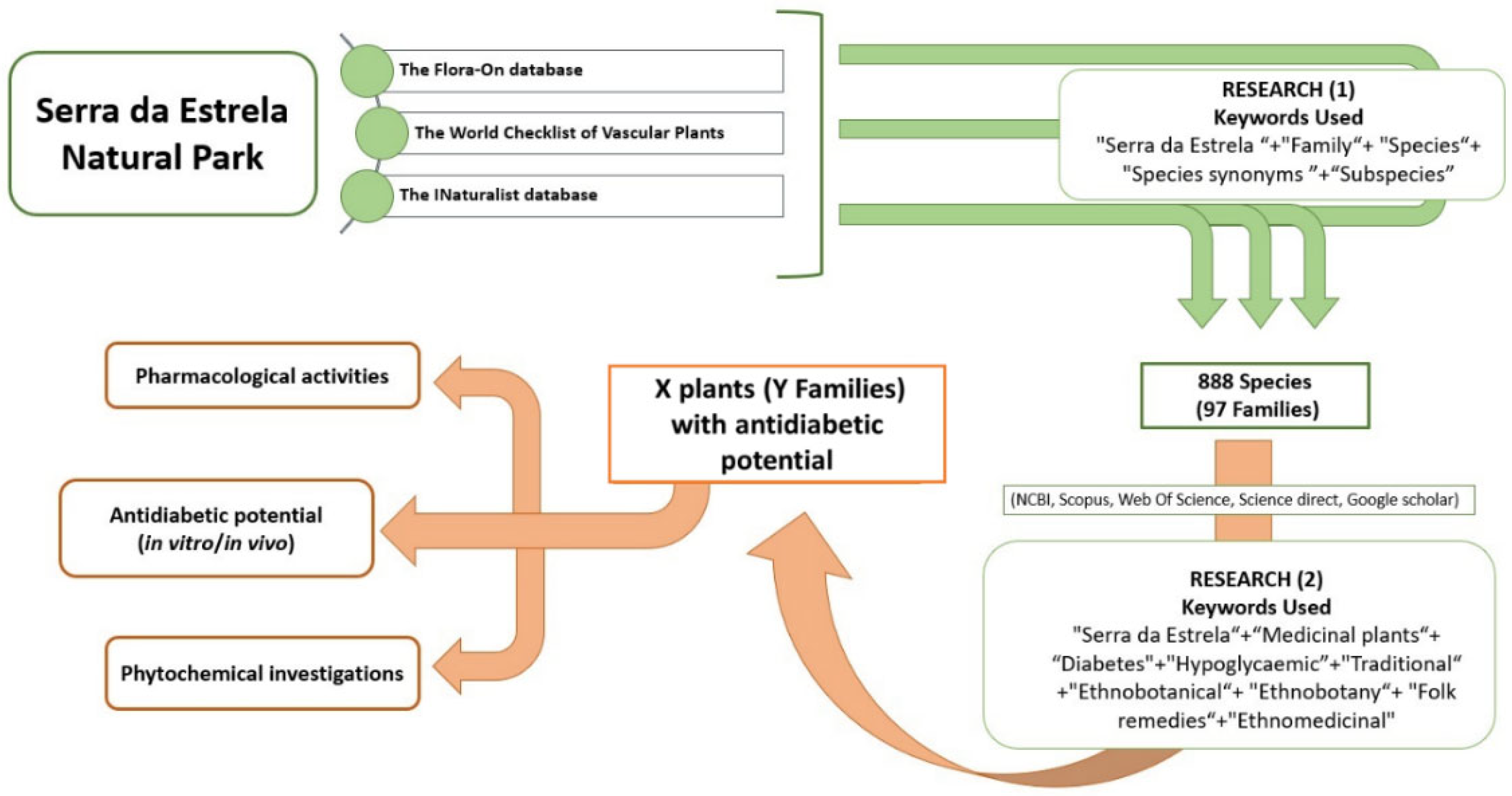
Figure 3.
Families containing the highest number of species with antidiabetic potential (out of 888) in NPSE.
Figure 3.
Families containing the highest number of species with antidiabetic potential (out of 888) in NPSE.
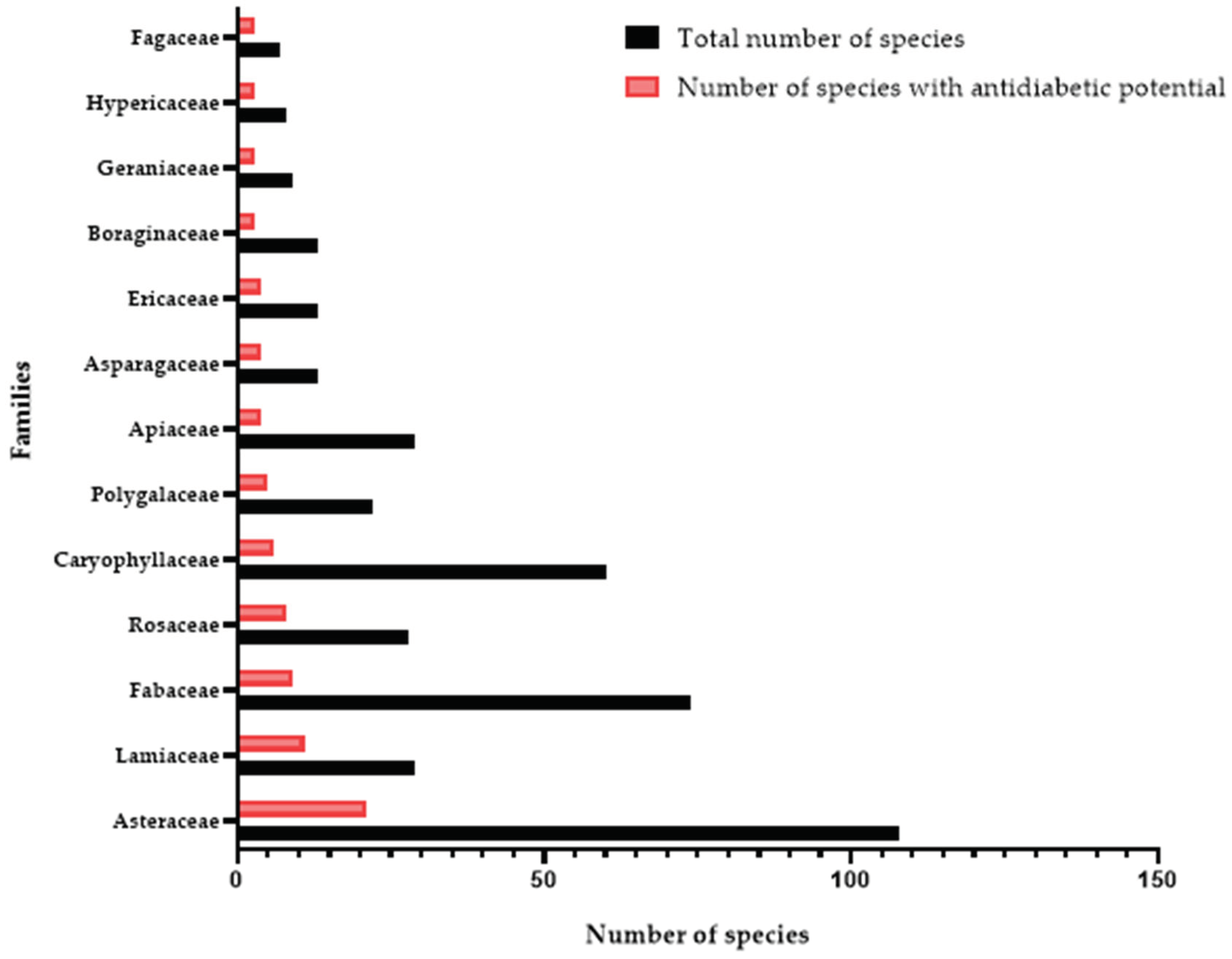
Disclaimer/Publisher’s Note: The statements, opinions and data contained in all publications are solely those of the individual author(s) and contributor(s) and not of MDPI and/or the editor(s). MDPI and/or the editor(s) disclaim responsibility for any injury to people or property resulting from any ideas, methods, instructions or products referred to in the content. |
© 2024 by the authors. Licensee MDPI, Basel, Switzerland. This article is an open access article distributed under the terms and conditions of the Creative Commons Attribution (CC BY) license (http://creativecommons.org/licenses/by/4.0/).
Copyright: This open access article is published under a Creative Commons CC BY 4.0 license, which permit the free download, distribution, and reuse, provided that the author and preprint are cited in any reuse.


