
Submitted:
18 February 2024
Posted:
19 February 2024
You are already at the latest version
Abstract
Introduction: Wound healing is a complex cascade involving the orchestration of inflammatory cells, skin fibroblasts, keratinocytes, and endothelial cells. These cells achieve tissue repair through an interchange of cytokines and growth factors through a controlled inflammation, angiogenesis, and remodeling process. Disruptions in the wound-healing process by comorbid conditions lead to significant morbidity and mortality. Stem cell therapy has emerged as a new strategy to facilitate the wound healing process. Stem cells harvested from different sources can be used for wound repair and regeneration (endothelial progenitor cells, bone marrow, adipose tissue, dermal and inducible pluripotent stem cells). We aimed to review clinical, translational, and primary literature on stem cell therapies in chronic wounds and summarize practical clinical applications to dermatological care. Materials and Methods: A comprehensive search of Pubmed, Embase, Web of Science, Google Scholar, and Cochrane Library was conducted for studies relating to stem cells and wound healing. Duplicate results were removed using Covidence. Titles and abstracts were screened by two independent researchers (BF and KR), with discrepancies resolved through discussion and mutual agreement. Articles were excluded if they were review-type or commentary-type articles. Results: A total of 22,454 articles were retrieved from the search. Deduplication removed 905, and automation removed an additional 18,363 articles. After an initial screening of titles and abstracts, 170 articles remained. Full-text screening resulted in an additional 126 articles being removed, leaving 44 included studies. Discussion: Stem cells used for wound repair and regeneration include endothelial progenitor stem cells and adult stem cells, in the forms of bone marrow-derived mesenchymal stem cells, adipose tissue stem cells, and inducible pluripotent stem cells. Among these sources, adipose tissue-derived mesenchymal stem cells (AD-MSCs) are ideally used due to the abundant supply of fat tissue, ease of isolation, extensive proliferative capacities ex vivo, and their ability to secrete pro-angiogenic factors. AD-MSCs have been used to enhance wound healing in peripheral arterial disease, diabetic wounds, hypertensive ulcers, bullous diabeticorum, venous ulcers, and postsurgical wounds after Mohs micrographic surgery. These cells have been delivered to the tissue topically, with scaffolds, combined with plasma-rich proteins, and through atelocollagen in various human studies. These approaches, when combined with local wound care practices, resulted in decreased pain, shorter wound healing times, and better cosmesis. Conclusions: Stem cell transplantation is a potential therapeutic approach in the wound healing process. Transplanted stem cells do not only differentiate into multiple skin cell types but also provide cytokine and growth factors required for wound healing resulting in increased angiogenesis. Thus, this approach may be regarded as an attractive option for intractable wounds that cause major clinical problems, especially chronic lower leg wounds. It can also be used after Mohs micrographic surgery for defects left to heal by secondary intention. Stem cell application may reduce the overall cost burden on the healthcare system and improve the quality of life for patients.
Keywords:
stem cells
; chronic wound
; wound healing
; dermatology
1. Introduction
The stem cells have been used to modulate the progression of neurodegenerative diseases, ischemic damage in the tissues (peripheral artery disease, chronic diabetic wounds, venous ulcers, etc.), as well as skin rejuvenation (1-3). Recent studies have also shown the secreted factors from stem cells (or cell-free extracts from stem cells) enhance various disorders through paracrine function in the tissues (4-6).
In the last decade, a significant rise in interest in chronic wound healing occurred due to the increasing incidence of chronic ulcers, an aging population, an increasing incidence of diabetes and healthcare inequalities. Non-healing ulcers significantly reduce the patient’s quality of life and increase healthcare costs due to recurrent admissions due to chronically infected wounds, increased risk of osteomyelitis and prolonged intravenous antibiotic treatments, amputation, and eventually the need for supportive care measures for these individuals (7,8).
In contemporary healthcare, the alarming statistic that 70% of amputations stem from unhealed wounds underscores the urgency to address this pervasive issue. With an estimated 6 million individuals in the United States alone grappling with the consequences of non-healing wounds, the associated healthcare expenditures have surged to a staggering $25 billion. In the light of these compelling figures, there is a critical need to explore innovative research avenues to mitigate the human suffering and economic burden imposed by chronic wounds. It is within this context that the exploration of stem cell therapy emerges as a promising and potentially transformative approach. Researching into the unique regenerative capabilities of stem cells, further research in this domain has the potential to revolutionize wound care and contribute substantially to ameliorating the impact of non-healing wounds on both individuals and the healthcare system (8-11).
Stem cells harvested from different sources can be used for wound repair and regeneration such as endothelial progenitor cells (EPC), adult stem cells in the forms of bone marrow-derived mesenchymal stem cells (BM-MSC), adipose tissue stem cells (ASC), dermal stem cells (DSC) and inducible pluripotent stem cells (iPS). These stem cells enhance wound healing by tissue regeneration through paracrine signaling and growth factor release resulting in fibroblast proliferation and tissue remodeling (2).
Wound healing is a complex cascade involving the interaction of inflammatory cells, skin fibroblasts, keratinocytes, and endothelial cells in injured tissue. These cells contribute to wound healing by releasing various chemo-cytokines, growth factors promoting cell migration to the injured area and stimulate inflammation, angiogenesis, wound contraction, and remodeling resulting in a healthy wound healing process. The first phase of wound healing starts with inflammation phase which starts within 6-8 hours after injury. During this phase platelets migrate to the tissue and release chemo attractive cytokines, next macrophages arrive and phagocyte/debride the tissue/organisms and set stage for proliferative phase. The proliferative phase starts around 5-7 days and initiated by cytokines released from macrophages (PDGF, TGF-α/β, FGF, etc.) In this stage, formation of granulation tissue occurs with fibroblast proliferation and extracellular matrix deposition. During this phase, angiogenesis occurs which allows leukocyte migration and provides nutrients and oxygen to develop granulation tissue. The final stage is tissue remodeling, in which wound contraction and extracellular matrix reorganization occurs over several months to years transitioning into mature scar formation. Overall, an efficient wound healing process results from a sufficient supply of growth factors, nutrients, cell-cell interaction, and adequate oxygenation to the tissue. Disruptions in these mechanisms, caused by conditions such as infection, malnutrition, chronic disease, or diabetes, can lead to delayed wound healing and chronic wound formation. Despite addressing systemic factors (controlling blood glucose levels, optimizing oxygenation to the tissue), providing local wound care, chronic wound care has only moderate success and treatment options are limited (12).
This systematic review aims to explore the use of stem cells in the treatment of chronic wounds as an ancillary method and investigate the current evidence in clinical utilization of stem cells and stem cell products from various resources.
2. Materials and Methods
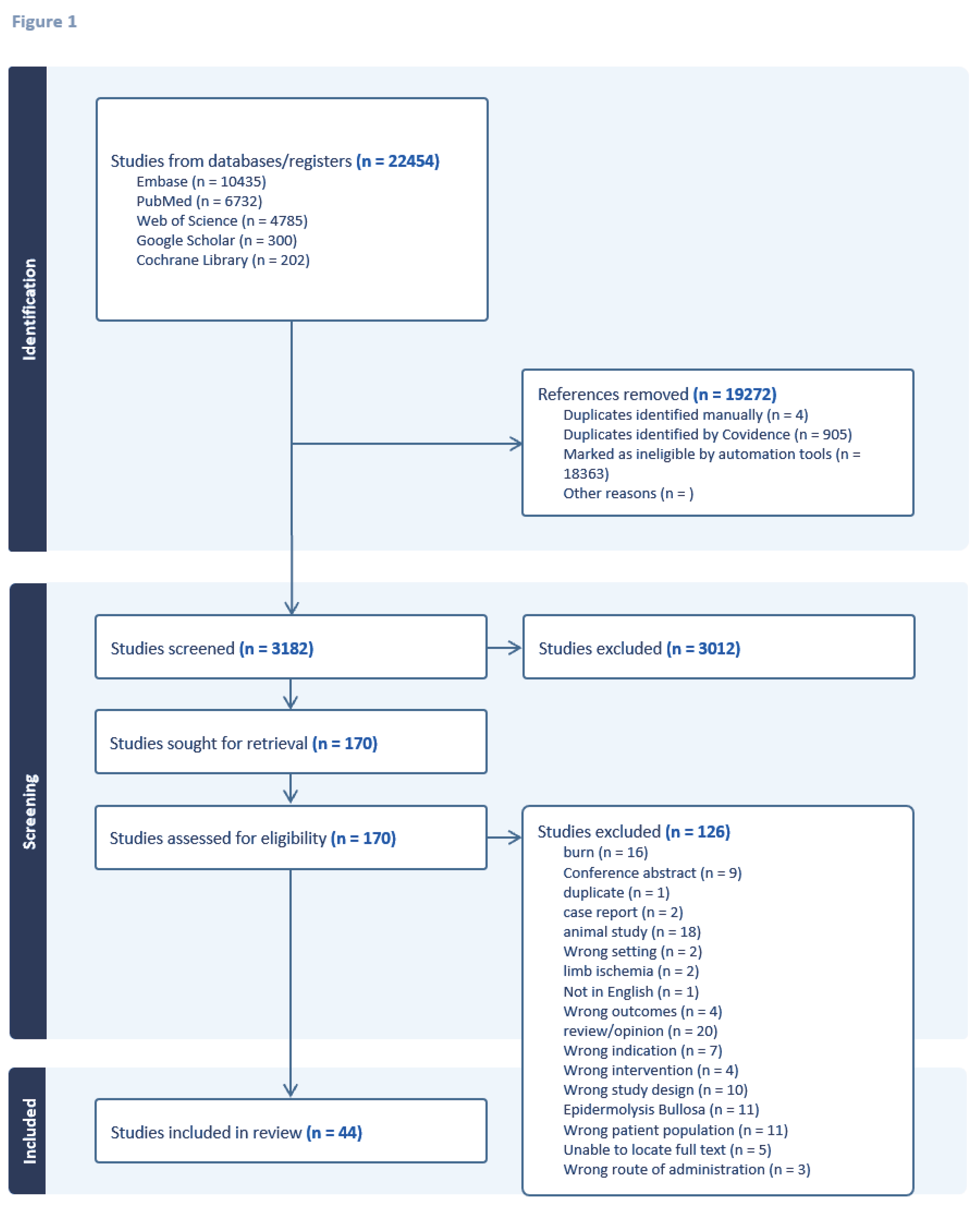
This review follows the PRISMA-S 2021 reporting standards for systematic reviews. KT and BF developed the search strategy. KT executed the searches (Figure 1) (13).
2.1. Information Sources
A comprehensive search of the literature for primary sources was undertaken in the following sources: Medline and PubMed Central (PubMed), Embase (Elsevier), Web of Science (Clarivate), Cochrane Library, and Google Scholar.
2.2. Search Strategy
Each source was searched independently. The search strategy was developed using keywords and subject headings, where available, related to the concepts of stem cells AND wound healing AND humans. Complete search strings for each database used are included in Appendix A.
No limits were applied to the searches. The search filter, NOT ("Animals"[Mesh] NOT ("Animals"[Mesh] AND "Humans"[Mesh]), or an equivalent specific to each database, was applied to the searches to reduce the number of animal studies in the results.
The search strategy for each source was peer-reviewed by two additional academic medical librarians.
The final search was run, and references were exported on December 18, 2023. Appendix A includes the exact date and number of results for each source searched.
2.3. Eligibility Criteria
We established specific criteria for study inclusion in this systematic review. Studies of any design that reported on stem cell therapy and chronic wounds were considered eligible. Clinical trials, including randomized clinical trials, involving human subjects were included. Animal studies or studies that did not focus on human patients were excluded.
References were uploaded into Covidence for deduplication and screening. Two independent reviewers (BF and KR) screened the abstracts of the identified studies and reviewed the full texts of those studies that were deemed potentially eligible. Disagreements between the reviewers were resolved through consensus discussions.
2.4. Data Collection and Extraction Process
Data related to the application of stem cell therapy in the context of chronic wound healing was systematically extracted from each included study. The following information was recorded, if reported: study type, year of publication, treatment groups, number of participants and the results of the study. All extracted data was documented in a Microsoft Excel spreadsheet for further analysis (Appendix B). Meta-analysis was not conducted due to the substantial heterogeneity observed among the included studies.
3. Results
Figure 1 shows the search results as a flow diagram. It shows the initial 22,454 results retrieved from five sources were narrowed via deduplication and automation to 3,186 articles for title and abstract screening. Upon applying the exclusion and inclusion criteria, 3,012 articles were excluded as irrelevant, leaving 170 articles for full-text screening. After reviewing the full-text articles, 126 articles were excluded, leaving 44 included studies. Excluded articles include; burn scars, conference abstracts, duplicate papers, case reports, animal studies, studies with wrong setting (such as stem cell application in scar revision, chronic radiation dermatitis, tissue augmentation), wrong intervention (such as platelet rich plasma application to the chronic wounds), wrong outcomes (outcomes scar revisions), wrong patient population (patients with scars, patients underwent subcision, patients with alopecia), wrong intervention (stem cell application to prevent critical limb ischemia), wrong route of administration (application of fat tissue without any intervention), epidermolysis bullosa, and papers that we were not able to locate their full text articles.
3.1. Skin-Derived Stem Cells
3.1.1. Autologous keratinocytes
In an initial study which demonstrated the effectiveness of keratinocytes and fibroblasts in wound healing, 12 diabetic ulcers were treated with keratinocyte epithelium and fibroblast-gelatin sponge weekly and all but one ulcer completely healed (14). This led to a multicenter study spanning 24 U.S. centers, 208 patients were randomly assigned to receive Graftskin (human fibroblasts and human epithelium) every 4 weeks (112 patients) or saline-moistened gauze (control group, 96 patients). At the 12-week follow-up, 56% of Graftskin-treated patients achieved complete wound healing compared to 38% in the control group (P = 0.0042). The median time to complete closure was significantly lower for Graftskin (65 days) than the control group (90 days) (P = 0.0026). Adverse reactions were similar, except for fewer cases of osteomyelitis and lower-limb amputations in the Graftskin group (15). Similarly, in a randomized, controlled, multicenter study involving 314 patients, Dermagraft (human fibroblast–derived dermal substitute) was compared to control. By week 12, 30% of Dermagraft patients achieved complete wound closure compared to 18.3% in the control group (P = 0.023) (16).
A study with 40 participants having grade II and III ulcers showed that the treatment group, receiving allogenic keratinocytes on polyethylene and silica microcarriers every 3 days, had a remarkable 92% reduction in wound area after 30 days, compared to 32% in the control group (p<0.001). The treatment group also required fewer dressings for complete healing (9.2+3.2) compared to the control group (16.5+2.3) (p<0.001) (17).
In randomized, single-blinded study by Moustafa et al. (2007), autologous keratinocytes on transfer discs were compared to a cell-free placebo for healing diabetic neuropathic ulcers. Although 18 of the 21 initially resistant ulcers responded positively to autologous keratinocyte applications, the response at 6 weeks did not achieve statistical significance (18).
In a larger multicenter trial by Ukat et al. (2007), a commercially available combination of autologous keratinocytes with fibrin sealant was compared to standard care for healing recalcitrant venous leg ulcers. After 3 months, 38.3% of patients who received autologous keratinocyte treatment achieved complete healing, in contrast to 22.4% of patients who received standard treatment (chi-square test: p=0.0106) (19).
Han et al. (2009) demonstrated that primary fresh dermal fibroblasts embedded in fibrin resulted in complete healing in 83.8% of cases at 8 weeks (20). Similarly, You et al. (2012), found that a primary foreskin keratinocyte sheet on vaseline gauze resulted in complete healing in 100% of treated wounds (21). Marchesi et al. (2014) reported a 70% reduction in wound area at the end of 70 days in 11 patients using primary keratinocytes in a hyaluronic acid scaffold (22). Hwang et al (2019) treated ulcers in 71 patients with primary foreskin keratinocyte for 12 weeks, resulting in 64.7% of patients had complete healing within an average of 6.1 weeks (23).
3.1.2. Hair Follicle Derived Stem cells
Hair follicle outer root sheath (ORS) is a putative source of stem cells with regeneration capacity. ORS contains several multipotent stem cells, especially on the distal compartment of the bulge region. These stem cells can give rise to neuroectodermal and mesenchymal stem cell populations which makes them an easily accessible source for stem cell niche (24). Renner et al. reported using tissue engineered autologous epidermal sheets derived from ORS cells of patient’s hair in chronic wound healing. In their study, the sheets were placed in the wound bed and they found that complete wound closure was significantly higher in the intervention group, especially that patients with small ulcer area (< 25cm2) (25).
3.2. Peripheral Blood-Derived Cells/ Endothelial progenitor stem cells
EPC are endothelial precursor cells involved in revascularization of injured tissue and tissue repair and have been extensively studied in ischemic heart diseases, stroke and peripheral arterial disease (26). Suh et al. reported that intradermal injection of EPC stimulates production of various cytokines that accelerate the wound healing process in murine dermal excisional wound model. When wounds were analyzed immunohistochemically, it was shown that EPC stimulated wounds exhibited significantly increased monocytes/macrophages at the 5th day of injury and promoted neovascularization (27). Di Santo et al. showed in vitro, EPC conditioned medium (EPC-CM) significantly inhibited apoptosis of mature endothelial cells and promoted angiogenesis after 72 hours of hypoxia in a rat aortic ring assay In the same study, the authors demonstrated that the regenerative potential of EPC-CM was equivalent to EPC transplantation in hindlimb ischemia by leading substantial increase in blood flow, enhancing neovascularization, vascular maturation and muscle function in rat models (28).
In 2020, a randomized double-blind clinical trial was conducted to assess the efficacy of using umbilical cord blood-derived platelet gel for treating diabetic foot ulcers. The study included 244 patients randomly assigned to Intervention (PRP gel), Placebo (platelet-poor plasma gel), or Control (lubricant gel) groups. No significant differences were observed among the three groups in terms of wound recovery and tissue regeneration. The platelet gel group exhibited similar efficacy to both the placebo (platelet-poor plasma) and the control group. The authors concluded that while growth factors in platelet granules may aid in the wound healing process, achieving a statistically better clinical outcome may require consideration of other factors. They also suggested that diabetic patients might not respond sufficiently, potentially due to underlying pathologic or genetic conditions (29).
In 2022 Tanaka et al., treated nine patients treated with MNC-QQ therapy. At the end of 12 weeks, six out of ten treated ulcers achieved complete wound closure with an average closure rate of 73.2% + 40.1%. Although there was no control group for comparison, the authors concluded that the treatment increased vascular perfusion, skin perfusion pressure, and decreased pain intensity in all patients. Notably, several adverse events were observed, including cellulitis at an injection site (N=1), restenosis (N=4), chronic subdural hematoma (N =1), bedsore (N=1), heterotopic ulcer (N=1), urinary tract infection (N=1), arthralgia (N=1), dyspnea (N=1), hypoperfusion (N=1), labial herpes simplex (N=1), diarrhea (N=1), patellofemoral joint pain (N=1), fever due to respiratory infection (N=1), ALP elevation (hepatic-cystic system failure) (N=1), and CRP elevation (N=1) (30).
In 2023, Johnson et al. tested the use of platelet extracellular vesicles (pEVs) from activated platelets for treating healing biopsy sites. The investigators created two biopsy wounds on the upper arms of 11 individuals. One arm received a single subcutaneous injection of clinical pEVs (100 μg in 340 μL), while the other arm underwent standard wound care. Notably, there was no discernible distinction in the overall duration for wound closure between the treated and untreated arms. Both groups exhibited a mean healing period of 22.8 ± 8.7 days, and all wounds had fully healed within a 30-day timeframe (31).
3.3. Bone marrow-derived mesenchymal stem cells (BM-MSC)
Bone marrow-derived stem cells, encompassing both Hematopoietic Stem Cells (HSCs) responsible for blood cell formation and Mesenchymal Stem Cells (MSCs) with the capacity to differentiate into various cell types, including bone, cartilage, and fat cells, originate from the spongy tissue within bone cavities. While MSCs are not exclusive to bone marrow and are also found in tissues like adipose tissue, umbilical cord tissue, and synovial fluid, the bone marrow, particularly within long bones like the femur and tibia, serves as a valuable reservoir for these stem cells, making them instrumental for tissue repair across the body. Extraction of autologous bone marrow-derived cells involves a minimally invasive procedure known as bone marrow aspiration, with a specialized needle accessing the posterior iliac crest under local anesthesia. This aspirate, comprising a mix of hematopoietic and mesenchymal stem cells, holds promise for regenerative medicine. The unique regenerative properties of bone marrow-derived stem cells are crucial to optimizing their selection for the development of efficient wound healing therapies.
Distinctive characteristics set bone marrow-derived stem cells apart from those derived from other tissues, especially in the realm of wound healing therapy. Bone marrow serves as a rich source of diverse stem cells, including hematopoietic stem cells (HSCs) and mesenchymal stem cells (MSCs), each playing a unique role in blood cell formation and tissue regeneration. The advantageous attributes of bone marrow-derived stem cells lie in their innate ability to modulate the immune response, stimulate angiogenesis, and promote tissue repair, making them particularly well-suited for wound healing applications. While stem cells can be sourced from alternative sites such as adipose tissue or umbilical cord, the unique regenerative properties and versatile differentiation potential of bone marrow-derived stem cells position them as pivotal players in the development of effective therapeutic strategies for wound healing. Understanding these distinctions is essential for optimizing the selection of stem cell sources in the pursuit of targeted and efficient wound-healing therapies.
Bone marrow-derived mesenchymal stem cells (BM-MSC) have emerged as a promising candidate for enhancing wound healing. These cells possess the ability to differentiate into multiple lineages, including cartilage, muscle, connective tissue, and adipose cells. Recent studies have demonstrated their capability to differentiate into various skin cell types, contributing significantly to wound repair. Additionally, research has shown that the application of BM-MSC, whether through injection or topical occlusive dressing, accelerates wound healing by releasing proangiogenic factors and differentiating them into critical cell types. The therapeutic efficacy of BM-MSC has been confirmed in human patients with chronic leg ulcers, demonstrating reduced wound size, increased vascularity, and dermal thickness. Notably, BM-MSC application has shown significant reductions in wound area as early as 2 weeks after application in chronic lower extremity wound patients. These findings highlight the potential of BM-MSC as a valuable and effective approach to advancing wound healing therapies (8, 9, 12, 32).
In a case-control study involving 75 patients with chronic wounds, 50 were treated with autologous bone marrow (BM) aspirate, either fresh or cultured, while 25 received daily saline dressings. Notably, both fresh and cultured BM aspirate, even without specific identification, isolation, and selective application of stem cells, led to a significant reduction in wound surface area compared to the control group at day 7 and week 4 (11). Another study by Bonora et al. focused on patients with diabetes and ischemic wounds. They were randomized to receive a single subcutaneous injection of plerixafor or saline in addition to standard medical and surgical therapy. The trial was terminated after an interim analysis of 50% of the target population revealed a significantly lower healing rate in the plerixafor group compared to the placebo group. In the final analysis, the plerixafor group exhibited a healing rate of 38.5%, while the placebo group showed a higher rate of 69.2% (33).
Badiavas et al. aimed to establish proof of principle that bone marrow-derived cells applied to chronic wounds could lead to closure. Autologous bone marrow cells were applied to chronic wounds in three patients with wounds lasting over a year. The results demonstrated complete closure, dermal rebuilding, reduced scarring, and successful engraftment (34).
Vojtassak et al. tested a new technique for treating chronic non-healing wounds, specifically diabetic ulcers. They utilized an autologous biograft composed of autologous skin fibroblasts on a biodegradable collagen membrane (Coladerm) combined with autologous mesenchymal stem cells (MSC) derived from the patient’s bone marrow. The combined treatment resulted in a steady overall decrease in wound size, increased vascularity of the dermis, and an augmented dermal thickness of the wound bed after 29 days (35).
Rogers et al. presented results from a case series study involving three cases where bone marrow aspirate containing marrow-derived cells was applied or injected locally into complex lower extremity chronic wounds of varying etiologies. The findings suggest that topically applied and injected bone marrow aspirate may be a useful adjunct to wound simplification and ultimate closure, as evidenced by reductions in chronic wound size (36).
In a prospective randomized clinical study, Jain et al. compared the healing rates of chronic lower limb wounds in diabetic patients treated with topically applied and locally injected bone marrow-derived cells versus whole blood (control). The study demonstrated a significant decrease in wound area by 17.4% compared to 4.84% in the control group at 2 weeks, with an average decrease of 36.4% versus 27.24% (37).
Andersen et al. conducted a study aimed at examining the safety of using a mesenchymal stem cell product for the treatment of diabetic foot ulcers. The study involved participants who received a singular application of an allogeneic cellular product topically, which comprised mesenchymal stem cells enriched with CD362 and suspended in collagen solution. Although two individuals experienced increased exudation, it was resolved within a week of application with no further complications noted (38).
Additionaly, Falanga et al. reported a successful clinical trial involving the topical application of autologous mesenchymal stem cells (MSC) to accelerate the healing of both human and experimental murine wounds. The cultured autologous MSCs were applied using a fibrin polymer spray system, showing a strong correlation between the number of cells applied and the subsequent decrease in chronic wound size. This approach also stimulated the closure of full-thickness wounds in diabetic mice. In this particular study, there were 4 human subjects, who underwent Mohs micrographic surgery (MMS), and the wounds were not suitable for primary closure. These patients underwent bone marrow aspiration 2 weeks prior to the procedure to allow proper in vitro establishment if MSC cultures by the day of surgery. These cells were applied immediately after removal of the skin cancer with MMS, as well as later within 12 weeks of wound closure led to complete closure to the area with persistent wound closure (39).
Another indication of the use of BM-MSCs has been reported as chronic graft versus host disease (GvHD). Zhou et al. reported regression of symptoms (pain, ulceration of the skin, pliability) after intravenous application of autologous MSCs to chronic GvHD patients without recurrence of leukemia (40). Supporting the latter study, Boberg et al. reported successful use of intravenous allogeneic MSC infusion for treatment refractory chronic GvHD in 11 patients with durable responses over 12 months (41).
3.3.1. Bone marrow-derived mononuclear stem cells
Bone marrow derived mononuclear stem cells (BMMCs) are a heterogenous group of cells that include mature B and T cells, monocytes, endothelial progenitor cells and embryonic-like cells, and positive for CD 133, CD 117, and CD 34. Due to their abundance in both peripheral blood and bone marrow, BMMCs, they do not need in vitro expansion, therefore a feasible source of stem cells (42). They have been clinically applied for chronic ulcers and their most notable property is the ability to secrete angiogenic factors. Jain et al. reported significant decrease in wound area in diabetic ulcers after application of BMMCs via spraying the wound bed after debridement compared to control group (34). Yamaguchi et al. reported rapid healing of intractable diabetic foot ulcers with exposed bones after treatment and grafting epidermal sheets. In this interesting study, authors debrided the wound with a scalpel, followed by partial excision with a bone scraper to expose BMMC. Later, the area was covered with epidermal graft of skin harvested from suction blisters. This combination led to healing of diabetic ulcers without occurrence of osteomyelitis or necessity of amputation (43).
In a pilot study by Wettstein et al., autologous hematopoietic CD34+ selected stem cell suspension was injected into sacral pressure III-IV stage ulcers in three complete para- or tetraplegic patients and monitored for three weeks. 3D laser scanning was used to assess wound volume and circumference and showed about 50% reduction in volume versus 40% in the control side of the wound. In addition, this study did not find signs of malignant transformation at 2 years after cell application and re-debridement prior to flap coverage of wound, a possible concern as the mechanism of healing skin wounds can be similar to that of malignancy (44).
Similarly, another study evaluated the use of bone marrow mononuclear cells (BM-MNCs) in patients with spinal cord injury and stage IV pressure ulcers. MNCs were isolated from autologous bone marrow aspiration and injected into the pressure ulcers after debridement and forming sutured-closed pouches. 19 out of 22 patients treated achieved complete closure of the wound in a mean time of 21 days (45).
3.4. Adipose tissue-derived mesenchymal stem cells
Adipose tissue-derived stem cells (ADSCs) are found in the stromal fraction of the adipose tissue. ADSCs were defined as CD45-negative, CD90, CD73 and CD105 positive cells (41). Unlike other stem cells, ADSCs can easily be collected without any ethical problems and differentiate into different cell lines including adipogenic, osteogenic, chondrogenic and myogenic cells. Thus, they are studied extensively as one of the leading sources in stem cell therapy for regenerative medicine (46, 47).
ADSCs can easily adhere to plastic culture flasks, expand in vitro and they have capacity to differentiate into different cell lines. They have been reported to be effective in wound healing, acute graft versus host disease, hematologic and immunologic disorders via their immunomodulatory properties (48). In contrast to intrusive methods required for harvesting BM-MSCs, adipose tissue is plentiful and can be easily obtained through liposuction, resulting in a less invasive process. Although flow cytometry is the conventional method for isolating AD-MSCs, autologous fat grafting can also be a valuable and practical alternative (49).
Impact of ADSCs on neovascularization in ischemic tissue in animal models have been shown and these cells can release many potent angiogenic factors as well as they are capable of to differentiate into endothelial cells, thus increasing tissue vascularization (15). ADSCs secrete TGF-β, vascular endothelial growth factor (VEGF), keratinocyte growth factor (KGF), fibroblast growth factor 2 (FGF2), PDGF, HGF, fibronectin, and collagen I, which have been previously shown to stimulate wound healing process previously (16). Studies suggested that ADSCs can promote wound healing with paracrine activity with the latter mentioned chemokines (17-19). Skin wounds treated with ADSCs have shown to have enhanced healing rate and less scar formation. It was shown that human epidermal keratinocyte migration rate was increased when cocultured with ADSCs (20).
Exosomes or extracellular vesicles have been defined as “particles naturally released from the cell that are delineated by lipid bilayer that cannot replicate or do not contain a functional nucleus” (50). They can be secreted from various cell types and act as intercellular communications, delivering bioactive cargos, such as proteins, lipids, nucleic acids, miRNAs and growth factors. They regulate cell-to-cell communication and regulate metabolism and homeostasis. Increasing evidence shows that exosomes derived from ADSCs have anti-inflammatory properties by inducing polarization of macrophages to M2 type through STAT-3 pathway and reduce inflammation. Exosomes contain microRNSAs which reinforce the acceleration of wound healing (51). Additionally, ADSCs exosomes promote scarless wound healing by preventing fibroblasts from differentiating into myofibroblasts and resulting in better cosmesis. These exosomes can be delivered to the tissue with injections, by being loaded to alginate hydrogel or loaded to wound dressings (52).
It was shown that autologous ADSCs combined with atelocollagen accelerated wound healing in a diabetic chronic wound model on mice via increasing healing time, epithelization rate, granulation tissue and vascular formation compared to control group (53). Altman et al. reported enhanced wound healing when full-thickness wounds were sutured with ADSCs- seeded silk sutures in mice (54). Furthermore, Blanton et al. reported better cosmesis and vascularization on porcine skin when ADSC and platelet rich plasma applied together topically compared to only ADSCs or PRP applications in mice (55). ADSCs appear to enhance wound healing via differentiation to other cells lines and paracrine activity.
In a pilot study by Chopinaud et al. reported that fat grafting to hypertensive leg ulcers resulted in decreased wound healing time with better cosmesis when compared with a control ulcer in the same patient. The median wound closure rate was 73.2% and 93.1% at 3 months and 6 months of follow up. They also reported significant increase in granulation tissue and reduction in pain without any adverse events (56).
Moon et al. examined the potential of allogeneic ADSC sheets for diabetic wound treatment. 59 patients were randomized to either control group or ADSC sheet group. After 12 weeks of evaluation, they found that 82% of ADSC sheet-applied patients achieved a complete wound closure, whereas 53% of patients in control group achieved complete closure of their wounds. Parallel to later findings, the times to complete closure from the beginning of application were 28.5 and 63 days respectively in ADSC sheet applied group and control group. They argued that this technique is a safe and effective method to treat diabetic ulcers (57).
Han et al. used processed lipoaspirate (adipose tissue cells incubated with collagenase and centrifuged) to promote diabetic wound healing. In their study, they used processed lipoaspirate (PLA) to 26 patients with diabetes and after 8 weeks of treatment cell proliferation and collagen synthesis was higher than the control group (44% vs 28%). Additionally, the PLA applied group achieved 100% complete wound closure, whereas only 62% of the control group achieved complete closure of their wounds (58). Lee et al. similarly used autologous PLA from thromboangiitis obliterans (TAO) (n:12) and diabetes mellitus (DM) patients (n:3) with critical limb ischemia. In their study they also had a control group to assess the quantity of stromal vascular fraction (SVF) of adipose tissue in different groups. In colony forming unit assay, the SVF of TAO and DM patients yielded less colonies and lesser proliferative capacity than that of healthy controls. Multiple intramuscular injections of PLA were done with a mean follow up of 6 months resulted clinical improvement in 66.7% of the patients. Only five patients required minor amputations and all amputation sites healed completely. All diabetic patients’ wounds completely healed (100% wound closure rate in this subgroup) and pain significantly decreased in all patients. Digital subtraction angiography showed significant collateral vascular network in critically ischemic limbs by the end of 6 months (59). Maslowski et al. used autologous ADSCs for treatment of chronic venous stasis ulcers. The SVF was injected subcutaneously to the periphery of the ulcers and to the wound bed in addition to traditional local treatments and wound have been measured planimetrically for 6 months. Overall, improvement was observed in 75% of the patients and no serious side effects were observed. Eight of the patients were healed completely, 9 of the patients showed >50% reduction of the ulcer area and 5 patients showed no improvement (60). Marino et al. used autologous ADSCs to chronic arterial leg ulcers. They applied the SVF to the edges of the wound with a depth of 1 cm. All patients showed a reduction in diameter and depth of their ulcers and 6 out of 10 patients had a complete closure of their wounds (61).
Recently, an automatized processing system for isolating ADSC has been developed. The system simply standardizes the processing of human adipose tissue to harvest and concentrate in a real-time bedside manner. The system reliably and reproducibly generates ADSC from collected adipose tissues via liposuction and it allows application of the product in approximately in 1.5 hours. This system has been used in cosmetic field to rejuvenate the face, breast augmentation and reconstruction in small studies with success (62). Moreover, this system can simply be utilized for acute radiation dermatitis at bedside to prevent radiation related skin side effects in cancer patients (63).
In a randomized controlled trial, 32 individuals suffering from chronic plantar ulcers due to leprosy were enrolled. Following thorough clinical examination and initial debridement, participants were randomly assigned to two cohorts. One group (n = 16) received topical application of conditioned medium from adipose mesenchymal stem cells, while the other group (n = 16) was treated solely with framycetin gauze dressing applied every three days for a duration of eight weeks. Throughout this period, ulcer size, as well as any adverse reactions or complications, were monitored on a weekly basis. The findings revealed a consistent increase in the healing percentage across all groups each week. Notably, statistical distinctions between the two groups (P < 0.05) became apparent from the second week onward for the reduction in mean ulcer size and from the third week onward for the reduction in mean ulcer depth. Importantly, no adverse reactions or complications were reported throughout the study period (64).
Furthermore, Del Papa et al. reported ADSC fraction injection to the treatment resistant digital ulcers (DUs) in systemic sclerosis resulted in reduction in pain and analgesic consumption, faster healing rate of DUs, significant increase in the number of capillaries, and reduction in arterial resistance led to favorable outcomes in the intervention group making stem cell treatment as a promising method (65).
A new indication for use of SVF of adipose tissue has been reported as vulvar lichen sclerosus. Autologous fat grafting enriched with adipose tissue derived SVF cell injection to the vulvar skin resulted in a significant decrease in global scoring for burning, pain, dryness and distress associated with sexuality and resulted better cosmesis in the area (66).
3.5. Placental /Umbilical/Wharton jelly derived mesenchymal stem cells (WJSCs)
Human embryo fibroblast, Wharton jelly, umbilical cord, and human placenta-derived stem cells have shown significant potential in improving healing rates and reducing wound size.
3.5.1. Human Embryo Fibroblast
Sedov’s (2006) study focused on the efficacy of "Foliderm" in treating venous trophic ulcers. Patients either had postthrombophlebitic disease or varicose disease, and the treatment group was 23 patients and the control group had Control Group: 25 patients (67). The treatment group demonstrated a 100% healing rate, compared to 86% for varicose and 78% for post-thrombophlebitic ulcers in the control group. Additionally, the treatment group experienced significantly shorter average healing periods (67).
3.5.2. Wharton’s jelly derived stem cells
Between the tenth and twelfth weeks of embryogenesis, the extraembryonic mesoderm undergoes development, creating a protective extracellular structure for the umbilical cord known as Wharton’s jelly (68). Wharton’s jelly, a gel-like substance within the umbilical cord, is abundant in proteoglycans, particularly hyaluronic acid, and chondroitin sulfate. Mesenchymal stem cells present in Wharton’s jelly express c-KIT and exhibit telomerase activity, characteristics indicative of their role as a source of stem cells (59). The ease of extraction of these cells post-delivery makes them a convenient stem cell source, accompanied by fewer ethical considerations compared to alternative stem cell resources (69).
In a 2019 study conducted in Iran (Hashemi, 2019), the healing potential of Wharton’s jelly stem cells was evaluated in five patients with chronic diabetic wounds. There was no control, but the authors conclude that acellular amniotic membrane seeded with WJ-MSCs significantly reduced wound healing time and size on days 6 and 9 (70).
3.5.3. Human Umbilical Cord Mesenchymal Stem Cells
Zhang (2022) investigated the topical and intravenous administration of human umbilical cord mesenchymal stem cells (hUC-MSC) for diabetic foot ulcers (DFU) and peripheral arterial disease (PAD). The study involved 14 patients with PAD and incurable DFU. In this phase I pilot study, the safety and efficacy of hUC-MSC administration were assessed against conservative treatments. Topical and intravenous hUC-MSCs at 2 × 10⁵ cells/kg (up to 1×10⁷ cells per dose) were administered to all 14 patients. There was no control, but the authors report that the treatment resulted in ulcer closure and symptom alleviation, but no direct evidence of vessel obstruction improvement was observed. Rehospitalization for DFUs lasted 2.0 + 0.6 years, and all patients survived without amputation within three years post-treatment. Safety assessments recorded only two cases of transient fever (71).
Expanding on these findings, Tan’s (2023) study centered on a 10% secretome from human umbilical cord mesenchymal stem cells (SM-hUCMSC) in gel form. The study involved forty-one patients with chronic ulcers, including diabetic ulcers. The Patient’s ulcers demonstrated significant improvement in ulcer length, width, and area after the intervention. There was no control for comparison, but the change between the beginning and end of the intervention was statistically significant (p value < 0.05) (72).
Furthermore, the value of hUCMSC as a systemic infusion has shown to be a promising agent in Stevens-Johnson syndrome (SJS). According to the study by Li and colleagues, three patients with SJS with severe skin and mucosa lesions, unresponsive to systemic prednisolone and supportive therapies showed interruption on skin blistering as well as recovery of the blood chemical abnormalities within 12 days of hUCMSC infusion. They concluded the potential capacity of blocking the apoptosis in keratinocytes of hUCMSC. Therefore, hUCMSC infusion treatment might be a new salvage therapy for cutaneous severe drug eruptions to prevent further damage (73).
3.5.4. Human placenta/Human Amniotic Membrane
In 2017, Dehghani et al. conducted a randomized clinical trial to assess the efficacy of using Grafting with Cryopreserved Amniotic Membrane for the Treatment of Pressure Ulcers in 24 patients with second and third-stage pressure ulcers who required split-thickness skin grafts. Patients were assigned to the treatment group or the control group which received treatment with local Dilantin powder application. The amnion group demonstrated a significantly higher rate of complete healing (p<0.001) (74).
In a prospective study, Farivar’s (2019) study using cryopreserved placental tissue wound matrix achieved complete healing in 53% of refractory venous leg ulcers (VLUs) among 21 patients (75).
In 2020, Suzdaltseva conducted a trial on Locally Delivered Umbilical Cord Mesenchymal Stromal Cells (UCMSC) to Reduce Chronic Inflammation in Long-Term Nonhealing Wounds, involving 108 patients with chronic wounds of various etiologies. The treatment group (n = 59) received a single local subcutaneous infusion of UCMSCs around the wound periphery, while the placebo group (n = 49) received a placebo. Patients treated with UCMSCs exhibited notable growth of granulation tissue, improved blood microcirculation, and a reduction in wound size compared to the placebo group (76).
Expanding on these findings, Meamar et al 2021 conducted a clinical trial of twenty-eight patients with diabetic foot ulcers (DFUs) randomly assigned to three groups over a 12-week period. The two treatment groups involved the use of human placental-derived mesenchymal stem cells (hPDMSCs) without PRP gel (Group A) and with PRP gel (Group B), while the third group received standard wound care as the Control group (Group C). Wound size reduction was 66% in Group A, 71% in Group B, and 36% in the Control group (Group C). Significant differences in wound closure and pain-free walking distance were observed in Groups A and B compared to the Control group (Group C) (p < 0.05). There was no significant difference between Groups A and B (77).
Similarly, Rezaei-Nejad et al (2023) tested the efficacy of using Freeze-Dried Human Amniotic Membrane Allograft Gel (LAMG) in a clinical trial among 18 patients with chronic diabetic foot ulcers (DFUs) included in the study. The patients were randomly assigned to two groups: the LAMG group (n = 9) and the Placebo group (n = 9). After 9 weeks, the study revealed that Freeze-Dried Human Amniotic Membrane Allograft led to a mean reduction of 73.4%, in contrast to the control group’s mean reduction of 13.1% + 10.1% (78).
3.6. Inducible pluripotent stem cells
Inducible pluripotent stem cells (iPSCs) are pluripotent stem cells derived from somatic donor cells that were generated via overexpression of Oct4, Klf4, Sox2, and c-myc transcription factors in adult somatic cells harvested from healthy objects. iPSCs have the capacity to differentiate into and repopulate all cell types found in the skin (9). Although there are some concerns regarding their safety and regenerative capacity, iPCs have been investigated in clinical trials of disease modeling including cardiomyopathy, autism spectrum disorder, coronary artery disease, and cystic fibrosis (79). Human-induced pluripotent fibroblasts, human-induced pluripotent mesenchymal stem cells, and human-induced pluripotent stem-cell derived vesicles have a potential to accelerate wound healing (80). Recent two studies from Sebastiano et al. and Umegaki-Arao et al. reported successful use of human keratinocyte-derived iPSCs to reconstitute skin in vitro for recessive dystrophic epidermolysis bullosa (81, 82). Clayton et al. showed that injection of iPSC-derived endothelial cells promoted angiogenesis and accelerated the wound closure in murine excisional wound model (82). These findings can enable iPSCs-based generation of cutaneous substitutes that include epidermal appendages and may be attracted as a therapeutic option in wound healing. However, there are concerns regarding iPCs safety, since iPCs can differentiate into cells from any of the three germ layers, they carry a risk for teratoma formation. Different strategies, including using viral vectors to deliver induced pluripotent stem cell vesicles are under investigation to overcome this risk (80).
4. Discussion
Additional research is needed to understand the complex molecular mechanisms through which MSCs are involved in wound healing (83). In vivo, MSCs migrate towards injury sites when prompted by chemotactic signals that modulate inflammation. The differentiation of MSCs aids in the regeneration of damaged tissue and their paracrine signaling regulates local cellular responses to injury (84). MSC paracrine signaling is thought to be one of the primary mechanisms underlying the beneficial effects of MSCs in wound healing wounds, leading to inflammation reduction, promotion of angiogenesis, and stimulation of cell migration and proliferation. Moreover, the migration of mesenchymal cells, as well as the interplay between pro and anti-inflammatory cytokines plays an important role in regulating wound repair processes (85).
Numerous chemical and natural compounds have been identified for their potential to enhance the migratory capabilities of cells while maintaining their regenerative potential and differentiation capacity (40, 86). Chemical compounds like sphingosine-1-phosphate (S1P) and stromal cell-derived factor-1 (SDF-1) have shown promise in promoting cell migration by activating specific signaling pathways involved in cell movement, such as the PI3K/Akt and MAPK pathways (87). Additionally, natural compounds such as epigallocatechin-3-gallate (EGCG) found in green tea and curcumin derived from turmeric have demonstrated the ability to modulate cell migration through their anti-inflammatory and antioxidant properties without compromising stem cell identity (88).
To address the issue of fetal bovine serum (FBS) in stem cell transplantation, serum-free or defined media formulations supplemented with synthetic growth factors and nutrients can be used in stem cell transplantation (89). Similarly, exploring xenogeneic-free components and human-derived alternatives will help eliminate animal-origin products (90). Bioreactor culture systems are also being utilized for large-scale stem cell expansion in these serum-free or defined media, promoting standardized and reproducible culture conditions for therapeutic applications (89).
Conclusions
Stem cell transplantation is a potential therapeutic approach in wound healing process. Transplanted stem cells not only differentiate into multiple skin cell types, but also provide cytokine and growth factors required for wound healing resulting increased angiogenesis. Thus, this approach may be regarded as an attractive option in intractable wounds which cause major clinical problems, especially chronic lower leg wounds and decrease the overall cost effect on the system and improves patients’ quality of life.
Supplementary Materials
The following supporting information can be downloaded at the website of this paper posted on Preprints.org. Appendices A and B.
Conflicts of Interest
None declared.
References
- Kim, W.S.; Park, B.S.; Park, S.H.; Kim, H.K.; Sung, J.H. Antiwrinkle effect of adipose-derived stem cell: activation of dermal fibroblast by secretory factors. J Dermatol Sci. 2009, 53, 96–102. [Google Scholar] [CrossRef]
- Nakagami, H.; Maeda, K.; Morishita, R.; Iguchi, S.; Nishikawa, T.; Takami, Y.; et al. Novel autologous cell therapy in ischemic limb disease through growth factor secretion by cultured adipose tissue-derived stromal cells. Arterioscler Thromb Vasc Biol. 2005, 25, 2542–2547. [Google Scholar] [CrossRef] [PubMed]
- Kosaric, N.; Kiwanuka, H.; Gurtner, G.C. Stem cell therapies for wound healing. Expert Opin Biol Ther. 2019, 19, 575–585. [Google Scholar] [CrossRef]
- Loretelli, C.; Ben Nasr, M.; Giatsidis, G.; Bassi, R.; Lancerotto, L.; D’Addio, F.; et al. Embryonic stem cell extracts improve wound healing in diabetic mice. Acta Diabetol. 2020, 57, 883–890. [Google Scholar] [CrossRef] [PubMed]
- Huldani, H.; Kozlitina, I.A.; Alshahrani, M.; Daabo, H.M.A.; Almalki, S.G.; Oudaha, K.H.; et al. Exosomes derived from adipose stem cells in combination with hyaluronic acid promote diabetic wound healing. Tissue Cell. 2023, 85, 102252. [Google Scholar] [CrossRef] [PubMed]
- Na, Y.K.; Ban, J.J.; Lee, M.; Im, W.; Kim, M. Wound healing potential of adipose tissue stem cell extract. Biochem Biophys Res Commun. 2017, 485, 30–34. [Google Scholar] [CrossRef] [PubMed]
- Gould, L.; Abadir, P.; Brem, H.; Carter, M.; Conner-Kerr, T.; Davidson, J.; et al. Chronic wound repair and healing in older adults: current status and future research. J Am Geriatr Soc. 2015, 63, 427–438. [Google Scholar] [CrossRef]
- Sen, C.K.; Gordillo, G.M.; Roy, S.; Kirsner, R.; Lambert, L.; Hunt, T.K.; et al. Human skin wounds: a major and snowballing threat to public health and the economy. Wound Repair Regen. 2009, 17, 763–771. [Google Scholar] [CrossRef]
- Sen, C.K. Human Wound and Its Burden: Updated 2020 Compendium of Estimates. Adv Wound Care (New Rochelle). 2021, 10, 281–292. [Google Scholar] [CrossRef]
- Dash, B.C.; Korutla, L.; Vallabhajosyula, P.; Hsia, H.C. Unlocking the Potential of Induced Pluripotent Stem Cells for Wound Healing: The Next Frontier of Regenerative Medicine. Adv Wound Care (New Rochelle). 2022, 11, 622–638. [Google Scholar] [CrossRef]
- Gupta, G.J.; Karki, K.; Jain, P.; Saxena, A.K. Autologous Bone Marrow Aspirate Therapy for Skin Tissue Engineering and Tissue Regeneration. Adv Wound Care (New Rochelle). 2017, 6, 135–142. [Google Scholar] [CrossRef]
- Werdin, F.; Tenenhaus, M.; Rennekampff, H.O. Chronic wound care. Lancet 2008, 372, 1860–1862. [Google Scholar] [CrossRef]
- Page, M.J.; McKenzie, J.E.; Bossuyt, P.M.; Boutron, I.; Hoffmann, T.C.; Mulrow, C.D.; et al. The PRISMA 2020 statement: an updated guideline for reporting systematic reviews. Syst Rev. 2021, 10, 89. [Google Scholar] [CrossRef]
- Harvima, I.T.; Virnes, S.; Kauppinen, L.; Huttunen, M.; Kivinen, P.; Niskanen, L.; Horsmanheimo, M. Cultured allogeneic skin cells are effective in the treatment of chronic diabetic leg and foot ulcers. Acta Derm Venereol. 1999, 79, 217–220. [Google Scholar] [CrossRef]
- Veves, A.; Falanga, V.; Armstrong, D.G.; Sabolinski, M.L.; Study, t.A.D.F.U. Graftskin, a Human Skin Equivalent, Is Effective in the Management of Noninfected Neuropathic Diabetic Foot Ulcers: A prospective randomized multicenter clinical trial. Diabetes Care. 2001, 24, 290–295. [Google Scholar] [CrossRef]
- Marston, W.A.; Hanft, J.; Norwood, P.; Pollak, R. The efficacy and safety of Dermagraft in improving the healing of chronic diabetic foot ulcers: results of a prospective randomized trial. Diabetes Care. 2003, 26, 1701–1705. [Google Scholar] [CrossRef]
- Bayram, Y.; Deveci, M.; Imirzalioglu, N.; Soysal, Y.; Sengezer, M. The cell based dressing with living allogenic keratinocytes in the treatment of foot ulcers: a case study. Br J Plast Surg. 2005, 58, 988–996. [Google Scholar] [CrossRef] [PubMed]
- Moustafa, M.; Bullock, A.J.; Creagh, F.M.; Heller, S.; Jeffcoate, W.; Game, F.; et al. Randomized, controlled, single-blind study on use of autologous keratinocytes on a transfer dressing to treat nonhealing diabetic ulcers. Regen Med. 2007, 2, 887–902. [Google Scholar] [CrossRef] [PubMed]
- Vanscheidt, W.; Ukat, A.; Horak, V.; Brüning, H.; Hunyadi, J.; Pavlicek, R.; et al. Treatment of recalcitrant venous leg ulcers with autologous keratinocytes in fibrin sealant: a multinational randomized controlled clinical trial. Wound Repair Regen. 2007, 15, 308–315. [Google Scholar] [CrossRef] [PubMed]
- Han, S.K.; Kim, H.S.; Kim, W.K. Efficacy and safety of fresh fibroblast allografts in the treatment of diabetic foot ulcers. Dermatol Surg. 2009, 35, 1342–1348. [Google Scholar] [CrossRef] [PubMed]
- You, H.J.; Han, S.K.; Lee, J.W.; Chang, H. Treatment of diabetic foot ulcers using cultured allogeneic keratinocytes--a pilot study. Wound Repair Regen. 2012, 20, 491–499. [Google Scholar] [CrossRef] [PubMed]
- Andrea, M.; Marco, B.; Pier Camillo, P.; Matteo, M.; Roberto, B.; Luca, V. Allogeneic epidermal substitutes in the treatment of chronic diabetic leg and foot ulcers. Plastic and Aesthetic Research. 2014, 1, 74–80. [Google Scholar] [CrossRef]
- Hwang, Y.G.; Lee, J.W.; Park, K.H.; Han, S.H. Allogeneic keratinocyte for intractable chronic diabetic foot ulcers: A prospective observational study. Int Wound J. 2019, 16, 486–491. [Google Scholar] [CrossRef] [PubMed]
- Li, H.; Masieri, F.F.; Schneider, M.; Bartella, A.; Gaus, S.; Hahnel, S.; et al. The Middle Part of the Plucked Hair Follicle Outer Root Sheath Is Identified as an Area Rich in Lineage-Specific Stem Cell Markers. Biomolecules. 2021, 11. [Google Scholar] [CrossRef] [PubMed]
- Renner, R.; Harth, W.; Simon, J.C. Transplantation of chronic wounds with epidermal sheets derived from autologous hair follicles--the Leipzig experience. Int Wound J. 2009, 6, 226–232. [Google Scholar] [CrossRef] [PubMed]
- Shantsila, E.; Watson, T.; Lip, G.Y. Endothelial progenitor cells in cardiovascular disorders. J Am Coll Cardiol. 2007, 49, 741–752. [Google Scholar] [CrossRef]
- Suh, W.; Kim, K.L.; Kim, J.M.; Shin, I.S.; Lee, Y.S.; Lee, J.Y.; et al. Transplantation of endothelial progenitor cells accelerates dermal wound healing with increased recruitment of monocytes/macrophages and neovascularization. Stem Cells. 2005, 23, 1571–1578. [Google Scholar] [CrossRef]
- Santo, S.D.; Seiler, S.; Andres, R.; Widmer, H.R. Endothelial Progenitor Cells Conditioned Medium Supports Number of GABAergic Neurons and Exerts Neuroprotection in Cultured Striatal Neuronal Progenitor Cells. Cell Transplant. 2019, 28, 367–378. [Google Scholar] [CrossRef]
- Hosseini, S.E.; Molavi, B.; Goodarzi, A.; Alizadeh, A.; Yousefzadeh, A.; Sodeifi, N.; Arab, L.; Aghdami, N. The efficacy of platelet gel derived from umbilical cord blood on diabetic foot ulcers: A double-blind randomized clinical trial. Wound Medicine. 2020, 28. [Google Scholar] [CrossRef]
- Tanaka, R.; Fujimura, S.; Kado, M.; Fukuta, T.; Arita, K.; Hirano-Ito, R.; et al. Phase I/IIa Feasibility Trial of Autologous Quality- and Quantity-Cultured Peripheral Blood Mononuclear Cell Therapy for Non-Healing Extremity Ulcers. Stem Cells Transl Med. 2022, 11, 146–158. [Google Scholar] [CrossRef] [PubMed]
- Johnson, J.; Law, S.Q.K.; Shojaee, M.; Hall, A.S.; Bhuiyan, S.; Lim, M.B.L.; et al. First-in-human clinical trial of allogeneic, platelet-derived extracellular vesicles as a potential therapeutic for delayed wound healing. Journal of Extracellular Vesicles. 2023, 12, 12332. [Google Scholar] [CrossRef]
- Abadeh, A.; Herman, S.M.; Abdalian, R. The prevalence of gastrointestinal symptoms and cobalamin deficiency in patients with chronic urticaria. Allergy Asthma Clin Immunol. 2023, 19, 14. [Google Scholar] [CrossRef]
- Bonora, B.M.; Cappellari, R.; Mazzucato, M.; Rigato, M.; Grasso, M.; Menegolo, M.; et al. Stem cell mobilization with plerixafor and healing of diabetic ischemic wounds: A phase IIa, randomized, double-blind, placebo-controlled trial. Stem Cells Transl Med. 2020, 9, 965–973. [Google Scholar] [CrossRef]
- Badiavas, E.V.; Falanga, V. Treatment of chronic wounds with bone marrow-derived cells. Arch Dermatol. 2003, 139, 510–516. [Google Scholar] [CrossRef]
- Vojtassák, J.; Danisovic, L.; Kubes, M.; Bakos, D.; Jarábek, L.; Ulicná, M.; Blasko, M. Autologous biograft and mesenchymal stem cells in treatment of the diabetic foot. Neuro Endocrinol Lett. 2006, 27 (Suppl 2), 134–137. [Google Scholar]
- Rogers, L.C.; Bevilacqua, N.J.; Armstrong, D.G. The use of marrow-derived stem cells to accelerate healing in chronic wounds. Int Wound J. 2008, 5, 20–25. [Google Scholar] [CrossRef]
- Jain, P.; Perakath, B.; Jesudason, M.R.; Nayak, S. The effect of autologous bone marrow-derived cells on healing chronic lower extremity wounds: results of a randomized controlled study. Ostomy Wound Manage. 2011, 57, 38–44. [Google Scholar] [PubMed]
- Askø Andersen, J.; Rasmussen, A.; Frimodt-Møller, M.; Engberg, S.; Steeneveld, E.; Kirketerp-Møller, K.; et al. Novel topical allogeneic bone-marrow-derived mesenchymal stem cell treatment of hard-to-heal diabetic foot ulcers: a proof of concept study. Stem Cell Res Ther. 2022, 13, 280. [Google Scholar] [CrossRef] [PubMed]
- Falanga, V.; Iwamoto, S.; Chartier, M.; Yufit, T.; Butmarc, J.; Kouttab, N.; et al. Autologous bone marrow-derived cultured mesenchymal stem cells delivered in a fibrin spray accelerate healing in murine and human cutaneous wounds. Tissue Eng. 2007, 13, 1299–1312. [Google Scholar] [CrossRef] [PubMed]
- Zhou, H.; Guo, M.; Bian, C.; Sun, Z.; Yang, Z.; Zeng, Y.; et al. Efficacy of bone marrow-derived mesenchymal stem cells in the treatment of sclerodermatous chronic graft-versus-host disease: clinical report. Biol Blood Marrow Transplant. 2010, 16, 403–412. [Google Scholar] [CrossRef] [PubMed]
- Boberg, E.; von Bahr, L.; Afram, G.; Lindström, C.; Ljungman, P.; Heldring, N.; et al. Treatment of chronic GvHD with mesenchymal stromal cells induces durable responses: A phase II study. STEM CELLS Translational Medicine. 2020, 9, 1190–1202. [Google Scholar] [CrossRef]
- Amato, B.; Compagna, R.; Amato, M.; Butrico, L.; Fugetto, F.; Chibireva, M.D.; et al. The role of adult tissue-derived stem cells in chronic leg ulcers: a systematic review focused on tissue regeneration medicine. International Wound Journal 2016, 13, 1289–1298. [Google Scholar] [CrossRef] [PubMed]
- Yamaguchi, Y.; Kubo, T.; Murakami, T.; Takahashi, M.; Hakamata, Y.; Kobayashi, E.; et al. Bone marrow cells differentiate into wound myofibroblasts and accelerate the healing of wounds with exposed bones when combined with an occlusive dressing. Br J Dermatol. 2005, 152, 616–622. [Google Scholar] [CrossRef] [PubMed]
- Wettstein, R.; Savic, M.; Pierer, G.; Scheufler, O.; Haug, M.; Halter, J.; et al. Progenitor cell therapy for sacral pressure sore: a pilot study with a novel human chronic wound model. Stem Cell Res Ther. 2014, 5, 18. [Google Scholar] [CrossRef] [PubMed]
- Sarasúa, J.G.; López, S.P.; Viejo, M.A.; Basterrechea, M.P.; Rodríguez, A.F.; Gutiérrez, A.F.; et al. Treatment of pressure ulcers with autologous bone marrow nuclear cells in patients with spinal cord injury. J Spinal Cord Med. 2011, 34, 301–307. [Google Scholar] [CrossRef] [PubMed]
- Dicker, A.; Le Blanc, K.; Aström, G.; van Harmelen, V.; Götherström, C.; Blomqvist, L.; et al. Functional studies of mesenchymal stem cells derived from adult human adipose tissue. Exp Cell Res. 2005, 308, 283–290. [Google Scholar] [CrossRef] [PubMed]
- Gimble, J.M.; Katz, A.J.; Bunnell, B.A. Adipose-derived stem cells for regenerative medicine. Circ Res. 2007, 100, 1249–1260. [Google Scholar] [CrossRef] [PubMed]
- Si, Z.; Wang, X.; Sun, C.; Kang, Y.; Xu, J.; Wang, X.; Hui, Y. Adipose-derived stem cells: Sources, potency, and implications for regenerative therapies. Biomed Pharmacother. 2019, 114, 108765. [Google Scholar] [CrossRef] [PubMed]
- Bellini, E.; Grieco, M.P.; Raposio, E. The science behind autologous fat grafting. Ann Med Surg (Lond). 2017, 24, 65–73. [Google Scholar] [CrossRef]
- Théry, C.; Witwer, K.W.; Aikawa, E.; Alcaraz, M.J.; Anderson, J.D.; Andriantsitohaina, R.; et al. Minimal information for studies of extracellular vesicles 2018 (MISEV2018): a position statement of the International Society for Extracellular Vesicles and update of the MISEV2014 guidelines. J Extracell Vesicles. 2018, 7, 1535750. [Google Scholar] [CrossRef]
- Deng, H.; Chen, Y. The role of adipose-derived stem cells-derived extracellular vesicles in the treatment of diabetic foot ulcer: Trends and prospects. Front Endocrinol (Lausanne) 2022, 13, 902130. [Google Scholar] [CrossRef]
- Hu, L.; Wang, J.; Zhou, X.; Xiong, Z.; Zhao, J.; Yu, R.; et al. Exosomes derived from human adipose mensenchymal stem cells accelerates cutaneous wound healing via optimizing the characteristics of fibroblasts. Sci Rep. 2016, 6, 32993. [Google Scholar] [CrossRef]
- Nambu, M.; Kishimoto, S.; Nakamura, S.; Mizuno, H.; Yanagibayashi, S.; Yamamoto, N.; et al. Accelerated wound healing in healing-impaired db/db mice by autologous adipose tissue-derived stromal cells combined with atelocollagen matrix. Ann Plast Surg. 2009, 62, 317–321. [Google Scholar] [CrossRef]
- Altman, A.M.; Yan, Y.; Matthias, N.; Bai, X.; Rios, C.; Mathur, A.B.; et al. IFATS collection: Human adipose-derived stem cells seeded on a silk fibroin-chitosan scaffold enhance wound repair in a murine soft tissue injury model. Stem Cells. 2009, 27, 250–258. [Google Scholar] [CrossRef]
- Blanton, M.W.; Hadad, I.; Johnstone, B.H.; Mund, J.A.; Rogers, P.I.; Eppley, B.L.; March, K.L. Adipose stromal cells and platelet-rich plasma therapies synergistically increase revascularization during wound healing. Plast Reconstr Surg. 2009, 123 (2 Suppl), 56s–64s. [Google Scholar] [CrossRef]
- Chopinaud, M.; Labbé, D.; Creveuil, C.; Marc, M.; Bénateau, H.; Mourgeon, B.; et al. Autologous Adipose Tissue Graft to Treat Hypertensive Leg Ulcer: A Pilot Study. Dermatology 2017, 233, 234–241. [Google Scholar] [CrossRef] [PubMed]
- Moon, K.C.; Suh, H.S.; Kim, K.B.; Han, S.K.; Young, K.W.; Lee, J.W.; Kim, M.H. Potential of Allogeneic Adipose-Derived Stem Cell-Hydrogel Complex for Treating Diabetic Foot Ulcers. Diabetes. 2019, 68, 837–846. [Google Scholar] [CrossRef] [PubMed]
- Han, S.K.; Kim, H.R.; Kim, W.K. The treatment of diabetic foot ulcers with uncultured, processed lipoaspirate cells: a pilot study. Wound Repair Regen. 2010, 18, 342–348. [Google Scholar] [CrossRef]
- Lee, H.C.; An, S.G.; Lee, H.W.; Park, J.S.; Cha, K.S.; Hong, T.J.; et al. Safety and effect of adipose tissue-derived stem cell implantation in patients with critical limb ischemia: a pilot study. Circ J. 2012, 76, 1750–1760. [Google Scholar] [CrossRef]
- Masłowski, L.; Paprocka, M.; Czyżewska-Buczyńska, A.; Bielawska-Pohl, A.; Duś, D.; Grendziak, R.; et al. Autotransplantation of the Adipose Tissue-Derived Mesenchymal Stromal Cells in Therapy of Venous Stasis Ulcers. Arch Immunol Ther Exp (Warsz). 2020, 68, 5. [Google Scholar] [CrossRef] [PubMed]
- Marino, G.; Moraci, M.; Armenia, E.; Orabona, C.; Sergio, R.; De Sena, G.; et al. Therapy with autologous adipose-derived regenerative cells for the care of chronic ulcer of lower limbs in patients with peripheral arterial disease. J Surg Res. 2013, 185, 36–44. [Google Scholar] [CrossRef]
- Doi, K.; Tanaka, S.; Iida, H.; Eto, H.; Kato, H.; Aoi, N.; et al. Stromal vascular fraction isolated from lipo-aspirates using an automated processing system: bench and bed analysis. J Tissue Eng Regen Med. 2013, 7, 864–870. [Google Scholar] [CrossRef]
- Fraser, J.K.; Hicok, K.C.; Shanahan, R.; Zhu, M.; Miller, S.; Arm, D.M. The Celution(®) System: Automated Processing of Adipose-Derived Regenerative Cells in a Functionally Closed System. Adv Wound Care (New Rochelle). 2014, 3, 38–45. [Google Scholar] [CrossRef]
- Alinda, M.D.; Christopher, P.M.; Listiawan, M.Y.; Endaryanto, A.; Suroto, H.; Rantam, F.A.; et al. The efficacy of topical adipose mesenchymal stem cell-conditioned medium versus framycetin gauze dressing in chronic plantar ulcer of leprosy: A randomized controlled trial. Indian J Dermatol Venereol Leprol. 2023, 89, 656–664. [Google Scholar] [CrossRef]
- Del Papa, N.; Di Luca, G.; Sambataro, D.; Zaccara, E.; Maglione, W.; Gabrielli, A.; et al. Regional implantation of autologous adipose tissue-derived cells induces a prompt healing of long-lasting indolent digital ulcers in patients with systemic sclerosis. Cell Transplant. 2015, 24, 2297–2305. [Google Scholar] [CrossRef]
- Monreal, J. Safety and Efficacy of Stromal Vascular Fraction Enriched Fat Grafting Therapy for Vulvar Lichen Sclerosus. Cureus 2020, 12, e7096. [Google Scholar] [CrossRef]
- Sedov, V.M.; Andreev, D.; Smirnova, T.D.; Paramonov, B.A.; En’kina, T.N.; Sominina, A.A.; et al. Cell therapy in treatment of trophic ulcers of lower extremities. Vestn Khir Im I I Grek. 2006, 165, 90–94. [Google Scholar]
- Hegazy, A.A. Anatomy and embryology of umbilicus in newborns: a review and clinical correlations. Front Med. 2016, 10, 271–277. [Google Scholar] [CrossRef]
- Heil, J.R.; Bordoni, B. Embryology, Umbilical Cord. StatPearls. Treasure Island (FL): StatPearls Publishing Copyright © 2023, StatPearls Publishing LLC.; 2023.
- Hashemi, S.S.; Mohammadi, A.A.; Kabiri, H.; Hashempoor, M.R.; Mahmoodi, M.; Amini, M.; Mehrabani, D. The healing effect of Wharton’s jelly stem cells seeded on biological scaffold in chronic skin ulcers: A randomized clinical trial. J Cosmet Dermatol. 2019, 18, 1961–1967. [Google Scholar] [CrossRef]
- Zhang, J.; Lv, S.; Liu, X.; Song, B.; Shi, L. Umbilical Cord Mesenchymal Stem Cell Treatment for Crohn’s Disease: A Randomized Controlled Clinical Trial. Gut Liver. 2018, 12, 73–78. [Google Scholar] [CrossRef]
- Tan, S.T.; Aisyah, P.B.; Firmansyah, Y.; Nathasia, N.; Budi, E.; Hendrawan, S. Effectiveness of Secretome from Human Umbilical Cord Mesenchymal Stem Cells in Gel (10% SM-hUCMSC Gel) for Chronic Wounds (Diabetic and Trophic Ulcer) - Phase 2 Clinical Trial. J Multidiscip Healthc. 2023, 16, 1763–1777. [Google Scholar] [CrossRef]
- Li, X.; Wang, D.; Lu, Z.; Chen, J.; Zhang, H.; Sun, L. Umbilical cord mesenchymal stem cell transplantation in drug-induced Stevens-Johnson syndrome. J Eur Acad Dermatol Venereol. 2013, 27, 659–661. [Google Scholar] [CrossRef]
- Dehghani, M.; Azarpira, N.; Mohammad Karimi, V.; Mossayebi, H.; Esfandiari, E. Grafting with Cryopreserved Amniotic Membrane versus Conservative Wound Care in Treatment of Pressure Ulcers: A Randomized Clinical Trial. Bull Emerg Trauma. 2017, 5, 249–258. [Google Scholar] [CrossRef]
- Farivar, B.S.; Toursavadkohi, S.; Monahan, T.S.; Sharma, J.; Ucuzian, A.A.; Kundi, R.; et al. Prospective study of cryopreserved placental tissue wound matrix in the management of chronic venous leg ulcers. J Vasc Surg Venous Lymphat Disord. 2019, 7, 228–233. [Google Scholar] [CrossRef] [PubMed]
- Suzdaltseva, Y.; Zhidkih, S.; Kiselev, S.L.; Stupin, V. Locally Delivered Umbilical Cord Mesenchymal Stromal Cells Reduce Chronic Inflammation in Long-Term Nonhealing Wounds: A Randomized Study. Stem Cells Int. 2020, 2020, 5308609. [Google Scholar] [CrossRef]
- Meamar, R.; Ghasemi-Mobarakeh, L.; Norouzi, M.R.; Siavash, M.; Hamblin, M.R.; Fesharaki, M. Improved wound healing of diabetic foot ulcers using human placenta-derived mesenchymal stem cells in gelatin electrospun nanofibrous scaffolds plus a platelet-rich plasma gel: A randomized clinical trial. Int Immunopharmacol. 2021, 101 Pt B, 108282. [Google Scholar] [CrossRef]
- Rezaei-Nejad, A.; Amirkhani, M.A.; Ebrahimi, A.; Ghorani, S.M.; Alamoutifard, E.; Nilforoushzadeh, M.A.; Mollapour-Sisakht, M. The Therapeutic Efficacy of Freeze-Dried Human Amniotic Membrane Allograft Gel for Diabetic Foot Ulcers: A Phase-1 Clinical Trial. Int J Low Extrem Wounds. 2023, 15347346231204246. [Google Scholar] [CrossRef]
- Singh, V.K.; Kalsan, M.; Kumar, N.; Saini, A.; Chandra, R. Induced pluripotent stem cells: applications in regenerative medicine, disease modeling, and drug discovery. Front Cell Dev Biol. 2015, 3, 2. [Google Scholar] [CrossRef] [PubMed]
- Gorecka, J.; Kostiuk, V.; Fereydooni, A.; Gonzalez, L.; Luo, J.; Dash, B.; et al. The potential and limitations of induced pluripotent stem cells to achieve wound healing. Stem Cell Res Ther. 2019, 10, 87. [Google Scholar] [CrossRef]
- Sebastiano, V.; Zhen, H.H.; Haddad, B.; Bashkirova, E.; Melo, S.P.; Wang, P.; et al. Human COL7A1-corrected induced pluripotent stem cells for the treatment of recessive dystrophic epidermolysis bullosa. Sci Transl Med. 2014, 6, 264ra163. [Google Scholar] [CrossRef]
- Clayton, Z.E.; Tan, R.P.; Miravet, M.M.; Lennartsson, K.; Cooke, J.P.; Bursill, C.A.; et al. Induced pluripotent stem cell-derived endothelial cells promote angiogenesis and accelerate wound closure in a murine excisional wound healing model. Biosci Rep. 2018, 38. [Google Scholar] [CrossRef]
- Lau, K.; Paus, R.; Tiede, S.; Day, P.; Bayat, A. Exploring the role of stem cells in cutaneous wound healing. Exp Dermatol. 2009, 18, 921–933. [Google Scholar] [CrossRef]
- Newman, R.E.; Yoo, D.; LeRoux, M.A.; Danilkovitch-Miagkova, A. Treatment of inflammatory diseases with mesenchymal stem cells. Inflamm Allergy Drug Targets. 2009, 8, 110–123. [Google Scholar] [CrossRef]
- Gnecchi, M.; Zhang, Z.; Ni, A.; Dzau, V.J. Paracrine mechanisms in adult stem cell signaling and therapy. Circ Res. 2008, 103, 1204–1219. [Google Scholar] [CrossRef]
- Zhou, T.; Yuan, Z.; Weng, J.; Pei, D.; Du, X.; He, C.; Lai, P. Challenges and advances in clinical applications of mesenchymal stromal cells. J Hematol Oncol. 2021, 14, 24. [Google Scholar] [CrossRef]
- Smith, P.; O’Sullivan, C.; Gergely, P. Sphingosine 1-Phosphate Signaling and Its Pharmacological Modulation in Allogeneic Hematopoietic Stem Cell Transplantation. Int J Mol Sci. 2017, 18. [Google Scholar] [CrossRef]
- Almatroodi, S.A.; Almatroudi, A.; Khan, A.A.; Alhumaydhi, F.A.; Alsahli, M.A.; Rahmani, A.H. Potential Therapeutic Targets of Epigallocatechin Gallate (EGCG), the Most Abundant Catechin in Green Tea, and Its Role in the Therapy of Various Types of Cancer. Molecules 2020, 25. [Google Scholar] [CrossRef] [PubMed]
- Duarte, A.C.; Costa, E.C.; Filipe, H.A.L.; Saraiva, S.M.; Jacinto, T.; Miguel, S.P.; et al. Animal-derived products in science and current alternatives. Biomater Adv. 2023, 151, 213428. [Google Scholar] [CrossRef]
- Díez, J.M.; Bauman, E.; Gajardo, R.; Jorquera, J.I. Culture of human mesenchymal stem cells using a candidate pharmaceutical grade xeno-free cell culture supplement derived from industrial human plasma pools. Stem Cell Res Ther. 2015, 6, 28. [Google Scholar] [CrossRef]
Disclaimer/Publisher’s Note: The statements, opinions and data contained in all publications are solely those of the individual author(s) and contributor(s) and not of MDPI and/or the editor(s). MDPI and/or the editor(s) disclaim responsibility for any injury to people or property resulting from any ideas, methods, instructions or products referred to in the content. |
© 2024 by the authors. Licensee MDPI, Basel, Switzerland. This article is an open access article distributed under the terms and conditions of the Creative Commons Attribution (CC BY) license (http://creativecommons.org/licenses/by/4.0/).
Copyright: This open access article is published under a Creative Commons CC BY 4.0 license, which permit the free download, distribution, and reuse, provided that the author and preprint are cited in any reuse.


