Submitted:
24 January 2024
Posted:
25 January 2024
You are already at the latest version
Abstract
The currently available anti-cancer therapies, such as gamma-radiation and chemotherapeutic agents, induce cell death and cellular senescence not only in cancer cells but also in the adjacent normal tissue. New anti-tumor approaches focus on limiting the side effects on normal cells. In this frame, the potential anti-tumor properties of Pulsed Electromagnetic Fields (PEMFs) through irradiation of breast cancer epithelial cells (MCF-7 and MDA-MB-231) and normal fibroblasts (FF95) were investigated. PEMFs had a frequency of 8 Hz, full-square wave type, magnetic flux density of 0.011 T and applied twice daily for 5 days. Data collected showcases that PEMF application decreases the proliferation rate and viability of breast cancer cells while having the opposite effect on normal fibroblasts. Moreover, PEMF irradiation induced cell death and cellular senescence only in breast cancer cells without any effect in the non-cancerous cells. These findings suggest PEMF irradiation as a novel, non-invasive anti-cancer strategy in combination with senolytic drugs, to eliminate both cancer and the remaining senescent cells while simultaneously avoiding the side effects of the current treatments.
Keywords:
Pulsed Electromagnetic Fields (PEMFs)
; cancer therapeutics
; cell viability
; cell death
; senescence
1. Introduction
Cancer is one of the most prevalent causes of death worldwide. This entity represents a multifaceted disorder commonly characterized by heterogeneity [1]. Thus, complex treatments are required while the continuous development of novel approaches that either eliminate or delay the growth of cancer cells, comprises an imperative therapeutic goal [2].
Chemotherapy, radiation therapy [3], radionuclide therapy [4], immunotherapy, hormone therapies along with surgery are traditionally applied, solely or in combination, and are considered as powerful tools in our arsenal against cancer. As far as radiation therapy is concerned, ionizing irradiation [characterized by very low wavelength, high frequency, low linear energy transmission (LET)] [5] is conventionally utilized as the treatment of choice, resulting in the shrinkage or remission of tumors [6]. However, following this intervention, serious side effects usually occur at the local and systematic level, e.g., secondary cancer, Radiation-Induced Coronary Artery Disease (RICAD) [7], etc. Locally, adjacent tissues are also affected, given that this type of irradiation is harmful not exclusively to cancer cells but also normal ones. Furthermore, radiation therapy is not indicated for all patients, due to several endogenous vulnerabilities of some patients’ categories [8].
Compared to normal cells, cancer cells harbor a variety of structural abnormalities [9,10]. Such defects are associated with alterations in the genome (genomic instability) and a plethora of altered domains/organelles such as the nuclear envelope, the cytoskeleton, mitochondria, centrosomes and the cell membrane. Collectively, they reflect the architectural “anarchy” that cancer cells exhibit, rendering them susceptible to mild interventions that would putatively not harm normal cells [9,10,11,12,13,14,15]. Various studies have previously focused on the use of Photodynamic Therapy (PDT) [16] and Photothermal Therapy (PTT) [17] in parallel with the conventional treatments, applying Infrared (IR) or Ultraviolet (UV) light in order to target cancer cells, avoiding any harmful effect on the normal ones. In this context, we hypothesized whether low-intensity radiation might be applied for effective cancer therapeutic purposes, exploiting the specific characteristics of the cancer cells, while at the same time leaving normal, adjacent to the tumor, cells and tissues unaffected.
Pulsed Electromagnetic Fields (PEMFs) is a type of low-intensity irradiation. According to the general categorization of these electromagnetic frequencies by the IEEE (Institute of Electrical and Electronics Engineers), ULF are Ultra Low electromagnetic Frequencies (<3 Hz), ELF are Extremely Low electromagnetic Frequencies (3 Hz–30 kHz), and VLF (Very Low electromagnetic Frequencies (30 kHz–300 kHz) [18]. In the past few years, PEMFs have been a point of interest for the medical community, since they appear to have a wide range of applications. Importantly, they have been used as a supporting therapy, improving the healing rate, and accelerating the recovery period in post-surgery patients [19], since PEMFs facilitate biophysical interactions. It is established that PEMFs might be beneficial at a cellular/organismal level, such as recharging of the Trans- Membrane Potential (TMP), increase of adenosine triphosphate (ATP) production in the mitochondria, enhancement of the sodium-potassium pump, increase of cellular pH, improvement of oxygen uptake and its assimilation into the cells, reduction of blood viscosity and improved circulation [20]. In this context, radiation of low energy and frequency might carry various beneficial effects in treating different pathologies. Focusing on the ELF-PEMFs, as a pathology treatment approach, it seems that they have mainly been used as a complementary type of therapy, coupled with chemo-/radiotherapy etc., and rarely on its own, as a monotherapy. There are sparse indications in the literature regarding their putative promising role in cancer treatment [21]. Consequently, considering the fragile profile of cancer cells, and the beneficial potential of PEMFs acting as an anticancer agent, traditional radiation therapy might also incorporate PEMF treatment sessions, in order to decrease the resistance of the cancer cells to other therapies, strengthening the killing effect on cancer cells, while also nullifying potentially side effects on normal cells. In the current work, we examined the aforementioned working hypothesis applying ELF-PEMFs on breast cancer epithelial cells as well as normal fibroblasts.
2. Materials and Methods
2.1. Cell Cultures
FF95 normal dermal fibroblasts, MDA-MB-231 and MCF-7 breast cancer cell lines, purchased from ATCC (MDA-MB-231: HTB-26™, MCF7: HTB-22™) (LGC Standards GmbH, ATCC, Wesel, Germany), were used for the in vitro experiments of this study. MDA-MB-231 is a human breast cancer cell line with absent estrogen, progesterone and glucocorticoid receptors while MCF-7 is a less aggressive and invasive than MDA-MB-231 breast cancer line. Cells were cultured in Dulbecco’s modified Eagle’s high glucose medium (DMEM) (Gibco BRL, Life Technologies, ThermoScientific, Paisley, UK), 10% FBS with antibiotics (1% penicillin/streptomycin) (Gibco BRL, Life Technologies, Thermo Scientific, Paisley, UK), at 37°C, 5% CO2 and were grown up to 70% confluence [22].
2.2. In Vitro PEMF Irradiation Device
Multi Pulse Magnetic Field Therapy Device Alphatron 4100-MW
Τhe device for cell treatments was manufactured by JCD Technology GmbH (Hamburg, Germany) and offers eight different preset programs as well as the ability to create custom ones through various different parameters. More precisely, it is possible to manually adjust the desired treatment by selecting the suitable pulse wave form, desired frequency, treatment time, and the intensity of the treatment. This device at first was programmed with the preset program C4 in order to test the effect of PEMFs and some other manual programs (Table 1). These programs were applied to the tested cells twice per day (8 hours gap between treatments) for 30 minutes. After applying these programs in all cell lines, a manual program with the following parameters was investigated: frequency 8Hz, wave type Full Square, treatment time 30 min (2 times per day, for 5 days) and magnetic flux density of 0.011 T. These settings were used for all following experiments. A parallel plate platform was inserted perpendicularly to a coil ring. The ring was the source of PEMF irradiation. Treated cells were placed on the designed platform for irradiation while at the same time the control samples were placed in a culture cell hood.
2.3. Cytotoxicity Test (MTT Assay)
In order to investigate the possible cytotoxic effect of PEMFs in cells, MTT colorimetric assay (3-(4,5-dimethylthiazol-2-yl)-2,5-diphenyl-tetrazolium bromide) was performed (Thiazolyl Blue Tetrazolium Bromide M5655, Sigma-Aldrich, Darmstadt, Germany). A multi-well scanning spectrophotometer (enzyme-linked immunosorbent assay reader) was used to quantify cell viability based on the MTT Assay. Briefly, the higher the number of viable cells in each well the higher the optical density value [23]. MCF-7, MDA-MB-231 and FF95 cells were seeded at approximately 10.000 cells/well in 96-well plates. On the day of the MTT assay, culture medium in each well was replaced with 100 μl of fresh medium. 10 μl of 3-(4,5-dimethylthiazol-2-yl)-2,5- diphenyl-terazolium bromide (MTT) solution [5 mg/ml in Phosphate-buffered saline (PBS) (Gibco BRL, Life Technologies, ThermoScientific, Paisley, UK)] were added to each well. Samples were incubated at 37 °C for 2 h. Afterwards, all supernatant was removed from each well and 100 μl of dimethyl sulfoxide (DMSO) were gradually supplemented and incubated on a shaker for 30 minutes in room temperature (RT). For background regularization, the optical density was measured at 570 nm and 650 nm respectively [24]. The percentage of the PEMF-treated cells’ viability was calculated and compared to the viability of the untreated control.
2.4. Immunocytochemistry for Anti-Biotin, p21WAF1/Cip1, and Ki67 Staining
Cells from each cell line were seeded on coverslips, in 100 mm Petri dishes at 50% confluence, hence keeping the cultures at approximately 70-85% confluence by the end of the 5-day PEMF treatment. Cells were then fixed using 4% Paraformaldehyde (PFA) for 10 minutes, in 4 °C, washed with PBS for a total of three times. For the Immunocytochemistry (ICC) process cell membranes were permeabilized using Triton-X 0.3%/PBS for 15 minutes in RT, followed by blocking of non-specific binding sites with goat serum (Abcam ab138478, in 1:40) for 1 h in RT. Cells were then incubated with one of the following primary antibodies: anti-biotin (Cat.no: ab201341, dilution 1:300, Hyb-8, Abcam, Cambridge, UK), p21WAF1/Cip1 (Cat.no: 2947S, dilution 1:400, 12D1, Cell Signaling, Danvers, US), Ki67 (Cat.no: ab16667, dilution 1:250, SP-6, Abcam, Cambridge, UK), respectively, for 1 hour in RT. Development of the staining was accomplished using the Dako REAL EnVision Detection System kit, (Cat.no: K5007, Santa Clara, US) according to the manufacturer’s instructions using 3,3′-Diaminobenzidine (DAB) (brown color). Coverslips were counterstained with hematoxylin and positive cells were counted. Finally, coverslips were sealed and observed under a ZEISS Axiolab5 (Munich, Germany) optical microscope at 20x or 40x objectives [25].
2.5. GL13 (SenTraGorTM) Staining
For SenTraGorTM staining, as defined previously, after the step of blocking of non- specific binding sites, the coverslips were incubated in 50% and 70% ethanol for 5 minutes, respectively [26]. After applying GL13 on each coverslip, the samples were incubated at 37°C for 10 minutes. At the end of this step, the coverslips were washed with 50% ethanol for 30-60 seconds, with PBS and then Triton-X 0.3%/PBS was applied for 5 minutes in order to remove any reagent precipitates. Cells were washed again with PBS and anti-biotin antibody (Cat.no: K5007, in dilution 1:300, Hyb-8, ab201341, Abcam, Cambridge, UK) was applied and incubated for 1 hour in RT [25]. The mean percentage of GL13-positive cells in ≥ 5 high-power fields (Objection 40x) per sample was quantified.
2.6. Estimation of Cell Proliferation Rate
The effect of PEMFs on the proliferation rate of the cultured cells was studied. To facilitate cell counting, approximately 400,000 cells/ petri dish were seeded for MCF-7 and FF95, while for MDA-MB-231 the number of the cells was 250.000. For each cell line two different groups of samples were tested, the PEMF-treated cells and the control group that included cells that remained in the laminar hood (during PEMF application). Following every day treatment cells of both groups were daily counted. Trypan Blue staining and cell counting through a hemocytometer (Neubauer, Corning, The Netherlands) using optical microscope (OLYMPUS IM, Olympus Deutschland GmbH, Hamburg, Germany) were performed every 24 hours to generate proliferation rate graphs [27].
2.7. Statistical Analysis
Wilcoxon signed-ranks test (non-parametric equivalent of the paired t-test) was applied for the statistical analysis of the results (MTT cytotoxicity assay, immunohistochemical stainings). Also, Two Way RM (repeated measures) ANOVA test was applied for the evaluation of proliferation rate. p < 0.05 was considered statistically significant.
3. Results and Discussion
3.1. Experimental Design
Cancer’s complexity restricts therapeutics to a handful of generalized non-invasive approaches. Chemo-/radiotherapy are two of the most established strategies in the anti- cancer arsenal, they are, however, far from flawless. Their mercurial nature, although effective against cancer cells, has proven equally harmful towards the normal, surrounding tissue. In spite of cancer’s unpredictable character, what the pathology gains in numbers through enhanced proliferating rates it lacks in stability and cellular component integrity. This key characteristic could be utilized in order to identify the break-point at which a therapeutic approach could be as detrimental against cancer as it is harmless towards non- pathological cells. Since high energy/short wavelength radiation (radiotherapy) has deleterious effects on normal cells, it was only logical to investigate the effect of low energy/high-wavelength radiation. Hence, various pre-set programs of the Alphatron machine were put to the test. Data gathered from these experiments led to the development of a manual PEMF irradiation program that consists of frequency: 8Hz, wave type: full square, magnetic flux density of 0.011 T and duration: 30 min two times per day for 5 consecutive days (Table 1) (Figure 1A). Since, breast cancer is the most common cancer type in women overall [28], the corresponding low grade (MCF-7) and high grade (MDA-MB-231) cell lines, along with normal fibroblast cells (FF95) were selected in the current study. For each cellular group, PEMFs were applied in cells or remained in laminar flow hood as non-treated control (Figure 1B). With the aforementioned aim in mind, the experiment was conceptualized around the investigation of the in vitro PEMFs’ effects on cell viability, proliferation rate and induction of cellular senescence in breast cancer and normal fibroblasts (Figure 1C).
3.2. Pulse Electromagnetic Fields (PEMFs) Trigger Cell Death and Cellular Senescence
Effect on Cell Proliferation Rate
In order to investigate the potential effects of PEMF application as an anti-tumor strategy, we estimated the proliferation rate of breast cancer cells as well as normal fibroblasts. Cells were stained with Trypan Blue and counted following PEMF treatment or in the absence of it. MCF-7 and MDA-MB-231 cells displayed a statistically significant decrease in their proliferation capacity compared to controls (Figure 2A,B). Interestingly, the irradiated FF95 cells showed an increased proliferation rate (Figure 2C). Collectively, these data indicate that PEMF irradiation exhibited not only anti-cancer properties but also beneficial effects for the normal cells.
3.3. Effect on Cytotoxicity
Cell viability was measured by MTT colorimetric assay in all PEMF-treated and control cellular systems. Notably, we found decreased levels in the viability of breast cancer cells, particularly a 10% and 16% reduction were observed in MDA-MB-231 and MCF-7, respectively (Figure 3A,B). Interestingly, the corresponding results in FF95 cells indicated not only a non-cytotoxic effect in cell viability but also a 20% significantly increase of it (Figure 3C). The latter finding in combination with the decreased proliferation rate of FF95 cells revealed another aspect of PEMFs that could be advantageous in the field of surgery regarding the tissue repair and wound healing [29]. Particularly, the observed increased cell viability of fibroblasts induced by PEMFs could prove useful for post-operation due to the boost in their regenerative capacity.
3.4. Detection of Cellular Senescence
Subsequently, we evaluated the proliferation rate and cellular senescence of PEMF-irradiated cells compared to unirradiated one. In more detail, proliferation rates results were confirmed through Ki67 immunostaining and cellular senescence by the presence of lipofuscin (by GL13 staining) and the overexpression of p21WAF1/Cip1. Cellular senescence is a stress response mechanism characterized by a state of generally irreversible cell cycle arrest. Apart from absence of proliferation, senescent cells exhibit distinct morphological and molecular characteristics, including macromolecular damage, disrupted metabolism, and the secretion of a unique set of molecules collectively known as the senescence-associated secretory phenotype (SASP) [30]. Cellular senescence plays a crucial role in preventing proliferation of stressed, damaged and potentially harmful cells, such as those with extensive DNA damage. While senescence contributes to maintaining tissue homeostasis and suppressing the development of cancer, it also plays an important role in development, aging and age-related diseases. The accumulation of senescent cells over time contributes to tissue dysfunction and various age-related pathologies [30,31]. Lipofuscin is a pigment of oxidized proteins, lipids and metals that accumulates within the lysosomes and cytoplasm of senescent cells. Lipofuscin is considered as a hallmark of senescence and is recognized by GL13 (SenTraGorTM) reagent [32]. Additionally, GL13 staining is the first step of the proposed algorithm from senescence community for the accurate detection of senescent cells. The second step includes co-detection of other markers that imply cell cycle arresting such as p16INK4A and p21WAF/Cip1 or the absence of proliferation markers such as Ki67 [26].
In this report, PEMF application demonstrated a decrease of proliferation, as indicated by Ki67 expression, in MCF-7 and MDA-MB-231 cells compared to controls (Figure 4A–B). Quantification analysis revealed a statistically significant equal decrease of Ki67 in both cancer cell lines (Figure 4C). Moreover, this treatment increased senescence assessed by GL13 and p21WAF/Cip1 staining in MCF-7 and MDA-MB-231 cells opposite to the corresponding untreated cells (Figure 4D–I). Results showed statistically increased levels of cellular senescence following PEMF irradiation only in irradiated cells. As expected according to the proposed algorithm, GL13 overlapped with p21WAF1/Cip1 staining, but exhibited a mutually exclusive pattern with Ki67.
Notably, any significant change in the proliferation rate and cellular senescence was not observed in the FF95 cells (Figure 5A–D). This comparative study shows that exposure of different cells to a low intensity and frequency (8 Hz) electromagnetic field, which is the exact frequency of the earth’s magnetic field, could have a potential therapeutic window between the normal and the cancer cells. Numerous investigations have demonstrated that symmetrical, low- intensity electromagnetic fields (PEMFs) can inhibit cancer cells’ proliferation [33,34,35]. Our findings are in accordance with a previous study that used low-intensity, frequency-modulated (25-6 Hz) Thomas-Electromagnetic field (EMF) pattern [26]. Thomas-EMF was able to inhibit the growth of cancer cell lines including B16-BL6, MCF-7, MDA-MB-231, and HeLa via increased Ca2+ uptake through T-type Ca2+ channels but did not affect the growth of normal cells. Additionally, Crocetti and colleagues showed similar results in an in vitro PEMF treatment study using cancer and non-cancer breast cell lines. Importantly, the novelty of this study is that apart from the decreased proliferation rate as well as the reduction of cell viability, we revealed for the first time the induction of senescence in breast cancer PEMF-treated cells [34].
The multiple ways that PEMFs apply their effects include the transmission of electric signals on the different ions of the cells, the activation of specific pathways, the monitoring of the expression of molecules, the compromise of the plasma membrane integrity, the changes in the mitochondrial pathways of energy and the inhibition or the activation of specific cell receptors [35,36,37,38]. Numerous theories including variations in temperature, flux density, or energy input have been examined to explain these changes in cellular responses (5). Alphatron 4100-MW used in our experimental procedure, is a complex time, frequency, intensity and wave type modulated instrument which could change during exposure. We propose that the aforementioned pattern (8Hz, full square, magnetic flux density of 0.011 T) is critical for its effects on cells. Since the energy profiles of these exposures are equal, the effect of the PEMFs was not provokes by an augmentation in energy exposure and was more consistent with models of stochastic resonance.
Evidently, a clear window of vulnerability of cancer cells to PEMFs exists. The validity of the described window effect is implicitly substantiated within the context of our data presented herein, not only due to the fact that the measuring of cell viability gave the identical result, but also because there is a reduction in the proliferation rate of all the malignant cell lines. Though they have been documented and widely debated in the literature, similar window phenomena in electromagnetics have not been satisfactorily explained by any established model [39]. The interpretation of several observed biological effects of AM (amplitude modulated) electromagnetic fields is further complicated by the apparent existence of window of response in both power density and the frequency domains, according to the Protection Guidelines Report of the International Commission on Non-Ionizing Radiation. No established theories can fully account for this occurrence and that’s the reason why we count this scientific work as a first step to approach this phenomenon, but there will be more investigations in the future about the biochemical and molecular events that take place in cells after the PEMF irradiation [40].
The cardinal outcomes of the current study include the non-cytotoxity effect of PEMFs in non-cancer cells and the PEMF-treated cancer cells exhibited high levels of cellular death and senescence. Both of these processes are considered as anti-tumor barriers as cellular senescence has been recently characterized as a hallmark of cancer [41,42]. The ultimate goal of the current anti-cancer strategies is the elimination of cell proliferation of cancer cells or the delay of carcinogenesis progress. In that context, Therapy Induced Senescence (TIS) is among the main scopes of the standard anti-cancer treatments and can be activated by different chemicals, chemotherapeutic drugs or gamma- irradiation [42]. However, these approaches lead to a plethora of side effects on adjacent normal cells [43,44]. According to our findings, PEMFs could be a useful tool to precondition the cancer tissue by inducing senescence in cancer cells without interrupting the homeostasis of non cancer ones. The last stage of TIS, includes the treatment of senescent cells with senolytics drugs that interfere with the anti-apoptotic pathways of senescent cells, allowing them to undergo apoptosis [45]. Our team has revealed the crucial role of senescent cells in different pathological entities including cancer, marking them as therapeutic targets [40,46]. Cellular senescence is an anti-tumor barrier nevertheless senescent cells exhibit a dual nature, since it could support the progress of malignancy by the expression of SASP molecules or due to their ability to escape from senescence. SASP molecules contribute to neo-angiogenesis; promote metastases by inducing changes in the tissue microenvironment via production of metalloproteinases (MMPs) and create an immunosuppressive tissue microenvironment supporting the cancer progression in vivo and in vitro [47]. Senescent cells are associated with a generally permanent cell cycle arrest but under certain conditions re-entry in a proliferative status can occur, this phenomenon termed escape- from senescence [48]. Apart from the SASP, escaped cells have been associated with tumor relapse because they acquire highly aggressive features. In this context, application of senolytic drugs at the proper time not only can selectively remove them preventing the phenomenon of escape, but also reduce the SASP factors avoiding their harmful effects. In order to achieve that, we need to have in our arsenal reliable tools for the detection and visualization of senescent cells. Recently, our team created a new reagent termed GLF16 enables for the in vitro and in vivo detection, isolation and live tracking of senescent cells [49]. GLF16 embedded in a nanocarrier (m-GLF16) permits the detection of living senescent cells. m-GLF16 linking with senolytic drugs suggests an innovative and appealing strategy in the field of theranostics reducing the undesirable side effects of the traditional therapeutic approaches. Collectively, PEMF treatment in cancer cells/ tissues following the application of senolytics and ideally with tools such as m-GLF16 could be a novel, prominent and efficient non-invasive strategy for tumor elimination.
4. Conclusions
Although our recommended approach based on PEMF irradiation exhibited obviously advantages, needs improvement. Regarding the optimization of the whole process, in order to establish a possible therapeutic protocol based on PEMF irradiation, further investigation of the effect of the associated parameters, such as time of exposure, time interval between two consecutive exposures, intensity, wave type, other cellular models, etc. should be implemented to ensure the effectiveness on cancer cells.
In conclusion, PEMF irradiation is a promising pre-conditioning non-invasive strategy for tumor elimination limiting the side effects of traditional radiotherapy (Figure 6A) as revealed by our findings. PEMF-treated cancer cells not only displayed cell death but also senescence. On the contrary, normal fibroblasts were not affected by the possible harmful effects of PEMFs but increased their cellular viability (Figure 3C). These findings suggested that PEMFs reduced the viability of cancer cells without having side effects in the adjacent normal tissue (Figure 6B), a fact that is observed during the traditional radiation therapy. Finally, a two-step treatment consists of PEMF irradiation following the targeted administration of a senolytic drug could alter the route of cancer therapeutics (Figure 6C).
Author Contributions
Conceptualization, V.G.G., N.K. and N.L.; methodology, P.P., G.T., V.G.G. and N.L.; validation, P.P., G.T., D.V. and I.-A.V; formal analysis, P.P., G.T., D.V., I.-A.V. and A.P.; investigation, P.P., G.T., D.V. and I.-A.V.; resources, V.G.G. and N.K.; writing—original draft preparation, P.P., G.T., D.V., I.-A.V., K.E. and N.L.; writing—review and editing, P.P., G.T., D.V., I.-A.V., D.-F.T., E.K., A.K., K.E., N.L., V.G. and N.K.; visualization, G.T. and A.P.; supervision, V.G. and N.L.; project administration, N.L.; funding acquisition, V.G.G. and N.K. All authors have read and agreed to the published version of the manuscript.
Funding
This research was financially supported by Sonia Kotopoulos.
Institutional Review Board Statement
Not applicable.
Informed Consent Statement
Not applicable.
Acknowledgments
In the memory of Sonia Kotopoulos and Eleni Gorgouli. The authors would like to thank Dr. Johannes Drzyzga (president and developer of JCD Technology GmbH) for the fruitful discussion regarding the Alphatron apparatus.
Conflicts of Interest
The authors declare no conflict of interest.
References
- World Health Organization, Health Topics, Cancer. Available online: https://www.who.int/health-topics/cancer#tab=tab_1 (accessed on 19/12/2023).
- Fisher, R.; Pusztai, L.; Swanton, C. Cancer heterogeneity: implications for targeted therapeutics. Br. J. Cancer 2013, 108, 479–485. [Google Scholar] [CrossRef] [PubMed]
- Jaffray, D.A.; Gospodarowicz, M.K. Radiation Therapy for Cancer. In Radiation Therapy for Cancer Cancer: Disease Control Priorities, Washington (DC): The International Bank for Reconstruction and Development, 3rd ed.; Gelband, H., Jha, P., Sankaranarayanan, R., Horton, S., Eds.; The World Bank 2015; Volume 3.
- Lyra, M.; Lagopati, N.; Charalambatou, P.; Vamvakas, I. Patient-specific dosimetry in radionuclide therapy. Radiat. Prot. Dosim. 2011, 147, 258–263. [Google Scholar] [CrossRef] [PubMed]
- Russ, E.; Davis, C.M.; Slaven, J.E.; Bradfield, D.T.; Selwyn, R.G.; Day, RM. Comparison of the Medical Uses and Cellular Effects of High and Low Linear Energy Transfer Radiation. Toxics 2022, 10, 628. [Google Scholar] [CrossRef] [PubMed]
- Baskar, R.; Dai, J.; Wenlong, N.; Yeo, R.; Yeoh, K.W. Biological response of cancer cells to radiation treatment. Front. Mol. Biosci. 2014, 1, 24. [Google Scholar] [CrossRef] [PubMed]
- Kirresh, A.; White, L.; Mitchell, A.; Ahmad, S.; Obika, B.; Davis, S.; Ahmad, M.; Candilio, L. Radiation-induced coronary artery disease: a difficult clinical conundrum. Clin Med (Lond) 2022, 22, 251–256. [Google Scholar] [CrossRef] [PubMed]
- Chen, H.H.W.; Kuo, M.T. Improving radiotherapy in cancer treatment: Promises and challenges. Oncotarget 2017, 37, 62742–62758. [Google Scholar] [CrossRef] [PubMed]
- Suresh, S. Biomechanics and biophysics of cancer cells. Acta Biomater. 2007, 413, 38. [Google Scholar] [CrossRef]
- Simpson, B.S.; Pye, H.; Whitaker, H.C. The oncological relevance of fragile sites in cancer. Commun Biol 2021, 4, 567. [Google Scholar] [CrossRef]
- Carew, J.S.; Huang, P. Mitochondrial defects in cancer. Mol Cancer 2002, 1, 9. [Google Scholar] [CrossRef]
- Aseervatham, J. Cytoskeletal Remodeling in Cancer. Biology 2020, 9, 385. [Google Scholar] [CrossRef]
- Denais, C.; Lammerding, J. Nuclear mechanics in cancer. Adv. Exp. Med. Biol. 2014, 773, 435–470. [Google Scholar] [CrossRef]
- Byun, S.; Son, S.; Amodei, D.; Cermak, N.; Shaw, J.; Kang, J.H.; Hecht, V.C.; Winslow, M.M.; Jacks, T.; Mallick, P.; Manalis, S.R. Characterizing deformability and surface friction of cancer cells. Proc. Natl. Acad. Sci. USA 2013, 110, 7580–7585. [Google Scholar] [CrossRef]
- Wu, Q.; Li, B.; Liu, L. Centrosome dysfunction: a link between senescence and tumor immunity. Sig Transduct. Target. Ther. 2020, 5, 107. [Google Scholar] [CrossRef] [PubMed]
- Lagopati, N.; Kotsinas, A.; Veroutis, D.; Evangelou, K.; Papaspyropoulos, A.; Arfanis, M.; Falaras, P.; Kitsiou, P. V.; Pateras, I.; Bergonzini, A.; Frisan, T.; Kyriazis, S.; Tsoukleris, D. S.; Tsilibary, E. C.; Gazouli, M.; Pavlatou, E. A.; Gorgoulis, V. G. Biological Effect of Silver-modified Nanostructured Titanium Dioxide in Cancer. Cancer Genom. Proteom. 2021, 3, 425–439. [Google Scholar] [CrossRef]
- Vines, J.B.; Yoon, J.H.; Ryu, N.E.; Lim, D.J.; Park, H. Gold Nanoparticles for Photothermal Cancer Therapy. Front. Chem. 2019, 7, 167. [Google Scholar] [CrossRef] [PubMed]
- Moslemi, S.; Ghotbi Ravandi, M.R.; Zare, S.; Tohidi, N.H. Measuring and assessing the effects of extremely low-frequency electromagnetic fields (ELF-EMF) on blood parameters and liver enzymes of personnel working in high voltage power stations in a petrochemical industry. Heliyon 2023, 14, 9–15414. [Google Scholar] [CrossRef]
- Kirson, E. D.; Gurvich, Z.; Schneiderman, R.; Dekel, E.; Itzhaki, A.; Wasserman, Y.; Schatzberger, R.; Palti, Y. Disruption of cancer cell replication by alternating electric fields. Cancer Res. 2004, 64, 3288–3295. [Google Scholar] [CrossRef] [PubMed]
- Meyers, B.A. PEMF The Fifth Element of Health: Learn Why Pulsed Electromagnetic Field (PEMF); Balboa Press, 2013. [Google Scholar]
- Wang, M.H.; Chen, K.W.; Ni, D.X.; Fang, H.J.; Jang, L.S.; Chen, C.H. Effect of extremely low frequency electromagnetic field parameters on the proliferation of human breast cancer. Electromagn. Biol. Med. 2021, 40, 384–392. [Google Scholar] [CrossRef]
- Papadopoulou-Fermeli, N.; Lagopati, N.; Pippa, N.; Sakellis, E.; Boukos, N.; Gorgoulis, V.G.; Gazouli, M.; Pavlatou, E.A. Composite Nanoarchitectonics of Photoactivated Titania-Based Materials with Anticancer Properties. Pharmaceutics 2023, 15, 135. [Google Scholar] [CrossRef]
- Lagopati, N.; Tsilibary, E. P.; Falaras, P.; Papazafiri, P.; Pavlatou, E. A.; Kotsopoulou, E.; Kitsiou, P. Effect of nanostructured TiO₂ crystal phase on photoinduced apoptosis of breast cancer epithelial cells. Int. J. Nanomed. 2014, 9, 3219–3230. [Google Scholar] [CrossRef]
- Plumb, J.A. Cell sensitivity assays: the MTT assay. Methods Mol. Med. 2004, 88, 165–169. [Google Scholar] [CrossRef] [PubMed]
- Evangelou, K.; Lougiakis, N.; Rizou, S.V.; Kotsinas, A.; Kletsas, D.; Muñoz-Espín, D.; Kastrinakis, N.G.; Pouli, N.; Marakos, P.; Townsend, P.; Serrano, M.; Bartek, J.; Gorgoulis, V.G. Robust, universal biomarker assay to detect senescent cells in biological specimens. Aging Cell 2017, 16, 192–197. [Google Scholar] [CrossRef] [PubMed]
- Kohli, J.; Wang, B.; Brandenburg, S.M.; Basisty, N.; Evangelou, K.; Varela-Eirin, M.; Campisi, J.; Schilling, B.; Gorgoulis, V.G; Demaria, M. Algorithmic assessment of cellular senescence in experimental and clinical specimens. Nat. Protoc. 2021, 16, 2471–2498. [Google Scholar] [CrossRef] [PubMed]
- Piccinini, F.; Tesei, A.; Arienti, C.; Bevilacqua, A. Cell Counting and Viability Assessment of 2D and 3D Cell Cultures: Expected Reliability of the Trypan Blue Assay. Biol. Proced. Online 2017, 19, 12. [Google Scholar] [CrossRef] [PubMed]
- Kashyap, D.; Pal, D.; Sharma, R.; Garg, V. K.; Goel, N.; Koundal, D.; Zaguia, A.; Koundal, S.; Belay, A. Global Increase in Breast Cancer Incidence: Risk Factors and Preventive Measures. Biomed. Res. Int. 2022, 9605439. [Google Scholar] [CrossRef] [PubMed]
- Caliogna, L.; Medetti, M.; Bina, V.; Brancato, A.M.; Castelli, A.; Jannelli, E.; Ivone, A.; Gastaldi, G.; Annunziata, S.; Mosconi, M.; Pasta, G. Pulsed Electromagnetic Fields in Bone Healing: Molecular Pathways and Clinical Applications. Int. J. Mol. Sci. 2021, 1 4, 7403. [Google Scholar] [CrossRef]
- Gorgoulis, V.G; Adams, P.D.; Alimonti, A.; Bennett, D.C.; Bischof, O.; Bishop, C.; Campisi, J.; Collado, M.; Evangelou, K.; Ferbeyre, G.; Gil, J.; Hara, E.; Krizhanovsky, V.; Jurk, D.; Maier, A.B.; Narita, M.; Niedernhofer, L.; Passos, J.F.; Robbins, P.D.; Schmitt, C.A.; Sedivy, J.; Vougas, K.; von Zglinicki, T.; Zhou, D.; Serrano, M.; Demaria, M. Cellular Senescence: Defining a Path Forward. Cell 2019, 179, 813–827. [Google Scholar] [CrossRef] [PubMed]
- Muñoz-Espín, D.; Serrano, M. Cellular senescence: from physiology to pathology. Nat. Rev. Mol. Cell Biol. 2014, 15, 482–496. [Google Scholar] [CrossRef] [PubMed]
- Georgakopoulou, E.A.; Tsimaratou, K.; Evangelou, K.; Fernandez-Marcos, P.J.; Zoumpourlis, V.; Trougakos, I.P.; Kletsas, D.; Bartek, J.; Serrano, M.; Gorgoulis, V.G. Specific lipofuscin staining as a novel biomarker to detect replicative and stress-induced senescence. A method applicable in cryo-preserved and archival tissues. Aging (Albany NY) 2013, 5, 37–50. [Google Scholar] [CrossRef]
- Buckner, C.A.; Buckner, A.L.; Koren, S.A.; Persinger, M.A.; Lafrenie, RM. Inhibition of cancer cell growth by exposure to a specific time-varying electromagnetic field involves T-type calcium channels. PLoS ONE 2015, 4, 0124136. [Google Scholar] [CrossRef]
- Wang, M.H.; Chen, K.W.; Ni, DX.; Fang, HJ.; Jang, LS.; Chen, CH. Effect of extremely low frequency electromagnetic field parameters on the proliferation of human breast cancer. Electromagn. Biol. Med. 2021, 40(3), 384–392. [Google Scholar] [CrossRef] [PubMed]
- Omote, Y.; Hosokawa, M.; Komatsumoto, M.; Namieno, T.; Nakajima, S.; Kubo, Y.; Kobayashi, H. Treatment of Experimental Tumours with a Combination of a Pulsing Magnetic Field and an Antitumor Drug. Jpn. J. Cancer Res. 1990, 9, 81,9,956–961. [Google Scholar] [CrossRef]
- Ivancsits, S.; Pilger, A.; Diem, E.; Jahn, O.; Rüdiger, H.W. Cell type-specific genotoxic effects of intermittent extremely low-frequency electromagnetic fields. Mutat. Res. 2005, 583, 184–8. [Google Scholar] [CrossRef] [PubMed]
- Nie, K.; Henderson, A. MAP kinase activation in cells exposed to a 60 Hz electromagnetic field. J. Cell Biochem. 2003, 90, 1197–1206. [Google Scholar] [CrossRef] [PubMed]
- Pawluk, W.; Layne, *!!! REPLACE !!!*; Caitlin, J. Power tools for health, How Pulsed Magnetic Fields (PEMFs) Help You; Friesen Press, 2017. [Google Scholar]
- Crocetti, S.; Beyer, C.; Schade, G.; Egli, M.; Fröhlich, J.; Franco-Obregón, A. Low intensity and frequency pulsed electromagnetic fields selectively impair breast cancer cell viability. PLoS ONE 2013, 11, 72944. [Google Scholar] [CrossRef] [PubMed]
- Halazonetis, T.D.; Gorgoulis, V.G.; Bartek, J. An oncogene-induced DNA damage model for cancer development. Science 2008, 319, 1352–1355. [Google Scholar] [CrossRef]
- Hanahan, D. Hallmarks of Cancer: New Dimensions. Cancer Discov. 2022, 12, 31–46. [Google Scholar] [CrossRef] [PubMed]
- Ewald, J.A.; Desotelle, J.A.; Wilding, G.; Jarrard, D.F. Therapy-induced senescence in cancer. J. Natl. Cancer Inst. 2010, 102, 1536–1546. [Google Scholar] [CrossRef] [PubMed]
- De Boer-Dennert, M.; de Wit, R.; Schmitz, P.; Djontono, J.; Beurden, V.v.; Stoter, G.; Verweij, J. Patient perceptions of the side-effects of chemotherapy: the influence of 5HT3 antagonists. Br. J. Cancer 1997, 76, 1055–1061. [Google Scholar] [CrossRef]
- Bentzen, S. Preventing or reducing late side effects of radiation therapy: radiobiology meets molecular pathology. Nat Rev Cancer 6 2006, 702–713. [Google Scholar] [CrossRef]
- Myrianthopoulos, V.; Evangelou, K.; Vasileiou, PVS; Cooks, T.; Vassilakopoulos, T.P.; Pangalis, G.A.; Kouloukoussa, M.; Kittas, C.; Georgakilas, A.G.; Gorgoulis, V.G. Senescence and senotherapeutics: a new field in cancer therapy. Pharmacol. Ther. 2019, 193, 31–49. [Google Scholar] [CrossRef] [PubMed]
- Veroutis, D.; Argyropoulou, O.D.; Goules, A.V.; Kambas, K.; Palamidas, D.A.; Evangelou, K.; Havaki, S.; Polyzou, A.; Valakos, D.; Xingi, E.; Karatza, E.; Boki, KA.; Cavazza, A.; Kittas, C.; Thanos, D.; Ricordi, C.; Marvisi, C.; Muratore, F.; Galli, E.; Croci, S.; Salvarani, C.; Gorgoulis, V.G.; Tzioufas, A.G. Senescent cells in giant cell arteritis display an inflammatory phenotype participating in tissue injury via IL-6-dependent pathways. Ann. Rheum. Dis. 2023, 24, 2023–224467. [Google Scholar] [CrossRef] [PubMed]
- Coppé, J.P.; Desprez, P.Y.; Krtolica, A.; Campisi, J. The senescence-associated secretory phenotype: the dark side of tumor suppression. Annu. Rev. Pathol. 2010, 5, 99–118. [Google Scholar] [CrossRef] [PubMed]
- Evangelou, K.; Belogiannis, K.; Papaspyropoulos, A.; Petty, R.; Gorgoulis, V.G. Escape from senescence: molecular basis and therapeutic ramifications. J. Pathol. 2023, 260, 649–665. [Google Scholar] [CrossRef]
- Magkouta, S.; Veroutis, D.; Pousias, A.; Papaspyropoulos, A.; Pippa, N.; Lougiakis, N.; Kambas, K.; Lagopati, N.; Polyzou, A.; Georgiou, M.; Chountoulesi, M.; Pispas, S.; Foutadakis, S.; Pouli, N.; Marakos, P.; Kotsinas, A.; Verginis, P.; Valakos, D.; Mizi, A.; Papantonis, A.; Vatsellas, G.; Galanos, P.; Bartek, J.; Petty, R.; Serrano, M.; Thanos, D.; Roussos, C.; Demaria, M.; Evangelou, K.; Gorgoulis, V.G. A fluorophore-conjugated reagent enabling rapid detection, isolation and live tracking of senescent cells. Mol. Cell 2023, 83, 3558–3573. [Google Scholar] [CrossRef]
Figure 1.
Experimental design. (A) Settings of applied manual program 3 for cells’ treatment. (B) Schematic representation of PEMF treatment. Cells irradiated with PEMFs or not remaining in the laminar flow cabinet (control) (C) Treated cells and corresponding controls evaluated for cell viability, proliferation rate and cellular senescence following PEMF treatment by the MTT assay, Trypan blue staining and immunocytochemical staining for Ki67, p21WAF1/Cip1 and GL13, respectively.
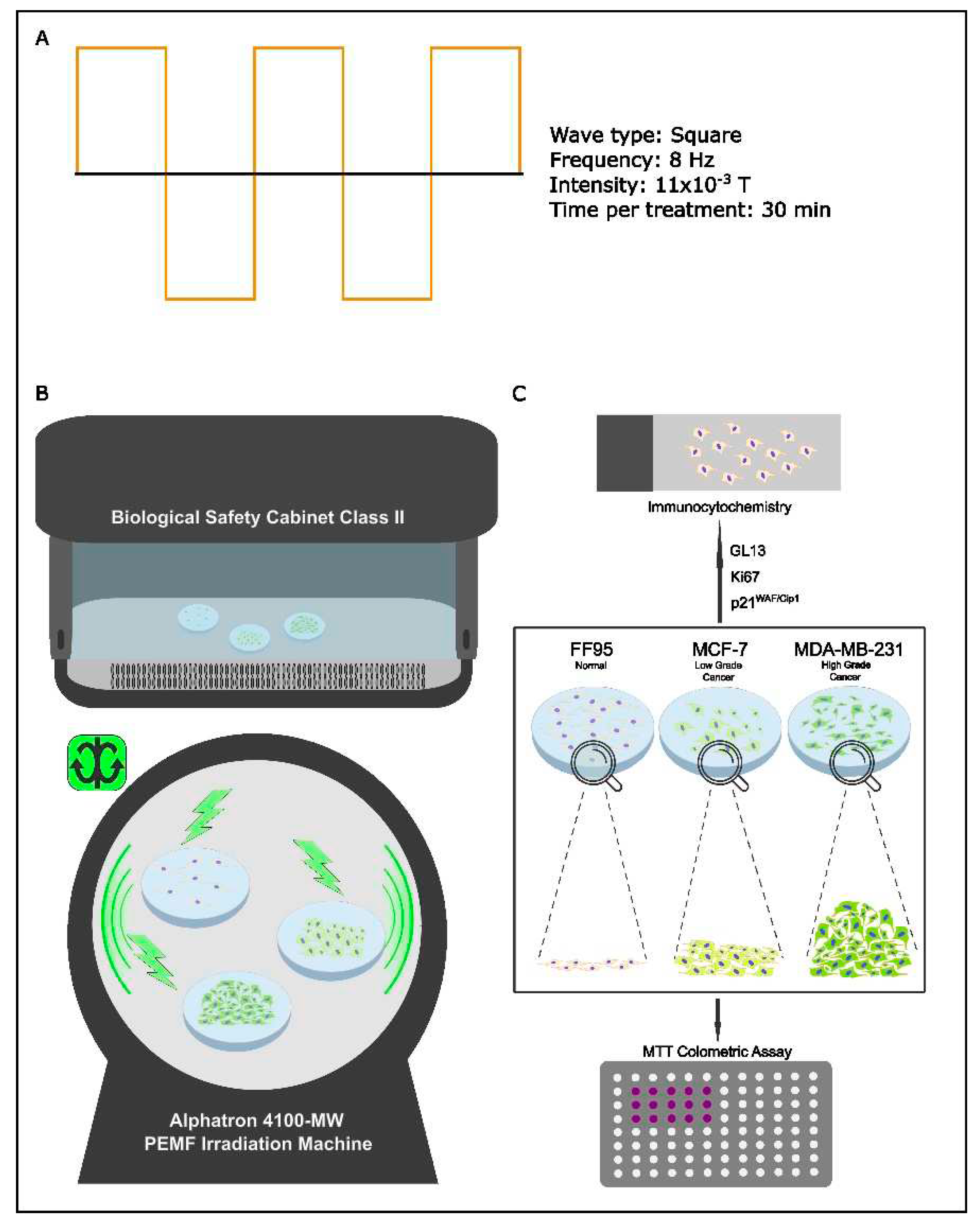
Figure 2.
Effect of PEMF irradiation on cell proliferation. Trypan blue staining and cell counting was performed to estimate the proliferation rate of PEMF-treated and control cells. (A) MDA-MB-231 and (B) MCF-7 cells exhibited decreased proliferation capacity following PEMF application in comparison with untreated. (C) PEMF irradiation in FF95 cells was accompanied by a statistically significant increase in proliferation in contrast to unirradiated controls. Statistical analysis was performed through the Kruskal–Wallis nonparametric test. The obtained data represent means ± standard deviation from three experiments. *p < 0.05, **p < 0.005.
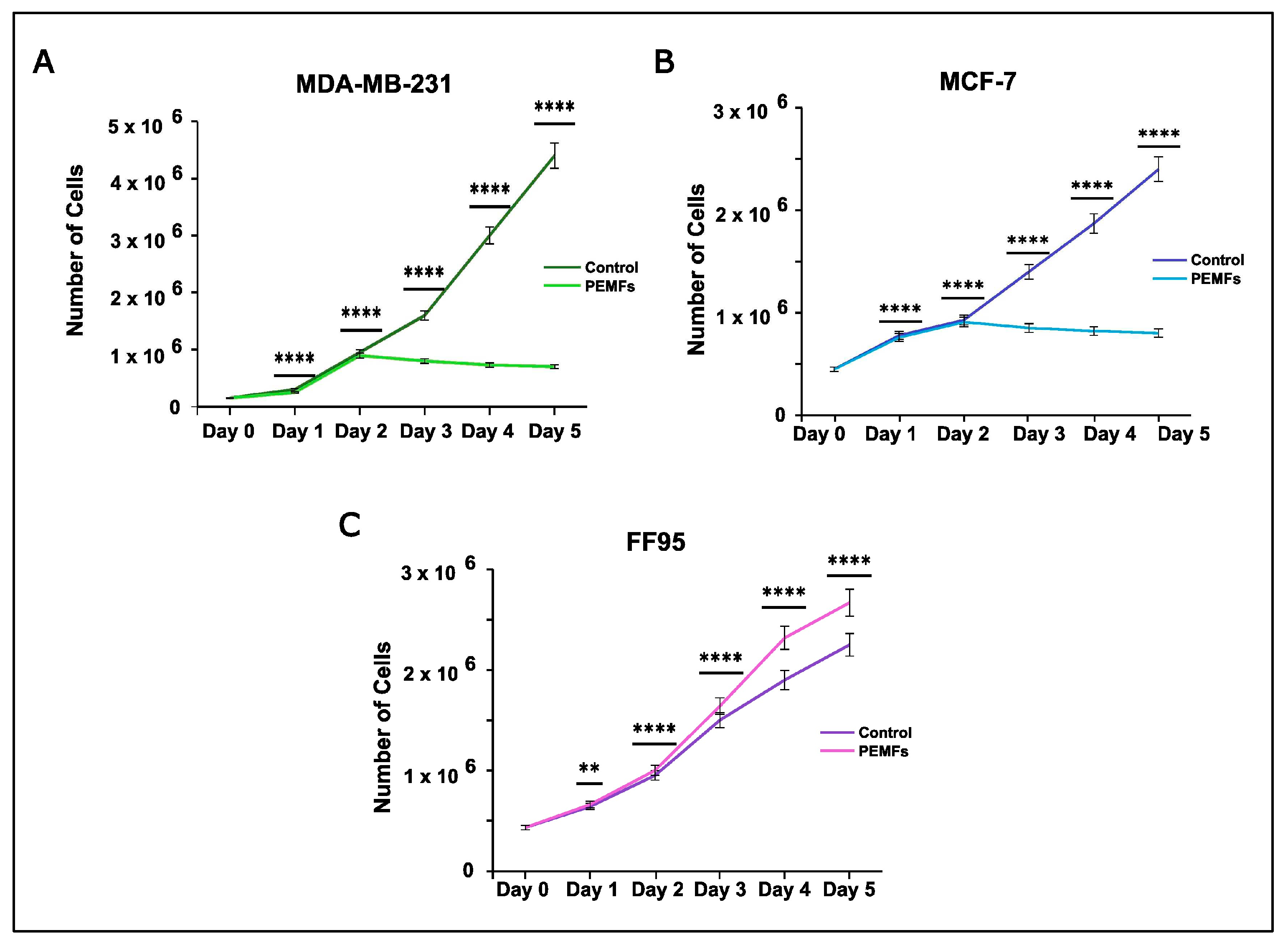
Figure 3.
Evaluation of cytotoxic effect after PEMF treatment in breast cancer cells and normal fibroblasts. MTT colorimetric assay was employed to estimate the percentage of cell viability of MDA-MB-231, MCF-7 and FF95 cells in the presence or absence of PEMF irradiation. PEMF treatment gradually decreased the cell viability of (A) MDA-MB-231 and (B) MCF-7 compared to the corresponding controls, while this phenomenon was opposite in FF95 cell (C). Statistical analysis was performed through the Wilcoxon nonparametric test. The obtained data represent means ± standard deviation from four experiments. *p < 0.05, **p < 0.005.
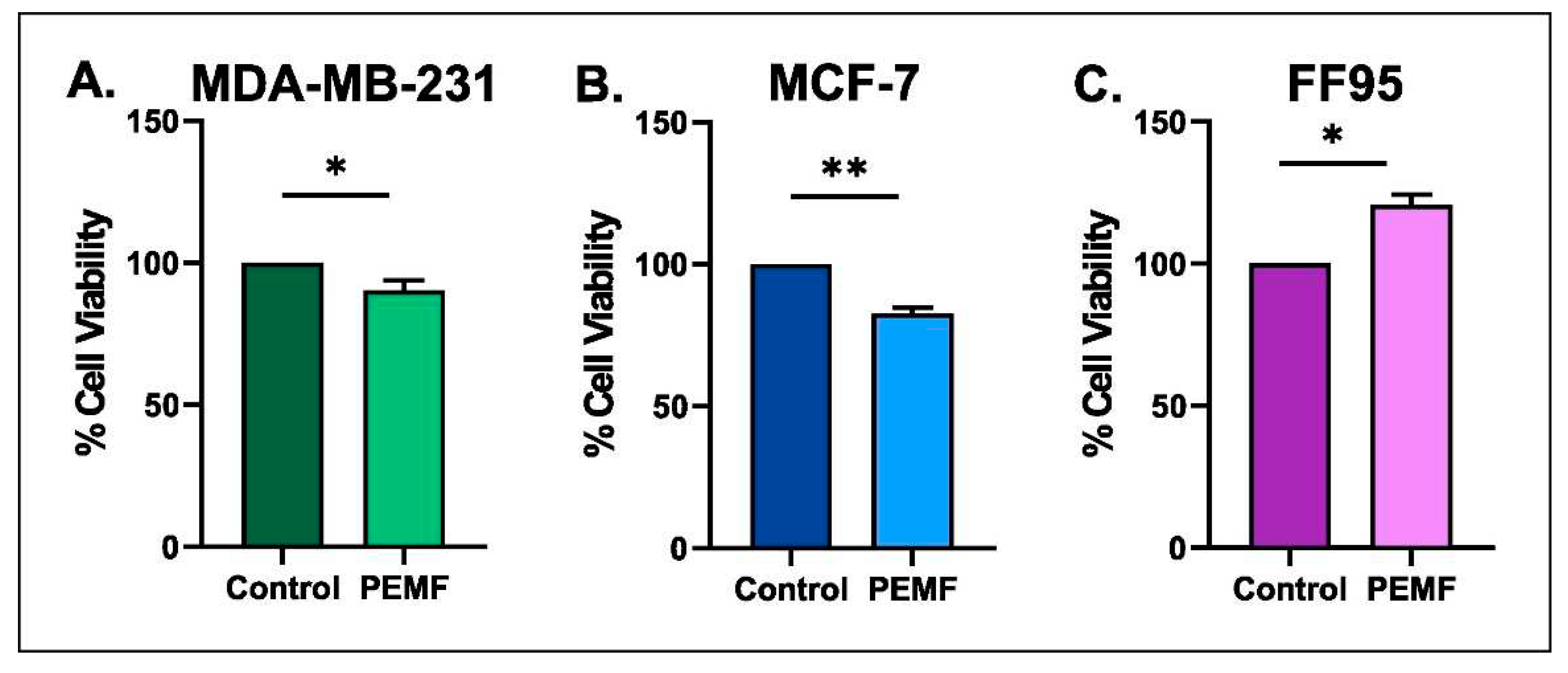
Figure 4.
Evaluation of proliferation and cellular senescence in breast cancer cells following PEMF treatment. Representative images of Ki67 (A,B), GL13 (D,E) and p21WAF1/Cip1 (G,H) immunocytochemical staining. Positive cells were calculated by strong brown nuclear signal for Ki67 and p21WAF1/Cip1. GL13 staining developed a strong brown perinucleus and/or cytoplasmic signal. Graphs depicted the percentage of positive cells (%) for Ki67 (C), GL13 (F) and p21WAF1/Cip1 (I) in PEMF-treated and control cells. Approximately 100 cells per optical field were counted and ≥5 high-power fields per sample were used for the quantification. Statistical analysis was performed through the Wilcoxon nonparametric test. The obtained data represent means ± standard deviation from four experiments. *p < 0.05, **p < 0.005. Objective 20x, 40x. Scale bars: (A,B,G,H) 30 μm, (D,E) 60 μm.
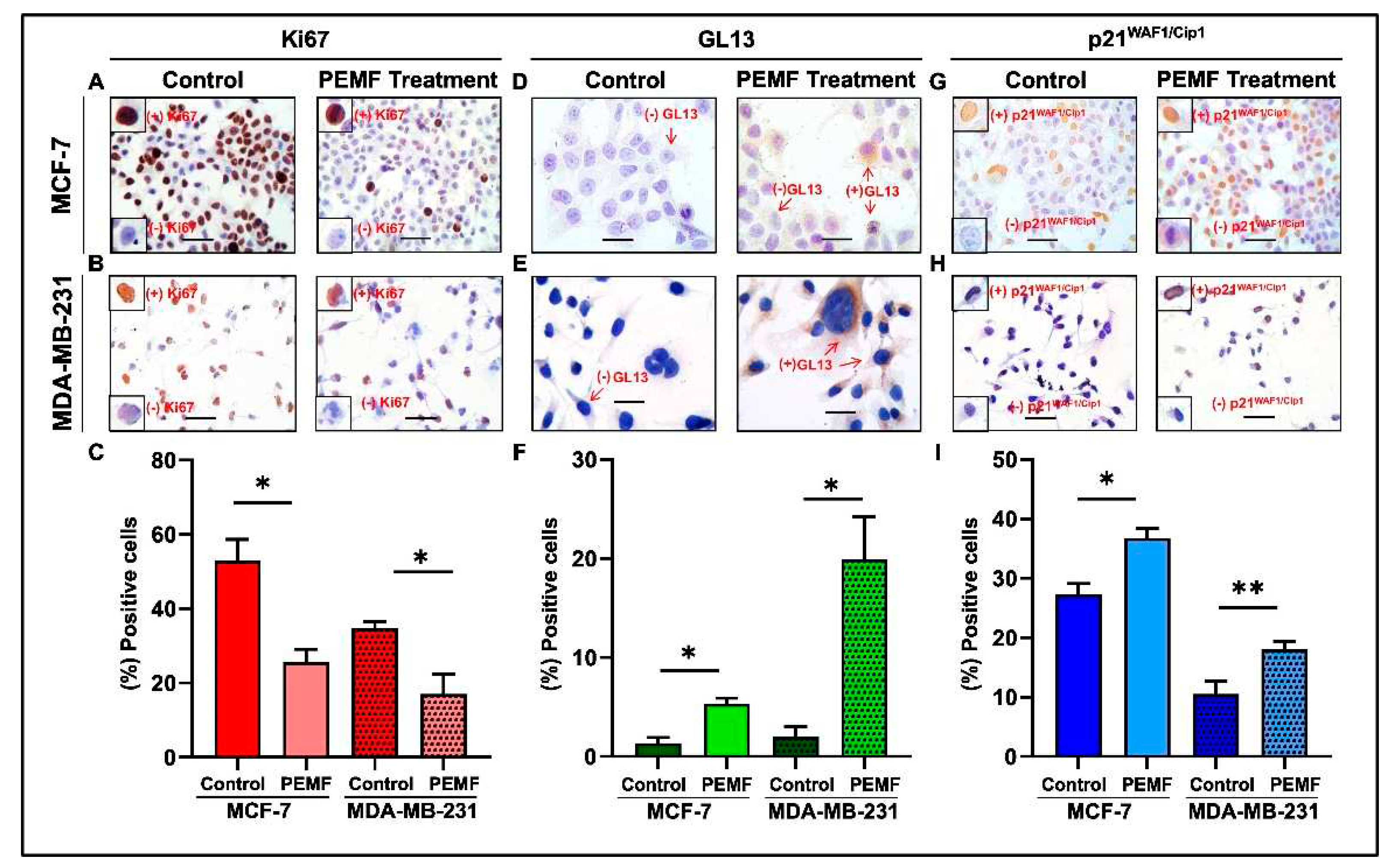
Figure 5.
Evaluation of proliferation and cellular senescence in normal fibroblasts (FF95) following PEMF treatment. Representative images of Ki67 (A), GL13 (B) and p21WAF1/Cip1 (C) immunocytochemical staining. Ki67 and p21WAF1/Cip1 positivity was determined by strong brown nuclear signal. GL13 staining developed a strong brown perinuclear and/or cytoplasmic signal. (D) Graph depicting the percentage of positive cells (%) for Ki67, GL13 and p21WAF1/Cip1 in PEMF-treated and control cells. Approximately 100 cells per optical field were counted and ≥ 5 high-power fields per sample were used for the quantification. Statistical analysis was performed through the Wilcoxon nonparametric test. The obtained data represent means ± standard deviation from four experiments. ns: non-significant. Objective 20x. Scale bar: 30 μm.
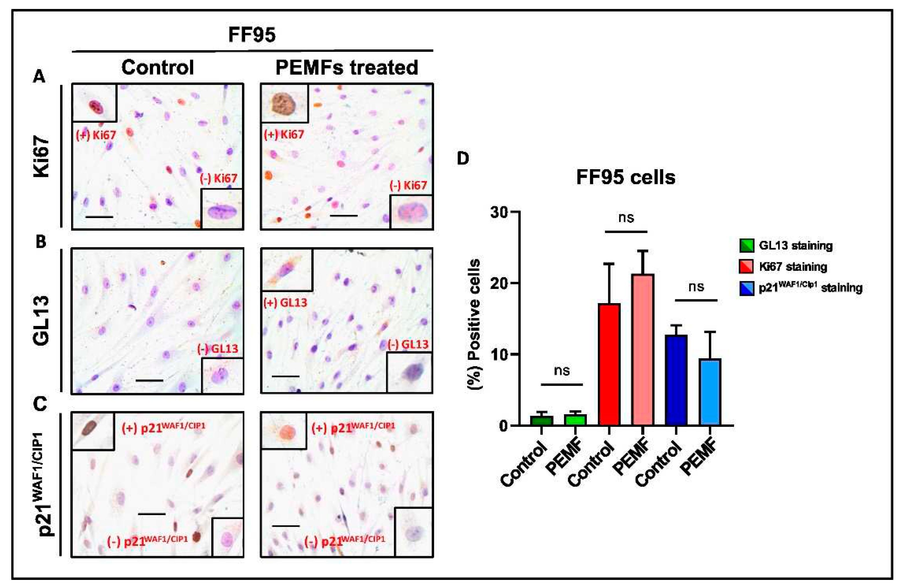
Figure 6.
Proposed model for the anti-tumor combined therapy applying PEMFs and senolytic drugs. (A) Traditional radiotherapy inducing cell death and/or cellular senesce in both cancer and adjacent normal tissue. (B) PEMF irradiation enhancing proliferation of normal cells while simultaneously pre-conditioning cancer tissue, through induction of senescence and apoptosis. (C) Administration of senolytic drugs eliminates the remaining senescent cancer cells following PEMF treatment.
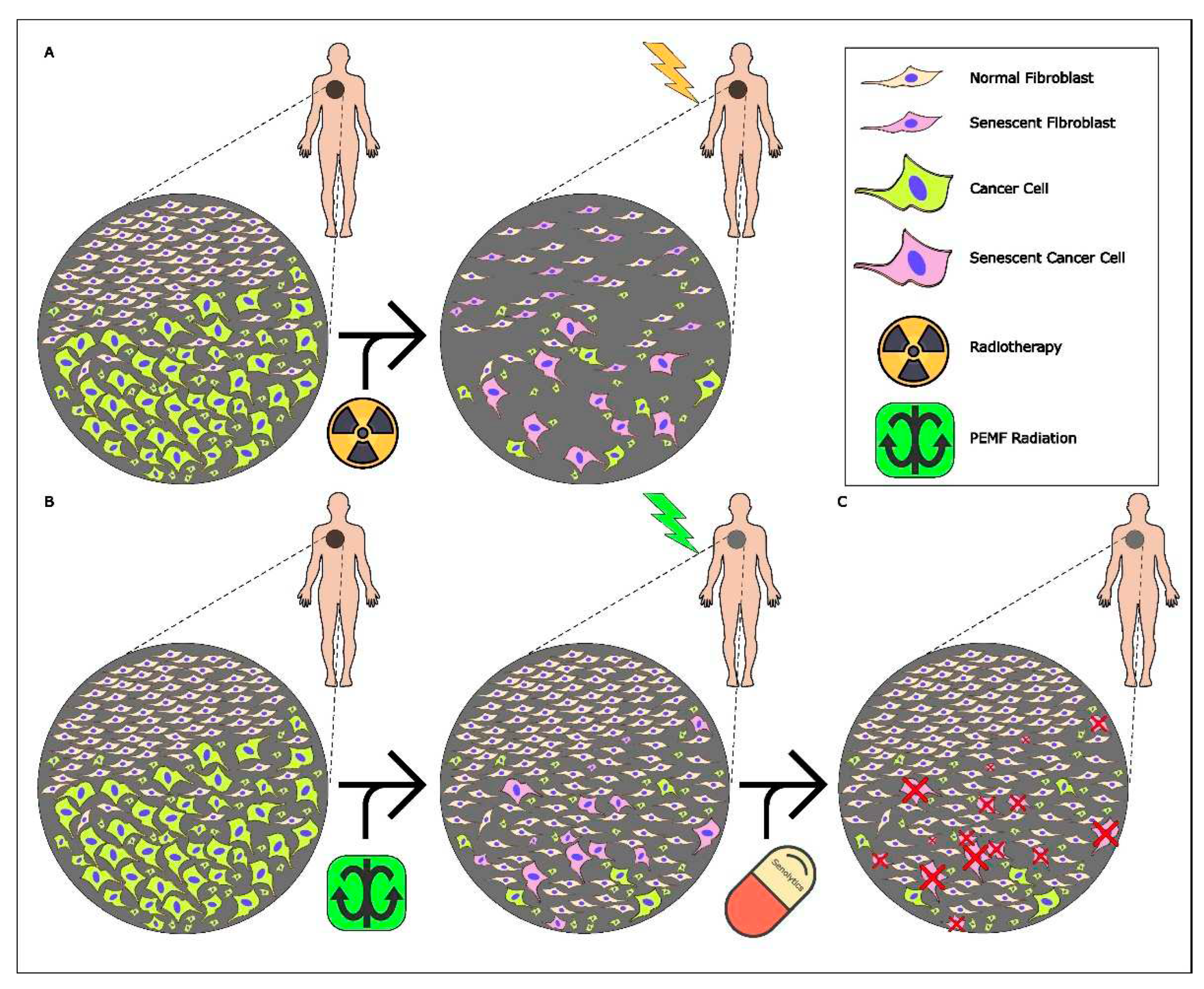

Table 1.
Settings of the different tested PEMFs programs that applied in the experimental procedure.
Table 1.
Settings of the different tested PEMFs programs that applied in the experimental procedure.
| Pre-set Program C4 | Manual Program 1 | Manual Program 2 | Manual Program 3 | |
|---|---|---|---|---|
| Frequency | 8-31 Hz | 8 Hz | 8 Hz | 8 Hz |
| Wave Type | Sinus-Rechteck | Full square | Full square | Full square |
| Intensity | 0.0033-0.0055 T | 0.0088 T | 0.0044 T | 0.011 T |
| Duration | 30 min (2 times)/ 5 days |
1 h (2 times)/ 5 days |
30 min (2 times)/ 5 days |
30 min (2 times)/ 5 days |
Disclaimer/Publisher’s Note: The statements, opinions and data contained in all publications are solely those of the individual author(s) and contributor(s) and not of MDPI and/or the editor(s). MDPI and/or the editor(s) disclaim responsibility for any injury to people or property resulting from any ideas, methods, instructions or products referred to in the content. |
© 2024 by the authors. Licensee MDPI, Basel, Switzerland. This article is an open access article distributed under the terms and conditions of the Creative Commons Attribution (CC BY) license (http://creativecommons.org/licenses/by/4.0/).
Copyright: This open access article is published under a Creative Commons CC BY 4.0 license, which permit the free download, distribution, and reuse, provided that the author and preprint are cited in any reuse.


