Submitted:
07 January 2024
Posted:
08 January 2024
You are already at the latest version
Abstract
Transdermal drug delivery systems (TDDS) are designed to administer a consistent and effective dose of medication through the patient's skin. These pharmaceutical preparations are self-contained, discrete dosage forms designed to be placed topically on intact skin to release the active component at a controlled rate by penetrating the skin barriers. This medication provides a continuous and prolonged administration of a substance at a consistent rate. TDDS, or transdermal drug delivery system, has gained significant attention as a non-invasive method of administering drugs to vulnerable patient populations, such as pediatric and geriatric patients. This approach is considered easy to administer and helps overcome bioavailability issues associated with conventional drug delivery, which can be hindered by poor absorption and metabolism. TDDS has various advantages compared to conventional methods of drug administration. It is less intrusive, more patient-friendly, and can circumvent first-pass metabolism and the corrosive acidic environment of the stomach that happens when medications are taken orally. Multiple methodologies have been devised to augment the transdermal permeability of diverse pharmaceutical substances. Recent advancements in transdermal drug delivery systems have facilitated the precise delivery of medications to the intended site of action by improving the penetration of drugs through the outermost layer of the skin, known as the stratum corneum, resulting in enhanced drug availability throughout the body. This review seeks to provide a concise overview of the several methods employed in the production of TDDS, as well as their evaluation, therapeutic uses, clinical considerations, and current advancements. These advancements have resulted in the development of intelligent, biodegradable, and highly efficient transdermal drug delivery systems.
Keywords:
transdermal drug delivery system
; skin permeation
; permeation enhancers
; quality by design ap-proach
; therapeutic applications
1. Introduction
The transdermal drug delivery system (TDDS) has garnered significant attention due to its numerous advantages compared to conventional drug delivery systems. These advantages include its simplicity, predetermined dosing, ease of handling, and the ability for self-administration [1]. Transdermal delivery provides a competitive advantage over injectable and oral formulations by enhancing patient compliance and bypassing the first-pass impact. TDDS is a versatile pharmaceutical preparation consisting of many layers, which can vary in size, and contain one or more active chemicals. It is designed to be applied to the intact skin in order to be absorbed into the bloodstream [2]. The medicine is transdermally delivered into the bloodstream at a predetermined rate and then distributed throughout the body before reaching the intended location. Most transdermal patches are designed to gradually release the active ingredients over many hours to days once they are placed on the skin. The medicine initially permeates the stratum corneum and subsequently traverses the underlying epidermis and dermis without any drug build-up in the dermal layer [3]. Transdermal delivery offers both controlled and continuous administration of pharmaceuticals, ensuring a steady supply of medication. It is particularly beneficial for drugs with short biological half-lives, as it prevents the intermittent release of drugs into the bloodstream, which can lead to unwanted side effects. The quantifiable concentrations of the medication in the blood, detectable elimination of the drug and its byproducts in the urine, and the patient’s clinical reaction to the provided pharmacological treatment can be employed to ascertain the percutaneous drug absorption [4].
Nevertheless, the restricted practical applicability of TDDS is due to the low skin permeability of certain medicines. The layered physiological composition of the skin serves as the primary obstacle to transdermal drug delivery (TDD), and only certain medicines with particular physicochemical characteristics can permeate the skin and enter the systemic circulation [5]. The percutaneous absorption is influenced by various elements, including the drug’s physical and chemical qualities such as molecular weight, solubility, partitioning coefficient, and dissociation constant. Additionally, the type of the carrier vehicle and the circumstances of the skin also have a role [6].
Various approaches and formulation strategies have been developed to enhance the transdermal permeability of medications by overcoming the skin’s physiological barrier. TDDS is composed of pressure-sensitive adhesives that ensure the adhesion of the preparation to the skin [7]. The product consists of a backing sheet that is impervious to both the active chemicals and water. Before placing the patch on the skin, it is necessary to remove the protective liner that covers the releasing surface of the patch. Transdermal patches have long been a subject of interest and have been utilized to administer medications such as nicotine, fentanyl, nitroglycerin, and clonidine for the treatment of diverse ailments [8]. The constraints and disadvantages related to the bioavailability of conventional formulations justify the need for the development of TDDS to improve drug delivery and penetration. The numerous advantages of various nanocarriers used in TDDS have attracted significant interest from pharmaceutical experts and dermatologists [9]. Various medications are offered in the form of transdermal patches, and the specific location where these medications are applied can differ according to their therapeutic classification [10]. The duration of drug release is also variable depending on the administration of these medications. The advantages and limitations of TDDS are shown in Table 1 and Table 2 respectively.
1.1. Skin Permeation as a Barrier
It is essential to provide a concise overview of the skin’s function as a protective barrier to enhance comprehension of the fundamental principles underlying the advancement of new technologies associated with TDDS. The skin, which makes up around 16% of an adult’s overall body weight, is the biggest organ in the body. Therefore, it has a crucial function in preserving homeostasis and serves as a barrier against external environmental dangers, both chemically, physically, and biologically [15]. The skin has a surface area of approximately 2 square meters. It consists of five layers in the epidermis, namely the stratum corneum (SC), stratum lucidum, stratum granulosum, stratum spinosum, and stratum basale. The outermost layer is formed by the stratum corneum, as shown in Figure 1a,b. The SC is a dense structure composed of fully matured keratinocytes that are spread out in a lipid-rich environment. It has a thickness of around 10 μm and is recognized as the step that controls the speed of absorption through the skin [16]. An undamaged and healthy stratum corneum serves as a barrier against both hydrophilic substances and big molecules. SC covers the viable epidermis consisting of numerous skin layers made of viable keratinocytes, which is followed by the dermal layer consisting of connective tissues, fibroblasts and other extracellular components such as hair follicles and glands. In order for a medicine included in the TDDS to enter the bloodstream, it must initially permeate through the layers of the skin. Several variables can influence the transdermal transfer of medications, including skin permeability, surface area, application duration, and metabolic activity of the skin. Non-ionic and moderately lipophilic medications effectively accomplish sufficient absorption and penetration through the skin [17]. The transdermal medication delivery is restricted to medicines with a molecular weight of no more than 500 Daltons due to the SC’s resistance to molecular diffusion. Nano-drug delivery systems with advanced capabilities can effectively penetrate the epidermis and specifically target locations for treating skin-related illnesses [18]. These nanocarriers effectively bypass the stratum corneum barrier by enhancing drug solubility and penetrating the skin, enabling the precise delivery of the desired dosage of the drug to the intended location [19].
1.2. Factors Affecting Transdermal Permeability
Transdermal drug delivery is not suited for all medicinal compounds. The physical and chemical features of the drug, including its molecular weight, solubility, partition coefficient, and dissociation constant, as well as the type of the carrier vehicle and the condition of the skin, all contribute to percutaneous absorption. Drugs within the molecular weight range of 100-800 kDa, possessing sufficient lipid and water solubility, are capable of penetrating the skin [20]. The optimal molecular weight for a medication intended for TDDS should be approximately 400 kDa or lower [21].
1.2.1. Physico-Chemical Properties of Parent Molecule
- Solubility and Partition Coefficient
The solubility of a medication directly affects its capacity to permeate the skin and impact the rate and degree of drug absorption. pKa is a measure of the drug’s ability to dissolve in a solvent, and the stratum corneum affects the movement of the drug from the solvent to the skin [22]. Enhancing the lipophilic nature of the medicine can increase its ability to permeate the skin, allowing it to enter through the stratum corneum. However, it does not pass through the epidermis since its water solubility has been reduced [23].
- pH Condition and Penetrant Concentration
An optimal pH level is beneficial and solutions with either high or low pH might cause skin damage. The absorption rate increases as the concentration of the drug in the vehicle increases [24].
1.2.2. Physico-Chemical Properties of Drug Delivery System
- Release characteristics and Composition of drug delivery system
The release rate of a drug is determined by its solubility in the vehicle. The rate of release of drugs will be influenced, as well as the permeability of the stratum corneum, through moisture and interaction with skin lipids. The more the lipophilicity, the greater the drug absorption [25].
1.2.3. Physiological and Pathological Conditions of Skin
- Lipid Film and Skin Hydration
The lipid film functions as a barrier to inhibit the loss of moisture from the skin. The defeat of this coating will reduce the rate at which medications are absorbed via the skin [26]. One way to obtain skin hydration is by applying plastic sheeting to the skin, which results in the buildup of sweat, condensed water vapors, improved porosity, and enhanced hydration [27].
- Effect of Vehicle
The absorption of the medicine and its effect on the skin can be influenced by a vehicle through its impact on their physical state [28]. Paraffin-based products have a higher level of occlusiveness, whereas water-in-oil based products have a lower level of occlusiveness. The presence of humectants in bases will cause skin dehydration and reduce the rate of absorption through the skin [29].
1.2.4. Biological Factors
- Skin Age and Skin Condition
The skin of a fetus, young individuals, and elderly individuals is more porous than the skin of adults. The viability of the dermis is higher than that of the dermis. The local and systemic bioavailability of a topically administered medicine can be influenced if it undergoes metabolism during penetration [30].
2. Kinetics of Transdermal Permeation
An understanding of the kinetics of skin permeation is crucial for the effective creation of transdermal therapeutic systems [31]. The process of transdermal medication penetration consists of the following sequential steps, shown in Figure 2:
- (a)
- Absorption by the outermost layer of the skin, known as the stratum corneum
- (b)
- Drug permeation through the viable outer layer of the skin
- (c)
- Absorption of the drug by the network of small blood vessels in the upper layer of the dermis [32]
Permeation is only possible if the medication exhibits specific physicochemical features. The equation provided represents the rate at which a substance passes through the skin, known as permeation rate (dQ/dt):
Where and are the concentrations of skin penetrant in the donor compartment (on the surface of stratum corneum) and in the receptor compartment (body) respectively. is the overall permeability coefficient of the skin tissues. The permeability coefficient is given by the relationship:
represents the partition coefficient for the transfer of the penetrant molecule from a transdermal therapeutic system to the stratum corneum. is the apparent diffusivity for the steady-state diffusion of the penetrant molecule through the skin tissues, and is the total thickness of the skin tissues. Given that and remain constant under the above conditions, it may be inferred that the permeability coefficient remains constant as well.
A constant rate of drug permeation can be obtained only when >> . i.e., the concentration of drug at the surface of the stratum corneum is consistently and substantially greater than the drug concentration in the body Then the equation becomes:
The rate of skin permeation ( is constant provided the magnitude of remains constant throughout skin permeation [33]. Hence, the drug should be released from the device at a rate () that is either constant or greater than the rate of skin uptake (). i.e., >> , which is shown in the Figure 3.
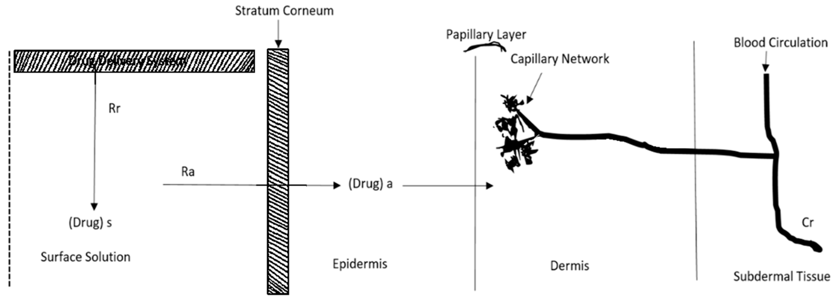
Figure 2.
Schematic representation of the relationship between the rate of drug release (Rr) from a transdermal drug delivery system (TDDS) and the rate of drug absorption (Ra) by the skin.
Figure 2.
Schematic representation of the relationship between the rate of drug release (Rr) from a transdermal drug delivery system (TDDS) and the rate of drug absorption (Ra) by the skin.
Since is greater than , the drug concentration on the skin surface is maintained at a level equal to or greater than the equilibrium or saturation solubility of the drug in the stratum corneum . i.e., >> . Hence a maximum rate of skin permeation [ is obtained and is given by the equation [34]:
The equation above demonstrates that the highest rate of skin permeation is determined by the skin permeability coefficient () and its equilibrium solubility in the stratum corneum (). Drug absorption via the skin seems to be restricted by the stratum corneum. The kinetics of skin permeation can be analyzed using in-vitro investigations utilizing a freshly excised skin sample affixed to a precisely calibrated skin permeation cell [35].
3. Basic Components and Classification of TDDS
The TDDS consists of many layers to facilitate the transport of medication from the skin to the bloodstream [36]. The Figure 3 below illustrates the fundamental elements of a TDDS. The backing layer functions as the outermost layer of the TDDS and protects the other layers from the surrounding environment. The backing layer must possess impermeability to both the medication and penetration enhancers. The typical composition of this layer consists of a pliable and impermeable substance, such as polyethylene, polypropylene, or polyester [37]. The adhesive layer functions to affix the patch onto the skin and secure its position. The product is composed of a potent, pressure-responsive, hypoallergenic adhesive that is mild on the skin. Widely utilized adhesives include silicone adhesives, polyisobutylene, and polyacrylate-based adhesives [38]. The drug reservoir is the fundamental component of TDDS. It comprises pharmaceutical substances that are administered transdermally. The formulation is designed to gradually release the medication at a consistent pace throughout a specific duration. The drug-release membrane regulates the rate of drug release from the patch [39]. Membranes consist of semipermeable substances that enable regulated passage of medicines. Polyethylene sheets, ethylene vinyl acetate copolymer, and cellulose acetate serve as membranes that limit the rate of a process.
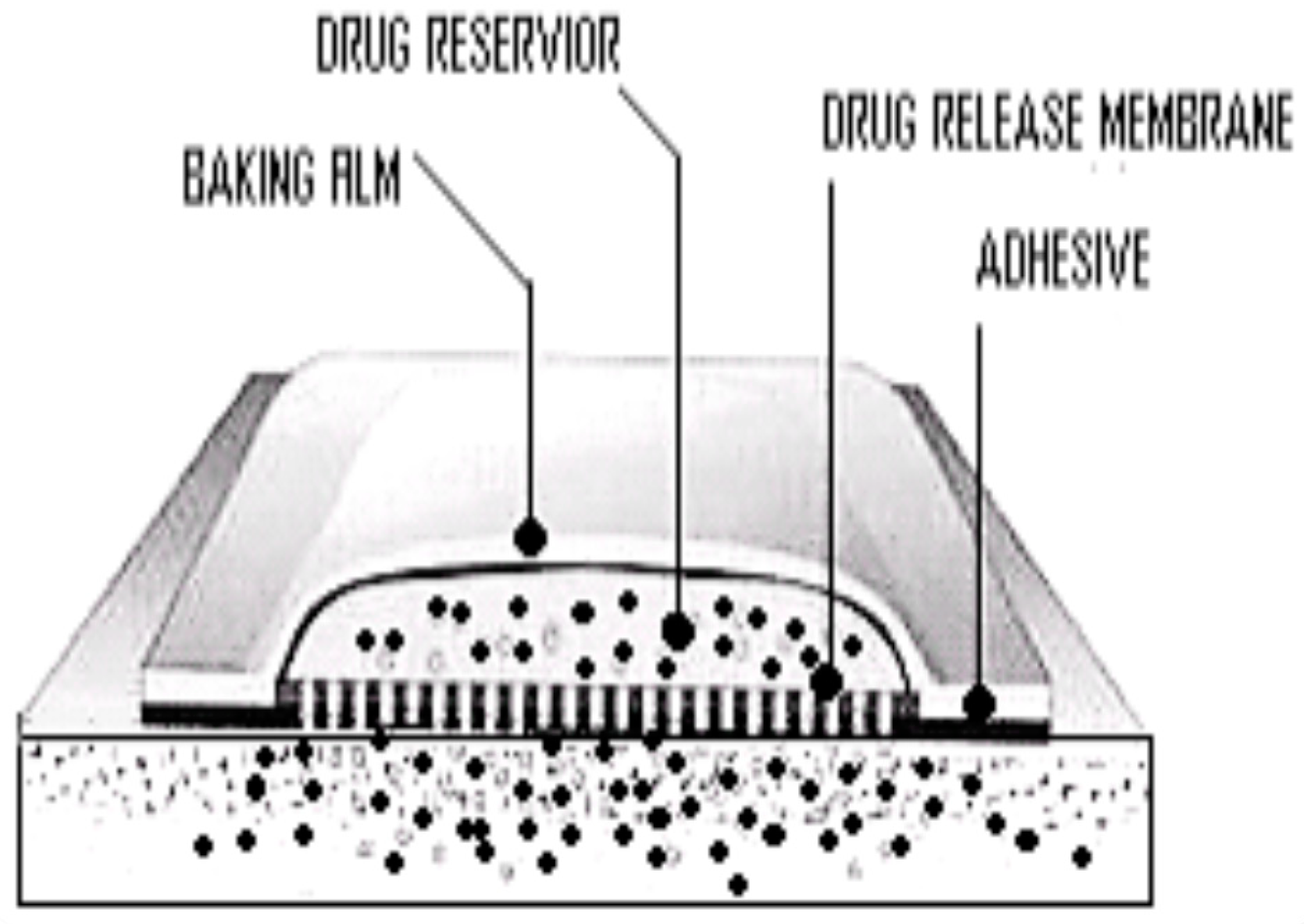
Figure 3.
Basic components of a TDDS.
3.1. Polymer Matrices
The polymer regulates the release of the medication from the device. The polymer’s molecular weight, glass transition temperature, and chemical activity must be carefully selected to ensure optimal diffusion and release of the specific medication [40]. The ideal characteristics of the material include stability, non-reactivity with the medicine, ease of manufacturing and fabrication, and cost-effectiveness. The polymer and its byproducts must be biocompatible with the host. The polymer’s mechanical characteristics should not undergo substantial deterioration when significant quantities of active substances are integrated into it [41]. The polymers utilized in TDDS encompass natural polymers, synthetic elastomers, and synthetic polymers are shown in Table 3.
3.2. Drug Reservoir
The drug reservoir comprises drug particles that are either dissolved or dispersed throughout the matrix, facilitated by solvents and cosolvents. The selection of the drug for the effective development of TDDS should be done with meticulous consideration [43]. The Table 4 below shows some of the ideal characteristics of a drug for transdermal delivery.
3.3. Permeation Enhancers
Permeation enhancers are chemicals that modify the skin’s barrier to allow for increased penetration of desired substances. The flux J, of a drug across the skin can be written as [45]:
The diffusion coefficient, denoted as D, is influenced by various factors such as the size, shape, and flexibility of the diffusing molecule, as well as the resistance of the membrane. The concentration of the diffusing molecules is represented by C, while x represents the spatial coordinate [46]. Permeation enhancers are believed to influence one or many layers of the skin in order to promote the penetration of substances into the skin. Several compounds have been examined for their capacity to increase the permeability of the stratum corneum [47]. They are categorized in the following manner, shown in Table 5. List of transdermal chemical penetration enhancers with active ingredients and mechanism of action is given in Table 6.
3.4. Classification of TDDS
4. Quality by Design (QbD) Approach of TDDS
The optimization of transdermal drug delivery systems (TDDS) is achieved through the use of experimental design techniques [59]. The study focuses on investigating the impact of temperature and occlusion on drug flux by examining the stability of thermodynamic changes, crystallization, and polymer crosslinking. Utilizing the Quality by Design (QbD) methodology in the production of TDDS assures the excellence, security, and effectiveness of the end pharmaceutical product, resulting in an effective formulation optimization [60]. Design of experiment (DoE) enhances the quality of the final formulation in this particular instance by minimizing the number of necessary trials. These experiments have been verified using various mathematical models. The models employed include the Box-Behnken design, central composite rotatable design, surface response composite design, and randomized fractional factorial design [61]. The ANOVA statistically confirmed the factors that had a substantial impact on the dependent variables. The implementation of QbD in the creation of pharmaceutical products leads to enhanced manufacturing and development efficiency, as well as the identification of potential dangers and characteristics that may impact product quality [62].
Recently, a number of researchers have been utilizing the QbD methodology in the creation of TDDS in order to get superior pharmaceutical goods [63]. This is accomplished by comprehending and managing the formulation, materials, and manufacturing variables. The application of QbD has been utilized to enhance the drug release and targeting, as well as to optimize the pharmacokinetics and pharmacodynamics features of TDDS [64].
Critical Quality Attributes (CQAs)
5. Approaches Used in the Development of TDDS
Transdermal dosage forms are composed of multiple layers, each with a distinct purpose, forming a fundamental structure. An impermeable covering that keeps the system from becoming wet during use [66]. The second layer functions as a drug reservoir, providing a consistent supply of medication for a predetermined duration. Adjacent to the reservoir is the rate-controlling polymeric membrane that governs the rate at which a medicine is released over a predetermined period. The medicine administered by the system permeates the skin and reaches the systemic circulation [67]. The drug delivery rate from the TDDS typically exceeds the maximum absorption capacity of the skin. Therefore, despite potential differences in skin permeability, a consistent rate of medication absorption into the bloodstream is attained [68]. There exist four distinct approaches for acquiring TDDS:
5.1. Membrane Permeation-Controlled Systems
The drug reservoir in this system is enclosed within a shallow compartment made of a drug-impermeable metallic plastic laminate, as shown in Figure 6. Additionally, there is a rate-controlling polymeric membrane that can either be microporous or non-porous, and it has a certain drug permeability characteristic [69]. The medication molecules are exclusively allowed to be released via the rate-controlling membrane. The drug particles in the drug reservoir compartment are either disseminated within a solid polymer matrix or suspended in an unleachable, viscous liquid medium, such as silicone fluid, resulting in the formation of a paste-like suspension [70]. To ensure close contact between the TDDS and the skin, a thin coating of adhesive polymer that is compatible with drugs and hypoallergenic, such as silicone or polyacrylate adhesive, can be placed on the outer surface of the membrane that controls the rate of drug release. The drug release rate of this particular TDDS can be adjusted by altering the polymer composition, permeability coefficient, thickness of the rate-limiting membrane, and adhesives [71]. The continuous dispensation of medications is the primary benefit of this approach. However, there is a possibility of an unintentional rupture of the rate-controlling membrane, leading to dose dumping or the sudden release of the entire drug content [72]. A few examples of this system are shown in Table 8.
The intrinsic rate of drug release from this type of drug delivery system is explained by:
Where CR is the concentration of the drug in the reservoir compartment. Pa and Pm are the permeability coefficients of the adhesive layer and the rate-controlling membrane respectively. Pa and Pm are defined as follows,
and are the partition coefficients for the interfacial partitioning of the drug from the reservoir to the membrane and from the membrane to the adhesive respectively; and are the diffusion coefficients in the rate-controlling membrane and adhesive layer respectively; and are the thickness of the rate-controlling membrane and adhesive layer respectively. Substituting the equations for and , the equation for the intrinsic rate of drug release () becomes [77],
The above equation defines the intrinsic rate of drug release from a membrane-moderated drug delivery system.
5.2. Adhesive Dispersion-Type Systems
In this particular transdermal drug delivery system (TDDS), the drug reservoir is produced by directly dispersing the drug in an adhesive polymer, such as poly(isobutylene) or poly(acrylate) adhesive [78]. The medicated adhesive is then spread onto a flat sheet of drug-impermeable metallic plastic backing using either solvent casting or hot melt techniques, forming a thin drug reservoir layer. Above the layer containing the drugs, thin layers of non-medicated, adhesive polymer with a specified permeability and consistent thickness are applied to develop an adhesive diffusion-controlled drug delivery system, shown in Figure 7.
An example of this type of system is Isosorbide dinitrate releasing TDDS for a once-a-day medication of angina pectoris and transdermal controlled administration of verapamil. The rate of drug release in this system is defined by:
is the partition coefficient for the interfacial partitioning of the drug from the reservoir layer to the adhesive layer [79].
This particular TDDS can also be altered to allow for the manipulation of the drug loading level in small increments, resulting in the formation of a gradient of drug reservoir across multilaminate adhesive layers, as shown in Figure 8. An example of this system is the nitroglycerin-releasing TDDS.
The drug release rate in this system is defined by:
is the thickness of the adhesive layer for the diffusion of drug molecules with time. To compensate for this time-dependent increase in diffusional path due to the depletion of drug dose by release, the drug loading level is also increased with the thickness of diffusional path to achieve a constant drug release profile [80].
5.3. Matrix Diffusion-Controlled Systems
The drug reservoir in this system is produced by evenly distributing drug particles within a polymer matrix that is either hydrophilic or lipophilic. Subsequently, the medicated polymer is moulded into a medicated disc, possessing a specific surface area and controlled thickness [81]. The drug particles can be dispersed in the polymer matrix through two methods: either by uniformly mixing finely ground drug particles with a liquid polymer or a highly viscous base polymer, and then crosslinking the polymer chains, or by homogeneously blending drug solids with a rubbery polymer at a high temperature. The drug reservoir can also be created by dissolving the drug and polymer in a common solvent and then evaporating the solvent in a mould at a higher temperature or in a vacuum [82]. The polymer disc, which holds the drug, is then attached to an occlusive base plate within a compartment made of a drug-impermeable plastic backing. The adhesive polymer is thereafter applied evenly around the circumference to create a band of the adhesive rim around the medicated disc, as depicted in Figure 9. A good example of this technology is the transdermal therapeutic system that releases nitroglycerin and delivers it continuously through the skin [83].
The rate of drug release from this system is defined as:
where A is the initial loading dose dispersed in the polymer matrix. are the solubility and diffusivity of the drug in the polymer respectively.
5.4. Microreservoir Type or Micro-Sealed DissolutionCcontrolled Systems
This system is a hybrid of the reservoir and matrix diffusion drug delivery methods. The drug reservoir is produced by combining the drug solids with a water-soluble liquid polymer solution, and then uniformly dispersing the drug suspension within a lipophilic polymer using a high-energy dispersion technique [84]. This process results in the formation of distinct and non-leachable microspheres that serve as the drug reservoir. The rapid stabilization of this thermodynamically unstable dispersion is achieved by promptly cross-linking the polymer chains in-situ, resulting in the formation of a medicated polymer disc with a consistent surface area and fixed thickness. If necessary, the device might be additionally coated with a biocompatible polymer layer to alter the method and rate of drug release [85]. A transdermal therapeutic system is created by placing the medicated disc in the middle and surrounding it with an adhesive rim, which is shown in Figure 10. A specific example of this kind of technology is the TDDS which releases nitroglycerin for the treatment of angina pectoris once a day. The microreservoir technology exhibits zero-order release kinetics while mitigating the risk of dose dumping [86].
The rate of release of drugs from the micro-reservoir system is defined by:
Where m = a/b, here a represents the ratio of drug concentration in the bulk of the elution medium, while b represents the drug concentration at the outer edge of the polymer coating [87]. n represents the quotient of the drug concentration at the inner boundary of the interfacial barrier divided by the drug solubility in the polymer matrix. The drug diffusivities in the liquid layer surrounding the drug particles, the polymer coating membrane covering the polymer matrix, and the hydrodynamic diffusion layer surrounding the polymer coating are represented by accordingly. The thickness of these layers is denoted by , . are the partition coefficients that describe the distribution of the drug between different compartments, specifically from the liquid compartment to the polymer matrix, from the polymer matrix to the polymer coating membrane, and from the polymer coating membrane to the elution solution, respectively. represent the drug’s solubilities in the liquid compartment and the polymer matrix, respectively. This drug delivery system follows either a partition control or matrix diffusion-control mechanism [88].
6. Production of TDDS
The production of TDDS on a large scale requires the use of manufacturing technologies that are either novel or adapted from the conventional pharmaceutical sector.
6.1. Asymmetric TPX Membrane Method
The backing membrane will consist of a heat-sealable polyester film, specifically type 1009, with a diameter of 1cm and a concave shape. The drug sample is placed into the concave membrane, which is then coated with a TPX poly (4-methyl-1-pentene) asymmetric membrane and sealed with an adhesive layer [89]. To produce a TPX asymmetric membrane, either a wet inversion method or a dry inversion approach is employed.
6.2. Circular-Teflon Mould Method
Polymers are dissolved in an organic solvent in different proportions to create solutions. The determined quantity of the drug is dissolved in half the volume of the identical organic solvent. The permeation enhancers are dissolved in the remaining portion of the organic solvent and subsequently combined [90]. Di-N-butyl phthalate serves as a plasticizer and is included in a drug-polymer solution. The entire mixture should be agitated for a duration of 12 h and thereafter transferred into a round Teflon mould. The moulds should be positioned on a flat surface and covered with an inverted funnel to regulate the evaporation of the solvent in a laminar flow hood model with an air velocity of 0.5 m/s. The solvent is allowed to evaporate for a duration of 24 h. The films that have been stored need to be kept for a further 24 h at a temperature of 25± 0.5˚ in a desiccator that contains silica gel. This is done in order to eradicate any impacts caused by ageing before evaluation [91]. The films must be assessed within a week of their preparation.
6.3. Mercury-Substrate Method
The medicament is dissolved or dispersed in a solution of polymer comprising of plasticizer. The solution was then agitated for 10–15 min to ensure a consistent, homogeneous mixture and poured into a flat surface of mercury with a covering of an inverted funnel to avoid evaporation of the solvent [92].
6.4. By Using the IPM Membrane Method
This technique involves dispersing the medicine in a solution composed of water and propylene glycol, which also contains carbomer 940 polymers. The mixture is then agitated for a duration of 12 h using a magnetic stirrer. To counteract the dispersion, triethanolamine is added to neutralize it and increase its viscosity. A solution gel can be generated by using a buffer with a pH of 7.4, particularly when the drug’s solubility in an aqueous solution is extremely low. The gel that is produced will be incorporated into the IPM membrane [93].
6.5. By Using the (Ethylene Vinyl Acetate Copolymer) EVAC Membrane Method
To develop the desired transdermal therapeutic system using this method, one can utilize a 1% Carbopol reservoir gel along with polyethylene (PE) and EVAC membranes as rate-controlling membranes. When the medicine is not able to dissolve in water, propylene glycol is employed to create the gel formulation. The medication is solubilized in propylene glycol. Carbopol resin will be introduced into the aforementioned solution and neutralized using a 5% w/w solution of sodium hydroxide. The gel-based drug is applied onto a backing layer sheet that covers the designated area. A rate-limiting membrane will be applied onto the gel and the edges will be sealed using heat to achieve a device that is impermeable to leaks [94].
6.6. Aluminium-Backed Adhesive Film Method
The aluminium-backed adhesive film approach is appropriate when the initial dose of the medication exceeds 10mg. Chloroform is employed as a solvent in this procedure due to its ability to dissolve a wide range of medicines and adhesives. The drug will be dissolved in chloroform, and an adhesive material will be added to the drug solution and dissolved. An individually crafted aluminium mould is coated with aluminium foil and sealed at the ends with snugly fitting cork blocks [95].
6.7. By Using the Free-Film Method
The production of cellulose acetate-free film involves the application of chloroform over a mercury surface. A polymer solution with a concentration of 2% w/w is prepared. A plasticizer is incorporated into the polymer at a concentration of 40% by weight. A glass ring is positioned on a glass petri dish, and then mercury is poured onto the surface. Subsequently, dispense 5ml of polymer solution onto the glass ring. Position a funnel on a Petri plate to regulate the rate of solvent evaporation. A thin layer developed on the surface of mercury once the solvent had fully evaporated. The dehydrated coating was thereafter divided and preserved in a desiccator, sandwiched between sheets of wax paper until it was required [96].
7. Enhancement of Transdermal Drug Delivery
In order to improve the absorption via the skin and increase the effectiveness of therapy, medications need to possess a low molecular weight (less than 1kDa), an affinity towards both lipid-based and water-based substances, a short duration of action, and a non-irritating effect on the skin. Several variables influence the ability of drugs to pass through the skin, including variations across species, length of exposure, age and site of the skin, temperature of the skin, moisture levels in the skin, pre-treatment techniques, application region, condition of the skin, and physical properties of the substance being absorbed [97].
7.1. Active Drug Delivery
External stimuli, such as electrical, mechanical, or physical stimulation, have been found to increase the ability of medications and biomolecules to pass through the skin, compared to applying drugs topically. Active transdermal delivery, also known as TDDS with appropriate equipment, efficiently and consistently delivers medications into the skin at a rapid pace. This kind of improved TDDS can expedite the therapeutic effectiveness of administered medications [98]. The advantages of various delivery methods are shown in Table 9.
7.2. Passive Drug Delivery
Chemical penetration enhancers (CPEs) facilitate the diffusion of drugs through the skin or enhance their ability to dissolve in the skin, resulting in the development of highly potent formulations, microemulsions, and vesicles. Their action is accomplished by disrupting the structured lipid bilayer, interacting with proteins in the cell membrane and intercellular proteins, disrupting lipids between cells, increasing moisture in the outermost layer of the skin, and improving the partition coefficient of drugs. Penetration enhancers can be utilized either independently or in conjunction with chemical penetration enhancers to provide enhanced skin penetration, surpassing the effectiveness of individual chemicals. Over 300 chemical penetration enhancers have been employed in various TDDS to aid the transportation of pharmaceuticals over the stratum corneum. An ideal enhancer should possess the qualities of being non-toxic and biocompatible, while also exhibiting predictable and consistent activity and duration of effect [99].

| Active delivery | Methods | Advantages |
| Iontophoresis | Improves the delivery of polar molecules and high molecular weight compounds, easy to administer, continuous or pulsatile delivery of drugs | |
| Sonophoresis | Strict control of transdermal diffusion rates, greater patient approval, less risk of systemic absorption, non-sensitizing | |
| Electroporation | Highly effective, reproducible, rapid termination of drug delivery, not sensitizing | |
| Photomechanical waves | Improve the transfer of molecules across the plasma membrane without loss of viability, do not cause pain or discomfort | |
| Microneedle | Painless administration of the active pharmaceutical ingredient, faster healing at the injection site, no fear of needle, specific delivery of the drugs | |
| Thermal ablation | Avoid pain, bleeding and infection, better control and reproducibility, low cost and disposable device | |
| Passive delivery | Nanoemulsion | Long-term thermodynamic stability, excellent wettability, high solubilization capacity and physical stability |
| Polymeric nanoparticles | Targeted and controlled release behaviour, high mechanical strength, and both hydrophilic and lipophilic drugs can be loaded | |
| Vesicles | Sustained drug release behaviour, controls the absorption rate through a multilayered structure. |
8. Evaluation of TDDS
The development of a transdermal drug delivery system necessitates a thorough and systematic evaluation at several phases. Various evaluation processes are shown in Table 10.
8.1. Physico-Chemical Evaluation–Adhesive Evaluation
The pressure-sensitive adhesives are evaluated for the following properties:
8.1.1. Peel Adhesion Properties [112]
Peel adhesion refers to the amount of force needed to detach an adhesive covering from a designated surface for testing purposes. An essential characteristic of TDDS is that the adhesive must ensure sufficient adherence of the device to the skin without causing any harm when removed. The molecular weight of the adhesive polymer, the kind and quantity of additives, and the composition of the polymer all have an impact on these qualities. The testing involves quantifying the amount of force needed to extract a solitary tape with a coating, which is affixed to a substrate, at a 180˚ inclination. The absence of any residue on the substrate signifies ’adhesive failure’, which is a desirable outcome for TDDS. The presence of remnants on the substrate suggests a "cohesive failure," which indicates a lack of cohesive strength in the coating, shown in Figure 11.
8.1.2. Tack Properties [113]
Tack refers to the capacity of a polymer to stick to a surface with minimal applied pressure. Finger pressure is crucial in TDDS. The adhesive strength of tack is influenced by the molecular weight and content of the polymer, as well as the incorporation of tackifying resins into the polymer. Tests conducted to evaluate the performance of tack include:
- (a)
- Thumbtack Test [114]
This is a subjective test in which evaluation is done by pressing the thumb briefly into the adhesive.
- (b)
- Rolling Ball Tack Test [115]
This experiment measures the distance of a stainless steel ball as it moves along an adhesive surface facing upwards. The lesser the tackiness of the adhesive, the greater the distance the ball will travel, as given in Figure 12.
- (c)
- Quick-Stick or Peel Tack Test [116]
The peel force necessary to separate the adhesive from the substrate is quantified by applying a 90˚ angle force to the tape, peeling it away from the substrate at a rate of 12 inches per minute, shown in Figure 13. The force is measured and documented as the tack value, which is denoted in ounces or grams per inch width. Higher values correspond to more tackiness.
- (d)
- Probe Tack Test [117]
The probe makes contact with the glue, resulting in the formation of a bond between them. Tack is the measurement of the force, expressed in grams, needed to separate a probe from an adhesive at a constant rate, shown in Figure 14.
8.1.3. Shear Strength Properties [118]
It quantifies the adhesive polymer’s ability to stick together. The sufficient adhesive strength of a device ensures that it remains in place during application and does not leave any residue upon removal. The molecular weight, as well as the type and quantity of tackifier used, have an impact on it. Shear strength or creep resistance is assessed by measuring the duration required to detach an adhesive-coated tape from a stainless steel plate while applying a predetermined weight that pulls the tape in a direction parallel to the plate, shown in Figure 15.
8.2. Patch Width [119]
The width of the patch is determined using a digital micrometer, while the average thickness ensures the desired thickness of the created patch. The film thickness is measured using a screw gauge and a microscope dial gauge at various locations on the film.
8.3. Folding Endurance [120]
A section of TDDS has been cut and subsequently folded multiple times in a manner like a plug until it eventually fractures. The durability of the patch is determined by the number of times it can be folded from the same spot until it breaks.
8.4. Percentage of Moisture Content [121]
Every individual transdermal patch is individually weighed and then kept at room temperature for a duration of 24 h in desiccators with fused calcium chloride. After 24 h, the patches are weighed again and the moisture content percentage is determined using the following formula:
8.5. Moisture Uptake [122]
The patches are stored in a desiccator at room temperature for a duration of 24 h. After a duration of 24 h, the patches are taken off and exposed to a concentrated solution of potassium chloride in desiccators at a relative of 84% until they attain a stable weight. The calculation of moisture uptake percentage is determined using the following formula:
8.6. Content Uniformity Test [123]
A total of 10 patches have been selected for the purpose of this study, and the specific content of each patch has been determined. The content uniformity test is considered acceptable if 9 out of 10 patches have a content uniformity ranging from 85% to 115%, and if the remaining individual patch falls within the limit of 75% to 125%. An additional examination is conducted on 20 patches, with their acceptable range set at 85% to 115%. If the results fall within this range, the test is considered successful.
8.7. Drug Content [124]
Dissolve the patch in a solvent following its cutting to the desired dimensions. Subsequently, the solution is passed through a filter media and the constituents are identified using an appropriate analytical technique, such as a UV visible spectrophotometer or High-performance liquid chromatography.
8.8. In-Vitro Drug release [125]
In-vitro studies assist in the design and development of TDDS. These investigations can aid in investigating how the drug is absorbed via the skin before its formulation into a transdermal therapeutic system. The data acquired from in-vitro investigations of the developed TDDS can be utilized to optimize the formulation prior to conducting more expensive in-vivo tests.
For this study, excised skin is placed on skin permeation cells. The use of excised skin is commonly preferred for in-vitro investigations due to the fact that the stratum corneum, which is a biologically inactive tissue, serves as the primary barrier for drug permeation. The diffusion through the stratum corneum occurs through a passive process. The use of hairless mouse skin is advantageous due to the absence of any potentially harmful hair removal procedures. Additionally, this species is readily accessible and allows for the excision of skin specimens immediately prior to the permeation study. An appropriately designed in-vitro apparatus ensures that the drug delivery mechanism originates exclusively from the TDDS. Some examples of in-vitro membrane permeation apparatus are the Valia-Chien (V-C) cell, Ghannam-Chien (G-C) membrane permeation cell, Jhawer-Lord (J-L) rotating disc system, Franz diffusion cell, and the Keshary-Chien (K-C) cell. The Franz diffusion cell and the K-C cell are the most commonly utilized among them. The K-C cell, a modified iteration of the Franz diffusion cell, possesses a receptor volume of 12 ml and a skin surface area of 3.14 cm2, shown in Figure 16. The receptor solution is agitated by a star-head magnet that rotates at a consistent pace of 600 revolutions per minute, powered by a 3-watt synchronous motor. The heater, which is regulated by a thermostat, maintains a temperature of 32±0.5˚C. Periodically, a specific amount of the sample is taken from the receptor compartment and analyzed with a spectrophotometer.
8.9. Skin Irritation Study [126]
Skin irritation and sensitization studies can be carried out on healthy rabbits. The hair of the animal has been removed from the dorsal side. Clean the surface with a rectified spirit and place the patch on the skin. After 24 h, the patch is removed. Depending on the degree of skin injury, the skin condition has been assessed.
8.10. Stability Study [127]
These studies are conducted in compliance with ICH guidelines for 6 months. The samples are kept at 40 ± 5 °C and 75 ± 5% relative humidity and are analysed at various time intervals such as 0, 30, 60, 120 and 180 days.
8.11. In-Vivo Evaluation
In-vivo evaluation of TDDS can be carried out using animal models, human volunteers and biophysical models.
8.11.1. Animal Models [128]
Animal models are favoured due to the substantial time and resources needed to conduct investigations in humans. The species utilized for both in- vivo and in-vitro testing encompass mouse, rat, guinea pig, rabbit, hairless mouse, hairless rat, cat, dog, tiny pig, horse, goat, squirrel, and rhesus monkey. Multiple experiments have been conducted to ascertain which animal models offer the most precise predictions of the device’s performance when tested on humans. The conducted experiments have resulted in the subsequent findings:
Small, hirsute animals are not highly effective prediction models for human in- vivo transdermal drug administration. The penetration values seen in these animals exceed those observed in humans. This observation is corroborated by in-vitro measurements. Shaving or depilating the skin of these animals for investigation can cause alterations in the skin’s resilience. The rhesus monkey is the most reliable model for in-vivo assessment of TDDS. The application site is typically the forearm or abdomen, as these areas tend to have the least amount of hair on the animal’s body. Drawbacks associated with utilizing this particular animal species encompass cost , necessary handling skills, and challenges related to accessibility. The utilization of rhesus monkeys is further restricted due to ethical reasons. Promising alternative animal models for predicting transdermal medication distribution in humans include weanling pigs and nude mice with human skin grafts. The challenge faced while utilizing these animals lies in their unavailability and the necessary facilities and expertise needed for their handling.
8.11.2. Human Models [129]
The last phase in the advancement of a transdermal device entails gathering pharmacokinetic and pharmacodynamics data subsequent to the administration of the device on human participants. A human-based in-vivo assessment should provide relevant data with minimal risk to the participants within a suitable timeframe. This method entails assessing percutaneous absorption by an indirect approach of quantifying radioactivity in waste products from the body after applying the labelled drug topically. To determine the absorbed drug percentage, it is necessary to assess the elimination that occurs after the medication is administered parenterally. Optimal patient compliance is essential for the successful completion of the process, since it typically spans a duration of 5-7 days. This approach can be extremely beneficial for researching penetrants, including steroids and cosmetics. The percentage of transdermal absorption rate is subsequently determined as:
8.12. Cutaneous toxilogical evaluation [130]
This can be done by evaluating the contact dermatitis. Contact dermatitis can be either, contact irritant dermatitis or contact allergic dermatitis.
Contact irritant dermatitis: Which results from direct toxic injury to cell membrane, cytoplasm or nuclei. Two types of protocols are used for evaluation. Ten days primary irritation test and twenty one days irritation test.
Contact allergic dermatitis: It involves a host immunological reaction to an antigen. It can be screened by the guinea pig maximization test.
9. Regulatory Guidance
The design of the systems might vary from drug-in-adhesive matrix systems to more intricate systems that necessitate microelectronics. Several guidelines were issued about the advancement of TDDS concerns. Transdermal products possess characteristics that can potentially cause skin irritation and/or sensitization. These effects may be attributed to either the delivery method itself or the system working together with the drug component. In order to reduce these adverse occurrences, the guidance presented recommendations that were necessary to carry out during the process of drug development [131].
Current transdermal drug delivery systems and topical patches contain an excessive quantity of active pharmaceutical ingredients compared to the desired amount to be administered to the patient. An additional quantity of the drug substance is required to ensure efficient delivery of the desired drug dosage to the patient. This excess drug remains in the system after use. The presence of residual drug substances in TDDS and topical patches has the potential to significantly affect the quality, effectiveness, and safety of these products, including the risk of abuse. Therefore, it is imperative to guarantee the utilization of a suitable scientific approach in the design and development of these items. The strategy should aim to decrease the remaining drug substance to the greatest extent possible while taking into account the current technological capabilities [132].
If the active ingredient may achieve the desired clinical impact through a different pharmacokinetic profile than that of an immediate-release form, then a prolonged-release dosage form may be deemed suitable. A sustained-release formulation may provide several benefits compared to an immediate-release form. This guidance offers suggestions for the planning and execution of research aimed at assessing the adhesive efficacy of a TDDS or a topical patch, which is being presented to support an Abbreviated New Drug Application (ANDA) [133].
Implementing a Quality by Design (QbD) strategy can streamline the process of developing TDDS and topical patches that have been specially designed to meet the needs of patients and address post-use considerations. Specifically, it can assist in the development of a product that delivers the optimal dosage of medication through the skin while decreasing the drug load, hence reducing the remaining amount of medication. Quality by Design (QbD) applies to both the development of new products and the process of reformulating current products. Furthermore, it can result in enhanced comprehension and continual improvement of the product during its entire lifespan [134].
10. Potential Applications of Transdermal Products
With the progress of technology and research, a wide range of prospective uses for transdermal patches have been investigated. The TDDS significantly influences the administration of different medications in the areas of pain control, central nervous system ailments, cardiovascular illnesses, and hormone treatments [135]. The United States approved the first transdermal patch containing scopolamine for systemic delivery in 1979. This patch was specifically designed to alleviate motion sickness. The Transderm-Scop four-layer system is a round, flat patch with a thickness of 0.2mm and an area of 2.5cm2. The product includes 1.5 milligrams of scopolamine and is specifically formulated to administer roughly 1mg of scopolamine consistently through the microporous membrane that controls the release rate [136]. Additionally, there are other over-the-counter (OTC) transdermal drug delivery system products, including nicotine, capsaicin, and menthol. Nitroglycerin is used as a preventive therapy for angina. The substance has a short plasma half-life and reaches elevated levels in the bloodstream. When ingested orally, it undergoes fast hepatic metabolism and the transdermal approach circumvents this first-pass effect. A number of nitroglycerin-containing TDDSs have been developed, including Minitran, Nitro-Dur, Transder-Nitro and NItrodisc. These products maintain nitroglycerin drug delivery for 24 h after application [137].
The Japanese market approved the first antihistamine transdermal patch, emedastine difumarate, in 2018 for the treatment of allergic rhinitis. Its impact is enduring, persisting for a duration of 24 h following administration. The Food and Drug Administration (FDA) approved the first Parkinson’s patch containing rotigotine in 2007. This patch is designed to be applied once a day. The FDA has recently granted approval for the use of ruxolitinib cream, the first topical Janus Kinase Inhibitor (JAK), in the treatment of mild to moderate atopic dermatitis. Hyaluronic-based systems are gaining popularity due to their widespread use in the pharmaceutical industry, where they are valued for their improved permeability and biocompatibility. The initial transdermal system for hypertension, Catapres TTS (clonidine transdermal therapeutic system), is offered in several sizes, wherein the quantity of medication discharged is directly related to the dimensions of the patch [138]. The clonidine stored in the reservoir starts to pass through the rate-controlling membrane and the skin, entering the systemic circulation. Nicotine TDDS deliver a continuous supply of nicotine into the bloodstream, serving as a replacement therapy to aid patients in achieving and maintaining cessation from smoking. The patches available commercially contain nicotine doses ranging from 7 to 21mg. These patches are meant to be used daily for a period of treatment lasting between 6 to 12 weeks [139]. Examples of various TDDSs are summarized in Table 11.
Methylphenidate is a transdermal device with an adhesive-based matrix. It is specifically used to treat attention deficit hyperactivity disorder (ADHD) in children. It is offered in different dosages and administers the specified dose over a duration of 9 h. The ortho Evra transdermal system is a contraceptive patch of the thin matrix type that includes 6mg of norelgestromin and 0.75mg of ethinyl estradiol. It releases the specified amount into the bloodstream every 24 h [140]. The testosterone TDDSs known as Testoderm and Androderm are offered in different dosages for the purpose of hormone replacement therapy in males with testosterone insufficiency. The estradiol hormone, namely in the form of the Estraderm TDDS, is used for managing moderate to severe vasomotor symptoms related to menopause, female hypogonadism, primary ovarian failure, and atrophic diseases. The TDDS provides a constant release of estradiol when applied to unbroken skin through the rate-limiting barrier [141]. Various new drugs approved as TDDS are given in Table 12.
| Drugs | Indications |
|---|---|
| Nicotine | Cessation of tobacco smoking |
| Fentanyl CII (Duragesic) | Moderate/severe pain |
| Buprenorphine CIII (Bu Trans) | Relief for severe pain |
| Oestrogen, Levonorgestrel, Estradiol | Treat menopausal syndromes, postmenopausal osteoporosis |
| Ortho Evra or Evra (norelgestromin, ethinyl estradiol) | Contraceptive |
| Nitroglycerin | Angina pectoris and relieves pain after surgery |
| Scopolamine | Motion sickness |
| Clonidine | Anti-hypertensive |
| MAOI selegiline | Anti-depressant |
| Methylphenidate | Attention deficit hyperactivity disorder (ADHD) |
| Asenapine | Antipsychotic agent |
| Vitamin B12 (Cyanocobalamin) | Supplement |
| Rivastigmine, Donepezil | Alzheimer’s disease |
| Asenapine | Bipolar disorder |
| Bisoprolol | Atrial fibrillation |
| Clonidine | Hypertension, Tourette syndrome, ADHD |
| Dextroamphetamine | ADHD |
| Granisetron | Anti-emetic |
| Lidocaine | Treatment of pain |
| Oxybutynin | Overactive bladder |
| Rotigotine | Parkinson’s disease |
| Testosterone | Hypogonadism in males |
| Selegiline | Depression |
11. Clinical Considerations of TDDS
- The extent of percutaneous absorption can differ depending on the location of the application. The intended primary application location is indicated in the package insert for each product. The patient should be informed about the significance of utilizing the prescribed location. After a period of one week, it is possible to reuse skin sites.
- The use of TDDS should be limited to skin that is clean, dry, and devoid of hair. Additionally, the skin should not be oily, irritable, inflamed, damaged, or callused. Increased skin moisture can enhance the rate of drug penetration beyond the targeted level. The presence of excess sebum on the skin can hinder the ability of the patch to stick to the intended area.
- Avoid applying skin lotion at the location of application. Lotions alter the moisture of the skin and have the potential to modify the partition coefficient between the medication and the skin.
- It is not advisable to cut TDDSs (to decrease the dose) as this compromises the integrity of the system.
- The unit should be removed from its protective packaging, taking caution to avoid any tearing or cutting. Remove the protective backing carefully to reveal the sticky layer, making sure not to touch the adhesive surface with your fingertips. To achieve consistent contact and adherence, it is necessary to firmly apply pressure to the skin location using the heel of the hand for approximately 10 seconds.
- The TDDS should be positioned in a location where it is not susceptible to friction from clothing or bodily motion. It is permissible to keep it on while showering, bathing, or swimming. If a TDDS becomes dislodged before the intended time, one can either try to reapply it or replace it with a new system.
- It is important to wear a TDDS for the entire duration specified in the product’s instructions. Subsequent to that time frame, it ought to be eliminated and substituted with a new system as directed.
- The patient must be advised to properly cleanse their hands before and after applying a TDDS. Using precautions and avoiding touching the eyes or mouth while handling the system is important.
- If the patient experiences sensitivity or intolerance to a TDDS, or if skin irritation occurs, the patient should seek reevaluation.
- When removing a used TDDS, it should be folded in half with the adhesive layer to prevent any possibility of reuse. The patch that has been used, and still contains traces of medication, should be placed inside the pouch of the replacement patch and disposed of in a way that is safe for children and pets.
12. Conclusion and Future Challenges
This article provides valuable insights into the TDDS, its evaluation techniques and therapeutic applications serving as a convenient reference for research scientists engaged in trasndermal drug delivery system development.
TDDS is a highly valuable means of administering drugs, offering numerous advantages compared to alternative delivery routes. They have the ability to circumvent the digestive system and initial metabolism in order to achieve sustained medication release over a prolonged duration, resulting in enhanced therapeutic effectiveness. TDDS is a method of delivering medication that does not require intrusive procedures. It can be used on age groups who are more susceptible to harm, such as children and elderly patients. The large surface area and easy access to the skin make it a handy and patient-friendly target for drug delivery. The physical methods employed in the administration of transdermal patches are designed to deliver a diverse array of medications, particularly those that are water-soluble and large in size. Transdermal patches offer a handy and effective method of delivering drugs for various medical conditions. But these drug delivery systems encompass the potential for toxicity resulting from incorrect dosage, inadequate adherence, limited drug permeation, skin irritation, or patch malfunction. It is limited to lipophilic, low molecular weight medicines due to the barrier properties of human skin.
The transdermal method of drug administration is gaining widespread acceptance due to recent technological advancements and the ability to deliver drugs directly to the site of action without breaking the skin membrane. Nevertheless, transdermal technologies are constrained by the relatively impenetrable thickness of the outer stratum corneum layer. Scientists are attempting to overcome this obstacle of low permeability by physical and chemical methods. These technologies have demonstrated considerable potential in recent years and are increasingly widespread in the field of healthcare. The intricate nature of chemical TDDS that hinders their commercialization. Furthermore, chemical techniques have a significant limitation in terms of transporting hydrophilic macromolecules, such as proteins. However, only a limited number of commercially available TDDS have employed physical methods, particularly for hydrophilic medicines and macromolecules. They possess substantial potential as they enable the development of effective treatments using both hydrophobic and hydrophilic active ingredients.
Enhancing our understanding of diverse biological interactions and polymer mechanisms is necessary to optimize this drug delivery system. They have garnered significant attention due to its numerous advantages compared to traditional drug delivery methods. Further research and development are necessary to enhance the safety and effectiveness of this drug delivery technology.
Funding
This research received no external funding.
Data Availability Statement
Not applicable.
Conflicts of Interest
The author declares no conflict of interest.
References
- Chien, Y.W.; Liu, J.C. Transdermal drug delivery systems. J. Biomater. Appl. 1986, 1, 183–206. [Google Scholar] [CrossRef] [PubMed]
- Lasagna, L.; Greenblatt, D.J. More than skin deep: Transdermal drug-delivery systems. N. Engl. J. Med. 1986, 314, 1638–1639. [Google Scholar] [CrossRef]
- Berner, B.; John, V.A. Pharmacokinetic characterisation of transdermal delivery systems. Clin. Pharmacokinet. 1994, 26, 121–134. [Google Scholar] [CrossRef] [PubMed]
- Sahoo, D.; Bandaru, R.; Samal, S.K.; Naik, R.; Kumar, P.; Kesharwani, P.; Dandela, R. Chapter 9—Oral drug delivery of nano medicine. In Theory and Applications of Nonparenteral Nanomedicines; Kesharwani, P., Taurin, S., Greish, K., Eds.; Academic Press: Cambridge, MA, USA, 2021; pp. 181–207. [Google Scholar] [CrossRef]
- He, M.; Zhu, L.; Yang, N.; Li, H.; Yang, Q. Recent advances of oral film as platform for drug delivery. Int. J. Pharm. 2021, 604, 120759. [Google Scholar] [CrossRef]
- Kaur, G.; Arora, M.; Ravi Kumar, M.N.V. Oral Drug Delivery Technologies-A Decade of Developments. J. Pharmacol. Exp. Ther. 2019, 370, 529–543. [Google Scholar] [CrossRef] [PubMed]
- Kang, H.; Zhang, Y.F.; Jiao, F.Y.; Guo, X.Y.; Gao, X.M. Efficacy of clonidine transdermal patch for treatment of Tourette’s syn drome in children. Chin. J. Contemp. Pediatr. 2009, 11, 537–539. [Google Scholar]
- Ke, G.M.; Wang, L.; Xue, H.Y.; Lu, W.L.; Zhang, X.; Zhang, Q.; Guo, H.Y. In vitro and in vivo characterization of a newly developed clonidine transdermal patch for treatment of attention deficit hyperactivity disorder in children. Biol. Pharm. Bull. 2005, 28, 305–310. [Google Scholar] [CrossRef]
- Song, P.P.; Jiang, L.; Li, X.J.; Hong, S.Q.; Li, S.Z.; Hu, Y. The Efficacy and Tolerability of the Clonidine Transdermal Patch in the Treatment for Children with Tic Disorders: A Prospective, Open, Single-Group, Self-Controlled Study. Front. Neurol. 2017, 8, 32. [Google Scholar] [CrossRef]
- Pastore, M.N.; Kalia, Y.N.; Horstmann, M.; Roberts, M.S. Transdermal patches: History, development and pharmacology. Br. J. Pharmacol. 2015, 172, 2179–2209. [Google Scholar] [CrossRef] [PubMed]
- Prausnitz, M.R.; Langer, R. Transdermal drug delivery. Nat. Biotechnol. 2008, 26, 1261–1268. [Google Scholar] [CrossRef] [PubMed]
- Sharma, G.; Alle, M.; Chakraborty, C.; Kim, J.C. Strategies for transdermal drug delivery against bone disorders: A preclinical and clinical update. J. Control. Release 2021, 336, 375–395. [Google Scholar] [CrossRef] [PubMed]
- Kim, Y.H.; Choi, H.Y.; Lim, H.S.; Lee, S.H.; Jeon, H.S.; Hong, D.; Kim, S.S.; Choi, Y.K.; Bae, K.S. Single dose pharmacokinetics ofthe novel transdermal donepezil patch in healthy volunteers. Drug Des. Dev. Ther. 2015, 9, 1419–1426. [Google Scholar] [CrossRef] [PubMed]
- Yoon, S.K.; Bae, K.S.; Hong, D.H.; Kim, S.S.; Choi, Y.K.; Lim, H.S. Pharmacokinetic evaluation by modeling and simulation analysis of a donepezil patch formulation in healthy male volunteers. Drug Des. Devel. Ther. 2020, 14, 1729. [Google Scholar] [CrossRef]
- Aguirre, W.; Chedraui, P.; Mendoza, J.; Ruilova, I. Gabapentin vs. low-dose transdermal estradiol for treating post-menopausal women with moderate to very severe hot flushes. Gynecol. Endocrinol 2010, 26, 333–337. [Google Scholar] [CrossRef]
- Clemente, C.; Caruso, M.G.; Berloco, P.; Buonsante, A.; Giannandrea, B.; Di Leo, A. alpha-Tocopherol and beta-carotene serum levels in post-menopausal women treated with transdermal estradiol and oral medroxyprogesterone acetate. Horm. Metab. Res. 1996, 28, 558–561. [Google Scholar] [CrossRef]
- Liu, X.; Kruger, P.; Maibach, H.; Colditz, P.B.; Roberts, M.S. Using Skin for Drug Delivery and Diagnosis in the Critically Ill. Adv. Drug Deliv. Rev. 2014, 77, 40–49. [Google Scholar] [CrossRef] [PubMed]
- Williams, A.C.; Barry, B.W. Penetration Enhancers. Adv. Drug Deliv. Rev. 2012, 64, 128–137. 19. Benson, H.A.; Watkinson, A.C. Topical and Transdermal Drug Delivery: Principles and Practice; Wiley: Hoboken, NJ, USA, 2012.
- Wokovich, A.M.; Shen, M.; Doub, W.H.; Machado, S.G.; Buhse, L.F. Evaluating elevated release liner adhesion of a transdermal drug delivery system (TDDS): A study of Daytrana methylphenidate transdermal system. Drug Dev. Ind. Pharm. 2011, 37, 1217–1224. [Google Scholar] [CrossRef] [PubMed]
- Chen, H.K.; Lan, T.H.; Wu, B.J. A double-blind randomized clinical trial of different doses of transdermal nicotine patch for smoking reduction and cessation in long-term hospitalized schizophrenic patients. Eur. Arch. Psychiatry Clin. Neurosci. 2013, 263, 75–82. [Google Scholar] [CrossRef] [PubMed]
- Akhtar, N.; Singh, V.; Yusuf, M.; Khan, R.A. Non-invasive drug delivery technology: Development and current status of transdermal drug delivery devices, techniques and biomedical applications. Biomed. Tech. Eng. 2020, 65, 243–272. [Google Scholar] [CrossRef] [PubMed]
- Walters, K.A. Dermatological and Transdermal Formulations; CRC Press: Boca Raton, FL, USA, 2002. [Google Scholar]
- Alexander, A.; Dwivedi, S.; Giri, T.K.; Saraf, S.; Saraf, S.; Tripathi, D.K. Approaches for Breaking the Barriers of Drug Permeation through Transdermal Drug Delivery. J. Control. Release 2012, 164, 26–40. [Google Scholar] [CrossRef] [PubMed]
- Perng, R.P.; Hsieh, W.C.; Chen, Y.M.; Lu, C.C.; Chiang, S.J. Randomized, double-blind, placebo-controlled study of transdermal nicotine patch for smoking cessation. J. Formos. Med. Assoc 1998, 97, 547–551. [Google Scholar]
- Rich, J.D. Transdermal nicotine patch for smoking cessation. N. Engl. J. Med. 1992, 326, 344–345. 38. Dahlstrom, C.G.; Rasmussen, K.; Bagger, J.P.; Henningsen, P.; Haghfelt, T. Transdermal nitroglycerin (Transiderm-Nitro) in the treatment of unstable angina pectoris. Dan. Med. Bull. 1986, 33, 265–267.
- Archer, D.F.; Furst, K.; Tipping, D.; Dain, M.P.; Vandepol, C. A randomized comparison of continuous combined transdermal delivery of estradiol-norethindrone acetate and estradiol alone for menopause. CombiPatch Study Group. Obstet. Gynecol. 1999, 94, 498–503. [Google Scholar]
- Danso, M.O.; Berkers, T.; Mieremet, A.; Hausil, F.; Bouwstra, J.A. An ex vivo human skin model for studying skin barrier repair. Exp. Dermatol, 2014; 24, 48–54. [Google Scholar] [CrossRef]
- Danso, M.O.; van Drongelen, V.; Mulder, A.; Gooris, G.; van Smeden, J.; El Ghalbzouri, A.; Bouwstra, J.A. Exploring the potentials of nurture: 2nd and 3rd generation explant human skin equivalents. J. Dermatol. Sci, 2015; 77, 102–109. [Google Scholar] [CrossRef]
- Andrews, S.N.; Jeong, E.; Prausnitz, M.R. Transdermal delivery of molecules is limited by full epidermis, not just stratum corneum. Pharm. Res. 2013, 30, 1099–1109. [Google Scholar] [CrossRef]
- Jepps, O.G.; Dancik, Y.; Anissimov, Y.G.; Roberts, M.S. Modeling the human skin barrier— Towards a better understanding of dermal absorption. Adv. Drug Deliv. Rev. 2013, 65, 152–168. [Google Scholar] [CrossRef]
- Cho, C.W.; Shin, S.C. Enhanced transdermal delivery of atenolol from the ethylene-vinyl acetate matrix. Int. J. Pharm. 2004, 287, 67–71. [Google Scholar] [CrossRef] [PubMed]
- Dull, P. Transdermal oxybutynin (oxytrol) for urinary incontinence. Am. Fam. Physician 2004, 70, 2351–2352. [Google Scholar]
- Ho, C. Transdermally-delivered oxybutynin (Oxytrol(R) for overactive bladder. Issues Emerg Health Technol, 2001; 24, 1–4. [Google Scholar] [CrossRef]
- Kurz, A.; Farlow, M.; Lefevre, G. Pharmacokinetics of a novel transdermal rivastigmine patch for the treatment of Alzheimer’s disease: A review. Int. J. Clin. Pract. 2009, 63, 799–805. [Google Scholar] [CrossRef] [PubMed]
- Lefevre, G.; Pommier, F.; Sedek, G.; Allison, M.; Huang, H.L.; Kiese, B.; Ho, Y.Y.; Appel-Dingemanse, S. Pharmacokinetics and bioavailability of the novel rivastigmine transdermal patch versus rivastigmine oral solution in healthy elderly subjects. J. Clin. Pharmacol. 2008, 48, 246–252. [Google Scholar] [CrossRef] [PubMed]
- Lee, J.W.; Park, J.-H.; Prausnitz, M.R. Dissolving microneedles for transdermal drug delivery. Biomaterials 2008, 29, 2113–2124. [Google Scholar] [CrossRef]
- Finnin, B.C.; Morgan, T.M. Transdermal penetration enhancers: Applications, limitations, and potential. J. Pharm. Sci. 1999, 88, 955–958. [Google Scholar] [CrossRef]
- Pastore, M.N.; Kalia, Y.N.; Horstmann, M.; Roberts, M.S. Transdermal patches: History, development and pharmacology. Br. J. Pharmacol. 2015, 172, 2179–2209. [Google Scholar] [CrossRef]
- Prausnitz, M.R.; Langer, R. Transdermal drug delivery. Nat. Biotechnol. 2008, 26, 1261–1268. [Google Scholar] [CrossRef]
- Sharma, G.; Alle, M.; Chakraborty, C.; Kim, J.C. Strategies for transdermal drug delivery against bone disorders: A preclinical and clinical update. J. Control. Release 2021, 336, 375–395. [Google Scholar] [CrossRef]
- Eckert, R.W.; Wiemann, S.; Keck, C.M. Improved dermal and transdermal delivery of curcumin with smartfilms and nanocrystals. Molecules 2021, 26, 1633. [Google Scholar] [CrossRef]
- Kermode, M. Unsafe Injections in Low-Income Country Health Settings: Need for Injection Safety Promotion to Prevent the Spread of Blood-Borne Viruses. Health. Promot. Int. 2004, 19, 95–103. [Google Scholar] [CrossRef]
- Donnelly, R.F.; Singh, T.R.R.; Morrow, D.I.; Woolfson, A.D. Microneedle-Mediated Transdermal and Intradermal Drug Delivery; Wiley: Hoboken, NJ, USA, 2012. [Google Scholar]
- Kretsos, K.; Kasting, G.B. A Geometrical Model of Dermal Capillary Clearance. Math. Biosci. 2007, 208, 430–453. [Google Scholar] [CrossRef]
- Donnelly, R.F.; Singh, T.R.R.; Garland, M.J.; Migalska, K.; Majithiya, R.; McCrudden, C.M.; Kole, P.L.; Mahmood, T.M.T.; McCarthy, H.O.; Woolfson, A.D. Hydrogel-Forming Microneedle Arrays for Enhanced Transdermal Drug Delivery. Adv. Funct. Mater. 2012, 22, 4879–4890. [Google Scholar] [CrossRef]
- Arora, A.; Prausnitz, M.R.; Mitragotri, S. Micro-Scale Devices for Transdermal Drug Delivery. Int. J. Pharm. 2008, 364, 227–236. [Google Scholar] [CrossRef]
- Wadhawan, J.; Parmar, P.K.; Bansal, A.K. Nanocrystals for improved topical delivery of medium soluble drug: A case study of acyclovir. J. Drug Deliv. Sci. Technol. 2021, 65, 102662. [Google Scholar] [CrossRef]
- Khan, B.A.; Rashid, F.; Khan, M.K.; Alqahtani, S.S.; Sultan, M.H.; Almoshari, Y. Fabrication of capsaicin loaded nanocrystals: Physical characterizations and in vivo evaluation. Pharmaceutics 2021, 13, 841. [Google Scholar] [CrossRef] [PubMed]
- Tekko, I.A.; Permana, A.D.; Vora, L.; Hatahet, T.; McCarthy, H.O.; Donnelly, R.F. Localised and sustained intradermal delivery of methotrexate using nanocrystal-loaded microneedle arrays: Potential for enhanced treatment of psoriasis. Eur. J. Pharm. Sci. 2020, 152, 105469. [Google Scholar] [CrossRef] [PubMed]
- Johnson, P.; Hansen, D.; Matarazzo, D.; Petterson, L.; Swisher, C.; Trappolini, A. Transderm Scop for prevention of motion sickness. N. Engl. J. Med. 1984, 311, 468–469. [Google Scholar] [PubMed]
- Swaminathan, S.K.; Strasinger, C.; Kelchen, M.; Carr, J.; Ye, W.; Wokovich, A.; Ghosh, P.; Rajagopal, S.; Ueda, K.; Fisher, J.; et al. Determination of Rate and Extent of Scopolamine Release from Transderm Scop(R) Transdermal Drug Delivery Systems in Healthy Human Adults. AAPS PharmSciTech 2020, 21, 117. [Google Scholar] [CrossRef] [PubMed]
- Bhasin, S.; Storer, T.W.; Asbel-Sethi, N.; Kilbourne, A.; Hays, R.; Sinha-Hikim, I.; Shen, R.; Arver, S.; Beall, G. Effects of testosterone replacement with a nongenital, transdermal system, Androderm, in human immunodeficiency virus-infected men with low testosterone levels. J. Clin. Endocrinol. Metab. 1998, 83, 3155–3162. [Google Scholar] [CrossRef] [PubMed]
- Alkilani, A.Z.; McCrudden, M.T.; Donnelly, R.F. Transdermal Drug Delivery: Innovative Pharmaceutical Developments Based on Disruption of the Barrier Properties of the stratum corneum. Pharmaceutics 2015, 7, 438–470. [Google Scholar] [CrossRef] [PubMed]
- Bajaj, S.; Whiteman, A.; Brandner, B. Transdermal drug delivery in pain management. Contin. Educ. Anaesth. Crit. Care Pain 2011, 11, 39–43. [Google Scholar] [CrossRef]
- Chacko, I.A.; Ghate, V.M.; Dsouza, L.; Lewis, S.A. Lipid vesicles: A versatile drug delivery platform for dermal and transdermal applications. Colloids Surf. B 2020, 195, 262–276. [Google Scholar] [CrossRef] [PubMed]
- Sawant, R.R.; Torchilin, V.P. Challenges in development of targeted liposomal therapeutics. AAPS J. 2012, 14, 303–315. [Google Scholar] [CrossRef] [PubMed]
- Kraft, J.C.; Freeling, J.P.; Wang, Z.; Ho, R.J.Y. Emerging research and clinical development trends of liposome and lipid nanoparticle drug delivery systems. J. Pharm. Sci. 2014, 103, 29–52. [Google Scholar] [CrossRef] [PubMed]
- Gillet, A.; Evrard, B.; Piel, G. Liposomes and parameters affecting their skin penetration behaviour. J. Drug Deliv. Sci. Technol. 2011, 21, 35–42. [Google Scholar] [CrossRef]
- Abrams, L.S.; Skee, D.M.; Natarajan, J.; Wong, F.A.; Anderson, G.D. Pharmacokinetics of a contraceptive patch (Evra/Ortho Evra) containing norelgestromin and ethinyloestradiol at four application sites. Br. J. Clin. Pharmacol. 2002, 53, 141–146. [Google Scholar] [CrossRef]
- Thakur, R.; Anwer, M.K.; Shams, M.S.; Ali, A.; Khar, R.K.; Shakeel, F.; Taha, E.I. Proniosomal transdermal therapeutic system of losartan potassium: Development and pharmacokinetic evaluation. J. Drug Target. 2009, 17, 442–449. [Google Scholar] [CrossRef] [PubMed]
- Marsh, N.; Marsh, A. A short history of nitroglycerine and nitric oxide in pharmacology and physiology. Clin. Exp. Pharmacol. Physiol. 2000, 27, 313–319. [Google Scholar] [CrossRef] [PubMed]
- Giannos, S. Skin Microporation: Strategies to Enhance and Expand Transdermal Drug Delivery. J. Drug Deliv. Sci. Technol. 2014, 24, 293–299. [Google Scholar] [CrossRef]
- Brown, M.B.; Traynor, M.J.; Martin, G.P.; Akomeah, F.K. Transdermal drug delivery systems: Skin perturbation devices. In Drug Delivery Systems; Springer: New York, NY, USA, 2008. [Google Scholar] [CrossRef]
- Gomez, C.; Costela, A.; García-Moreno, I.; Llanes, F.; Teijon, J.M.; Blanco, D. Laser Treatments on Skin Enhancing and Controlling Transdermal Delivery of 5-fluorouracil. Lasers Surg. Med. 2008, 40, 6–12. [Google Scholar] [CrossRef]
- Nelson, J.S.; McCullough, J.L.; Glenn, T.C.; Wright, W.H.; Liaw, L.L.; Jacques, S.L. Mid-Infrared Laser Ablation of Stratum Corneum Enhances in Vitro Percutaneous Transport of Drugs. J. Investig. Dermatol. 1991, 97, 874–879. [Google Scholar] [CrossRef] [PubMed]
- Nicholls, M. Nitric oxide discovery Nobel Prize winners. Eur. Heart J. 2019, 40, 1747–1749. [Google Scholar] [CrossRef] [PubMed]
- Noonan, P.K.; Gonzalez, M.A.; Ruggirello, D.; Tomlinson, J.; Babcock-Atkinson, E.; Ray, M.; Golub, A.; Cohen, A. Relative bioavailability of a new transdermal nitroglycerin delivery system. J. Pharm. Sci. 1986, 75, 688–691. [Google Scholar] [CrossRef]
- Balfour, J.A.; Heel, R.C. Transdermal estradiol. A review of its pharmacodynamic and pharmacokinetic properties, and therapeutic efficacy in the treatment of menopausal complaints. Drugs 1990, 40, 561–582. [Google Scholar] [CrossRef]
- Le Roux, Y.; Borg, M.L.; Sibille, M.; Thebault, J.; Renoux, A.; Douin, M.J.; Djebbar, F.; Dain, M.P. Bioavailability Study of Menorest(R), a New Estrogen Transdermal Delivery System, Compared with a Transdermal Reservoir System. Clin. Drug Investig. 1995, 10, 172–178. [Google Scholar] [CrossRef]
- Ogiso, T.; Hata, T.; Iwaki, M.; TANINO, T. Transdermal absorption of bupranolol in rabbit skin in vitro and in vivo. Biol. Pharm. Bull. 2001, 24, 588–591. [Google Scholar] [CrossRef]
- Van Zyl, L.; Du Preez, J.; Gerber, M.; Du Plessis, J.; Viljoen, J. Essential fatty acids as transdermal penetration enhancers. J. Pharm. Sci. 2016, 105, 188–193. [Google Scholar] [CrossRef]
- Ghosh, T.K.; Jasti, B.R. Theory and Practice of Contemporary Pharmaceutics; CRC press: Boca Raton, FL, USA, 2004. [Google Scholar]
- Choy, Y.B.; Prausnitz, M.R. The Rule of Five for Non-Oral Routes of Drug Delivery: Ophthalmic, Inhalation and Transdermal. Pharm. Res. 2011, 28, 943–948. [Google Scholar] [CrossRef]
- Andrews, S.N.; Jeong, E.; Prausnitz, M.R. Transdermal delivery of molecules is limited by full epidermis, not just stratum corneum. Pharm. Res. 2013, 30, 1099–1109. [Google Scholar] [CrossRef] [PubMed]
- Jepps, O.G.; Dancik, Y.; Anissimov, Y.G.; Roberts, M.S. Modeling the human skin barrier—Towards a better understanding of dermal absorption. Adv. Drug Deliv. Rev. 2013, 65, 152–168. [Google Scholar] [CrossRef]
- Flaten, G.E.; Palac, Z.; Engesland, A.; Filipović-Grčić, J.; Vanić, Ž.; Škalko-Basnet, N. In vitro skin models as a tool in optimization of drug formulation. Eur. J. Pharm. Sci. 2015, 75, 10–24. [Google Scholar] [CrossRef]
- Sala, M.; Diab, R.; Elaissari, A.; Fessi, H. Lipid nanocarriers as skin drug delivery systems: Properties, mechanisms of skin interactions and medical applications. Int. J. Pharm. 2018, 535, 1–17. [Google Scholar] [CrossRef]
- Khan, D.; Qindeel, M.; Ahmed, N.; Khan, A.U.; Khan, S.; Rehman, A.U. Development of novel pH-sensitive nanoparticle-based transdermal patch for management of rheumatoid arthritis. Nanomedicine 2020, 15, 603–624. [Google Scholar] [CrossRef]
- Rai, V.K.; Mishra, N.; Yadav, K.S.; Yadav, N.P. Nanoemulsion as pharmaceutical carrier for dermal and transdermal drug delivery: Formulation development, stability issues, basic considerations and applications. J. Control. Release 2018, 270, 203–225. [Google Scholar] [CrossRef]
- Goyal, R.; Macri, L.K.; Kaplan, H.M.; Kohn, J. Nanoparticles and nanofibers for topical drug delivery. J. Control. Release 2016, 240, 77–92. [Google Scholar] [CrossRef]
- Buch, A.; Shen, L.; Kelly, S.; Sahota, R.; Brezovic, C.; Bixler, C.; Powell, J. Steady-state bioavailability of estradiol from two matrix transdermal delivery systems, Alora and Climara. Menopause 1998, 5, 107–112. [Google Scholar] [CrossRef]
- Rozenbaum, H.; Birkhauser, M.; De Nooyer, C.; Lambotte, R.; Pornel, B.; Schneider, H.; Studd, J. Comparison of two estradiol transdermal systems (Oesclim 50 and Estraderm TTS 50). I. Tolerability, adhesion and efficacy. Maturitas 1996, 25, 161–173. [Google Scholar] [CrossRef]
- Youngkin, E.Q. Estrogen replacement therapy and the estraderm transdermal system. Nurse Pract. 1990, 15, 19–26. [Google Scholar] [CrossRef]
- Wokovich, A.M.; Prodduturi, S.; Doub, W.H.; Hussain, A.S.; Buhse, L.F. Transdermal drug delivery system (TDDS) adhesion as a critical safety, efficacy and quality attribute. Eur. J. Pharm. Biopharm. 2006, 64, 1–8. [Google Scholar] [CrossRef]
- Lipinski, C.A.; Lombardo, F.; Dominy, B.W.; Feeney, P.J. Experimental and Computational Approaches to Estimate Solubility and Permeability in Drug Discovery and Development Settings. Adv. Drug Deliv. Rev. 2012, 64, 4–17. [Google Scholar] [CrossRef]
- Barry, B. Novel Mechanisms and Devices to Enable Successful Transdermal Drug Delivery. Eur. J. Pharm. Sci. 2001, 14, 101–114. [Google Scholar] [CrossRef]
- Park, D.; Park, H.; Seo, J.; Lee, S. Sonophoresis in Transdermal Drug Deliverys. Ultrasonics 2014, 54, 56–65. [Google Scholar] [CrossRef]
- Naik, A.; Kalia, Y.N.; Guy, R.H. Transdermal Drug Delivery: Overcoming the skin’s Barrier Function. Pharm. Sci. Technol. Today 2000, 3, 318–326. [Google Scholar] [CrossRef] [PubMed]
- Stott, P.W.; Williams, A.C.; Barry, B.W. Mechanistic study into the enhanced transdermal permeation of a model β-blocker, propranolol, by fatty acids: A melting point depression effect. Int. J. Pharm. 2001, 219, 161–176. [Google Scholar] [CrossRef]
- Klimentová, J.; Kosák, P.; Vávrová, K.; Holas, T.; Hrabálek, A. Influence of terminal branching on the transdermal permeationenhancing activity in fatty alcohols and acids. Bioorganic Med. Chem. 2006, 14, 7681–7687. [Google Scholar] [CrossRef] [PubMed]
- Marty, J.P. Menorest: Technical development and pharmacokinetic profile. Eur. J. Obstet. Gynecol. Reprod. Biol. 1996, 64, S29–S33. [Google Scholar]
- Frampton, J.E. Rotigotine Transdermal Patch: A Review in Parkinson’s Disease. CNS Drugs 2019, 33, 707–718. [Google Scholar] [CrossRef]
- Schaer, D.H.; Buff, L.A.; Katz, R.J. Sustained antianginal efficacy of transdermal nitroglycerin patches using an overnight 10-hour nitrate-free interval. Am. J. Cardiol. 1988, 61, 46–50. [Google Scholar] [CrossRef]
- Vartan, C.M. Buprenorphine Transdermal Patch: An Overview for Use in Chronic Pain. US Pharm 2014, 10, 16. [Google Scholar]
- Kanikkannan, N.; Singh, M. Skin permeation enhancement effect and skin irritation of saturated fatty alcohols. Int. J. Pharm. 2002, 248, 219–228. [Google Scholar] [CrossRef] [PubMed]
- Maibach, H.I.; Feldmann, R.J. The effect of DMSO on percutaneous penetration of hydrocortisone and testosterone in man. Ann. N. Y. Acad. Sci. 1967, 141, 423–427. [Google Scholar] [CrossRef]
- Hadgraft, J.; Lane, M.E. Transdermal delivery of testosterone. Eur. J. Pharm. Biopharm. 2015, 92, 42–48. [Google Scholar] [CrossRef] [PubMed]
- Jaiswal, J.; Poduri, R.; Panchagnula, R. Transdermal delivery of naloxone: Ex vivo permeation studies. Int. J. Pharm. 1999, 179, 129–134. [Google Scholar] [CrossRef] [PubMed]
- Liu, C.; Guan, Y.; Tian, Q.; Shi, X.; Fang, L. Transdermal enhancement strategy of ketoprofen and teriflunomide: The effect of enhanced drug-drug intermolecular interaction by permeation enhancer on drug release of compound transdermal patch. Int. J. Pharm. 2019, 572, 118800. [Google Scholar] [CrossRef]
- Perez, V.L.; Wirostko, B.; Korenfeld, M.; From, S.; Raizman, M. Ophthalmic drug delivery using iontophoresis: Recent clinical applications. J. Ocul. Pharmacol. 2020, 36, 75–87. [Google Scholar] [CrossRef] [PubMed]
- Schoellhammer, C.M.; Blankschtein, D.; Langer, R. Skin permeabilization for transdermal drug delivery: Recent advances and future prospects. Expert Opin. Drug Deliv. 2014, 11, 393–407. [Google Scholar] [CrossRef]
- Zorec, B.; Préat, V.; Miklavčič, D.; Pavšelj, N. Active Enhancement Methods for Intra-and Transdermal Drug Delivery: A Review. Zdravniški Vestnik 2013, 82, 339–356. (In Slovenian) [Google Scholar]
- Paudel, K.S.; Milewski, M.; Swadley, C.L.; Brogden, N.K.; Ghosh, P.; Stinchcomb, A.L. Challenges and Opportunities in dermal/transdermal Delivery. Ther. Deliv. 2010, 1, 109–131. [Google Scholar] [CrossRef]
- Krishnan, G.; Roberts, M.S.; Grice, J.; Anissimov, Y.G.; Moghimi, H.R.; Benson, H.A. Iontophoretic skin permeation of peptides: An investigation into the influence of molecular properties, iontophoretic conditions and formulation parameters. Drug Deliv. Transl. Res. 2014, 4, 222–232. [Google Scholar] [CrossRef]
- Karpinski, T.M. Selected medicines used in iontophoresis. Pharmaceutics 2018, 10, 204. [Google Scholar] [CrossRef] [PubMed]
- Aqil, M.; Kamran, M.; Ahad, A.; Imam, S.S. Development of clove oil based nanoemulsion of olmesartan for transdermal delivery: Box–Behnken design optimization and pharmacokinetic evaluation. J. Mol. Liq. 2016, 214, 238–248. [Google Scholar] [CrossRef]
- Sharma, A.; Singh, A.; Harikumar, S. Development and optimization of nanoemulsion based gel for enhanced transdermal delivery of nitrendipine using box-behnken statistical design. Drug Dev. Ind. Pharm. 2020, 46, 329–342. [Google Scholar] [CrossRef]
- Shakeel, F.; Ramadan, W. Transdermal delivery of anticancer drug caffeine from water-in-oil nanoemulsions. Colloids Surf. B Biointerfaces 2010, 75, 356–362. [Google Scholar] [CrossRef]
- Birks, J. Cholinesterase inhibitors for Alzheimer’s disease. Cochrane Database Syst. Rev. 2006. [Google Scholar] [CrossRef]
- Marucci, G.; Buccioni, M.; Ben, D.D.; Lambertucci, C.; Volpini, R.; Amenta, F. Efficacy of acetylcholinesterase inhibitors in Alzheimer’s disease. Neuropharmacology 2021, 190, 108352. [Google Scholar] [CrossRef]
- Rogers, S.L.; Friedhoff, L.T. Pharmacokinetic and pharmacodynamic profile of donepezil HCl following single oral doses. Br. J. Clin. Pharmacol. 1998, 46 (Suppl. 1), 1–6. [Google Scholar] [CrossRef] [PubMed]
- Bickel, U.; Thomsen, T.; Weber, W.; Fischer, J.P.; Bachus, R.; Nitz, M.; Kewitz, H. Pharmacokinetics of galanthamine in humans and corresponding cholinesterase inhibition. Clin. Pharmacol. Ther. 1991, 50, 420–428. [Google Scholar] [CrossRef] [PubMed]
- Farlow, M.R. Clinical pharmacokinetics of galantamine. Clin. Pharmacokinet. 2003, 42, 1383–1392. [Google Scholar] [CrossRef] [PubMed]
- Mitragotri, S. Devices for Overcoming Biological Barriers: The use of physical forces to disrupt the barriers. Adv. Drug Deliv. Rev. 2013, 65, 100–103. [Google Scholar] [CrossRef] [PubMed]
- Lee, J.W.; Gadiraju, P.; Park, J.; Allen, M.G.; Prausnitz, M.R. Microsecond Thermal Ablation of Skin for Transdermal Drug Delivery. J. Control. Release 2011, 154, 58–68. [Google Scholar] [CrossRef]
- Azagury, A.; Khoury, L.; Enden, G.; Kost, J. Ultrasound Mediated Transdermal Drug Delivery. Adv. Drug Deliv. Rev. 2014, 72, 127–143. [Google Scholar] [CrossRef] [PubMed]
- Ameen, D.; Michniak-Kohn, B. Development and in vitro evaluation of pressure sensitive adhesive patch for the transdermal delivery of galantamine: Effect of penetration enhancers and crystallization inhibition. Eur. J. Pharm. Biopharm. 2019, 139, 262–271. [Google Scholar] [CrossRef]
- Islam, M.R.; Uddin, S.; Chowdhury, M.R.; Wakabayashi, R.; Moniruzzaman, M.; Goto, M. Insulin Transdermal Delivery System for Diabetes Treatment Using a Biocompatible Ionic Liquid-Based Microemulsion. ACS Appl. Mater. Interfaces 2021, 13, 42461–42472. [Google Scholar] [CrossRef]
- Dhote, V.; Bhatnagar, P.; Mishra, P.K.; Mahajan, S.C.; Mishra, D.K. Iontophoresis: A Potential Emergence of a Transdermal Drug Delivery System. Sci. Pharm. 2012, 80, 1–28. [Google Scholar] [CrossRef]
- Jorge, L.R.; Harada, L.K.; Silva, E.C.; Campos, W.F.; Moreli, F.C.; Shimamoto, G.; Pereira, J.F.B.; Oliveira, J.M., Jr.; Tubino, M.; Vila, M.; et al. Non-invasive Transdermal Delivery of Human Insulin Using Ionic Liquids: In vitro Studies. Front. Pharmacol. 2020, 11, 243. [Google Scholar] [CrossRef] [PubMed]
- Lin, X.; Choi, D.; Hong, J. Insulin particles as building blocks for controlled insulin release multilayer nano-films. Mater. Sci. Eng. C Mater. Biol. Appl. 2015, 54, 239–244. [Google Scholar] [CrossRef] [PubMed]
- Maciel, V.B.V.; Yoshida, C.M.P.; Pereira, S.; Goycoolea, F.M.; Franco, T.T. Electrostatic Self-Assembled Chitosan-Pectin Nano- and Microparticles for Insulin Delivery. Molecules 2017, 22, 1707. [Google Scholar] [CrossRef] [PubMed]
- Tanner, E.E.L.; Ibsen, K.N.; Mitragotri, S. Transdermal insulin delivery using choline-based ionic liquids (CAGE). J. Control. Release 2018, 286, 137–144. [Google Scholar] [CrossRef]
- Kahraman, E.; Kaykın, M.; ¸Sahin Bektay, H.; Güngör, S. Recent Advances on Topical Application of Ceramides to Restore Barrier Function of Skin. Cosmetics 2019, 6, 52. [Google Scholar] [CrossRef]
- Bariya, S.H.; Gohel, M.C.; Mehta, T.A.; Sharma, O.P. Microneedles: An Emerging Transdermal Drug Delivery System. J. Pharm. Pharmacol. 2012, 64, 11–29. [Google Scholar] [CrossRef] [PubMed]
- Gill, H.S.; Prausnitz, M.R. Coated Microneedles for Transdermal Delivery. J. Control. Release 2007, 117, 227–237. [Google Scholar] [CrossRef] [PubMed]
- Hong, X.; Wei, L.; Wu, F.; Wu, Z.; Chen, L.; Liu, Z.; Yuan, W. Dissolving and biodegradable microneedle technologies for transdermal sustained delivery of drug and vaccine. Drug Des. Dev. Ther. 2013, 945–952. [Google Scholar] [CrossRef]
- Chen, M.-C.; Huang, S.-F.; Lai, K.-Y.; Ling, M.-H. Fully embeddable chitosan microneedles as a sustained release depot for intradermal vaccination. Biomaterials 2013, 34, 3077–3086. [Google Scholar] [CrossRef]
- Howell, J.; Smeets, J.; Drenth, H.J.; Gill, D. Pharmacokinetics of a granisetron transdermal system for the treatment of chemotherapy-induced nausea and vomiting. J. Oncol. Pharm. Pract. 2009, 15, 223–231. [Google Scholar] [CrossRef] [PubMed]
- Le, T.N.; Adler, M.T.; Ouillette, H.; Berens, P.; Smith, J.A. Observational Case Series Evaluation of the Granisetron Transdermal Patch System (Sancuso) for the Management of Nausea/Vomiting of Pregnancy. Am. J. Perinatol. 2017, 34, 851–855. [Google Scholar] [CrossRef] [PubMed]
- Badoi, D.; Crauciuc, E.; Rusu, L.; Luca, V. Therapy with climara in surgical menopause. Rev. Med. Chir. Soc. Med. Nat. Iasi 2012, 116, 828–833. [Google Scholar] [PubMed]
- Taggart, W.; Dandekar, K.; Ellman, H.; Notelovitz, M. The effect of site of application on the transcutaneous absorption of 17-beta estradiol from a transdermal delivery system (Climara). Menopause 2000, 7, 364–369. [Google Scholar] [CrossRef] [PubMed]
- Guo, J.M.; Shi, X.X.; Yang, S.W.; Qian, Q.F.; Huang, Y.; Xie, Y.Q.; Ou, P. Efficacy of clonidine transdermal patch in treatment of moderate to severe tic disorders in children. Chin. J. Contemp. Pediatr. 2017, 19, 786–789. [Google Scholar]
- Kang, H.; Zhang, Y.F.; Jiao, F.Y.; Guo, X.Y.; Gao, X.M. Efficacy of clonidine transdermal patch for treatment of Tourette’s syndrome in children. Chin. J. Contemp. Pediatr. 2009, 11, 537–539. [Google Scholar]
- Ke, G.M.; Wang, L.; Xue, H.Y.; Lu, W.L.; Zhang, X.; Zhang, Q.; Guo, H.Y. In vitro and in vivo characterization of a newly developed clonidine transdermal patch for treatment of attention deficit hyperactivity disorder in children. Biol. Pharm. Bull. 2005, 28, 305–310. [Google Scholar] [CrossRef]
- . McAllister, D.V.; Wang, P.M.; Davis, S.P.; Park, J.H.; Canatella, P.J.; Allen, M.G.; Prausnitz, M.R. Microfabricated Needles for Transdermal Delivery of Macromolecules and Nanoparticles: Fabrication Methods and Transport Studies. Proc. Natl. Acad. Sci. USA 2003, 100, 13755. [Google Scholar] [CrossRef] [PubMed]
- Kovacik, A.; Kopecna, M.; Vavrova, K. Permeation enhancers in transdermal drug delivery: Benefits and limitations. Expert Opin. Drug Deliv. 2020, 17, 145–155. [Google Scholar] [CrossRef] [PubMed]
- Mitragotri, S. Modeling skin permeability to hydrophilic and hydrophobic solutes based on four permeation pathways. J. Control. Release 2003, 86, 69–92. [Google Scholar] [CrossRef] [PubMed]
- Cho, C.W.; Shin, S.C. Enhanced transdermal delivery of atenolol from the ethylene-vinyl acetate matrix. Int. J. Pharm. 2004, 287, 67–71. [Google Scholar] [CrossRef]
- Yung, K.; Xu, Y.; Kang, C.; Liu, H.; Tam, K.; Ko, S.; Kwan, F.; Lee, T.M.H. Sharp Tipped Plastic Hollow Microneedle Array by Microinjection Moulding. J. Micromech. Microeng. 2012, 22, 015016. [Google Scholar] [CrossRef]
- Park, J.H.; Allen, M.G.; Prausnitz, M.R. Biodegradable Polymer Microneedles: Fabrication, Mechanics and Transdermal Drug Delivery. J. Control. Release 2005, 104, 51–66. [Google Scholar] [CrossRef]
- Caffarel-Salvador, E.; Tuan-Mahmood, T.-M.; McElnay, J.C.; McCarthy, H.O.; Mooney, K.; Woolfson, A.D.; Donnelly, R.F. Potential of hydrogel-forming and dissolving microneedles for use in paediatric populations. Int. J. Pharm. 2015, 489, 158–169. [Google Scholar] [CrossRef] [PubMed]
- Ita, K. Transdermal delivery of drugs with microneedles: Strategies and outcomes. J. Drug Deliv. Sci. Technol. 2015, 29, 16–23. [Google Scholar] [CrossRef]
- Buch, A.; Shen, L.; Kelly, S.; Sahota, R.; Brezovic, C.; Bixler, C.; Powell, J. Steady-state bioavailability of estradiol from two matrix transdermal delivery systems, Alora and Climara. Menopause 1998, 5, 107–112. [Google Scholar] [CrossRef] [PubMed]
- Berner, B.; John, V.A. Pharmacokinetic Characterisation of Transdermal Delivery Systems. Clin. Pharmacokinet. 1994, 26, 121–134. [Google Scholar] [CrossRef] [PubMed]
- Marwah, H.; Garg, T.; Goyal, A.K.; Rath, G. Permeation enhancer strategies in transdermal drug delivery. Drug Delivery 2016, 23, 564–578. [Google Scholar] [CrossRef] [PubMed]
- Manca, M.L.; Zaru, M.; Manconi, M.; Lai, F.; Valenti, D.; Sinico, C.; Fadda, A.M. Glycerosomes: A new tool for effective dermal and transdermal drug delivery. Int. J. Pharm. 2013, 455, 66–74. [Google Scholar] [CrossRef] [PubMed]
- Mitragotri, S.; Burke, P.A.; Langer, R. Overcoming the challenges in administering biopharmaceuticals: Formulation and delivery strategies. Nat. Rev. Drug Discov. 2014, 13, 655–672. [Google Scholar] [CrossRef]
- Anselmo, A.C.; Mitragotri, S. An overview of clinical and commercial impact of drug delivery systems. J. Control. Release 2014, 190, 15–28. [Google Scholar] [CrossRef]
- Parhi, R.; Mandru, A. Enhancement of skin permeability with thermal ablation techniques: Concept to commercial products. Drug Deliv. Transl. Res. 2021, 11, 817–841. [Google Scholar] [CrossRef] [PubMed]
- Ita, K. Dissolving microneedles for transdermal drug delivery: Advances and challenges. Biomed. Pharmacother. 2017, 93, 1116–1127. [Google Scholar] [CrossRef]
- Lee, J.W.; Park, J.H.; Prausnitz, M.R. Dissolving microneedles for transdermal drug delivery. Biomaterials 2008, 29, 2113–2124. [Google Scholar] [CrossRef] [PubMed]
- Zhang, S.; Liu, C.; Song, Y.; Ruan, J.; Quan, P.; Fang, L. High drug-loading and controlled-release hydroxyphenyl-polyacrylate adhesive for transdermal patch. J. Control. Release 2023, 353, 475–489. [Google Scholar] [CrossRef] [PubMed]
- Yang, D.; Liu, C.; Piao, H.; Quan, P.; Fang, L. Enhanced Drug Loading in the Drug-in-Adhesive Transdermal Patch Utilizing a Drug-Ionic Liquid Strategy: Insight into the Role of Ionic Hydrogen Bonding. Mol. Pharm. 2021, 18, 1157–1166. [Google Scholar] [CrossRef] [PubMed]
- Yang, D.; Liu, C.; Quan, P.; Fang, L. Molecular mechanism of high capacity-high release transdermal drug delivery patch with carboxyl acrylate polymer: Roles of ion-ion repulsion and hydrogen bond. Int. J. Pharm. 2020, 585, 119376. [Google Scholar] [CrossRef] [PubMed]
- Stead, L.F.; Perera, R.; Bullen, C.; Mant, D.; Hartmann-Boyce, J.; Cahill, K.; Lancaster, T. Nicotine replacement therapy for smoking cessation. Cochrane Database Syst. Rev. 2012, 11, 46–272. [Google Scholar] [CrossRef] [PubMed]
- Caraceni, A.; Hanks, G.; Kaasa, S. Use of opioid analgesics in the treatment of cancer pain: Evidence-based recommendations from the EAPC. Lancet Oncol. 2012, 13, 58–68. [Google Scholar] [CrossRef] [PubMed]
- Qindeel, M.; Ullah, M.H.; Fakharud, D.; Ahmed, N.; Rehman, A.U. Recent trends, challenges and future outlook of transdermal drug delivery systems for rheumatoid arthritis therapy. J. Control. Release 2020, 327, 595–615. [Google Scholar] [CrossRef] [PubMed]
- Carter, P.; Narasimhan, B.; Wang, Q. Biocompatible nanoparticles and vesicular systems in transdermal drug delivery for various skin diseases. Int. J. Pharm. 2019, 555, 49–62. [Google Scholar] [CrossRef] [PubMed]
- El-Tokhy, F.S.; Abdel-Mottaleb, M.M.A. Transdermal delivery of second-generation antipsychotics for management of schizophrenia; disease overview, conventional and nanobased drug delivery systems. J. Drug Deliv. Sci. Technol. 2021, 61, 102–104. [Google Scholar] [CrossRef]
- Schoellhammer, C.M.; Blankschtein, D.; Langer, R. Skin permeabilization for transdermal drug delivery: Recent advances and future prospects. Expert Opin. Drug Deliv. 2014, 11, 393–407. [Google Scholar] [CrossRef]
- Carter, P.; Narasimhan, B.; Wang, Q. Biocompatible nanoparticles and vesicular systems in transdermal drug delivery for various skin diseases. Int. J. Pharm. 2019, 555, 49–62. [Google Scholar] [CrossRef]
- Schoellhammer, C.M.; Blankschtein, D.; Langer, R. Skin permeabilization for transdermal drug delivery: Recent advances and future prospects. Expert Opin. Drug Deliv. 2014, 11, 393–407. [Google Scholar] [CrossRef]
Figure 1.
(a) Layers of the skin-schematic sectional view. (b) The epidermis layer of human skin.
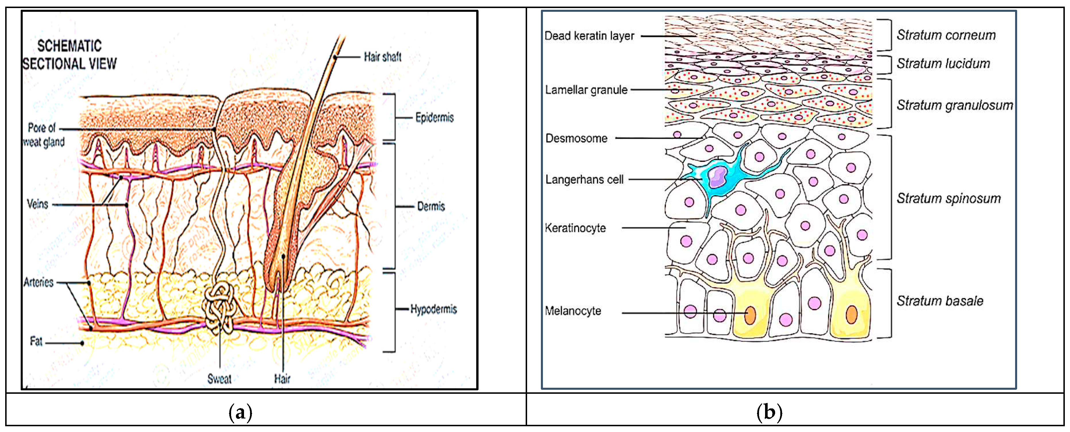
Figure 4.
Classification of TDDS.
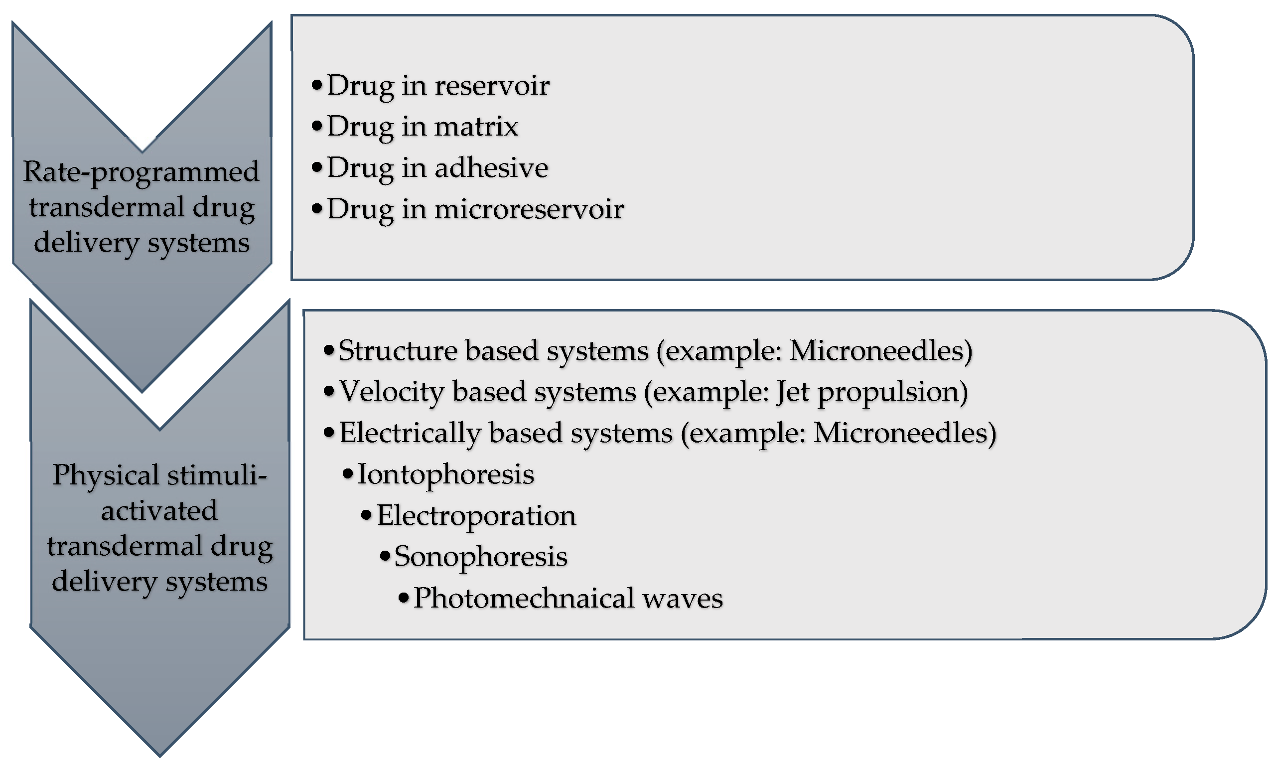
Figure 5.
Ishikawa (Fishbone) diagram to assess critical quality attributes (CQAs) of TDDS.
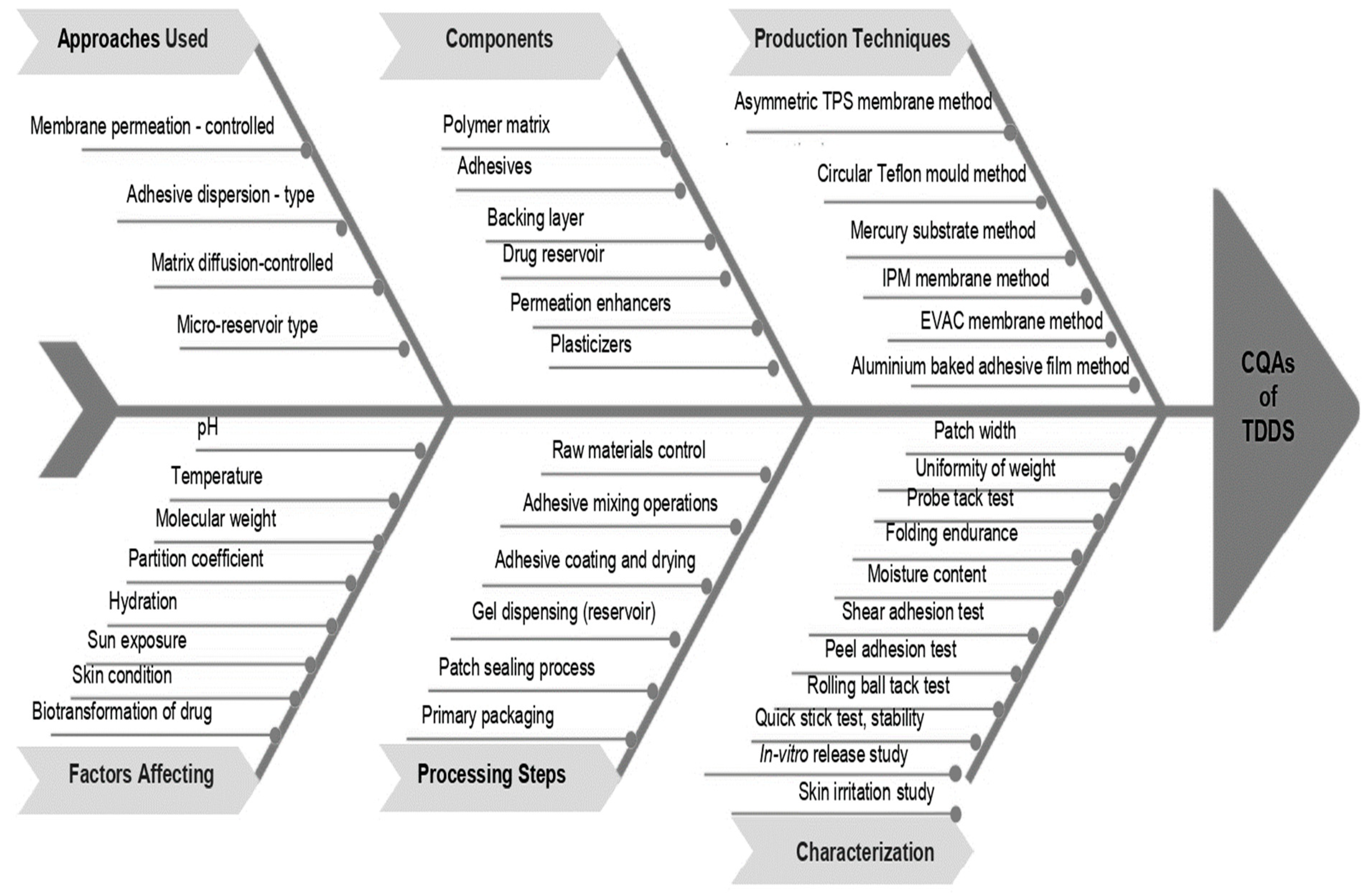
Figure 6.
Membrane-moderated transdermal drug delivery system.
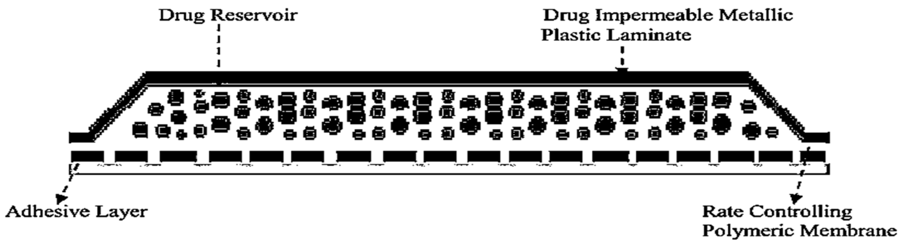
Figure 7.
Adhesive-dispersion type transdermal drug delivery system.
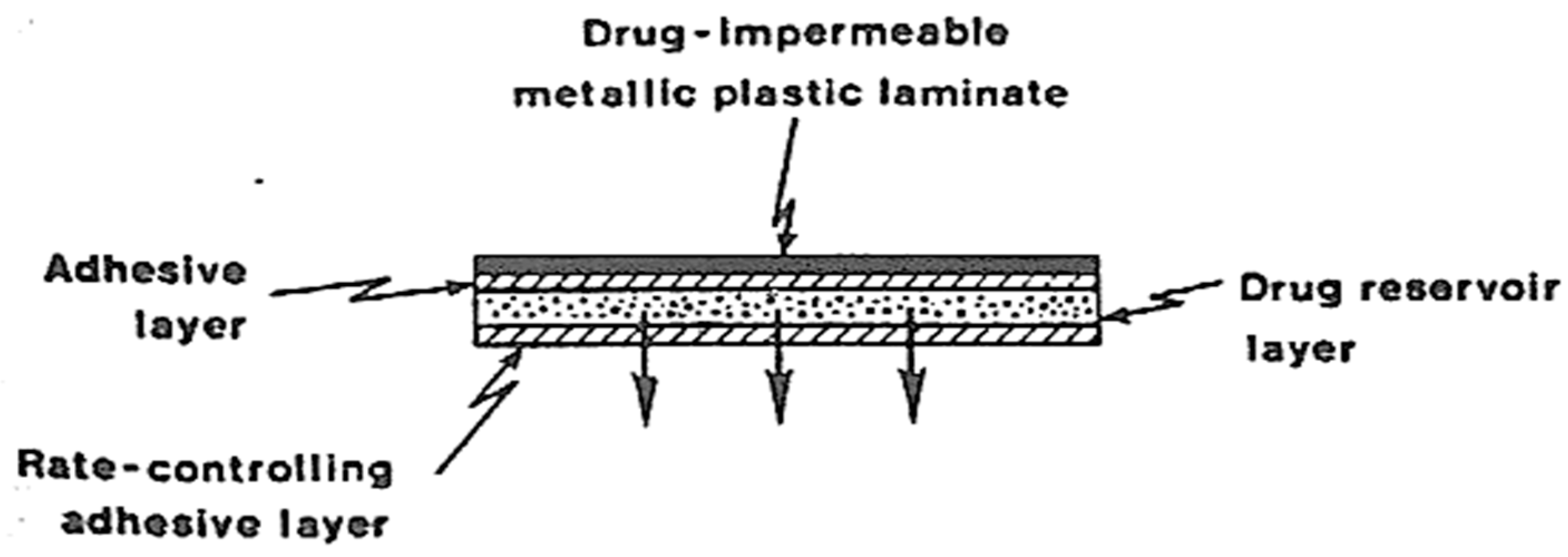
Figure 8.
Drug reservoir gradient-controlled TDDS.
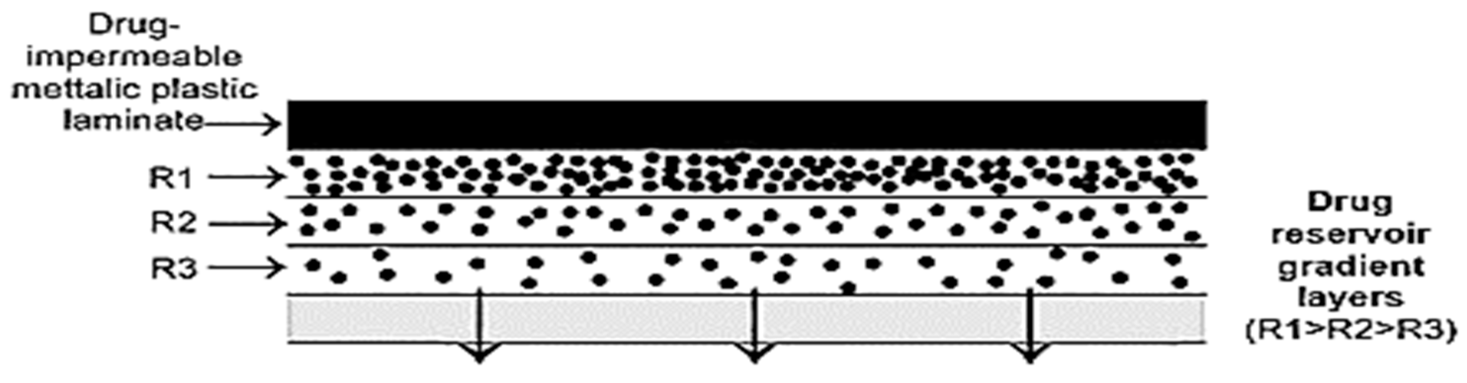
Figure 9.
Matrix diffusion-controlled TDDS.
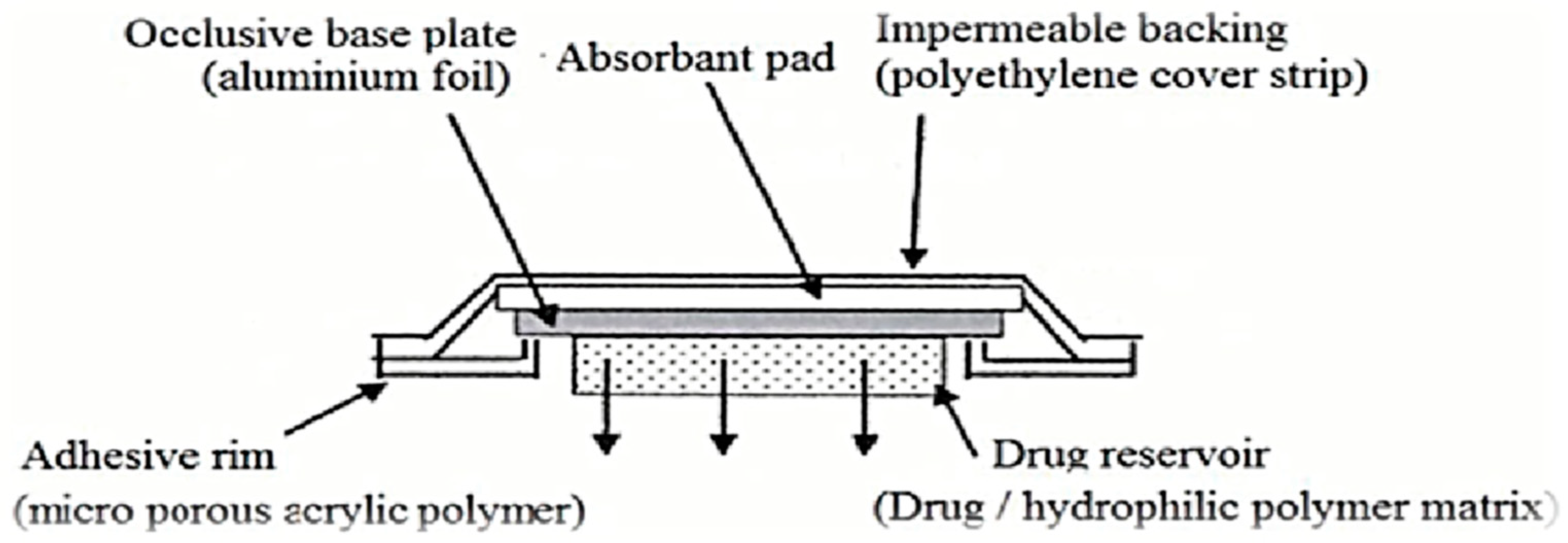
Figure 10.
Microreservoir dissolution-controlled TDDS.
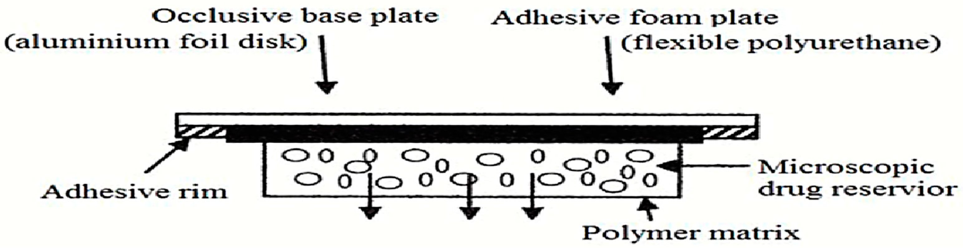
Figure 11.
Peel adhesion test.
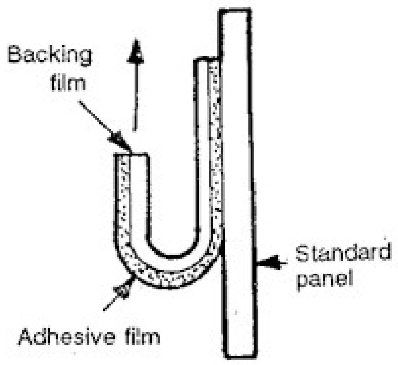
Figure 12.
Rolling ball tack test.
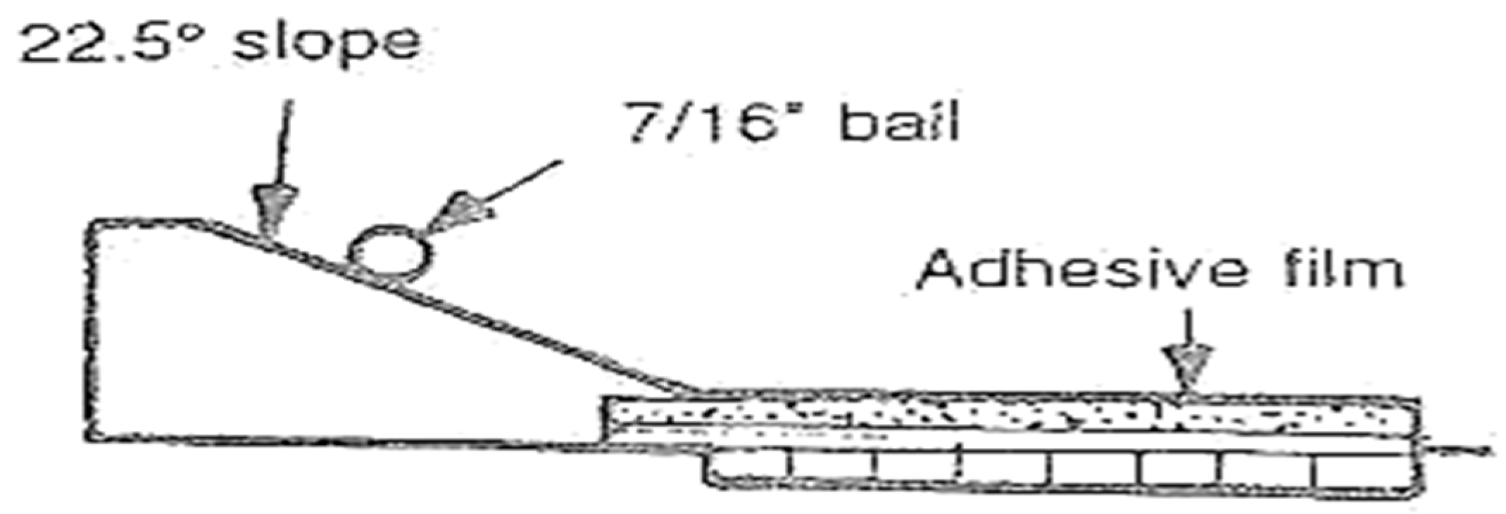
Figure 13.
Peel-tack test.
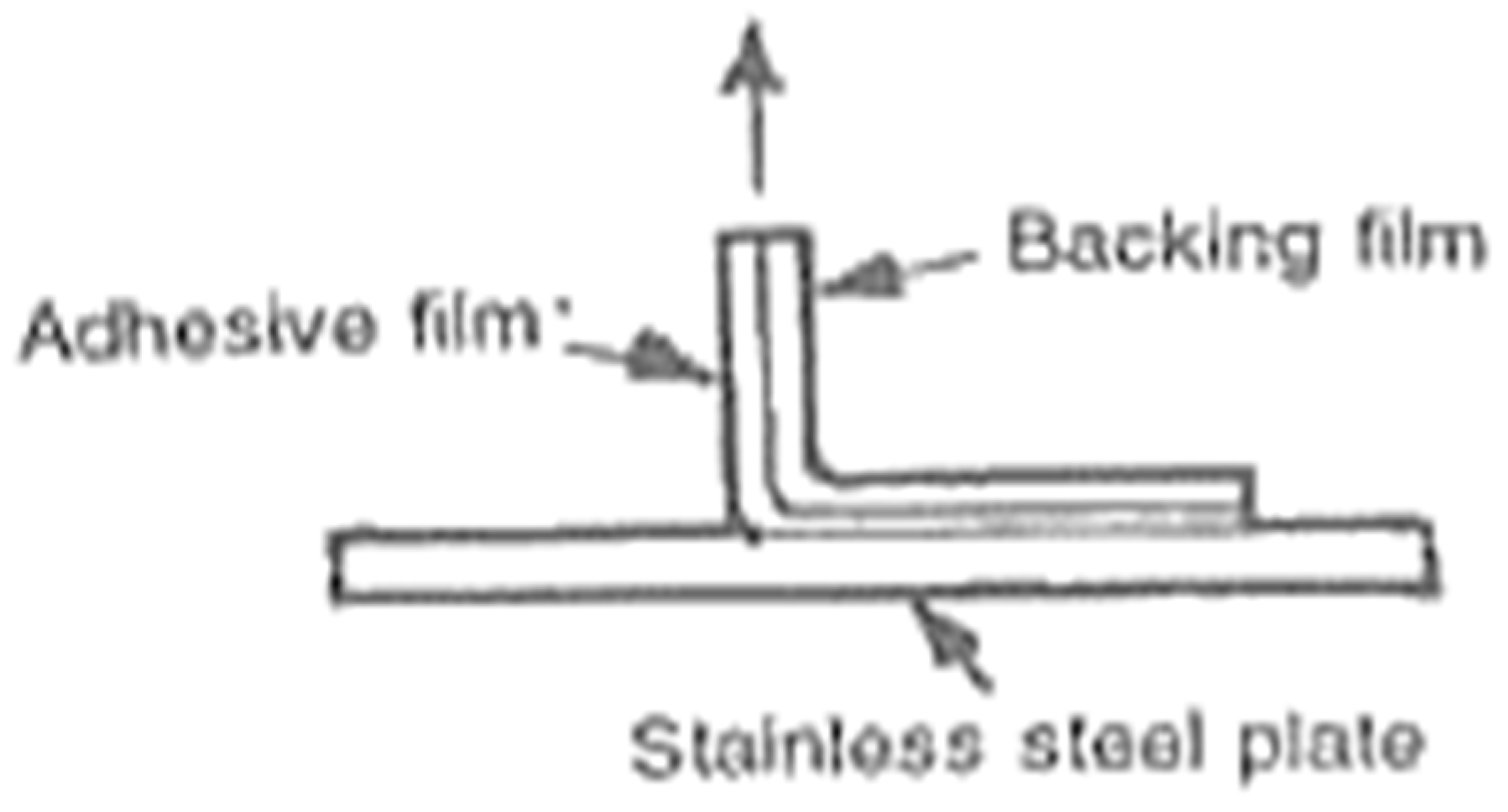
Figure 14.
Probe tack test.
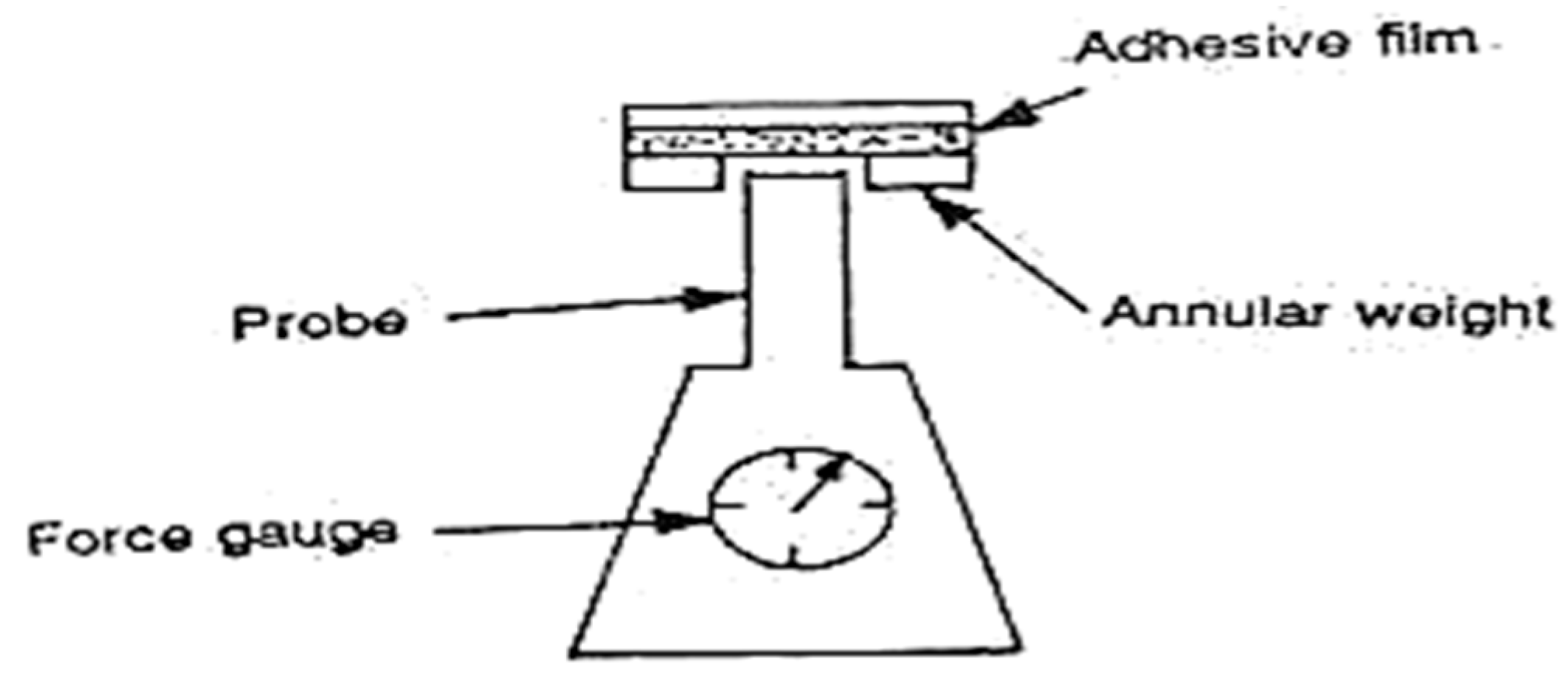
Figure 15.
Shear strength test.
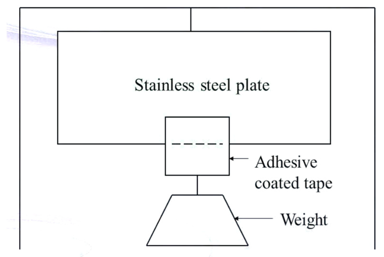
Figure 16.
Keshary-Chien (K-C) cell.
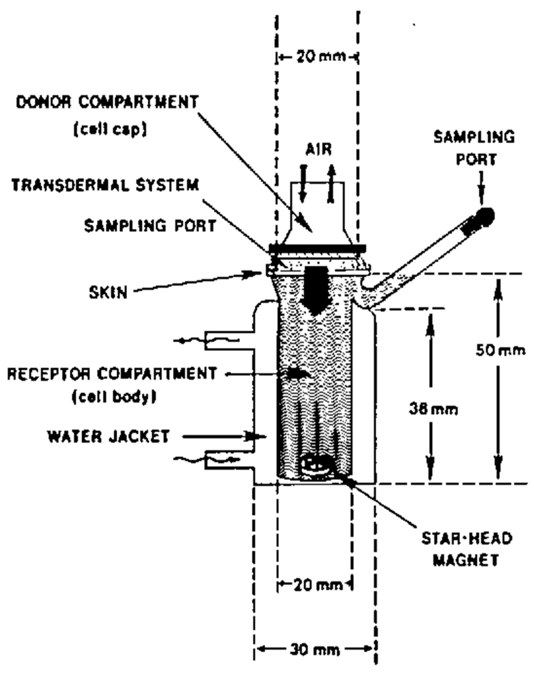
| Advantages |
|---|
| The concentration of the drug can be reduced due to improved bioavailability, protecting sensitive drugs from the harsh conditions of GIT |
| A simplified medication regimen leads to improved patient compliance and reduced inter and intra-patient variability |
| Dose frequency can be reduced, circumvents first pass effect of drugsCan be used for chronic conditions that require drug therapy for a long period of time |
| Prevent the hassle of parenteral therapy since TDDS are non-invasive |
| Sustained release of drugs, Offers long duration of action, maintains a more uniform plasma drug concentration |
| It delivers a steady-state infusion of a drug over an extended time.Therapeutic failures associated with intermittent dosing can also be avoided |
| Reduces the systemic drug interactions, self-administration is possibleThe drug can be terminated at any point of time by removing the transdermal patch |
| Limitations |
|---|
| The drug must have some desirable physicochemical properties for penetration through the stratum corneum |
| Skin irritation or contact dermatitis at the site of application due to the drug, excipients and enhancers of the drug used to increase percutaneous absorption |
| The barrier function of the skin changes from one site to another on the same person, person to person and with age. Variability of application site conditions |
| Only potent drugs are suitable for transdermal drug delivery |
| Unsuitable for large molecules (M.wt above 500 Daltons), drugs metabolized in the skin and undergo protein binding in the skin |
| The therapeutic efficacy of the medication can be affected by cutaneous metabolism |
| The dosing option is limited. Inconsistent absorption |
Table 3.
Polymers used in TDDS. [42].
Table 3.
Polymers used in TDDS. [42].
| Natural Polymers | Synthetic Elastomers | Synthetic Polymers |
|---|---|---|
| Cellulose derivatives, zein, gelatin, waxes, shellac, gums and their derivatives, proteins, natural rubber and starch | Polybutadiene, hydrin rubber, polysiloxane, silicone rubber, butyl rubber, acetonitrile, styrene-butadiene rubber, neoprene and nitrile | Polyvinyl chloride, polyvinyl alcohol, polyacrylate, polypropylene, polyethylene, polyamide, polyuria, polymethyl methacrylate, epoxy and polyvinyl pyrrolidone |
Table 4.
Ideal characteristics of a drug [44].
Table 4.
Ideal characteristics of a drug [44].
| Physicochemical Properties | Biological Properties |
|---|---|
| The molecular weight of the drug should be less than 1000 Daltons | Only potent medications with a daily dosage in the range of a few milligrams per day are appropriate |
| The drug should have an affinity for both hydrophilic and lipophilic phases | The half-life of the drug should be short and he drug should not induce an allergic response |
| The drug should possess a low melting point | Drugs that undergo degradation in the gastrointestinal tract or are rendered inactive by the hepatic first-pass effect are appropriate choices for TDDS |
| Drugs that require prolonged administration or have adverse effects on tissues other than the intended target can also be developed as TDDS |
| Solvents | Surfactants | Binary systems | Miscellaneous compounds |
|---|---|---|---|
| They increase the penetration by swelling the polar pathway and by fluidizing lipids. Examples: Water, alcohols, alkyl methyl sulfoxides, dimethyl acetamide, dimethyl formamide, pyrrolidones, propylene glycol, glycerol, silicone fluids, and isopropyl palmitate. |
They are used to enhance polar pathway transport, especially of hydrophilic drugs. These compounds are, however, skin irritants. Anionic surfactants can penetrate and interact strongly with the skin and can induce large alterations in the skin. Cationic surfactants are more irritant than anionic surfactants, hence they have not been widely used as skin permeation enhancers. Of the three classes of surfactants, nonionic surfactants have been recognized as those with the least potential for irritation and are widely used. Examples: Anionic surfactants: Dioctyl sulphosuccinate, sodium lauryl sulphate, decodecylmethyl sulphoxide. Nonionic surfactants: Pluronic F127, Pluronic F68. Bile salts: Sodium taurocholate, sodium deoxycholate, sodium tauroglycocholate. |
These systems open up the heterogeneous multilaminate pathway as well as the continuous pathways. Examples: Propylene glycol-oleic acid, 1, 4 butane diol-linoleic acid. |
These include urea (hydrating and keratolytic agent), N, N dimethyl-m-toluamide, calcium thioglycolate, eucalyptol and soyabean casein. |
| Chemical penetration Enhancers |
Drugs used | Mechanism of action |
|---|---|---|
| Terpenes | Zidovudine Imipramine hydrochloride |
Disrupt the lipid bilayer of the stratum corneum (SC), increased drug partitioning in the SC |
| Dimethyl sulphoxide | Naloxone Hydrocortisone Naloxone |
Disrupt the lipid bilayer of the stratum corneum (SC), denature the proteins of the SC |
| Pyrrolidone | Bupranolol Ketoprofen |
Change the solubility properties of the SC |
| Alcohols | Lidocaine Thymoquinone |
Alter the drug solubility in the SC, extract the lipids of SC |
| Fatty acids | Propranolol Flurbiprofen Theophylline |
Interact with the lipid bilayer. |
| Urea | Metronidazole Indomethacin |
Disrupt the lipid bilayer of the SC, increase the hydration of the SC |
| Adhesives | Backing Membrane |
|---|---|
| The attachment of transdermal drug delivery systems (TDDS) to the skin has been achieved through the utilization of a pressure-sensitive adhesive. The adhesive is applied either on the front or rear of the device and extends around the edges. It must meet the following criteria: | It is a material that is impervious and provides protection to the product when applied to the skin. Example: Metallic plastic laminate, plastic backing with absorbent pad, occlusive base plate, adhesive foam pad (flexible polyurethane) |
| Should not cause irritation or sensitization to the skin upon contact. The medication should firmly adhere to the skin throughout the dosing period without being displaced by activities. | They are flexible and provide a good bond to the drug reservoir |
| It should have a simple removal process and should not leave a permanent residue on the skin. Should come into direct contact with the skin. Commonly employed pressure-sensitive adhesives encompass polyisobutylenes, acrylics, and silicones. | Prevent drug from leaving the dosage form through the top |
| TDDS | Use |
|---|---|
| Nitroglycerin-releasing transdermal patch (Transderm-Nitro) | Once a day medication in anginal pectoris |
| Clonidine-releasing transdermal patch (Catapres) |
7 days of therapy for hypertension |
| Estradiol-releasing transdermal patch (Estraderm) |
Treatment of menopausal syndrome for 3-4 days |
| Scopolamine-releasing transdermal patch (Transderm-Scop) |
72 hours of prophylaxis for motion sickness |
| Physico-Chemical Evaluation | In-Vitro Evaluation | In-Vivo Evaluation |
|---|---|---|
| Compatibility study | In-vitro release study | Animal models |
| Thickness test | Skin irritation study | Human volunteers |
| Uniformity of weight | Stability study | Toxicological evaluation |
| Drug content | ||
| Moisture content | ||
| Adhesive evaluation | ||
| Tensile strength | ||
| Folding endurance | ||
| Water vapour transmission study | ||
| Microscopic study |
| Drug name | Formulations | Approval year | Use |
|---|---|---|---|
| Tazarotene | Lotion | 2019 | Acne |
| Asenapine | Transdermal system | 2019 | Schizophrenia |
| Trifarotene | Cream | 2019 | Acne |
| Tribanibulin | Ointment | 2020 | Actinic keratosis |
| Clascoterone | Cream | 2020 | Acne |
| Abametapira | Topical lotion | 2020 | Head lice removal |
| Calcipotriene and betamethasone dipropionate | Cream | 2020 | Plaque, psoriasis |
| Minocycline | Topical foam | 2020 | Rosacea |
| Lactic acid, citric acid and potassium bitartrate | Vaginal gel | 2020 | Contraceptive |
| Ethinylesyradiol and levonoegesterol | Transdermal system | 2020 | Contraceptive |
| Ruxolitinib | Cream | 2021 | Atopic dermatitis |
| Butenafine hydrochloride | Cream | 2021 | Fungal skin infection |
| Fentanyl | Patch | 2021 | Pain |
| Tretinoin benzoyl peroxide | Cream | 2021 | Acne |
Disclaimer/Publisher’s Note: The statements, opinions and data contained in all publications are solely those of the individual author(s) and contributor(s) and not of MDPI and/or the editor(s). MDPI and/or the editor(s) disclaim responsibility for any injury to people or property resulting from any ideas, methods, instructions or products referred to in the content. |
© 2024 by the authors. Licensee MDPI, Basel, Switzerland. This article is an open access article distributed under the terms and conditions of the Creative Commons Attribution (CC BY) license (http://creativecommons.org/licenses/by/4.0/).
Copyright: This open access article is published under a Creative Commons CC BY 4.0 license, which permit the free download, distribution, and reuse, provided that the author and preprint are cited in any reuse.


