Submitted:
27 December 2023
Posted:
27 December 2023
You are already at the latest version
Abstract
Healthcare-Associated infections (HAIs) affect more than 100 million patients each year worldwide. These infections are mainly caused by multi-resistant bacterial strains (MDR) such as Acinetobacter Baumanii which is responsible of high mortality rate in immunocompromised individuals. However, the treatment is further aggravated by the abuse of antibiotics, the lack of vaccine and the emergence of resistance posing a huge challenge for the discovery of new therapeutic drugs to overcome this nosocomial infection. In this context, antimicrobial peptides with potential antibacterial proprieties could be an alternative. In this research, we describe the synthesis and the bioactivity of dermaseptins and their derivatives against Acitenobacter baumanii. The cytotoxicity of these DS was investigated on the HEp-2 cell line by the MTT cell viability assay. Thereafter, we studied morphological alterations caused by the action of one of the active peptides on the bacterial membrane using Atomic Force Microscopy (AFM). The cytotoxicity of dermaseptins was concentration-dependent at microgram concentrations. It was observed that all tested analogs exhibit antibacterial activity with MICs ranging from 3.125 to 12.5 μg/mL and MBCs ranging from 6.25 to 25 μg/mL. Microscopic images obtained by AFM revealed morphological changes on the surface of treated bacteria caused by K4S4(1-16), as well as significant surface alterations. Overall, these findings demonstrate that DS might constitute new lead structures for the development of potent antibacterial agents against Acinetobacter infections.
Keywords:
Dermaseptin B2
; Dermaseptin S4
; analogs
; Acinetobacter baumannii
; Healthcare-Associated Infections
; Antibacterial activity
1. Introduction
Healthcare-Associated infections (HAIs), also known as nosocomial infections are infections acquired within a healthcare establishment, which were not initially present or incubating at the time of patient admission. They generally manifest 48 hours or more after hospitalization [1]. HAIs can be transmitted by patient contact with healthcare personnel, patients contact, medical devices, or procedural interventions by the means of hand contact, oral transmission, parental, aerial and vector borne [2]. These infections are caused by various microorganisms, such as multidrug-resistant bacteria (MDR) and other agents responsible for their occurrence. World Health Organization (WHO) data estimates that HAIs lead to approximately 40,000 deaths per year. These nosocomial infections have rates of up to 25% in developing countries unlike developed countries which show lower values of around 5 and 15% [3,4]. HAIs represent important public health concerns requiring global attention, given their impact on morbidity and mortality rates, as well as the considerable associated cost of medical care. Additionally, Cases of nosocomial COVID-19 infections have been identified and acquired in many medical facilities around the world [5]. Most of HAIs are attributed to catheter-associated urinary tract infections (CAUTIs), hospital-acquired pneumonias (HAPs), bloodstream infections (BSIs), and surgical site infections (SSIs) [6].
Acinetobacter baumanni, a Gram-negative coccobacillus, is the predominant human pathogen in hospital settings presents considerable viability on human hands, contributing to significant cross-contamination rates in nosocomial infections due to this characteristic [7]. Nowadays, A. baumannii holds the highest rank on the WHO priority pathogen list as "critical," highlighting its importance as a nosocomial causative pathogen, particularly in cases where it presents resistance to carbapenem, the "last resort" antibiotic [8]. Previous study has been reported the Acinetobacter baumannii as a responsible agent of ocular infections. Ophthalmic complications in burn victims have been observed due to corneal injuries or an associated trauma. Acinetobacter ophtalmic infections are more common in patients requiring respiratory assistance [9]. Therefore, this multi-drug resistant bacillus is the top priority for antibiotic research and development efforts due to their ineffectiveness. The evolution of antimicrobial resistance of Acinetobacter strains to antibiotics began in 1932 [10]. These antibiotics include: beta-lactams [11], Carbapenems [12], Colistin [13], Tigecycline [14] and quinolones [15].
For years, the medical field has witnessed the emergence of new resistance mechanisms in A. baumannii against antimicrobial treatments, leading to a reduction in effectiveness of therapeutic interventions. Significant research has been conducted on the development of preventive vaccines. Several promising vaccine methods have been studied in preclinical trials over the past decade, showing varying degrees of success. However, to our knowledge, none of these investigated candidates has progressed beyond preclinical evaluation [16]. For these reasons, the development of new drugs for HAIs caused by A. baumanii is immediately required, preferably employing compounds effective in low concentrations, non-toxic and selective against this bacteria. Antimicrobial peptides, especially dermaseptins could be potential candidates for such assays. Cationic antimicrobial peptides (AMPs) have attracted considerable attention in the search of new therapeutic options. These small peptides, composed of 5 to 100 amino acid residues, present a wide range of molecular weights (<10 kDa) [17,18]. Various organisms produce these peptides through their innate immune system; they are derived from various sources such as: macrophages, neutrophils, epithelial cells, haemocytes, fat body, reproductive tract, and similar origins [19]. These PAMs are potentially active against a broad spectrum of microorganisms against bacteria, fungi, eukaryotic parasites and/or viruses [19]. The majority of these peptides are positively charged, amphipathic and act according to various mechanisms: the formation of pores in the bacterial membrane, dissolving the membrane, or targeting crucial bacterial processes like cell wall synthesis or protein production [20]. Among these antimicrobial peptides are Dermaseptins, a family of cationic AMPs initially identified in the skin of South American Phyllomedusa frog. These DS are secreted by amphibians' skin as part of their defense mechanism against microbes. These polycationic-amphipathic peptides are linear and generally composed of 28–34 amino acids, exhibiting significant variability between different peptides. Nevertheless, in apolar solvents, they tend to adopt an amphibathic α-helices [21]. The Dermaseptins exhibit substantial variation in both their peptide sequences and lengths. However, they share structural simlarities, such as the presence of conserved Trp residue at position 3, a conserved sequence of AA(G)KAALG(N)A in the middle region, and a net positive charge [22]. Overall, DS-S peptides isolated from the secretions of Phyllomedusa sauvagei have not been widely used for various human antimicrobial applications. These applications are particularly interesting due to their ability to effectively eliminate microbes without encountering resistance and their rapid mechanism for killing pathogens [23]. So far, a total of thirteen dermaseptins (DS1 to 13) have been discovered and characterized [24]. Considering the existing data regarding dermaseptins' action against bacteria, we can identify two distinct types of mechanisms: The initially proposed mechanism involves the "barrel-stave" model; it is mediated by the attachment of dermaseptins to membrane phospholipids. This interaction disrupts the osmotic balance of the cell, leading to membrane permeabilization. Subsequently, transmembrane pores or channels form, eventually causing membrane rupture. Another second mechanism, called the "carpet-like" mechanism, involves the binding of positively charged lytic peptides to the negatively charged surface. This destruction method allows complete surface coverage, leading to the impregnation and disintegration of the membrane [25].
Dermaseptins S have generated substantial interest due to their potential as a clinical approach against microbial resistance to existing antibiotics and antifungals. Certain dermaseptins exhibit an impressive ability to efficiently, rapidly, and irreversibly inhibit microbial cells, without causing toxicity on mammalian cells [23]. Besides their large spectrum of activity, DRS are not hemolytic [24], excepting for dermaseptin S4, which demonstrates a potent hemolytic effect [26]. In the same context, DRS-B2, extracted from the secretion of Phyllomedusa bicolor [27], is alternatively referred to as adenoregulin due to its capacity to modulate the binding of adenosine A1 receptor agonists [28]. Among the peptides of DRS family, DRS-B2 stands out for its highest level of activity, making it the subject of extensive research. This amphipathic (+3) cationic polypeptide composed of 33 amino acids, with a molecular weight of approximately 3180 Da, and a tryptophan residue at position 3, and six lysine residues [29]. This α-helical peptide has the ability to disrupt the membranes of a wide range of microorganisms, such as bacteria, yeast, fungi, and protozoa. However, its specific mechanism of action remains unclear [30]. There are reports of administration of (DRS-Bs) to humans in non-experimental settings; comprehensive data on the pharmacokinetics, efficacy, and safety in humans are currently unavailable [31].
To our knowledge, no studies have been carried out to evaluate the antibacterial effect of the native B2 and our derivatives from both dermaseptin families against Acinetobacter baumanii. Thus, in the current research, we report for the first time the in vitro antibacterial activity of these peptides against nosocomial infections. Dermaseptin derivatives were synthesized, purified and evaluated for their antibacterial activity. Their cytotoxicity towards Hep-2 cells was evaluated and then the atomic force microscopy (AFM) was performed to describe morphological changes on bacteria.
2. Materials and Methods
2.1. Synthesis, Purification, and Preparation of Peptides
Peptides were prepared by stepwise solid phase synthesis using Fmoc polyamide-active ester chemistry on a Milligen 9050 pepsynthesizer. All Fmoc-amino acids were from Milligen–Waters (France). 4-(Hydroxymethyl) phenoacetic acid-linked polyamide/kieselguhr resin (pepsin kA), Fmoc-aminoacidpentafluorophenyl (Pfp), and 3-hydroxy- 2,3-dehydro-4-oxo-benzotriazine (Dhbt) esters were from Milligen/Bioresearch. Cleavage of peptidyl-resin and side chain deprotection were carried out using 5 mg of peptidyl-resin in 1 ml of a mixture composed of trifluoroacetic acid, para-cresol, thioanisol, water, and ethyl methyl sulfide (82.5%, 5.5%, and 2.5% (v/v)) for 2 hr at room temperature. After filtering to remove the resin and ether extraction, the crude peptides were purified by a combination of Sephadex gel filtration, ion exchange chromatography, and preparative high performance liquid chromatography (HPLC). The homogeneity of synthetic peptides was assessed by analytical HPLC, amino acid analysis, solid phase sequence analysis, and mass spectrometry [21]. All peptides were stored frozen as stock solutions at 3.5 mM in double-distilled water at -20°C.
2.2. Calculation of Peptide Physicochemical and Structural Parameters
Properties of our peptides such as Length, net charge (Z) and Molecular Weights (MW) were calculated using BACHEM peptide calculator tool. Both hydrophobicity (H) and hydrophobic moment (μH) were calculated using Heliquest software [32]. The total trend of aggregation (aggregation) in aqueous solution was predicted using TANGO software [33]. While the helicity (α-helix %) of each peptide was calculated using AGADIR software [34].
2.3. Bacterial Strains and Inoculum Standardization
The bacterial strain used in the present study is from the stock culture of the microbiology laboratory of the Federal University of the Delta of Parnaíba - UFDPar, Parnaíba - PI. The strain used in the tests is: Acinetobacter baumannii clinical specimen MDR (multidrug resistant). For the study, these isolates were subjected to susceptibility test according to the standards of the Clinical Laboratory Standards Institute [35]. Before performing all experiments, the selected strains were seeded in Petri dishes containing Mueller-Hinton agar (Difco™), and then, under aerobic conditions, they were incubated in a bacteriological oven for 24 hours at 35 ± 2 °C. After this time, the colonies grown alone were collected with a disposable bacteriological loop and suspended in sterile saline solution (0.85% NaCl (w/v)), in order to reach an absorbance pattern between 0.08 and 0.13, at 625 nm in UV-vis spectrophotometer (Shimadzu, Japan), thus corresponding to 0.5 McFarland scale (1 - 2 x 10⁸ CFU/mL), as recommended by CLSI. Once standardized, the bacterial suspension obtained was subsequently used to prepare the bacterial inoculum used in the execution of Minimum Inhibitory Concentration (MIC) determination protocols [35].
2.4. Antibacterial Experiments
In accordance with the standards recommended by CLSI [35], the antibacterial potential of peptides was evaluated with the method of determining the Minimum Inhibitory Concentration MIC, through broth microdilution method. Using a 96-well microdilution plate (KASVI), the antibacterial effect was analyzed against the bacterial strains. Therefore, 5 μL of each peptide were added to the first line of the microtiter wells containing 195 μL Mueller-Hinton broth followed by 2-fold serial dilutions with final concentrations ranging from 25 µg/mL to 0.19 µg/mL for all peptides. The volume of the bacterial inoculum was equal to 50 µL, it was added to the test wells with the M-H broth at the beggining of the experience to give the final concentration of 5 x 10⁵ CFU/mL and to reach the final volume of 100 μL in each well after discarding the last additional volume of 100 μL following the serial dilution. MIC was defined as the lowest concentration of an antibacterial agent expressed in mg/L (μg/mL) which, under strictly controlled in vitro conditions, completely prevents visible bacterial growth after incubation for 24 h at 37°C in aerobic conditions. The MBC is complementary to the MIC; the MBC demonstrates the lowest concentration of antimicrobial agent that inhibited growth of bacterial colonies on the agar. MBC was verified by seeding 10 μL of the wells that showed results equal to or greater than the MIC onto Mueller-Hinton Agar (MHA), with the assistance of the Drigalski spatula. All tests were performed in triplicate. In order to guarantee the quality and safety of the protocols of this study, the manipulation of bacterial strains was performed under recommended aseptic conditions. In addition, all procedures for the execution of the experimental protocols were performed in a class II B2 biological safety cabinet (Buzattos, MG, Brazil).
2.5. Cell Culture
The HEp-2 cell line contains HeLa marker chromosomes and was derived via HeLa contamination. It was obtained from the American Type Culture Collection (ATCC, USA). Cells were routinely maintained in a humidified atmosphere of 5% CO2 at 37 °C. The Culture Medium Dulbecco's Modified Eagle Medium (DMEM) was supplemented with 1% L-glutamine, 1% penicillin/streptomycin and 10% (v/v) heat inactivated fetal bovine serum (FBS).
2.6. MTT Assay and Cytotoxicity Analysis
The cytotoxicity test consists of measuring the viability of cells in culture when they are brought into contact with the peptides to be tested. Cytotoxicity is determined using the 3-(4, 5-dimethylthiazol-2-yl)-2, 5-diphenyl tetrazolium bromide (MTT) colorimetric assay on cultured cells (HEp-2 lines). The lyphosized peptides are diluted in distilled water to obtain a final concentration of 1 mg/ml. ½ dilutions of the different peptides were then prepared in Eppendorf tubes with the fresh medium DMEM. These concentrations range from 100 µg/ml to 1.5 µg/ml. The HEp-2 cells in suspension (MEM medium with 10% FCS) are distributed in 96-well plates, at a rate of 100 μl containing 105 cells/well, and incubated in the oven at 37°C at 5% CO2 for 24 hours. Subsequently, the medium is removed and replaced with 50 μl DMEM medium with 2% FCS. 50 µl of each peptide are added to all wells, and a 1/2 dilution series is carried out from 100 µg/ml to a concentration of 1.5 µg/ml. The test is carried out in triplicate and in three different times. Untreated cells will serve as a negative control. After 72 hours of incubation, the supernatant is collected and the viability of the cells treated with the peptides is determined by the MTT method. Briefly 50 µl of the MTT solution (5 mg/ml) are added to each well. After 4 hours of incubation at 37°C, the optical density (OD) is measured at 570 nm using an ELISA reader (Multiskan EX, Labsystems), after adding 100 μl of DMSO to be able to dissolve the crystals of formazan trained. The results are expressed as a percentage of viability relative to the negative control control without peptide tested, according to the formula: (Viability percentage = (DO544 nm peptide / DO544 nm control) ×100). The results were expressed in percentage of viable HEp-2 cells and the half maximal cytotoxic concentrations to the HEp-2 CC50 values were calculated with GraphPad Prism® (version 9.0).
2.7. Atomic Force Microscopy (AFM)
With the objective of observing possible morphological alterations caused by the action of an active peptide from dermaseptin families, bacterial growth control and bacteria treated with K4S4 (1-16) were evaluated, using the technique of Atomic Force Microscopy (AFM). Therefore, the selected bacterial strain, Acinetobacter baumannii MDR, was submitted to this experimental assay. The bacterial inoculum used to obtain the images was 5×10⁵ CFU/mL and, for sample preparation, procedures similar to those described by Araújo et al [36] were used. Briefly, after 24 hours of incubation of the MIC determination assay, 20 µL of culture medium from the wells of the treated and untreated (control) group were deposited on the surface of a glass slide. Then, the samples were submitted to drying in an oven at 37 °C for 10 minutes. After this time, the samples were then carefully washed with distilled water and subjected to drying under the same conditions described above. After this preparation, AFM images of 6 x 6 µm were obtained using the model TT-AFM microscope (AFM Workshop - USA), in intermittent contact mode (tapping mode), using TAP300-G10 tips (TED PELLA, INC.), with a resonance frequency of approximately 239 kHz. Multiple areas (n=10, 0.3 x 0.3 µm) of each sample were examined in order to verify the average roughness of treated and untreated bacteria, using the program Gwyddion 2.60.
2.8. Statistical Analysis
The statistical analyses were performed with the GraphPad Prism 9.0 software (GraphPad software Inc.) for cell viability assays. The difference between the average roughnesses (Ra) was statistically analyzed using T Test, with GraphPad Prism 8.0 software. Differences were considered as statistically significant when p < 0.05.
3. Results and Discussion
3.1. Design of Dermaseptin S4 and B2 Derivatives
In our study, the peptides were designed by making three main steps of structural modifications: substitutions and/or deletions to S4 and B2 sequences (Table 1). K4K20S4 is a derivative that was formed by replacing methionine (M) in position 4 and asparagin in position 20 with a lysine (K) and designating it as M4K and N20K. Peptides K4S4(1-16) have the same M4K substitution with the deletion of 12 C-terminal residues. The K3K4B2 derivative was obtained by a double substitution, a tryptophan residue (W) with a lysine (K) at position 3(W3K) and a serine residue (S) with a lysine at position 4 (S4K) of B2. The one-letter code was used to identify the amino-acid sequence of these peptides, taking as reference the sequence and length of the native peptides, dermaseptins S4 and B2 (Table 1).
The first modification step was designed to increase hydrophilic characteristics and decrease hydrophobicity. Since S4 and B2 exhibited some levels of functional activity with cationic residues, our initial focus in modifying this peptide was to enhance its hydrophilic properties by adding basic amino acids such as lysine and the choice of lysine was also made to prevent an increase in cytotoxicity. In fact, it was previously shown that increasing the net positive charge and decreasing the hydrophobicity of dermaseptin S4 led to decreased hemolytic activity while maintaining high biological activity [26,37,38]. The S4 sequence is minimized by truncating 12 amino acids in the secondary structure. It has been reported that the use of AMPs as therapeutic agents faces challenges attributed to their long peptide sequences, limited efficacy, instability, systemic toxicities, and the potential to compromise innatedefense immunity of the host. These factors have hampered the progress and clinical implementation of AMPs [39]. Several approaches have been created in the design of analogous peptides to surmount these obstacles. These methods include motif hybridization, aimed at enhancing antimicrobial efficacy and functionality [40], truncation/ substitution intended to reduce toxicity [41], and de novo design, aimed at shortening peptide length and eliminating host defense immunogenicity [42]. Previous data suggested that the N-terminal domain of dermaseptin demonstrates selectivity during the interaction with the bacterial cell membrane, while the C-terminal helix mainly exhibits nonspecific membrane lytic activity [43,44]. Previous studies on N-terminal peptide fragments of dermaseptins revealed that truncated peptides ranging from 16- to 19- mer, maintain comparable antimicrobial activity. However, shorter sequences containing less than 13 amino acid residues experience a significant reduction in antimicrobial activity [45,46]. In summary, the criteria selection of peptides was examined as a means of defining the structural requirements for biological activity. Regarding B2, to the best of our knowledge it’s the first time that the derivative K3K4B2 is used. Indeed, modifications made to the native B2 molecule primarily involved truncation, such as the design of a C-terminal truncated analog known as [1-23]-Drs B2. Despite maintaining the net cationic charge of the native peptide B2, this truncated analog was found to be inactive against bacteria [30]. Combining DRS-B2 with alginate nanoparticles (Alg NPs) results in a formulation (Alg NPs + DRS-B2) that creates novel B2 derivatives displaying significant antibacterial efficacy against both sensitive and resistant strains of Escherichia coli and E. coli to colistin. The antibacterial activity achieved with this innovative formulation exceeds that of DRS-B2 when used alone [29]. Moreover, our synthesized peptides offer the advantage of being configured with D-amino acids. Unlike, peptides in their natural form consist of L-amino acids, which pose a challenge due to their susceptibility to degradation by proteases, thereby limiting their clinical applications owing to poor stability [47]. As previous research pointed out, a feasible approach to adress these constraints involves replacing the L-amino acids at the most susceptible site with D-amino acids [48]. Obviously, substituting with D-amino acids does not alter the net positive charge of the native peptide. However, it does impact the structure and function associated with recognizing chiral targets [49,50].
3.2. Structural and Physicochemical Properties of Peptides
According to Heliquest, we noted that K4K20S4 peptide had the highest value of Hydrophobicity (H) among S4 peptide derivatives (0.451), whereas K4S4(1–16) had the lowest value (0.426) (Table 1). In general, all peptide derivatives of dermaseptin S4 had a lower H than the native peptide (S4). This is also confirmed for dermaseptin B2 and its derivative, where the H value is higher (0.199) than K3K4B2 value (0.072). This hydrophobicity is a crucial physicochemical characteristic of peptides. Typically, it is determined by analyzing the peptide's sequence [51]. It plays an important role on their conformational modifications, stability and their molecular interactions [52]. The μH ranged from 0.159 to 0.526, where K4S4(1-16) showed the highest value (μH = 0.526) and K3K4B2 showed the lowest (μH = 0.159) (Table 1). The hydrophobic moment μH is established through the calculation of the vector sum of hydrophobicity values for each individual amino acid [32,53]. This factor significantly influences the interfacial binding of peptides with the membrane [54]. Based on the TANGO algorithm [33], we noticed that dermaseptin S4 presents a greater tendency to aggregation than its derivatives due to the presence of two N-terminal and C-terminal hydrophobic domains (Table 1, Figure 1). These results agreed with Feder et al, who demonstrated that aggregation in dermaseptin S4 and its derivatives occurs through hydrophobic interactions. Indeed, the M4KN20K substitution in the N-terminal and C-terminal domains induced a decrease in the aggregation tendency of K4S4, which was in accordance with other studies [26,55]. However, the deletion of C-terminal domain of S4 and the insertion of positive charges in these regions result in a loss of the aggregation. K4(1-16)S4 had a value of aggregation equal to zero (Table 1). Hence, the deletion of the hydrophobic domains and/or the insertion of positive charges in these regions will probably decrease the aggregation, either due to the absence of such hydrophobic domains or by electrostatic repulsion between cationic residues [26,31]. These results revealed that the aggregation of peptides is influenced not only by their hydrophobicity, but also by their charge distribution. Thus, the introduction of cationic amino acids into one of the hydrophobic domains, and/or the elimination of hydrophobic domains, adversely affects aggregation. Regarding dermaseptin B2, we noticed that neither substitution nor deletion induce a loss or a decrease of aggregation since the native molecule had a very low degree of aggregation (9.681) comparing to S4 (Table 1). Previously, it has been demonstrated that the tendency to aggregation in aqueous solution is another important parameter for antibacterial activity and cell selectivity [56]. This property can be explained by the ability of peptides to form aggregates (oligomers) in aqueous solution, establishing hydrophobic interactions with other monomers. Due to the increase in size and the loss of flexibility, such aggregates are unable to pass through polysaccharide capsules, outer membranes and bacterial cell walls, being unable to interact with the plasma membrane, which is the action target. Consequently, peptides with a high tendency for aggregation are weakly active against bacteria; on the other hand, for a monomeric peptide, it is easier to reach the plasma membrane and present an effective antibacterial activity [57]. Therefore, in designing antimicrobial peptides, it is usually desirable to reduce aggregation to favor antibacterial activity [38,57]. In our study, we noticed that all our peptides’ derivatives have a reduced aggregation, which provide them a potential biological activity such as antileishmanial activity.
To estimate the helicity of the studied peptides, we used the AGADIR algorithm [34] which indicated that the native molecule S4 has the greater a helicity (as α-helix %) approximately eightfold greater than K4S4(1-16), and similar to K4K20S4. However, DS-B2 and its derivative had the same helicity (10.2 % and 9.85 % respectively) (Table 1). It has been reported that helicity was assessed to investigate the relationships of secondary structure and the selectivity against microbial cells of α-helical antimicrobial peptides [58]. Therefore, the high potency of the dermaseptins may be attributed not only to a higher net positive charge, but also to the stabilization of the helical conformation [59].
3.3. In Vitro Toxicity of Dermaseptin and Derivatives against HEp-2 Cells
Considering that the respiratory tract is the most commonly linked site for A. baumannii infection [60]. HEp-2 cells originating from the respiratory tract exhibit higher susceptibility to A. baumannii cell invasion compared to HeLa cells that were not derived from the respiratory tract [61]. This cell line contains HeLa marker chromosomes and was derived via HeLa contamination. Thus, all our peptides were evaluated for their cytotoxicity on HEp-2 cells using the MTT viability assay and the results are shown as survival rates after 72 h of treatment with the compounds (Table 2).
Considering our results, the tested cationic derivatives exhibit variable toxicity towards Hep-2 cells. Cells were exposed to increasing concentrations of peptides ranging from 1.5 to 100 μg/ml. The cytotoxicity of dermaseptins was concentration dependent (data not shown), and peptide 50% cytotoxic concentration (CC50) values were determined (Table 2). The highest cytotoxicity values for all S4 and B2 derivatives were recorded at concentrations higher than 61.25 μg/ml (CC50). Interestingly, results showed that shortening the peptide in the C-terminal extremity of K4S4(1–16) which present an CC50 equal to 68.9 μg/ml; and increasing its positive charge with different substitutions (K4K20S4 or K3K4B2) led to peptides with low toxicity, with CC50 of about: 75.71 μg/ml and 61.25 μg/ml respectively. Belaid et al demonstrated that maximal non-cytotoxic concentrations were 32 μg/ml for dermaseptin S1, S2; 16 μg/ml for dermaseptin S3 and S4, and 64 μg/ml for dermaseptin S5 to HEp-2 cells [62]. Besides, the study of Gourkhede et al indicate that other AMPs like Cecropin A (1–7)-Melittin and lactoferricin (17–30)a exhibited negligible cytotoxicity at lower concentrations (1X and 2X MIC); however, at 4X MIC, slightly higher cytotoxicity was observed, given that MIC values are equal to 64 μg/ml and 128 μg/ml respectively [63]. Sruthy et al confirm that at the highest concentration tested of histone H2A-derived antimicrobial peptide (200 μM), growth inhibition of 89% was observed in HEp2 cell lines [64]. Another study conducted by Hazime et al. tested the cytotoxic effects of DRS-B2, and also that of the new formulation (Alg NPs + DRS-B2) on the human erythrocytes and eukaryotic cell line types HT29 (human) and IPEC-1 (animal) and their safety have been verified [29]. Zairi et al. found that dermaseptin K4S4 exhibited an enhanced toxicity profile, against human endometrial epithelial cells displaying lower sensitivity to dermaseptins' toxic effects compared to other cell types [65]. While, dermaseptin S4 and its derivatives demonstrate significant cytotoxicity against SW620 cell line, it remains challenging to discern their cellular selectivity or mode of action [66]. Moreover, Lorin et al demonstrated that dermaseptin K4S4(1–16)a exhibited comparable effect on both HeLa P4-CCR5 cells and primary PBMCs, showed no toxicity in mice, and revealed its reduced cell toxicity at high concentrations [67]. All this work has shown that synthesized and modified peptides are less toxic to HEp-2 cells compared to the native molecules S4 and B2.
3.4. Antibacterial Activity of Dermaseptins Derivatives against Acinetobacter baumannii
The susceptibility of bacteria to dermaseptin S4 and B2 derivatives was assessed by measuring the peptide Minimum Inhibitory Concentration (MICs) against the clinical isolate: Acinetobacter baumannii, the multidrug-resistant gram-negative bacteria. The resulted data are summarized in (Table 2). The data shows that all peptides tested inhibited A. baumannii growth was dependent on the nature of the peptide and the highly charged molecules were the most active. The MICs ranged from 3.125-12.5 μg/ml. Thereby, demonstrating the portential of these peptides as antibacterial agents. Thus, in Table 2 we can notice that the most effective peptide is K4K20S4, this peptide displayed potent antibacterial activity with MICs of about 3.125 μg/ml. Likewise, The mono-substituted truncate peptide K4S4(1-16) present a neary homogenous potency with a MICs walue equal to 6.25 μg/ml. The truncation of the C-terminal extremity results in a less toxic peptide and did not affect its potency. As Kustanovich suggested, the interaction with cell membrane of N-terminal domain mainly depends on the net charge state while the C-terminal domain also contributes to the binding affinity [38]. The native dermaseptin S4 has a MICs of 12.5 μg/ml and was the less potent against A. baumannii, but S4 presents the same MICs value as the native B2. The analog K3K4B2 is likewise more active compared to the native B2, the peptide exhibits a MICs equal to 6.25 μg/ml (same value as K4S4(1-16)). The increase of the positive charge for the derivatives of both of dermaseptin S and B is essential to increase their antibacterial activities. Therefore, K4K20S4 and K3K4B2 are the most active analogs against Acinetobacter baumannii since they have the lowest MICs values. In the same context, our result showed that native molecules such as B2 and S4 remain less actives comparing to their cationic analogs.
After determining the MIC, the Minimal Bactericidal Concentration (MBC) was assessed against A. baumannii MDR. Meropenem was used as antibacterial control drug. The solvent saline loaded served as negative control. However, meropenem which is used as a reference presented an MIC of 32 µg/ml and an MBC value of 64 µg/ml. Meropenem, belonging to the carbapenem class of beta-lactam antibiotics, exhibits a broad spectrum of activity and minimal toxicity. This antimicrobial agent provides effective coverage against various microorganisms, making it a valuable and frequently prescribed treatment for managing severe and nosocomial infections [68]. Meropenem targets Gram-positive and Gram-negative bacteria as well as anaerobic bacteria. Similar to other carbapenems, meropenem disrupts the synthesis of bacterial cell walls, inhibiting growth and leading to cell death [69]. Inadequate concentrations of meropenem may result in treatment failure and heighten the risk of microbial resistance emergence [70].
MBC showed us results equal to or greater than the MIC values, values ranging from 6.25 μg/ml to 25 μg/ml for both dermaseptins derivatives. The highest MBC found (25 μg/ml) present the native molecules S4 and B2 values. The lowest MBC (6.25) was for K4K20S4. For K4S4(1-16) and K3K4B2, MBC is equal to 12.5 μg/ml. However, it has been reported in a previous study of Jiang et al, that some dermaseptins S4 and Piscidin 1 and their derivatives (piscidin 1 (I9K) and dermaseptin S4 (L7K, A14K)) were active against the gram-negative pathogen A. baumannii (11 strains) [71]. In the case of D-dermaseptin S4, the geometric mean of MIC values for A. baumannii decreased from 1.8 µM for D-dermaseptin S4 to 1.1 µM for D-dermaseptin S4 L7K, A14K indicating a small improvement in antimicrobial activity [71]. D-dermaseptin S4 L7K, A14K, the most selective peptide of dermaseptin S4 analogs. For A. baumannii, the therapeutic index improved by 730-fold from 0.3 for native D-dermaseptin S4 to 219 for this analog [71]. To the best of our knowledge, it’s the first time that the antibacterial activity of dermaseptin B2 and its derivative against Acinetobacter baumannii was evaluated. Previous study demonstrated that DRS-B1 and S1 exhibit in vitro activity against both gram-positive and gram-negative bacteria, demonstrating varied specificities [72]. Furthermore, The literature shows that derivatives of DRS-S4, DRS-CA1, DRS-DU1 and DRS-PH present in vitro activity against Staphylococcus aureus (including the methicillin resistant strain), Pseudomonas aeruginosa and E. coli, even when they are formed in biofilm [45,73,74,75]. Due to Zairi et al, DRS-S4 derivatives have been shown to be less cytotoxic than conventional antibiotics [73].
Understanding the peptide-membrane interaction and resistance mechanisms of Acinetobacter baumannii is crucial for the development of new antimicrobial agents or other alternative tools to combat this public health challenge. The mechanisms of drug resistance can be broadly categorized into several groups, such as drug inactivation or alteration, modification of drug binding sites or targets, alterations in cell permeability leading to decreased intracellular drug accumulation, and the formation of biofilms [76]. AMPs are known for their primary interaction with the bacterial cytoplasmic membrane, affecting both membrane integrity and electrical potential [77]. The Gram-negative bacteria consist of two layers of membranes; at the moment of interaction AMPs must first penetrate the outer membrane before exerting their effects on the cytoplasmic membrane or further acting on bacteria [78]. While interacting with the membrane, AMPs might additionally influence the trans-membrane voltage, referred to as the membrane potential, which typically regulates ATP synthesis, membrane transport, and cell division [78]. On the other hand, Jiang et al suggest that AMPs interact with the negatively charged prokaryotic cell membranes employs a detergent-like mechanism (also known as the carpet mechanism [79], where antimicrobial activity does not necessitate tans-membrane insertion [71]. the cationic peptides can align parallel to the bacterial membrane surface, where the positively charged residues on the polar face interact with the negatively charged phospholipid headgroups of the bilayer and the ε-amino group of the Lys side chain of the “specificity determinant(s)” can be long enough to prevent hydrophobicity of the bilayer when lying parallel to the membrane surface, even if they are on the nonpolar face of the AMP. These peptides conserve their ability to disrupt the lipid bilayer, leading to cytoplasmic leakage and cell death [71].
Our study aimed also at improving the antibacterial activity of our peptides and has focused on the morphological alterations due to the interaction of our modified peptide K4S4(1-16) to the outer membrane of Acinetobacter baumannii.
3.5. The Morphological Effect of K4S4(1-16) on the Treated Bacteria
In order to understand how our cationic peptide eliminates bacteria, we must consider its effect on the outer membrane found in Gram-negative bacteria. Here, we use atomic force microscopy (AFM) to directly investigate K4S4(1-16) interaction with the outer membrane of Acinetobacter baumanii and characterize the biophysical consequences of K4S4(1-16) treatment. However, Gram-negative bacteria are complex organisms with two membranes and molecular machinery dedicated to maintaining membrane stability. The images show the morphology of the bacteria before and after treatment of the clinical strain of Acinetobacter MDR with the derivative. AFM provides high resolution images, AFM imaging reveals that the membrane becomes pitted, more flexible and more adhesive after K4S4(1-16) treatment (Figure 2), it shows also changes in the appearance in the cell envelope of treated bacteria, the changes become more pronounced including more variable cell shape with shrinkage and membrane disruption. The cell differs considerably from the cocci-bacilli-shaped cell observed without peptide treatment. Therefore, this cationic peptide appears to have a highly disruptive effect. This interaction causes changes in elasticity and adhesion as well as increased roughness on the cell surface after treatment, from 25.77 nm to 43.41 nm, compared to the smooth surface of the non-treated bacteria (Figure 3).
These changes observed during AFM may be caused by a disruption of membrane integrity, which could lead in cell osmolarity without the occurrence of lysis in the observed images [36]. Few studies have focused on the membrane changes observed in treated A. baumannii. Eales et al demonstrated that atomic force microscopy (AFM) revealed significant alterations in both the size and surface conformity of A. baumannii cells when treated with different peptide concentrations (Bicarinalin and BP100) equal to or surpassing the minimum bactericidal concentration (MBC) [80].
4. Conclusion
In conclusion, the present findings showed that our dermaseptines from the family of S4 and B2 have significant and selective antibacterial effects against Acinetobacter baumannii. This study also revealed that the cytotoxicity of these modified peptides was concentration dependent. We conclude that the bi-substituted peptide, K4K20S4, which has the highest CC50 (75.71 μg/ml), the highest net positive charge (+6) and the lower values of MIC and MBC (3.125 μg/ml and 6.25 μg/ml respectively), is the best candidate against antibacterial activity. Collectively, these small molecules may have the potential for being safe antibacterial compounds. Furthermore, AFM images obtained revealed morphological changes and alterations as well as increased roughness.
Author Contributions
This manuscript is the overall collaborative work of the participants. Writing, Methodology, Conceptualization, H.H.; Review-Validation, R.M..; Analysis, Methodology, Validation, A.R.A.; Supervision, Review & Editing, A.Z.
Informed Consent Statement
Not applicable.
Funding statement
This research received no external funding.
Conflicts of Interest
The authors have no conflict of interest to declare.
Institutional Review Board Statement
Not applicable.
Data Availability Statement
Materials, data, and associated protocols are available to readers without undue qualifications regarding material transfer agreements. For data retrieval, please contact (email: zairi_amira@yahoo.fr).
Acknowledgments
We would like to thank all members of BIOTEC Laboratory at the UFDPar Parnaiba. We thank also the Head of BIOLIVAL laboratory Pr. Lotfi ACHOUR.
Abbreviations
| HAIs | Healthcare-Associated infections |
| MDR | Multi-resistant bacterial strains |
| MTT | (3-[4,5-dimethylthiazol-2-yl]-2,5 diphenyl tetrazolium bromide) |
| WHO | World Health Organization |
| CAUTIs | Catheter-associated urinary tract infections |
| HAPs | Hospital-acquired pneumonias |
| BSIs | Bloodstream infections |
| SSIs | Surgical site infections |
| AMPs | Antimicrobial peptides |
| DS | Dermaseptin |
| AFM | Atomic force microscopy |
| Pfp | Fmoc-aminoacidpentafluorophenyl |
| Dhbt | 3-hydroxy- 2,3-dehydro-4-oxo-benzotriazine |
| HPLC | High performance liquid chromatography |
| MHA | Mueller Hinton Agar |
| DMEM | Modified Eagle Medium |
| FBS | Fetal Bovine Serum |
| Alg NPs | Alginate nanoparticles |
References
- Monegro, A.F.; Muppidi, V.; Regunath, H. Hospital-Acquired Infections. In: StatPearls. Treasure Island (FL,. StatPearls Publishing. 2023. PMID: 28722887.
- Naveed, S.; Sana, A.; Sadia, H.; Qamar, F.; Aziz, N. Nosocomial Infection: Causes Treatment and Management. Am. J. Biomed. Sci. Res. 2019, 5, 3. [Google Scholar] [CrossRef]
- Larypoor, M.; Frsad, S. Evaluation of nosocomial infections in one of hospitals of Qom, 2008. Iran. J. Med. Microbiol. Persian. 2011, 5, 7–17. [Google Scholar] [CrossRef]
- Olise, C.C.; Simon-Oke, I.A. Fomites: Possible vehicle of nosocomial infections. J. Pub. Health. Catalog. 2018, 1, 16–16. [Google Scholar]
- 5- Du, Q. Zhang, D. Hu, W. Li, X. Xia, Q. Wen, T. Jia, H. Nosocomial infection of COVID-19: A new challenge for healthcare professionals (Review,. Int. J. Mol. Med. 2021, 47, 31. [CrossRef]
- Khan, H.A.; Baig, F.K.; Mehboob, R. Nosocomial infections: epidemiology, prevention, control and surveillance. Asian. Pac. J. Trop. Biomed. 2017, 7, 478–82. [Google Scholar] [CrossRef]
- Houang, E.T.S. Sormunen, R.T. Lai, L. Chan, C.Y. Leong, A.S.Y. Effect of desiccation on the ultrastructural appearances of Acinetobacter baumannii and Acinetobacter lwoffii. J. Clin. Path. 1998, 51, 786–788. [Google Scholar] [CrossRef] [PubMed]
- World Health Organization, WHO Global Antimicrobial Resistance Surveillance System (GLASS, Report: Early Implementation 2016–2017, World Health, Organization, 2017. ISBN 978-92-4-151344-9. (Accessed 20 November 2023.
- Talreja, D.; Muraleedharan, C.; Gunathilaka, G.; Zhang, Y.; Kaye, K.S.; Walia, S.K.; Kumar, A. Virulence properties of multidrug resistant ocular isolates of Acinetobacter baumannii. Curr. Eye. Res. 2014, 39(7), 695–704. [Google Scholar] [CrossRef] [PubMed]
- Roy, S.; Chowdhury, G.; Mukhopadhyay, A.K.; Dutta, S.; Basu, S. ; Convergence of Biofilm Formation and Antibiotic Resistance in Acinetobacter baumannii Infection. Front. Med. 2022, 9, 793615. [Google Scholar] [CrossRef] [PubMed]
- Ia, K.; Diene, S.M.; Goderdzishvili, M.; Rolain, J.M. Molecular detection of OXA carbapenemase genes in multidrug-resistant Acinetobacter baumannii isolates from Iraq and Georgia. Int. J. Antimicrob. Agents. 2011, 38, 164–168. [Google Scholar] [CrossRef]
- Hamidian, M.; Nigro, S.J. Emergence, molecular mechanisms and global spread of carbapenem-resistant Acinetobacter baumannii. Microb. Genom. 2019, 5, e000306. [Google Scholar] [CrossRef] [PubMed]
- Cai, Y.; Chai, D.; Wang, R.; Liang, B.; Bai, N. Colistin resistance of Acinetobacter baumannii: clinical reports, mechanisms and antimicrobial strategies. J. Antimicrob. Chemoth. 2012, 67, 1607–15. [Google Scholar] [CrossRef]
- Navon-Venezia, S.; Leavitt, A.; Carmeli, Y. High tigecycline resistance in multidrug-resistant Acinetobacter baumannii. J. Antimicrob. Chemoth. 2007, 59, 772–774. [Google Scholar] [CrossRef] [PubMed]
- Geisinger, E.; Vargas-Cuebas, G.; Mortman, N.J.; Syal, S.; Dai, Y.; Wainwright, E.L.; Lazinski, D.; Wood, S.; Zhu, Z.; Anthony, J. et al. The Landscape of Phenotypic and Transcriptional Responses to Ciprofloxacin in Acinetobacter baumannii: Acquired Resistance Alleles Modulate Drug-Induced SOS Response and Prophage Replication. mBio. 2019, 10, e01127–e01219. [CrossRef]
- Gellings, P.S.; Wilkins, A.A.; Morici, L.A. Recent Advances in the Pursuit of an Effective Acinetobacter baumannii Vaccine. Pathogens. 2020, 9, 1066. [Google Scholar] [CrossRef]
- Bahar, A.A.; Ren, D. Antimicrobial peptides. Pharmaceuticals. 2013, 6, 1543–1575. [Google Scholar] [CrossRef] [PubMed]
- Mwangi, J. Hao, X. Lai, R. Zhang, Z.Y. Antimicrobial peptides: New hope in the war against multidrug resistance. Zool. Res. 2019, 40, 488–505. [CrossRef]
- Brown, K.L.; Hancock, R.E. Cationic host defense (antimicrobial, peptides. Curr. Opin. Immunol. 2006, 18, 24–30. [Google Scholar] [CrossRef]
- Hargraves, S. Greub, G. Jacquier, N. Peptides antimicrobiens: une alternative aux antibiotiques? Education. 2020, 1, 14–16. [Google Scholar]
- Mor, A. Nguyen, V.H. Delfour, A. Migliore-Samour, D. Nicolas, P. Isolation, amino acid sequence, and synthesis of dermaseptin, a novel antimicrobial peptide of amphibian skin. Biochemistry, 1991; 30, 8824–8830. [Google Scholar] [CrossRef]
- Amiche, M. Ladram, A. Nicolas, P. A consistent nomenclature of antimicrobial peptides isolated from frogs of the subfamily Phyllomedusinae. Peptides. 2008, 29, 2074–2082. [Google Scholar] [CrossRef]
- Zairi, A. Tangy, F. Saadi, S. Hani, K. In vitro activity of dermaseptin S4 derivatives against genital infections pathogens. Regul. Toxicol. Pharmacol. 2008, 50, 353–358. [Google Scholar] [CrossRef] [PubMed]
- Nicolas, P. El Amri, C. The dermaseptin superfamily: a gene-based combinatorial library of antimicrobial peptides. Biochim. Biophys. Acta. 2009, 1788, 1537–1550. [CrossRef]
- Shai, Y. Mode of action of membrane active antimicrobial peptides. Biopolymers. 2002, 66, 236–248. [Google Scholar] [CrossRef]
- Feder, R.; Dagan, A.; Mor, A. Structure-activity relationship study of antimicrobial dermaseptin S4 showing the consequences of peptide oligomerization on selective cytotoxicity. J. Biol. Chem. 2000, 275, 4230–4238. [Google Scholar] [CrossRef] [PubMed]
- Amiche, M.; Ducancel, F.; Mor, A.; Boulain, J.; Menez, A.; Nicolas, P. Precursors of vertebrate peptide antibiotics dermaseptin b and adenoregulin have extensive sequence identities with precursors of opioid peptides dermorphin, dermenkephalin, and deltorphins. J. Biol. Chem. 1994, 269, 17847–17852. [Google Scholar] [CrossRef] [PubMed]
- Daly, J.W.; Caceres, J.; Moni, R.W.; Gusovsky, F.; Moos, M.; Seamon, K.B.; Milton, K.; Myers, C.W. Frog secretions and hunting magic in the upper Amazon: Identification of a peptide that interacts with an adenosine receptor. Proc. Natl. Acad. Sci. 1992, 89, 10960–10963. [Google Scholar] [CrossRef]
- Hazime, N.; Belguesmia, Y.; Barras, A.; Amiche, M.; Boukherroub, R.; Drider, D. Enhanced Antibacterial Activity of Dermaseptin through Its Immobilization on Alginate Nanoparticles—Effects of Menthol and Lactic Acid on Its Potentialization. Antibiotics. 2022, 11, 787. [Google Scholar] [CrossRef]
- Galanth, C.; Abbassi, F.; Lequin, O.; Ayala-Sanmartin, J.; Ladram, A.; Nicolas, P.; Amiche, M. Mechanism of antibacterial action of dermaseptin B2: interplay between helix-hinge-helix structure and membrane curvature strain. Biochemistry. 2009, 48, 313–27. [Google Scholar] [CrossRef]
- Bartels, E.J.H.; Dekker, D.; Amiche, M. Dermaseptins, Multifunctional Antimicrobial Peptides: A Review of Their Pharmacology, Effectivity, Mechanism of Action, and Possible Future Directions. Front. Pharmacol. 2019, 10, 1421. [Google Scholar] [CrossRef] [PubMed]
- Gautier, R.; Douguet, D.; Antonny, B.; Drin, G. HELIQUEST: a web server to screen sequences with specific α-helical properties. Bioinformatics. 2008, 24, 2101–2102. [Google Scholar] [CrossRef] [PubMed]
- Fernández-Escamilla, A.M.; Rousseau, F.; Schymkowitz, J.; Serrano, L. Prediction of sequence-dependent and mutational effects on the aggregation of peptides and proteins. Nat. Biotec. 2004, 22, 1302–1306. [Google Scholar] [CrossRef]
- Muñoz, V.; Serrano, L. Elucidating the folding problem of helical peptides using empirical parameters. Nat. Struc. Mol. Bio. 1994, 1, 399–409. [Google Scholar] [CrossRef]
- Clinical and Laboratory Standards Institute, CLSI Performance standards for antimicrobial susceptibility testing; approved standard— 28th ed M100. Clinical and Laboratory Standards Institute, 2018, Wayne, PA.
- de Araujo, A.R.; Quelemes, P.V.; Perfeito, M.L.; de Lima, L.I.; Sá, M.C.; Nunes, P.H.; Joanitti, G.A.; Eaton, P.; Soares, M.J.; de Souza de Almeida Leite, J.R. Antibacterial, antibiofilm and cytotoxic activities of Terminalia fagifolia Mart. extract and fractions. Ann. Clin. Microbiol. Antimicrob. 2015, 14, 25. [Google Scholar] [CrossRef] [PubMed]
- Efron, L.; Dagan, A.; Gaidukov, L.; Ginsburg, H.; Mor, A. Direct interaction of dermaseptin S4 aminoheptanoyl derivate with intraerythrocytic malaria parasite leading to increased specific antiparasitic activity in culture. J. Biol. Chem. 2002, 277, 24067–24072. [Google Scholar] [CrossRef]
- Kustanovich, I.; Shalev, D.E.; Mikhlin, M.; Gaidukov, L.; Mor, A. Structural requirements for potent versus selective cytotoxicity for antimicrobial dermaseptin S4 derivatives. J. Biol. Chem. 2002, 277, 16941–16951. [Google Scholar] [CrossRef]
- Ong, Z.Y.; Wiradharma, N.; Yang, Y.Y. Strategies employed in the design and optimization of synthetic antimicrobial peptide amphiphiles with enhanced therapeutic potentials. Adv. Drug. Deliv. 2014, 78, 28–45. [Google Scholar] [CrossRef]
- Ma, Z.; Wei, D.; Yan, P.; Zhu, X.; Shan, A.; Bi, Z. Characterization of cell selectivity, physiological stability and endotoxin neutralization capabilities of alpha-helix-based peptide amphiphiles. Biomaterials. 2015, 52, 517–530. [Google Scholar] [CrossRef] [PubMed]
- Lyu, Y.; Yang, Y.; Lyu, X.; Na, D.; Shan, A. Antimicrobial activity, improved cell selectivity and mode of action of short PMAP-36-derived peptides against bacteria and Candida. Sci. Rep. 2016, 6, 27258. [Google Scholar] [CrossRef] [PubMed]
- Dong, N.; Zhu, X.; Chou, S.; Shan, A.; Li, W.; Jiang, J. Antimicrobial potency and selectivity of simplified symmetric-end peptides. Biomaterials. 2014, 35, 8028–8039. [Google Scholar] [CrossRef]
- Van Zoggel, H.; Carpentier, G.; Dos Santos, C.; Hamma-Kourbali, Y.; Courty, J.; Amiche, M.; Delbé, J. Antitumor and angiostatic activities of the antimicrobial peptide dermaseptin B2. PLoS. One. 2012, 7, e44351. [Google Scholar] [CrossRef] [PubMed]
- Irazazabal, L.N.; Porto, W.F.; Ribeiro, S.M.; Casale, S.; Humblot, V.; Ladram, A.; Franco, O.L. Selective amino acid substitution reduces cytotoxicity of the antimicrobial peptide mastoparan. Biochim. Biophys. Acta. 2016, 1858, 2699–2708. [Google Scholar] [CrossRef]
- Navon-Venezia, S.; Feder, R.; Gaidukov, L.; Carmeli, Y.; Mor, A. Antibacterial properties of dermaseptin S4 derivatives with in vivo activity. Antimicrob. Agents. Chemother. 2002, 46, 689–694. [Google Scholar] [CrossRef]
- Krugliak, M.; Feder, R.; Zolotarev, V.Y.; Gaidukov, L.; Dagan, A.; Ginsburg, H.; Mor, A. Antimalarial activities of dermaseptin S4 derivatives. Antimicrob. Agents Chemother. 2000, 44, 2442–2451. [Google Scholar] [CrossRef]
- Walter, R.; Neidle, A.; Marks, N. Significant differences in the degradation of pro-leu-gly-nH2 by human serum and that of other species (38484). Proc. Soc. Exp. Biol. Med. 1975, 148, 98–103. [Google Scholar] [CrossRef]
- Hong, S.Y.; Oh, J.E.; Lee, K.H. Effect of D-amino acid substitution on the stability, the secondary structure, and the activity of membrane-active peptide. Biochem. Pharmacol. 1999, 58, 1775–1780. [Google Scholar] [CrossRef]
- Braunstein, A.; Papo, N.; Shai, Y. In vitro activity and potency of an intravenously injected antimicrobial peptide and its DL amino acid analog in mice infected with bacteria. Antimicrob. Agents. Chemother. 2004, 48, 3127–3129. [Google Scholar] [CrossRef] [PubMed]
- Zhao, Y.; Zhang, M.; Qiu, S.; Wang, J.; Peng, J.; Zhao, P.; Zhu, R.; Wang, H.; Li, Y.; Wang, K.; et al. Antimicrobial activity and stability of the D-amino acid substituted derivatives of antimicrobial peptide polybia-MPI. AMB. Express. 2016, 6, 122. [Google Scholar] [CrossRef] [PubMed]
- Vaezi, Z.; Bortolotti, A.; Luca, V.; Perilli, G.; Mangoni, M.L.; Khosravi-Far, R.; Bobone, S.; Stella, L. Aggregation determines the selectivity of membrane-active anticancer and antimicrobial peptides: The case of killer FLIP. Biochim. Biophys Acta. Bio. 2020, 1, 183107. [Google Scholar] [CrossRef] [PubMed]
- Al Musaimi, O.; Valenzo, O.M.M.; Williams, D.R. Prediction of peptides retention behavior in reversed-phase liquid chromatography based on their hydrophobicity. J. Sep. Sci. 2023, 46, e2200743. [Google Scholar] [CrossRef] [PubMed]
- Eisenberg, D.; Weiss, R.M.; Terwilliger, T.C. The hydrophobic moment detects periodicity in protein hydrophobicity. Proc. Nat. Acad. Sci. 1984, 81, 140–144. [Google Scholar] [CrossRef] [PubMed]
- Eisenberg, D.; Weiss, R.M.; Terwilliger, T.C. The helical hydrophobic moment: a measure of the amphiphilicity of a helix. Nature. 1982, 299, 371–374. [Google Scholar] [CrossRef] [PubMed]
- Dennison, S.R.; Phoenix, D.A. Influence of C-terminal amidation on the efficacy of modelin-5. Biochemistry. 2011, 50, 1514–23. [Google Scholar] [CrossRef] [PubMed]
- Zou, R.; Zhu, X.; Tu, Y.; Wu, J.; Landry, M.P. Activity of Antimicrobial Peptide Aggregates Decreases with Increased Cell Membrane Embedding Free Energy Cost. Biochemistry. 2018, 57, 2606–2610. [Google Scholar] [CrossRef] [PubMed]
- Torres, M.; Sothiselvam, Sh.; Lu, T.K.; de la Fuente-Nunez, C. Peptide Design Principles for Antimicrobial Applications. J. Mol. Bio. 2019, 431, 3547–3567. [Google Scholar] [CrossRef] [PubMed]
- Huang, Y.; He, L.; Li, G.; Zhai, N.; Jiang, H.; Chen, Y. Role of helicity of α-helical antimicrobial peptides to improve specificity. Prot. Cell. 2014, 5, 631–42. [Google Scholar] [CrossRef]
- Zelezetsky, I.; Tossi, A. Alpha-helical antimicrobial peptides—using a sequence template to guide structure–activity relationship studies. Bioch. Biophy. Acta. 2006, 1758, 1436–1449. [Google Scholar] [CrossRef] [PubMed]
- Choi, C.H.; Lee, E.Y.; Lee, Y.C.; Park, T.I.; Kim, H.J.; Hyun, S.H.; Kim, S.A.; Lee, S.K.; Lee, J.C. Outer membrane protein 38 of Acinetobacter baumannii localizes to the mitochondria and induces apoptosis of epithelial cells. Cell. Microbiol. 2005, 7, 1127–11238. [Google Scholar] [CrossRef] [PubMed]
- Choi, C.H.; Lee, J.S.; Lee, Y.C.; et al. Acinetobacter baumannii invades epithelial cells and outer membrane protein A mediates interactions with epithelial cells. BMC. Microbiol. 2008, 8, 216. [Google Scholar] [CrossRef] [PubMed]
- Belaid, A.; Aouni, M.; Khelifa, R.; Trabelsi, A.; Jemmali, M.; Hani, K. In vitro antiviral activity of dermaseptins against herpes simplex virus type 1. J. Med. Virol. 2002, 66, 229–234. [Google Scholar] [CrossRef] [PubMed]
- Gourkhede, D.P.; Bhoomika, S.; Pathak, R.; Yadav, J.P.; Nishanth, D.; Vergis, J.; Malik, S.V.S.; Barbuddhe, S.B.; Rawool, D.B. Antimicrobial efficacy of Cecropin A (1-7,- Melittin and Lactoferricin (17-30, against multi-drug resistant Salmonella Enteritidis. Microb. Pathog. 2020, 147, 104405. [Google Scholar] [CrossRef] [PubMed]
- Sruthy, K.S.; Nair, A.; Antony, S.P.; Puthumana, J.; Singh, I.S.B.; Philip, R. A histone H2A derived antimicrobial peptide, Fi-Histin from the Indian White shrimp, Fenneropenaeus indicus: Molecular and functional characterization. Fish. Shellfish. Immunol. 2019, 92, 667–679. [Google Scholar] [CrossRef] [PubMed]
- Zairi, A.; Serres, C.; Tangy, F.; Jouannet, P.; Hani, K. In vitro spermicidal activity of peptides from amphibian skin: dermaseptin S4 and derivatives. Bioorg. Med. Chem. 2008, 16, 266–75. [Google Scholar] [CrossRef]
- Belaid, A.; Braiek, A.; Alibi, S.; Hassen, W.; Beltifa, A.; Nefzi, A.; Mansour, H.B. Evaluating the effect of dermaseptin S4 and its derivatives on multidrug-resistant bacterial strains and on the colon cancer cell line SW620. Env. Sci. Pollut. Res. Int. 2021, 28, 40908–40916. [Google Scholar] [CrossRef]
- Lorin, C.; Saidi, H.; Belaid, A.; Zairi, A.; Baleux, F.; Hocini, H.; Bélec, L.; Hani, K.; Tangy, F. The antimicrobial peptide dermaseptin S4 inhibits HIV-1 infectivity in vitro. Virology. 2005, 334, 264–75. [Google Scholar] [CrossRef]
- Streit, F.; Perl, T.; Schulze, M.H.; Binder, L. Personalised beta-lactam therapy: basic principles and practical approach. Laboratoriums. Medizin. 2016, 40, 385397. [Google Scholar] [CrossRef]
- MerremRM, IV (meropenem for injection,: US Prescribing Information, AstraZeneca. 2007.
- Steffens, N.A.; Zimmermann, E.S.; Nichelle, S.M.; Brucker, N. Meropenem use and therapeutic drug monitoring in clinical practice: a literature review. J. Clin. Pharm. Ther. 2021, 46, 610–621. [Google Scholar] [CrossRef] [PubMed]
- Jiang, Z.; Vasil, A.I.; Vasil, M.L.; Hodges, R.S. “Specificity Determinants” Improve Therapeutic Indices of Two Antimicrobial Peptides Piscidin 1 and Dermaseptin S4 Against the Gram-negative Pathogens Acinetobacter baumannii and Pseudomonas aeruginosa. Pharmaceuticals. 2014, 7, 366–391. [Google Scholar] [CrossRef] [PubMed]
- Strahilevitz, J.; Mor, A.; Nicolas, P.; Shai, Y. Spectrum of antimicrobial activity and assembly of dermaseptin-b and its precursor form in phospholipid membranes. Biochemistry. 1994, 33, 10951–10960. [Google Scholar] [CrossRef] [PubMed]
- Zairi, A.; Ferrieres, L.; Latour-Lambert, P.; Beloin, C.; Tangy, F.; Ghigo, J.M.; et al. In vitro activities of dermaseptins K4S4 and K4K20S4 against Escherichia coli, Staphylococcus aureus, and Pseudomonas aeruginosa planktonic growth and biofilm formation. Antimicrob. Agents. Chemother. 2014, 58, 2221–2228. [Google Scholar] [CrossRef] [PubMed]
- Liu, J.; Wu, Q.; Li, L.; Xi, X.; Wu, D.; Zhou, M.; et al. Discovery of phylloseptins that defense against gram-positive bacteria and inhibit the proliferation of the non-small cell lung cancer cell line, from the skin secretions of Phyllomedusa frogs. Molecules. 2017, 22, 1428. [Google Scholar] [CrossRef]
- Zhu, H.; Ding, X.; Li, W.; Lu, T.; Ma, C.; Xi, X.; et al. Discovery of two skin derived dermaseptins and design of a TAT-fusion analogue with broad-spectrum antimicrobial activity and low cytotoxicity on healthy cells. Peer. J. 2018, 6, e5635. [Google Scholar] [CrossRef] [PubMed]
- Santajit, S.; Indrawattana, N. Mechanisms of Antimicrobial Resistance in ESKAPE Pathogens. Biomed. Res. Int. 2016, 2475067. [Google Scholar] [CrossRef]
- O'Shea, P. Intermolecular interactions with/within cell membranes and the trinity of membrane potentials: kinetics and imaging. Biochem. Soc. Trans. 2003, 31, 990e996. [Google Scholar] [CrossRef] [PubMed]
- Lin, B.; Hung, A.; Li, R.; Barlow, A.; Singleton, W.; Matthyssen, T.; Sani, M.A.; Hossain, M.A.; Wade, J.D.; O'Brien-Simpson, N.M.; Li, W. Systematic comparison of activity and mechanism of antimicrobial peptides against nosocomial pathogens. Eur. J. Med. Chem. 2022, 231, 114135. [Google Scholar] [CrossRef] [PubMed]
- Pouny, Y.; Rapaport, D.; Mor, A.; Nicolas, P.; Shai, Y. Interaction of antimicrobial dermaseptin and its fluorescently labeled analogues with phospholipid membranes. Biochemistry. 1992, 31, 12416–12423. [Google Scholar] [CrossRef]
- Eales, M.G.; Ferrari, E.; Goddard, A.D.; Lancaster, L.; Sanderson, P.; Miller, C. Mechanistic and phenotypic studies of bicarinalin, BP100 and colistin action on Acinetobacter baumannii. Res. Microbiol. 2018, 169, 296–302. [Google Scholar] [CrossRef] [PubMed]
Figure 1.
Helical structure of dermaseptines and their derivatives. These peptides were represented as the 2-dimensional axial projection of an ideal α-helix. A) Helical structure of the native S4; B) Helix structure of K4K20S4; C) Helical structure of K4S4(1-16); D) Helical structure of K3K4 B2; E) Helical structure of the native B2.The figures use the one-letter code for amino acids. Figure built using Heliquest software (Gautier et al, 2008).
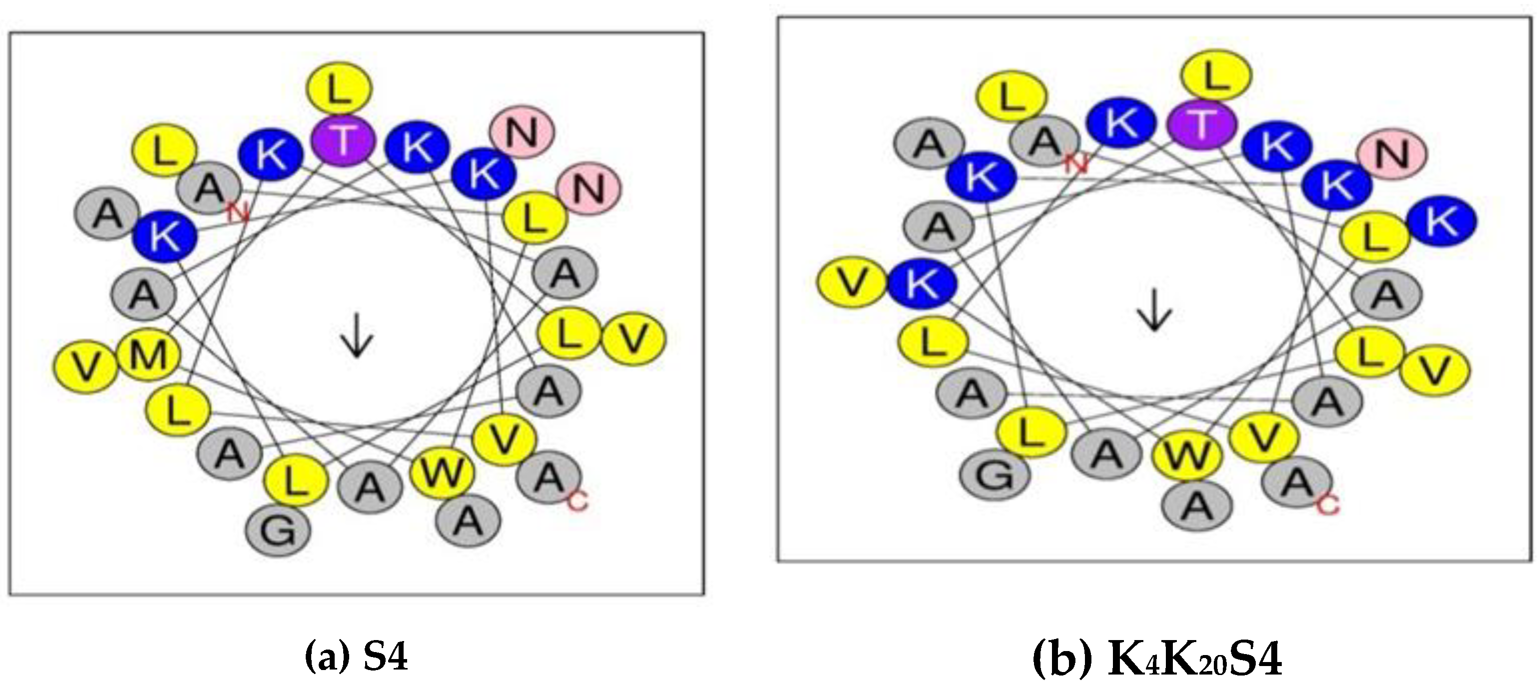
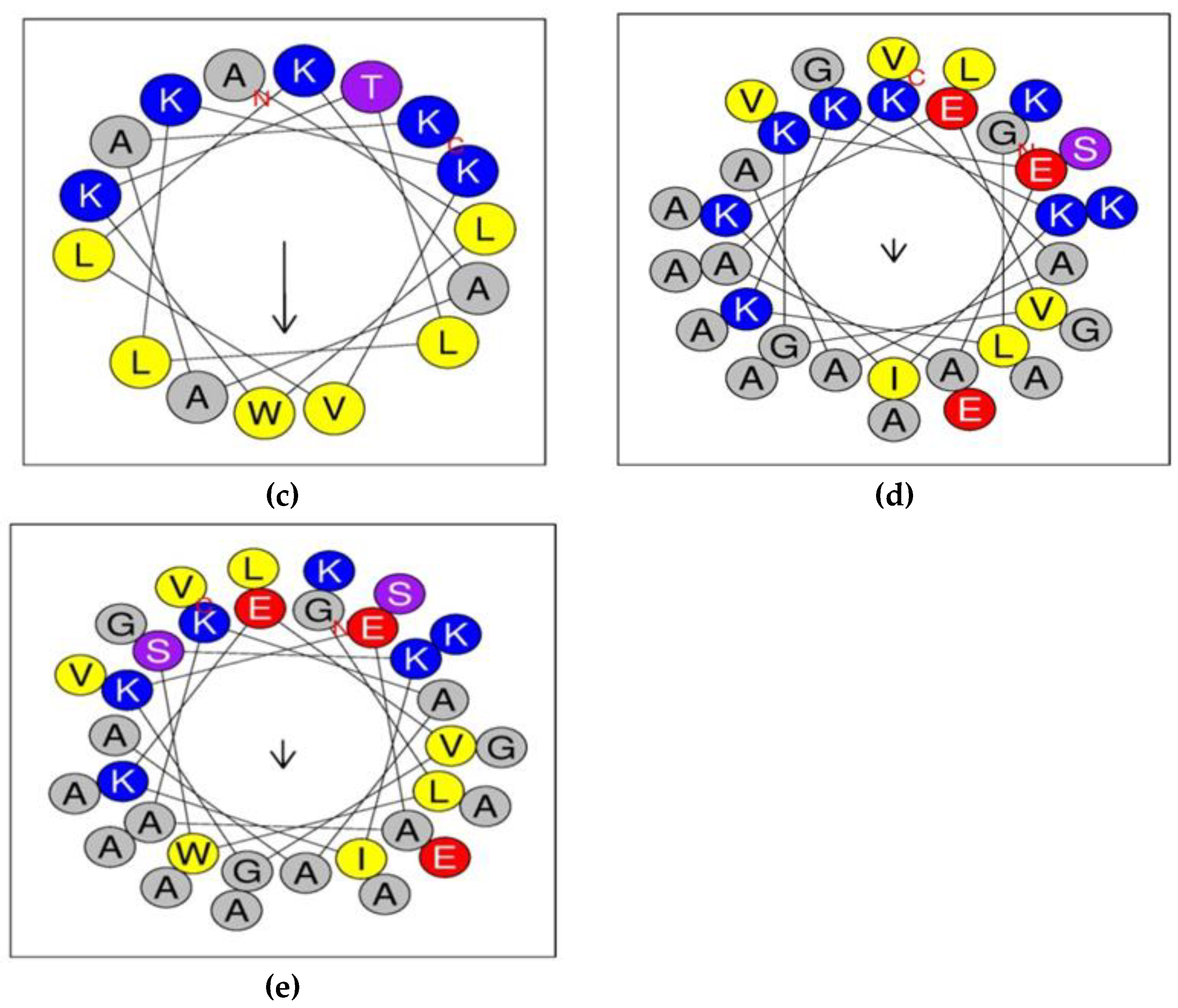
Figure 2.
AFM images of Acinetobacter baumannii before and after treatment with K4S4 (1-16)peptide.
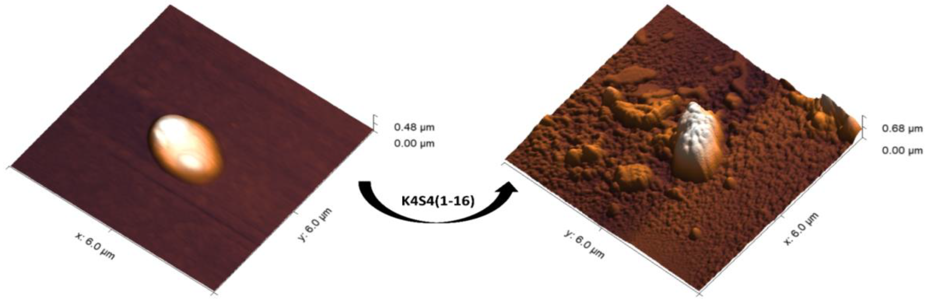
Figure 3.
Average roughness of Acinetobacter baumannii before (control) and after treatment with K4-S4 peptide (1-16) by AFM technique.
Figure 3.
Average roughness of Acinetobacter baumannii before (control) and after treatment with K4-S4 peptide (1-16) by AFM technique.
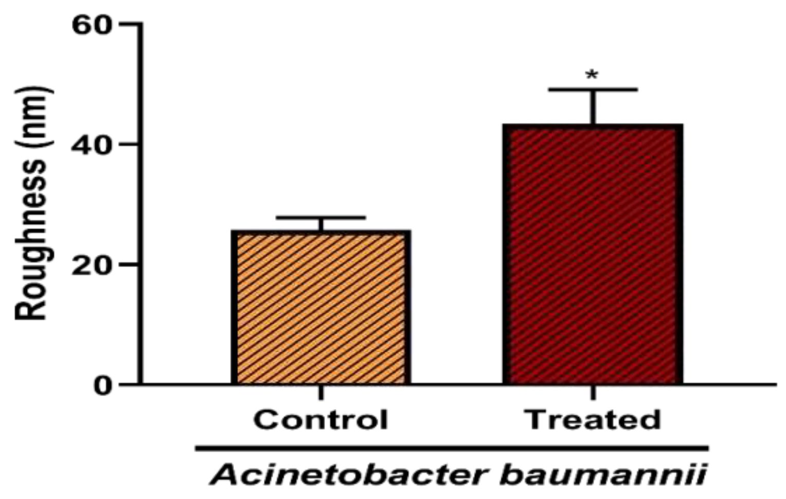

Table 1.
Structural and physicochemical properties of dermaseptins and their derivatives.
| Peptides | Sequence* | Parameters** | ||||||
|---|---|---|---|---|---|---|---|---|
| Length | MW |
Net Charge |
H | Aggregation | μH | α-Helix % | ||
| S4 (Native) | ALWMTLLKKVLKAAAKAALNAVLVGANA | 28 | 2.850 | +4 | 0.544 | 183.33 | 0.248 | 16.55 |
| K4K20S4 | ALWKTLLKKVLKAAAKAALKAVLVGANA | 28 | 2.861 | +6 | 0.451 | 112.02 | 0.246 | 11.8 |
| K4S4(1-16) | ALWKTLLKKVLKAAAK | 16 | 1.782 | +5 | 0.426 | 0 | 0.526 | 2.41 |
| B2 (Native) | GLWSKIKEVGKEAAKAAAKAAGKAALGAVSEAV | 33 | 3.181 | +3 | 0.199 | 9.681 | 0.204 | 10.02 |
| K3K4B2 | GLKKKIKEVGKEAAKAAAKAAGKAALGAVSEAV | 33 | 3.164 | +5 | 0.072 | 9.681 | 0.159 | 9.85 |
*The sequences are shown using the one letter code for the amino acids. ** Parameters: MW (kDa); H: Hydrophobicity; Aggregation: total trend of aggregation; μH: Hydrophobic moment; α-Helix %: Helicity.
Table 2.
Antimicrobial activities and dose-dependent effects of different dermaseptins and their derivatives.
Table 2.
Antimicrobial activities and dose-dependent effects of different dermaseptins and their derivatives.
|
Peptides |
CC50 Hep-2 cells (μg/ml) |
Acinetobacter baumannii MIC ( μg/ml) |
Acinetobacter baumannii MBC ( μg/ml) |
|---|---|---|---|
| S4 | 16.51 | 12.5 | 25 |
| K4S4(1-16) | 68.9 | 6.25 | 12.5 |
| K4K20S4 | 75.71 | 3.125 | 6.25 |
| B2 | 30.4 | 12.5 | 25 |
| K3K4B2 | 61.25 | 6.25 | 12.5 |
| meropenem | ND | 32 | 64 |
CC50: peptide concentration that causes 50% cytotoxicity in HEp-2 cells for dermaseptin S4 and derivatives (μg/ml); ND, not determined; MIC: Minimum Inhibitory Concentration (μg/ml); MBC: Minimal Bactericidal Concentration (μg/ml).
Disclaimer/Publisher’s Note: The statements, opinions and data contained in all publications are solely those of the individual author(s) and contributor(s) and not of MDPI and/or the editor(s). MDPI and/or the editor(s) disclaim responsibility for any injury to people or property resulting from any ideas, methods, instructions or products referred to in the content. |
© 2023 by the authors. Licensee MDPI, Basel, Switzerland. This article is an open access article distributed under the terms and conditions of the Creative Commons Attribution (CC BY) license (http://creativecommons.org/licenses/by/4.0/).
Copyright: This open access article is published under a Creative Commons CC BY 4.0 license, which permit the free download, distribution, and reuse, provided that the author and preprint are cited in any reuse.


