1. Introduction
Rift Valley fever (RVF) is a viral zoonotic disease that affects domestic and wild ungulates. The disease leads to high abortion rates in herds and neonatal mortality, making it a significant threat to livestock and public health. The disease has a significant economic impact, particularly in rural communities that depend on local livestock for their livelihoods, especially in developing countries. The causative virus, RVFV, is transmitted by the bite of mosquitoes, mainly of the genera
Aedes and
Culex. The disease is currently endemic in many countries of sub-Saharan Africa, the Middle East and Indian Ocean islands, and numerous cases of seropositive animals have been reported in North African countries. Humans can be severely affected by the disease if exposed to infected mosquitoes, contaminated food or animal by-products. Though the disease has been fundamentally restricted to the African continent, the spread of the virus is also possible by several means, including the import of viremic livestock [
1] or by humans returning to their home countries from RVF endemic areas [
2].
RVFV belongs to the order
Bunyavirales, family
Phenuiviridae and genus Phlebovirus. At least 15 different genetic lineages have been described [
3] but for the moment only one serotype is recognized. Structurally, the RVFV virions are icosahedral, with a diameter of approximately 80-100 nm [
4], composed of heterodimers of two membrane glycoproteins (Gn and Gc) [
5,
6,
7] enclosing a nucleocapsid that packs at least three single-stranded RNA segments of different sizes (L, M, S), and negative and ambisense polarity [
8]. The large (L) RNA segment encodes the RNA-dependent RNA polymerase (RdRp) [
9,
10], while the medium (M) segment encodes a polyprotein that upon protease processing gives rise to the two membrane glycoproteins (Gn and Gc) [
11] and a non-structural protein (NSm) related to apoptosis modulation [
12]. The messenger RNA of the M segment can translate at least an additional protein of 78kDa protein comprising the NSm and Gn ORFs [
13]. Finally, the small (S) segment encodes the virus nucleoprotein N and the virulence associated factor NSs in opposite orientations, separated by an intergenic region (IGR) [
14,
15,
16].
An effective method to prevent the potential spread of RVF is vaccination, and to date three types of vaccines have been licensed for use in livestock, both attenuated and inactivated [
17]. There are also several experimental vaccines based on rational deletion of non-structural genes associated with virulence or on mutagenisation [
18]. In many cases, these vaccines retain some residual virulence when tested in more sensitive animal models or in early stage pregnant animals, so there is still room for the improvement of their safety profile.
As a result of previous research, a mutagenised variant of the South African 56/74 virus isolate was obtained by cell culture propagation in the presence of favipiravir, a purine nucleoside analogue with antiviral properties whose incorporation into nascent RNA strands causes base mispairing and increases the mutation rate of the virus. After the 8
th serial passage in the presence of a suboptimal dose of favipiravir (40µM), a resistant virus variant was selected. More specifically, sequence analysis showed the inclusion of a total of 47 base mutations along the three RNA segments, 23 of which were non-synonymous (NS) changes. Most NS mutations (14) accumulated in the M-segment encoding the GnGc polyprotein, 7 in the viral polymerase gene and 2 in the non-structural protein NSs [
19]. The most interesting finding was the highly attenuated nature of the resulting virus, even in immunodeficient A129 (IFNARKO) mice. Accordingly, we proposed the name “hyper attenuated RVFV variant 40Fp8” [
20].
The structure of the RVFV viral polymerase has recently been described [
21] and contains all the catalytic motifs described for negative-polarity RNA virus RNA polymerases [
22]. To characterise the role of key mutations in the attenuation of the RVFV-40Fp8 virus, we focused first on those mutations that could be related to favipiravir resistance, which intuitively should correspond to the viral polymerase (RdRp). Three mutations found in the catalytic core of the 40Fp8 RdRp were close to these canonical motifs responsible for the entry of template RNA, nucleotides and metal ions, which cooperate together for polymerisation. Residues 924 and 1303 are highly conserved among phleboviruses and the introduction of G924S and A1303T mutations into the viral RdRp of the ZH548 RVFV strain was enough to fully attenuate the virus, as demonstrated upon intraperitoneal inoculation into mice, indicating a predominant role in virus attenuation [
23].
As for the two mutations found in the S segment affecting the non-structural protein NSs, known as the main virulence factor, we focused on the P82L mutation because it constitutes a drastic change and is also found in one of the proline-rich areas previously linked to the ability of NSs to form typical intranuclear filaments in infected cells [
24]. In this case, the introduction of P82L into the NSs protein of ZH548 resulted in partial attenuation of the virus when inoculated into mice [
25]. As expected, a triple mutant G924S, A1303T, P82L virus (termed ZH548-rA2) was also highly attenuated. The results obtained in mice prompted us to test whether this level of attenuation would be maintained in the natural target species, as well as the ability of the triple mutant to induce protection upon challenge with the parental virus. Here, we show that these positions can be targeted to increase the safety of attenuated RVF vaccines in livestock.
2. Materials and Methods
Cells and Viruses
The Vero cell line (African green monkey kidney cells, ATCC CCL-81) was used for this study. Cells were grown in Dulbecco’s modified Eagle medium (Bio-West) supplemented with 10% fetal bovine serum (Gibco-FBS), 2 mM L glutamine, 1% 100X non-essential amino-acids and 100 U/ml penicillin/100 μg/ml streptomycin. All cells were incubated at 37º C in the presence of 5% CO
2. Viruses used in this work are the rescued ZH548 (wild-type, short named as rC1), and the ZH548 triple mutant virus (short named as rA2) carrying G924S and A1303T mutations in the L-polymerase and a full-length P82L mutated NSs protein as described [
23]. Infections and titrations were performed as described [
20].
Animal study design
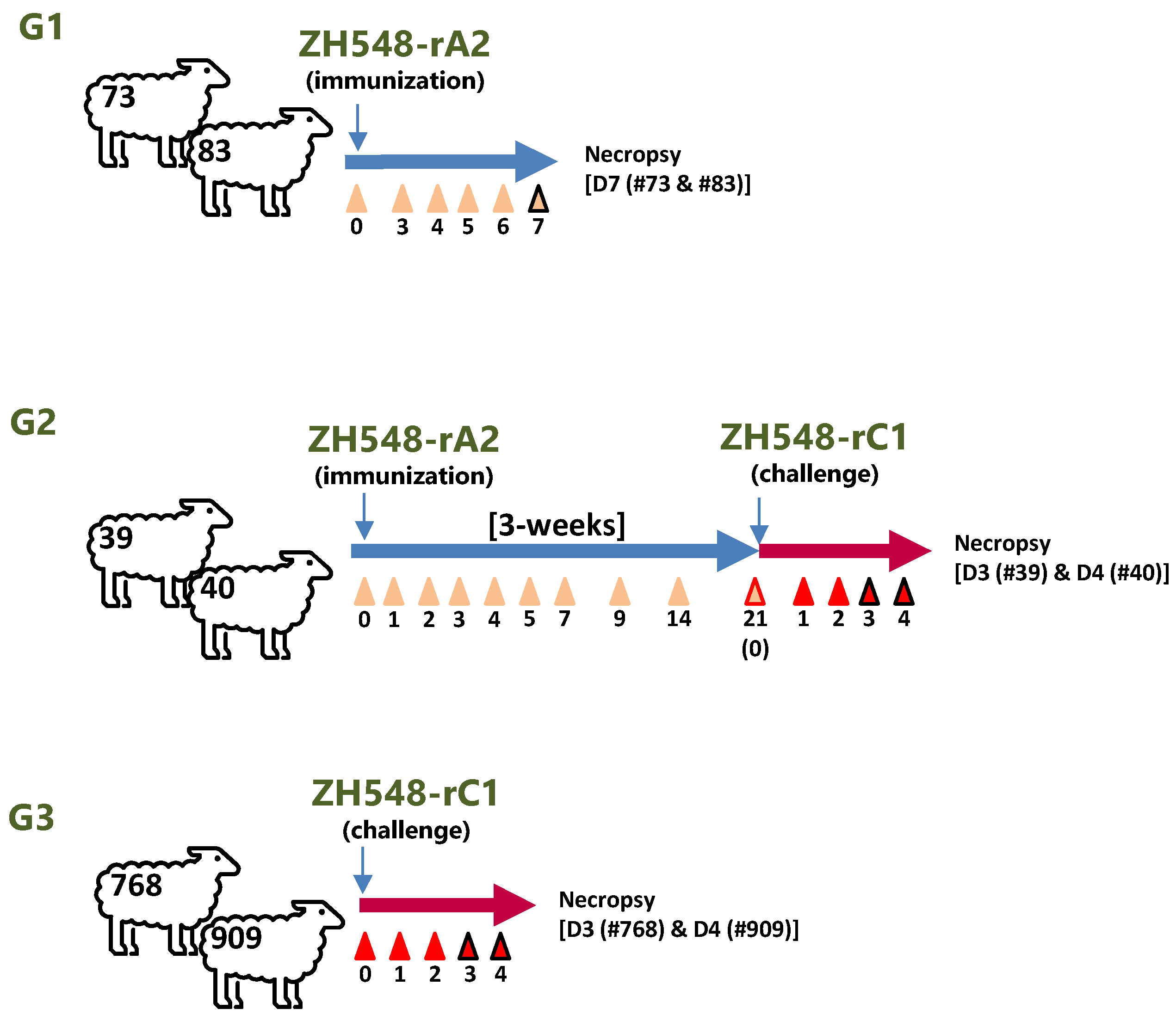
A total of six, 4-month-old, female Churra sheep (Spanish native breed), weighing about 30-40kg, were supplied by a farm with a high sanitary status. The sheep were housed at the CISA-BSL3 facilities and acclimatized for one week before the commencement of the experiment. In addition to a straw bed, animals were provided with water and food ad libitum. The animals were randomly divided into three groups, each consisting of two animals. The day of experimental inoculation was designated as day 0. Before inoculation all animals were bled. Throughout the experiment, rectal temperatures and clinical signs were monitored daily. In group 1 (G1), animals were inoculated subcutaneously in the neck area near the pre-scapular lymph node with 1 ml containing a high dose of ZH548-rA2 virus (107pfu). Whole blood samples were taken from the jugular vein at 0, 3, 4, 5, 6 and 7 days after inoculation (dpi). At 7dpi both animals were euthanized and necropsied to assess macroscopic lesions. On the other hand, the immunostimulatory capacity (i.e the induction of humoral or cellular responses) and protective efficacy of rA2 virus was assessed in another two sheep (G2). Three weeks after the inoculation with ZH548-rA2, following the same procedure described for G1, animals were challenged by the subcutaneous route with 1 ml containing 107 pfu of the rescued parental virus (ZH548-rC1). Sheep belonging to G2 were sampled at 0, 1, 2, 3, 4, 5, 7, 14, and 21 dpi. A control group of two non-immunised sheep (G3) was also infected subcutaneously with 1 ml containing 107 pfu of rC1 virus. After challenge, blood samples were taken at 0, 1, 2, 3 and 4 dpi from G2 and G3 sheep until they were euthanized and necropsied between days 3 and 4 post-challenge (dpc). Animal experiments were performed following EC guidelines (Directive 86/609) upon approval by the Ethical Review and Animal Care Committees of INIA and Comunidad de Madrid (authorization decision PROEX 192/17).
Blood chemistry
Whole blood samples from sheep in G2 and G3 were collected in BD Vacutainer tubes and centrifuged at 1267 × g for 10 min for serum and/or plasma separation. Serum and plasma samples were stored at −80 °C until use. Samples were then tested with a VetScan VS2 analyzer using a large animal profile reagent rotor (Abaxis) for the measuring of several parameters including ALB, ALP, AST, BUN, Ca, CK, GGT, GLOB, Mg, PHOS, and TP levels.
Virus detection in blood samples
RNA was extracted from EDTA blood samples collected at the indicated time points using an RNA virus extraction kit (Speedtools, 180 Biotools BM, Madrid, Spain) as described [
31]. Viremia levels were then assessed using a real-time RT-qPCR specific to the RVFV L-segment [
32]. The correspondence between Cq values and plaque forming units was estimated using sheep blood spiked with 10
1 to 10
5 pfu of a RVFV virus stock previously titrated by plaque assay. For virus isolation, 100 μl of defibrinated blood or plasma were diluted with DMEM and added onto freshly plated Vero cell monolayers for one hour in a 5% C0
2 incubator at 37ºC. Upon extensive washing, the cultures where further incubated for 5-6 days. Cell lysates were generated after three consecutive freeze-thawing cycles and clarified supernatants incubated onto fresh Vero cell monolayers for a total of three rounds.
Sequencing analysis
The whole genome sequence of rA2 virus was determined by next-generation sequencing (NGS). Briefly, RNA extracted from the supernatant of infected cells was used as template for Double Strand cDNA synthesis using the PrimeScript™ Double Strand cDNA Synthesis Kit (Takara) following manufacturer’s instructions. A pool of 10 RVFV specific primers covering the whole viral genome was used [
20]. After analysis and quantification of the final product by a high sensitivity D5000 Screen Tape (Agilent), libraries were generated using the NEBNext® Ultra™ II DNA Library Prep Kit and sequenced with MiSeq instrument, using protocols and reagents from Illumina (Illumina®, San Diego, CA, USA) at the Unidad Genómica FPCM” (Madrid, Spain). Analysis of the reads was performed as described [
23,
33]. Reads were aligned against the available sequences corresponding to the 3 segments of RVFV strain ZH548 (accession. numbers. DQ375403 (L), DQ380206 (M) and DQ380151(S) [
34,
35]).
Tissue sampling, histopathological and immunohistochemical analysis
During necropsies, macroscopic evaluations of lesions were performed. In addition, different tissue samples were fixed by immersion in 4% buffered formalin solution for 72 hours, routinely processed and embedded in paraffin wax. 4 µm tissue sections were stained with haematoxylin and eosin (H&E) and then microscopically evaluated.
To visualise the viral antigen, immunohistochemistry was performed on tissue sections. Briefly, sections were deparaffinised and rehydrated. Endogenous peroxidase activity was inhibited by incubation with 3% hydrogen peroxide in methanol for 30 minutes (min) at room temperature (RT). Samples were then subjected to heat-induced epitope retrieval by immersion in citrate buffer (pH 3.2) at sub boiling for 10 min. After treatment, sections were cooled to RT and rinsed in TBS buffer (pH 7.2) for 5 min. Nonspecific binding sites were blocked with 5% normal goat serum in TBS buffer for 30 min. After the blocking step, sections were incubated with an in-house polyclonal rabbit anti-RVFV serum (diluted 1:500-1:1000 in TBS buffer) overnight at 4ºC. Next, the sections were washed with TBS 1x – Tween20 (0,2%) (1 x 5 min) and TBS (2 x 5 min), followed by 1 hour incubation at RT with a secondary antibody (DAKO EnVision labelled polymer goat anti-rabbit). A working solution of 3,3´-diaminobenzidine tetrahydrochloride (DAB) (Dako REAL EnVision Detection System) was applied as the chromogen for 45 seconds to the slides to be stained. Slides were rinsed with distilled water, counterstained with Mayer’s haematoxylin, dehydrated and mounted. Tissue samples from infected and non-infected sheep were included as test controls. In addition, an in-house irrelevant rabbit antiserum was used as a primary antibody negative technique control.
Expression of recombinant antigens
The gene corresponding to the RVFV ZH548 Gn glycoprotein ectodomain sequence (encoding amino acids 154-580) was cloned into a pFastBac HT donor transfer vector and used to transform DH10B Bac E.coli cells (Bac to Bac expression system ThermoFisher). The resombinant baculovirus generated was used to infect highfive cells (H5). After threee days, infected cells were collected, lysed and processed following established protocols. Gn protein was purified under denaturing conditions using HisTrap FF columns (GE Healthcare) to the Akta prime plus (GE Healthcare). Collected fractions were analyzed by SDS-PAGE, pooled, dyalized against phosphate buffer and further concerntrated (Vivaspin 6 100 kDa; GE Healthcare) . Finally the Gn protein was quantified using BCA Protein Assay Kit (Pierce™)
A RVFV N full length construct was cloned by recombination (inPhusion cloning, Promega) into the pOPIN-E vector (Oxford Protein Production Facility, UK), which adds a C-terminal hexahistidine tag. The expression of recombinant N in IPTG induced E.coli BL21 cells was confirmed by SDS-PAGE and western blot. A larger prep of affinity purified recombinant N protein was kindly provided by Dr Hani Boshra (Abrescia´s lab , CIC-BioGune) using a Ni-NTA column (GE Biosciences) followed by size-exclusion gel-filtration chromatography using a HiLoad Superdex 200 pg column (GE Healthcare).
Antibody Assays
A seroneutralization assay was performed in multiwell 96 plates using sheep sera (in quadruplicates) that were 2-fold diluted starting from 1/10, mixed with an equal volume of infectious virus containing 100 TCID50 and incubated for 30 minutes at 37ºC. Then, a Vero cell suspension was added, and plates were incubated in a 5% CO2 atmosphere for 4 days. Monolayers were then monitored for the development of cytopathic effect (CPE), fixed with a 10% solution of formaldehyde and stained with a 2% Crystal Violet solution. Neutralization titers are expressed as the highest dilution of serum (in log10) rendering a 50% reduction in infectivity.
Interferon-γ detection in sheep plasma and upon in vitro re-stimulation analysis
An antibody sandwich ELISA using capture anti- bovine IFNγ antibody was performed as described [
36]. 100µl of fresh plasma samples from sheep in groups G2 and G3 collected after rA2 immunization and after rC1 virus challenge were added to ELISA plate wells previously coated with purified mAb anti-IFNγ (MT17.1, Mabtech). Upon incubation and washing steps, a detector anti-bovine IFNγ mAb conjugated with biotin (MT307, Mabtech) was added. Streptavidin-HRPO (Becton-Dickinson) was used for detection of immunocomplexes upon addition of TMB peroxidase substrate (Sigma-Aldrich). Absorbance values were determined at 450 nm by an automated reader (BMG, Labtech). For the
in vitro re-stimulation assay 0.5 mL of whole blood samples were incubated in MW24 plate wells and 1µg/ml of recombinant purified proteins Gn, or N, or 5µg of ZH548-Gn derived peptides #19 (
203-FQSYAHHRTLLEAVH-
217) and #21 (
211-TLLEAVHDTIIAKAD-
225), or ZH548-Gc-derived peptides #226 (
1031-AAFLNLTGCYSCNAG-
1045) and 253 (
1139-SWNFFDWFSGLMSWF-
1153) added to each well and incubated at 37ºC in a 5%CO
2 incubator for three days. Plasma was recovered from wells by plate centrifugation (300 x g)and tested as described above in the IFNγ capture mAb sandwich assay.
3. Results
3.1. Clinical and pathological findings
To evaluate the safety and efficacy of the triple mutant virus ZH548-rA2 in sheep, one of the natural hosts of RVFV, a small pilot trial was conducted (
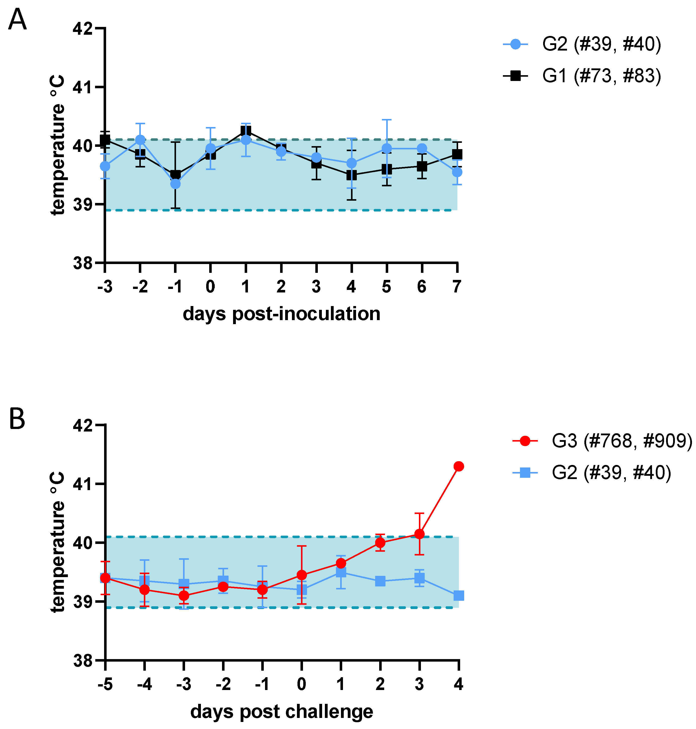
Figure 1). After inoculation with rA2 virus, none of the sheep included in G1 and G2 groups showed any apparent signs of disease. Only a transient and mild increase in temperature at 1 dpi was recorded in sheep #73, #83, #39 and #40 (
Figure 2A). A more evident increase in temperature was observed at 3 dpi in sheep included in G3 that received the same dose of the parental rC1 virus (#768, #909) (
Figure 2B), with sheep #909 showing the highest temperature by 4 dpi. Despite the increase in temperature, no other clinical signs were evident. The challenge with rC1 virus did not cause temperature raise in the sheep #39 and #40 (G2) that had received a previous inoculation with rA2 virus.
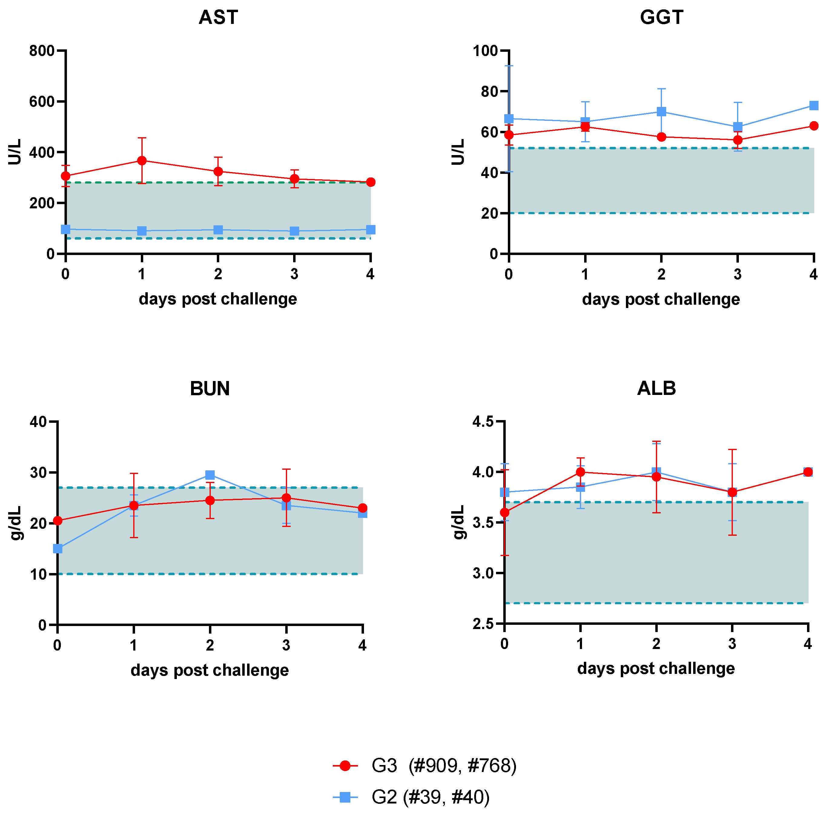
Since one of the main affected organs after a RVFV infection is the liver, different enzymes indicative of hepatic injury were evaluated after challenge with rC1 virus by blood chemistry using serum samples. Aspartate amino transferase (AST) levels, normally present in low concentrations, were elevated in G3 sheep compared to G2, sheep, where levels remained normal. However, gamma-glutamyl transferase (GGT) levels remained elevated in both groups (
Figure 3A). No differences were found with respect to plasma protein degradation, as suggested by the detected levels of plasma albumin (ALB) or blood urea nitrogen (BUN) (
Figure 3B), both indicative of liver or kidney disease. Additional biochemical parameters were measured and found to be within normal ranges (data not shown).
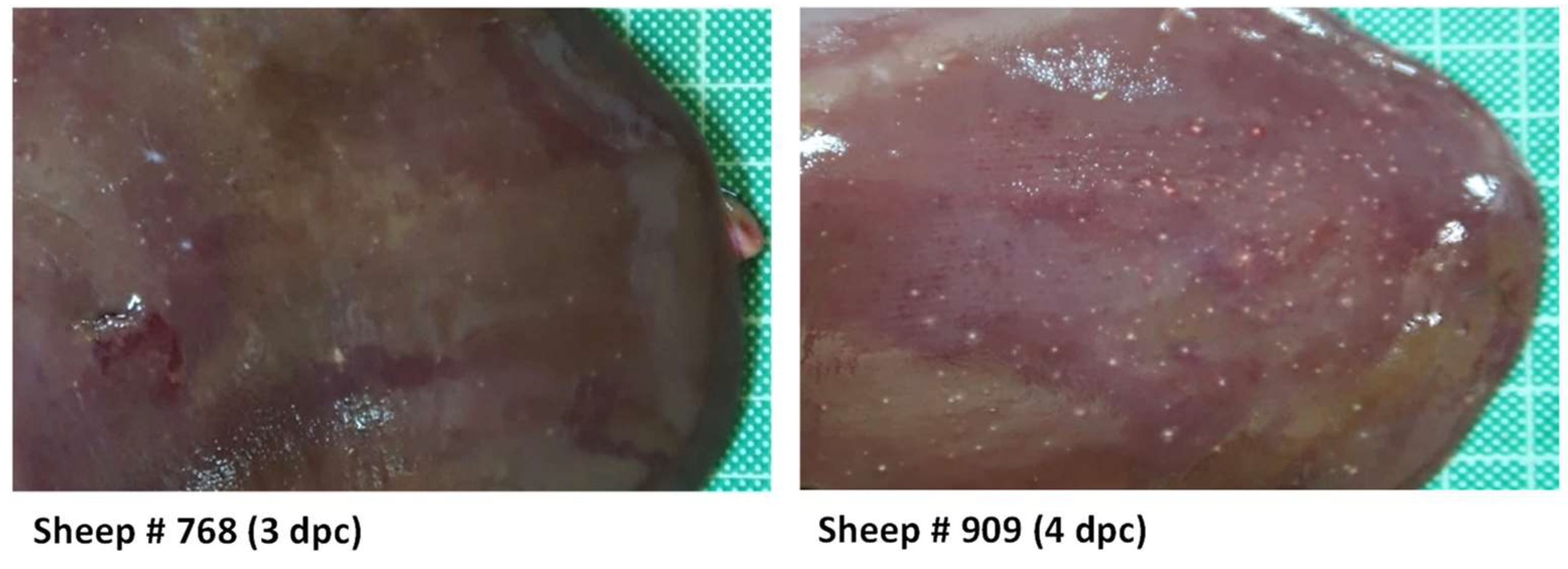
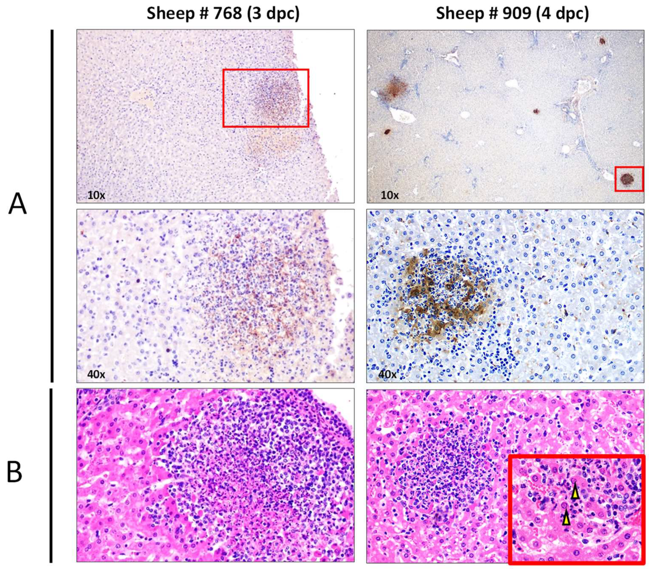
During necropsies, none of the rA2-inoculated sheep included in either G1 or G2 showed any relevant macroscopic lesions. However, the livers of both animals inoculated with rC1 (G3) showed the most remarkable macroscopic findings characteristic of RVFV infection. The liver parenchyma showed a loss of consistency together with the presence of greyish-white foci, 0.5-2 mm in diameter, consisting of scattered necrotic lesions, many of them surrounded by a reddish halo. The number of foci was more abundant in sheep #909 necropsied on day 4 (
Figure 4).
Histopathological evaluation of the livers of G3 sheep revealed findings characteristic of RVFV infection, with multifocal necrotic foci filled with cellular debris and both degenerated and viable neutrophils, surrounded by mononuclear cells, mainly lymphocytes. Hepatocytes with eosinophilic intranuclear inclusion bodies were also present. Viral infection was also confirmed by immunohistochemistry, with hepatocytes within the necrotic foci appearing as the main cells immunolabelled against viral antigen (
Figure 5). Immunolabelled Kupffer cells and circulating monocytes were also occasionally observed in the hepatic sinusoids.
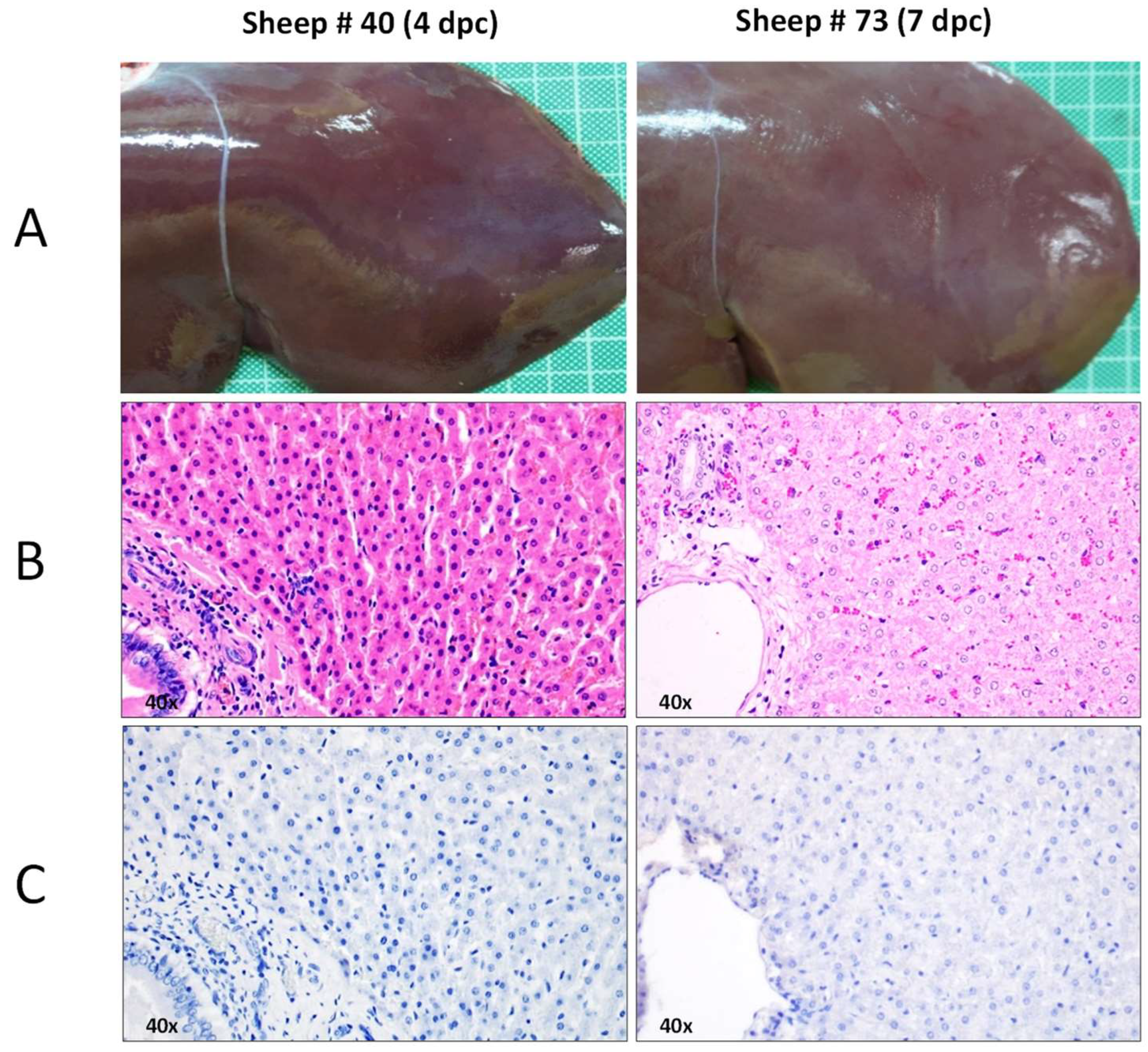
In contrast to what was observed in G3 sheep, histopathological evaluations revealed no liver lesions in any of the rA2-inoculated sheep (G1) euthanised on day 7 pi. Likewise, animals that were first immunized with rA2 and then challenged with rC1 virus (G2) showed no liver injury (
Figure 6). Collectively all these data indicate that rA2 inoculation in sheep is safe and able to avoid the pathology induced by the inoculation of rC1.
3.2. Analysis of viremia levels by RT-qPCR and virus isolation
The presence of viral genome was tested in blood samples taken at different times post inoculation. Consistent with the absence of pathology observed in the livers, no positive detection above the detection threshold was found in the blood of any of rA2-inoculated sheep (G1 and G2). Attempts to isolate virus upon blind passages in cell cultures yielded also negative results. Surprisingly, no viral genome was detected in the blood of rC1-infected sheep taken at 3 or 4 dpi despite the clear liver pathology observed both macro- and microscopically. Known amounts of virus were spiked into sheep blood that rendered positive Cq, ruling out the possibility of PCR inhibition by some components upon the RNA extraction procedure. Attempts to isolate virus from rC1 blood samples upon serial blind passage in cell culture rendered also negative. These data may indicate that the replication of ZH548 rescued viruses in sheep is limited, conditioning the molecular detection of viral genomes as well as viral isolation in cell cultures.
3.3. Analysis of immune responses
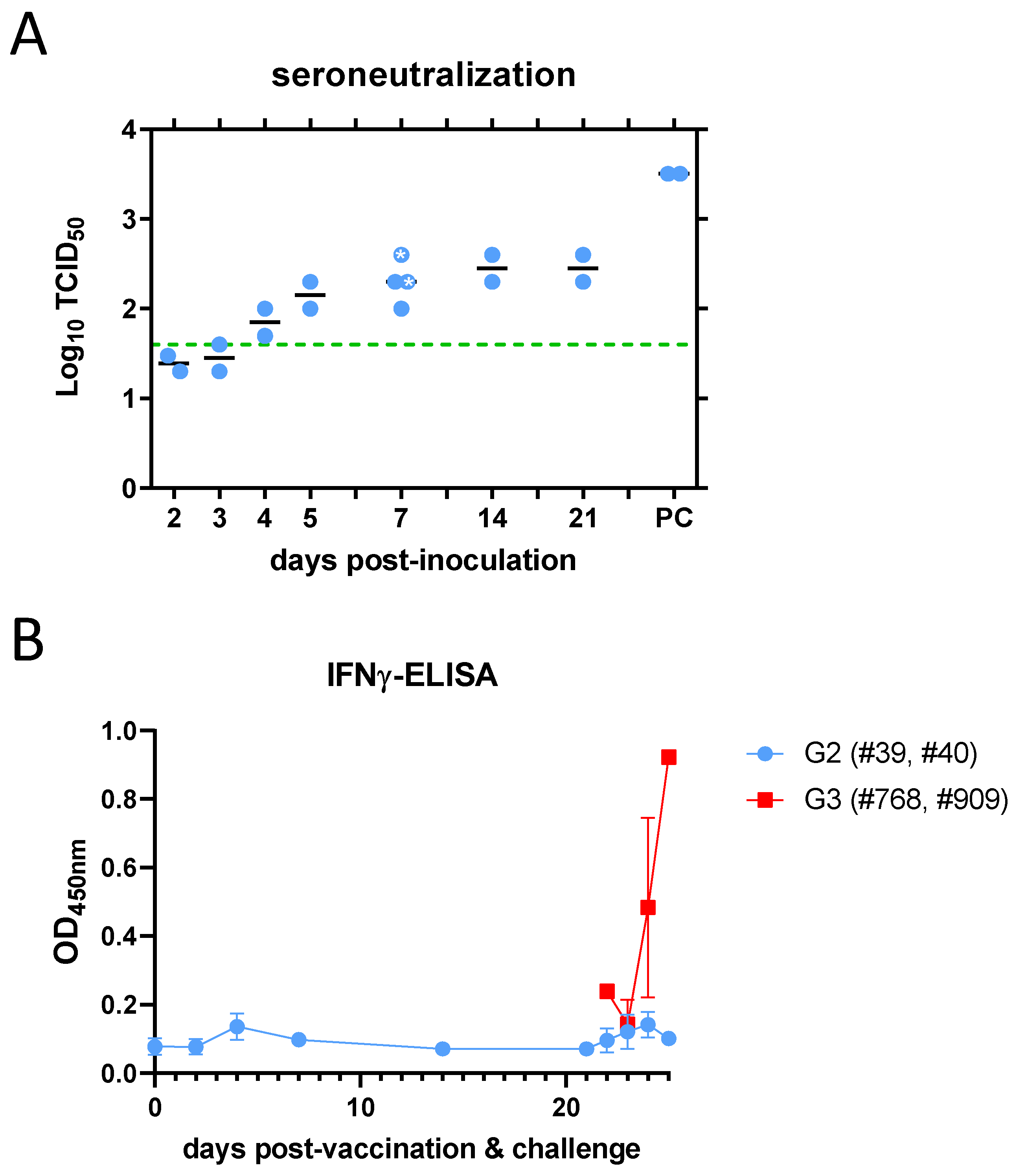
Upon rA2 virus inoculation (G2), neutralizing antibodies were detected in serum samples from sheep #39 and #40 as early as day 4 post inoculation (pi), with a peak titre on day 21 pi. In the animals that were further challenged with rC1 virus, a booster effect was observed in 3 days post-challenge samples (
Figure 7A). Neutralizing antibodies were also tested and detected in sheep #73 and #83 (G1) on day 7 pi. Increased interferon gamma concentrations were also detected in plasma samples from sheep inoculated with rC1 virus (G3), but not in those that had been previously inoculated (vaccinated) with rA2 virus (G2) after rC1 virus challenge (
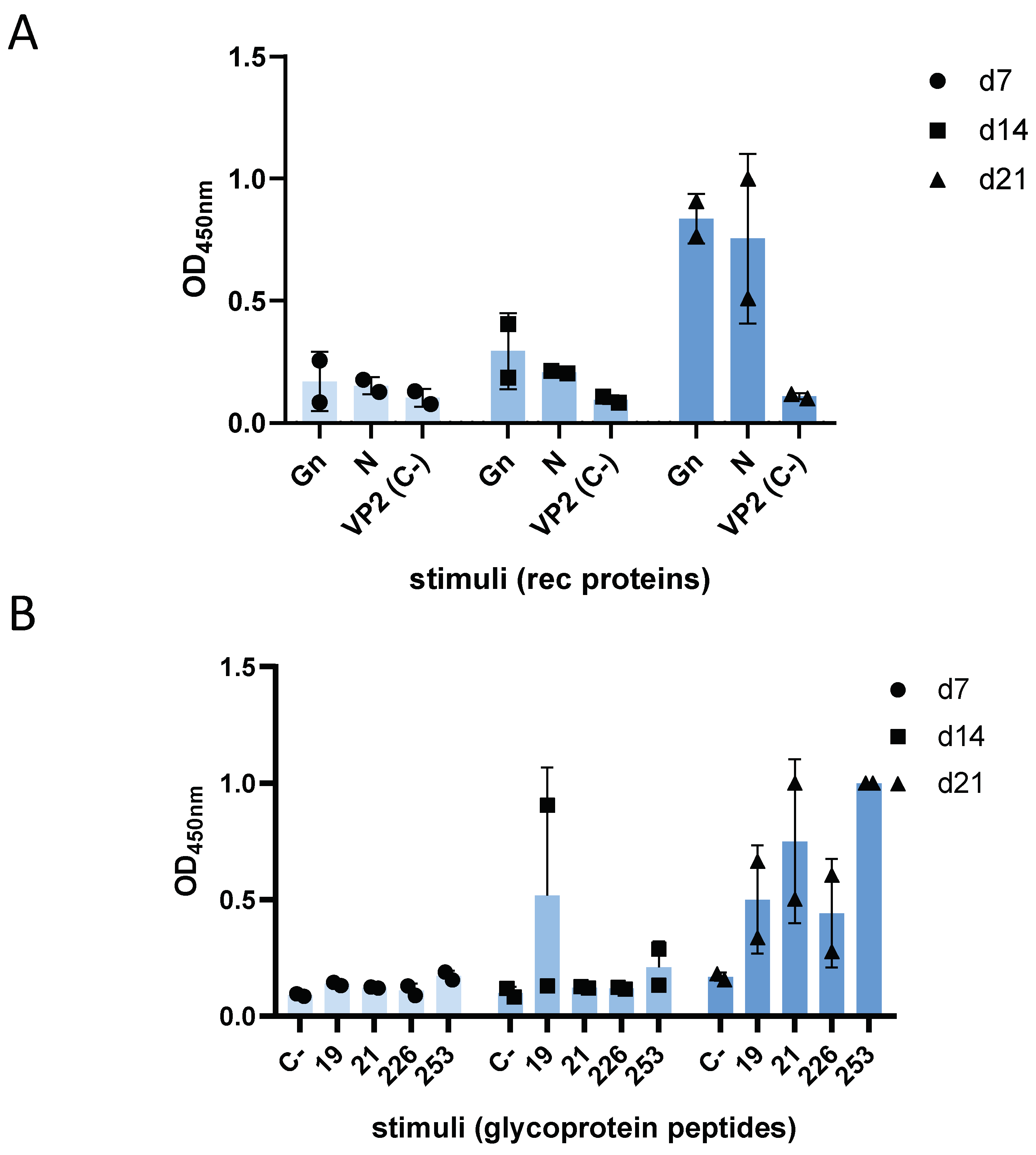
Figure 7B). However, IFNγ secretion was re-stimulated in these sheep using recombinant Gn or N antigens in whole blood samples taken at 7, 14- and 21-days after rA2 inoculation (G2) (
Figure 8A). The specificity of this result was further confirmed by re-stimulation of sheep blood with four 12-mer peptides corresponding to Gn and Gc glycoproteins, indicating the presence of RVFV-specific IFNγ-secreting T cells (
Figure 8B). These results support the proper induction of an inmmune response in sheep to levels that can be considered protective in spite of the clear attenuation of the rA2 virus.
4. Discussion
In a previous study [
23] we showed that the triple mutant ZH548-rA2 virus was completely attenuated in mice. The high susceptibility of laboratory mice to virulent RVFV infection makes this infection model suitable for testing the efficacy of a vaccine approach. The inoculation of rA2 protected mice upon a lethal challenge with rC1 by means of a neutralising antibody response at levels correlated with protection. To confirm whether this positive result could be extrapolated to RVFV target species, we conducted a preliminary exploratory study in sheep, a model commonly used for RVF vaccine efficacy studies. To comply with animal welfare regulatory requirements, we minimised animal use by using only six animals, four of which received a dose of rA2 virus for pathological and immunological assessments and four of which were challenged with the parental virus rC1 to test the protective effect of rA2 as a vaccine. Although the reduced sample size hampers statistical significance, the results obtained suggest that rA2 virus in sheep retains the phenotype observed in mice, i.e., a marked attenuation compared to the parental virus and the ability to induce a protective immune response.
We used two rescued pure virus populations (rC1 and rA2) that were confirmed by NGS data, so that phenotypic differences could be attributed to only three amino acid residues. Our results showed that the rA2 virus was unable to cause pathology in sheep even at high doses of up to 10
7 pfu. This suggests that the virus retains a high level of attenuation. Assessment of virus replication by RT-qPCR in blood samples suggests that rA2 is poorly disseminated since no virus isolation could be achieved after three blinded serial passages in cell culture. However, we cannot rule out a minimal level of replication as serum neutralising antibodies were readily detected. Another interesting observation was the lack of detectable viremia in sheep inoculated with the rescued rC1 virus where, as with rA2, no virus could be isolated in cell culture. However, rC1 caused remarkable liver damage, also inducing elevated AST levels and increased plasma IFNγ levels, indicating an ongoing response to infection. One possibility is that rC1, derived from a pure rescued viral clone with reduced genetic diversity compared to a natural isolate, represents a virus with low pathogenicity and reduced ability to spread. It is important to note that in previous experiments in mice, rC1 was fully virulent [
23] and liver damage was massive by day 3-4 post inoculation (unpublished observation). In sheep, the observation of scattered necrotic foci in the liver may be a consequence of better control of viral spread once in the liver, preventing further systemic spread of the virus and precluding detection of viremia. On the other hand, to our knowledge, the strain ZH548 has never been inoculated in sheep, so we have no previous data on the virulence of this human-origin isolate in sheep. Previous reports suggested that the ZH548 virus isolate was less virulent than ZH501 [
27], a contemporary isolate of identical lineage. Another possibility is that the virulent effect in sheep is manifested after 4 days post inoculation, as the animals inoculated with rC1 were sacrificed on days 3 and 4 post challenge. It is conceivable that delayed viremia peaks may have impeded early detection of viral genomes or isolation. As ZH548 has not been previously inoculated in sheep, its kinetics of infection were unknown. We posited that the progression of the infection in sheep with ZH548 would resemble that of other RVFV isolates, manifested as peak viremia between 2-3 days post inoculation. Our findings indicate that this assumption needs to be revised. Nonetheless, the use of ZH548 for the challenge was solely conducted to enable a direct comparison with the rA2 virus and to attribute any differences in phenotype to the three specific mutations. It would be interesting to examine whether the rA2 virus offers protection against challenge with a heterologous isolate. Nonetheless, the exploratory nature of our preliminary experiment precluded the inclusion of more animals in the trial.
Nevertheless, inoculation with rA2 was able to induce neutralising antibodies, explaining the absence of liver lesions observed in vaccinated sheep challenged with rC1 virus. Since the sole introduction of three amino acid changes caused this effect, it can be concluded that these three residues could be targeted for improvement of live attenuated RVFV vaccines, provided that they do not interfere with the growth or replication of the virus to the extent of compromising the induction of immune responses.
One of the best characterized RVF vaccine candidates is the mutagenized attenuated variant MP12. In MP12, the combination of three changes (Y259H (Gn), R1182G (Gc), and R1029K (L) were sufficient to attenuate the ZH501 virus [
28]. Therefore, a number of residue positions are being identified that could be targeted to improve the safety of live attenuated vaccine candidates generated by reverse genetics. It would be interesting to test for instance, whether the attenuating mutations carried by rA2 would reduce MP12 or clone 13 vaccines residual virulence. Both vaccines retain the ability to kill type-I Interferon receptor KO mice (A129) and can compromise the safety of ruminant foetuses in the initial steps of pregnancy when used at high doses [
29,
30]. Conversely, the introduction of MP12 attenuating mutations in the rA2 virus could perhaps reduce the virulence observed in A129 mice. Thus, the identification of attenuating mutations in the genome of RVFV constitutes a valuable knowledge to increase the safety of RVF vaccines.
Author Contributions
Conceptualization, A.B., B.B.; validation, B.B., A.B., C.A.; formal analysis, A.L-V., J.I.; investigation, S.M, G.L, P.S-C., B.B., C.A., E.C, N.L.; data curation, S.M, G.L, P.S-C., B.B., C.A; writing—original draft preparation, A.B.; writing—review and editing, B.B., P.S-C., S.M.,G.L.; visualization, A.B., P.S-C., G.L. B.B.; supervision, A.B., B.B.; project administration, A.B.; funding acquisition, A.B., B.B. All authors have read and agreed to the published version of the manuscript.”
Funding
This research was funded by grants AGL2017-83226-R and PID2021-122567OB-I00 funded by MCIN/AEI/ 10.13039/501100011033 and by ERDF “A way of making Europe”.
Institutional Review Board Statement
The animal study protocol was approved by the Ethics Committee of INIA (ORCEA 2017/15; date of approval 09-01-2017) and by the Director-General of Agriculture and Livestock from Comunidad de Madrid (authorization decision PROEX192/17, date of approval 25-01-2018).
Informed Consent Statement
Not applicable
Data Availability Statement
The data presented in this study are available in a Supplementary Excel file.
Acknowledgments
We thank Antonia González Guirado and Laura Fernández del Ama for their excellent technical assistance. We also thank to the animal care staff, veterinary services and biosafety staff at CISA for their support with the in vivo experiments. We thank Dr George Warimwe (ILRI, Nairobi) for the collection of overlapping RVFV peptide sequences.
Conflicts of Interest
The authors declare no conflict of interest. “The funders had no role in the design of the study; in the collection, analyses, or interpretation of data; in the writing of the manuscript; or in the decision to publish the results”.
References
- Napp, S., et al., Understanding the legal trade of cattle and camels and the derived risk of Rift Valley Fever introduction into and transmission within Egypt. PLoS Negl Trop Dis 2018, 12, e0006143. [CrossRef]
- Gibson, S., et al., Rift Valley fever virus: movement of infected humans threatens global public health and agriculture. CABI Reviews 2022, 17. [CrossRef]
- Grobbelaar, A.A., et al., Molecular epidemiology of Rift Valley fever virus. Emerg Infect Dis, 2011, 17, 2270–2276. [CrossRef] [PubMed]
- Freiberg, A.N., et al., Three-dimensional organization of Rift Valley fever virus revealed by cryoelectron tomography. J Virol, 2008, 82, 10341–10348. [CrossRef] [PubMed]
- Huiskonen, J.T., et al., Electron cryo-microscopy and single-particle averaging of Rift Valley fever virus: evidence for GN-GC glycoprotein heterodimers. J Virol, 2009, 83, 3762–3769. [CrossRef] [PubMed]
- Sherman, M.B., et al., Single-particle cryo-electron microscopy of Rift Valley fever virus. Virology, 2009, 387, 11–15. [CrossRef] [PubMed]
- Rusu, M., et al., An assembly model of rift valley Fever virus. Front Microbiol, 2012, 3, 254. [CrossRef]
- Terasaki, K., et al., Mechanism of tripartite RNA genome packaging in Rift Valley fever virus. Proc Natl Acad Sci U S A, 2011, 108, 804–809. [CrossRef]
- Muller, R., et al., Completion of the genome sequence of Rift Valley fever phlebovirus indicates that the L RNA is negative sense and codes for a putative transcriptase-replicase [corrected]. Nucleic Acids Res, 1991, 19, 5433. [CrossRef]
- Muller, R., et al., Rift Valley fever virus L segment: correction of the sequence and possible functional role of newly identified regions conserved in RNA-dependent polymerases. J Gen Virol 1994, 75 Pt 6, 1345–1352. [CrossRef]
- Collett, M.S., et al., Complete nucleotide sequence of the M RNA segment of Rift Valley fever virus. Virology, 1985, 144, 228–245. [CrossRef]
- Won, S., et al., NSm protein of Rift Valley fever virus suppresses virus-induced apoptosis. J Virol, 2007, 81, 13335–13345. [CrossRef] [PubMed]
- Kreher, F., et al., The Rift Valley fever accessory proteins NSm and P78/NSm-GN are distinct determinants of virus propagation in vertebrate and invertebrate hosts. Emerg Microbes Infect, 2014, 3, e71. [CrossRef]
- Ikegami, T., et al., Characterization of Rift Valley fever virus transcriptional terminations. J Virol, 2007, 81, 8421–8438. [CrossRef] [PubMed]
- Lara, E., et al., Characterization of wild-type and alternate transcription termination signals in the Rift Valley fever virus genome. J Virol, 2011, 85, 12134–12145. [CrossRef] [PubMed]
- Giorgi, C., et al., Sequences and coding strategies of the S RNAs of Toscana and Rift Valley fever viruses compared to those of Punta Toro, Sicilian Sandfly fever, and Uukuniemi viruses. Virology, 1991, 180, 738–753. [CrossRef] [PubMed]
- Wright, D., et al., Rift Valley fever: biology and epidemiology. J Gen Virol, 2019, 100, 1187–1199. [CrossRef] [PubMed]
- Wichgers Schreur, P.J., et al., Perspectives of Next-Generation Live-Attenuated Rift Valley Fever Vaccines for Animal and Human Use. Vaccines 2023, 11. [CrossRef]
- Borrego, B., et al., Lethal Mutagenesis of Rift Valley Fever Virus Induced by Favipiravir. Antimicrob Agents Chemother 2019, 63. [CrossRef]
- Borrego, B. and A. Brun, A hyper-attenuated variant of Rift Valley fever virus (RVFV) generated by a mutagenic drug (favipiravir) unveils potential virulence markers Frontiers in Microbiology (submitted), 2020. [CrossRef]
- Wang, X., et al., Structure of Rift Valley Fever Virus RNA-Dependent RNA Polymerase. J Virol, 2022, 96, e0171321. [CrossRef]
- Te Velthuis, A.J.W., J.M. Grimes, and E. Fodor, Structural insights into RNA polymerases of negative-sense RNA viruses. Nat Rev Microbiol, 2021, 19, 303–318. [CrossRef] [PubMed]
- Borrego, B., et al., Identification of Single Amino Acid Changes in the Rift Valley Fever Virus Polymerase Core Domain Contributing to Virus Attenuation In Vivo. Front Cell Infect Microbiol, 2022, 12, 875539. [CrossRef]
- Billecocq, A., et al., NSs protein of Rift Valley fever virus blocks interferon production by inhibiting host gene transcription. J Virol, 2004, 78, 9798–9806. [CrossRef] [PubMed]
- Borrego, B., et al., The Change P82L in the Rift Valley Fever Virus NSs Protein Confers Attenuation in Mice. Viruses 2021, 13. [CrossRef]
- Lorenzo, G., et al., Efficacy of different DNA and MVA prime-boost vaccination regimens against a Rift Valley fever virus (RVFV) challenge in sheep 12 weeks following vaccination. Vet Res, 2018, 49, 21. [CrossRef] [PubMed]
- Caplen, H., C.J. Peters, and D.H. Bishop, Mutagen-directed attenuation of Rift Valley fever virus as a method for vaccine development. J Gen Virol 1985, 66 Pt 10, 2271–2277. [CrossRef]
- Ikegami, T., et al., Rift Valley Fever Virus MP-12 Vaccine Is Fully Attenuated by a Combination of Partial Attenuations in the S, M, and L Segments. J Virol, 2015, 89, 7262–7276. [CrossRef]
- Hunter, P., B.J. Erasmus, and J.H. Vorster, Teratogenicity of a mutagenised Rift Valley fever virus (MVP 12) in sheep. Onderstepoort J Vet Res, 2002, 69, 95–98.
- Makoschey, B., et al., Rift Valley Fever Vaccine Virus Clone 13 Is Able to Cross the Ovine Placental Barrier Associated with Foetal Infections, Malformations, and Stillbirths. PLoS Negl Trop Dis, 2016, 10, e0004550. [CrossRef]
- Moreno, S., et al., Recombinant Rift Valley fever viruses encoding bluetongue virus (BTV) antigens: Immunity and efficacy studies upon a BTV-4 challenge. PLoS Negl Trop Dis, 2020, 14, e0008942. [CrossRef]
- Mwaengo, D., et al., Detection and identification of Rift Valley fever virus in mosquito vectors by quantitative real-time PCR. Virus Res, 2012, 169, 137–143. [CrossRef]
- Lopez-Valinas, A., et al., Identification and Characterization of Swine Influenza Virus H1N1 Variants Generated in Vaccinated and Nonvaccinated, Challenged Pigs. Viruses 2021, 13. [CrossRef]
- Habjan, M., et al., T7 RNA polymerase-dependent and -independent systems for cDNA-based rescue of Rift Valley fever virus. J Gen Virol, 2008, 89 Pt 9, 2157–2166. [CrossRef]
- Bird, B.H., et al., Complete genome analysis of 33 ecologically and biologically diverse Rift Valley fever virus strains reveals widespread virus movement and low genetic diversity due to recent common ancestry. J Virol, 2007, 81, 2805–2816. [CrossRef]
- Lorenzo, G., et al., Understanding Rift Valley fever: contributions of animal models to disease characterization and control. Mol Immunol, 2015, 66, 78–88. [CrossRef]
|
Disclaimer/Publisher’s Note: The statements, opinions and data contained in all publications are solely those of the individual author(s) and contributor(s) and not of MDPI and/or the editor(s). MDPI and/or the editor(s) disclaim responsibility for any injury to people or property resulting from any ideas, methods, instructions or products referred to in the content. |
© 2023 by the authors. Licensee MDPI, Basel, Switzerland. This article is an open access article distributed under the terms and conditions of the Creative Commons Attribution (CC BY) license (http://creativecommons.org/licenses/by/4.0/).
