Submitted:
29 June 2024
Posted:
01 July 2024
Read the latest preprint version here
Abstract
The aim of this article is to identify the best available evidences analyzing liver samples, normall and pathological. In the study were used liver samples, collected during necropsy, from healthy patients and from patients diagnosed with cirrhosis .This mentioned, are known as routinally practice for hepatitis C diagnosis, which conduct to hepatic cirrhosis. Were made permanent preparations and used Hematoxylin–Eosin staining and other special stainings for structural observations at optical microscope using lens with differents magnifications, such us x10 and x40.
Keywords:
liver
; hepatitis C
; cirrhosis
; diagnosis
; laboratory technique
Introduction
Currently , HCV infection could be consider as one of bad results after percutaneous blood administration to ill patients. Also good to mention that HCV infection it is known as one of the most commonly through injection drug use. [1] The first and most important step in the care cascade is testing for HCV. Actually, is an enlarge number of expansion of populations eligible for testing for HCV prevention. [1,2,3]
It is know about care cascade, including multiple key points in diagnosis HCV infection. An HCV care cascade is consider as a model for identifying opportunities and barriers in order to improve laboratory tests, linkage to care, and proper treatment access. [4,5] Relativelly recently, there have been increases in HCV detection among women of childbearing age. [6,7] Reinfection with HCV after curative therapy in illness status in different patients, is an important key point for medical team. [8,9,10] The proper laboratory techniques include immunoglobulin (Ig) G antibody enzyme immunoassays (anti-HCV) and nucleic acid tests (NAT), as modern methods, in conection with blood tests.[11] In case that need to distinguish between two directions such as true or false positivity of the anti-HCV antibody result, previously mentined tests, may be done with a second FDA-approved HCV antibody assay that is different from previously used for testing [11,12] Morphologically, HCV is an enveloped, positive-sense, single-stranded RNA virus of the Flavivirdae family.[13,14,15] The great key point knowing as a start of the direct-acting antiviral (DAA) era was in 2011. The important role in this direction was the introduction of two NS3/4A protease inhibitors. Both previously mentioned were used in combination with interferon-based regimens for chronic HCV treatment to ill patients diagnosed.16] Results of studies show that the HCV replication process is error prone. Finally results practically could be observe in variant viruses knowing as quasispecies.[17,18] Nowadays there are 7 genotypes of HCV. So there are known 6 major genotypes and the recent addition of genotype 7. This last 7 genotip has been found only in a few cases diagnosed to HCV positive patients. [19] Hepatitis C virus (HCV) infection is a great cause of various liver diseases as cirrhosis and hepatocellular carcinoma. Following promising news, significant scientific discovering things remain in attention for reducing morbidity and mortality, associated to HCV. [20,21] Understanding the properties of hepatitis C virus (HCV) viral RNA and proteins facilitates the development of diagnosis methods and also a proper treatment, including antivirals.[22,23,24] In addition we can mention that HCV genotyping assays approved for in vitro diagnostic use are commercially available[25,26] Cirrhosis, as a nowadays disease, is characterized by fibrosis and nodule formation of the liver. In the secondary plan, it is known as a chronic injury, which leads to alteration of the normal lobular organization of the liver. A complex of factors, such as life style, or environmentals, can injure the liver, and beside also including viral infections, toxins, hereditary. With each injury, the liver suffer alterations as fibrosis. Finally but after a long-standing injury, liver functionalteration, develop in time cirrhosis as a complex diseases. Ethiology of the chronic liver diseases usually progress unfortunately in cirrhosis, following pathological mechanisms.[27] Scientific knowledges referring to the severity of liver cirrhosis, as a disease with a bad prognostic on the public health, is still not well characterized. [28] Liver diseases are without doubts, the most common in the world. [29] Reserchers must be carefully to ideea refering to demonstration that drug injury is present inliver structure injury that conduct to pathology. [30]
Material and methods
Following the study purpose, were made permanent preparations that were stained with hematoxylin and eosin for observation at optical microscope and with special stainings.The process of the permanent microscopic preparations was based on prior knowledge of the steps from the classical method, using a standard H&E staining technique. Optical microscope examination were used lens with magnification x10 and x40. In order to assist medical staff in understanding the concerns outlined, a series of digital images have been prepared. The operative pieces are intended to bring in the pathological anatomy service for macroscopic examination for diagnostic purposes. Most important is to analyse and to diagnosed liver pathology observing structural changes.
Results
Normal liver structure, using an optical microscope analyse and a special staining namely Masson. We can observe hepatocytes and interlobular spaces and septa.[Figure 1]
The functional unit of the liver is the lobule with hexagonal form. Kienann space is specific for liver strucutre, including a portal triad (portal vein, hepatic artery, bile duct) sits at each corner of the hexagon. Mitochondri as points observing with lens x40. Portal vein with enlarge lumen. [Figure 2]
Based on function and perfusion, hepatocytes are divided into three zones.
- Zone I is considered to be the periportal region of hepatocytes and are the best perfused and first to regenerate due to their proximity to oxygenated blood and nutrients. Implication in oxidative metabolisms.
- Zone II is defined as the pericentral region of the hepatocytes.
- Zone III has the lowest perfusion due to its distance from the portal triad. Implication role in detoxification.
Cirrhosis is a result of continuous liver injury, inflammation, fibrosis, and necrosis. Commonly cause cirrhosis are chronic hepatitis B and C and also life style including alcoholism. The fibrosis present in cirrhosis occurs from the secretion of TGF-beta from the Ito cells in the space of Disse. [Figure 3]
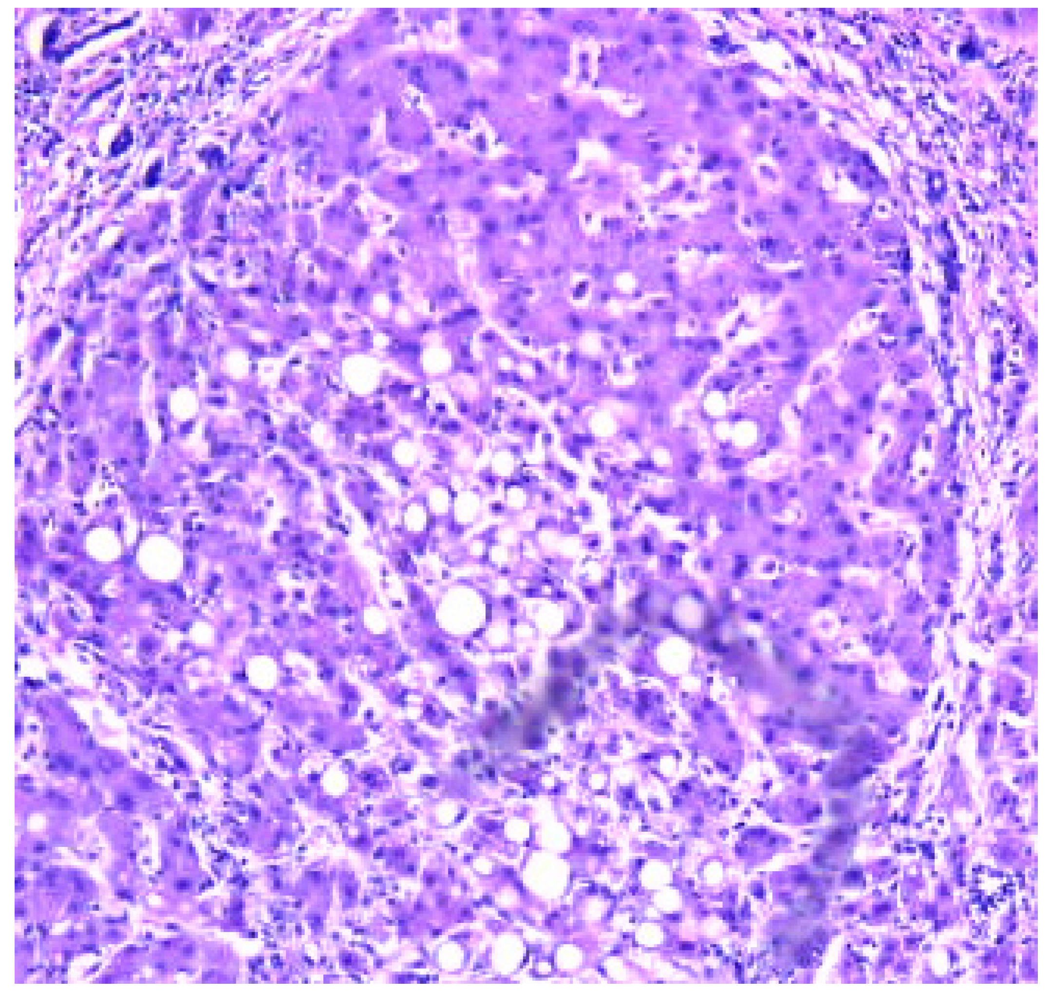
Cirrhosis usually represents with end-stage liver disease. Hepatitis C is the most damaging. Cirrhosis develops after a period of inflammation. The ill liver has parenchyma with fibrotic tissue and regenerative nodules. [Figure 4]
Clinically, liver cirrhosis is the severe period of chronic liver diseases. Early prevention and treatment of the causes of development and progression and pathogenic mechanism may slow down or reverse liver cirrhosis and its severe complications.
Decompensated liver cirrhosis and its complications, take attention to the clinicians. Various clinically signs as ascites, esophagogastric variceal bleeding, hepatic encephalopathy, acute kidney injury, and hepatocellular carcinoma, could be observing at the medical examination. Clearly that patients' quality of life is affected in liver cirrhosis.
Discussions
Patient lifestyle changes, unfortunately cannot cure cirrhosis. Complications accompanying hepatic cirrhosis include, portal hypertension, edema in the abdomen and lower extremities, splenomegaly, infections, hepatic encephalopathy.
Behavioral modifications can prevent or at least delay disease progression and provide symptomatic relief.
Lifestyle changes, include factors, as eliminating ethanol consumption and dietary interventions as possible low-sodium diet, in order to reduce water retention. Regulate protein intake according to their doctor's directions and some medical recommandations, will be proper in the treatment of cirrhosis.
Cirrhosis secondary to HBV and HCV is one of the common risk factor for liver degeneration in cirrhosis.. Practically monitoring of cirrhotic patients is recommended, with at least six monthly screenings. Liver biopsy is the gold standard technique highly promising non-invasive methodology under development, that are used in diagnosis. Liver transplantation (LT) is also an effective therapeutic option for the management of cirrhosis end-stage. Relatively recently research investigations try to elucidate the signal transduction pathways that link hepatocytes alterations including cellular disfunctionality. Diferential diagnosis of cirrhosis include research directions reffering to neonatal iron storage diseases, HELLP(hemolysis, elevated liver enzymes, low platelets) syndrome of pregnancy, idiopathic drug reaction. More than, other diseases are included in the diferential diagnosis of cirrhosis. This are Tyrosinemia, Galactosemia, Fructose intolerance.
Conclusions
Research studies predict about not so a good prognosis of patients diagnosed with cirrhosis, knowing laboratory results and clinical points.. Knowing the diagnostic in the ill patients, medical specialists could applay the proper treatment, carefully to the comorbidities.In idea that hepatic cirrhosis is hard or impossible to cure, we are waiting from future research dirrections and plans.
References
- AASLD-IDSA. HCV Guidance: Recommendations for Testing, Managing, and Treating Hepatitis C 2016. Available from: http://hcvguidelines.org/sites/default/files/HCV-Guidance_October_2016_a.pdf. Guidance produced by the American Association for the Study of Liver Disease and the Infectious Disease Society of America (AASLD-IDSA) covering recommendations for hepatitis C testing, clinical management and treatment.
- Recommendations for prevention and control of hepatitis C virus (HCV) infection and HCV-related chronic disease. Centers for Disease Control and Prevention. MMWR Recomm Rep. 1998;47(Rr-19):1–39. Recommendations from the Centers for the Disease Control and Prevention definng populations for risk-based testing of hepatitis C.
- Moyer VA. Screening for hepatitis C virus infection in adults: U.S. Preventive Services Task Force recommendation statement. Ann Intern Med. 2013;159(5):349–57. Hepatitis C testing recommendations for adults from the United States Preventive Services Task Force including risk-based testing and testing for adults born from 1945–1965.
- Holmberg SD, Spradling PR, Moorman AC, Denniston MM. Hepatitis C in the United States. N Engl J Med. 2013;368(20):1859–61. Description of a care cascade integrating data from the Chronic Hepatitis Cohort Study and the National Health and Nutrition Examination Survey.
- Viner K, Kuncio D, Newbern EC, Johnson CC. The continuum of hepatitis C testing and care. Hepatology. 2015;61(3):783–9. Description of an HCV continuum of care in a US urban center highlighting important areas where patients become lost at each stage. [CrossRef]
- Koneru A, Nelson N, Hariri S, Canary L, Sanders KJ, Maxwell JF, et al. Increased Hepatitis C Virus (HCV) Detection in Women of Childbearing Age and Potential Risk for Vertical Transmission - United States and Kentucky, 2011–2014. MMWR Morb Mortal Wkly Rep. 2016;65(28):705–10. Analysis finding that the proportion of infants born to HCV-infected mothers increased 68% nationally and 124% in Kentucky. [CrossRef]
- Kuncio DE, Newbern EC, Johnson CC, Viner KM. Failure to Test and Identify Perinatally Infected Children Born to Hepatitis C Virus-Infected Women. Clin Infect Dis. 2016;62(8):980–5. Analysis of surveillance data from the Philadelphia Department of Public Health finding a significiant number of women giving birth in Philadelphia tested positive for HCV but that most of their children were not tested for HCV. [CrossRef]
- Lambers FA, Prins M, Thomas X, Molenkamp R, Kwa D, Brinkman K, et al. Alarming incidence of hepatitis C virus re-infection after treatment of sexually acquired acute hepatitis C virus infection in HIV-infected MSM. AIDS. 2011;25(17):F21–7.
- Martin TC, Martin NK, Hickman M, Vickerman P, Page EE, Everett R, et al. Hepatitis C virus reinfection incidence and treatment outcome among HIV-positive MSM. AIDS (London, England) 2013;27(16):2551–7. [CrossRef]
- Ingiliz P, Martin TC, Rodger A, Stellbrink HJ, Mauss S, Boesecke C, et al. HCV reinfection incidence and spontaneous clearance rates in HIV-positive men who have sex with men in Western Europe. J Hepatol. 2016. [CrossRef]
- Testing for HCV infection: an update of guidance for clinicians and laboratorians. MMWR Morb Mortal Wkly Rep. 2013;62(18):362–5. Updated guidance for clinicians and laboratorians from the Centers for Disease Control and Prevention on diagnostic testing procedures for hepatitis C.
- Moyer VA. Screening for hepatitis C virus infection in adults: U.S. Preventive Services Task Force recommendation statement. Ann Intern Med. 2013;159(5):349–57. Hepatitis C testing recommendations for adults from the United States Preventive Services Task Force including risk-based testing and testing for adults born from 1945–1965.
- Thomas DL, Seeff LB. Natural history of hepatitis C. Clin Liver Dis. 2005;9(3):383–98. vi.
- Mack CL, Gonzalez-Peralta RP, Gupta N, Leung D, Narkewicz MR, Roberts EA, et al. NASPGHAN practice guidelines: Diagnosis and management of hepatitis C infection in infants, children, and adolescents. J Pediatr Gastroenterol Nutr. 2012;54(6):838–55.
- Thein HH, Yi Q, Dore GJ, Krahn MD. Estimation of stage-specific fibrosis progression rates in chronic hepatitis C virus infection: a meta-analysis and meta-regression. Hepatology. 2008;48(2):418–31. [CrossRef]
- Conteduca V, Sansonno D, Russi S, Pavone F, Dammacco F. Therapy of chronic hepatitis C virus infection in the era of direct-acting and host-targeting antiviral agents. J Infect. 2014;68(1):1–20. [CrossRef]
- Gomez J, Martell M, Quer J, Cabot B, Esteban JI. Hepatitis C viral quasispecies. J Viral Hepat. 1999;6(1):3–16. [CrossRef]
- Duffy S, Shackelton LA, Holmes EC. Rates of evolutionary change in viruses: patterns and determinants. Nat Rev Genet. 2008;9(4):267–76. [CrossRef]
- Bukh J. The history of hepatitis C virus (HCV): Basic research reveals unique features in phylogeny, evolution and the viral life cycle with new perspectives for epidemic control. J Hepatol. 2016;65(1 Suppl):S2–s21. S: Suppl). [CrossRef]
- Ly KN, Hughes EM, Jiles RB, Holmberg SD. Rising Mortality Associated With Hepatitis C Virus in the United States, 2003–2013. Clin Infect Dis. 2016;62(10):1287–8. Describes rising HCV-associated mortality in the United States from 2003–2013. During that time period, HCV-associated deaths surpassed 60 other nationally natofiable infectious conditions combined. [CrossRef]
- Allison RD, Tong X, Moorman AC, Ly KN, Rupp L, Xu F, et al. Increased incidence of cancer and cancer-related mortality among persons with chronic hepatitis C infection, 2006–2010. J Hepatol. 2015;63(4):822–8. Analysis of a cohort of HCV-infected persons that found the the incidence of liver cancer and many types of non-liver cancers were higher, and age at diagnosis and death younger, in patients with chronic HCV infection compared to the general population. [CrossRef]
- Alberti A, Chemello L, Benvegnù L. Natural history of hepatitis C. J Hepatol. 1999;31 Suppl 1:17–24.
- Hoofnagle JH. Course and outcome of hepatitis C. Hepatology. 2002;36:S21–S29.
- >Chen SL, Morgan TR. The natural history of hepatitis C virus (HCV) infection. Int J Med Sci. 2006;3:47–52. [CrossRef]
- Chevaliez S, Pawlotsky JM. Virology of hepatitis C virus infection. Best Pract Res Clin Gastroenterol. 2012;26:381–389. [CrossRef]
- Saludes V, González V, Planas R, Matas L, Ausina V, Martró E. Tools for the diagnosis of hepatitis C virus infection and hepatic fibrosis staging. World J Gastroenterol. 2014;20:3431–3442.
- Naveau S, Perlemuter G, Balian A. [Epidemiology and natural history of cirrhosis]. Rev Prat. 2005 Sep 30;55(14):1527-32.
- Kleinbloesem CH, van Harten J, Wilson JP, Danhof M, van Brummelen P, Breimer DD. Nifedipine: kinetics and hemodynamic effects in patients with liver cirrhosis after intravenous and oral administration. Clin Pharmacol Ther. 1986;40:21–8. [CrossRef]
- Bruno S, Maisonneuve P, Castellana P, et al. Incidence and risk factors for nonalcoholic steatohepatitis: prospective study of 5408 women enrolled in Italian tamoxifen chemoprevention trial. BMJ 2005;330(7497):932. [CrossRef]
- Fontana RJ, Hayashi PH, Barnhart H, et al. Persistent liver biochemistry abnormalities are more common in older patients and those with cholestatic drug induced liver injury. Am J Gastroenterol 2015;110(10):1450–9. [CrossRef]
Figure 1.
Normal Liver x10 Masson staining.
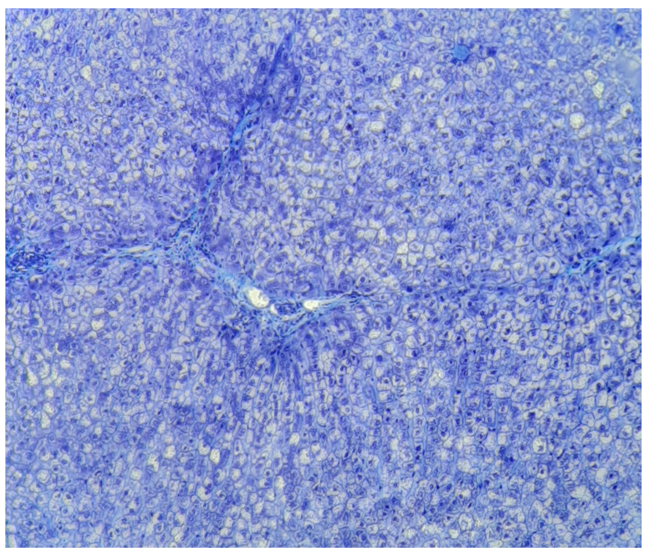
Figure 2.
Normal liver x 40 Goldner Szekely stain.
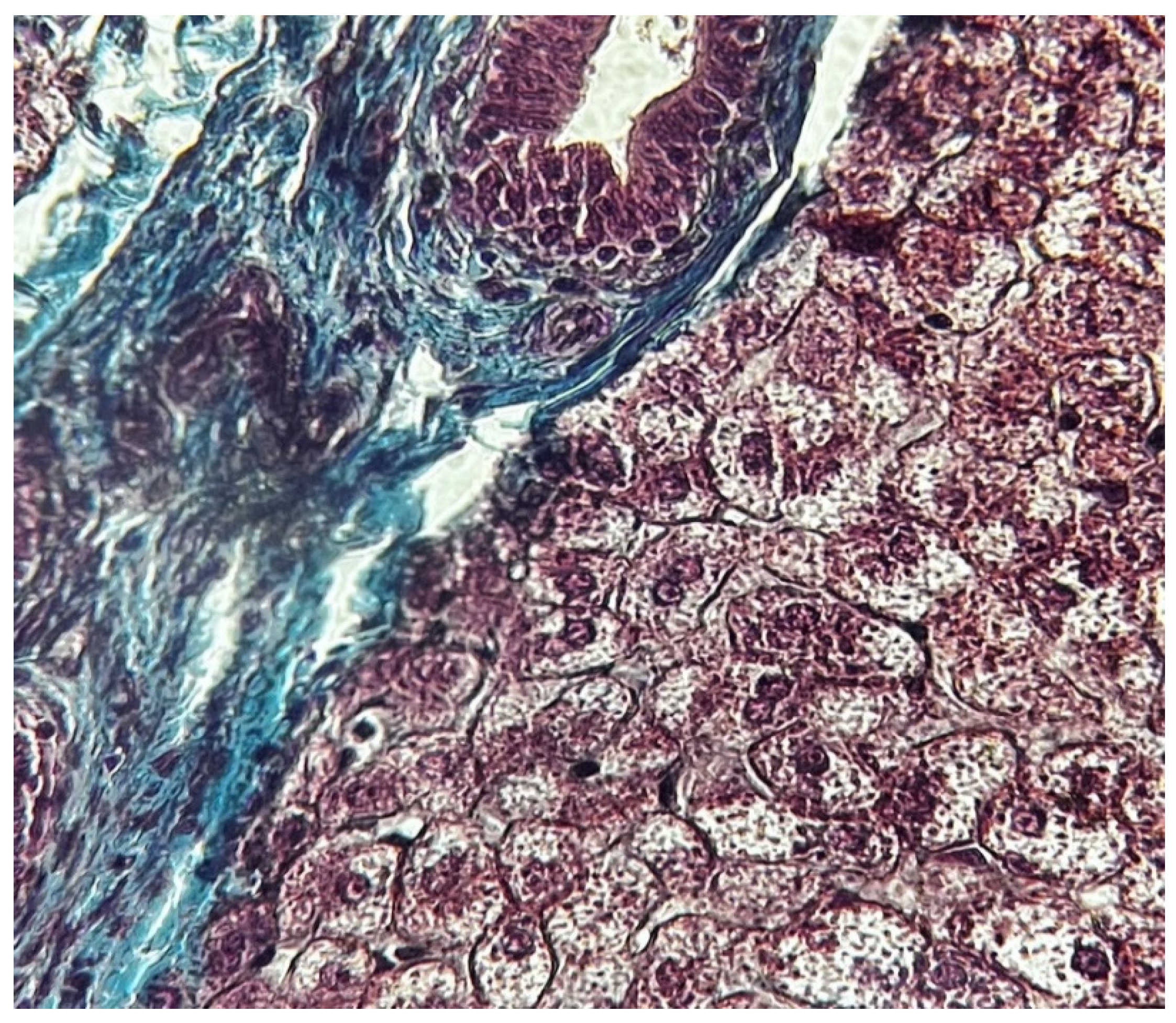
Figure 3.
Cirrhosis liver x10 H&E stain.
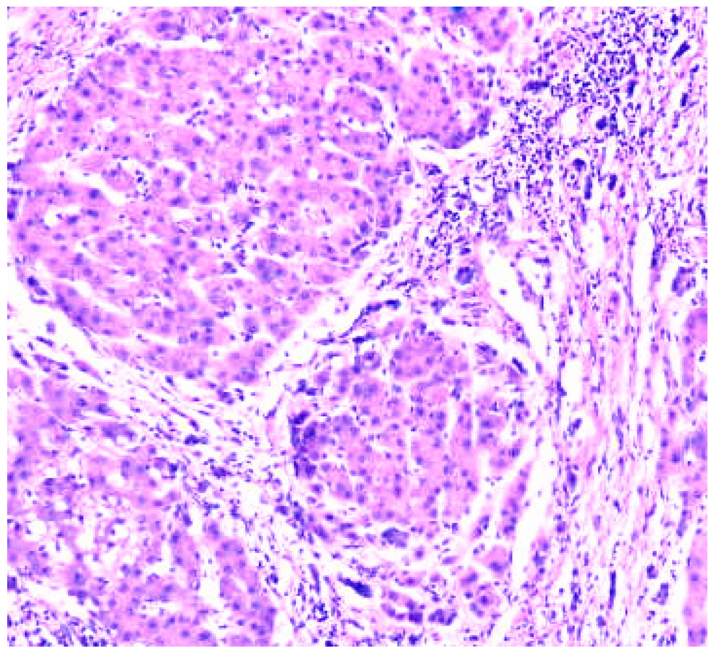
Figure 4.
Cirrhosis liver x10 H&E stain.
Disclaimer/Publisher’s Note: The statements, opinions and data contained in all publications are solely those of the individual author(s) and contributor(s) and not of MDPI and/or the editor(s). MDPI and/or the editor(s) disclaim responsibility for any injury to people or property resulting from any ideas, methods, instructions or products referred to in the content. |
© 2024 by the authors. Licensee MDPI, Basel, Switzerland. This article is an open access article distributed under the terms and conditions of the Creative Commons Attribution (CC BY) license (http://creativecommons.org/licenses/by/4.0/).
Copyright: This open access article is published under a Creative Commons CC BY 4.0 license, which permit the free download, distribution, and reuse, provided that the author and preprint are cited in any reuse.


