Preprint
Article

Phytochemical Constituents and Antibacterial Activity of Bark and Flowers Extracts of Four Magnolia Plant Species

Altmetrics

Downloads

213

Views

162

Comments

0

A peer-reviewed article of this preprint also exists.

Submitted:

07 December 2023

Posted:

07 December 2023

You are already at the latest version

Alerts
Abstract
This research focuses on exploring the bioactive and antibacterial properties of extracts from the flowers and bark of four Magnolia species (Magnolia champaca, Magnolia denudata, Magnolia grandiflora, and Magnolia officinalis) for possible cosmetic applications. We used ethanol to extract compounds from these plants and conducted various tests including spectrophotometry, HPLC, GC-MS, and microbiological analyses. The extracts, particularly rich in polyphenols (55.18 mg GAE/g), displayed significant antioxidant capabilities, with IC 50 values ranging between 9.99 mg/mL and 23.23 mg/mL. We quantified different compounds: phenolic acids (6.259 to 27.883 mg/g dry weight), aglycone flavonoids (61.224 to 135.788 mg/g dw), glycosidic flavonoids (17.265 to 57.961 mg/g dw), and lignans (150.071 to 374.902 mg/g dw). We identified 76 volatile compounds, predominantly oxygenated monoterpenes and sesquiterpene hydrocarbons, which contribute to the antibacterial effectiveness of the extracts. These extracts showed greater antibacterial activity against Gram-negative bacteria than Gram-positive bacteria. The diverse chemical compounds and their demonstrated activities suggest these extracts could be valuable in cosmetic, pharmaceutical, or food industries.
Keywords: 
Subject: Biology and Life Sciences  -   Plant Sciences

1. Introduction

Currently, there is an upward trend in the use of biological and ecological products in the cosmetics industry to obtain valuable, biodegradable, and environmentally friendly compounds. Many of these products are derived from biological sources, extracts from microorganisms, or plants.
A series of plant extracts can provide protection against pathogenic or facultative pathogenic microorganisms, and as a result, studies have been conducted, especially in the cosmetic field, where products come into direct contact with consumers. Bactericidal effects are exhibited by skincare products or disinfectants for the skin, soaps, or creams, many of which straddle the line between cosmetic treatment and pharmaceuticals themselves.
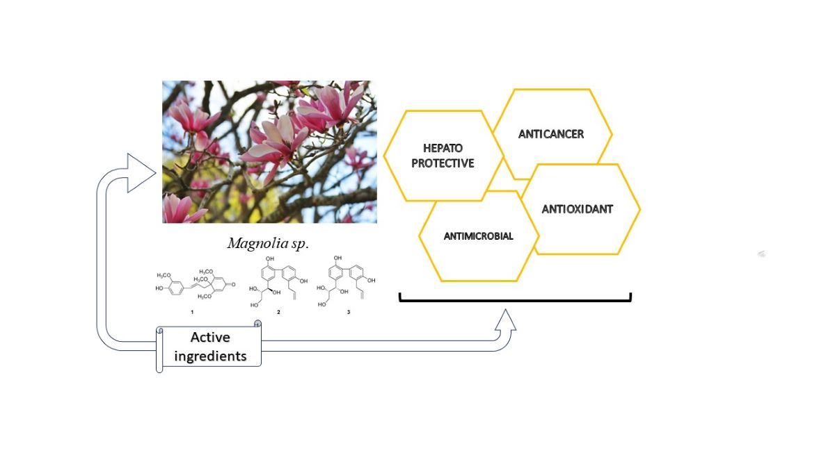
In these cases, a careful evaluation of the active principles that define the product and their scientific certification is necessary [1,2,3]. One of the plants appreciated for its complex biological activity is Magnolia. Magnolias are ornamental and medicinal plants known and used since ancient times, with the variety of species exceeding two hundred. They are native to America (Central and South America) and Asia, and they have been acclimatized in most warmer regions of Europe, including Romania. The plant belongs to the Magnoliaceae family and prefers clayey, less humid soils, a friendly, sunny climate, and mild winters. The leaves are oval with a smooth or rarely lobed edge and short petioles. The flowers are large, solitary, hermaphroditic, displaying various colors depending on the species, with fruit in the form of cones. Magnolia plants are known in traditional Chinese medicine and in other cultures, being valued for the properties of their bark and their flowers, with a particular emphasis on the buds. M. champaca (yellow-orange flowers) has spasmolytic properties, relaxation of respiratory pathways, and vasodilator mediated due to the blocking of Ca²⁺ channels, thus validating its therapeutic use in diarrhea, asthma, and hypertension [4]. Approximately 65 phytochemical compounds, including sesquiterpene hydrocarbons, sesquiterpene alcohols, and β-caryophyllene, have been identified, leading to their use in preventing or treating various ailments [5,6,7,8,9]. Valuable compounds, including phenolic compounds, were identified by Yoon [10] in Magnolia flower extracts (M. denudata), with his study focusing on the variation of bioactive elements depending on their processing parameters. Studies on the leaves and flowers of this pale pink plant have demonstrated the existence of phytochemicals, including lignans, phenolic compounds, and primary and secondary metabolites leading to antioxidant and anti-inflammatory properties [10,11,12,13,14].
Huang et al. [15] established that the creamy white flower extract of M. grandiflora L. leads to the reduction of tyrosinase and TRP-1 and inhibits melanogenesis in B16F10 cells. The flower extract also has antioxidant capacities and depleted reactive oxygen species (ROS) in cells. Therefore, the flower extract of M. grandiflora L. could be applied as a type of dermatological whitening agent in skincare products. Magnolia grandiflora L. is abundant in sesquiterpene lactones and lignans, such as magnolol and honokiol. Preparations derived from it have proven effective in treating a range of conditions, including coughs, ulcers, gastritis, diarrhea, nausea, vomiting, anxiety, and the reduction of allergies and asthmatic reactions. Additionally, they exhibit hepatoprotective and anticancer properties [16,17,18,19,20,21,22,23,24,25,26,27,28]. In a preliminary phytochemical study conducted by Rivas-Garcia et al. [29], it was found that extracts from Magnolia grandiflora in Mexico primarily contain flavonoids, terpenes, tannins, and alkaloids. These extracts also contain organic chromophores like flavonoids. Importantly, the extracts demonstrated antioxidant properties and were non-cytotoxic, suggesting their suitability for both medicinal and cosmetic purposes.
Furthermore, Magnolia officinalis is utilized as a tonic during the convalescent period, with its buds known to alleviate discomfort and intestinal issues while also exhibiting antiviral effects [30,31,32,33,34]. Magnolol, a natural compound isolated from M. officinalis, exhibits a range of biological activities through the NF-kB/MAPK, Nrf2/HO-1, and PI3K/Akt pathways, which are involved in its mediated biological functions. As a result, magnolol is regarded as a promising candidate for clinical research, despite its challenges related to low water solubility and rapid metabolism. Its biological activity is evident in various aspects, including anti-inflammatory, antimicrobial, antioxidant, anticancer, neuroprotective, antiepileptic, cardiovascular protection, management of gastrointestinal disorders, regulation of metabolism, and mediation of ion activity [28,35,36,37,38,39,40,41,42]. Both magnolol and honokiol are phenolic compounds from the lignan group [43,44]. They exhibit antioxidant and antimicrobial activity, playing a beneficial role in treating various ailments [36,45,46,47,48,49]. Magnolia extracts are used in treating depression, sinusitis, and cerebral congestion, being among others, an anticoagulant, a natural antioxidant, and an anti-cancer agent [24,27,50,51,52,53,54,55,56,57,58,59,60,61,62]. The antimicrobial activity of Magnolia extracts was examined against various types of microorganisms, including bacteria, yeasts, and molds, and their effectiveness was observed across all three major categories of microorganisms. In a study conducted by Ho et al. [63], the antimicrobial properties of two primary compounds found in Magnolia officinalis, honokiol and magnolol, were investigated. The results were particularly significant against microorganisms such as Actinobacillus actinomycetemcomitans, Porphyromonas gingivalis, Prevotella intermedia, Micrococcus luteus, and Bacillus subtilis, with minimum inhibitory concentration (MIC) values of 25 µg/mL. This suggests their potential use as adjuncts in treatments for periodontitis.
Additionally, the beneficial antimicrobial effects of magnolia extracts extended to commonly encountered strains in human pathology, including Klebsiella pneumonia, Staphylococcus aureus, Escherichia coli, Streptococcus mutans, Candida albicans, methicillin-resistant Staphylococcus aureus (MRSA), Propionibacterium sp., and against species like Porphyromonas gingivalis, which contribute to the development of dental caries [26,35,45,64,65,66,67,68,69,70,71,72,73,74,75,76,77,78,79].

2. Materials and Methods

2.1. Materials. Magnolia Samples, Reagents, Microbial Strains, Culture Media

In May-June 2023, flowers, and bark from four Magnolia species - Magnolia champaca, Magnolia denudata, Magnolia grandiflora, and Magnolia officinalis - were collected from the Nursery of Ornamental Shrubs and Plants. These samples were then identified by experts at the CCBIA Research Center, part of the Faculty of SAIAPM/Lucian Blaga University of Sibiu. Each sample was assigned a unique registration voucher number, falling within the range of 390/1 to 390/4. The reagents used include 96% ethanol, Folin-Ciocâlteu reagent, sodium carbonate, gallic acid, 2,2-diphenyl-1-picrylhydrazyl, Trolox, acetonitrile, acetic acid, supplied by Sigma-Aldrich GmbH, Steinheim, Germany.
To assess antibacterial effectiveness, we utilized specific reference bacterial strains. For Gram-positive bacteria, these included Staphylococcus aureus ATCC 43300 (MRSA), Staphylococcus epidermidis ATCC 12228, Streptococcus faecalis ATCC 19443, Streptococcus pyogenes ATCC 12347, Streptococcus sanguinis ATCC 10556, Actinomyces israelii ATCC 12102, and Propionibacterium acnes ATCC 6921/4311. For Gram-negative bacteria, we used Enterobacter aerogenes ATCC 13048, Escherichia coli ATCC 35218, Klebsiella pneumoniae ATCC 13883, Prevotella intermedia ATCC 25611, Porphyromonas gingivalis ATCC 33277, Proteus vulgaris ATCC 13315, and Pseudomonas aeruginosa ATCC 27853. We also employed standard antibiotics such as ampicillin, gentamicin, and tetracycline for comparison. The bacterial strains were grown and activated on Mueller Hinton agar and Mueller Hinton broth, both sourced from Sigma-Aldrich GmbH in Steinheim, Germany.

2.2. Methods

The bark and flowers of the selected plants were dried over a period of 10 days, maintaining a temperature of 40°C±0.5°C. This process continued until the materials consistently weighed the same. The dried material, weighing 100 grams, was then finely ground to a size between 300 and 500 microns and stored at a cool temperature of 4⁰C ±1⁰C.
The dried material was then macerated for four days in a solvent mixture, consisting of 500 ml of an ethanol: water solution (70:30 v/v), with the temperature maintained steadily at 18°C. Following the maceration period, the samples were filtered and concentrated using a rotary evaporator. This extraction process was carried out three times. Finally, the concentrated extracts were re-dissolved in distilled water at a 1:1 ratio for further analysis or application.

2.2.1. Identification of Total Polyphenols and Determination of Antioxidant Activity

To measure total polyphenol concentration, we adapted the Folin-Ciocâlteu method. This involved mixing 0.20 mL of the plant extract with 0.80 mL Folin-Ciocâlteu reagent (10% v/v) and 1 mL of a sodium carbonate solution (7.5% w/v). These samples were incubated for an hour at room temperature, ensuring they were shielded from light [80]. The polyphenol levels were measured using a UV-1900 SHIMADZU spectrophotometer (Shimadzu Corporation, Kyoto, Japan) at a wavelength of 750 nm. Results were expressed as milligrams of gallic acid equivalent per gram of the dried matter (mg GAE/g).
Antioxidant activity was determined using the DPPH free radical scavenging method, slightly modified by Popescu et al. [82]. This method involves preparing a stock methanolic solution of DPPH (25:100) and samples composed of dried extract and methanol in a 1:1 ratio. The working solution for the DPPH assay was prepared by mixing 10 mL of the stock solution with 90 mL of methanol. Subsequently, 25 µL of the sample was reacted with 175 µL of this DPPH working solution for 30 minutes at 20° C in the dark, followed by measuring the absorbance at 517 nm using a UV-1900 SHIMADZU spectrophotometer (Shimadzu Corporation, Kyoto, Japan). The control sample will be conducted following the same procedure, with the extract replaced by methanol. The calibration curve is made using Trolox, with the results expressed in milligrams of Trolox equivalent per gram of dried extract (mg TE / g DE).
The inhibition percentage (Formula (1)) is calculated according to the equation:
%   I = A b A a A b x   100
Where Ab is the absorbance of the control, and Aa is the absorbance of the reaction between the sample and radicals.

2.2.2. Identification and Quantification of Phenolic Compounds

Phenolic compounds were identified and quantified using a modified version of the LC-ESI-QTOF-MS method [81], using Agilent 1200 HPLC equipment (Agilent Technologies, Santa Clara, CA, USA). The mobile phase A was a mixture of water and acetic acid (95/5 v/v), and mobile phase B consisted of acetonitrile, water, and acetic acid (100/95/5 v/v/v), previously degassed at 20°C for 20 minutes. The chromatographic column used was Zorbax SB-Aq: 250 mm × 4.6 mm i.d., 5.0 μm particle size. The gradient elution program was established following a mix between A and B, the program being also used in the quantification of phenolic compounds. Using the ESI ionization system, positive and negative modes of droplets were established, with mass spectra identified in the m/z 50-1300 range.
Quantification of phenolic compounds was performed using the Agilent 1200 HPLC system (Agilent Technologies, Santa Clara, CA, USA), equipped with a PDA detector, an automatic injection system, and quaternary pump. A C18 chromatographic column was used (Zorbax SB-Aq: 250 mm × 4.6 mm i.d., 5.0 μm particle size). The mobile phase A was a mixture of water and acetic acid solution (95/5 v/v), and mobile phase B consisted of acetonitrile/water/acetic acid (100/95/5 v/v/v). The sample injection volume was set at 20 µL, with a flow rate of 0.8 mL/minute. Both mobile phase A and B were degassed at 20°C for 20 minutes. The gradient elution program mixed mobile phases A and B in varying proportions: initially 15% B for the first quarter-hour; 15–25 min, 25% B; 40% B from 25–40 minutes, 45% B during 40–65 minutes, 60% B for 65–70 minutes, 80% B from 70–75 minutes, and finally 100% B for 75–80 minutes, before returning to 15% B for the last 5 minutes. Phenolic compounds were detected in the 190 nm to 400 nm range. Data analysis was conducted using Agilent LC-MS-QTOF/HPLC MassHunter software version B.03.01.

2.2.3. Identification of Volatile Compounds

To analyze and identify the volatile compounds in Magnolia extracts, we used GC-MS (Gas Chromatography-Mass Spectrometry) technology, specifically a slightly adapted version of the method outlined by Popescu et al. [82]. The primary analysis of these volatile compounds was carried out using a Varian CP-3800/Saturn 2000 gas chromatograph, manufactured by Varian in California, USA, equipped with a Zebron ZB-5 MSI capillary. The column measures 30 m × 0.25 mm × 0.25 µm (Phenomenex, Torrance, CA, USA). This setup allows for the precise separation and identification of the various volatile compounds present in the Magnolia extracts.
In the GC-MS analysis of Magnolia extracts, we adhered to specific temperature settings for different components: the ion source was maintained at 230°C, the quadrupole at 150°C, and the injector at 220°C. We injected a 1µL sample using a splitter. The temperature program for the run was methodically set: initially, the temperature was held at 30°C for 10 minutes, then increased at a rate of 5°C per minute up to 160°C. It was held at 160°C for 15 minutes, after which it was raised at a rate of 15°C per minute to 250°C. Subsequently, the temperature was increased at a rate of 5°C per minute to 270°C and maintained at 270°C for a final duration of 10 minutes.
The ionization energy used in the process was 70 eV. Helium served as the carrier gas, flowing at a rate of 0.5 mL/min. To accurately identify the volatile compounds, we compared the resulting mass spectra with those in the Wiley 275 library and the NIST 17 Mass Spectral and Retention Index Libraries (NIST17), as well as the NIST WebBook and our laboratory’s own database.

2.2.4. Antibacterial Activity/Determination of MIC (Minimum Inhibitory Concentration)

To establish the minimum inhibitory concentration (MIC) of Magnolia samples against selected microorganisms, a series of dilutions was prepared, ranging from 0.625 µg/mL to 30 µg/mL extract (0.625, 1.25, 2.5, 5, 7.5, 10, 12.5, 15, 30 µg/mL). Dilutions were made in Mueller-Hinton Broth (MHB) following a slightly adapted version of the method by Ibrahim et al. [83].
We tested the Magnolia extract dilutions on various bacterial strains, including Staphylococcus aureus ATCC 43300 (MRSA), Staphylococcus epidermidis ATCC 12228, Streptococcus faecalis ATCC 19443, Streptococcus pyogenes ATCC 12347, Streptococcus sanguinis ATCC 10556, Actinomyces israelii ATCC 12102, Propionibacterium acnes ATCC 6921/4311, Enterobacter aerogenes ATCC 13048, Escherichia coli ATCC 35218, Klebsiella pneumoniae ATCC 13883, Prevotella intermedia ATCC 25611, Porphyromonas gingivalis ATCC 33277, Proteus vulgaris ATCC 13315, Pseudomonas aeruginosa ATCC 27853. These bacterial strains were activated by growing them for 24 hours in the MHB culture medium.
To conduct the MIC test, each test tube was inoculated with 10 µL of a bacterial strain suspension (density 0.5 McF = 1.5 × 10⁸ CFU/mL). 1 mL of diluted extract was added in the established order of dilutions. The tubes were thoroughly mixed to ensure uniform distribution of the bacterial suspension and extract. Following this, the tubes were placed in a Memmert incubator, set at 36°C. After a 24-hour incubation period, each set of tubes was examined to assess the growth of bacteria. The MIC was determined by observing the level of turbidity in each tube, which correlates with bacterial growth. The presence or absence of turbidity in the different dilutions helped to establish the lowest concentration of extract that effectively inhibited bacterial growth. To ensure accuracy and reproducibility, all these determinations were performed in triplicate.

2.2.5. Multivariate Statistical Analyses

Multivariate statistical analyses were performed using Addinsoft XLSTAT software, version 2014.5.03 (Addinsoft Inc., New York, NY, USA). The primary goal of this analysis was to uncover significant associations between the quality parameters of the Magnolia extracts and the identified volatile compounds. For this purpose, Pearson correlation analysis was used, allowing us to determine the strength and direction of the relationships between all the variables we had measured. We considered correlations statistically significant if they had a p-value less than 0.05 and highly significant if the p-value was less than 0.01. By doing this, we could understand which volatile compounds were most closely related to the quality parameters of the extracts, providing valuable insights into their characteristics and potential applications.

3. Results

The identification of total polyphenols and their associated antioxidant activity in the study revealed significant insights into the properties of hydroalcoholic extracts. Polyphenols, known for their antioxidant capabilities, also contribute to inhibiting the activity of various microorganisms. According to the data presented in Table 1, the concentration of polyphenols in the hydroalcoholic extracts was found to be directly proportional to both the source of extraction and the specific species of plant from which the samples were derived. The findings from this research, which focused on assessing the total polyphenol content and antioxidant activity in different parts of Magnolia species, demonstrate that these naturally occurring chemical compounds are found in both the bark and flowers of these plants. Particularly noteworthy is the bark of Magnolia champaca, which was found to contain a significant number of polyphenols, approximately 73.12 mg of gallic acid equivalent (GAE) per gram. Additionally, this part of the plant exhibited notable antioxidant properties. This was evidenced by its performance in the DPPH antioxidant test, where it showed a relatively strong ability to neutralize free radicals, as indicated by its IC50 value of 19.50 mg/mL.
The study’s results also indicate a variation in the polyphenol content and antioxidant activity between different parts of the Magnolia species. In Magnolia flowers, the polyphenol concentration is slightly lower, at around 68.62 mg GAE/g, compared to the bark. However, the antioxidant activity of the flowers is like that of the bark, with an IC50 value of 18.66 mg/mL in the DPPH test. In the case of Magnolia denudata, the hydroalcoholic bark extracts contain 65.18 mg GAE/g of polyphenols and show moderate antioxidant activity, with an IC50 value of 17.01 mg/mL. The flowers of this species have a lower polyphenol content, approximately 55.23 mg GAE/g, and exhibit reduced antioxidant activity, reflected in a lower IC50 value of 9.99 mg/mL. For Magnolia grandiflora, the bark has an average polyphenol content of 72.52 mg GAE/g, demonstrating relatively good antioxidant activity, with an IC50 value of 19.07 mg/mL in the DPPH test. The flowers, in contrast, contain fewer polyphenols, about 55.18 mg GAE/g, and exhibit lower antioxidant activity, with an IC50 value of 10.55 mg/mL. Among the Magnolia species studied, M. officinalis stood out for having the highest polyphenol content in its hydroalcoholic bark extracts, approximately 98.44 mg GAE/g. This was the greatest concentration observed among all the species examined. Additionally, it demonstrated the most potent antioxidant activity, with an IC50 value of 23.23 mg/mL in the DPPH test.
In contrast, the flowers of M. officinalis had a lower polyphenol content compared to its bark, about 66.02 mg GAE/g. Despite this, they still exhibited significant antioxidant activity, with an IC50 value of 18.78 mg/mL.
Further supporting these observations, other researchers have also identified polyphenols in Magnolia flowers. Park et al. [13] reported polyphenol levels ranging from 14 mg/g to 17 mg/g, and Yoon et al. [10] found even higher concentrations, between 86 mg GAE/g and 192 mg GAE/g, with variations depending on factors like moisture and temperature. The antioxidant activity in these studies was recorded to range between 84 and 311 mg VCE/g MFE, demonstrating the significant antioxidant potential of Magnolia species.

3.1. Identification and Quantification of Phenolic Compounds in Magnolia Extracts

Table 2 presents detailed data on the content of various types of compounds - namely phenolic acids, aglycone flavonoids, glycosidic flavonoids, and lignans - in different Magnolia species. This table specifically focuses on hydroethanolic extracts obtained from both the bark and flowers of these plants. The table helps in identifying which Magnolia species might be more suitable for specific uses based on their chemical profiles.
In the analysis of the four Magnolia species, gallic acid was found to be a common phenolic compound across all of them, with its highest abundance observed in M. champaca, reaching 5.021 mg/g.
Additionally, 4-hydroxybenzoic acid, another phenolic compound, was identified in all the Magnolia species. Notably, its concentration was highest in the bark extracts. In M. grandiflora, the concentration of 4-hydroxybenzoic acid in the bark was measured at 5.667 mg/g, while in M. denudata, it reached up to 11.097 mg/g. On the other hand, the flower extracts of these species also contained this compound, but in lower amounts. For instance, M. grandiflora flowers had 1.224 mg/g, and M. officinalis flowers had a higher concentration of up to 6.127 mg/g.
p-Coumaric acid was found in significant amounts in M. champaca, particularly in its flowers at a concentration of 2.671 mg/g. In M. denudata, this acid was notably prevalent in the bark extracts, with a concentration of 3.108 mg/g. Salicylic acid, another phenolic compound with well-known benefits, was detected in the bark extracts of M. champaca, M. grandiflora, and M. officinalis. However, it was absent in the flower extracts of these species. This suggests a variation in the distribution of this compound within the plants. Caffeic acid, often praised for its anti-inflammatory and antioxidant effects, showed higher concentrations in flower extracts. It was most abundant in the flowers of M. grandiflora, with a level of 1.044 mg/g. In addition, caftaric acid was quantified in all the studied Magnolia species. Interestingly, there were negative values reported for this acid in the bark extracts of M. champaca and M. grandiflora, as well as in the flower extracts of M. denudata.
Cinnamic acid was found in the bark of M. champaca, M. denudata, and M. officinalis, ranging from 0.002 mg/g to 0.092 mg/g. Chlorogenic acid was detected in all species, both in bark and flower extracts, being most abundant in the bark of M. officinalis, with a concentration of 5.619 mg/g. Ellagic acid, another compound with potential health benefits, was notably absent in the flower extracts of M. champaca and M. officinalis. Similarly, ferulic acid was not found in the flower extracts of M. grandiflora and M. officinalis.
From the Figure 1, it’s evident that the highest concentration of phenolic acids was observed in the bark extracts of M. denudata, reaching up to 27.883 mg/g. This is closely followed by the bark of M. champaca, which showed a significant phenolic acid content of 26.296 mg/g.
In contrast, when looking at the flower extracts, M. officinalis stood out with the highest number of phenolic acids, measuring 12.582 mg/g. This suggests that while the bark of certain Magnolia species may have higher overall concentrations of phenolic acids, the flowers of other species, like M. officinalis, also contain substantial amounts of these compounds.
Catechin, an aglycone flavonoid known for its potent antioxidant properties, was found to be the most abundant of its kind in all the Magnolia extracts studied. The concentrations of catechin were notably high, particularly in both the bark and flower extracts of certain species. For instance, in M. officinalis, catechin levels reached 81.034 mg/g in the bark and 32.333 mg/g in the flowers. In M. denudata, these values were even higher, with 91.227 mg/g in the bark and 78.936 mg/g in the flowers. Additionally, the study found that myricetin and luteolin, two other types of aglycone flavonoids, were present in the bark extracts of all four Magnolia species. However, these compounds were absent in the flower extracts of M. grandiflora. The presence of taxifolin was observed specifically in the flower extracts of the Magnolia species studied. However, it was notably absent in the bark extracts of both M. denudata and M. grandiflora. Additionally, eriodictyol was found in very low quantities in certain parts of the Magnolia species. The concentrations ranged between 0.001 mg/g and 0.021 mg/g. Eriodictyol was detected in the flower extracts of M. champaca and M. grandiflora, as well as in the bark of M. grandiflora and M. officinalis.
Apigenin and quercetin are present in all species, with values ranging from 0.991 mg/g - 1.283 mg/g in bark and 2.342 mg/g - 12.002 mg/g in flowers, respectively, 0.615 mg/g-0.995 mg/g and 0.357 mg/g-1.911 mg/g, the most significant quantities being found in M. denudata. Epicatechin was identified in all species, with values in bark extracts ranging from 20.361 mg/g for M. denudata to 34.562 mg/g for M. officinalis. In the flower extracts of the studied Magnolia species, epicatechin was found in particularly high concentrations in M. grandiflora. The maximum value of epicatechin in M. grandiflora’s flower extracts was remarkably high, at 39.397 mg/g.
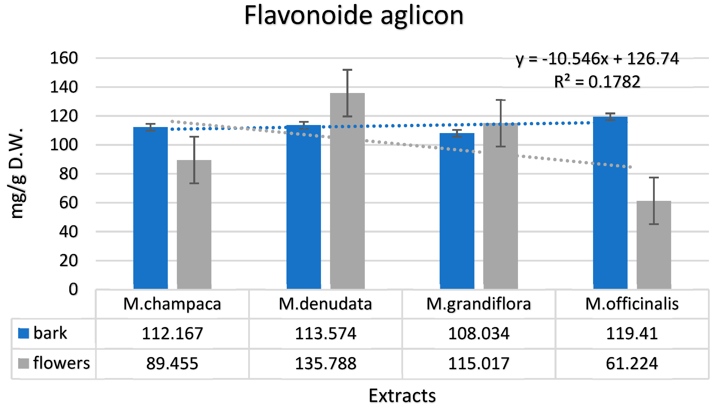
According to Figure 2, the total content of aglycone flavonoid compounds in Magnolia species exhibits significant variation between bark and flower extracts. For the bark extracts, the total aglycone flavonoids ranged from 108.034 mg/g to 119.41 mg/g. In the case of flower extracts, the total aglycone flavonoid content varied between 61.224 mg/g and 135.788 mg/g. Again, M. officinalis and M. denudata stood out with the highest values in this category.
Glycoside flavonoids are most significantly represented by rutin, which was identified in bark extracts at maximum values of 51.103 mg/g in M. officinalis and in flower extracts at a value of 56.782 mg/g in M. grandiflora. The minimum values are present in the bark of M. champaca (27.035 mg/g), and in the flowers of M. denudata (15.075 mg/g). Luteolin-7-O-glucoside is present in all species, with the highest quantities in the flower extracts of M. officinalis. Isoquercetin and hyperoside were present in relatively small quantities. Additionally, it was noted that these compounds were absent in the bark extracts of M. denudata, M. grandiflora, and M. officinalis. Kaempferol-3-O-rutinoside and Apigenin-7-O-glucoside were detected in all the Magnolia species studied, though in relatively small amounts. The highest concentrations of these compounds were found in the hydroethanolic extracts derived from the flowers of these species. Isorhamnetin-3-O-glucoside, on the other hand, was not found in any of the extracts from M. officinalis, neither in the bark nor in the flowers.
However, trace amounts of this compound, ranging from 0.001 mg/g to 0.002 mg/g, were identified in the bark of M. champaca, M. denudata, and M. grandiflora. Naringenin-7-O-glucoside was quantified in the Magnolia species, with a concentration of 0.001 mg/g in the bark extracts of M. champaca. In the flower extracts of M. denudata, this compound was found at a slightly higher concentration of 0.002 mg/g. Astragalin was detected exclusively in the flower extracts of M. denudata, present at a concentration of 0.001 mg/g. Quercitrin was also identified in these studies, with a concentration of 0.001 mg/g in both the bark of M. officinalis and the flowers of M. champaca. Isorhamnetin-3-O-rutinoside was found in various concentrations: in M. champaca, it was present in both bark and flowers, with concentrations of 0.003 mg/g and 0.004 mg/g, respectively; in M. denudata, this compound was found in the flowers at a concentration of 0.021 mg/g. For M. grandiflora and M. officinalis, the concentrations were 0.021 mg/g to 0.114 mg/g and 0.018 mg/g to 0.007 mg/g, respectively.
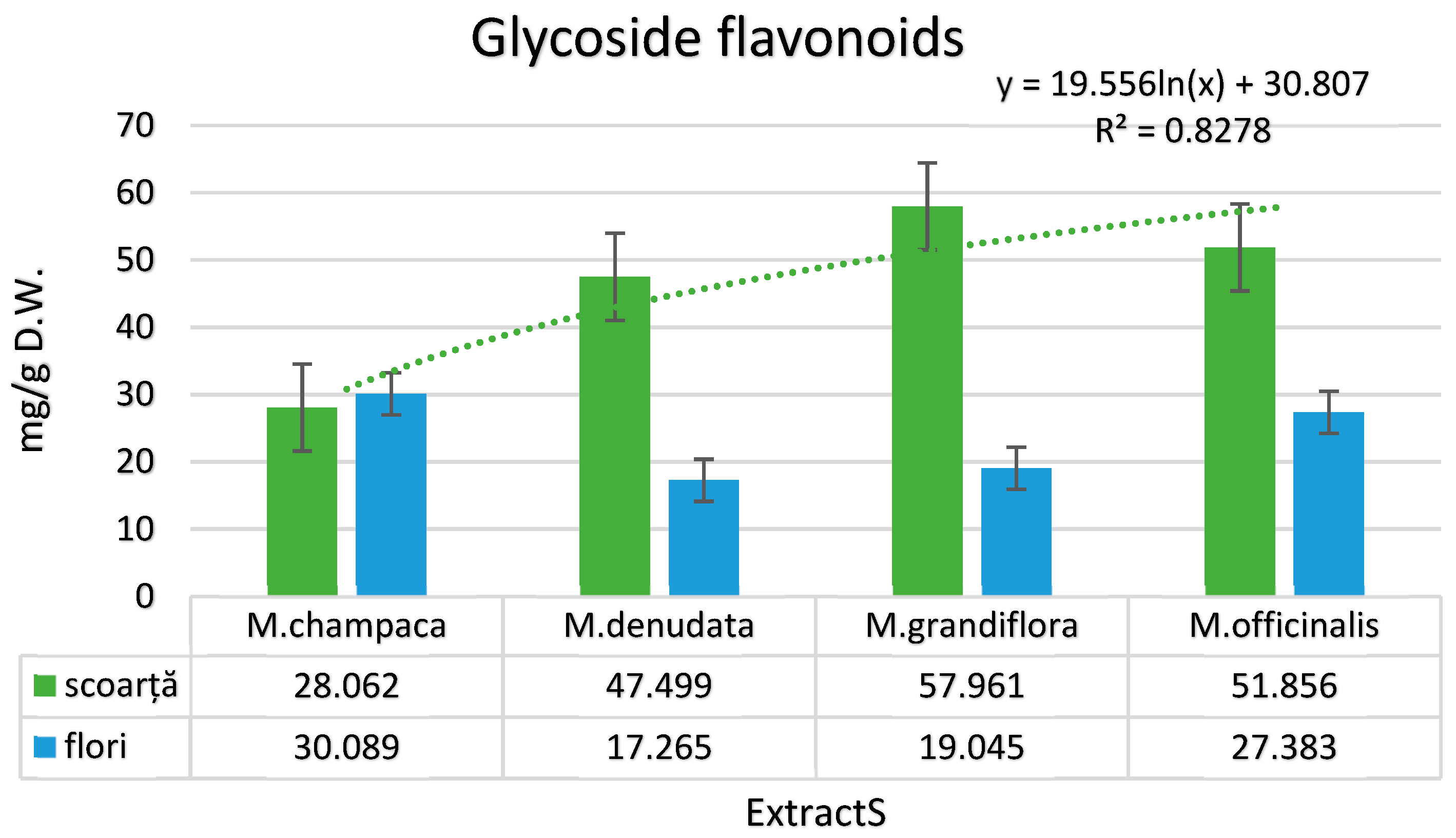
Additionally, the total values of glycoside flavonoids (Figure 3) in these Magnolia species varied significantly. In the bark extracts, these values ranged from 28.062 mg/g in M. champaca to 57.961 mg/g in M. grandiflora. For the flower extracts, the range was between 17.265 mg/g in M. denudata and 30.089 mg/g in M. champaca.
Lignans were found in both the bark and flower extracts of the studied Magnolia species. One such lignan, 4’-O-methylhonokiol, was identified across all species, displaying a range of concentrations. In the bark extracts, the concentration of 4’-O-methylhonokiol varied notably, with the lowest value being 2.011 mg/g in M. champaca and the highest reaching 21.021 mg/g in M. officinalis. Similarly, in the flower extracts, the levels of 4’-O-methylhonokiol also showed variability. The lowest concentration was found in M. denudata at 1.021 mg/g, while the highest was in M. grandiflora, with a concentration of 18.098 mg/g. Magnolol, a compound characteristic of Magnolia plants, was found in significant amounts, especially in M. officinalis.
In this species, the concentration of magnolol was remarkably high, with 97.093 mg/g in the bark and 23.021 mg/g in the flowers. Across all the studied species, magnolol was present in varying concentrations. In the bark extracts, its levels ranged from 7.021 mg/g to 9.999 mg/g.
For the hydroethanolic flower extracts, the range was between 2.001 mg/g and 19.021 mg/g. Honokiol, another lignan found in Magnolia species, was also identified in all the species included in the study.
The highest concentration of honokiol was in the bark extracts of M. officinalis, with a value of 56.785 mg/g. Conversely, the lowest concentration was observed in the flower extracts of M. denudata, at 2.001 mg/g. 3-Methoxymagnolol and isomagnolol, two lignan compounds, were detected in all the Magnolia species studied.
The most notable concentrations of these compounds were found in the bark and flower extracts of M. grandiflora and M. officinalis, indicating a higher accumulation of these lignans in these species.
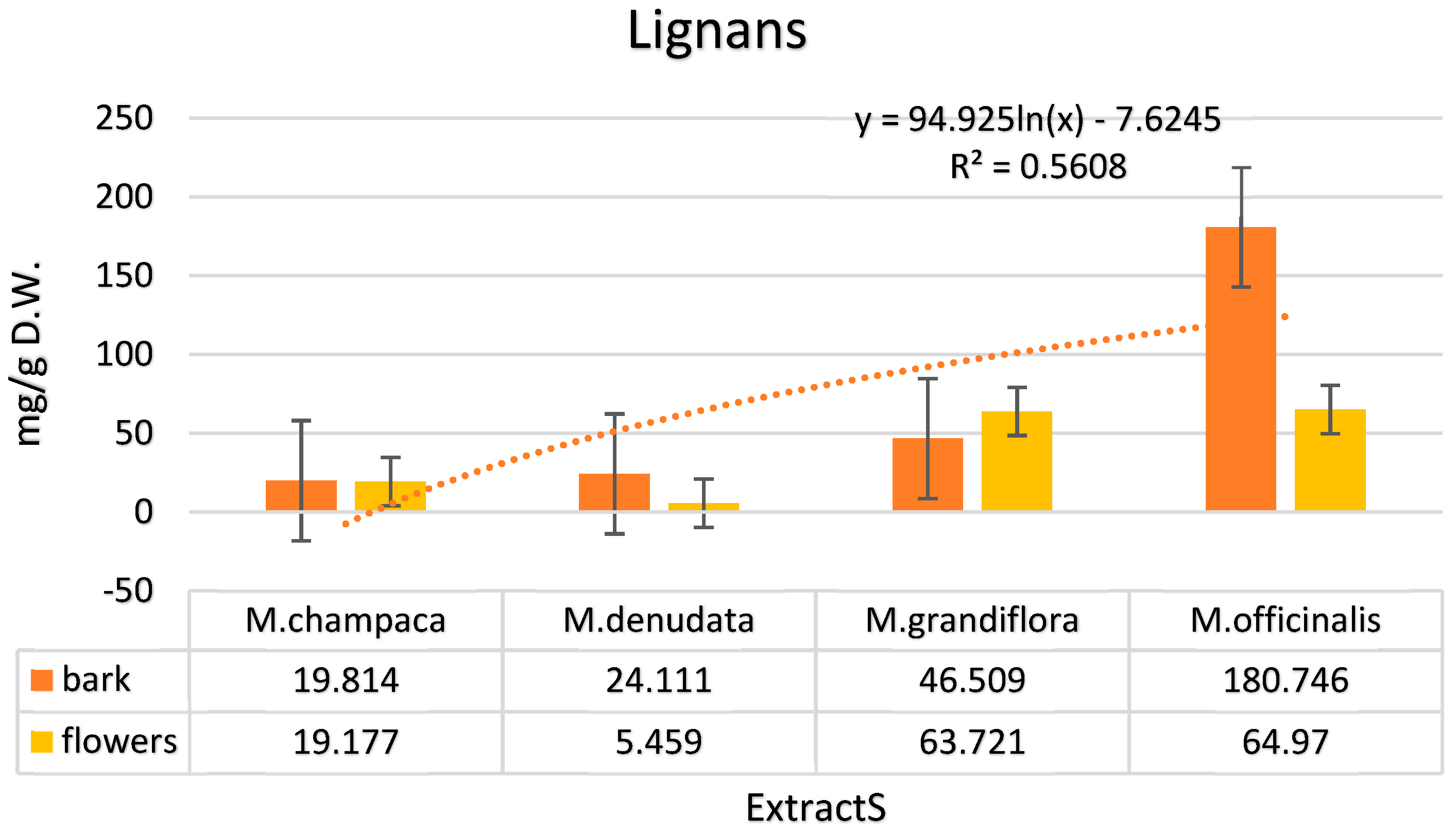
According to Figure 4, the total lignan content varied significantly between the bark and flower extracts. In the bark extracts, the total lignan concentration ranged from 19.814 mg/g to an impressive 180.746 mg/g. In the flower extracts, this range was slightly lower, spanning from 5.459 mg/g to 64.97 mg/g.
The data reveals that the chemical composition of Magnolia plants varies significantly between the bark and the flowers. Notably, the bark of these plants generally contains higher amounts of several phenolic acids compared to the flowers. These compounds include gallic acid, 4-hydroxybenzoic acid, p-coumaric acid, salicylic acid, caffeic acid, caftaric acid, cinnamic acid, chlorogenic acid, ellagic acid, ferulic acid, and syringic acid. Among the species studied, M. champaca and M. grandiflora stand out for having a richer content of these phenolic acids in both the bark and flowers, compared to M. denudata and M. officinalis. This suggests that M. champaca and M. grandiflora may be particularly valuable for applications that benefit from high levels of phenolic compounds, given their abundance in both key parts of the plant. The analysis of the Magnolia species shows that catechin, myricetin, luteolin, taxifolin, apigenin, and epicatechin are typically found in higher concentrations in the bark compared to the flowers. However, there are some notable exceptions to this trend: M. denudata is distinct for having significant amounts of catechin, luteolin, and apigenin in its flower extracts, indicating a unique distribution of these flavonoids in this species; M. grandiflora, on the other hand, exhibits a higher content of myricetin and epicatechin in both the bark and flowers, suggesting a more uniform distribution of these compounds across different parts of the plant. The analysis of flavonoid glycosides in the Magnolia species reveals that compounds such as rutin, luteolin-7-O-glucoside, kaempferol-3-O-rutinoside, apigenin-7-O-glucoside, and isorhamnetin-3-O-rutinoside are generally found in higher concentrations in the bark compared to the flowers.
However, there are exceptions to this trend in certain species: M. champaca is characterized by a higher content of rutin in both the bark and flowers. This indicates a relatively uniform distribution of rutin throughout the plant; in the case of M. grandiflora, luteolin-7-O-glucoside is found in significant amounts in both the bark and flowers, suggesting a consistent presence of this compound across different parts of the plant. The comparative analysis of Magnolia species indicates that the bark generally contains higher concentrations of lignans, such as 4’-O-methylhonokiol, magnolol, honokiol, 3-methoxymagnolol, and isomagnolol, compared to the flowers. Notably, M. officinalis stands out for its particularly high lignan content in the bark, especially in terms of magnolol and honokiol concentrations. This abundance of lignans in the bark of Magnolia species, especially M. officinalis, correlates with findings from studies by Ho et al. [63], Shen et al. [84], and Sun et al. [85]. These studies have highlighted the presence and significant impact of these compounds in Magnolia extracts, attributing to them properties such as antibacterial, antioxidant, and antitumor effects. Additionally, the total phenolic compound values in the bark extracts were found to vary widely, ranging from 186.339 mg/g in M. champaca to as high as 374.902 mg/g in M. officinalis. In comparison, the flower extracts contained lower total phenolic values, ranging between 150.071 mg/g in M. champaca and 204.042 mg/g in M. grandiflora.

3.2. Identification and Quantification of Volatile Compounds in Magnolia Extracts

Table 3 provides a detailed account of the content of various chemical compounds found in the bark and flowers of four Magnolia species: M. champaca, M. denudata, M. grandiflora, and M. officinalis. The table likely compares the concentrations of these compounds, highlighting significant variations not only between the species but also between the two plant parts (bark and flowers). This comparative analysis is essential for understanding the unique chemical makeup of each species and how it varies within the plant. Regarding the volatile compounds, the study focuses on comparing these compounds in the bark of the four Magnolia species. Common volatile compounds identified across all species include α-Thujene and α-Pinene. The concentrations of these compounds vary among the species: 0.1%, 0.1%, 0.4%, 0.5%, respectively, 0.4%, 1.1%, 0.2%, 0.3%. Similar relative concentrations were presented by all bark extracts for the compound Camphene (1.1%, 1.1%, 1.1%, 1.3%).
Significant variations were observed in the case of Limonenes, where the percentages ranged between 0.5% in M. grandiflora and 2.2% in the bark extracts of M. denudata. 1,8-Cineole is present in all four species, with variable concentrations (0.5%, 2.1%, 0.5%, 1.7%).
Variable concentrations are noted for the compounds β-Pinene and Camphor, which can reach up to 6.6%, lower in the case of Phenylacetaldehyde and Borneol, where they are between 1.1% and 2.2%. Bornyl acetate is present in all four species, with concentrations of 25.6% (M. champaca), 14.9% (M. denudata), 14.2% (M. grandiflora), 17.8% (M. officinalis). Significant results were obtained for the compound Ε-Caryophyllene, with obtained values in variable concentrations (11.1%, 22.4%, 21.1%, 15.6%). β-Caryophyllene was identified in all species with values ranging from 1.2%, 1.8%, 3.4%, to 5.6%, close to α-Selinene and Viridiflorene. 9-epi-(E)-Caryophyllene, (3.7%, 2.9%, 3.8%, 3.1%), presents values close to n-Hexadecanol and n-Heneicosane. Germacrene D shows a maximum value in the bark extracts of M. champaca of 11.2%, followed by M. officinalis with 7.7%, M. denudata with 5.3%, and M. grandiflora with 4.6%. Present in all four species, (0.9%, 0.5%, 1.5%, 1.7%) is also δ-Cadinene, n-Tricosene (7.7%, 5.5%, 1.7%, 4.2%), and n-Pentacosane (1.4%, 4.9%, 1.1%, 2.9%). Some of the volatile compounds identified are specific to each species, namely: M. champaca contains Heptanal (0.1%) and 1-Octanol (0.4%). M. denudata contains 6-Methyl-5-hepten-2-one (0.1%). M. grandiflora contains α-Phellandrene (0.1%) and p-Cymene (0.1%). M. officinalis contains (Z)-β-Farnesene (0.1%), γ-Gurjunene (0.1%), and γ-Muurolene (0.2%). To compare the volatile compounds in the four species (M. champaca, M. denudata, M. grandiflora, and M. officinalis) in terms of their content in flowers, we will analyze the presence and relative concentrations of these compounds: α-Thujene and α-Pinene are present in all four species, with variations in concentration of 1.2%, 1.5%, 0.7%, 2.1%, and 0.6%, 0.9%, 0.8%, 1.5%, respectively.
The volatile compound 1,8-Cineole exhibits its lowest values in M. grandiflora and M. officinalis, around 0.1%. In contrast, higher concentrations of this compound are found in the hydroethanolic flower extracts of M. champaca and M. denudata, at 0.9% and 1.1% respectively. Phenylacetaldehyde and (E)-β-Ocimene are identified in all four Magnolia species, showing a range of concentrations from 1.1% to 5.7%. The concentration of 1-Octanol varies notably among these species. It is found to be 2.8% in M. champaca, and it reaches 4.6% in M. denudata. In M. grandiflora, the concentration is 1.2%, while M. officinalis has a slightly higher value at 1.9%. Other volatile compounds such as cis-Linalool oxide (furanoid) are present in a range between 0.1% and 0.6%. Terpinolene, another volatile compound, is found in concentrations varying from 0.2% to 0.8% among these species.
Linalool is found in substantial amounts in the flower extracts of the studied Magnolia species. The identified concentrations are notably high, with 25.1% in M. champaca, 20.4% in M. denudata, 15.8% in M. grandiflora, and 21.2% in M. officinalis. Geraniol and α-Terpineol, two other volatile compounds, vary in concentration between 1.6% and 9.1% across these species. Bornyl acetate is another compound present in all four Magnolia species, showing significant variations in its concentration: 2.1%, 5.3%, 4.3%, and 11.1%. Each Magnolia species also contains specific volatile compounds in their flower extracts: M. champaca is characterized by the presence of Hexanal (0.1%) and Myrtenal (0.3%); M. denudata includes trace amounts of cis-Sabinene hydrate and trans-Nerolidol (0.1%); M. grandiflora contains trace amounts of β-Pinene, as well as Perillene (0.1%), α-trans-Bergamotene (0.1%), and n-Eicosane (0.3%); M. officinalis is distinguished by 6-Methyl-5-hepten-2-one (0.5%) and (E)-β-Farnesene (0.3%). When comparing the data across the species M. champaca, M. denudata, M. grandiflora, and M. officinalis, and analyzing the content of volatile compounds in both bark and flowers, several observations emerge: In M. champaca, the flower extracts contain significantly higher amounts of volatile compounds compared to the bark extracts.
Notably, Linalool and Limonene are dominant in the flower extracts of this species, contributing to its distinct aromatic profile. For M. denudata, like in M. champaca, the flower extracts exhibit a notably higher concentration of volatile compounds compared to the bark. Linalool and Limonene are prominent in the flowers of M. denudata, contributing significantly to their characteristic aroma. This suggests that the flowers of M. denudata are particularly enriched with these compounds, which are known for their distinctive fragrances. In the case of M. grandiflora, there are significant differences in the volatile compound profiles between the bark and flowers. The flowers of M. grandiflora are characterized by a high content of Linalool, whereas the bark is notable for its substantial amounts of Camphor. This indicates a distinct chemical composition in different parts of the plant, each contributing uniquely to the overall aromatic and phytochemical profile of M. grandiflora. Like M. champaca and M. denudata, the flowers of M. officinalis also contain a greater variety and concentration of volatile compounds than the bark. Notably, Linalool, Limonene, and Camphor are key compounds in the flowers of this species. This pattern reinforces the trend observed across these Magnolia species, where the flower extracts generally have a richer and more diverse array of volatile compounds compared to the bark.
In the gas chromatography analyses conducted for this study, a total of 76 volatile components were identified across the Magnolia species. Using statistical models, the main components were grouped based on the aromatic profile of the extracts from the bark and flowers of the four Magnolia species (M. champaca, M. denudata, M. grandiflora, and M. officinalis). The concentration of oxygenated monoterpenes in the bark extracts ranged between 38.6% and 49.6%, while in the flower extracts, these values were significantly higher, ranging between 52.8% and 68.4%. This indicates a more pronounced presence of oxygenated monoterpenes in the flowers compared to the bark. Hydrocarbon monoterpenes varied between 7.2% and 19.6%. These values are notably lower than those reported in previous studies by Yahaya et al. [86], Morshedloo et al. [27], and Farag et al. [19], where hydrocarbon monoterpenes were found to be between 40.7% and 43.8%. The concentration of oxygenated sesquiterpenes in the extracts ranged between 2.3% and 4.6%. In contrast, hydrocarbon sesquiterpenes varied more widely, comprising between 33.1% and 40.4% in the bark extracts and between 11.8% and 19.7% in the flower extracts.
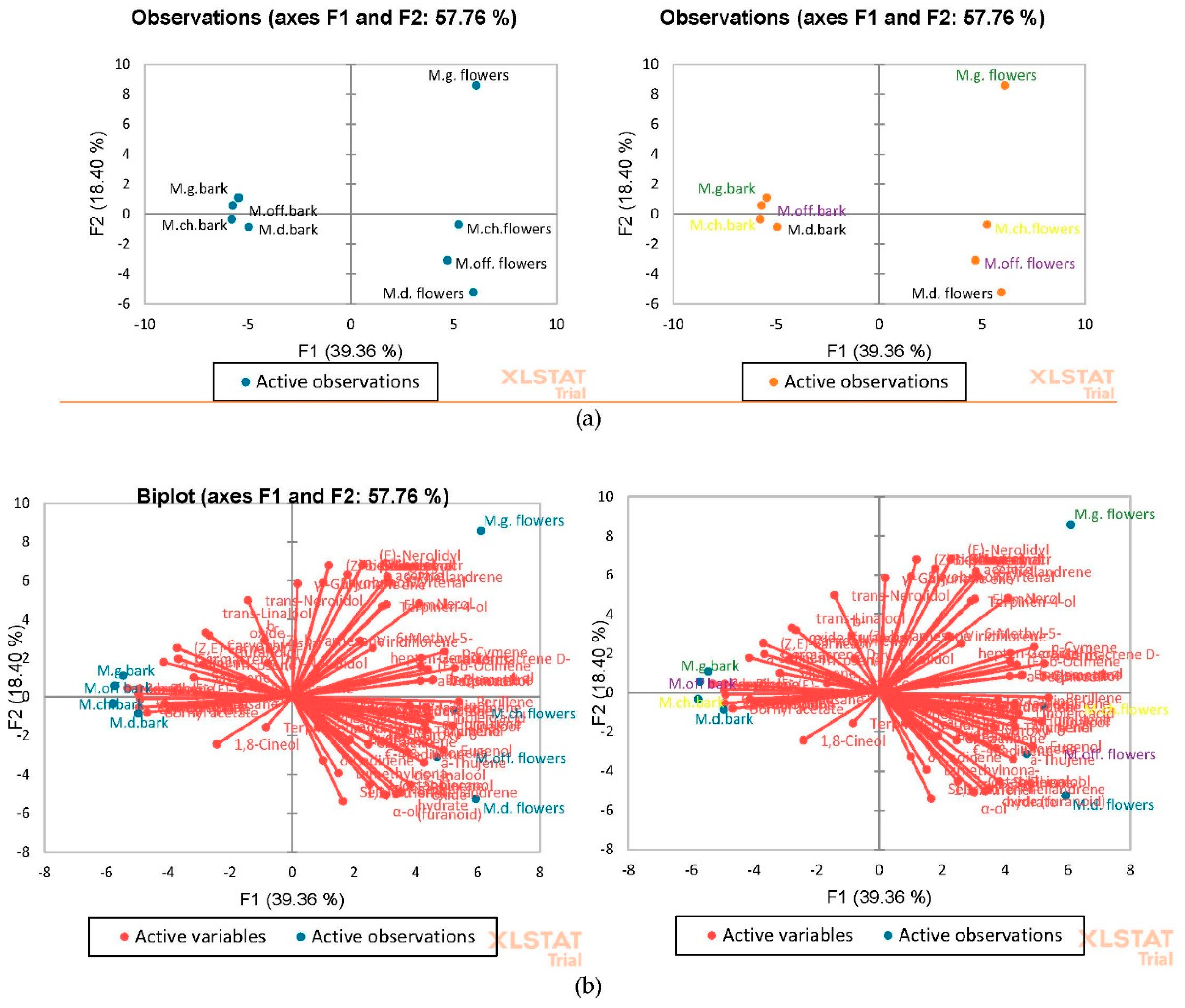
Figure 5 in the study provides a graphical representation, likely a scatter plot or similar type of chart, showing the separation of extracts based on species (M. grandiflora and M. officinalis) and plant part (bark or flowers). This separation is depicted along positive axes in the graph.
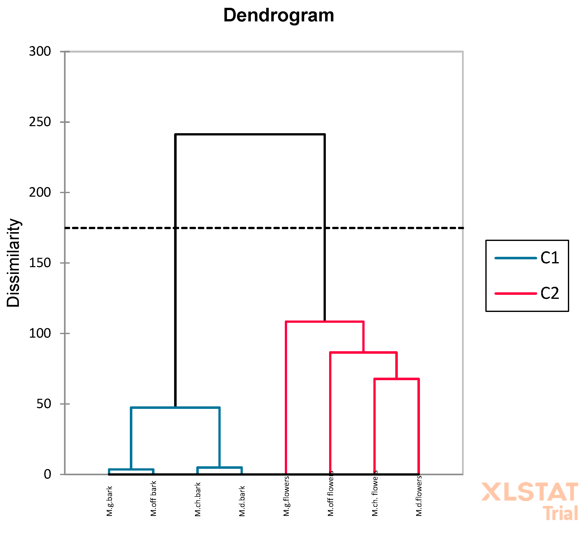
In Figure 5a, the flower extracts of M. grandiflora are particularly distinct, positioned in the second quadrant, and are characterized by a rich composition of various volatile compounds. These include α-Thujene, α-Pinene, α-Terpinene, Limonene, (E)-β-Ocimene, γ-Terpinene, 1-Octanol, Linalool, α-Terpineol, Myrtenol, Nerol, and Geraniol. Their location in the second quadrant suggests a unique volatile profile that sets them apart from the other extracts studied. Bark extracts of M. grandiflora and M. officinalis are near the axis dividing the quadrants, being rich in volatile compounds such as α-Thujene, α-Pinene, β-Pinene, Limonene, 1-Octanol, Bornyl acetate, β-Caryophyllene, 9-epi-(E)-Caryophyllene, Germacrene D. In quadrant one, on the negative semi-axis, are the bark extracts of M. champaca (M.ch.) and M. denudata (M.d.), represented by compounds like β-Pinene, Limonene, Camphor, Bornyl acetate, E-Caryophyllene, Germacrene D, Viridiflorene, Spathulenol, etc. In quadrant two, on the negative semi-axis, are the flower extracts of M. champaca (M.ch.), rich in α-Thujene, Limonene, (E)-β-Ocimene, γ-Terpinene, 1-Octanol, Linalool, and M. denudata (M.d.) and M. officinalis (M.off.) with significant values in volatile compounds such as α-Thujene, α-Pinene, Limonene, 1-Octanol, Linalool, β-Citronellol, Geraniol, etc. (Figure 5b). The multivariate analysis was complemented by a cluster analysis that minimized variation within the group, revealing two distinct clusters for bark and flowers (Figure 6). In the study, a wide range of metabolites was observed, with the quantity of volatile compounds differing based on both the source and the species from which the extracts were derived. The vectors depicted in the graphical representations illustrate the contribution of each compound to the overall distribution of variables, highlighting how individual components influence the compositional makeup of the extracts. Additionally, the multivariate analysis was further enhanced by conducting a cluster analysis. This analysis focused on minimizing the variation within each group, leading to the identification of two distinct clusters corresponding to bark and flower extracts. This differentiation is clearly depicted in Figure 6, effectively showcasing the inherent compositional differences between the bark and flower extracts of the Magnolia species studied.
The observed differences in the data reflect the variability in aromatic profiles both between different Magnolia species and between various parts of the plant. It is evident from the analysis that the flowers typically serve as the primary source of volatile compounds in magnolias. Furthermore, the concentration and composition of these volatile compounds can vary significantly from one species to another. It is important to recognize that these diverse volatile compounds are key contributors to the unique aromas and characteristics specific to each Magnolia species. The distinct aromatic profiles resulting from these compounds have potential applications across various industries, including cosmetics, perfumery, and medicine. Their unique properties may be harnessed for developing fragrances, therapeutic products, or other specialized applications, underscoring the importance of understanding, and characterizing these compounds in magnolias.

3.3. Antibacterial Action of Hydroethanolic Magnolia Extracts

The study involved testing both bark and flower extracts of Magnolia species against selected bacterial strains to determine their minimum inhibitory concentration (MIC). The purpose of this testing was to evaluate the potential of these extracts for use in developing natural cosmetic products with antibacterial properties. As detailed in Table 4, a range of both Gram-positive and Gram-negative bacteria were chosen for this assessment. The inclusion of different types of bacteria, both Gram-positive and Gram-negative, ensures a thorough assessment of the extracts’ broad-spectrum antibacterial capabilities.
The data from table 4 in the study clearly demonstrates the varying sensitivity of different bacterial strains to extracts from the bark and flowers of four Magnolia species: M. champaca, M. denudata, M. grandiflora, and M. officinalis. Specifically, the study focused on the sensitivity of these extracts to the Staphylococcus aureus ATCC 43300 (MRSA) strain, a commonly known resistant bacteria. The results indicate that the MRSA strain showed relative insensitivity to both conventional antibiotics (ampicillin, tetracycline) and the Magnolia extracts from both bark and flowers. Notably, this strain exhibited no sensitivity to the bark extracts of M. champaca and M. grandiflora. In contrast, the bark extracts from M. denudata and M. officinalis showed some effectiveness, with MIC values recorded at 30 µg/mL. Similarly, the flower extracts from M. grandiflora and M. officinalis also demonstrated MIC values of 30 µg/mL.
These findings are significant in the context of previous research, such as the studies by Chung et al. [70], which reported antibacterial activity against Staphylococcus aureus strains but focused on extracts from Magnolia coco. The results from Table 4 contribute to a broader understanding of the antibacterial properties of different Magnolia species and their potential applicability in addressing antibiotic-resistant bacterial strains. The study also examined the effectiveness of Magnolia extracts against Staphylococcus epidermidis ATCC 12228, a bacterium known to show moderate resistance to antibiotics. The bark extracts from the Magnolia species displayed varied effectiveness against this strain: M. officinalis bark extract showed the highest efficacy with an MIC value of 5 µg/mL, while M. champaca and M. denudata had MIC values of 15 µg/mL. On the other hand, the flower extracts of M. champaca and M. denudata were less effective, exhibiting weaker action with MIC values of 30 µg/mL.
However, flower extracts from M. grandiflora and M. officinalis were more potent, with MIC values of 7.5 µg/mL and 5 µg/mL, respectively. Furthermore, the study revealed that Streptococcus faecalis ATCC 19443 responded effectively to Magnolia extracts. The bark extracts from the Magnolia species had MIC values ranging from 5 µg/mL to 15 µg/mL, whereas the flower extracts showed a range of 2.5 µg/mL to 15 µg/mL.
Notably, the extracts from M. officinalis demonstrated the best results, with the bark extract having an MIC of 5 µg/mL and the flower extract even lower at 2.5 µg/mL. These findings highlight the potential of Magnolia extracts as effective antibacterial agents against specific bacterial strains, including those resistant to conventional antibiotics. Streptococcus pyogenes ATCC 12347, a bacterium sensitive to all three tested antibiotics, exhibited varying responses to Magnolia extracts. It showed no reaction to the bark extracts from M. denudata and the flower extracts from both M. grandiflora and M. officinalis. However, a slight sensitivity was observed with an MIC value of 30 µg/mL for both the bark and flower extracts of M. champaca, as well as the flower extracts from M. denudata. In tests involving Streptococcus sanguinis ATCC 10556, no substantial antibacterial activity was observed with most of the Magnolia extracts. Only very low sensitivity was detected in the case of the bark and flower extracts from M. officinalis and the flower extracts from M. grandiflora, each with an MIC of 30 µg/mL.
These results indicate that while Magnolia extracts can be effective against certain bacterial strains, their efficacy varies significantly depending on the species of the extract and the bacterial strain. The Magnolia extracts tested in the study exhibited a range of effectiveness, from moderate to sensitive, against the bacterial strains Actinomyces israelii ATCC 12102 and Propionebacterium acnes ATCC 6921/4311. The minimum inhibitory concentration (MIC) values for these extracts against these strains varied, with the most notable values ranging between 2.5 µg/mL and 15 µg/mL. However, an exception was noted in the case of the flower extracts from M. champaca, which displayed a higher MIC of 30 µg/mL against the strain Actinomyces israelii ATCC 12102. In the assessment of Gram-negative bacteria, the study found varying levels of effectiveness of the Magnolia extracts against Enterobacter aerogenes ATCC 13048. Most extracts exhibited medium to low inhibitory activity, with minimum inhibitory concentration (MIC) values ranging between 10 µg/mL and 30 µg/mL. However, an exception was noted in the case of the bark extracts from M. officinalis, which demonstrated intense antibacterial activity with a MIC of 7.5 µg/mL. In the case of Escherichia coli ATCC 35218, the extracts generally showed moderate to low antibacterial action, with MIC values again falling within the range of 10 µg/mL to 30 µg/mL. Notably, the extracts from both the bark and flowers of M. champaca did not inhibit this strain of bacteria.
The study revealed that the Magnolia extracts generally did not exhibit inhibitory activity against the Klebsiella pneumoniae ATCC 13883 strain, as no significant antibacterial effects were observed in most cases. However, there was a notable exception with the bark extract from M. officinalis, which demonstrated a mild effect on this strain, reflected in a minimum inhibitory concentration (MIC) of 30 µg/mL. In contrast, the bark and flower extracts from various Magnolia species showed medium to very good antibacterial activity against Prevotella intermedia ATCC 25611 and Porphyromonas gingivalis ATCC 33277. The MIC values for these strains ranged from as low as 0.625 µg/mL to 15 µg/mL. Particularly effective were the bark extracts from M. champaca and M. officinalis, which yielded the best results against these strains. The extracts also displayed medium antibacterial activity against Proteus vulgaris ATCC 13315, with MIC values ranging between 5 µg/mL and 15 µg/mL.
The study showed that Magnolia officinalis, including both bark and flower extracts, exhibited very good antimicrobial activity against the Pseudomonas aeruginosa ATCC 27853 strains, with a minimum inhibitory concentration (MIC) of 2.5 µg/mL. This indicates a high level of effectiveness in inhibiting the growth of this bacterial strain. In contrast, the extracts from the other three Magnolia species (M. champaca, M. denudata, M. grandiflora) displayed moderate antibacterial activity against the same strain, with MIC values ranging between 7 µg/mL and 15 µg/mL. The lower MIC levels observed in M. officinalis extracts suggest a greater efficacy in inhibiting bacterial growth compared to the extracts from the other species. This effectiveness can be attributed to the specific volatile compounds identified in the extracts. The presence and concentration of these compounds are directly linked to the strength of the antibacterial effects, ranging from strong to moderate or weak.
These findings highlight the potential of M. officinalis as a particularly effective source for antimicrobial applications and emphasize the importance of understanding the composition of volatile compounds in Magnolia extracts for their targeted use in combating bacterial infections. The antimicrobial properties of magnolol and honokiol, compounds extracted from M. officinalis, were studied by Ho et al. in 2001. Their research focused on testing these compounds against a range of bacterial strains, including Actinobacillus actinomycetemcomitans, Porphyromonas gingivalis, Prevotella intermedia, Micrococcus luteus, and Bacillus subtilis.
The results showed a minimum inhibitory concentration (MIC) of 25 µg/mL for these strains, indicating effective antimicrobial activity. However, the same study found that magnolol and honokiol were less effective against other bacteria such as Shigella flexneii, Staphylococcus epidermidis, Enterobacter aerogenes, Proteus vulgaris, Escherichia coli, and Pseudomonas aeruginosa, with MIC values of 100 µg/mL or higher, indicating relatively lower efficacy against these strains. Subsequent studies by Shen et al. [84], Lovecká et al. [87], and Sun et al. [85] have also recognized and confirmed the antimicrobial qualities of magnolol and honokiol.

4. Discussion

The identification of total polyphenols and their associated antioxidant activity in the study revealed significant insights into the properties of hydroalcoholic extracts. Polyphenols, known for their antioxidant capabilities, also contribute to inhibiting the activity of various microorganisms.
Although flower extracts generally exhibit slightly lower polyphenol concentrations than bark extracts, their antioxidant activities are comparable. Further supporting these observations, other researchers have also identified polyphenols in Magnolia flowers. Park et al. [13] reported polyphenol levels ranging from 14 mg/g to 17 mg/g, and Yoon et al. [10] found even higher concentrations, between 86 mg GAE/g and 192 mg GAE/g, with variations depending on factors like moisture and temperature. The polyphenolic content is characterized by a significant presence of phenolic acids such as gallic acid, ferulic acid, and 4-hydroxybenzoic acid. Additionally, catechin and epicatechin are prominent as aglycone flavonoids, along with rutin as a glycosidic flavonoid, and lignans like honokiol and magnolol. In terms of volatile compounds, there are both similarities and notable differences among the four Magnolia species, particularly concerning their content in flowers and bark. Compounds such as α-Thujene, α-Pinene, β-Pinene, Limonene, Camphor, Bornyl acetate, E-Caryophyllene, Germacrene D, as well as linalool, n-Tricosene, or linoleic acid show varying concentrations. Terpenoids like linalool and limonene, which are commonly found in essential oils and known for their floral and citrus aromas, are present in significant quantities in all four Magnolia species. These compounds are typical for essential oils and have distinct floral and citrus aromas. Aldehydes, including heptanal and octanal, are found in modest amounts in the bark and flowers of Magnolia species. These aldehydes play a role in contributing to the specific aromas of the extracts. Alongside these, alcohols such as linalool and terpineol are significantly present in the flowers of Magnolia species, imparting fresh and floral scents to the extracts. Ketones are generally present in small quantities or are absent in most Magnolia species, with the notable exception of camphor, which is abundant and falls under this chemical category. Esters, including bornyl acetate and myrtenyl acetate, are found in considerable quantities in both the bark and flowers of Magnolia species. These esters likely contribute to the fruity and sweet aromas of the extracts. Hydrocarbons, particularly α- and β-pinene, are variably present across all Magnolia species. These compounds are known for adding fresh and woody aromas to the extracts. The varying presence and concentration of these hydrocarbons across different species contribute to the diversity in scent profiles. The data reveals that the chemical composition of Magnolia plants varies significantly between the bark and the flowers. Notably, the bark of these plants generally contains higher amounts of several phenolic acids compared to the flowers. These compounds include gallic acid, 4-hydroxybenzoic acid, p-coumaric acid, salicylic acid, caffeic acid, caftaric acid, cinnamic acid, chlorogenic acid, ellagic acid, ferulic acid, and syringic acid. Among the species studied, M. champaca and M. grandiflora stand out for having a richer content of these phenolic acids in both the bark and flowers, compared to M. denudata and M. officinalis. This suggests that M. champaca and M. grandiflora may be particularly valuable for applications that benefit from high levels of phenolic compounds, given their abundance in both key parts of the plant. The analysis of the Magnolia species shows that catechin, myricetin, luteolin, taxifolin, apigenin, and epicatechin are typically found in higher concentrations in the bark compared to the flowers. However, there are some notable exceptions to this trend: M. denudata is distinct for having significant amounts of catechin, luteolin, and apigenin in its flower extracts, indicating a unique distribution of these flavonoids in this species; M. grandiflora, on the other hand, exhibits a higher content of myricetin and epicatechin in both the bark and flowers, suggesting a more uniform distribution of these compounds across different parts of the plant. The analysis of flavonoid glycosides in the Magnolia species reveals that compounds such as rutin, luteolin-7-O-glucoside, kaempferol-3-O-rutinoside, apigenin-7-O-glucoside, and isorhamnetin-3-O-rutinoside are generally found in higher concentrations in the bark compared to the flowers.
However, there are exceptions to this trend in certain species: M. champaca is characterized by a higher content of rutin in both the bark and flowers. This indicates a relatively uniform distribution of rutin throughout the plant; in the case of M. grandiflora, luteolin-7-O-glucoside is found in significant amounts in both the bark and flowers, suggesting a consistent presence of this compound across different parts of the plant. The comparative analysis of Magnolia species indicates that the bark generally contains higher concentrations of lignans, such as 4’-O-methylhonokiol, magnolol, honokiol, 3-methoxymagnolol, and isomagnolol, compared to the flowers. Notably, M. officinalis stands out for its particularly high lignan content in the bark, especially in terms of magnolol and honokiol concentrations. This abundance of lignans in the bark of Magnolia species, especially M. officinalis, correlates with findings from studies by Ho et al. [65], Shen et al. [84], and Sun et al. [85]. These studies have highlighted the presence and significant impact of these compounds in Magnolia extracts, attributing to them properties such as antibacterial, antioxidant, and antitumor effects. Additionally, the total phenolic compound values in the bark extracts were found to vary widely, ranging from 186.339 mg/g in M. champaca to as high as 374.902 mg/g in M. officinalis. In comparison, the flower extracts contained lower total phenolic values, ranging between 150.071 mg/g in M. champaca and 204.042 mg/g in M. grandiflora.
The identified volatile compounds show significant variations not only between species but also between the two parts of the plant (bark and flowers). This comparative analysis is essential for understanding the unique chemical makeup of each species and how it varies within the plant. When comparing the data across the species M. champaca, M. denudata, M. grandiflora, and M. officinalis, and analyzing the content of volatile compounds in both bark and flowers, several observations emerge: In M. champaca, the flower extracts contain significantly higher amounts of volatile compounds compared to the bark extracts.
Linalool and Limonene are dominant in the flower extracts of this species, contributing to its distinct aromatic profile. For M. denudata, like in M. champaca, the flower extracts exhibit a notably higher concentration of volatile compounds compared to the bark. Linalool and Limonene are prominent in the flowers of M. denudata, contributing significantly to their characteristic aroma. This suggests that the flowers of M. denudata are particularly enriched with these compounds, which are known for their distinctive fragrances. In the case of M. grandiflora, there are significant differences in the volatile compound profiles between the bark and flowers. The flowers of M. grandiflora are characterized by a high content of Linalool, whereas the bark is notable for its substantial amounts of Camphor. This indicates a distinct chemical composition in different parts of the plant, each contributing uniquely to the overall aromatic and phytochemical profile of M. grandiflora. Like M. champaca and M. denudata, the flowers of M. officinalis also contain a greater variety and concentration of volatile compounds than the bark. Notably, Linalool, Limonene, and Camphor are key compounds in the flowers of this species. This pattern reinforces the trend observed across these Magnolia species, where the flower extracts generally have a richer and more diverse array of volatile compounds compared to the bark. Using statistical models, the main components were grouped based on the aromatic profile of the extracts from the bark and flowers of the four Magnolia species (M. champaca, M. denudata, M. grandiflora, and M. officinalis). The obtained values are notably lower than those reported in previous studies by Yahaya et al. [86], Morshedloo et al. [27], and Farag et al. [19].
However, the extent of this antibacterial activity varies based on both the plant part from which the extract is derived (bark versus flowers) and the specific Magnolia species. Among the extracts tested, the strongest antibacterial activity was observed in the bark extracts. These extracts were particularly effective against a range of bacterial strains, including Staphylococcus epidermidis ATCC 12228, Streptococcus faecalis ATCC 19443, Propionebacterium acnes ATCC 6921/4311, Prevotella intermedia ATCC 25611, Porphyromonas gingivalis ATCC 33277, Proteus vulgaris ATCC 13315, and Pseudomonas aeruginosa ATCC 27853. These findings highlight the potential of Magnolia bark extracts as a source of natural antibacterial agents. The presence and concentration of these compounds are directly linked to the strength of the antibacterial effects, ranging from strong to moderate or weak.
These findings highlight the potential of M. officinalis as a particularly effective source for antimicrobial applications and emphasize the importance of understanding the composition of volatile compounds in Magnolia extracts for their targeted use in combating bacterial infections. These findings are significant in the context of previous research, such as the studies by Chung et al. [70], which reported antibacterial activity against Staphylococcus aureus strains but focused on extracts from Magnolia coco.The antimicrobial properties of magnolol and honokiol, compounds extracted from M. officinalis, were studied by Ho et al. [63]. Their research focused on testing these compounds against a range of bacterial strains, including Actinobacillus actinomycetemcomitans, Porphyromonas gingivalis, Prevotella intermedia, Micrococcus luteus, and Bacillus subtilis.
The results showed a minimum inhibitory concentration (MIC) of 25 µg/mL for these strains, indicating effective antimicrobial activity. However, the same study found that magnolol and honokiol were less effective against other bacteria such as Shigella flexneii, Staphylococcus epidermidis, Enterobacter aerogenes, Proteus vulgaris, Escherichia coli, and Pseudomonas aeruginosa, with MIC values of 100 µg/mL or higher, indicating relatively lower efficacy against these strains. Subsequent studies by Shen et al. [84], Lovecká et al. [87], and Sun et al. [85] have also recognized and confirmed the antimicrobial qualities of magnolol and honokiol.

5. Conclusions

Hydroethanolic extracts of Magnolia bark and flowers show significant concentrations of bioactive compounds: polyphenols, phenolic compounds, and volatile compounds. These studies establish the significance of these compounds as potential antimicrobial agents, although their effectiveness varies depending on the specific bacterial strains. The hydroalcoholic extracts derived from both the bark and flowers of Magnolia species are rich in polyphenols, making them valuable for various applications in cosmetics, medicine, the food industry, and pharmaceuticals due to their antioxidant activity.
The variation in efficacy between different species and plant parts underscores the importance of targeted selection and utilization of these extracts, depending on the specific bacterial strains to be targeted.
This knowledge is particularly valuable for applications in natural medicine, where the use of plant-based antimicrobial agents is increasingly sought after. In addition to the notable antibacterial properties of the bark extracts, the flower extracts from the Magnolia species also exhibited significant results, particularly against certain bacteria. Each Magnolia species possesses a unique olfactory profile due to the distinct composition of these aromatic compounds. This uniqueness in aroma profiles potentially influences not only the sensory attributes but also the specific properties of each species, making them suitable for different applications in areas like aromatherapy, perfumery, and natural product formulations. The study demonstrates that all four Magnolia species (M. champaca, M. denudata, M. grandiflora, and M. officinalis) possess notable antimicrobial potential against a variety of bacterial strains.
These flower extracts showed considerable effectiveness against Streptococcus faecalis ATCC 19443, Propionebacterium acnes ATCC 6921/4311, Prevotella intermedia ATCC 25611, Porphyromonas gingivalis ATCC 33277, and Pseudomonas aeruginosa ATCC 27853. However, the study also highlighted instances where the Magnolia extracts displayed very low or even no antibacterial activity. This was especially evident against strains such as Staphylococcus aureus ATCC 43300 (MRSA), Streptococcus pyogenes ATCC 12347, Escherichia coli ATCC 35218, and Klebsiella pneumoniae ATCC 13883.
These observations underscore the variability in the antibacterial efficacy of Magnolia extracts, which can be influenced by factors such as the specific bacterial strain and the part of the plant from which the extract is derived. While some extracts show promising results against certain pathogens, their effectiveness is not universal across all bacterial species. This highlights the need for selective and targeted use of Magnolia extracts in applications where antibacterial properties are desired, considering their specific range of activity.

Author Contributions

Conceptualization, R.M.C., C.S., C.C. and A.K.; methodology, R.M.C., C.S. and A.K.; software, R.M.C. and C.C.; validation, R.M.C., C.S., C.C. and A.K.; formal analysis, R.M.C., C.S. and A.K.; investigation, R.M.C., C.S. and A.K.; resources, R.M.C. and A.K.; data curation, R.M.C. and C.C.; writing—original draft preparation, R.M.C., C.S., C.C.; writing—review and editing, R.M.C., C.S., C.C. and A.K.; visualization, R.M.C., C.C. and C.S.; supervision, R.M.C. and C.S.; project administration, R.M.C.; funding acquisition, R.M.C. All authors have read and agreed to the published version of the manuscript.

Funding

This research was funded by GREEN CONSULT COMPANY through ”Effective Strategies for Management and Safe Disposal of Waste from Beauty Salons and Cosmetology Centers: A Responsible Approach to Human Health and Environmental Protection” Research Project (Project No. 2923/19.06.2023).

Data Availability Statement

Not applicable.

Conflicts of Interest

The authors declare no conflict of interest. The funders had no role in the of the study; in the collection, analyses, or interpretation of data; in the writing of the manuscript; or in the decision to publish the results.

References

  1. Halla, N.; Fernandes, I.P.; Heleno, S.A.; Costa, P.; Boucherit-Otmani, Z.; Boucherit, K.; Rodrigues, A.E.; Ferreira, I.C.F.R.; Barreiro, M.F. Cosmetics Preservation: A Review on Present Strategies. Molecules 2018, 23, 1571. [Google Scholar] [CrossRef]
  2. Gupta, P.L.; Rajput, M.; Oza, T.; Trivedi, U.; Sanghvi, G. Eminence of Microbial Products in Cosmetic Industry. Nat. Prod. Bioprospect. 2019, 9, 267–278.
  3. Poivre, M.; Duez, P. Biological Activity and Toxicity of the Chinese Herb Magnolia officinalis Rehder & E. Wilson (Houpo) and Its Constituents. J Zhejiang Univ Sci B 2017, 18, 194–214. [Google Scholar] [PubMed]
  4. Saqib, F.; Mushtaq, Z.; Janbaz, K.H.; Imran, I.; Deawnjee, S.; Zia-Ul-Haq, M. Pharmacological Basis for the Medicinal Use of Michelia champaca in Gut, Airways and Cardiovascular Disorders. Asian Pac J Trop Med 2018, 11, 292–296. [Google Scholar]
  5. Sinha, R.; Varma, R. Michelia champaca L. (Swarna Champa): A Review. Int. J. Enhanc. Res. Sci. Technol. Eng. 2016, 5, 78–83. [Google Scholar]
  6. Taprial, S.; Kashyap, D.; Mehta, V.; Kumar, S.; Kumar, D. Antifertility Effect of Hydroalcoholic Leaves Extract of Michelia champaca L.: An Ethnomedicine Used by Bhatra Women in Chhattisgarh State of India. J. Ethnopharmacol. 2013, 147, 671–675. [Google Scholar] [CrossRef] [PubMed]
  7. Jiang, D.; Li, Y.; Xia, B.; He, F.; Pan. H. Components and Antibacterial Functions of Volatile Organic Compounds from Leaves and Flowers of Michelia champaka. J. Northeast For. Univ. 2012, 40, 71–74. [Google Scholar]
  8. Al-Sagheer, N.A. Magnolia champaca (L.) Baill. Ex Pierre (Magnoliaceae): A First Report and a New Record in the Arabian Peninsula (Yemen). J. Saudi Soc. Agric. Sci. 2021, 20, 243–247. [Google Scholar] [CrossRef]
  9. Sahoo, C.; Champati, B.B.; Dash, B.; Jena, S.; Ray, A.; Panda, P.C.; Nayak, S.; Sahoo, A. Volatile Profiling of Magnolia champaca Accessions by Gas Chromatography Mass Spectrometry Coupled with Chemometrics. Molecules. 2022, 27, 7302.
  10. Yoon, H. Effects of Aging on the Phenolic Content and Antioxidant Activities of Magnolia denudata Flower Extracts. Food Sci Biotechnol. 2014, 23, 1715–1718. [Google Scholar]
  11. Du, J.; Wang, M.L.; Chen, R.Y.; Yu, D.Q. Chemical Constituents from the Leaves of Magnolia denudata. J Asian Nat Prod Res. 2001, 3, 313–319.
  12. Li, J.; Tanaka, M.; Kurasawa, K.; Ikeda, T.; Nohara, T. Lignan and Neolignan Derivatives from Magnolia denudata. Chem. Pharm. Bull. 2005, 53, 235–237. [Google Scholar] [CrossRef] [PubMed]
  13. Park, C.H.; Park, S.-Y.; Lee, S.Y.; Kim, J.K.; Park, S.U. Analysis of Metabolites in White Flowers of Magnolia Denudata Desr. and Violet Flowers of Magnolia Liliiflora Desr. Molecules. 2018, 23, 1558. [Google Scholar] [CrossRef]
  14. Zhang, J.; Wu, M.; Zhang, M.; Chen, X.; Yang, J. Analysis on aroma components of flowers of five Magnolia denudata cultivars. IOP Conf. Ser.: Earth Environ. Sci. 2021, 705.
  15. Huang, H.C.; Hsieh, W.Y.; Niu, Y.L.; Chang, T.M. Inhibition of Melanogenesis and Antioxidant Properties of Magnolia grandiflora L. Flower Extract. BMC Complement Altern Med. 2012, 12, 72. [Google Scholar] [CrossRef] [PubMed]
  16. Schühly, W.; Khan, I.; Fischer, N.H. The Ethnomedicinal Uses of Magnoliaceae from the Southeastern United States as Leads in Drug Discovery. Pharmaceutical Biology. 2001, 39, 63–69.
  17. Del Valle-Mondragón, L.; Tenorio-López, F.A.; Torres-Narváez, J.C.; Zarco-Olvera, G.; Pastelín-Hernández, G. Study of Magnolia grandiflora Extracts on Guinea Pig Cardiac Muscle. Archivos de cardiología de México. 2004, 74, 108–117.
  18. Dave, P.C.; Vogler, B.; Setzer, W.N. Composition of the Floral Essential Oil of Magnolia grandiflora L. (Magnoliaceae): Intraspecific and Floral Maturity Variations. Journal of Essential Oil Bear Plant.
  19. Farag, M.A.; Al-Mahdy, D.A. Comparative Study of the Chemical Composition and Biological Activities of Magnolia grandiflora and Magnolia virginiana Flower Essential Oils. Nat. Prod. Res. 2013, 27, 1091–1097. [Google Scholar] [CrossRef]
  20. Guerra-Boone, L.; Álvarez-Román, R.; Salazar-Aranda, R.; Torres-Cirio, A.; Rivas-Galindo, V. M.; de Torres, N. W.; González, G. M. G.; Pérez-López, L. A. Chemical Compositions and Antimicrobial and Antioxidant Activities of the Essential Oils from Magnolia grandiflora, Chrysactinia mexicana, and Schinus molle Found in Northeast Mexico. Nat. Prod. Commun. 2013, 8, 135–138. [Google Scholar] [CrossRef] [PubMed]
  21. Kuan, L.Y.; Chen, W.L.; Chen, J.H.; Hsu, F.T.; Liu, T.T.; Chen, W.T.; Wang, K.L.; Chen, W.C.; Liu, Y.C.; Wang, W.S. Magnolol Induces Apoptosis and Inhibits ERK-modulated Metastatic Potential in Hepatocellular Carcinoma Cells. In Vivo 2018, 32, 1361–1368. [Google Scholar] [CrossRef] [PubMed]
  22. Garza, B.; Echeverria, A.; Gonzalez, F.; Castillo, O.; Eubanks, T.; Bandyopadhyay, D. Phytochemical Investigation of Magnolia grandiflora Green Seed Cones: Analytical and Phytoceutical Studies. Food Science & Nutrition. 2019, 7, 1761–1767. [Google Scholar]
  23. Latif, A.; Du, Y.; Dalal, S.R.; Fernández-Murga, M.L.; Merino, E.F.; Cassera, M.B.; Kingston, D.G. Bioactive Neolignans and Other Compounds from Magnolia grandiflora L.: Isolation and Antiplasmodial Activity. Chemistry & Biodiversity. 2017, 14, 1002. [Google Scholar]
  24. Ma, H.; Bai, X.; Sun, X.; Li, B.; Zhu, M.; Dai, Y.; Wu, C.Z. 2020. Anti-Cancer Effects of Methanol-Ethyl Acetate Partitioned Fraction from Magnolia grandiflora in Human Non-Small Cell Lung Cancer H1975 Cells. Journal of Bioenergetics and Biomembranes. 2020, 52, 1–9. [Google Scholar] [CrossRef] [PubMed]
  25. Cho, H.M.; Park, E.J.; Park, Y.J.; Ponce-Zea, J.; Doan, T.P.; Ryu, B.; Oh, W.K. Sesquiterpene Lactone and Its Unique Proaporphine Hybrids from Magnolia grandiflora L. and Their Anti-Inflammatory Activity. Phytochemistry. 2022, 200, 1–10. [Google Scholar] [CrossRef]
  26. Martínez-Báez, A.Z.; Oranday-Cárdenas, M.A.; Verde-Star, M.J.; Arévalo-Niño, K.; González-González, G.M.; Rodríguez-Garza, R.G. Preliminary Study on the Antioxidant and Antibacterial Activity of Methanolic Extracts of Azadirachta indica, Juglans regia, Tecoma stans, and Magnolia grandiflora. Revista Mexicana de Ciencias Farmacéuticas. 2016, 47, 36–44. [Google Scholar]
  27. Morshedloo, M.R.; Quassinti, L.; Bramucci, M.; Lupidi, G.; Maggi, F. Chemical Composition, Antioxidant Activity and Cytotoxicity on Tumour Cells of the Essential Oil from Flowers of Magnolia grandiflora Cultivated in Iran. Nat Prod Res. 2017, 31, 2857–2864. [Google Scholar] [CrossRef] [PubMed]
  28. Chen, J.; Tao, C.; Huang, X.; Chen, Z.; Wang, L.; Li, X.; Ma, M.; Wu, Z. CT2-3, A Novel Magnolol Analogue Suppresses NSCLC Cells Through Triggering Cell Cycle Arrest and Apoptosis. Bioorganic Med. Chem. 2020, 28, 1–8. [Google Scholar] [CrossRef] [PubMed]
  29. Rivas-García, T.; Alejandre-Rosas, J. A. ; Ramos-Ligonio, Ángel; Castillo-Morales, M.; Reyes-Pérez, J. J.; Esquivel-Valenzuela, B. Composición Química, Actividad Antioxidante y Citotoxicidad de Extractos de Flores de Magnolia grandiflora L. Encontradas en el Sureste de México. Biotecnia. 2022, 25, 5–13.
  30. Chen, X.; Hu, Y.; Shan, L.; Yu, X.; Hao, K.; Wang, G. Xue. Magnolol and Honokiol from Magnolia officinalis Enhanced Antiviral Immune Responses Against Grass Carp Reovirus in Ctenopharyngodon idella Kidney Cells. Fish Shellfish Immunol. 2017, 63, 245–254. [Google Scholar] [CrossRef] [PubMed]
  31. Lee, Y. J.; Choi, D. Y.; Yun, Y. P.; Han, S. B.; Kim, H. M.; Lee, K. Ethanol Extract of Magnolia officinalis Prevents Lipopolysaccharide-Induced Memory Deficiency Via Its Antineuroinflammatory and Antiamyloidogenic Effects. Phyther. Res. 2013, 27, 438–447. [Google Scholar] [CrossRef] [PubMed]
  32. Lee, D.H.; Szczepanski, M.J.; Lee, Y.J. Magnolol Induces Apoptosis Via Inhibiting the EGFR/PI3K/Akt Signaling Pathway in Human Prostate Cancer Cells. J. Cell Biochem. 2009, 106, 1113–1122. [Google Scholar] [CrossRef] [PubMed]
  33. Li, G.; Lu, Y.; Fan, Y.; Ning, Q.; Li, W. Enhanced Oral Bioavailability of Magnolol Via Mixed Micelles and Nano-suspensions Based on Soluplus- Poloxamer 188. Drug Deliv. 2020, 27, 1010–1017. [Google Scholar] [CrossRef] [PubMed]
  34. Chen, C. R.; Tan, R.; Qu, W. M.; Wu, Z.; Wang, Y.; Urade, Y. Magnolol, A Major Bioactive Constituent of the Bark of Magnolia officinalis, Exerts Antiepileptic Effects Via the GABA/Benzodiazepine Receptor Complex in Mice. Br. J. Pharmacol. 2011, 164, 1534–1546. [Google Scholar] [CrossRef] [PubMed]
  35. Zhang, J.; Chen, Z.; Huang, X.; Shi, W.; Zhang, R.; Chen, M.; Wu, L. Insights on the Multifunctional Activities of Magnolol. BioMed Research International. 2019, 1-15.
  36. Baschieri, A.; Pulvirenti, L.; Muccilli, V.; Amorati, R.; Tringali, C. Chain-Breaking Antioxidant Activity of Hydroxylated and Methoxylated Magnolol Derivatives: The Role of H-Bonds. Org. Biomol. Chem. 2017, 15, 6177–6184. [Google Scholar] [CrossRef] [PubMed]
  37. Chao, L.; Cao, Y.; Chen, P.; Zhang, L. Study on the Effect of Magnoliae Officinalis Cortex on Gastrointestinal Motility Disorders. China Med. Her. 2018, 15, 31–34. [Google Scholar]
  38. Chei, S.; Oh, H.J.; Song, J.H.; Seo, Y.J.; Lee, K.; Lee, B.Y. Magnolol Suppresses TGF-β-Induced Epithelial-to-Mesenchymal Transition in Human Colorectal Cancer Cells. Front. Oncol. 2019, 9, 752. [Google Scholar] [CrossRef]
  39. Chen, Y. H.; Huang, P. H.; Lin, F. Y.; Chen, W. C.; Chen, Y. L.; Yin, W. H.; et al. Magnolol: A Multifunctional Compound Isolated from the Chinese Medicinal Plant Magnolia officinalis. Eur. J. Integr. Med. 2011, 3, 317–324. [Google Scholar] [CrossRef]
  40. Chen, C.H.; Hsu, F.T.; Chen, W.L.; Chen, J.H. Induction of Apoptosis, Inhibition of MCL-1, and VEGF-A Expression Are Associated with the Anti-Cancer Efficacy of Magnolol Combined with Regorafenib in Hepatocellular Carcinoma. Cancers. 2021, 13, 2066. [Google Scholar] [CrossRef]
  41. Jin, W.; Wang, X.; Zeng, Y.; Lan, Y.; Wang, X. Magnolol Suppressed Cell Migration and Invasion and Induced Cell Apoptosis via Inhibition of the NF-κB Signaling Pathway by Upregulating microRNA-129 in Multiple Myeloma. Neoplasma. 2021, 68, 404–415.
  42. Lin, H.L.; Cheng, W.T.; Chen, L.C.; Ho, H.O.; Lin, S.Y.; Hsieh, C.M. Honokiol/Magnolol-Loaded Self-Assembling Lecithin-Based Mixed Polymeric Micelles (lbMPMs) for Improving Solubility to Enhance Oral Bioavailability. Int. J. Nanomed. 2021, 16, 651–665.
  43. Sakaue, Y.; Domon, H.; Oda, M.; Takenaka, S.; Kubo, M.; Fukuyama, Y.; et al. Anti-Biofilm and Bactericidal Effects of Magnolia Bark-Derived Magnolol and Honokiol on Streptococcus mutans. Microbiol. Immunol. 2016, 60, 10–16.
  44. Usach, I.; Alaimo, A.; Fernández, J.; Ambrosini, A.; Mocini, S.; Ochiuz, L.; Peris, J.E. Magnolol and Honokiol: Two Natural Compounds with Similar Chemical Structure but Different Physicochemical and Stability Properties. Pharmaceutics. 2021, 13, 224.
  45. Behbehani, J.; Shreaz, S.; Irshad, M.; Karched, M. The Natural Compound Magnolol Affects Growth, Biofilm Formation, and Ultrastructure of Oral Candida Isolates. Microb. Pathog. 2017, 113, 209–217.
  46. Chang, B.; Lee, Y.; Ku, Y.; Bae, K.; Chung, C. Antimicrobial Activity of Magnolol and Honokiol against Periodontopathic Microorganisms. Planta Medica. 1998, 64, 367–369.
  47. Chang, H.; Chang, C. Y.; Lee, H. J.; Chou, C. Y.; Chou, T. C. Magnolol Ameliorates Pneumonectomy and Monocrotaline-Induced Pulmonary Arterial Hypertension in Rats through Inhibition of Angiotensin II and Endothelin-1 Expression. Phytomedicine. 2018, 51, 205–213.
  48. Hsieh, S. F.; Chou, C. T.; Liang, W. Z.; Kuo, C. C.; Wang, J. L.; Hao, L. J.; et al. The Effect of Magnolol on Ca2+ Homeostasis and Its Related Physiology in Human Oral Cancer Cells. Arch. Oral Biol. 2018, 89, 49–54.
  49. Kim, A.; Lee, S. Y.; Seo, C. S.; Chung, S. K. Ethanol Extract of Magnoliae cortex (EEMC) Limits Teratoma Formation of Pluripotent Stem Cells by Selective Elimination of Undifferentiated Cells through the p53-Dependent Mitochondrial Apoptotic Pathway. Phytomedicine. 2020, 69.
  50. Wu, L.; Chen, C.; Cheng, C.; Dai, H.; Ai, Y.; Lin, C.; Chung, Y. Evaluation of Tyrosinase Inhibitory, Antioxidant, Antimicrobial, and Antiaging Activities of Magnolia officinalis Extracts After Aspergillus niger Fermentation. BioMed Research International. 2018, 2018, 1–11. [Google Scholar] [CrossRef] [PubMed]
  51. Bai, Y.; Song, L.; Dai, G.; Xu, M.; Zhu, L.; Zhang, W.; et al. Antidepressant Effects of Magnolol in a Mouse Model of Depression Induced by Chronic Corticosterone Injection. Steroids. 2018, 135, 73–78. [Google Scholar] [CrossRef] [PubMed]
  52. Banik, K.; Ranaware, A.M.; Deshpande, V.; Nalawade, S.P.; Padmavathi, G.; Bordoloi, D.; Sailo, B.L.; Shanmugam, M.K.; Fan, L.; Arfuso, F.; et al. Honokiol for Cancer Therapeutics: A Traditional Medicine That Can Modulate Multiple Oncogenic Targets. Pharmacol. Res. 2019, 144, 192–209. [Google Scholar] [CrossRef] [PubMed]
  53. Wang, X.; Liu, Q.; Fu, Y.; Ding, R.-B.; Qi, X.; Zhou, X.; Sun, Z.; Bao, J. Magnolol as a Potential Anticancer Agent: A Proposed Mechanistic Insight. Molecules. 2022, 27, 6441. [Google Scholar] [CrossRef]
  54. Emran, A.A.; Chinna Chowdary, B.R.; Ahmed, F.; Hammerlindl, H.; Huefner, A.; Haass, N.K.; Schuehly, W.; Schaider, H. Magnolol Induces Cell Death Through PI3K/Akt-Mediated Epigenetic Modifications Boosting Treatment of BRAF- and NRAS-Mutant Melanoma. Cancer Med. 2019, 8, 1186–1196. [Google Scholar] [CrossRef]
  55. Fried, L. E.; Arbiser, J. L. Honokiol, A Multifunctional Antiangiogenic and Antitumor Agent. Antioxid. Redox Signal 2009, 11, 1139–1148. [Google Scholar] [CrossRef] [PubMed]
  56. Hwang, Y. H.; Kim, T.; Kim, R.; Ha, H. Magnolol Inhibits Osteoclast Differentiation via Suppression of RANKL Expression. Molecules. 2018, 23, 1598. [Google Scholar] [CrossRef]
  57. Lee, W. L.; Tang, Y. Q.; Yap, W. H. Honokiol: A Review of Its Anticancer Potential and Mechanisms. Cancers. 2020, 12, 48. [Google Scholar]
  58. Peng, C.Y.; Yu, C.C.; Huang, C.C.; Liao, Y.W.; Hsieh, P.L.; Chu, P.M.; Yu, C.H.; Lin, S.S. Magnolol Inhibits Cancer Stemness and IL-6/Stat3 Signaling in Oral Carcinomas. J. Formos. Med. Assoc. 2022, 121, 51–57. [Google Scholar] [CrossRef]
  59. Ranaware, A. M.; Banik, K.; Deshpande, V.; Padmavathi, G.; Roy, N. K.; Sethi, G.; et al. Magnolol: A Neolignan from the Magnolia Family for the Prevention and Treatment of Cancer. Int. J. Mol. Sci. 2018, 19, 2362. [Google Scholar] [CrossRef]
  60. Tsai, J.J.; Chen, J.H.; Chen, C.H.; Chung, J.G.; Hsu, F.T. Apoptosis Induction and ERK/NF-κB Inactivation Are Associated with Magnolol-Inhibited Tumor Progression in Hepatocellular Carcinoma in Vivo. Environ. Toxicol. 2020, 35, 167–175. [Google Scholar] [CrossRef] [PubMed]
  61. Tao, C.; Chen, J.; Huang, X.; Chen, Z.; Li, X.; Li, Y.; Xu, Y.; Ma, M.; Wu, Z. CT1-3, A Novel Magnolol-Sulforaphane Hybrid Suppresses Tumorigenesis Through Inducing Mitochondria-Mediated Apoptosis and Inhibiting Epithelial Mesenchymal Transition. Eur. J. Med. Chem. 2020, 199, 1–12. [Google Scholar] [CrossRef] [PubMed]
  62. Woo, S.M.; Min, K.J.; Kwon, T.K. Magnolol Enhances the Therapeutic Effects of TRAIL Through DR5 Upregulation and Downregulation of c-FLIP and Mcl-1 Proteins in Cancer Cells. Molecules. 2020, 25, 4591. [Google Scholar] [CrossRef]
  63. Ho, K.Y.; Tsai, C.C.; Chen, C.P.; et al. Antimicrobial Activity of Honokiol and Magnolol Isolated from Magnolia officinalis. Phytother. Res. 2001, 15, 139–141. [Google Scholar] [CrossRef]
  64. Bang, K.H.; Kim, Y.K.; Min, B.S.; Na, M.K.; Rhee, Y.H.; Lee, J.P.; Bae, K.H. Antifungal Activity of Magnolol and Honokiol. Archives of Pharmacal Research. 2000, 23, 46–49. [Google Scholar] [CrossRef] [PubMed]
  65. Ho, K.Y.; Tsai, C.C.; Chen, C.P.; Huang, J.S.; Lin, C.C. Antimicrobial Activity of Honokiol and Magnolol Isolated from Magnolia officinalis. Phytoterapy Research. 2004, 15, 139–141. [Google Scholar] [CrossRef] [PubMed]
  66. Park, J.; Lee, J.; Jung, E.; Park, Y.; Kim, K.; Park, B.; Jung, K.; Park, E.; Kim, J.; Park, D. In Vitro Antibacterial and Anti-Inflammatory Effects of Honokiol and Magnolol Against Propionibacterium sp. European Journal of Pharmacology. 2004, 496, 189–195. [Google Scholar] [CrossRef]
  67. Syu, W.J.; Shen, C.C.; Lu, J.J.; Lee, G.H.; Sun, C.M. Antimicrobial and Cytotoxic Activities of Neolignans from Magnolia officinalis. Chemistry & Biodiversity. 2004, 1, 530–537. [Google Scholar]
  68. Chiu, K. C.; Shih, Y. H.; Wang, T. H.; Lan, W. C.; Li, P. J.; Jhuang, H. S.; et al. In Vitro Antimicrobial and Anti-Proinflammation Potential of Honokiol and Magnolol Against Oral Pathogens and Macrophages. J. Formos. Med. Assoc. 2020, 120, 827–837. [Google Scholar] [CrossRef] [PubMed]
  69. Choi, E. J.; Kim, H. I.; Kim, J. A.; Jun, S. Y.; Kang, S. H.; Park, D. J.; et al. The Herbal-Derived Honokiol and Magnolol Enhance Immune Response to Infection with Methicillin-Sensitive Staphylococcus aureus (MSSA) and Methicillin-Resistant S. aureus (MRSA). Appl. Microbiol. Biotechnol. 2015, 99, 4387–4396. [Google Scholar] [CrossRef] [PubMed]
  70. Chung, N.T.; L. T. Huong; I. A. Ogunwande. Antimicrobial, Larvicidal Activities and Composition of the Leaf Essential Oil of Magnolia coco (Lour.) DC. Rec. Nat. Prod. 2020, 14, 372–377. [Google Scholar] [CrossRef]
  71. Deng, Y.; Han, X.; Tang, S.; Xiao, W.; Tan, Z.; Zhou, C.; et al. Magnolol and Honokiol Regulate the Calcium-Activated Potassium Channels Signaling Pathway in Enterotoxigenic Escherichia coli-Induced Diarrhea Mice. Eur. J. Pharmacol. 2015, 755, 66–73. [Google Scholar] [CrossRef] [PubMed]
  72. Dodds, M.W.; Tian, M.; Inui, T. Synergistic Antibacterial Effects of Magnolia Bark Extract and L-Arginine, N-Alpha-Lauroyl Ethyl Ester on Plaque Biofilm. US Patent 2019, 10, 265–367. [Google Scholar]
  73. Dong, J.; Ding, H.; Liu, Y.; Yang, Q.; Xu, N.; Yang, Y.; et al. Magnolol Protects Channel Catfish from Aeromonas hydrophila Infection via Inhibiting the Expression of Aerolysin. Vet. Microbiol. 2017, 211, 119–123. [Google Scholar] [CrossRef]
  74. Kim, J.Y.; Kim, K.H.; Kwag, E.H.; Seol, Y.J.; Lee, Y.M.; Ku, Y.; et al. Magnoliae Cortex and Maize Modulate Porphyromonas gingivalis-Induced Inflammatory Reactions. J. Periodontal Implant Sci. 2018, 48, 70–83. [Google Scholar] [CrossRef] [PubMed]
  75. Kim, S.Y.; Kim, J.; Jeong, S.Il.; Jahng, K.Y.; Yu, K.Y. Antimicrobial Effects and Resistant Regulation of Magnolol and Honokiol on Methicillin-Resistant Staphylococcus aureus. Biomed. Res. Int. 2015, 2015, 1–10. [Google Scholar]
  76. Li, T.; Yang, S.R.; Chen, M. Antibacterial Mechanism of Ginger Mix-Fried Magnolia Bark Extract Against Escherichia coli and Staphylococcus aureus. Mod Food Sci Technol 2016, 32, 84–92. [Google Scholar]
  77. Li, W.L.; Zhao, X.C.; Zhao, Z.W. In Vitro Antimicrobial Activity of Honokiol Against Staphylococcus aureus in Biofilm Mode. J Asian Nat Prod Res 2016, 18, 1178–1185. [Google Scholar] [CrossRef] [PubMed]
  78. Oufensou, S.; Scherm, B.; Pani, G.; Balmas, V.; Fabbri, D.; Dettori, M.A.; et al. Honokiol, Magnolol and Its Monoacetyl Derivative Show Strong Antifungal Effect on Fusarium Isolates of Clinical Relevance. PLoS One 2019, 14, 1–16. [Google Scholar] [CrossRef] [PubMed]
  79. Zuo, G.Y.; Zhang, X.J.; Han, J.; Li, Y.Q.; Wang, G.C. In Vitro Synergism of Magnolol and Honokiol in Combination with Antibacterial Agents Against Clinical Isolates of Methicillin-Resistant Staphylococcus aureus (MRSA). BMC Complement. Altern. Med. 2015, 15, 425. [Google Scholar] [CrossRef] [PubMed]
  80. Stegarus, D.I.; Lengyel, E.; Apostolescu, G.F.; Botoran, O.R.; Tanase, C. Phytochemical Analysis and Biological Activity of Three Stachys Species (Lamiaceae) from Romania. Plants 2021, 10, 2710. [Google Scholar] [CrossRef]
  81. Gu, C.; Howell, K.; Dunshea, F.R.; Suleria, H.A.R. LC-ESI-QTOF/MS Characterisation of Phenolic Acids and Flavonoids in Polyphenol-Rich Fruits and Vegetables and Their Potential Antioxidant Activities. Antioxidants 2019, 8, 405. [Google Scholar] [CrossRef]
  82. Popescu, D.I.; Lengyel, E.; Apostolescu, F.G.; Soare, L.C.; Botoran, O.R.; Șuțan, N.A. Volatile Compounds and Antioxidant and Antifungal Activity of Bud and Needle Extracts from Three Populations of Pinus mugo Turra Growing in Romania. Horticulturae 2022, 8, 952. [Google Scholar] [CrossRef]
  83. Ibrahim, N.; Kebede, A. In Vitro Antibacterial Activities of Methanol and Aqueous Leave Extracts of Selected Medicinal Plants Against Human Pathogenic Bacteria. Saudi J Biol Sci. 2020, 27, 2261–2268. [Google Scholar] [CrossRef] [PubMed]
  84. Shen, C.C.; Ni, C.L.; Shen, Y.C.; Huang, Y.L.; Kuo, C.H.; Wu, T.S.; Chen, C.C. Phenolic Constituents from the Stem Bark of Magnolia officinalis. J. Nat. Prod. 2009, 72, 168–171. [Google Scholar] [CrossRef] [PubMed]
  85. Sun, J.; Xie, Y.; Chen, Z.; Fan, Y.; Liu, Y.; Gao, Q.; Li, J.; Bai, J.; Yang, Y. Antimicrobial Activity and Mechanism of Magnolia officinalis Root Extract Against Methicillin-Resistant Staphylococcus aureus Based on Mannose Transporter. Industrial Crops and Products 2023, 201, 116953. [Google Scholar] [CrossRef]
  86. Yahaya, A.A.H.; Salleh, W.M.N.H.W.; Ghani, N.A. Magnolia Genus – A Systematic Review on the Composition and Biological Properties of Its Essential Oils. La Rivista Italiana delle Sostanze Grasse 2022, 99, 249-261.
  87. Lovecká, P.; Svobodová, A.; Macůrková, A.; Vrchotová, B.; Demnerová, K.; Wimmer, Z. Decorative Magnolia Plants: A Comparison of the Content of Their Biologically Active Components Showing Antimicrobial Effects. Plants (Basel). 2020, 9, 879. [Google Scholar] [CrossRef]
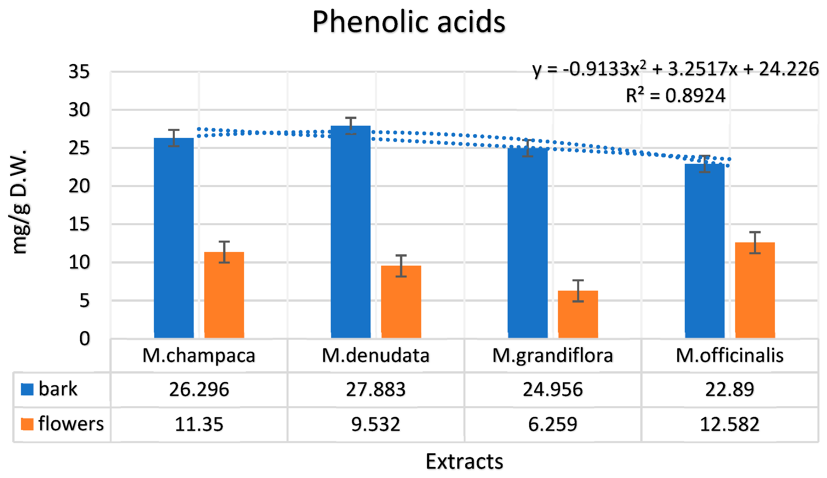
Figure 1. Assessment of phenolic acids in hydroethanolic extracts from the bark and flowers of Magnolia (M. champaca, M. denudata, M. grandiflora, M. officinalis).
Preprints 92595 g001
Figure 2. Assessment of aglycone flavonoids in hydroethanolic extracts from the bark and flowers of Magnolia (M. champaca, M. denudata, M. grandiflora, M. officinalis).
Preprints 92595 g002
Figure 3. Assessment glycoside flavonoids in hydroethanolic extracts from both the bark and flowers of Magnolia (M. champaca, M. denudata, M. grandiflora, M. officinalis).
Preprints 92595 g003
Figure 4. Assessment of lignans in hydroethanolic extracts from the bark and flowers of Magnolia (M. champaca, M. denudata, M. grandiflora, M. officinalis).
Preprints 92595 g004
Figure 5. Differentiation based on species of bark and flower extracts against compositional profile: (a) the PCA diagram illustrates the differentiation of bark and flower extracts based on their compositional profiles. The principal axes represent 57.76% of the total variance in the data set. (b) displays the graphical representation of PCA variation according to the species studied, in relation to compositional variability.
Preprints 92595 g005
Figure 6. Cluster distribution of bark and flower extracts from four Magnolia species (M. champaca, M. denudata, M.grandiflora, M.officinalis).
Preprints 92595 g006
Table 1. Total Polyphenols and Antioxidant Activity of Hydroethanolic Magnolia Extracts (M. champaca, M. denudata, M. grandiflora, M. officinalis).
Magnolia species Source of Extracts Total Polyphenols mg GAE/g DPPH IC50 mg/mL
M. champaca Bark 73.12±0.25 19.50±0.25
Flowers 68.62±0.21 18.66±0.21
M. denudata Bark 65.18±0.28 17.01±0.24
Flowers 55.23±0.34 9.99±0.25
M. grandiflora Bark 72.52±0.45 19.07±0.32
Flowers 55.18±0.38 10.55±0.18
M. officinalis Bark 98.44±0.49 23.23±0.48
Flowers 66.02±0.28 18.78±0.27
The values represent the average of the three determinations with the corresponding standard deviation; p < 0.05.
Table 2. Phenolic compounds identified in the four Magnolia species (M. champaca, M. denudata, M. grandiflora, and M. officinalis).
Compounds M. champaca M. denudata M. grandiflora M. officinalis
Bark Flowers Bark Flowers Bark Flowers Bark Flowers
Phenolic Acids (mg/g dry matter)
Gallic 5.021 1.191 1.278 1.782 2.371 0.984 1.999 2.035
4-hydroxybenzoic 9.289 4.133 11.097 3.191 5.667 1.224 10.012 6.127
p-coumaric 0.111 2.671 3.108 0.154 n.d. 1.793 0.010 1.119
Salicylic 7.789 n.d. 4.578 n.d. 9.123 n.d. 2.333 n.d.
Caffeic 0.019 0.662 0.027 0.276 0.113 1.044 0.022 0.993
Caftaric n.d. 0.011 0.001 n.d n.d. 0.015 0.017 0.024
Cinnamic 0.092 0.023 0.002 0.064 n.d. 0.048 0.076 n.d
Chlorogenic 1.357 2.543 4.504 3.934 4.441 1.147 5.619 2.281
Ellagic 0.487 n.d. 0.983 0.001 0.578 0.001 0.568 n.d.
Ferulic 1.129 0.111 1.222 0.127 1.341 n.d. 0.991 n.d.
Syringic 1.002 n.d. 1.083 n.d. 1.321 0.001 1.242 0.001
Vanillic n.d. 0.005 n.d. 0.003 0.001 0.002 0.001 0.002
Total 26.296 11.35 27.883 9.532 24.956 6.259 22.89 12.582
Aglycone Flavonoids (mg/g dry matter)
Catechin 85.333 52.744 91.227 78.936 71.772 66.033 81.034 32.333
Myricetin 1.024 0.221 0.023 2.003 0.992 n.d. 0.835 n.d.
Luteolin 2.001 0.001 0.779 0.287 1.429 n.d. 1.002 0.110
Taxifolin 0.004 1.983 n.d. 5.661 n.d. 1.111 0.002 2.003
Eriodictyol n.d. 0.001 n.d. n.d. 0.001 0.021 0.001 n.d.
Apigenin 0.772 4.229 0.189 12.002 0.991 7.456 1.283 2.342
Quercetin 0.615 0.357 0.995 1.911 0.937 0.999 0.691 1.001
Epicatechin 22.418 29.919 20.361 34.988 31.912 39.397 34.562 23.435
Total 112.167 89.455 113.574 135.788 108.034 115.017 119.41 61.224
Glycosidic flavonoids (mg/g dry matter)
Rutin 27.035 23.923 45.327 15.075 56.782 17.249 51.103 20.200
Luteolin-7-O-glucoside 1.002 5.552 2.033 2.003 1.023 1.279 0.435 6.771
Isoquercetin 0.004 0.213 n.d. 0.016 n.d. 0.111 n.d. 0.098
Hyperoside n.d. 0.111 n.d. 0.032 n.d. n.d. n.d. 0.011
Kaempferol-3-O-rutinoside 0.012 0.241 0.023 0.104 0.122 0.286 0.192 0.197
Apigenin-7-O-glucoside 0.003 0.044 0.115 0.011 0.012 0.101 0.011 0.099
Isorhamnetin-3-O-glucoside 0.002 n.d. 0.001 n.d. 0.001 0.001 n.d. n.d.
Naringenin-7-O-glucoside 0.001 n.d. n.d. 0.002 n.d. n.d. n.d. n.d.
Astragalin n.d. n.d. n.d. 0.001 n.d. n.d. n.d. n.d.
Quercitrin n.d. 0.001 n.d. n.d. n.d. n.d. 0.001 n.d.
Isorhamnetin-3-O-rutinoside 0.003 0.004 n.d. 0.021 0.021 0.018 0.114 0.007
Total 28.062 30.089 47.499 17.265 57.961 19.045 51.856 27.383
Lignans
4’-O-methylhonokiol 2.011 7.012 5.561 1.021 19.092 18.098 21.021 16.546
Magnolol 9.319 5.666 9.999 2.001 7.021 19.021 97.093 23.021
Honokiol 5.092 4.090 5.892 2.098 6.607 18.917 56.785 13.195
3-methoxymagnolol 2.367 2.311 1.285 0.227 7.000 5.662 2.368 4.432
Isomagnolol 1.025 0.098 1.374 0.112 6.789 2.023 3.479 7.776
Total 19.814 19.177 24.111 5.459 46.509 63.721 180.746 64.97
Total Phenolic Compounds 186.339 150.071 213.067 168.044 237.46 204.042 374.902 166.159
The analyses were conducted in triplicate; p < 0.05; n.d. not detected.
Table 3. Phenolic compounds identified in the four Magnolia species (M. champaca, M. denudata, M. grandiflora, and M. officinalis).
Compound RIa RIb M. champaca % M. denudata % M. grandiflora % M. officinalis %
bark flowers bark flowers bark flowers bark flowers
Hexanal 801 800 - 0.1 - - - - - -
Heptanal 902 902 - 0.2 - 0.1 0.1 0.1 0.3 0.9
α-Thujene 930 929 0.1 1.2 0.1 1.5 0.4 0.7 0.5 2.1
α-Pinene 939 932 0.4 0.6 1.1 0.9 0.2 0.8 0.3 1.5
Camphene 954 946 1.1 - 1.1 0.1 1.1 0.1 1.3 -
Sabinene 975 972 - tr - tr 0.1 tr 0.1 tr
β-Pinene 979 980 3.6 0.1 2.9 - 5.2 tr 2.3 tr
6-Methyl-5-hepten-2-one 986 985 - 1.1 0.1 - 0.1 0.5 - -
α-Phellandrene 1002 1001 - 0.1 - - - 0.2 - -
α-Terpinene 1016 1014 0.1 0.5 0.2 0.5 0.1 0.7 0.1 1.2
p-Cymene 1026 1024 - 0.2 - 0.1 - 0.2 - 0.1
Limonene 1029 1030 0.9 4.4 2.2 7.1 0.5 2.9 2.1 1.8
1,8-Cineol 1031 1031 0.5 0.9 2.1 1.1 0.5 0.1 1.7 0.1
β-Phellandrene 1034 1032 - 0.2 - 0.4 - - - 0.1
(Z)-β-Ocimene 1038 1044 0.1 0.1 0.1 0.1 0.1 0.3 0.1 -
Phenylacetaldehyde 1042 1043 1.1 1.8 1.4 3.9 2.2 1.6 1.7 -
(E)-β-Ocimene 1050 1052 0.1 5.7 0.1 1.9 0.1 3.4 0.1 1.1
γ-Terpinene 1060 1058 0.2 3.2 0.1 2.1 0.1 0.8 0.2 0.3
1-Octanol 1068 1066 0.4 2.8 2.0 4.6 2.4 1.2 1.6 1.9
cis-Sabinene hydrate 1070 1070 - tr - 0.1 tr - - 0.1
cis-Linalool oxide (furanoid) 1073 1074 0.2 0.4 0.1 0.6 0.2 0.1 - 0.3
trans-Linalool oxide (furanoid) 1087 1085 0.1 0.1 0.1 - 0.2 0.1 0.1 0.1
Terpinolene 1092 1093 0.5 0.2 0.4 0.3 0.7 0.3 0.2 0.8
Linalool 1095 1100 1.4 25.1 2.7 20.4 1.8 15.8 2.0 21.2
€-4,8-Dimethylnona-1,3,7-triene 1118 1120 0.1 1.2 1.0 4.4 1.1 0.4 1.3 -
Perillene 1122 1123 - 0.1 - 0.1 - 0.1 - 0.1
Camphor 1141 1140 2.5 1.1 3.1 1.3 6.3 0.8 6.6 3.4
Borneol 1163 1165 1.1 - 1.2 0.1 1.3 0.1 1.1 -
Terpinen-4-ol 1186 1185 - 0.1 0.1 0.1 0.1 0.2 0.1 0.1
α-Terpineol 1189 1190 0.2 2.2 0.5 0.8 0.8 1.6 0.7 2.1
Myrtenol 1195 1193 0.1 1.1 0.1 2.9 0.1 1.4 0.1 0.7
Myrtenal 1196 1197 - 0.3 tr - tr 0.5 tr -
β-Citronellol 1226 1223 0.4 1.8 0.9 1.3 0.7 2.3 0.5 2.9
Nerol 1230 1229 0.1 1.6 0.1 1.1 0.2 4.9 0.2 2.2
Neral 1238 1237 0.1 0.1 - - 0.1 - - -
Geraniol 1253 1255 1.1 5.2 0.5 1.9 0.9 7.7 0.8 9.1
Geranial 1257 1258 0.1 0.3 0.2 0.1 0.3 0.3 0.1 0.9
1-Decanol 1270 1273 0.2 0.3 0.1 0.6 0.1 0.2 0.1 0.7
Bornyl acetate 1289 1290 25.6 2.1 14.9 5.3 14.2 4.3 17.8 11.1
Myrtenyl acetate 1327 1325 0.5 2.1 1.0 1.5 1.1 0.9 1.3 0.4
Eugenol 1359 1360 0.1 0.9 0.2 1.4 0.2 0.8 0.1 1.5
α-Copaene 1388 1387 0.1 0.4 - - 0.1 - 0.1 0.4
β-Elemene 1404 1405 0.8 1.2 0.7 2.0 0.7 1.9 0.3 1.0
Ε-Caryophyllene 1417 1419 11.1 1.2 22.4 0.9 21.1 0.7 15.6 1.2
α-trans-Bergamotene 1435 1435 - 0.1 - 0.1 tr 0.1 - -
β-Caryophyllene 1437 1432 1.2 0.2 1.8 1.3 3.4 2.9 5.6 1.1
(Z)-β-Farnesene 1453 1447 - 0.1 - 0.1 0.1 0.3 0.7 -
α-Humulene 1455 1455 0.1 0.4 0.3 1.1 0.2 0.8 0.5 0.9
9-epi-(E)-Caryophyllene 1470 1470 3.7 0.1 2.9 1.8 3.8 0.9 3.1 0.3
γ-Gurjunene 1477 1476 - - - 0.1 0.1 - -
Germacrene D 1485 1481 11.2 2.4 5.3 1.5 4.6 5.2 7.7 4.8
α-Selinene 1488 1489 1.2 0.1 0.8 0.5 1.3 0.7 2.3 0.1
γ-Muurolene 1490 1494 0.1 - 0.1 0.8 0.2 0.5 0.1 0.3
Viridiflorene 1496 1497 1.8 2.1 1.5 1.8 2.2 2.6 - 1.2
Bicyclogermacrene 1500 1502 0.1 0.4 0.2 0.4 0.4 1.1 0.5 -
δ-Cadinene 1523 1522 0.9 2.0 0.5 0.8 1.5 0.3 1.7 3.5
Hedycaryol 1548 1545 - - - - - 0.1 - -
Elemol 1550 1552 - - - - - 0.8 - 0.5
trans-Nerolidol 1556 1555 0.1 0.1 - - 0.1 0.1 0.1 -
(E)-Nerolidol 1563 1563 0.2 0.5 - 0.1 0.6 0.2 0.3 0.1
Palustrol 1567 1569 - - - - - 0.1 - -
Germacrene D-4-ol 1576 1577 0.1 1.4 0.1 1.1 0.2 1.5 0.2 1.0
Spathulenol 1578 1579 3.6 0.1 2.8 0.5 1.7 0.1 2.2 0.1
Globulol 1585 1588 0.1 0.1 - - - 0.8 - -
β-Eudesmol 1650 1651 - - - - - 0.1 - -
α-Cadinol 1654 1653 0.1 0.1 0.1 1.0 0.3 1.1 0.1 1.7
Selin-11-en-4-α-ol 1659 1657 0.2 0.1 0.5 1.9 0.1 - 0.4 0.5
Shyobunol 1688 1688 - - - - 0.1 0.2 0.1 0.1
(E)-Nerolidyl acetate 1717 1713 - - - - - 0.1 - -
(Z,E)-Farnesol 1725 1722 0.3 0.4 0.5 - 0.9 0.3 0.6 0.2
14-Hydroxy-α-muurolene 1780 1777 0.2 0.5 0.2 0.6 0.5 0.3 0.4 0.1
14-Hydroxy-δ-cadinene 1802 1800 0.5 0.5 - 0.4 0.1 0.2 0.1 0.3
n-Hexadecanol 1875 1872 3.3 7.4 2.3 2.2 1.7 2.7 1.1 3.3
n-Heneicosane 2100 2100 2.1 0.2 2.7 0.2 1.6 0.3 0.5 0.5
Linoleic acid 2133 2130 0.1 0.6 0.1 2.2 0.1 1.4 0.4 1.0
n-Tricosene 2300 2300 7.7 1.9 5.5 1.8 1.7 4.1 4.2 2.8
n-Pentacosane 2500 2500 1.4 1.9 4.9 1.3 1.1 2.1 2.9 1.9
Total 95.3 96.0 96.9 95.1 94.1 91.1 97.5 97.7
Other compounds 4.7 4.0 3.1 4.9 5.9 8.9 2.5 2.3
RIa - calculated retention index; RIb - retention index from literature; determinations were performed in triplicate; p<0.05.
Table 4. Antibacterial activity of hydroethanolic Magnolia extracts (M. champaca, M. denudata, M. grandiflora, M. officinalis) expressed in MIC (µg/mL).
Bacterial strains MIC (µg/mL)
Ampi-cillin Genta-micin Tetracy-cline M. champaca M. denudata M. grandiflora M. officinalis
bark flo-wer bark flo-wer bark flo-wer bark flo-wer
Staphylococcus aureus ATCC 43300(MRSA) n.a. 30 n.a. n.a n.a 30 n.a. n.a. 30 30 30
Staphylococcus epidermidis ATCC 12228 10 5 5 15 30 15 30 7.5 7.5 5 5
Streptococcus faecalis ATCC 19443 2.5 2.5 2.5 15 2.5 7.5 15 7.5 7.5 5 2.5
Streptococcus pyogenes ATCC 12347 1.25 1.25 2.5 30 30 n.a. 30 15 n.a. 15 n.a.
Streptococcus sanguinis ATCC 10556 2.5 2.5 n.a. n.a n.a. n.a. n.a. n.a. 30 30 30
Actinomyces israelii ATCC 12102 0.625 0.625 0.625 15 30 12.5 15 10 10 7.5 7.5
Propionebacterium acnes ATCC 6921/4311 1.25 1.25 0.625 7.5 2.5 10 12.5 5 7.5 5 2.5
Enterobacter aerogenes ATCC 13048 0.625 0.625 30 15 10 30 30 10 12.5 7.5 10
Escherichia coli ATCC 35218 0.625 0.625 30 n.a. n.a. 15 30 15 15 10 12.5
Klebsiella pneumoniae ATCC 13883 0.625 0.625 30 n.a. n.a. n.a. n.a. n.a. n.a. 30 n.a.
Prevotella intermedia ATCC 25611 0.625 0.625 0.625 5 15 n.a. n.a. 10 2.5 15 15
Porphyromonas gingivalis ATCC 33277 0.625 30 15 0.625 5 7.5 10 5 2.5 0.625 1.25
Proteus vulgaris ATCC 13315 0.625 0.625 10 10 10 15 15 7.5 12.5 5 10
Pseudomonas aeruginosa ATCC 27853 n.a. 0.625 n.a. 5 12.5 12.5 15 7.5 10 2.5 2.5
n.a. / no activity.
Disclaimer/Publisher’s Note: The statements, opinions and data contained in all publications are solely those of the individual author(s) and contributor(s) and not of MDPI and/or the editor(s). MDPI and/or the editor(s) disclaim responsibility for any injury to people or property resulting from any ideas, methods, instructions or products referred to in the content.
Copyright: This open access article is published under a Creative Commons CC BY 4.0 license, which permit the free download, distribution, and reuse, provided that the author and preprint are cited in any reuse.
Prerpints.org logo

Preprints.org is a free preprint server supported by MDPI in Basel, Switzerland.

Subscribe

© 2024 MDPI (Basel, Switzerland) unless otherwise stated