
Submitted:
05 December 2023
Posted:
06 December 2023
You are already at the latest version
Abstract
Molecular docking, a computational method predicting how molecules bind to target proteins, is employed in this study to explore Polydatin's interactions with Ras-related protein Rab (Rab) proteins using the Mcule Database. This database validates the docking results and offers insights into the pharmacological properties of Polydatin. Notably, specific Rab proteins like Rab-5A, HRas, and Rab-18 exhibit robust interactions with Polydatin, indicated by favorable binding energies.These findings illuminate the potential roles of Polydatin in therapeutic and biological contexts, emphasizing its interaction with Rab proteins. The Mcule Database enhances the credibility of the study, providing additional perspectives on Polydatin's applications. The observed interactions, particularly with key proteins like HRas, present a promising avenue for modulating cellular signaling pathways. This modulation holds therapeutic potential, potentially addressing conditions associated with pathway overactivity, including specific cancer types.
Keywords:
Rab proteins
; Polydatin
; Docking analysis
; Autodock Vina
1. Introduction
Ras-related protein Rab (Rab) proteins, part of the Ras superfamily, play crucial roles in regulating intracellular vesicular transport. Specifically, Rab 2A and 3A have been linked to acrosomal exocytosis in spermatozoa, and Rab 2A is identified as a potential biomarker for male fertility [1,2,3].
Despite these findings, the precise mechanisms and comprehensive understanding of Rab proteins in spermatozoa remain unclear. Further research is needed to elucidate the specific roles and underlying mechanisms of Rab proteins in the context of male fertility and acrosomal exocytosis. This groundbreaking study marks the first computational exploration of Molecular Docking involving Ras-related proteins (Rab) and Polydatin [5]. Molecular Docking is a method used to predict the binding interactions between molecules, and in this case [6,7], it sheds light on how Rab proteins interact with Polydatin. Rab proteins, known regulators of intracellular vesicular transport, are of particular interest. Polydatin’s role in modulating these proteins, potentially influencing cellular processes, is a novel aspect explored. The findings from this study could contribute valuable insights into the molecular interactions between Rab proteins and Polydatin, offering potential applications in understanding and manipulating cellular functions. Molecular Docking investigations were performed by Mcul Service It stands out as an online drug discovery platform, offering a distinctive solution for pharmaceutical and biotech companies. The platform provides access to a top-tier purchasable compound database and molecular modeling tools, serving as a valuable resource for researchers and professionals in the field. By combining a high-quality compound database with advanced molecular modeling capabilities, Mcule.com facilitates efficient and informed drug discovery processes. This comprehensive approach enhances the ability of pharma and biotech companies to explore, analyze, and select compounds, ultimately contributing to the acceleration and optimization of drug development efforts [1,2,3].
2. Material and Methods
Ras-related protein Rab (Rab) proteins were performed by Mcule Database by Autodock Vina [9]. Several proteins were investigated in the Ligand Binding Site of these proteins:
- -
- Ras-related protein Rab-5A (PDB Code: 1n6k,RAB5A_HUMAN); Grid box Coordinates of binding Center X ( 9,9289), Y( -2,608), Z(42,2021)
- -
- Ras-related C3 botulinum toxin substrate 3(PDB Code: qme AC3_HUMAN); Grid box Coordinates of binding Center X ( -10,6359), Y( 7,5954), Z(23,1647)
- -
- Ras-related protein Rab-28 (PDB Code: 2hxs,RAB28_HUMAN); Grid box Coordinates of binding Center X ( 20,2841), Y( 20,0731), Z(11,1145)
- -
- Ras-related GTP-binding protein C(PDB Code: 3llu,RRAGC_HUMAN); Grid box Coordinates of binding Center X ( 37,8726), Y( 15,9775), Z(25,5165)
- -
- Ras-related protein Rab-9A (PDB Code: 1wmsRAB9A_HUMAN); Grid box Coordinates of binding Center X ( -1,0272), Y( 21,5378), Z(7,3575)
- -
- Ras-related protein Rab-18 (PDB Code: 1x3s RAB18_HUMAN); Grid box Coordinates of binding Center X ( 29,773), Y( 32,4934), Z(17,9592)
- -
- Ras-related protein Rab-31 (PDB Code: 2fg5 RAB31_HUMAN); Grid box Coordinates of binding Center X ( 19,2312), Y( 6,1773 ), Z((2,0914)
- -
- Ras-related protein Rap-2A(PDB Code: 3rap RAP2A_HUMAN); Grid box Coordinates of binding Center X ( 37,5957), Y( 10,1612), Z(65,4559)
- -
- Ras-related protein Rab-11 B (PDB Code: 2f9l RB11B_HUMAN); Grid box Coordinates of binding Center X ( 5,0846), Y( 2,0916 ), Z(6,1611)
- -
- Ras-related protein Rab-3D(PDB Code: 2gf9 RAB3D_HUMAN); Grid box Coordinates of binding Center X ( 3,0868), Y( 9,6966 ), Z(6,4)
- -
- Ras-related protein Rab-4A(PDB Code: 2bme RAB3D_HUMAN); Grid box Coordinates of binding Center X ( 73,0035), Y( 13,8161), Z(0,889)
- -
- GTPase HRas(PDB Code: 1qra RAB3D_HUMAN); Grid box Coordinates of binding Center X ( 12,0003), Y( 32,9114), Z(19,526)
- -
- Ras-related protein R-Ras(PDB Code: 2fn4 RRAS_HUMAN); Grid box Coordinates of binding Center X ( -6,1087), Y( 25,4637 ), Z(14,5476)
- -
- Ras-related protein Rab-1B(PDB Code: 3nkv RAB1B_HUMAN); Grid box Coordinates of binding Center X (-15,1987), Y( 19,8262), Z(-7,1299)
- -
- Ras-related protein Rab-6B(PDB Code: 2e9s RAB6B_HUMAN); Grid box Coordinates of binding Center X (3,6002), Y( 33,0027), Z(3,0894)
- -
- Ras-related protein Rab-6A(PDB Code: 1yzq RAB6A_HUMAN); Grid box Coordinates of binding Center X (25,1083), Y( 17,85 ), Z(13,8221)
3. Results and Discussion
The Ras protein family holds paramount significance in governing cell signaling and plays a pivotal role in diverse cellular processes, encompassing proliferation, differentiation, and cell survival. Functioning as small GTPases, Ras proteins serve as molecular switches activated through GTP (guanosine triphosphate) binding and deactivated by GTP hydrolysis to GDP (guanosine diphosphate) [1,2,3,4]. Implicated in various tumor types, the Ras gene mutations are frequently linked to Ras protein hyperactivation, fostering heightened cell proliferation. Ras proteins are integral players in signaling cascades that oversee the regulation of cell growth and division, rendering them substantial targets in cancer therapy research [1,2,3,4].
Rab proteins, part of the Ras superfamily of small GTPases, are pivotal regulators of intracellular vesicular transport. Key points about Rab proteins include their involvement in regulating vesicular transport between cellular compartments, such as the endoplasmic reticulum, Golgi apparatus, endosomes, lysosomes, and the plasma membrane. Acting as molecular switches with GTPase activity, Rab proteins cycle between active (GTP-bound) and inactive (GDP-bound) states, interacting with effector proteins to regulate vesicular transport steps. These proteins are associated with specific cell membranes, defining membrane identities and contributing to transport specificity. Rab proteins play vital roles in vesicle budding from donor membranes and fusion with target membranes, facilitating cargo sorting and delivery within the cell [1,2,3]. With over 60 diverse members in humans, each Rab protein is linked to specific membrane compartments, ensuring efficiency in intracellular transport. Dysregulation of Rab proteins is implicated in diseases like neurodegenerative disorders and cancer, underscoring their importance in maintaining cellular homeostasis. Studying Rab protein functions provides insights into the intricate mechanisms governing intracellular trafficking and contributes to understanding the organization and function of eukaryotic cells [1,2,3,4].
We presen a novel computational approach focused on the interaction between Polydatin and various Rab proteins using Molecular Docking, using the Mcule Database.
The results indicate favorable binding energies between Polydatin and several Rab proteins.
Here’s a summary of the key our finding between Polydatin Interaction with Rab Proteins:
Ras-related protein Rab-4A: Binding energy of -9.6 kcal/mol.
Ras-related protein Rab-5A: Binding energy of -10 kcal/mol.
GTPase HRas: Binding energy of -10.4 kcal/mol.
Ras-related C3 botulinum toxin substrate 3: Binding energy of -10.1 kcal/mol.
Ras-related protein Rab-6B: Binding energy of -9.6 kcal/mol.
Ras-related protein Rab-31: Binding energy of -9.6 kcal/mol.
Ras-related protein Rab-18: Binding energy of -10.5 kcal/mol.
The negative binding energies suggest favorable interactions between Polydatin and the mentioned Rab proteins. Lower binding energies generally indicate stronger binding affinity.
Specific Rab proteins, such as Rab-5A, HRas, and Rab-18, have shown particularly strong interactions with Polydatin based on the presented data. It’s essential to note that these findings provide valuable insights into the potential therapeutic or biological implications of Polydatin’s interaction with these Rab proteins. Further experimental validation would be crucial to confirm and expand upon these computational results. Additionally, the specific biological context and implications of these interactions would need to be explored in more detail.
Figure 1.
displays the docking outcomes of Ras-related protein Rab-4A in conjunction with Polydatin within the Ligand Binding Site, as analyzed by Autodock Vina through the Mcule Database. On the left side, 2D diagrams illustrate the residue interactions between the protein and Polydatin. Meanwhile, the right side exhibits the Ligand Binding Site of the protein, highlighting the specific location of Polydatin.
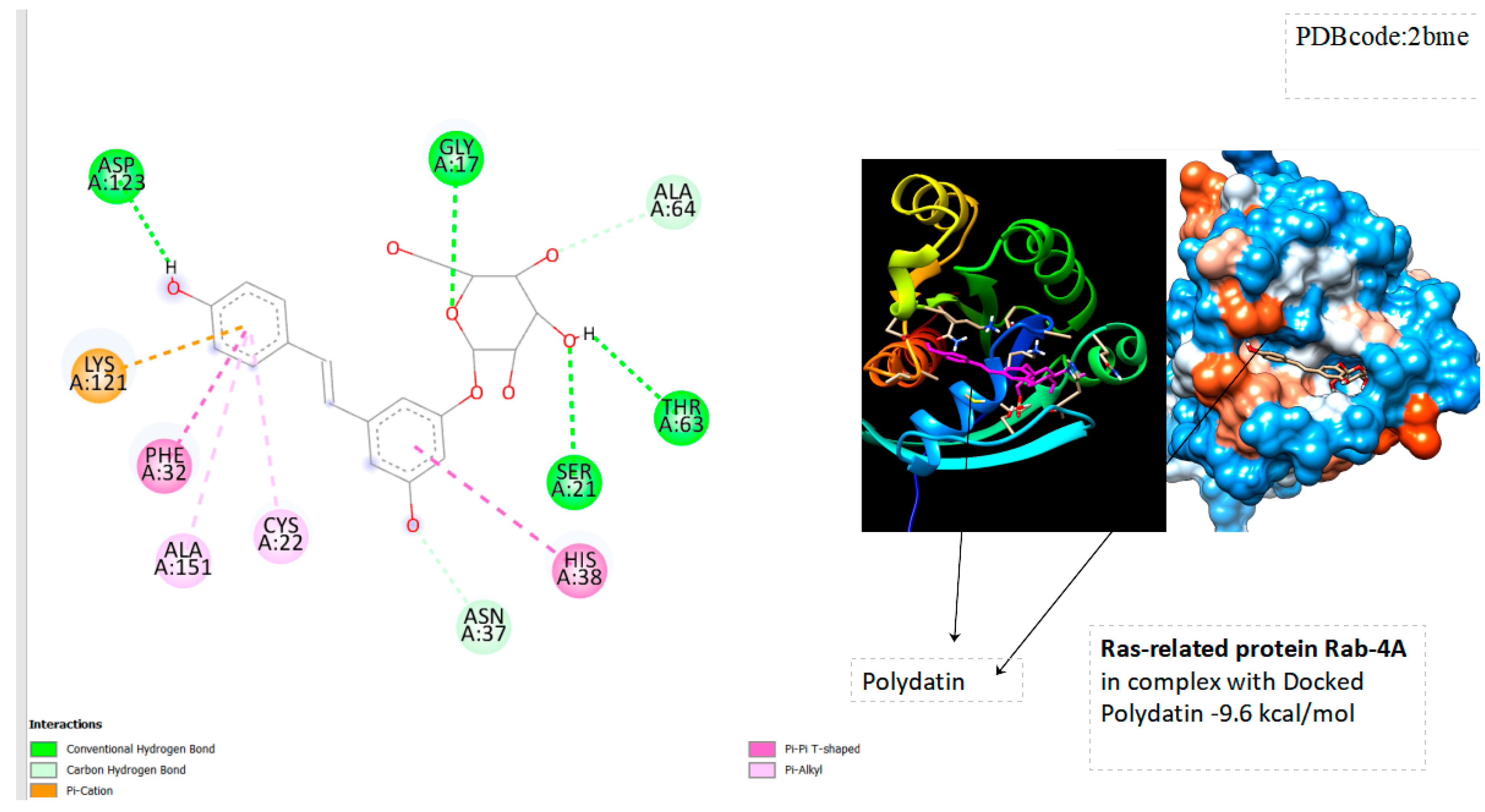
Figure 2.
Displays the docking outcomes of Ras-related protein Rab-5A in conjunction with Polydatin within the Ligand Binding Site, as analyzed by Autodock Vina through the Mcule Database. On the left side, 2D diagrams illustrate the residue interactions between the protein and Polydatin. Meanwhile, the right side exhibits the Ligand Binding Site of the protein, highlighting the specific location of Polydatin.
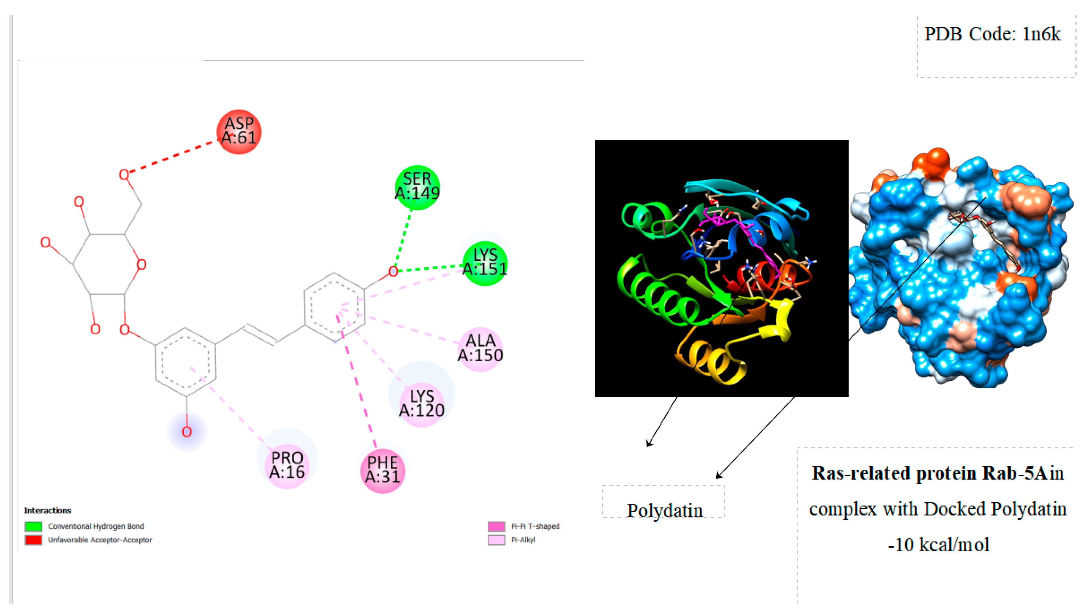
Figure 3.
Displays the docking outcomes of GTPase Hras with Polydatin within the Ligand Binding Site, as analyzed by Autodock Vina through the Mcule Database. On the left side, 2D diagrams illustrate the residue interactions between the protein and Polydatin. Meanwhile, the right side exhibits the Ligand Binding Site of the protein, highlighting the specific location of Polydatin.
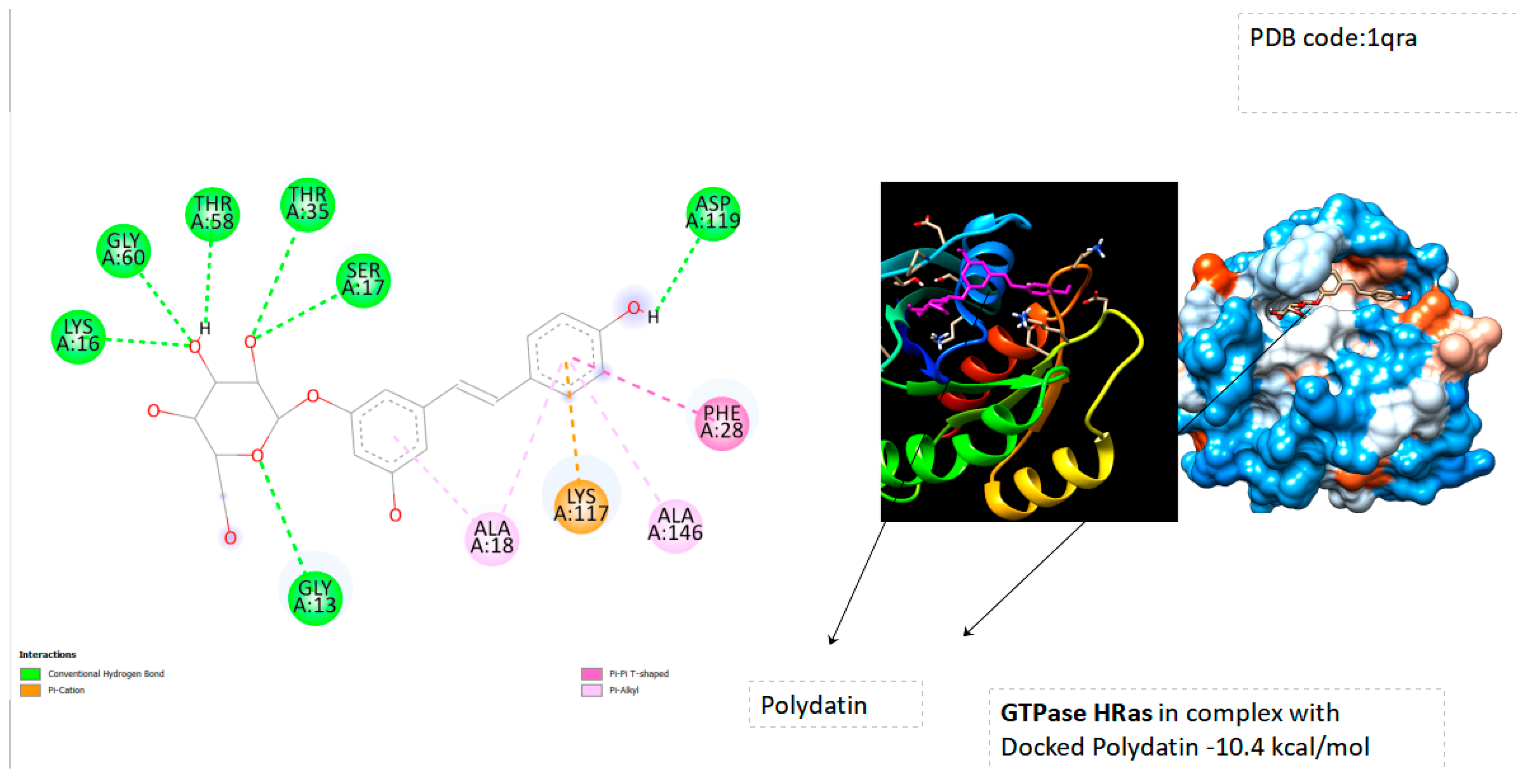
Figure 4.
Displays the docking outcomes of Ras-related C3 Botulinum toxin substrate with polydatin in Ligand Binding Site, as analyzed by Autodock Vina through the Mcule Database. On the left side, 2D diagrams illustrate the residue interactions between the protein and Polydatin. Meanwhile, the right side exhibits the Ligand Binding Site of the protein, highlighting the specific location of Polydatin.
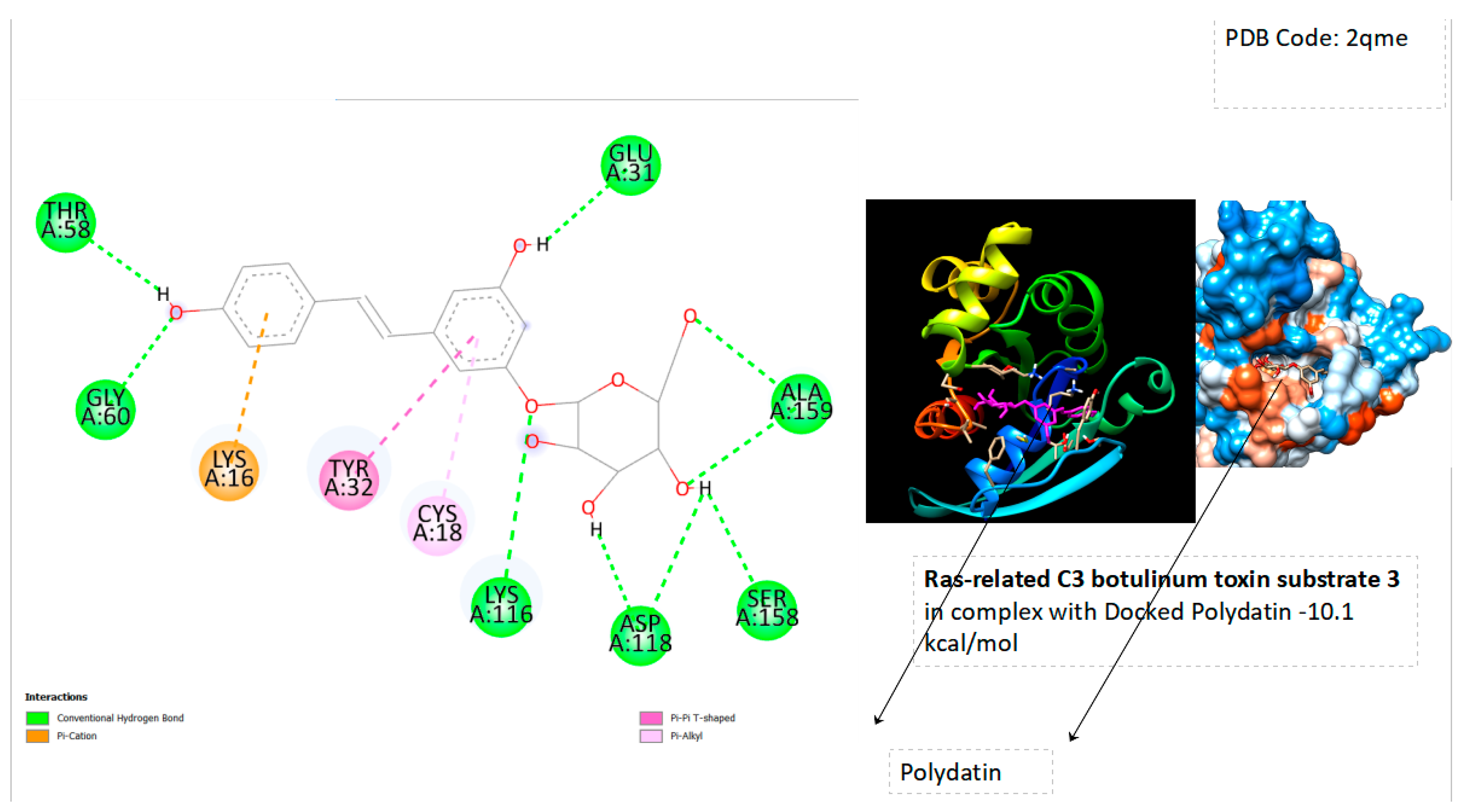
Figure 5.
Displays the docking outcomes of Ras-related protein Rab-18 with polydatin in Ligand Binding Site, as analyzed by Autodock Vina through the Mcule Database. On the left side, 2D diagrams illustrate the residue interactions between the protein and Polydatin. Meanwhile, the right side exhibits the Ligand Binding Site of the protein, highlighting the specific location of Polydatin.
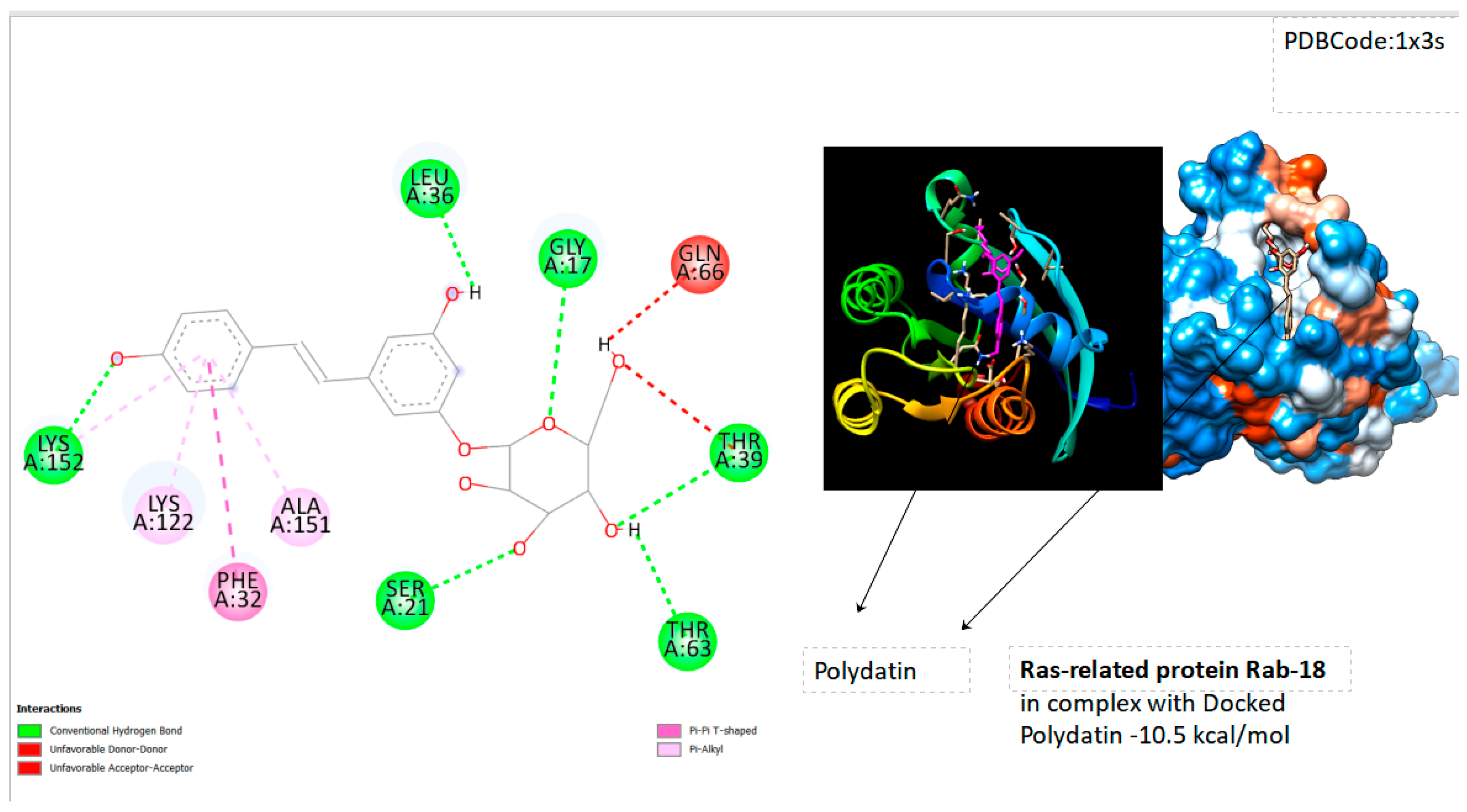
Figure 6.
Displays the docking outcomes of Ras-related protein Rab-31 with polydatin in Ligand Binding Site, as analyzed by Autodock Vina through the Mcule Database. On the left side, 2D diagrams illustrate the residue interactions between the protein and Polydatin. Meanwhile, the right side exhibits the Ligand Binding Site of the protein, highlighting the specific location of Polydatin.
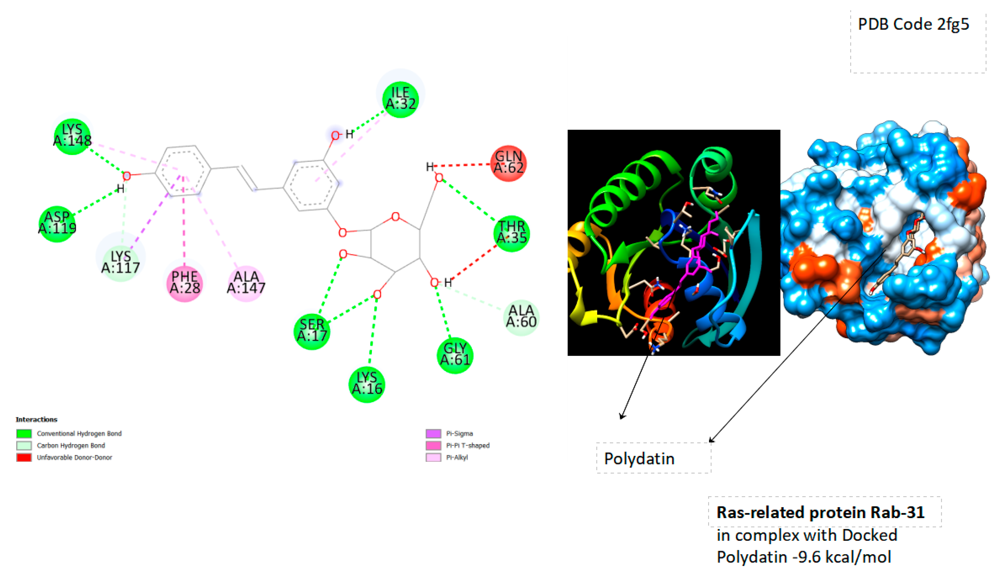
Figure 7.
Displays the docking outcomes of Ras-related protein Rab-6B with polydatin in Ligand Binding Site, as analyzed by Autodock Vina through the Mcule Database. On the left side, 2D diagrams illustrate the residue interactions between the protein and Polydatin. Meanwhile, the right side exhibits the Ligand Binding Site of the protein, highlighting the specific location of Polydatin.
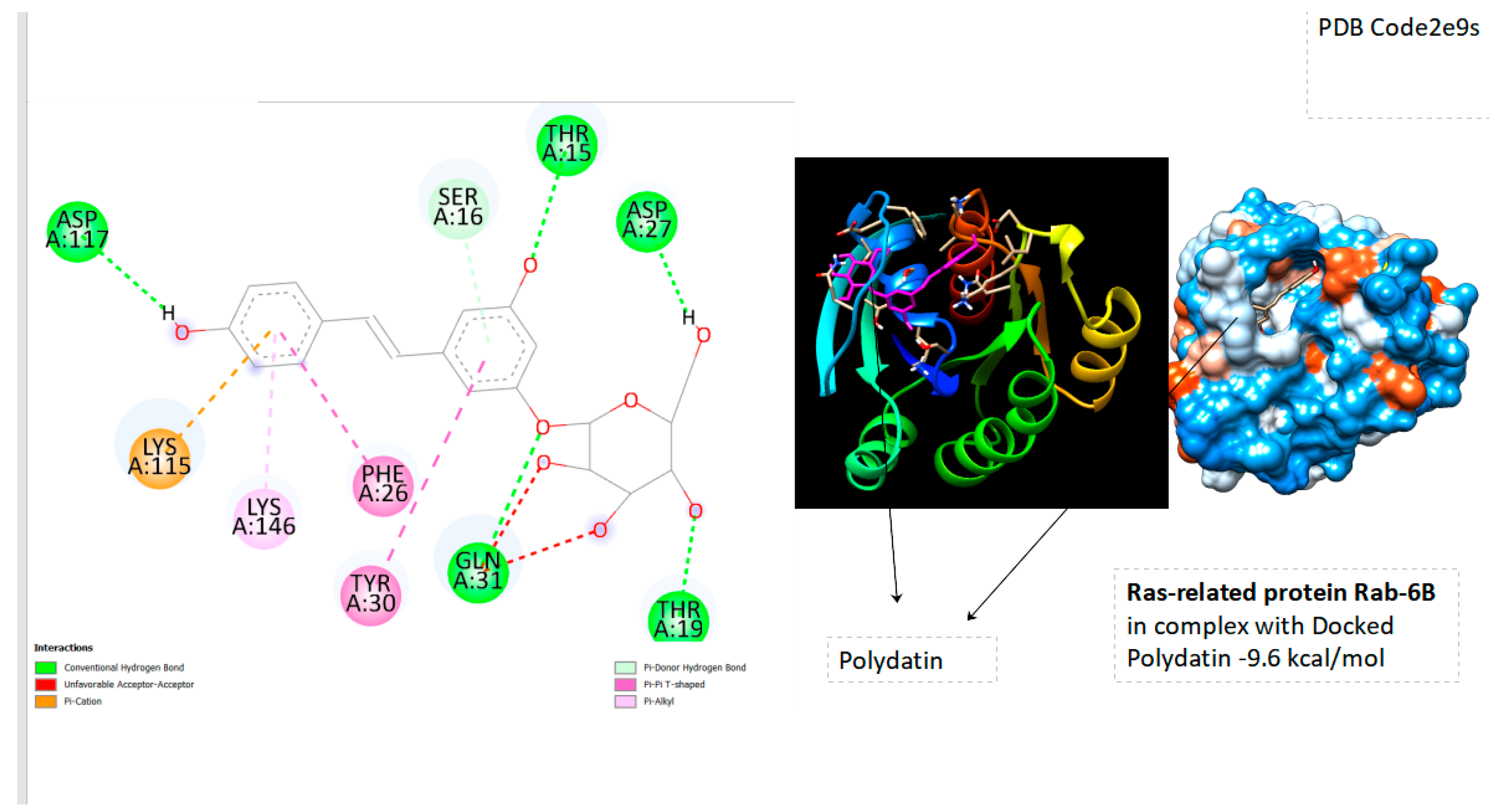

Table 1.
Comparison Binding Energies of Ras-related protein Rab-proteins.
| Ras-Related Protein Rab-Proteins | Binding Energy (kcal/mol) |
|---|---|
| Ras-related protein Rab-28 | -7.0 |
| Ras-related GTP-binding protein C | -9.0 |
| Ras-related protein Rab-9A | -8.1 |
| Ras-related protein Rab-18 | -10.5 |
| Ras-related protein Rab-31 | -9.6 |
| Ras-related protein Rap-2a | -8.1 |
| Ras-related protein Rab-11B | -9.1 |
| Ras-related protein Rab-5A | -10 |
| Ras-related protein Rab-3D | -9.2 |
| Ras-related protein Rab-4A | -9.6 |
| GTPase HRas | -10.4 |
| Ras-related protein R-Ras | -9.1 |
| Ras-related protein Rap-2A | -8.4 |
| Ras-related protein Rab-1B | -8.9 |
| Ras-related C3 botulinum toxin substrate 3 | -10.1 |
| Ras-related protein Rab-6B | -9.6 |
| Ras-related protein Rab-6A | -9.3 |
4. Conclusion
For the first time, we present a novel computational approach focused on Polydatin with Rab proteins by Molecular Docking , evaluated by Mcule Database. From these docking tests Polydatin has showed excellent results with Ras-related protein Rab-4A ( -9.6 kcal/mol), Ras-related protein Rab-5A( -10 kcal/mol),GTPase HRas( -10.4kcal/mol), Ras-related C3 botulinum toxin substrate 3( -10.1 kcal/mol),Ras-related protein Rab-6B( -9.6 kcal/mol),Ras-related protein Rab-31( -9.6 kcal/mol) and Ras-related protein Rab-18 ( -10.5 kcal/mol). It’s essential to note that these findings provide valuable insights into the potential therapeutic or biological implications of Polydatin’s interaction with these Rab proteins. Unraveling the interactions with Polydatin and other compounds involving Ras proteins holds therapeutic promise for modulating cellular signaling pathways and potentially addressing pathological conditions associated with the overactivity of these pathways, such as certain types of cancer. Further experimental validation would be crucial to confirm and expand upon these computational results. Additionally, the specific biological context and implications of these interactions would need to be explored in more detail.
References
- Bae, J. W., Kim, S. H., Kim, D. H., Ha, J. J., Yi, J. K., Hwang, S., ... & Kwon, W. S. (2019). Ras-related proteins (Rab) are key proteins related to male fertility following a unique activation mechanism. Reproductive biology, 19(4), 356-362.
- Martinez, O., & Goud, B. (1998). Rab proteins. Biochimica et Biophysica Acta (BBA)-Molecular Cell Research, 1404(1-2), 101-112.
- Barnekow, A., Thyrock, A., & Kessler, D. (2009). Rab proteins and their interaction partners. International review of cell and molecular biology, 274, 235-274.
- Bae, J. W., Yi, J. K., Jeong, E. J., Lee, W. J., Hwang, J. M., Kim, D. H., ... & Kwon, W. S. (2022). Ras-related proteins (Rab) play significant roles in sperm motility and capacitation status. Reproductive Biology, 22(2), 100617.
- Du, Q. H., Peng, C., & Zhang, H. (2013). Polydatin: a review of pharmacology and pharmacokinetics. Pharmaceutical biology, 51(11), 1347-1354. [CrossRef]
- Meng, X. Y., Zhang, H. X., Mezei, M., & Cui, M. (2011). Molecular docking: a powerful approach for structure-based drug discovery. Current computer-aided drug design, 7(2), 146-157. [CrossRef]
- Agarwal, S., & Mehrotra, R. J. J. C. (2016). An overview of molecular docking. JSM chem, 4(2), 1024-1028.
- Odhar, H. A., Rayshan, A. M., Ahjel, S. W., Hashim, A. A., & Albeer, A. A. M. A. (2019). Molecular docking enabled updated screening of the matrix protein VP40 from Ebola virus with millions of compounds in the MCULE database for potential inhibitors. Bioinformation, 15(9), 627.
- Trott, O., & Olson, A. J. (2010). AutoDock Vina: improving the speed and accuracy of docking with a new scoring function, efficient optimization, and multithreading. Journal of computational chemistry, 31(2), 455-461. [CrossRef]
Disclaimer/Publisher’s Note: The statements, opinions and data contained in all publications are solely those of the individual author(s) and contributor(s) and not of MDPI and/or the editor(s). MDPI and/or the editor(s) disclaim responsibility for any injury to people or property resulting from any ideas, methods, instructions or products referred to in the content. |
© 2023 by the authors. Licensee MDPI, Basel, Switzerland. This article is an open access article distributed under the terms and conditions of the Creative Commons Attribution (CC BY) license (http://creativecommons.org/licenses/by/4.0/).
Copyright: This open access article is published under a Creative Commons CC BY 4.0 license, which permit the free download, distribution, and reuse, provided that the author and preprint are cited in any reuse.


