Submitted:
27 October 2023
Posted:
02 November 2023
Read the latest preprint version here
Abstract
The incidence rate of articular injury is on the rise. With the help of imaging techniques, we classify the severity of the fracture and possible dislocations. Therapeutic interventions include the use of absorbable implants. The paper aims to present the surgical correction and post-operative treatment of osteochondral fractures with absorbable implants in three children. Affected areas are discussed as follows: lateral condyle of the femur, patella, and radial head. 1) A nine-year-old girl heard a sharp sound accompanied by pain after taking a careless step. Slight swelling of the knee was observed with the patella in place. Diagnostics revealed a broken piece of the femur’s lateral condyle located at the lateral recess. Under general anaesthesia, an arthrotomy was performed. The fragment was cleaned and reattached with absorbable nails. Postoperatively, a brace was applied for six weeks. 2) A 15-year-old boy was admitted due to a fall during ice skating, leading to rotational torsion of the knee. X-ray and CT imaging identified the osteochondral fracture of the patella. Based on these findings, an arthroscopy with arthrotomy was performed. 3) A 17-year-old girl fell while snowboarding, resulting in swelling of the proximal forearm. Imaging diagnostics confirmed the radial head fracture. Closed-fracture repositioning with absorbable implants was performed. The use of biodegradable implants eliminates the need for re-anaesthesia and reoperation, reducing the risks of complications. It also allows for earlier initiation of rehabilitation including physiotherapy. The efficiency of the method is further increased by a minimised hospital stay.
Keywords:
Osteochondral fracture
; Absorbable
; PLGA
; Osteosynthesis
; Articular fracture
; OCF
1. Introduction
Juvenile osteochondral fractures (OCF) account for approximately 1-10% of fractures and their incidence is on the rise [1,2]. This condition can be described by the separation of subchondral bone and the articular cartilage from the articular surface. Its incidence is 2-4 times higher in boys and is most common within the ages of 10-20 with a prevalence ranging from 8-30 per 100,000 cases [1,2,3]. The condition usually affects young athletes or children with an active lifestyle. These occur due to traumatic events or rotational torsion of the joint and present acutely. Most commonly these are the result of low-energy trauma or sports injuries [2,3,4,5,6]. The literature focuses mostly on chronic conditions, such as osteochondritis dissecans (OCD) or osteochondral lesions that develop over time. OCD was first described by Konig in 1887, and talks about loose bodies which were discovered in the knee joint [7,8,9,10]. Originally thought to occur due to major trauma, repetitive microtrauma or spontaneous necrosis. Today the term refers to an area of necrosis of the subchondral bone, which may or may not be separated from the articular surface[7,8,9,10]. In this report, we will focus on OCFs in an acute setting.
Osteochondral lesions are associated with joint instability, resulting in abnormal motion causing rotational, impaction, or shearing forces [1,3,4,11]. Further associated with sprains and dislocations, commonly in the elbow, ankle, knee - femoral condyles and patella - and hip joints [11]. At the knee joint, anterior cruciate ligament (ACL) injuries account for less than 1% of cases, however, it was found that the incidence of OCFs might be as high as 80% in the case of ACL injuries [1,11,12,13]. This is due to the valgus stress which leads to shearing forces that result in the damage of the cartilage of the femoral condyles[1,11]. The incidence of OCFs was found to be 70% in cases of patellar dislocation. As the patella transitions laterally, shearing forces act on the medial patellar facet and the lateral femoral condyle. Studies using MRI estimated the incidence of OCFs in case of lateral dislocations to be between 40-78% [11,14,15,16,17,18,19]. Diagnosis is challenging even with a comprehensive physical exam and X-ray imaging as symptoms are often masked by damage to local tissue and are non-specific. Pain is described to be the most common symptom, which exacerbates during or after weight bearing or activities. Mechanical symptoms, such as locking or catching, are caused by loose fragments in the joint space. Swelling and decreased range of motion (ROM) are typical signs. Hemarthrosis is highly indicative of OCF in skeletally immature patients. However, as the acute disability can be outweighed by ligamentous injury, minor pathologies or more severe injuries proper diagnostic evaluation is essential [1,11,13,14,18].
Imaging aims to confirm clinical suspicion, assess the extent of damage to the articular surface, look for instability or dislocations, and monitor disease progression [1,11,18]. Plain radiographs are part of the standard examinations after acute trauma of the joint. OCFs however, might be difficult to identify as small bony fragments cannot be visualised, as opposed to a large piece of detached articular cartilage. Features on plain radiographs include irregular bony contours, fragmentation, or a thin piece of radiodense subchondral bone. The sensitivity of X-rays for diagnosis is between 32-69% according to research, depending on the location. OCFs affecting the talus can be diagnosed with X-ray with 69% accuracy, while OCFs of the knee and hip reach only 32%, proven by McCarthy et al [1,20,21]. Radiographs should be extensively searched, in acute settings, for OCFs. Attention should be averted to prevalent locations of the lesions such as the talar dome or the lateral recess in case of patellofemoral injuries [3,14,21].
Other imaging modalities are recommended for fractures affecting the articular surfaces and are useful for surgical planning. For these cases computed tomography (CT) and magnetic resonance imaging (MRI) which are superior to plain radiographs for the detection of OCFs. CT is adequate in detecting small osteochondral fragments and thin bony plates with high-resolution images, but it lacks the capability to depict bone marrow oedema. As an addition, CT angiography can reveal small chondral pieces as well. MRI is also able to detect chondral injuries with high sensitivity and specificity, depending on the region, of about 75-93% [22,23,24,25]. It was noted by a study that the sensitivity of MRI is a little higher than CT, however, this is not statistically significant. A potential advantage of MR imaging is that it can detect other soft tissue abnormalities and injuries which are often associated with OCFs. This is beneficial in cases of sprains and ligament or tendon abnormalities which could lead to persistent instability and pain [1,22,23,24,25]. Signs of MRI which describe the instability of the lesion include the presence of fluid signals between the fragment and its origin. Other signs which suggest fragment instability include the collapse of the articular surface, extensive bone marrow oedema and cystic changes [1,21,22,23,24,26]. Contrast material can also be used as an indicator of its presence, if located between the fragment and the subchondral bone. For this reason, both CT and MR arthrography are adequate to evaluate the instability of the fracture and to gain proper staging [11,22,23,24,25,27,28].
The staging system, for osteochondral lesions, can be based on X-ray (Berndt and Harty), CT (Ferkel) MRI (Happle) or arthroscopic (Cheng-Ferkel) in cases affecting the talus [23,29,30,31,32,33]. However, depending on the size and location of the fragment and the amount of bone involvement, radiographs might not be able to show loose bodies. MRI attempts to grade the stability and severity of the injury and is used for surgical planning, a summary can be seen below:
- A.
-
Stage I:
- Injury limited to articular cartilage
- Subchondral Edema on MRI
- X-ray: Negative
- B.
-
Stage II:
- d.
- Cartilage damage with subchondral fracture without detachment
- e.
- Thin Sclerotic Margin
- f.
- X-ray: Negative or slight sclerosis
- g.
-
Subtypes:
- Type A: Cystic on CT and Edema on MRI
- Type B: Non-displaced and incompletely undercut by fluid (MRI) and lucency (CT) with an open connection to the articular cartilage.
- C.
-
Stage III:
- h.
- Detached, Non-Displaced
- i.
- MRI: Rim-sign - high signal around fragment
- j.
- X-ray: Lucency between fragment and normal bone.
- D.
-
Stage IV:
- k.
- Osteochondral fragment is displaced
- l.
- Joint effusion
- m.
- X-ray: Loose body, lucency
- E.
-
Stage V:
- n.
- Subchondral Cyst formation
- o.
- Secondary degenerative changes
- p.
- X-ray: Secondary Osteoarthritis
As the potential of the articular cartilage is limited for self-repair, proper and timely management is crucial in order to avoid the development of secondary osteoarthritis. Embedded within the extracellular matrix, chondrocytes make up about 1% of the cartilage volume [34,35,36]. The matrix is mainly made up of collagen, water and proteoglycans. Type II collagen forms a dense network of fibres which hold proteoglycans in place. Proteoglycans constitute 5-10% of the matrix and are formed by a protein core with attached glycosaminoglycan chains [11,34,35,36,37]. Three main forms are dermatin sulfate, chondroitin sulfate, and keratin sulfate. The function of these molecules is to provide structural rigidity to the cartilage. The mechanisms are as follows: 1)Hydrophil nature which draws in water 2)Strong negative charge which attracts cations and results in the Donnan Osmotic Effect 3)Swelling pressure provided by dense structure within the matrix which results in strong repulsive forces [11,34,36]. Another glycosaminoglycan, hyaluronate, acts as an anchor to bind together proteoglycans as hyaluronic acid. Chondrocytes, which produce extracellular matrix, are stimulated by mechanical load. However, their ability to divide past skeletal maturity declines and their numbers are reduced with age [36,38].
This report aims to describe the diagnosis, management and rehabilitation of osteochondral fractures with the use of bioabsorbable PLGA implants, without the need for a second surgery. The paper aspires to add to the literature as there is little written about the diagnosis and treatment of OCFs in acute settings. Affected areas are discussed as follows: lateral condyle of the femur, patella, and radial head.
2. Materials and Methods
- 1.
- PLGA Implants:
Biodegradable polymers were developed in the past decades for medical purposes, such as poly-glycolic acid (PGA) and poly-lactic acid (PLA) [39,40,41]. These degradable polyesters derive from monomers known as lactide and glycolide [42,43,44]. The newer generation of absorbable implants are made from PLGA: Poly L-lactide-co-glycolic acid. This biocompatible polymer can be sterilised by gamma radiation, which provides sterility and reduces the molecular weight of the implant. PLGA offers a distinct advantage for controlled bioabsorption of the implants over a span of two years. The oriented PLGA’s structure prevents premature breakage and the formation of sizable, jagged or sharp-edged fragments. In vivo, degradation gives rise to minute angular or spherical particles, which slowly disappear along with a mild inflammatory reaction. The speed of degradation depends on the size of the implant and its molecular properties - primarily depending on the ratio of copolymers within the implant [41,42,43,44,45]. The copolymerisation addresses the issue of PGA degrading too rapidly and PLA too slowly.
Degradation primarily occurs through hydrolysis. Secondarily, non-specific enzyme pathways also contribute to the bioabsorption process, albeit to a lesser degree. Absorption takes place via hydrolysis, giving rise to intermediary products such as glycolic acid and lactic acid. Subsequently, these intermediary products are then metabolised by the body to produce carbon dioxide and water, which are exhaled and excreted [41,42,43,44,45]. Prior to application, the implant exhibits visual transparency and slight malleability. From the point of implantation up to the sixth month, hydrolysis initiates, which can be seen as a decline in molecular weight and strength over time. In vitro, the appearance transitions from transparent to whitish during hydrolysis, signifying proper degradation. Following the initial six months, the implant retains its solidity; however, fragments can be broken off with substantial force. In vivo, a fortnight after being implanted into rabbit cranium, histological analysis reveals modest microvascularisation, along with fibroblast and osteoblast activity at the perimeters. By the 24th week, implants are predominantly fragmented, accompanied by notable osteoblast activity in the vicinity and macrophages in the surrounding area [39,40,41,46]. The implants maintain their mechanical strength and properties for a duration of at least eight weeks and eventually undergo complete absorption in about two years [39,42,43,45].An attribute of these implants, that makes them exceptionally suitable for clinical scenarios, is their ability to expand in diameter and contract in length by approximately 1-2% during the initial postoperative weeks. These effects stem from water absorption and structural relaxation of the molecular arrangement. Thanks to these dimensional alterations, the implant will be locked in place effectively. These characteristics provide intimate contact and firm compression of the fracture line [39,41,42,44,45]. A disadvantage of PLGA implants without tricalcium-phosphate tips (TCP), is that they are nearly invisible on X-ray. However, they are compatible with MR imaging.
- 2.
- Surgical Methods:
The clinical application of the technique was accepted and permitted in 2019 by our medical review board, the Hungarian Pediatric Trauma Committee, and the Hungarian Pediatric Surgery Committee. The work was performed in Pécs and Budapest at the Surgical Division, Department of Paediatrics, Medical School, University of Pécs, 7 József Attila Street, Pécs, H7623, Hungary and at the Department of Paediatric Traumatology, Péterfy Hospital, Manninger Jenő National Trauma Center, 1081, 17 Fiumei Street, Budapest, Hungary.
Prior to undergoing general anaesthesia, all patients received antibiotic prophylaxis (Cefazolin). All operations took place in exsanguinated conditions with the patient in a supine position and all patients received postoperative anticoagulant therapy (Clexane), as is used routinely. In all cases, implants used were produced by Bioretec, namely ActivaNailTM. The application of these implants requires drilling and guide-wire positioning marking the appropriate position for the implants. The implant is then removed from its container and inserted into the hole with the applicator, which is designed to sink the nail 1-2 mm below the cortical surface [47]. Sinking the nail is of utmost importance to avoid irritation of the surrounding tissue and to preserve functionality.
First patient
: A nine-year-old girl injured her knee during a careless step, resulting in a sprain. The left knee was slightly swollen, and the patient complained about pain, but other pathologies could not be identified with the physical examination. Therefore, an X-ray was requested. Results revealed an approximately 8 mm large broken piece of the femur’s lateral condyle, positioned at the border of the lateral recess (Figure 1).
After arthroscopy, a lateral arthrotomy was performed, and the fragment was repositioned with the aid of K-wires. The piece was stabilised to its proper position using three 1.5x15 mm resorbable nails. After applying a drain, the joint cap and subcutaneous layers were reconstructed (Figure 2).
The following day, the drain was removed, and physiotherapy was started immediately after the operation by CPM. A brace was worn for six weeks with no body-weight bearing initially, which increased throughout the recovery period gradually. Control examinations revealed good functional results, and the X-ray showed the ideal position of the fragment. Control MRI was performed one year after surgery (Figure 3).
Second
patient: A fifteen-year-old boy was admitted after he suffered a fall. The patient complained of pain and difficulty with weight-bearing. Knee extension was painful during the physical examination and a hematoma could be felt in the suprapatellar bursa. Primary X-rays showed the osteochondral fracture of the patella, which was confirmed by CT imaging, with the broken piece in the lateral recess (Figure 4).
An arthroscopy of the right knee was performed to identify the missing piece and exclude any associated injuries. After medial arthrotomy, the patella was repositioned, and the broken piece became visible on the lateral aspect of the knee joint, in close relationship to the tibial condyle. The fragment and patellar defect were covered by fibrin, so the coagulation was removed to yield a fresh spongious surface.
The approximately 2x2 cm piece was repositioned with three 1.5 mm K-wires - a proximal, a distal, and a central slightly medially - drilled until the opposing cortical surface. After measurements, three 1.5 mm in thickness and 15 mm in length absorbable nails were applied successfully for desired stability (Figure 5). The capsule and the medial patellofemoral ligament were reconstructed before complete closure. Swathing and elastic bandaging took place before the application of a brace, in a 15-degree position.
The patient was hospitalised for two days and received physiotherapy. Four weeks after the operation, the weight-bearing capacity was near 100%. The patient could walk without a limp with full extension and flexion. A control X-ray was performed (Figure 6).
Third
patient: A 17-year-old patient was admitted following a snowboarding injury, affecting her elbow. Although the patient reported pain and the swelling of the proximal forearm was visible upon inspection, the Moberg test was negative. The fracture of the radial head was confirmed by the initial X-ray, after which a CT was performed, which established the Mason type II fracture and dislocation of the radial head (Figure 7).
After disinfection and draping, a 4 cm incision was made radially just above the radial head. The fracture line could be seen clearly by passing the fascia and the joint cap, while the radial head fragment could be seen in a lateralised position. The fragment was temporarily placed into a wet dressing and repositioned using three 1.5 mm K-wires and then stabilised with four absorbable nails (Figure 8).
The stability was confirmed by pronation and supination without dislocation after which the joint cap was reconstructed. A 90° dorsal cast was applied and the patient was discharged after one day. Received physiotherapy in the hospital and continued exercise training at home. A month after surgery, follow-up X-rays demonstrated the correct positioning of the radial head without any articular surface incongruence (Figure 9).
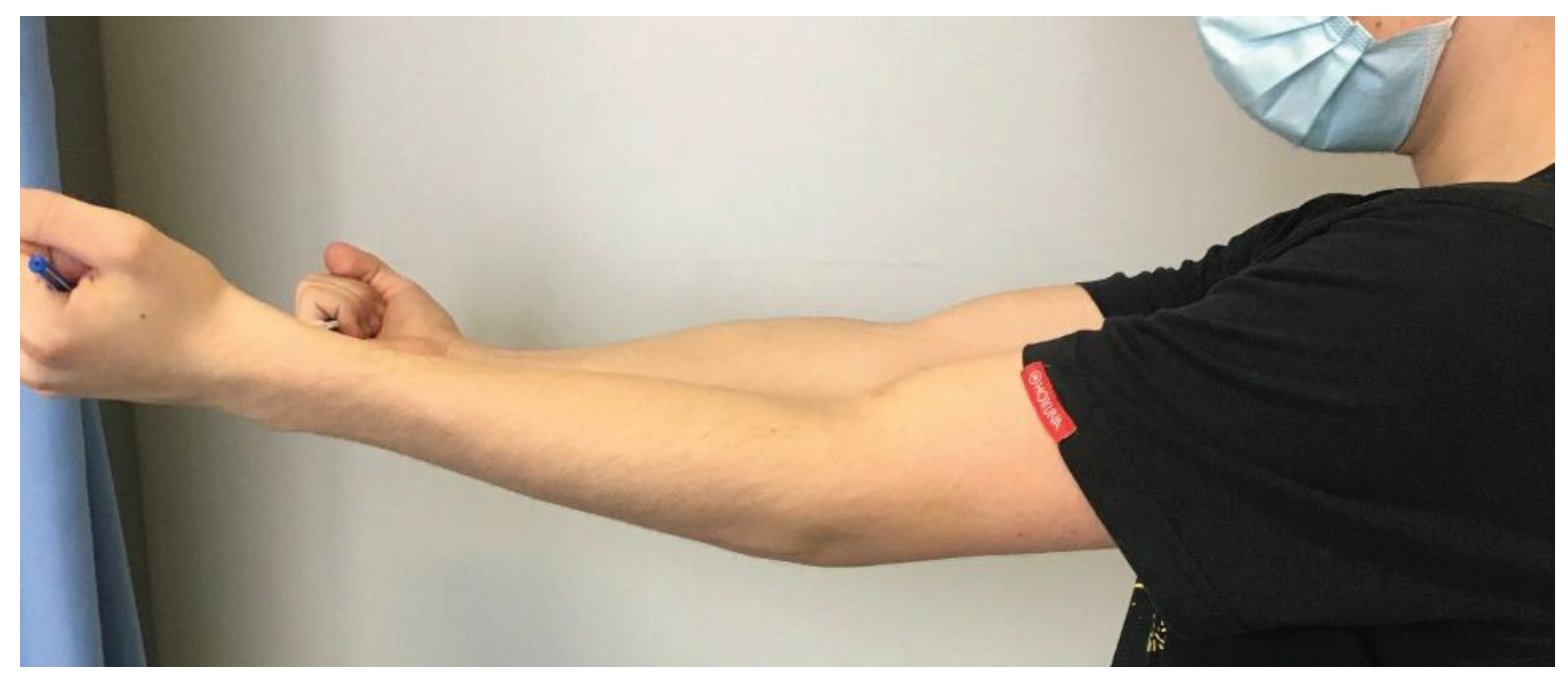
Six weeks later, the control X-ray revealed no signs of complications; however, the affected side was missing 15 degrees of extension at this point (Figure 10). With further physiotherapy over time, the extension was bettered and the joint movements were not limited in any way and were closely identical to the opposing side by week 12.
3. Discussion
Currently, the literature suggests that OCFs have a higher incidence in children, with active lifestyles including sports, dancing, or gymnastics. The mechanism of OCFs in the pediatric age group has been studied and was found to have lower resistance to sheer forces [1,13,48,49]. As discussed, diagnosis is challenging due to the difficulty of visualising small pieces on plain radiographs, non-specific symptoms, and access to diagnostic tools. As mentioned, symptoms of pain, swelling, locking and catching, and hemarthrosis are common and are suggested to be highly suspicious of articular surface injury [1]. A simple diagnostic algorithm was proposed in a 2015 paper written by Pedersen et al [11]. We suggest that this algorithm is adequate and can be used as a template when deciding on diagnostic evaluation of the injury.
Traditionally, the management of fractures affecting the articular surface involves osteosynthesis of the fragment with various methods. OCFs are to be treated operatively [1,49]. The literature describes the use of Herbert screws, compression or headless compression screws, magnesium screws, meniscus arrows, bone tunnel sutures, or bioabsorbable screws and pins [1,15]. In certain cases, when the fragment is too small for fixation (usually less than 5-10 mm) or if otherwise contraindicated by degenerative conditions, it is necessary to remove the fragment and manage the defect with cartilage restorative methods. Options include debridement, autologous chondrocyte implantation, mosaicplasty, allografts, or the use of biomaterials [1,11,15].
Delayed or improper therapy may seriously impact the quality of life. The aim is to achieve a stable fixation, restore articular congruity and joint stability, and allow for early passive motion. Complications due to implant protrusion or migration can result in injury of the surrounding meniscus, bone or cartilage. Malunion, nonunion, inflammation, infection, pain and functional deterioration can all occur in case of malposition and are more frequent with metal implants [1,11,48,49]. Metal implants might lead to more irritation or hardware reactions such as inflammation or foreign body reactions. Biodegradable instruments were revolutionary in this aspect as they are associated with few complications [47,50,51]. In agreement with other research, the size of the bony fragment should dictate the fixation tool (screw, pin, nail, adhesives) and the size of the chondral part is important to determine the treatment method. Taking into account the enhanced healing properties of the adolescent population, we suggest the operative treatment of OCFs if weight-bearing surfaces are affected, mechanical symptoms are present, diagnostic evidence can be found and the fragment is sufficient in size [29,30]. We suggest that operative treatment takes place immediately for OCFs, with special attention to the creation of articular surface congruency. We suggest that the fragments be cleaned of fibrin-coagulum with a curette and that smooth edges are formed with the underlying fresh spongious surface if possible for proper attachment without gaps or incongruency [1,11,29,30].
Timely and appropriate treatment is crucial to prevent long-term joint dysfunction, deformities and a restricted range of motion. The chondral part of the fragment may survive by diffusion from the synovium, however the same cannot be said about the bony element, which will have decreased ability to heal over time [1,30]. However, it will maintain its role as a scaffold. Bioabsorbable implants have been suggested as an appropriate treatment for OCFs by Schleter et al. and Hsu et al. and many others [1,9,52,53]. Our suggestions for the indication of absorbable implants are as follows:
- Osteochondral or chondral fragment
- Single, isolated body
- Sufficient size
- Instability, detachment or movement
The authors propose the arthroscopic removal of the fragments if:
- Fragment is less than 5 mm
- Bodies are multifragmented and small
In cases of pediatric intra-articular fractures, metal implants can be substituted for biodegradable implants. These are thought to be less harmful to the surrounding tissues and cartilage, however, they might provide less stability [50,51]. Beneficial mechanical properties, which are not provided by metal implants, include the diametric expansion of the implant and longitudinal contraction. An additional advantageous attribute resides in the bending modulus, which is closer to that of bone in the case of absorbable implants, in contrast to metal implants. This characteristic safeguards the fixation from adverse effects due to stress shielding [45]. Absorbable implants are less prone to interfere with growth, promote bone remodelling and lead to fewer biochemical reactions that might harm the patients' recovery [1,11,50,52].
Rehabilitation following surgical treatment involves physiotherapy and brace use for 2-6 weeks depending on severity. Children should start rehabilitation right after surgery with the help of physiotherapeutic training [48,54,55,56,57]. We advise early mobilisation during the immediate postoperative period as well as continuous passive movement (CPM) and ROM exercises, beginning on post-op day 1 and lasting until day 21. Physiotherapy and active motion exercises began after the first week, on day 8. In the case of weight-bearing joints, we suggest that weight-bearing is limited in the beginning with a gradual increase until week 6-8. We suggest the return to sports activities after the 4-6th months. The utilisation of these techniques allows for proper healing and helps decrease the occurrence of postop complications [1,11].
These implants do not need to be removed and thus, require no additional operation. A second anaesthesia would increase cost and prolong the length of hospital stay. Complications arising from a second surgery are eliminated. Because of this it greatly reduces the burden on the child and healthcare provider. The overall length of hospital stay is decreased. The expenses of the healthcare provider can be reduced as there is no need for staff, operating room or materials to be used at a second surgery. The child may recover from the comfort of their home and may begin physiotherapy sooner. Nonetheless, the real incidence, prevalence and outcome of the condition cannot be found in the literature. Neither can generalised guidelines be found regarding diagnostic and treatment algorithms for OCFs. More extensive studies are needed in both, pediatric and adult populations.
4. Conclusions
With the application of absorbable implants, the need to re-anaesthetise and reoperate was eliminated; thus, the strain on the patient was significantly reduced. Furthermore, possible risks relating to the surgery are also decreased - such as infections or other complications. Patients may begin physiotherapy sooner due to the reasons mentioned above.
Supplementary Materials
Information regarding the implants, their production, application, and other supporting information such as whitepapers, studies, operation videos and animations can be downloaded at: https://bioretec.com/educational-materials.
Author Contributions
Conceptualization, J.G. and N.H.; methodology, J.G.; software, N.H and A.L.; validation, J.G., N.H. and A.L.; formal analysis, N.H. and A.L.; investigation, N.H.; resources, J.G.; data curation, J.G.; writing—original draft preparation, N.H.; writing—review and editing, N.H. and A.L.; visualization, J.G., V.M., and K.T.; supervision, J.G.; project administration, J.G.; funding acquisition, J.G. All authors have read and agreed to the published version of the manuscript.
Funding
This work was supported, in part, by the …..(grant number) and by the Medical School, University of Pécs. The funders had no role in study design, data collection and analysis, the decision to publish, or manuscript preparation. The APC was funded by …….
Institutional Review Board Statement
The study was conducted in accordance with the Declaration of Helsinki, and approved by the Institutional Review Board (or Ethics Committee) of the Surgical Division, Department of Paediatrics, Medical School, University of Pécs (protocol code XXX and date of approval) for studies involving humans.
Informed Consent Statement
Informed consent was obtained from all subjects involved in the study. Written informed consent has been obtained from the patient’s guardians to publish this paper.
Data Availability Statement
The data is contained within this article.
Acknowledgments
The authors would like to extend thanks to Mahan Sayadinia for the revision of the document for better intellectual content.
Conflicts of Interest
The authors declare no conflict of interest.
References
- Gkiokas, A.; Morassi, L.G.; Kohl, S.; Zampakides, C.; Megremis, P.; Evangelopoulos, D.S. Bioabsorbable Pins for Treatment of Osteochondral Fractures of the Knee after Acute Patella Dislocation in Children and Young Adolescents. Adv. Orthop. 2012, 2012, e249687. [Google Scholar] [CrossRef] [PubMed]
- Sari, E. Delayed Osteochondral Fracture Fixation of the Knee in a Pediatric Patient. Turk. J. Trauma Emerg. Surg. 2020. [Google Scholar] [CrossRef] [PubMed]
- Badekas, T.; Takvorian, M.; Souras, N. Treatment Principles for Osteochondral Lesions in Foot and Ankle. Int. Orthop. 2013, 37, 1697–1706. [Google Scholar] [CrossRef] [PubMed]
- Agrawal, U.; Tiwari, V.; Selvanayagam, R. A Rare Case of Large Osteochondral Fracture of Patella. Cureus 15, e41245. [CrossRef]
- Alosaimi, M.N.; Almutairi, M.M.; Alshahrani, S.M.; Alqahtani, M.N.; Alghamdi, A.S. Osteochondral Fracture of the Patella without Soft Tissue Injury and with No Dislocation: A Case Report. Int. J. Surg. Case Rep. 2020, 78, 48–53. [Google Scholar] [CrossRef] [PubMed]
- Padaki, A.S.; Allahabadi, S.; Pandya, N.K. Adolescent Elbow Osteochondral Lesions Following Prior Elbow Fracture Pinning. J. Child. Orthop. 2022, 16, 475–480. [Google Scholar] [CrossRef] [PubMed]
- Weiss, J.M.; Nikizad, H.; Shea, K.G.; Gyurdzhyan, S.; Jacobs, J.C.; Cannamela, P.C.; Kessler, J.I. The Incidence of Surgery in Osteochondritis Dissecans in Children and Adolescents. Orthop. J. Sports Med. 2016, 4, 2325967116635515. [Google Scholar] [CrossRef] [PubMed]
- Wood, D.; Davis, D.D.; Carter, K.R. Osteochondritis Dissecans. In StatPearls; StatPearls Publishing: Treasure Island (FL), 2023. [Google Scholar]
- Wiktor, Ł.; Tomaszewski, R. Evaluation of Osteochondritis Dissecans Treatment with Bioabsorbable Implants in Children and Adolescents. J. Clin. Med. 2022, 11, 5395. [Google Scholar] [CrossRef] [PubMed]
- Tudisco, C.; Bernardi, G.; Manisera, M.T.; De Maio, F.; Gorgolini, G.; Farsetti, P. An Update on Osteochondritis Dissecans of the Knee. Orthop. Rev. 14, 38829. [CrossRef]
- Pedersen, M.E.; DaCambra, M.P.; Jibri, Z.; Dhillon, S.; Jen, H.; Jomha, N.M. Acute Osteochondral Fractures in the Lower Extremities - Approach to Identification and Treatment. Open Orthop. J. 2015, 9, 463–474. [Google Scholar] [CrossRef] [PubMed]
- Cordunianu, M.A.; Antoniac, I.; Niculescu, M.; Paltanea, G.; Raiciu, A.D.; Dura, H.; Forna, N.; Carstoc, I.D.; Cristea, M.B. Treatment of Knee Osteochondral Fractures. Healthcare 2022, 10, 1061. [Google Scholar] [CrossRef] [PubMed]
- Bauer, K.L. Osteochondral Injuries of the Knee in Pediatric Patients. J. Knee Surg. 2018, 31, 382–391. [Google Scholar] [CrossRef] [PubMed]
- Kennedy, J.C.; Grainger, R.W.; McGraw, R.W. Osteochondral Fractures of the Femoral Condyles. J. Bone Joint Surg. Br. 1966, 48, 436–440. [Google Scholar] [CrossRef]
- Kühle, J.; Angele, P.; Balcarek, P.; Eichinger, M.; Feucht, M.; Haasper, C.; Alexander, G.; Jung, T.; Lill, H.; Marquass, B.; et al. Treatment of Osteochondral Fractures of the Knee: A Meta-Analysis of Available Scientific Evidence. Int. Orthop. 2013, 37, 2385–2394. [Google Scholar] [CrossRef]
- Johnson, D.L.; Urban, W.P.; Caborn, D.N.; Vanarthos, W.J.; Carlson, C.S. Articular Cartilage Changes Seen with Magnetic Resonance Imaging-Detected Bone Bruises Associated with Acute Anterior Cruciate Ligament Rupture. Am. J. Sports Med. 1998, 26, 409–414. [Google Scholar] [CrossRef] [PubMed]
- Stanitski, C.L.; Paletta, G.A. Articular Cartilage Injury with Acute Patellar Dislocation in Adolescents. Am. J. Sports Med. 1998, 26, 52–55. [Google Scholar] [CrossRef] [PubMed]
- Elias, D.A.; White, L.M.; Fithian, D.C. Acute Lateral Patellar Dislocation at MR Imaging: Injury Patterns of Medial Patellar Soft-Tissue Restraints and Osteochondral Injuries of the Inferomedial Patella. Radiology 2002, 225, 736–743. [Google Scholar] [CrossRef] [PubMed]
- Gianotti, S.M.; Marshall, S.W.; Hume, P.A.; Bunt, L. Incidence of Anterior Cruciate Ligament Injury and Other Knee Ligament Injuries: A National Population-Based Study. J. Sci. Med. Sport 2009, 12, 622–627. [Google Scholar] [CrossRef] [PubMed]
- McCarthy, J.C.; Busconi, B. The Role of Hip Arthroscopy in the Diagnosis and Treatment of Hip Disease. Orthopedics 1995, 18, 753–756. [Google Scholar] [CrossRef] [PubMed]
- Bohndorf, K. Imaging of Acute Injuries of the Articular Surfaces (Chondral, Osteochondral and Subchondral Fractures). Skeletal Radiol. 1999, 28, 545–560. [Google Scholar] [CrossRef] [PubMed]
- Slaughter, A.J.; Reynolds, K.A.; Jambhekar, K.; David, R.M.; Hasan, S.A.; Pandey, T. Clinical Orthopedic Examination Findings in the Lower Extremity: Correlation with Imaging Studies and Diagnostic Efficacy. Radiogr. Rev. Publ. Radiol. Soc. N. Am. Inc 2014, 34, e41–55. [Google Scholar] [CrossRef] [PubMed]
- Mintz, D.N.; Tashjian, G.S.; Connell, D.A.; Deland, J.T.; O’Malley, M.; Potter, H.G. Osteochondral Lesions of the Talus: A New Magnetic Resonance Grading System with Arthroscopic Correlation. Arthrosc. J. Arthrosc. Relat. Surg. Off. Publ. Arthrosc. Assoc. N. Am. Int. Arthrosc. Assoc. 2003, 19, 353–359. [Google Scholar] [CrossRef] [PubMed]
- Disler, D.G.; McCauley, T.R.; Kelman, C.G.; Fuchs, M.D.; Ratner, L.M.; Wirth, C.R.; Hospodar, P.P. Fat-Suppressed Three-Dimensional Spoiled Gradient-Echo MR Imaging of Hyaline Cartilage Defects in the Knee: Comparison with Standard MR Imaging and Arthroscopy. AJR Am. J. Roentgenol. 1996, 167, 127–132. [Google Scholar] [CrossRef]
- Verhagen, R. a. W.; Maas, M.; Dijkgraaf, M.G.W.; Tol, J.L.; Krips, R.; van Dijk, C.N. Prospective Study on Diagnostic Strategies in Osteochondral Lesions of the Talus. Is MRI Superior to Helical CT? J. Bone Joint Surg. Br. 2005, 87, 41–46. [Google Scholar] [CrossRef] [PubMed]
- O’Donoghue, D.H. Chondral and Osteochondral Fractures. J. Trauma 1966, 6, 469–481. [Google Scholar] [CrossRef] [PubMed]
- Schmid, M.R.; Pfirrmann, C.W.A.; Hodler, J.; Vienne, P.; Zanetti, M. Cartilage Lesions in the Ankle Joint: Comparison of MR Arthrography and CT Arthrography. Skeletal Radiol. 2003, 32, 259–265. [Google Scholar] [CrossRef] [PubMed]
- El-Khoury, G.Y.; Alliman, K.J.; Lundberg, H.J.; Rudert, M.J.; Brown, T.D.; Saltzman, C.L. Cartilage Thickness in Cadaveric Ankles: Measurement with Double-Contrast Multi-Detector Row CT Arthrography versus MR Imaging. Radiology 2004, 233, 768–773. [Google Scholar] [CrossRef] [PubMed]
- Lan, T.; McCarthy, H.S.; Hulme, C.H.; Wright, K.T.; Makwana, N. The Management of Talar Osteochondral Lesions - Current Concepts. J. Arthrosc. Jt. Surg. 2021, 8, 231–237. [Google Scholar] [CrossRef] [PubMed]
- Steele, J.R.; Dekker, T.J.; Federer, A.E.; Liles, J.L.; Adams, S.B.; Easley, M.E. Republication of “Osteochondral Lesions of the Talus: Current Concepts in Diagnosis and Treatment”. Foot Ankle Orthop. 2023, 8, 24730114231192961. [Google Scholar] [CrossRef] [PubMed]
- Ferkel, R.D.; Zanotti, R.M.; Komenda, G.A.; Sgaglione, N.A.; Cheng, M.S.; Applegate, G.R.; Dopirak, R.M. Arthroscopic Treatment of Chronic Osteochondral Lesions of the Talus: Long-Term Results. Am. J. Sports Med. 2008, 36, 1750–1762. [Google Scholar] [CrossRef]
- Hepple, S.; Winson, I.G.; Glew, D. Osteochondral Lesions of the Talus: A Revised Classification. Foot Ankle Int. 1999, 20, 789–793. [Google Scholar] [CrossRef]
- Berndt, A.L.; Harty, M. Transchondral Fractures (Osteochondritis Dissecans) of the Talus. J. Bone Joint Surg. Am. 1959; 41-A, 988–1020. [Google Scholar]
- H, T. [The Structure, Physiology, and Biomechanics of Articular Cartilage: Injury and Repair]. Acta Orthop. Traumatol. Turc. 2007, 41 Suppl 2.
- Wilusz, R.E.; Sanchez-Adams, J.; Guilak, F. The Structure and Function of the Pericellular Matrix of Articular Cartilage. Matrix Biol. J. Int. Soc. Matrix Biol. 2014, 0, 25–32. [Google Scholar] [CrossRef]
- Lyndina, Y.; Romaniuk, A.; Lyndin, M.; Hyryavenko, N.; Kurochkina, V.; Tkach, G.; Gluschenko, N.; Sikora, V. Morphofunctional Features of Articular Cartilage Structure. Folia Medica Cracoviensia 2019 Vol 59 No 3 81-93 2019.
- Makris, E.A.; Gomoll, A.H.; Malizos, K.N.; Hu, J.C.; Athanasiou, K.A. Repair and Tissue Engineering Techniques for Articular Cartilage. Nat. Rev. Rheumatol. 2015, 11, 21–34. [Google Scholar] [CrossRef] [PubMed]
- Gilmore, R.S.; Palfrey, A.J. A Histological Study of Human Femoral Condylar Articular Cartilage. J. Anat. 1987, 155, 77–85. [Google Scholar] [PubMed]
- Gentile, P.; Chiono, V.; Carmagnola, I.; Hatton, P.V. An Overview of Poly(Lactic-Co-Glycolic) Acid (PLGA)-Based Biomaterials for Bone Tissue Engineering. Int. J. Mol. Sci. 2014, 15, 3640–3659. [Google Scholar] [CrossRef] [PubMed]
- Girón, J.; Kerstner, E.; Medeiros, T.; Oliveira, L.; Machado, G.M.; Malfatti, C.F.; Pranke, P. Biomaterials for Bone Regeneration: An Orthopedic and Dentistry Overview. Braz. J. Med. Biol. Res. 2021, 54, e11055. [Google Scholar] [CrossRef]
- Rocha, C.V.; Gonçalves, V.; da Silva, M.C.; Bañobre-López, M.; Gallo, J. PLGA-Based Composites for Various Biomedical Applications. Int. J. Mol. Sci. 2022, 23, 2034. [Google Scholar] [CrossRef] [PubMed]
- Hedelin, H.; Hebelka, H.; Brisby, H.; Laine, T. MRI Evaluation of Resorbable Poly Lactic-Co-Glycolic Acid (PLGA) Screws Used in Pelvic Osteotomies in Children—a Retrospective Case Series. J. Orthop. Surg. 2020, 15, 329. [Google Scholar] [CrossRef] [PubMed]
- Heye, P.; Matissek, C.; Seidl, C.; Varga, M.; Kassai, T.; Jozsa, G.; Krebs, T. Making Hardware Removal Unnecessary by Using Resorbable Implants for Osteosynthesis in Children. Children 2022, 9, 471. [Google Scholar] [CrossRef] [PubMed]
- Golubev, V.G.; Starostenkov, A.N. OPERATIVE TECHNIQUE FEATURES IN APPLICATION OF BIOABSORBABLE IMPLANTS FOR LIMB FRACTURES TREATMENT.
- Whitepapers - Bioretec Ltd. Available online: https://bioretec.com/educational-materials/whitepapers?medical_professional=on&category=12 (accessed on 5 August 2023).
- Singh, V.; Garg, V.; Parikh, S.N. Management of Physeal Fractures: A Review Article. Indian J. Orthop. 2021, 55, 525–538. [Google Scholar] [CrossRef]
- ActivaNailTM - Bioabsorbable Nail - Bioretec Ltd. Available online: https://bioretec.com/products/8/activanail-bioabsorbable-nail (accessed on 6 August 2023).
- Macken, A.A.; Eygendaal, D.; van Bergen, C.J. Diagnosis, Treatment and Complications of Radial Head and Neck Fractures in the Pediatric Patient. World J. Orthop. 2022, 13, 238–249. [Google Scholar] [CrossRef] [PubMed]
- Ackerson, R.; Nguyen, A.; Carry, P.M.; Pritchard, B.; Hadley-Miller, N.; Scott, F. Intra-Articular Radial Head Fractures In the Skeletally Immature Patient: Complications and Management. J. Pediatr. Orthop. 2015, 35, 443. [Google Scholar] [CrossRef]
- Li, Z.-H.; Yu, A.-X.; Guo, X.-P.; Qi, B.-W.; Zhou, M.; Wang, W.-Y. Absorbable Implants versus Metal Implants for the Treatment of Ankle Fractures: A Meta-Analysis. Exp. Ther. Med. 2013, 5, 1531–1537. [Google Scholar] [CrossRef] [PubMed]
- Noh, J.H.; Roh, Y.H.; Yang, B.G.; Kim, S.W.; Lee, J.S.; Oh, M.K. Outcomes of Operative Treatment of Unstable Ankle Fractures: A Comparison of Metallic and Biodegradable Implants. J. Bone Joint Surg. Am. 2012, 94, e166. [Google Scholar] [CrossRef] [PubMed]
- Schlechter, J.A.; Nguyen, S.V.; Fletcher, K.L. Utility of Bioabsorbable Fixation of Osteochondral Lesions in the Adolescent Knee: Outcomes Analysis With Minimum 2-Year Follow-Up. Orthop. J. Sports Med. 2019, 7, 2325967119876896. [Google Scholar] [CrossRef] [PubMed]
- Hsu, T.-L.; Lin, S.-M.; Chang, C.-H.; Lan, T.-Y. Neglected Pediatric Osteochondral Fracture Dislocation of the Patella. Case Rep. Orthop. 2019, 2019, 2904782. [Google Scholar] [CrossRef] [PubMed]
- Yoon, A.; King, G.J.W.; Grewal, R. Is ORIF Superior to Nonoperative Treatment in Isolated Displaced Partial Articular Fractures of the Radial Head? Clin. Orthop. 2014, 472, 2105–2112. [Google Scholar] [CrossRef] [PubMed]
- Ring, D. Open Reduction and Internal Fixation of Fractures of the Radial Head. Hand Clin. 2004, 20, 415–427, vi. [Google Scholar] [CrossRef] [PubMed]
- Gao, X.; Yin, H.; Zhou, G. Minimally Invasive Treatment of Mason Type II Radial Head Fracture by Intramedullary Pinning. Orthop. Surg. 2019, 11, 879–885. [Google Scholar] [CrossRef] [PubMed]
- Timofeev, I.V.; Виктoрoвич, Т.И.; Dyakonova, E.Y.; Юрьевна, Д.Е.; Gusev, A.A.; Андреевич, Г.А.; Romanova, E.A.; Алексеевна, Р.Е.; Khrolenko, P.V.; Владимирoвна, Х.П. Arthroscopic treatement of patella fractures in children. Pediatr. Traumatol. Orthop. Reconstr. Surg. 2017, 5, 53–57. [Google Scholar] [CrossRef]
Figure 1.
Preoperative X-ray shows the osteochondral fragment of the lateral condyle of the femur.
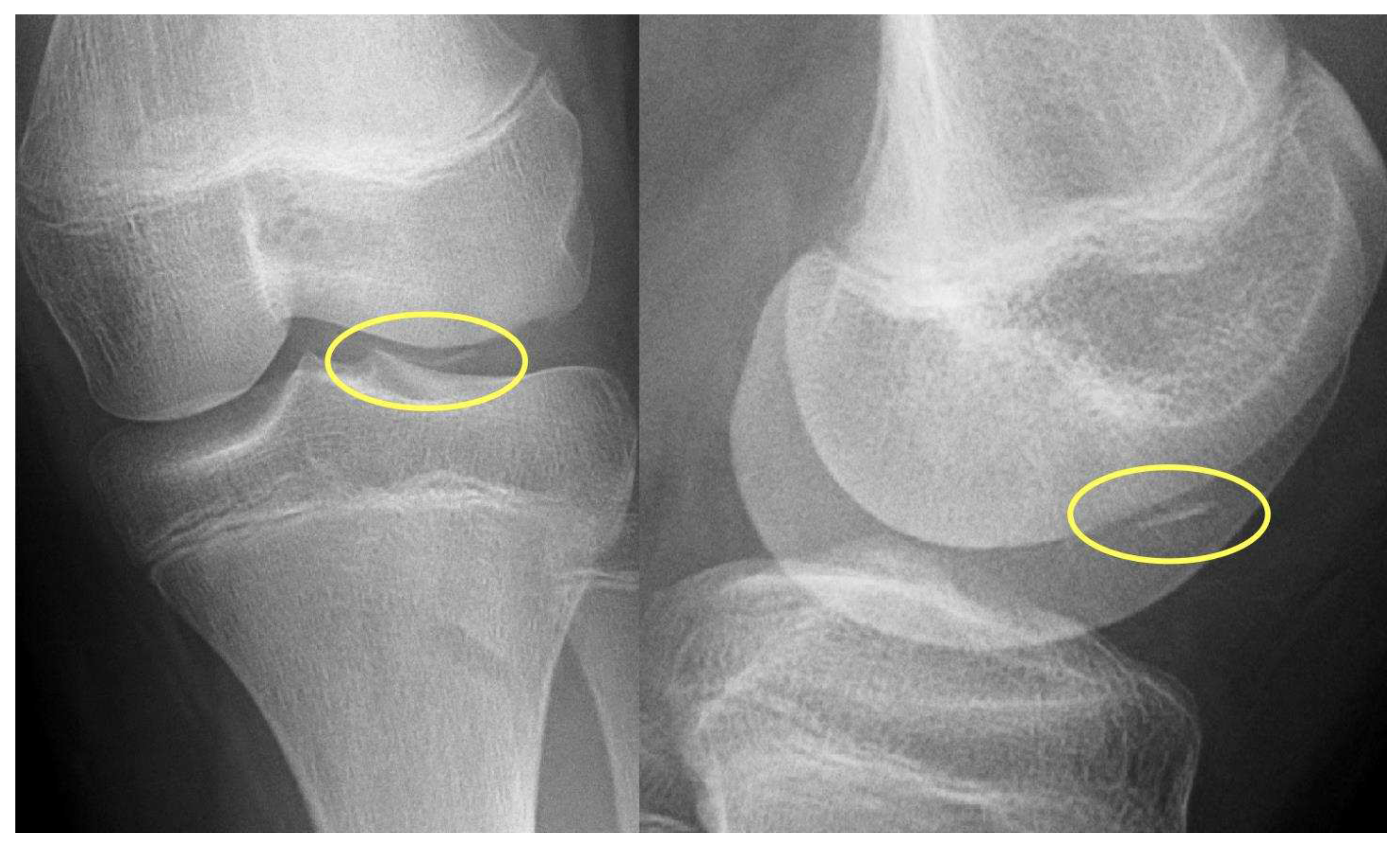
Figure 2.
Intraoperative pictures showing the defect (a), fixation of the fragment with K-wires (b), and the stabilised fragment with absorbable nails.
Figure 2.
Intraoperative pictures showing the defect (a), fixation of the fragment with K-wires (b), and the stabilised fragment with absorbable nails.
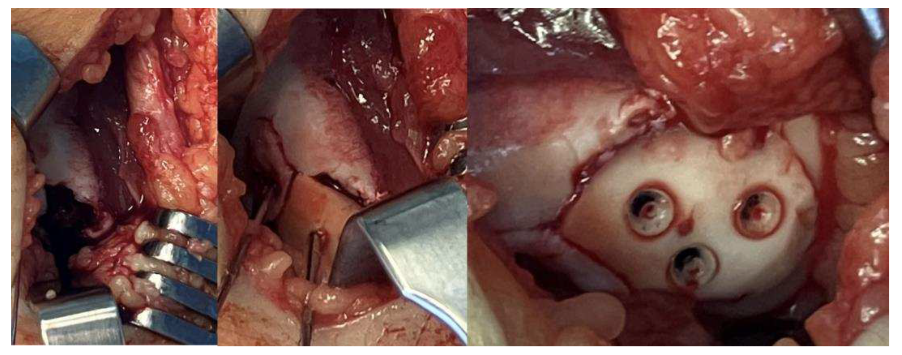
Figure 3.
Control MRI one year after surgery. The remainder of the absorbable nails can be seen inside the femoral condyle.
Figure 3.
Control MRI one year after surgery. The remainder of the absorbable nails can be seen inside the femoral condyle.
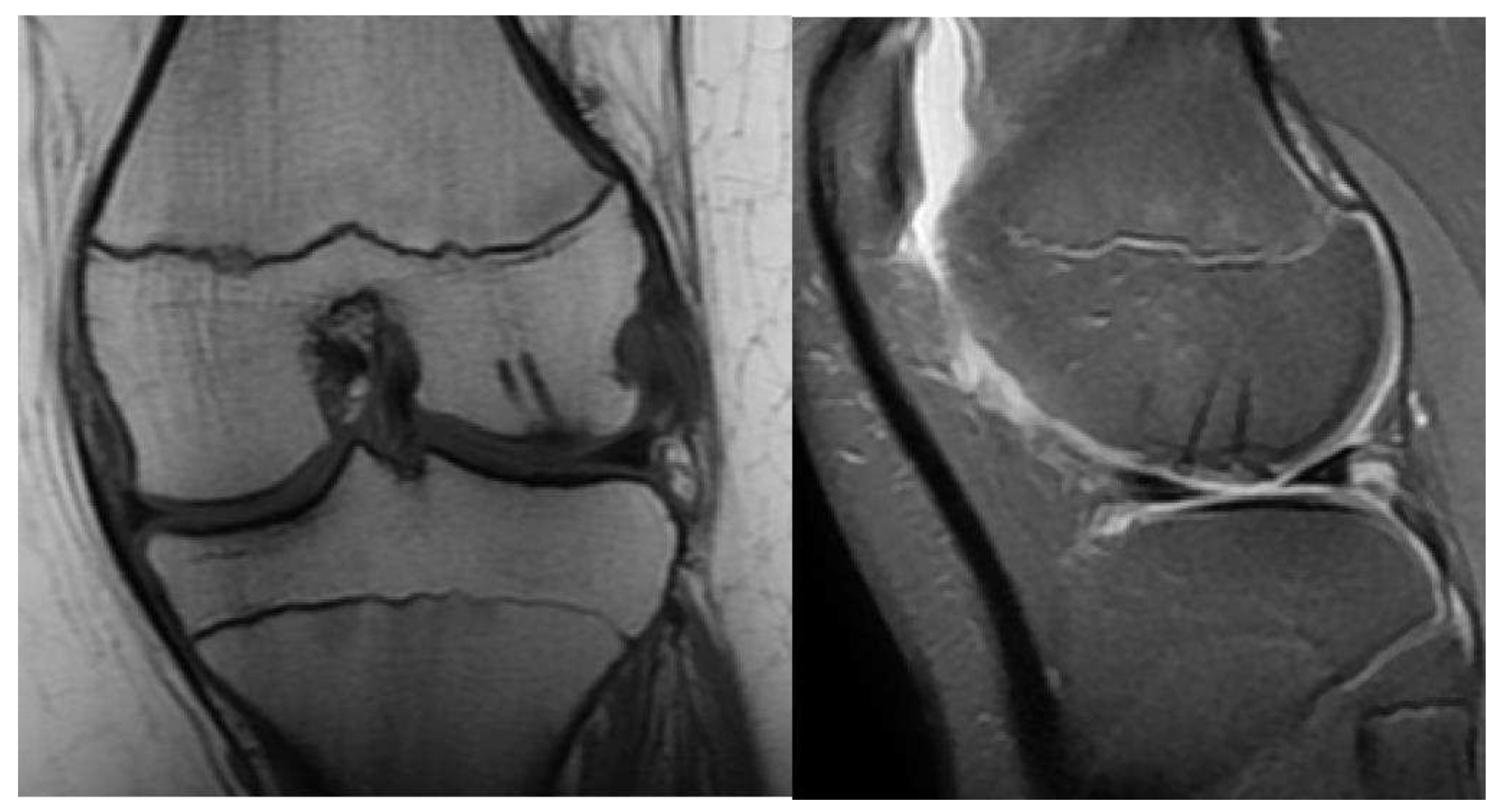
Figure 4.
CT images of the knee, showing the fragment (black arrow) and the location of the missing piece (red circle).
Figure 4.
CT images of the knee, showing the fragment (black arrow) and the location of the missing piece (red circle).
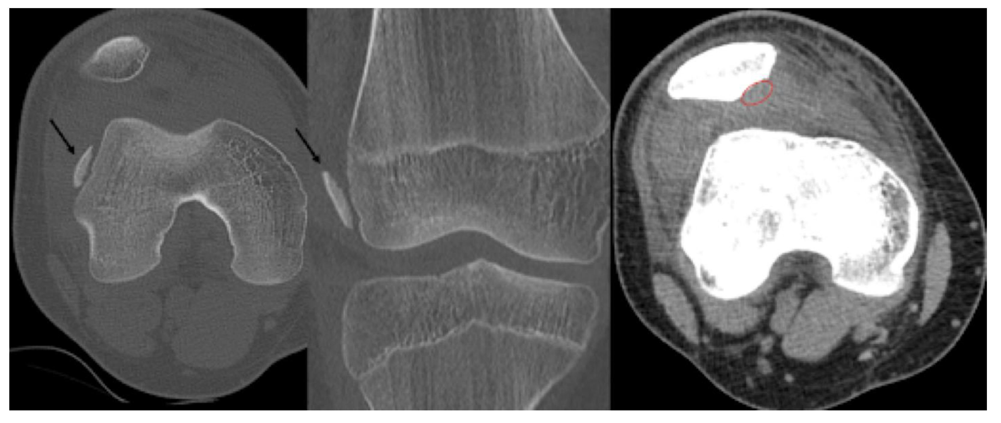
Figure 5.
Intraoperative pictures showing the defect of the patella before reduction and fixation of the osteochondral fragment (a). The fragment can be seen after removal and cleaning (b) and after stabilisation with absorbable implants (c).
Figure 5.
Intraoperative pictures showing the defect of the patella before reduction and fixation of the osteochondral fragment (a). The fragment can be seen after removal and cleaning (b) and after stabilisation with absorbable implants (c).
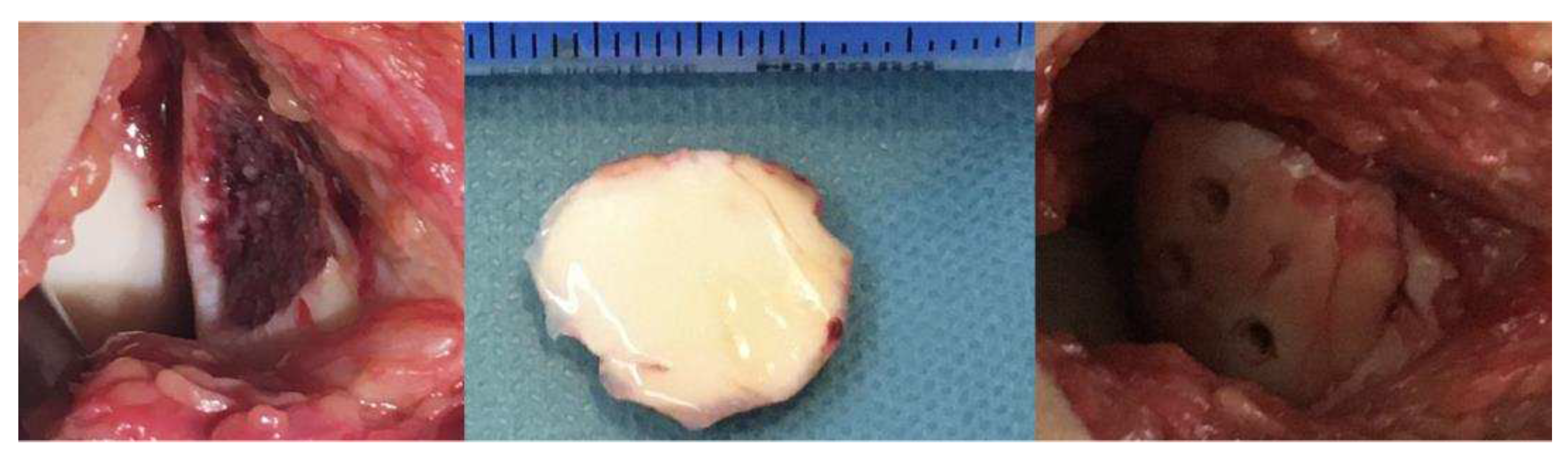
Figure 6.
Postoperative X-ray presents the proper position of the osteochondral fragment of the patella.
Figure 6.
Postoperative X-ray presents the proper position of the osteochondral fragment of the patella.
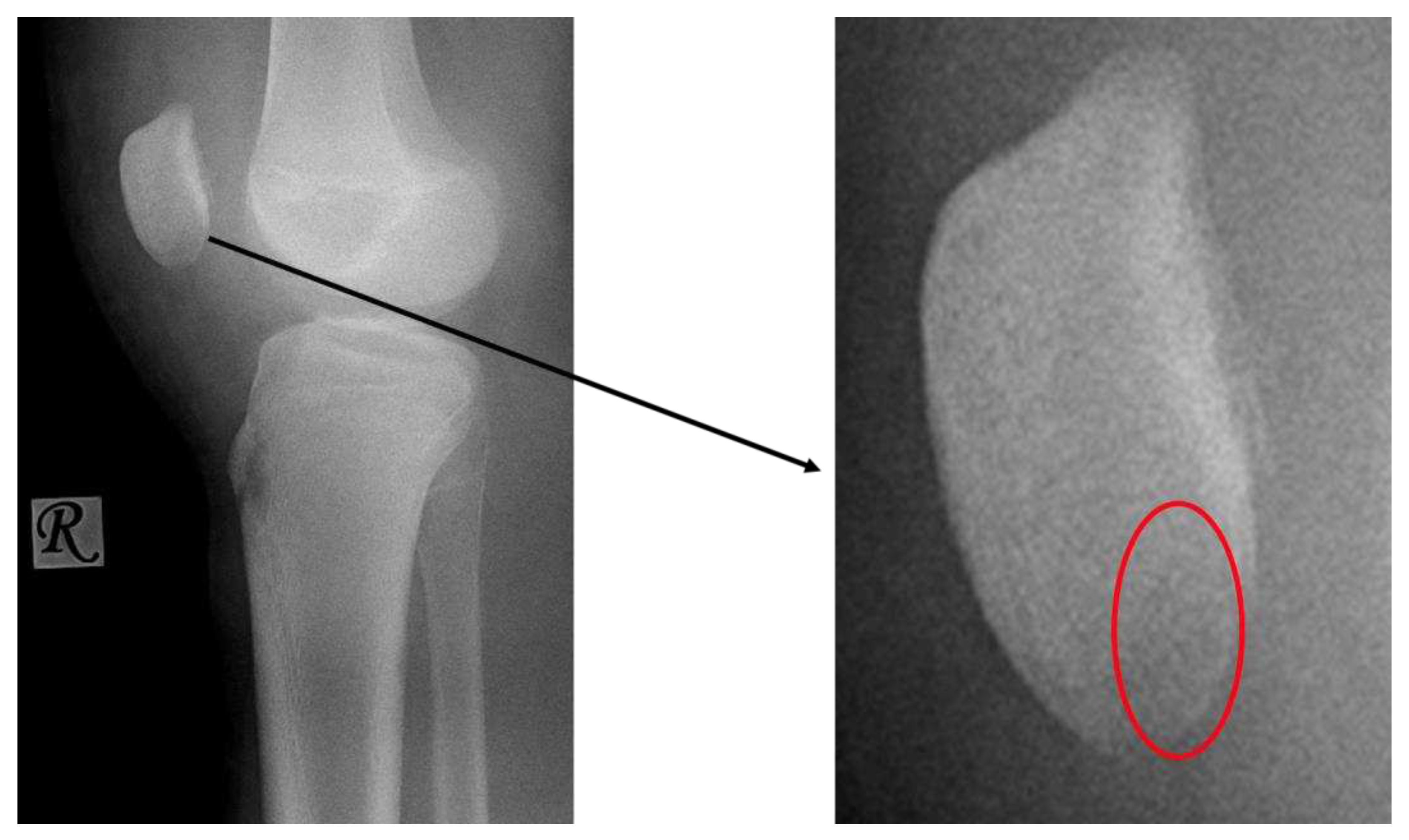
Figure 7.
The CT images confirm the Mason type II fracture and dislocation of the radial head.
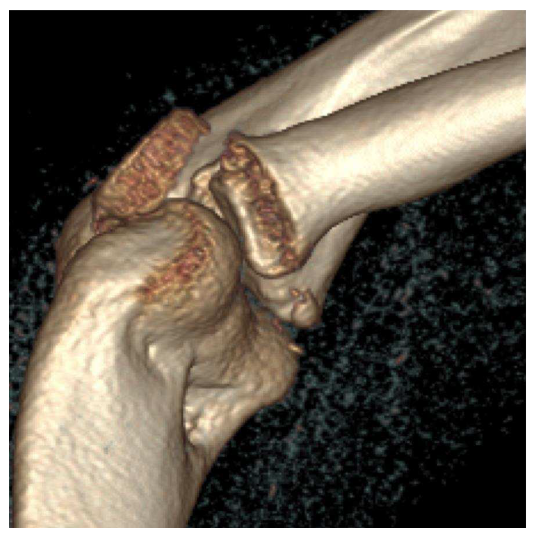
Figure 8.
Intraoperative images of the defect (a), the fragment (b) and the results of the correction (c).
Figure 8.
Intraoperative images of the defect (a), the fragment (b) and the results of the correction (c).
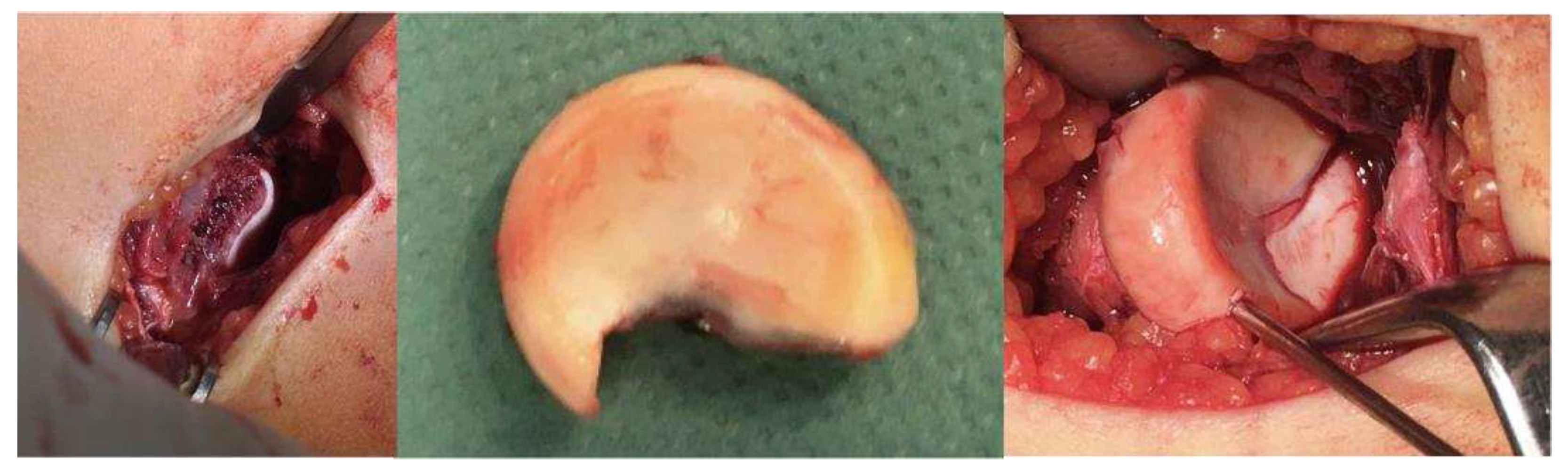
Figure 9.
Postoperative X-ray showing the perfect position of the radial head (c).
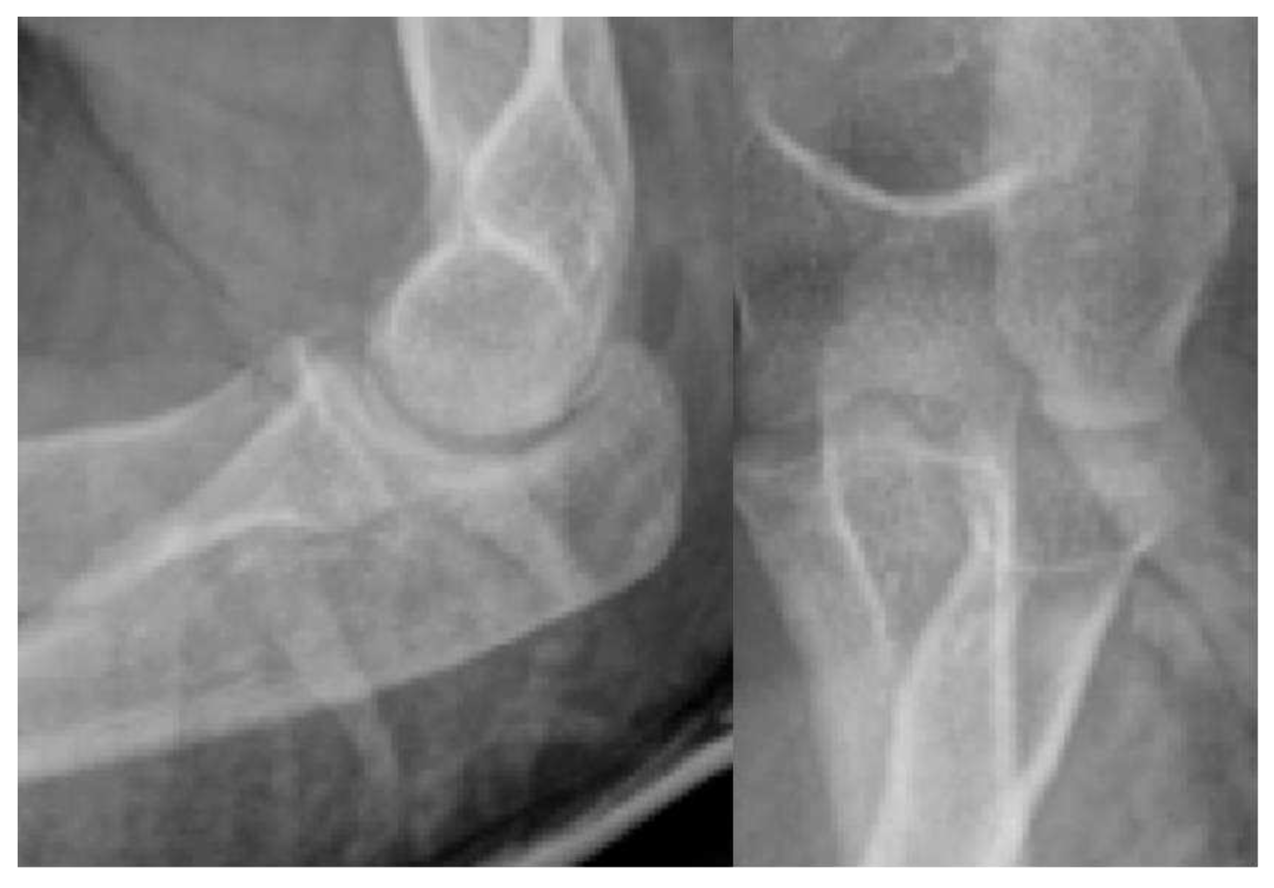
Figure 10.
Post-op image six weeks after surgery describing the 15-degree shortcoming of the affected side during extension.
Figure 10.
Post-op image six weeks after surgery describing the 15-degree shortcoming of the affected side during extension.
Disclaimer/Publisher’s Note: The statements, opinions and data contained in all publications are solely those of the individual author(s) and contributor(s) and not of MDPI and/or the editor(s). MDPI and/or the editor(s) disclaim responsibility for any injury to people or property resulting from any ideas, methods, instructions or products referred to in the content. |
© 2023 by the authors. Licensee MDPI, Basel, Switzerland. This article is an open access article distributed under the terms and conditions of the Creative Commons Attribution (CC BY) license (http://creativecommons.org/licenses/by/4.0/).
Copyright: This open access article is published under a Creative Commons CC BY 4.0 license, which permit the free download, distribution, and reuse, provided that the author and preprint are cited in any reuse.


