
Submitted:
27 October 2023
Posted:
30 October 2023
You are already at the latest version
Abstract
Abatacept (CTLA4-Ig) – a monoclonal antibody which restricts T cell activation – is an effective treatment for rheumatoid arthritis (RA). Herein, we aimed to investigate for biomarkers of response to abatacept. We performed a detailed immunological profiling of CTLA4-treated RA patients’ peripheral blood (PB) cells and sera using flow cytometry and proteomics analysis, respectively. At 6 months of treatment, 34.5% of patients attained clinical response. Notably, baseline levels of Th1 and myeloid cell populations were significantly elevated in PB of responders compared to non-responders (P-value<0.05). Additionally, proteomics analysis revealed 10 amongst 303 molecules which were associated with clinical responses. Should the present data be confirmed in a larger cohort could be of clinical value.
Keywords:
rheumatoid arthritis
; abatacept
; CTLA4-Ig
; biomarkers
; immune cells
; Th1
; proteomics
1. Introduction
Rheumatoid arthritis (RA) is a chronic autoimmune inflammatory disease, which may lead to articular bone and cartilage destruction, disability and reduced life expectancy [1,2]. RA develops upon aberrant activation of the immune system mainly due to failure of self-tolerance mechanisms [3]. T helper (Th) lymphocytes have been demonstrated to play a central role in disease pathogenesis and progression through the release of cytokines and chemokines [4,5]. Both Th1 and Th17 cells have been proved to contribute to RA inflammatory responses. On the other hand, CD4+ T regulatory cells (FoxP3+ Tregs) as well as myeloid-derived suppressor cells (MDSCs) that exert an immunoregulatory role and re-establish homeostasis are defective during active RA [6]. Restoration of immune regulation has been shown in RA patients upon effective treatment.
T cell proliferation and acquisition of effector functions require intracellular signals elicited by both the T cell receptor a co-receptor, CD28 [7]. CD28 delivers co-stimulatory signals upon engagement of its ligands, B7-1 (CD80) or B7-2 (CD86). Cytotoxic T lymphocyte antigen 4 (CTLA4), binds to the same ligands as CD28, with higher affinity and restricts T cell activation [8]. CTLA4 expression is induced in all T cells transiently after T cell receptor activation, to regulate T cell proliferation and maintenance of tolerance [9]. Abatacept (CTLA4-Ig) is a recombinant fusion protein comprising the extracellular domain of human CTLA4 and a fragment of the Fc domain of human IgG1, which has been modified to prevent complement fixation [10]. Abatacept, like CTLA4, competes with CD28 for CD80 and CD86 binding on DCs, and thereby can be used to selectively modulate T-cell activation [11,12]. Abatacept has been approved for the treatment of RA, based on the results of an extensive clinical development program assessing its effectiveness and safety in different RA populations [13,14,15].
Biologic therapies (bDMARDs) have revolutionized the treatment of RA patients. Nevertheless, data from registries and cohort studies have shown that in clinical practice, 50-60% of RA patients starting a bDMARD will stop treatment due to inefficacy or toxicity in the long-term, while clinical response (remission or low disease activity) is unpredictable and is achieved by 20-40% [16,17]. Development of predictive markers of response for everyday clinical practice will be an important step in optimizing the treatment of autoimmune diseases. Available data for predictors of abatacept response are rather limited [18].
In this study we propose that a detailed immunological profile early at treatment initiation with abatacept, could differentiate responders vs non-responders. Moreover, sera proteomics analysis revealed a signature of proteins associated with clinical responses. Immunological studies of peripheral blood (PB) and sera were performed at baseline, on 3rd and 6th month after treatment initiation further characterized the biological effects of abatacept. Based on the above data we propose a signature which could predict response to abatacept.
2. Materials and Methods
2.1. Study approval
This is an observational, prospective, single-center study of RA patients, starting treatment with abatacept, due to residual disease activity. All patients were recruited by the Rheumatology and Clinical Immunology Clinic at the University Hospital in Heraklion, Crete. Decision regarding treatment scheme was made by the treating rheumatologist and according to the guidelines for RA treatment of the Hellenic Society of Rheumatology and EULAR guidelines. The study was approved by the Ethics Committee of the University Hospital of Heraklion (protocol number:3601, 17/06/2015, Heraklion) and informed consent was obtained from all individuals prior to sample collection.
2.2. Human subjects
Peripheral blood (PB) samples were obtained from RA individuals before the initiation of abatacept treatment and at 3 and 6 months upon treatment. Subjects with RA fulfilled the 2010 EULAR/ACR classification criteria, at Rheumatology and Clinical Immunology Clinic at the University Hospital of Heraklion [19]. At the time of sampling all patients had active disease state (DAS28≥3.2) and were naïve for biological therapy. Patients were followed clinically every 3 months and all immunological studies were performed at the indicated time points.
2.3. Peripheral blood mononuclear cell isolation
Heparinized blood (20ml) was collected from individuals with RA. Peripheral blood mononuclear cells (PBMCs) were isolated on Histopaque-1077 (Sigma-Aldrich) density gradient. Briefly, blood was diluted 1:1 with PBS and layered over Histopaque medium. Tubes were centrifuged at 400g for 30 min at room temperature (RT) to obtain PBMC layer. PBMC layer was collected and cells were washed with PBS. Erythrocytes were eliminated by hypotonic lysis (1ml ddH2O for 35 seconds and 1ml 1.8% NaCl). Then, samples were processed for staining. Additionally, blood in appropriate serum separator tubes was collected from individuals with RA. Tubes were centrifuged at 2500g for 15 min at RT to obtain serum.
2.4. Antibodies
Fluorescence-conjugated antibodies against human with the following specificities were used to detect antigens by flow cytometry: CD4 (OKT4), IL-17 (BL168), CD127 (A019D5), CD25 (BC96), HLADR (L243), CD3 (OKT3), CD1α (HI149), CD14 (HCD14), CD15 (SSEA-1), CD33 (WM53) from Biolegend; IFN-γ (B27) from BD Pharmingen; and FoxP3 (PCH101) from eBioscience.
2.5. Flow cytometry
Single-cell suspensions were stained with fluorescence-conjugated antibodies and analyzed by flow cytometry. In brief, 3–5 × 10⁶ PBMCs were washed with FACS buffer (5% FBS in PBS), and cells were then stained with the indicated antibodies for 20 min at 4°C. Stained cells were washed twice with FACS buffer, resuspended and acquired on a FACS Calibur (BD Biosciences) and analyzed using the FlowJo software (Tree Star).
2.6. Cellular Cytokine Expression Assays
Cytokine production was assessed through intracellular staining. In brief, to study Th cell populations (Th1, Th17), PBMCs were resuspended into a concentration of 3 × 10⁶ cells/mL in RPMI 1640 supplemented with 10% FBS, 100IU/ml penicillin, and 100μg/ml streptomycin cell culture media in 96-well flat-bottom plates and stimulated with PMA (50 ng/ml) and ionomycin (2 µg/ml) for 6 hours in the presence of Brefeldin A (10 µg/ml) in a 5% CO2 atmosphere in a 37oC incubator. Stimulation was stopped by washing the cells with cold PBS. Then, cells resuspended in FACS buffer and stained with anti-CD4. For the intracellular staining, CD4-stained cells were fixed with formaldehyde solution (4%) and permeabilized using saponin (10% w/v) and stained with conjugated antibodies against human IFN-γ and IL-17. For FoxP3 intracellular staining, CD4-stained cells were permeabilized using the FoxP3 Staining Buffer Set (eBioscience), according to the manufacturer’s instructions. Finally, the cells were washed with FACS buffer before their flow cytometric analysis.
2.7. Sample preparation and Proteomics analysis
The protein concentration of plasma samples was measured by the Bradford assay. An appropriate volume containing 200 μg total protein per sample was processed with the filter aided sample preparation (FASP) protocol [20]. Briefly, proteins were reduced with DTE (0.1 M), alkylated with iodoacetamide (0.05 M), and digested overnight by trypsin in 50 mM NH4HCO3 pH 8.5. The peptides originating from tryptic digestion were lyophilized and kept at −80 °C. The peptides were further cleaned-up using the Sp3 protocol for peptides. Samples were analyzed on a liquid chromatography tandem mass spectrometry (LC-MS/MS) setup consisting of a Dionex Ultimate 3000 nanoRSLC coupled in line with a Thermo Q Exactive HF-X Orbitrap mass spectrometer. Peptidic samples were directly injected and separated on an 25 cm-long analytical C18 column (PepSep, 1.9μm3 beads, 75 µm ID) using an one-hour long run, starting with a gradient of 7% Buffer B (0.1% Formic acid in 80% Acetonitrile) to 35% for 40 min and followed by an increase to 45% in 5 min and a second increase to 99% in 0.5 min and then kept constant for equilibration for 14.5min. A full MS was acquired in profile mode using a Q Exactive HF-X Hybrid Quadrupole-Orbitrap mass spectrometer, operating in the scan range of 375- 1400 m/z using 120K resolving power with an AGC of 3x 10⁶ and maximum IT of 60 ms followed by data independent acquisition method using 8 windows (a total of 39 loop counts) each with 15K resolving power with an AGC of 3x 10⁵ and max IT of 22ms and normalized collision energy (NCE) of 26.
Orbitrap raw data were analyzed in DIA-NN 1.8.1 (Data-Independent Acquisition by Neural Networks) through searching against the Human Proteome (downloaded from Uniprot 20583 proteins entries, 8/11/2022) using the library free mode of the software, allowing up to two tryptic missed cleavages and a maximum of three variable modifications/peptide. A spectral library was created from the DIA runs and used to re-analyze them (double search mode). DIA-NN search was used with oxidation of methionine residues and acetylation of the protein N-termini set as variable modifications and carbamidomethylation of cysteine residues as fixed modification. The match between runs feature was used for all analyses and the output (precursor) was filtered at 0.01 FDR and finally the protein inference was performed on the level of genes using only proteotypic peptides.
The Perseus software (version1.6.15.0) was used for the statistical analysis of the raw data. The baseline proteomic data set of the cohort was grouped based on active swollen and tender joints count upon 6 months abatacept therapy. The groupings were responders and non-responders. The dataset was Log2 transformed and filtered based on a threshold of at least 70% valid values in at least one of the three groups. The groupings were statistically evaluated using an ANOVA test. The final list of significantly altered proteins contained ten proteins with P-values less than 0.05.
The mass spectrometry proteomics data have been deposited to the ProteomeXchange Consortium via the PRIDE partner repository with the dataset identifier PXD046112 [21].
2.8. Statistics
Unpaired t test, one-way ANOVA or Wilcoxon matched-pairs test were applied in all experimental settings. All data were analyzed using GraphPad Prism v8 software. Differences were considered statistically significant at P-value < 0.05.
3. Results
3.1. Patients’ characteristics at baseline and effect of treatment
We enrolled 29 RA patients who required initiation of a bDMARD due to active disease (median DAS28-ESR=5.48). Demographics and clinical characteristics at baseline and at 6 months of abatacept therapy of RA patients are given in Table 1. All patients received abatacept as the first biologic therapy. 89.7% of them were on concomitant treatment with MTX, while 34.5% were on steroids. Patients were followed clinically every 3 months and disease activity was documented based on active swollen and tender joints count. Patients with ≤2 swollen joints at 6 months were categorized as responders (referred as responders), the rest non-responders. We also applied the EULAR response criteria to categorize clinical response (as good, moderate or no response). After 6 months of therapy 10 patients were responders (≤2 swollen joints), while based on the EULAR criteria 7 patients were good and 10 moderate responders. PB and serum were collected before treatment, and then at 3 and 6 months after the start of abatacept and detailed immunological profiling as well as proteomic analysis were performed (Scheme 1).
3.2. High disease activity of RA is reflected in serum proteome
The expression pattern of serum proteins is altered in disease conditions and since it is known that proteins have an important role in pathogenesis of RA, we performed proteomics to characterize the immunological profile of RA patients. The serum proteomic analysis was filtered based on 50% total of valid values/protein and the dataset that was generated, through z-scoring normalization, identified 303 unique proteins (Figure 1a). Specifically, enrichment analysis showed that these proteins participated in pathways such as inflammatory responses, metabolic processes, complement system, macrophage markers, oxidative damage (Figure 1b), highlighting the active inflammation on the sera of the RA patients who enrolled the study.
3.3. CTLA4-Ig decreases the levels of CD4+ T cells in RA patients
Abatacept binds on co-stimulatory molecules on DCs and inhibits interaction with T cells, thus restricting T cell activation. However, the impact of abatacept on cell survival and proliferation remains elusive. So, firstly we aimed to explore the effect of abatacept on the frequency of PB cell populations of treated patients. Since abatacept is not a cell-specific depleting antibody, we studied cell populations that trigger inflammatory responses in RA, such as T-helper (Th) 1 (CD4+IFN-γ+) and Th17 (CD4+IL-17+), or have a regulatory role in immune responses, like FoxP3+ T cells (CD4+CD127-CD25+FoxP3+ T cells) and MDSCs (CD14+HLADRint/-CD15+CD33+ cells), as well as, DCs (CD3-HLADR+CD1α+ cells) that provide a crucial link between innate and adaptive immune responses (Figure 2a).
Thus, the proportions of CD4+ T cells subsets at baseline and at 6 months of abatacept treatment were evaluated. Notably, both pathogenic, Th1 and Th17, and regulatory FoxP3+ T cells were significantly reduced at CTLA4-treated RA patients. Furthermore, we studied the impact of CTLA4-Ig on myeloid cell populations, and we concluded with no significant differences on the levels of MDSCs and DCs after 6 months of abatacept treatment in the total cohort (Figure 2b).
3.4. Disease activity is positively correlated with the proportion of CD4+ T cell subsets
Then, we aimed to investigate the association of baseline levels of lymphocytic and myeloid cell populations with clinical characteristics of RA patients, such as seropositivity, ESR, CRP, joint counts, patient VAS and DAS28-ESR. We ended up that high levels of Th1 and FoxP3+ T cells were positively associated with disease activity based on Tender Joint 28 count, while proportion of MDSCs were elevated in seropositive RA patients (Table 2).
3.5. Increased baseline levels of Th1 cells are associated with response to abatacept therapy
Aiming to assess whether baseline immune cell populations may predict clinical responses, we compared cell subsets at baseline between responders and non-responders at 6 months. Since CD4+ T cells play a central role in the pathogenesis of RA, we determined their frequency and we observed lower levels of CD4+ T cells in responders compared to non-responders (Figure 3a). However, the levels of Th1 cells at baseline was significantly increased in responders compared non-responders at 6 months of abatacept treatment (Figure 3b). In accordance with this, the frequency of Th17 cells at baseline tended to be elevated in responders, but significant differences at baseline according to clinical response at 6 months were not observed (Figure 3c). Furthermore, the proportion of FoxP3+ T cells between responders and non-responders was comparable (Figure 3d). So, our findings suggest that the frequency of pathogenic Th1 cell population is associated with response at 6 months of abatacept therapy.
Then, we studied changes in the proportions of CD4+ T cell subsets at baseline, 3 and 6 months after abatacept treatment according to clinical response. We observed that pathogenic Th cells were reduced only in responders, while the levels of FoxP3+ T cells were not affected upon abatacept therapy both in responders and non-responders (Figure 3e-g).
3.6. Baseline myeloid cells are elevated in responders
We furthermore investigated the role of MDSCs as a potent predictor of the clinical response, as well as the effect of abatacept on the proportion of MDSCs separately on the two groups of patients and we found that the percentage of regulatory MDSCs at baseline was significantly higher in responders as compared to those with active disease at 6 months (Figure 4a). Interestingly, we showed that MDSCs were reduced at 6 months only in responders (Figure 4b).
Abatacept restricts T cell activation by blocking interaction of CD80/CD86 on DCs to CD28 on T cells. Thus, we assessed whether the frequency of DCs was associated with response to therapy. Interestingly, we found that the frequency of DCs was elevated in responders as compared to non-responders (Figure 4c). Notably, the levels of DCs were decreased at the PB of responders during abatacept therapy, but not at non-responders, showing that DC population was reduced accompanied with lower disease activity (Figure 4d,e). Overall, these results indicate that myeloid cell populations, MDSCs and DCs, in PB of RA patients at initiation of abatacept treatment are associated with clinical response after 6 months.
3.7. Inflammatory mediators are present in serum of responders before therapy initiation
Then, we further analyzed the serum proteome to identify proteins that would be associated with clinical response. Statistical comparison identified 10 differentially expressed proteins at baseline in responders compared to non-responders (P-value <0.05) (Figure 5a). Representative processes were presented in Table 3 with information on protein function and expression ratio on responders vs non-responders. These deregulated proteins were involved in processes such as response to stimulus, activation of immune response, metabolic processes, cell differentiation and cytoskeleton organization. Notably, most of the differentially expressed proteins are implicated in immune response pathways [22,23,24] (Figure 5b). Specifically, we found that elevated levels of CSF1R, GC, ADIPOQ, TNXB and CTBS were associated with response to abatacept. Moreover, we concluded that high levels of proteins such as immunoglobulins, apolipoproteins and complement system were negatively correlated with response to therapy. These findings suggest that serum proteome analysis of RA patients in combination with the immune profiling offers insights on the response to abatacept therapy.
3.8. A composite cellular and proteomic index predicts response to abatacept
Finally, to assess whether combining the cellular and proteomic data may better predict responses, we formulated a composite index. This was based on the following values: 1) Values greater than the highest quartile (75%) of the 5 higher expressed proteins. 2) Values of lowest quartile (25%) of the 5 proteins with lower baseline values. The sum of each one of the true high or low proteins was the “protein” score (range 0-10). 3) Values greater than the highest quartile (75%) of patients with Th1, MDSCs and DCs. The sum of each one of the true high cellular value was the “cellular” score (range 0-3). We finally combined the 3 above values in a “compo index” (range 0-13). We attributed as high “compo index” a value of ≥5. The performance of a score ≥5 of the “compo index” to predict response was high with (AUC=0.93, 95% CI 0.83 to 1), with 90% sensitivity and 88.24% specificity, and an odds therapy response of 67.5 (P-value<0.0001) (Figure S1).
4. Discussion
Although several trials have proven the clinical effectiveness of CTLA4-Ig, only 40-50% of patients attain desired clinical responses. Responses to abatacept as well as to most of the biologic agents used to treat RA are unpredictable, since no clinically reliable prognostic markers are available to assist individual agent selections. In this study, we investigated whether the PB immunological profiling and serum proteome of RA patients may be used as a biomarker to predict clinical responses to abatacept. We found a strong association of high levels of Th1, MDSCs and DCs with a better response to abatacept treatment at 6 months. Moreover, baseline levels of 10 out of 303 proteins in peripheral serum showed differential expression according to clinical responses at 6 months. Interestingly a composite index based on the above described cellular and protein markers showed a high discriminative ability to predict response to abatacept.
Several animal and human studies have shown that both Th1 and Th17 effector T cells contribute to RA pathogenesis. Earlier studies have shown that IFN-γ producing CD4+ cells are present in RA synovium [25,26], while more recent synovium RNA-seq analysis data support the presence of IFN-γ in early RA patients [27]. Mostly animal studies strongly support the contribution of Th17 cells in synovial inflammation [28,29,30]. On the contrary, FoxP3+ Tregs detected in RA tissues have compromised function, highlighting that the imbalance between pathogenic effector T cells and regulatory T cells is an essential factor in the development of RA [6]. Given that CTLA4 is expressed and regulates functionally the above effector and regulatory T cells, we herein questioned whether these subpopulations may affect response to abatacept. We found that responders had higher numbers of CD4+IFN-γ+ and a trend for higher CD4+IL-17+ T cells at baseline compared to patients resistant to abatacept. These findings indirectly support that the therapeutic effect of abatacept in RA patients in PB, could be partially attributed to a direct inhibitory effect on these effector T cells. Although several studies have assessed for soluble biomarkers to predict response to abatacept, only a few assessed whether immune cells in PB may be associated to clinical responses.
Microarray analysis of PB of RA patients before abatacept therapy revealed that high expression of type I IFN-related genes was associated with a clinical response to abatacept [31]. In our study, we did not assess for type-I IFN levels, however we showed that IFN-γ-producing CD4+ T cells are strongly associated with clinical response to abatacept in RA patients. Inamo et al have shown that elevated levels of Th17 and Treg cells could be associated with clinical responses to abatacept in seropositive early RA patients [32]. However, in our cohort seropositivity was not a co-factor – together with CD4+IFN-γ+ levels – which affected clinical responses. Differences in results in the above studies could be attributed to clinical and genetic differences between patients studied, stage of RA (early vs established) and methods applied to investigate peripheral immune cells.
Additionally, we found that CTLA4-Ig significantly reduces PB pathogenic CD4+ T cells (Figure 2b), nevertheless the effect was more significant and irrespective of clinical response for Th17 cells (Figure 3f), while the reduction in CD4+IFN-γ+ was significant only for clinical responders (Figure 3e). This finding is in accordance with data showing that abatacept decreased circulating CD8+CD28- T cells and other effector T cells [33], as well as CD25+ Tregs [32]. Nevertheless, the mechanisms that abatacept mediates these effects have not been investigated.
MDSCs are a heterogeneous population of myeloid cells which have a regulatory role through increase of regulatory cells and inhibition of pathogenic T cells [34]. Although some reports have shown that MDSCs can aggravate inflammatory arthritis in mice [35,36,37], in most of the studies a regulatory role of MDCSs has been found. MDSCs play crucial role in the suppression of collagen-induced arthritis (CIA) mouse model, by inhibiting CD4+ T cell proliferation, differentiation into Th17 cells and cytokine production [38]. In vitro studies have indicated that MDSCs regulate Th17/Treg cells, decrease Th1 and Th17 cells while Tregs are increased via IL-10, ameliorating inflammatory arthritis in vivo [39,40]. Furthermore, it has been found that RA patients with high disease activity have increased frequency of PB MDSCs and are also detectable in the synovial tissue of patients [41,42,43]. However, the impact of abatacept on MDSCs as well as any association between the proportion of MDSCs and therapeutic response are unknown. In this study, although we found no depleting effect in MDSCs population after 6 months of abatacept (Figure 2b, 4b), interestingly the baseline levels of MDSCs were positively associated with clinical responses at 6 months (Figure 4a). It has been shown that MDSCs express low levels of CD80 [37], but whether any effect of CTLA4-Ig on these cells could be mediated through reverse signaling in this mostly suppressive population is not known.
DCs are in the crossroad of immune responses through their crucial role in the antigen presentation process, and their dual role for both T cell activation or T cell inhibition. RA DCs have an increased capability to recruit macrophages, neutrophils and monocytes due to their enhanced secretion of chemokines, CXCL8 and CCL3, leading to exacerbated inflammation [44]. Moreover, DCs exist in high concentrations in RA synovial joint tissues and secrete increased amounts of the pro-inflammatory cytokines IL-12, IL-23, inducing the generation of Th17 cells [45]. Synovial DCs, also secrete high levels of chemokines CCL17, CXCL9 and CXCL10, attracting effector T cells which increase local inflammation, underling their role in the initiation of joint inflammation [46]. There are clinical trials with ex-vivo manipulated tolerogenic DCs in patients with RA, however they have limited efficacy [47,48]. Regarding abatacept, it is well studied that CTLA4-Ig induces its immunoregulatory function through binding to co-stimulatory molecules on DCs mediating a reverse signaling effect [49,50]. Our findings suggest that a higher frequency of DCs at abatacept initiation is positively correlated with response to abatacept therapy (Figure 4c). This finding further supports that the anti-inflammatory effect in RA patients may at least partially mediated through the above-described reverse signaling on DCs. Interestingly, our finding that DCs’ number is stable during the 6 months of therapy (Figure 2b, 4e), could support the concept of a continuous immunomodulatory effect through CTLA4-Ig on DCs which could be of clinical significance.
Herein, we performed serum proteomic analysis before abatacept treatment and we detected proteins considered to contribute to the inflammatory response in RA. Among the 303 proteins assessed, we concluded with 10 differentially expressed regarding the response to therapy after 6 months, and the majority of them are known to contribute to the pathogenesis of disease.
Interestingly, most of the proteins positively correlated to response have a pro-inflammatory role. Thus, CSF1R which binds to Colony-stimulating factor-1 (CSF-1) and promotes the proliferation and differentiation of myeloid cells, has been found to be expressed in synovial tissue of RA and PsA patients [51]. Pharmacological inhibitors of CSF1R reduce the inflammatory activation of RA synovial tissue and severity of experimental arthritis [52]. ADIPOQ which is a collagen-like protein shown to have a pro-inflammatory role in RA through the secretion of inflammatory mediators was found to be positively correlated to response [53]. Interestingly, high levels of adiponectin are detected in serum and synovial fluid of RA patients [54]. TNXB is a component of synovial membrane and immunofluorescence labeling in synovium from RA patients shows significant expression, suggesting that inflammatory mediators may increase TNXB production [55]. Finally, CTBS is a lysosomal glycosidase and participates in degradation of glycoproteins, was also increased in serum of responders. A previous report revealed increased levels of CTBS in RA patients after tocilizumab treatment [56].
Furthermore, 5 of the proteins detected in our analysis were shown to have a negative correlation with response to abatacept. ApoC3 is a regulator of lipoproteins and serves as a link between insulin resistance (IR) and beta-cell dysfunction that are present in RA patients. Notably, high ApoC3 serum levels have a positive and significant association with disease activity in RA patients [57]. IGHV2-70D and IGHD are immunoglobulin heavy chain (IGH) proteins that influence B cell receptor altering response to infections. A variety of IGH proteins, as well as polymorphisms in IGH loci are associated with autoimmune diseases, like RA and systemic lupus erythematosus (SLE) [58,59]. Regarding properdin complement system, it has been found that neutralization of properdin has a protective role in development arthritis in mice models [60]. Herein, we found that Complement Factor Properdin (CFP) is high in serum of non-responders to abatacept.
Although abatacept is an effective treatment for RA only 30-40% responds to therapy and individual responses are unpredictable. Research has been focused in the area of biomarkers to predict response in biological agents at the individual level, but reliable and clinically applicable biomarkers are not available. Herein we formulated a “composite index” based on the expression levels of the cellular signature and proteins found to be associated with clinical responses. The index was found to have a high discriminative value to predict response (AUC=0.93, 95% CI 0.83 to 1.00), with 90% sensitivity and 88.24% specificity for clinical responders (Figure S1). Nevertheless, we were not able to confirm these results in a replication cohort neither to assess its specificity towards another biologic agent. Should these data be confirmed in a larger cohort would be of clinical value to assist individual treatment choices and further optimize RA treatment.
Supplementary Materials
The following supporting information can be downloaded at the website of this paper posted on Preprints.org, Figure S1: Sensitivity analysis of the composite index as a predictor of clinical responses.
Author Contributions
Conceptualization, P.V. and P.S.; methodology, P.G., and G.P.; software, M.S.; validation, P.G.; formal analysis, P.G., and A.B.; investigation, P.G. and G.P.; resources, A.R., N.A., E.K., I.F., and G.B.; data curation, P.G., and J.Z.; writing—original draft preparation, P.G., and P.S.; writing—review and editing, M.S., P.V. and P.S.; visualization, P.G.; supervision, P.V. and P.S.; project administration, P.S.; funding acquisition, P.S. All authors have read and agreed to the published version of the manuscript.
Funding
This research is co-financed by “Greece and the European Union (European Social Fund- ESF) through the Operational Programme «Human Resources Development, Education and Lifelong Learning» in the context of the project “Strengthening Human Resources Research Potential via Doctorate Research – 2nd Cycle” (MIS-5000432), implemented by the State Scholarships Foundation (ΙΚΥ)” ,“BMS (Investigator initiated study, BMS PROTOCOL NUMBER: 2014-ORE-0033)”, “Hellenic Society of Rheumatology” and “Pancretan Health Association”.
Institutional Review Board Statement
The study was conducted in accordance with the Declaration of Helsinki and approved by the Ethics Committee of the University Hospital of Heraklion (protocol number 3601 and date of approval 17/06/2015).
Informed Consent Statement
Informed consent was obtained from all subjects involved in the study.
Data Availability Statement
The data that supports the findings of this study are available from the corresponding author upon reasonable request. Proteomics data are available via ProteomeXchange with identifier PXD046112.
Acknowledgments
We thank Rheumatology and Clinical Immunology Clinic at the University Hospital in Heraklion, Crete for sample collection, Xara Vlata for flow cytometry acquisition (IMBB-Forth) and all of the patients for participating in the study.
Conflicts of Interest
The authors declare no conflict of interest. The funders had no role in the design of the study; in the collection, analyses, or interpretation of data; in the writing of the manuscript; or in the decision to publish the results.
References
- B. McInnes and G. Schett, “The Pathogenesis of Rheumatoid Arthritis,” New England Journal of Medicine, vol. 365, no. 23, pp. 2205–2219, Dec. 2011, doi: 10.1056/NEJMra1004965. [CrossRef]
- F. Wolfe et al., “The mortality of rheumatoid arthritis,” Arthritis Rheum, vol. 37, no. 4, pp. 481–494, Dec. 1994, doi: 10.1002/art.1780370408. [CrossRef]
- B. McInnes and G. Schett, “Pathogenetic insights from the treatment of rheumatoid arthritis,” The Lancet, vol. 389, no. 10086, pp. 2328–2337, Jun. 2017, doi: 10.1016/S0140-6736(17)31472-1. [CrossRef]
- T. Namekawa, U. G. Wagner, J. J. Goronzy, and C. M. Weyand, “Functional subsets of CD4 T cells in rheumatoid synovitis,” Arthritis Rheum, vol. 41, no. 12, pp. 2108–2116, Dec. 1998, doi: 10.1002/1529-0131(199812)41:12<2108::AID-ART5>3.0.CO;2-Q. [CrossRef]
- M. Gizinski and D. A. Fox, “T cell subsets and their role in the pathogenesis of rheumatic disease,” Curr Opin Rheumatol, vol. 26, no. 2, pp. 204–210, Mar. 2014, doi: 10.1097/BOR.0000000000000036. [CrossRef]
- F. Behrens et al., “Imbalance in distribution of functional autologous regulatory T cells in rheumatoid arthritis,” Ann Rheum Dis, vol. 66, no. 9, pp. 1151–1156, Mar. 2007, doi: 10.1136/ard.2006.068320. [CrossRef]
- B. M. Carreno and M. Collins, “The B7 Family of Ligands and Its Receptors: New Pathways for Costimulation and Inhibition of Immune Responses,” Annu Rev Immunol, vol. 20, no. 1, pp. 29–53, Apr. 2002, doi: 10.1146/annurev.immunol.20.091101.091806. [CrossRef]
- T. L. Walunas, C. Y. Bakker, and J. A. Bluestone, “CTLA-4 ligation blocks CD28-dependent T cell activation.,” J Exp Med, vol. 183, no. 6, pp. 2541–50, Jun. 1996, doi: 10.1084/jem.183.6.2541. [CrossRef]
- E. A. Tivol, F. Borriello, A. N. Schweitzer, W. P. Lynch, J. A. Bluestone, and A. H. Sharpe, “Loss of CTLA-4 leads to massive lymphoproliferation and fatal multiorgan tissue destruction, revealing a critical negative regulatory role of CTLA-4,” Immunity, vol. 3, no. 5, pp. 541–547, Nov. 1995, doi: 10.1016/1074-7613(95)90125-6. [CrossRef]
- P. S. Linsley, W. Brady, M. Urnes, L. S. Grosmaire, N. K. Damle, and J. A. Ledbetter, “CTLA-4 is a second receptor for the B cell activation antigen B7.,” J Exp Med, vol. 174, no. 3, pp. 561–569, Sep. 1991, doi: 10.1084/jem.174.3.561. [CrossRef]
- K. Wing et al., “CTLA-4 Control over Foxp3 + Regulatory T Cell Function,” Science (1979), vol. 322, no. 5899, pp. 271–275, Oct. 2008, doi: 10.1126/science.1160062. [CrossRef]
- T. L. Walunas and J. A. Bluestone, “CTLA-4 regulates tolerance induction and T cell differentiation in vivo.,” J Immunol, vol. 160, no. 8, pp. 3855–60, Apr. 1998. [CrossRef]
- G. Ozen, S. Pedro, R. Schumacher, T. A. Simon, and K. Michaud, “Safety of abatacept compared with other biologic and conventional synthetic disease-modifying antirheumatic drugs in patients with rheumatoid arthritis: data from an observational study,” Arthritis Res Ther, vol. 21, no. 1, p. 141, Dec. 2019, doi: 10.1186/s13075-019-1921-z. [CrossRef]
- J. Bathon et al., “Sustained disease remission and inhibition of radiographic progression in methotrexate-naive patients with rheumatoid arthritis and poor prognostic factors treated with abatacept: 2-year outcomes,” Ann Rheum Dis, vol. 70, no. 11, pp. 1949–1956, Nov. 2011, doi: 10.1136/ard.2010.145268. [CrossRef]
- J. S. Smolen et al., “EULAR recommendations for the management of rheumatoid arthritis with synthetic and biological disease-modifying antirheumatic drugs: 2019 update,” Ann Rheum Dis, vol. 79, no. 6, pp. 685–699, Jun. 2020, doi: 10.1136/annrheumdis-2019-216655. [CrossRef]
- Flouri et al., “Comparative effectiveness and survival of infliximab, adalimumab, and etanercept for rheumatoid arthritis patients in the Hellenic Registry of Biologics: Low rates of remission and 5-year drug survival,” Semin Arthritis Rheum, vol. 43, no. 4, pp. 447–457, Feb. 2014, doi: 10.1016/j.semarthrit.2013.07.011. [CrossRef]
- M. L. Hetland et al., “Direct comparison of treatment responses, remission rates, and drug adherence in patients with rheumatoid arthritis treated with adalimumab, etanercept, or infliximab: Results from eight years of surveillance of clinical practice in the nationwide Danish DANBIO registry,” Arthritis Rheum, vol. 62, no. 1, pp. 22–32, Jan. 2010, doi: 10.1002/art.27227. [CrossRef]
- Mulhearn, Barton, and Viatte, “Using the Immunophenotype to Predict Response to Biologic Drugs in Rheumatoid Arthritis,” J Pers Med, vol. 9, no. 4, p. 46, Oct. 2019, doi: 10.3390/jpm9040046. [CrossRef]
- F. C. Arnett et al., “The american rheumatism association 1987 revised criteria for the classification of rheumatoid arthritis,” Arthritis Rheum, vol. 31, no. 3, pp. 315–324, Mar. 1988, doi: 10.1002/art.1780310302. [CrossRef]
- J. R. Wiśniewski, “Filter-Aided Sample Preparation for Proteome Analysis,” 2018, pp. 3–10. doi: 10.1007/978-1-4939-8695-8_1. [CrossRef]
- Y. Perez-Riverol et al., “The PRIDE database resources in 2022: a hub for mass spectrometry-based proteomics evidences,” Nucleic Acids Res, vol. 50, no. D1, pp. D543–D552, Jan. 2022, doi: 10.1093/nar/gkab1038. [CrossRef]
- M. Jiao et al., “Peroxisome proliferator-activated receptor α activation attenuates the inflammatory response to protect the liver from acute failure by promoting the autophagy pathway,” Cell Death Dis, vol. 5, no. 8, pp. e1397–e1397, Aug. 2014, doi: 10.1038/cddis.2014.361. [CrossRef]
- S. Xie, M. Chen, B. Yan, X. He, X. Chen, and D. Li, “Identification of a Role for the PI3K/AKT/mTOR Signaling Pathway in Innate Immune Cells,” PLoS One, vol. 9, no. 4, p. e94496, Apr. 2014, doi: 10.1371/journal.pone.0094496. [CrossRef]
- K. Katagiri, M. Hattori, N. Minato, S. Irie, K. Takatsu, and T. Kinashi, “Rap1 Is a Potent Activation Signal for Leukocyte Function-Associated Antigen 1 Distinct from Protein Kinase C and Phosphatidylinositol-3-OH Kinase,” Mol Cell Biol, vol. 20, no. 6, pp. 1956–1969, Mar. 2000, doi: 10.1128/MCB.20.6.1956-1969.2000. [CrossRef]
- R. J. E. M. Dolhain, A. N. van der Heiden, N. T. ter Haar, F. C. Breedveld, and A. M. M. Miltenburg, “Shift toward T lymphocytes with a T helper 1 cytokine-secretion profile in the joints of patients with rheumatoid arthritis,” Arthritis Rheum, vol. 39, no. 12, pp. 1961–1969, Dec. 1996, doi: 10.1002/art.1780391204. [CrossRef]
- B. Berner, D. Akça, T. Jung, G. A. Muller, and M. A. Reuss-Borst, “Analysis of Th1 and Th2 cytokines expressing CD4+ and CD8+ T cells in rheumatoid arthritis by flow cytometry.,” J Rheumatol, vol. 27, no. 5, pp. 1128–35, May 2000.
- M. J. Lewis et al., “Molecular Portraits of Early Rheumatoid Arthritis Identify Clinical and Treatment Response Phenotypes,” Cell Rep, vol. 28, no. 9, pp. 2455-2470.e5, Aug. 2019, doi: 10.1016/j.celrep.2019.07.091. [CrossRef]
- E. Lubberts, M. I. Koenders, and W. B. van den Berg, “ The role of T-cell interleukin-17 in conducting destructive arthritis: lessons from animal models.,” Arthritis Res Ther, vol. 7, no. 1, p. 29, 2005, doi: 10.1186/ar1478. [CrossRef]
- C. A. Murphy et al., “Divergent Pro- and Antiinflammatory Roles for IL-23 and IL-12 in Joint Autoimmune Inflammation,” J Exp Med, vol. 198, no. 12, pp. 1951–1957, Dec. 2003, doi: 10.1084/jem.20030896. [CrossRef]
- M. Ziolkowska et al., “High Levels of IL-17 in Rheumatoid Arthritis Patients: IL-15 Triggers In Vitro IL-17 Production Via Cyclosporin A-Sensitive Mechanism,” The Journal of Immunology, vol. 164, no. 5, pp. 2832–2838, Mar. 2000, doi: 10.4049/jimmunol.164.5.2832. [CrossRef]
- W. Yokoyama-Kokuryo et al., “Identification of molecules associated with response to abatacept in patients with rheumatoid arthritis,” Arthritis Res Ther, vol. 22, no. 1, p. 46, Dec. 2020, doi: 10.1186/s13075-020-2137-y. [CrossRef]
- J. Inamo, Y. Kaneko, J. Kikuchi, and T. Takeuchi, “High serum IgA and activated Th17 and Treg predict the efficacy of abatacept in patients with early, seropositive rheumatoid arthritis,” Clin Rheumatol, vol. 40, no. 9, pp. 3615–3626, Sep. 2021, doi: 10.1007/s10067-021-05602-0. [CrossRef]
- M. SCARSI, T. ZIGLIOLI, and P. AIRÒ, “Decreased Circulating CD28-negative T Cells in Patients with Rheumatoid Arthritis Treated with Abatacept Are Correlated with Clinical Response,” J Rheumatol, vol. 37, no. 5, pp. 911–916, May 2010, doi: 10.3899/jrheum.091176. [CrossRef]
- J. G. Navashenaq et al., “The role of myeloid-derived suppressor cells in rheumatoid arthritis: An update,” Life Sci, vol. 269, p. 119083, Mar. 2021, doi: 10.1016/j.lfs.2021.119083. [CrossRef]
- H. Zhang et al., “Myeloid-derived suppressor cells are proinflammatory and regulate collagen-induced arthritis through manipulating Th17 cell differentiation,” Clinical Immunology, vol. 157, no. 2, pp. 175–186, Apr. 2015, doi: 10.1016/j.clim.2015.02.001. [CrossRef]
- H. Zhang et al., “Myeloid-derived suppressor cells contribute to bone erosion in collagen-induced arthritis by differentiating to osteoclasts,” J Autoimmun, vol. 65, pp. 82–89, Dec. 2015, doi: 10.1016/j.jaut.2015.08.010. [CrossRef]
- C. Guo et al., “Myeloid-derived suppressor cells have a proinflammatory role in the pathogenesis of autoimmune arthritis,” Ann Rheum Dis, vol. 75, no. 1, pp. 278–285, Jan. 2016, doi: 10.1136/annrheumdis-2014-205508. [CrossRef]
- W. Fujii et al., “Myeloid-Derived Suppressor Cells Play Crucial Roles in the Regulation of Mouse Collagen-Induced Arthritis,” The Journal of Immunology, vol. 191, no. 3, pp. 1073–1081, Aug. 2013, doi: 10.4049/jimmunol.1203535. [CrossRef]
- M.-J. Park et al., “Interleukin-10 produced by myeloid-derived suppressor cells is critical for the induction of Tregs and attenuation of rheumatoid inflammation in mice,” Sci Rep, vol. 8, no. 1, p. 3753, Feb. 2018, doi: 10.1038/s41598-018-21856-2. [CrossRef]
- G. J. Walter et al., “Interaction with activated monocytes enhances cytokine expression and suppressive activity of human CD4+CD45ro+CD25+CD127 low regulatory T cells,” Arthritis Rheum, vol. 65, no. 3, pp. 627–638, Mar. 2013, doi: 10.1002/art.37832. [CrossRef]
- M. Li, D. Zhu, T. Wang, X. Xia, J. Tian, and S. Wang, “Roles of Myeloid-Derived Suppressor Cell Subpopulations in Autoimmune Arthritis,” Front Immunol, vol. 9, Dec. 2018, doi: 10.3389/fimmu.2018.02849. [CrossRef]
- J. Zhu et al., “The Expansion of Myeloid-Derived Suppressor Cells Is Associated with Joint Inflammation in Rheumatic Patients with Arthritis,” Biomed Res Int, vol. 2018, pp. 1–12, Jun. 2018, doi: 10.1155/2018/5474828. [CrossRef]
- J. Kurkó et al., “Identification of myeloid-derived suppressor cells in the synovial fluid of patients with rheumatoid arthritis: a pilot study,” BMC Musculoskelet Disord, vol. 15, no. 1, p. 281, Dec. 2014, doi: 10.1186/1471-2474-15-281. [CrossRef]
- C. PREVOSTO, J. C. GOODALL, and J. S. HILL GASTON, “Cytokine Secretion by Pathogen Recognition Receptor-stimulated Dendritic Cells in Rheumatoid Arthritis and Ankylosing Spondylitis,” J Rheumatol, vol. 39, no. 10, pp. 1918–1928, Oct. 2012, doi: 10.3899/jrheum.120208. [CrossRef]
- M. C. Lebre, S. L. Jongbloed, S. W. Tas, T. J. M. Smeets, I. B. McInnes, and P. P. Tak, “Rheumatoid Arthritis Synovium Contains Two Subsets of CD83−DC-LAMP− Dendritic Cells with Distinct Cytokine Profiles,” Am J Pathol, vol. 172, no. 4, pp. 940–950, Apr. 2008, doi: 10.2353/ajpath.2008.070703. [CrossRef]
- F. M. Moret et al., “Intra-articular CD1c-expressing myeloid dendritic cells from rheumatoid arthritis patients express a unique set of T cell-attracting chemokines and spontaneously induce Th1, Th17 and Th2 cell activity,” Arthritis Res Ther, vol. 15, no. 5, p. R155, 2013, doi: 10.1186/ar4338. [CrossRef]
- H. Benham et al., “Citrullinated peptide dendritic cell immunotherapy in HLA risk genotype–positive rheumatoid arthritis patients,” Sci Transl Med, vol. 7, no. 290, Jun. 2015, doi: 10.1126/scitranslmed.aaa9301. [CrossRef]
- G. M. Bell et al., “Autologous tolerogenic dendritic cells for rheumatoid and inflammatory arthritis,” Ann Rheum Dis, vol. 76, no. 1, pp. 227–234, Jan. 2017, doi: 10.1136/annrheumdis-2015-208456. [CrossRef]
- S. Qureshi et al., “Trans-Endocytosis of CD80 and CD86: A Molecular Basis for the Cell-Extrinsic Function of CTLA-4,” Science (1979), vol. 332, no. 6029, pp. 600–603, Apr. 2011, doi: 10.1126/science.1202947. [CrossRef]
- U. Grohmann et al., “CTLA-4–Ig regulates tryptophan catabolism in vivo,” Nat Immunol, vol. 3, no. 11, pp. 1097–1101, Nov. 2002, doi: 10.1038/ni846. [CrossRef]
- S. Garcia et al., “Colony-stimulating factor (CSF) 1 receptor blockade reduces inflammation in human and murine models of rheumatoid arthritis,” Arthritis Res Ther, vol. 18, no. 1, p. 75, Dec. 2016, doi: 10.1186/s13075-016-0973-6. [CrossRef]
- X. Hu et al., “Imatinib inhibits CSF1R that stimulates proliferation of rheumatoid arthritis fibroblast-like synoviocytes,” Clin Exp Immunol, vol. 195, no. 2, pp. 237–250, Jan. 2019, doi: 10.1111/cei.13220. [CrossRef]
- K. Szumilas, P. Szumilas, S. Słuczanowska-Głąbowska, K. Zgutka, and A. Pawlik, “Role of Adiponectin in the Pathogenesis of Rheumatoid Arthritis,” Int J Mol Sci, vol. 21, no. 21, p. 8265, Nov. 2020, doi: 10.3390/ijms21218265. [CrossRef]
- Y. H. Lee and S. Bae, “Circulating adiponectin and visfatin levels in rheumatoid arthritis and their correlation with disease activity: A meta-analysis,” Int J Rheum Dis, vol. 21, no. 3, pp. 664–672, Mar. 2018, doi: 10.1111/1756-185X.13038. [CrossRef]
- T. F. Li et al., “Distribution of tenascin-X in different synovial samples and synovial membrane-like interface tissue from aseptic loosening of total hip replacement,” Rheumatol Int, vol. 19, no. 5, pp. 177–183, Jul. 2000, doi: 10.1007/s002960000044. [CrossRef]
- M. Yanagida et al., “Serum Proteome Analysis in Patients with Rheumatoid Arthritis Receiving Therapy with Tocilizumab: An Anti-Interleukin-6 Receptor Antibody,” Biomed Res Int, vol. 2013, pp. 1–9, 2013, doi: 10.1155/2013/607137. [CrossRef]
- C. Martín-González et al., “Apolipoprotein C-III is linked to the insulin resistance and beta-cell dysfunction that are present in rheumatoid arthritis,” Arthritis Res Ther, vol. 24, no. 1, p. 126, Dec. 2022, doi: 10.1186/s13075-022-02822-w. [CrossRef]
- U. Hardt, M. M. Corcoran, S. Narang, V. Malmström, L. Padyukov, and G. B. Karlsson Hedestam, “Analysis of IGH allele content in a sample group of rheumatoid arthritis patients demonstrates unrevealed population heterogeneity,” Front Immunol, vol. 14, Jan. 2023, doi: 10.3389/fimmu.2023.1073414. [CrossRef]
- Y. Zhang and T.-Y. Lee, “Revealing the Immune Heterogeneity between Systemic Lupus Erythematosus and Rheumatoid Arthritis Based on Multi-Omics Data Analysis,” Int J Mol Sci, vol. 23, no. 9, p. 5166, May 2022, doi: 10.3390/ijms23095166. [CrossRef]
- Y. Kimura, L. Zhou, T. Miwa, and W.-C. Song, “Genetic and therapeutic targeting of properdin in mice prevents complement-mediated tissue injury,” Journal of Clinical Investigation, vol. 120, no. 10, pp. 3545–3554, Oct. 2010, doi: 10.1172/JCI41782. [CrossRef]
Scheme 1.
and 6 months of CTLA4-Ig therapy using flow cytometry and proteomic analysis (Created with BioRender.com).
Scheme 1.
and 6 months of CTLA4-Ig therapy using flow cytometry and proteomic analysis (Created with BioRender.com).
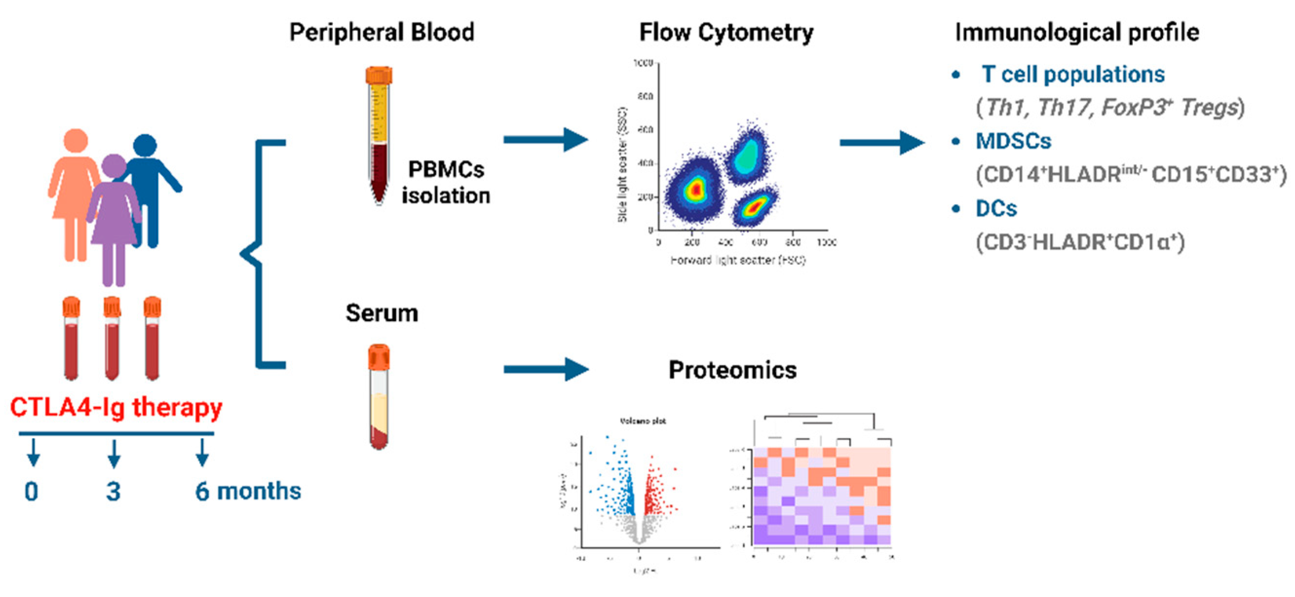
Figure 1.
Proteomics analysis of RA patients sera at baseline. (a) Heatmap of the 303 proteins that are identified at the serum from the proteomic analysis. (b) Bubble plot of enrichment analysis representing enriched pathways in the set of identified proteins. The color of the bubbles represents the statistical significance.
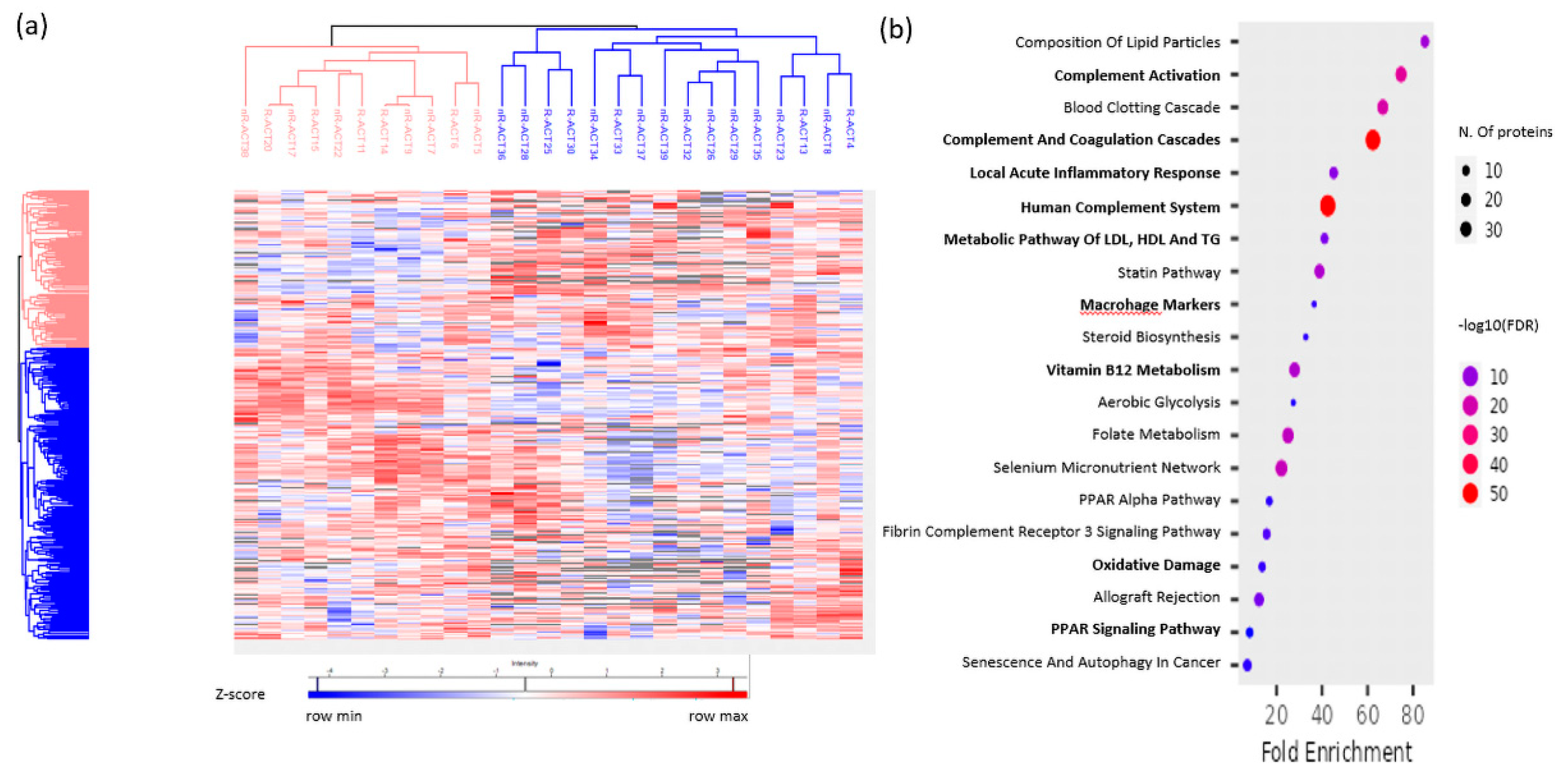
Figure 2.
(a) Gating strategy for the identification of PB IFN-γ and IL-17 expressing CD4+ T cell (Th1, Th17, respectively), CD4+CD127-CD25+FoxP3+ T cells (FoxP3+ T cells), CD14+HLADRint/-CD15+CD33+ cells (MDSCs) and CD3-HLADR+CD1α+ cells (DCs). (b) Frequencies of above-mentioned cell populations in the PB of patients at 0 and 6 months post treatment (**P-value=0.0087, ***P-value= 0.0004 and ***P-value= 0.0002, respectively). Patients n=27. Statistical significance was obtained by Wilcoxon matched-pairs test.
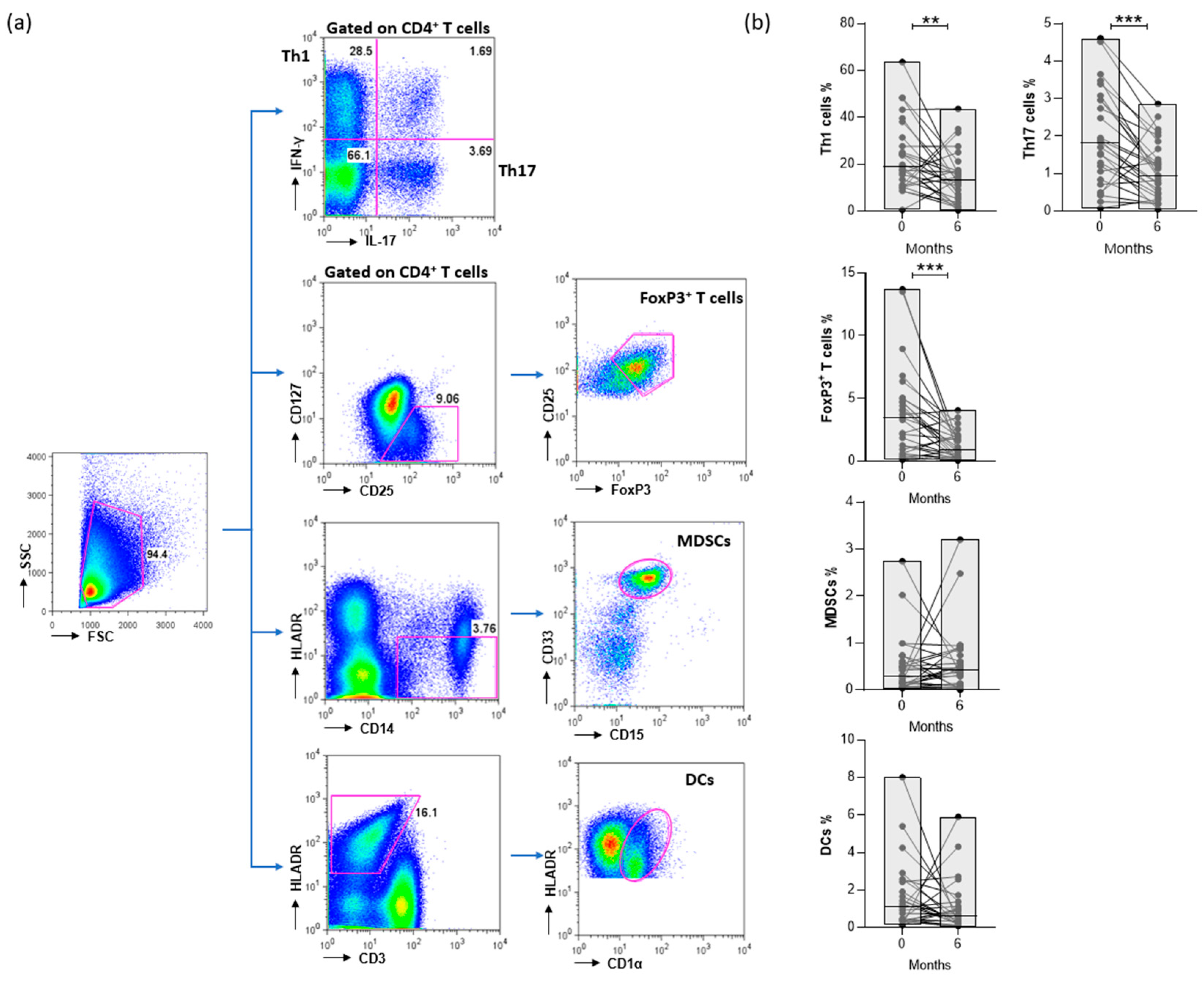
Figure 3.
Evaluation of Th1, Th17 and FoxP3+ T cells frequencies in the PB of RA patients in response to abatacept. Frequencies of (a) CD4+ T cells (b) Th1, (c) Th17 and (d) FoxP3+ T cells at baseline (0 months) of responders and non-responders at 6 months of abatacept therapy (*P-value=0.0224, **P-value=0.009). (e) Th1 (f) Th17 and (g) FoxP3+ T cells percentages in the PB of patients at 0, 3 and 6 months post treatment (*P-value=0.0277 and **P-value= 0.0042). Classification of patients (responders or non-responders) based on the “Swollen joints”, 6 months after abatacept. RA patients with “Swollen joints”≤2 belong to low disease activity group (responders). Responders n=9, non-responders n=18. Statistical significance was obtained by un-paired t test and one-way ANOVA.
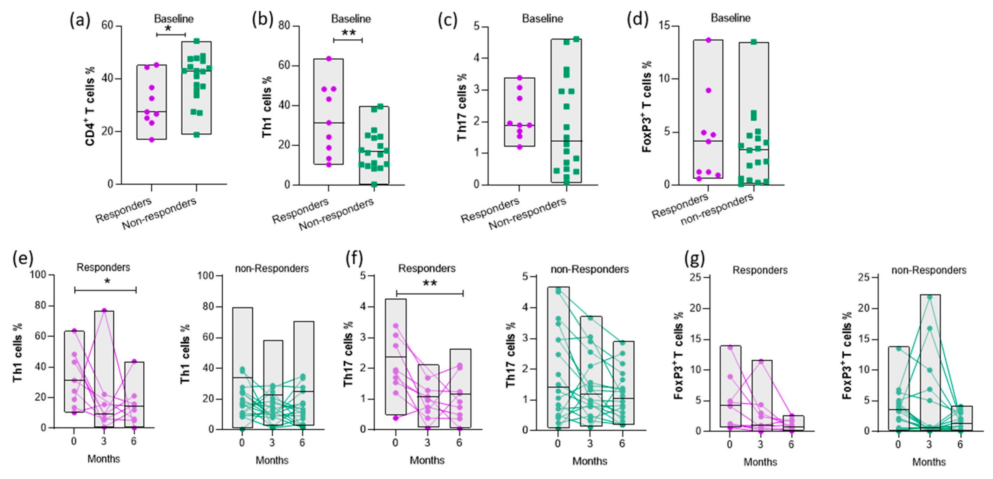
Figure 4.
Evaluation of myeloid cell frequencies in the PB of RA patients in response to abatacept. Frequencies of MDSCs in the PB of patients at (a) baseline, (b) 3 and 6 months post treatment (*P-value=0.0322). Responders n=9, non-responders n=18. (c) Frequencies of DCs at baseline (0 months) and (d) 6 months of responders and non-responders (*P-value=0.0339 and 0.0438, respectively). (e) Frequencies of DCs in the PB of patients at 0, 3 and 6 months post treatment. Responders n=8, non-responders n=18. Statistical significance was obtained by unpaired t test.
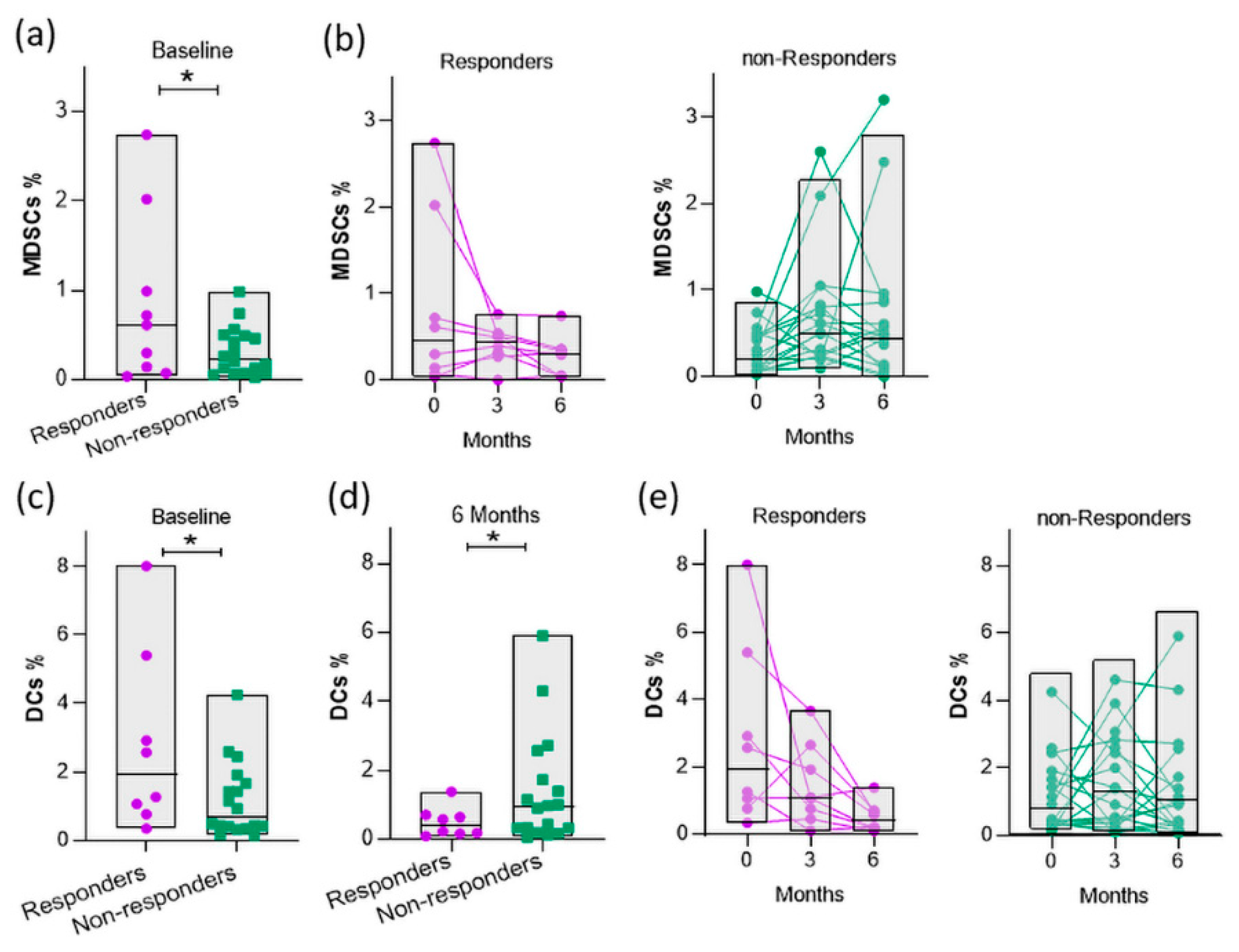
Figure 5.
Proteomics analysis of sera, at baseline, of responders vs non- responders to abatacept. (a) Volcano plots of the differential expression analysis comparing responders vs non-responders. Upregulated proteins are denoted by red and downregulated by blue. Proteins not reaching our significance threshold (P-value <0.05) depicted in gray. (b) Enrichment analysis of Kegg pathways for differentially expressed proteins of responders vs non-responders in 303 proteins background, sorted by P-value ranking. Color gradient correlates with statistical significance. Pathways not reaching significance threshold (P-value <0.05) are shown in gray.
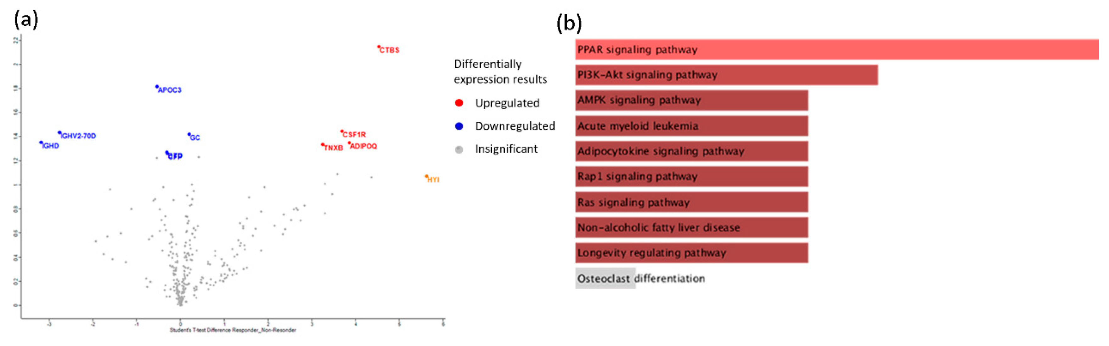

Table 1.
Clinical characteristics of patients.
| All | Responders | Non-responders | |
|---|---|---|---|
| Patients (n) | 29 | 10 | 19 |
| Gender (% females) | 25 (86.2%) | 7 (70.0%) | 18 (94.7%) |
| Age,Median (min,max;IQR) | 64 (32,77;17) | 58 (32,71;20) | 67 (40,77;18) |
| Disease duration,Median (min,max;IQR) | 23 (3,240;45) | 32.5 (8,120;50) | 22 (3,240;40) |
| R.F. positive (%) | 8 (27.6%) | 2 (20.0%) | 6 (31.6%) |
| Anti-CCP positive (%) | 15 (51.7%) | 5 (50.0%) | 10 (52.6%) |
| Methotrexate (%) | 26 (89.65%) | 9 (90%) | 17 (89.47%) |
| Steroids (%) | 10 (34.48%) | 4 (40%) | 6 (31.57%) |
| Disease characteristics before abatacept therapy (Baseline) | |||
| ESR,Median (min,max;IQR) | 26 (4,77;34) | 25.5 (4,69;34) | 27 (4,77;40) |
| CRP,Median (min,max;IQR) | 0.33 (0.24,6.03;0.53) | 0,52 (0.31,3.61;0.70) | 0.33 (0.24,6.03;0.53) |
| Swollen 28,Median (min,max;IQR) | 6 (2,18;5) | 9 (3,15;5) | 6 (2,18;4) |
| Tender 28,Median (min,max;IQR) | 7 (0,23;6) | 9 (2,23;8) | 6 (0,20;6) |
| VAS global,Median (min,max;IQR) | 70 (40,100;20) | 70 (40,100;60) | 60 (40,100;20) |
| DAS28-ESR,Median (min,max;IQR) | 5.48 (3.42,6.85; 1.90) | 5.91 (3.42,6.79;1.89) | 4.63 (3.94,6.85;1.89) |
| Disease characteristics after abatacept therapy (6 months) | |||
| ESR,Median (min,max;IQR) | 28 (6,68,21) | 24.5 (8,68;22) | 31 (6,59;18) |
| CRP,Median (min,max;IQR) | 0.36 (0.10,3.01;0.27) | 0.36 (0.10,1.45;0.18) | 0.36 (0.30,3.01;0.30) |
| Swollen 28,Median (min,max;IQR) | 4 (0,17;6) | 1.5 (0,2;2) | 6 (3,17;8) |
| Tender 28,Median (min,max;IQR) | 2 (0,27;6) | 1 (0,4;2) | 3 (0,27;9) |
| VAS global,Median (min,max;IQR) | 30 (0,100;35) | 30 (0,60;23) | 40 (10,100;50) |
| DAS28-ESR,Median (min,max;IQR) | 4.20 (2.22,8.04;2.13) | 2.93 (2.22,4.61;1.80) | 4.67 (2.85,8.04;2.05) |
| ΔDAS28,Median (min,max;IQR) | 1.30 (-1.84, 4.30; 2.61) | 2.42 (0.16, 4.30;2.48) | 0.48 (-1.84,3.44;2.76) |
| All values are medians (IQR) unless otherwise specified. R.F.: Rheumatoid Factor; anti-CCP: Anti-cyclic Citrullinated Peptide; ESR: Erythrocyte Sedimentation Rate; CRP: C-reactive protein; VAS: Visual Analogue Scale; DAS28: Disease activity score using 28 joints | |||
Table 2.
Baseline association of cell populations with clinical characteristics.
| TH1 % | TH17 % | FoxP3 % | MDSCs % | DCs % | |
|---|---|---|---|---|---|
| Mean (SD) | Mean (SD) | Mean (SD) | Mean (SD) | Mean (SD) | |
| Seropositive | |||||
| No | 27.9 (13.4) | 2.32 (1.43) | 4.58 (4.40) | 0.25 (0.22) | 1.18 (1.19) |
| Yes | 18.8 (15.8) | 1.62 (1.08) | 3.05 (2.43) | 0.77 (0.20) | 2.07 (2.21) |
| P-value | 0.120 | 0.167 | 0.273 | 0.050 | 0.224 |
| ESR (P-value) | -0.237 (0.233) | 0.028 (0.890) | -0.190 (0.343) | -0.143 (0.476) | -0.202 (0.321) |
| CRP (P-value) | 0.083 (0.681) | -0.087 (0.271) | -0.125 (0.535) | 0.261 (0.188) | 0.081 (0.692) |
| Swollen 28 (P-value) | 0.346 (0.077) | -0.220 (0.271) | -0.051 (0.799) | -0.151 (0.452) | -0.124 (0.547) |
| Tender 28 (P-value) | 0.386* (0.047) | 0.173 (0.389) | 0.440* (0.022) | -0.348 (0.075) | -0.133 (0.518) |
| VAS global (P-value) | -0.102 (0.614) | 0.097 (0.631) | 0.085 (0.674) | -0.274 (0.167) | -0.271 (0.180) |
| DAS28-ESR (P-value) | 0.184 (0.359) | 0.195 (0.329) | 0.272 (0.169) | -0.363 (0.063) | -0.204 (0.317) |
| Independent samples T-test (comparison of means) was performed on the upper part of the table and Spearman's correlation coefficient on the lower part. *Significant associations | |||||
Table 3.
Description of the 10 differentially expressed proteins at baseline in responders compared to non-responders.
Table 3.
Description of the 10 differentially expressed proteins at baseline in responders compared to non-responders.
| Protein Name | Ratio Responders/ non-Responders | P-value | Function |
|---|---|---|---|
| CSF1R (Macrophage colony-stimulating factor 1 receptor) | 2,21997 | 1,4471 | Biological regulation; cell differentiation |
| GC (Vitamin D-binding protein) | 2,19503 | 1,42407 | Lipid metabolic process |
| ADIPOQ (Adiponectin) | 2,11482 | 1,35079 | Adiponectin-mediated signaling pathway |
| TNXB (Tenascin-X) | 2,09943 | 1,33687 | Actin cytoskeleton organization |
| CTBS (Chitobiase, Di-N-Acetyl-) | 2,93148 | 2,14787 | Amine metabolic process |
| APOC3 (Apolipoprotein C-III) | -2,60697 | 1,81868 | Acylglycerol metabolic process |
| IGHV2-70D (Immunoglobulin heavy variable 2-70D) | -2,20792 | 1,43596 | Immune response |
| IGHD (Immunoglobulin heavy constant delta) | -2,11844 | 1,35407 | Immune response |
| BTD (Biotinidase) | -2,02902 | 1,27382 | Metabolic process |
| CFP (Complement factor properdin) | -2,01735 | 1,26348 | Activation of immune response |
Disclaimer/Publisher’s Note: The statements, opinions and data contained in all publications are solely those of the individual author(s) and contributor(s) and not of MDPI and/or the editor(s). MDPI and/or the editor(s) disclaim responsibility for any injury to people or property resulting from any ideas, methods, instructions or products referred to in the content. |
© 2023 by the authors. Licensee MDPI, Basel, Switzerland. This article is an open access article distributed under the terms and conditions of the Creative Commons Attribution (CC BY) license (http://creativecommons.org/licenses/by/4.0/).
Copyright: This open access article is published under a Creative Commons CC BY 4.0 license, which permit the free download, distribution, and reuse, provided that the author and preprint are cited in any reuse.


