
Submitted:
11 October 2023
Posted:
13 October 2023
You are already at the latest version
Abstract
The class I carcinogen aflatoxin B1 (AFB1) poses a significant risk to human health. The contamination of food sources contributes to the spread of AFB1, mainly through grains and nuts, from Aspergillus flavus. The high percentage of aflatoxin contamination in various imported nuts in the Republic of Kazakhstan has encouraged researchers to develop a local method of AFB1 detoxification. This research was focused on studying the ability of Ziziphora serpyllace leaf extract growing in the Akmola region of the Republic of Kazakhstan to inactivate aflatoxin B1 in various nuts. For the first time, an extract of Ziziphora serpyllace leaves growing in the territory of the Republic of Kazakhstan was used to detoxify AFB1 in nuts, and this demonstrated low cytotoxic activity, high antimicrobial and high antifungal activity, while preserving the qualitative characteristics of the nuts.
Keywords:
various nuts
; mycotoxins
; aflatoxin B1
; detoxification
; Ziziphora serpyllacea
1. Introduction
AFB1 poses a greater threat compared to other substances, primarily because of its potent carcinogenic, liver-damaging, birth-defect-causing, and immune-system-weakening impact on humans. It has earned a Group 1 carcinogen classification from the International Agency for Research on Cancer (IARC) [1].
According to ReportLinker Research estimations, global nut production will reach 22,550 thousand metric tons by 2026 from 20,930 thousand metric tons, and it is expected to grow by 1% per year. In this rating, the USA, Vietnam, and India ranked second, third, and fourth, respectively [2].
The presence of AFT pollution in numerous cases has raised apprehensions about international trade. This concern is exemplified by notifications issued by the European Union (EU) in their RASFF Annual Report of 2016, indicating instances where the allowable level of B1 in peanuts was exceeded. In light of the increasing awareness of serious health risks associated with AF, the EU has implemented stringent regulations stipulating that a product intended for human consumption must not contain more than 2 μg/kg of B1 and the total AF content must not exceed 4 μg/kg (as per EC 1881/2006). Although the Codex Alimentarius has established a Maximum Accepted Concentration (MAC) for AFB at 15 μg/kg (CODEX STAN 193-1995), the Food Safety Standards Authority of India has set it at 10 μg/kg for ready-to-eat nuts, 0.004 mg/kg in accordance with EU Commission Regulation No. 1881-2006, and 15 μg/kg for nuts [3].
A comprehensive search of scientific databases was carried out, covering the period from 2000 to 2020. The findings revealed that aflatoxin B1 (AFB1) was most frequently detected in samples of various nuts. The average concentrations of total aflatoxin and AFB1 in different types of nuts were as follows: peanuts (37.85, 32.82 µg/kg), pistachios (31.42, 39.44 µg/kg), almonds (3.54, 3.93 µg/kg), walnuts (42.27, 22.23 µg/kg), hazelnuts (17.33, 10.54 µg/kg), Brazil nuts (4.61, 3.35 μg/kg), and other nuts (26.22, 7.38 μg/kg).
The permissible exposure limits (MOE—Margin of Exposure) for adults varied depending on the country, with the ranking as follows: Argentina (21), Congo (67), India (117), Bangladesh (175), Cameroon (238), Iran (357), Bahrain (438), Brazil (447), Ghana (606), South Africa (1017), Egypt (1176), USA (1505), China (1526), and Cyprus (1588) [4].
In key literature references, the authors describe many physical and chemical methods of detoxication [5,6,7,8,9,10,11,12,13,14,15].
In the past, numerous plant extracts have undergone scrutiny for their potential to hinder aflatoxin production, both in laboratory settings and within food items. Extracts from plants such as Equisetum arvense, Zingiber officinalis, Stevia rebaudiana Oxalis corniculata, and Trigonella foenum-graecum have been documented for their antifungal and anti-aflatoxigenic characteristics. Additionally, research by Yazdani and colleagues has indicated that Piper betle L. possesses the capability to decrease the production of aflatoxin by A. flavus.
In the study by Kalli V. and his colleagues, they assessed the risk to consumers before and after adding C. incanus to inoculated nuts. In the study, they examined the antiaflatoxigenic efficacy of the herbaceous plant Cistus incanus L. toward macadamia nuts. Cistus incanus L. methanol extract in macadamia nuts reduced AFB1 by 72.5–85.9%. In addition, the presence of Cistus incanus extract was found to be safe for consumers [16].
Chen F. applied water-soluble tea flower extracts (Camellia sinensis L.), demonstrating excellent antioxidant ability and fungal inhibition against A. flavus due to the presence of 2-ketomobutyric acid, which suppressed aflatoxin biosynthesis genes and reduced aflatoxin production. A food additive containing 2-ketomasolic acid is safe for use in the food industry [17].
The study investigated the antifungal and anti-aflatoxigenic properties of four different plant species against Aspergillus flavus and Aspergillus parasiticus. Essential oils and methanol extracts were extracted from the aerial parts of Lippia javanica, Ocimum gratissimum, Satureja punctata, and the stem bark of Toddalia asiatica through hydrodistillation and maceration methods.
Among these extracts, the essential oils from Satureja punctata and Lippia javanica exhibited the most potent anti-aflatoxigenic effects against the tested fungal strains. Following closely in effectiveness was the essential oil from Ocimum gratissimum, while the essential oil from Toddalia asiatica displayed a moderate level of anti-aflatoxigenic activity. Conversely, the methanol extract from Toddalia asiatica exhibited moderate antifungal activity compared to its essential oil counterpart [18].
AFB1 production and Aspergillus flavus growth were studied using an aqueous extract of Aloysia citrodora, and it was discovered that polyphenols are molecules that are involved in inhibiting AFB1, achieving 99% inhibition of toxin production. One of the primary components of A. citrodora extract was identified as luteolin-7-diglucuronide, which also showed evidence of its ability to inhibit the production of AFB1 by 57%. This is the first study to show that this molecule has an antiaflatoxin effect, and other polyphenols unquestionably affect A. citrodora’s ability to combat AFB1 [19].
Clove, celac, and cinnamon extracts are extracted at different concentrations (3, 5, 7, and 9%) and then UV-sterilized with cinamaldehyde (25.74 mg/g), eugenol (12.15 mg/g), and a combination of timol (14.49 mg/g) and carvacrol (1.17 mg/g). In three months, the aflatoxin content of pistachios without plant extract exceeded 1000 g/kg. However, phenols in each extract at 9% concentration can inhibit 100% aflatcosin in each contaminated pistachio variety and effectively destroy aflatoxins even after 14 days in a refrigerator [20].
According to the results of monitoring studies, it was concluded [21] that over the period from 2021 to 2022, the largest share of imports of various nuts was from the PRC—582,946 tons (74.6% of the total import volume). The main types of nuts are as follows: walnut (434,823 tons), almonds (11,688 tons), macadamia (1596 tons), cashews (344.5 tons), pistachios (119.7 tons), chestnuts (0.2 tons), and pecans (1063.9 tons). The volume of nut imports from the United States amounted to 157,029 tons (almonds—13 712 ton; pistachios—1985.3 tons; pecans—4.9 tons), from Russia amounted to 21,262 tons (peanuts—16,938). Uzbekistan is one of the leading importers of walnut—741.9 tons; almonds—274.9 tons. A major importer of pistachios is Iran (4858 tons), Turkey (11,378 tons, including almonds—594.8 tons; pistachios—454.1 tons). Also, Tajikistan (953.9 tons), Ukraine (438.2 tons), and Kyrgyzstan (342.3 tons).
In Kazakhstan, imported nuts are not tested for aflatoxin B1 in customs zones. When importing nuts, according to the rules of protection of the territory of the Republic of Kazakhstan from quarantine facilities and foreign species, an act of quarantine phytosanitary control is issued for imported nuts since nuts are quarantine products [22]. Sampling for analysis and examination (barn pests) is carried out by state plant quarantine inspectors. Unfortunately, we do not have proper control and verification of quality certificates.
According to our research results, it was concluded that nuts are imported from various countries (China, Iran, Turkey, etc.), which are exceeded when imported into the European Union. According to the results of our monitoring studies, there is a high percentage of contamination of imported nuts; thus, there is an urgent need to develop new, local methods of detoxification for the manufacture of products free from contamination.
In this regard, the scientific community is looking for new, natural biologically active compounds that may possibly act as suppressors of aflatoxins.
The purpose of this study was, for the first time, to evaluate the effect of Ziziphora serpyllacea extract growing in the Akmola region on the detoxification of AFB1 from various nuts with the determination of their qualitative indicators.
2. Materials and Methods
2.1. Sсope of Research
The following samples were selected for the research: unshelled peanuts (Uzbekistan), unshelled walnuts (PRC), and pistachios (Iran). In total, 15 samples were examined from the harvests of 2021 and 2022 in the markets of Astana, the Republic of Kazakhstan.
2.2. Ziziphora serpyllacea Plant (Thyme ziziphora)
Ziziphora serpyllacea (Thyme ziziphora) is a species of plant in the genus Ziziphora of the Mint family (Lamiaceae).
It is a subshrub, 5–30 cm tall, branching at the base, with simple or many-branched stems, woody at the base, and densely covered with trichomes curved backwards. The leaves are oval, slightly leather-like, pilose below, and smooth-edged. Petioles are lush and slightly hairy; their bract leaves resemble cauline but are smaller; and their lateral branches are erect-trampled, curved, or marginally wavy. Ziziphora serpyllacea blooms from late June to late August and grows in the Akmola region.
2.3. Preparation of Ziziphora serpyllacea Plant Extract
The first step was to prepare the ethanol extract of Ziziphora serpyllacea. To obtain it, 100 g of raw vegetable materials was taken, which was then dissolved in 70% ethyl alcohol. The extraction was carried out for 2 h at the solvent’s reflux temperature using an ultrasound bath dubbed “Sapphire.” The extracted substances were evaporated using an IKA RV 10 rotary evaporator.
2.4. Determination of the Cytotoxic Activity of Ethanol Extract
A total of 200 mg of Artemia salina eggs were added to 500 mL of seawater. They were kept at 250 °C while supplying air until nauplii hatched from eggs. One side of the tank was covered with aluminum foil, and 5 min later, the larvae that were collected on the illuminated side were removed with a dispenser.
A 24-well plate was taken; 12 wells were intended for samples in 4 concentrations (3 wells for each concentration), and 12 for the control test. A total of 20–30 larvae were placed in 990 μL of seawater in each of the 24 wells.
Using a micropipette, certain volumes (3, 7.5, 15, and 30 μL) of samples were transferred from the initial solutions to test tubes to obtain the finishings of samples 1, 2.5, 5, and 10 μg/mL, respectively. The concentration of dimethyl sulfoxide (DMSO) solvent in these test tubes did not exceed 10 μL/mL. Actinomycin D was used as the comparator agent. Only 10 μL of DMSO was added for negative control.
After 24 h of incubation, the wells were examined, the number of survivors was counted in each well, and the results were recorded. Based on this, the percentage of the case-fatality rate was calculated for each concentration and for each sample.
Case-fatality rate P was determined by the following formula:
A – N – B
P = (-------------) × 100
Z
A = Number of dead nauplii after 24 h;
N = Number of dead nauplii before the test;
В = Average number of dead nauplii in negative control;
Z = Total number of nauplii.
2.5. Determination of Antimicrobial and Antifungal Activity
The following cultures were used to determine the antibacterial activity of Zizíphora serpyllácea extract: Staphylococcus aureus, Escherichia coli, Serratia marcescens, Candida albicans, and aspergillus flavus.
The antimicrobial activity of 5%, 10%, and 20% concentrations was tested on reference test microorganisms: facultative anaerobic Gram-positive cocci. Aerobic Gram-positive sporogenous rods, Gram-negative rods, Staphylococcus aureus ATCC 6538 Aerobic diffusion of the facultative anaerobes Escherichia coli ATCC 25922 and the thrush fungus Candida albicans ATCC 10231 into agar (wells). The American Type Culture Collection provided the microbial test strains of microorganisms used in the study.
Comparator agents are gentamicin for Staphylococcus aureus, Escherichia coli, and Serratia marcescens and nystatin for thrush fungi, Candida albicans, and Aspergillus flavus.
Pure cultures of test strains were taken for the study, which had previously grown on a fluid medium pH 7.3 0.2 at 30 to 370 °C for 24–48 h on a slant agar–meat infusion. Diluting a 1:1000 culture in a sterile solvent yielded a standard bacterial suspension.
For the test strains under study, 1.0 mL of the corresponding bacterial suspension was added to the plates with the corresponding elective, nutrient media, and inoculated using the “solid lawn” method. After drying, 50 L solutions of the test extracts in three concentrations (5%, 10%, and 20%) were introduced into 8.0 mm wells formed on the agar surface. In the control, water for injection was used to dilute samples in equi-volume amounts. The bacterium was incubated at 370 °C for 24 h, and the thrush fungi Candida albicans and Aspergillus flavus were incubated at 300 °C for 48 h.
The diameter of the inhibition zones of the test strains (mm) around the well, including the diameter of the well itself, was used to assess the antimicrobial activity of the samples. Absence of inhibition zone—the test culture is not sensitive to this sample concentration; diameter of inhibition zones is less than 10 mm, and confluent growth in the plate was evaluated as lack of antibacterial activity, 10–15 mm—weak activity, 15–20 mm—moderate activity, and more than 20 mm—severe activity. In three parallel experiments, each sample was tested at concentrations of 5, 10, 15, and 20, respectively. Antimicrobial solutions such as benzylpenicillin sodium salt, gentamicin, and nistatin were used to compare antimicrobial activity.
2.6. Aspergillus flavus Strain Cultivation and Nut Contamination
Aspergillus flavus strain provided by KWIK- STIKTM (France) was cultured on Chapek’s slant agar (HiMedia, India) in the thermostat at 28 °C for five days. This process provided optimal conditions for the growth and development of the microorganism.
The nuts were contaminated with a pure strain of Aspergillus flavus. This was achieved by diluting the strain culture in an aqueous solution, after which the solution was evenly distributed throughout the utensils to the surface of the nuts. After contamination, the nuts were cultured in a thermostat at 37 °C for 14 days until visible mold formed. The average concentration was 0.003 to 0.004 mg/kg.
Zizíphora serpyllácea leaf extract reacted with aflatoxin B1. A total of 500 μm of each extract was mixed with 100 μm of aflatoxin B1. After that, samples were kept at 370 °C in a thermostat for 48 h. The detoxification of aflatoxin B1 was then evaluated using high-performance liquid chromatography (HPLC). The HPLC method separates samples into components and enables their detection.
Next, a second method of detoxifying AFB1 was carried out. A dry extract of Zizíphora serpyllácea obtained after the removal of the solvent (ethanol) was dissolved in deionized water. Samples with extracts of Zizíphora serpyllácea plants were kept for 3 h at 250 °C and then for 30 min at 250 °C. They were then dried in a drying chamber at 1000 °C until complete dewatering.
2.7. Detection of Aflatoxin B1 by HPLC
To determine the aflatoxin content of B₁ in nuts, high-performance liquid chromatography was used according to all-Union State Standard 30711-2001 using the HPLC equipment of Water Corporation (USA) at Nutritest LLP at the Sharmanov Nutrition Academy in Almaty. The performers worked in accordance with the safety measures when working with methanol (certificate No. AS 568/1, No. AS 568/2, No. AS 567/1).
2.8. Organoleptic and Physical–Chemical Studies
In the organoleptic examination of the nuts, the appearance, color of the shell, size, surface, color, quality, taste, and smell of the kernel were specified. Physical–chemical studies of the nut included the following: the percentage of pests damaged, underdeveloped, and broken nuts; the presence of live pests and larvae; and the amount of moisture. The moisture content was determined by sequential drying at a temperature of (130 ± 2) °C for 40 min in a drying chamber (all-Union State Standard 32874-2014. Walnut nuts. Specification, all-Union State Standard 31788-2012. Unshelled pistachio nuts, unpeeled. Specification, all-Union State Standard 31788-2012. Peanut. Specification).
3. Results and Discussion
3.1. Determination of Cytotoxic Activity
The ethanol extract of Ziziphara serpyllacea showed low cytotoxic activity in concentrations of 10 mg/mL, 5 mg/mL, 2.5 mg/mL, and 1 mg/mL. Table 1 shows the results of exposure to 10 mg/mL, 5 mg/mL, 2.5 mg/mL, and 1 mg/mL% of surviving samples reached 96.5%.
3.2. Determination of Antimicrobial and Antifungal Activity
As a result of the antimicrobial study, it was found that the extract in concentrations of 20% showed moderate antibacterial activity to the ratio of the Gram-negative test strain Escherichia coli ATCC 25922, which minimally suppressed the concentration. At a concentration of 10%, the extracts showed weak antimicrobial activity.
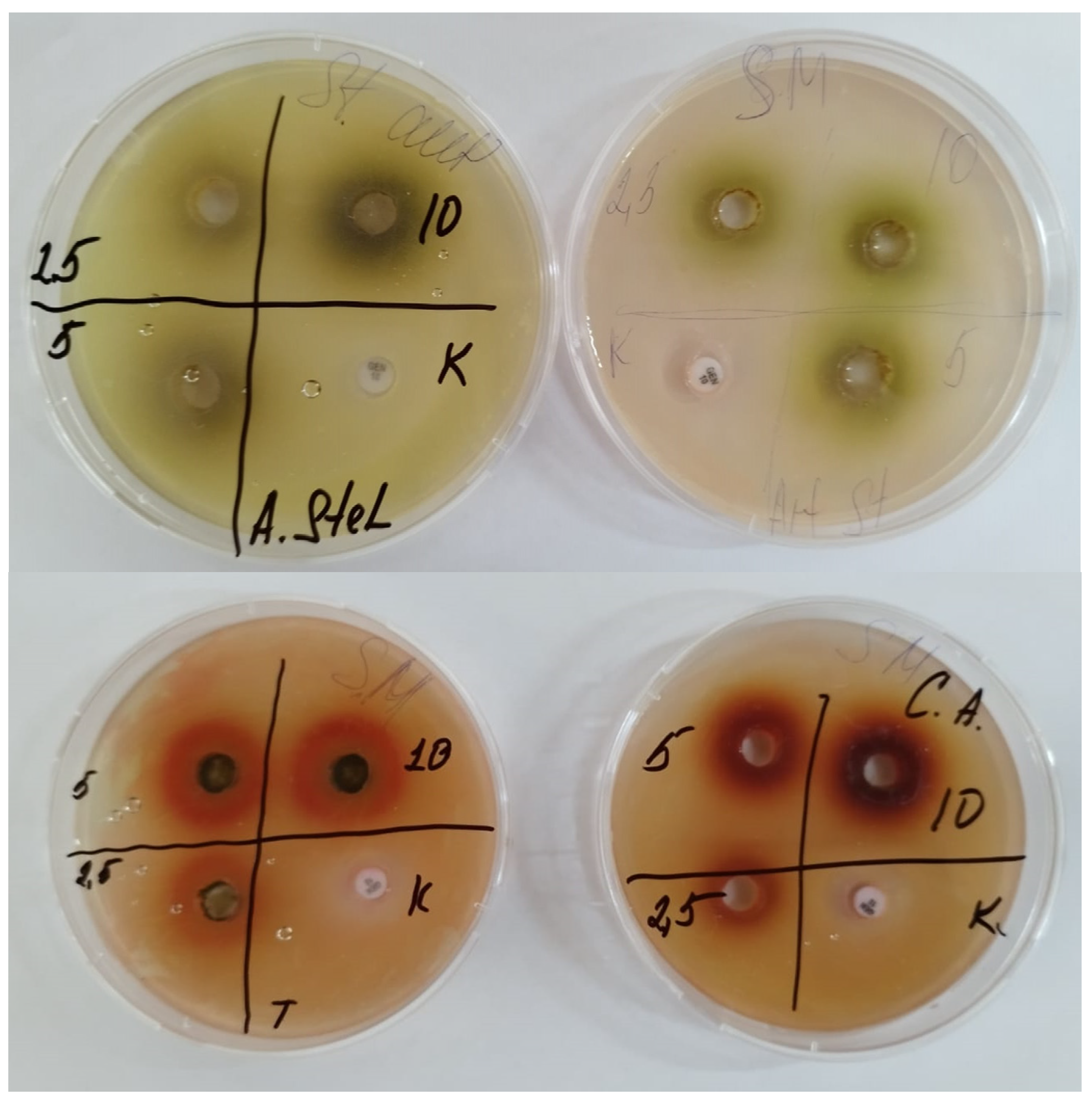
Zizíphora serpyllácea extract showed high antimicrobial and antifungal activity (Figure 1).
The results of the antimicrobial activity study of Ziziphora serpyllacea extracts are shown in Table 2.
Figure 2.
Chromatograms obtained by HPLC analysis illustrate the AFB1 detoxification of various contaminated nuts.
Figure 2.
Chromatograms obtained by HPLC analysis illustrate the AFB1 detoxification of various contaminated nuts.
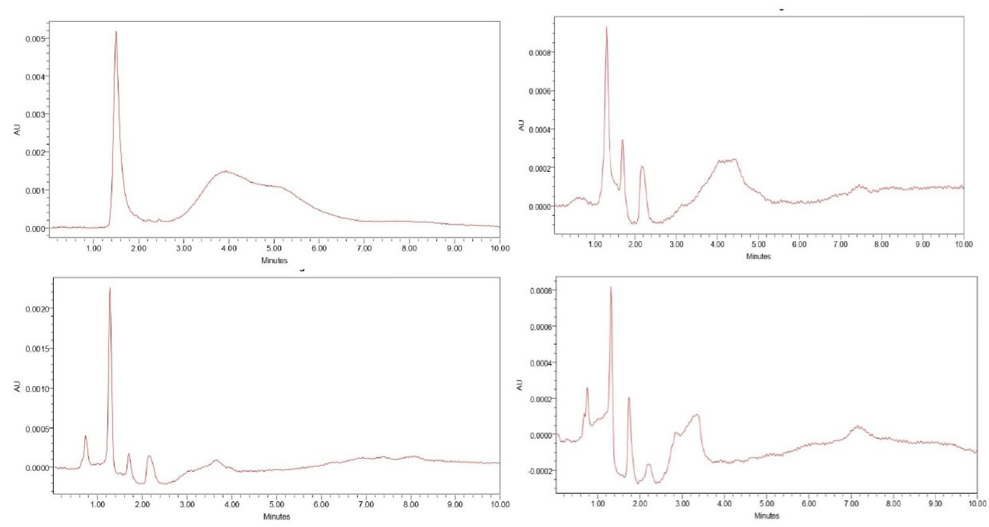
Figure 3.
Antimicrobial activity of Ziziphora serpyllacea extracts diffusion methods into agar.
The results obtained from the HPLC spectra are consistent with the results of the antifungal activity of the extract.
Figure 4 shows no peak of aflatoxin-specific B1, which is also consistent with the data (Table 2) on the high antifungal activity of Zizíphora serpyllácea extract.
The chromatograms presented in Figure 4 showed the effect of the extract on samples of nuts contaminated with AFB1. No peak AFB1 was detected in the chromatograms.
The initial contamination of the studied nuts ranges from 0.002 to 0.004 mg/kg. The concentration of AFB1 in the test samples after detoxification decreased to 0. Several types of studies of traditional aflatoxin detoxification methods are available in the literature. However, the end point in previous studies was only the degradation of AFB1, but the detoxified product was not explained.
The detoxification process of Ziziphora serpyllacea leaf extract is associated with high levels of antimicrobial and antifungal activity. In foreign sources, the leaves are described under the name of Zizipora clinopodioides, which are native to the territories of other countries. We first used the extract of Ziziphora serpyllacea leaves native to our country.
Zizipora clinopodioides contains pulegone (31.21%), menth-3-en-8-01 (23.82%), menthol (7.21%), borneol (2.25%), carvacrol (5.38%), and piperitone (5.55%).
Essential oils of this plant are rich in compounds such as carvacrol; as is widely known, they have high levels of antimicrobial activity [22].
There were 27 different essential oil compounds identified in Z. clinopodioides, with pulegone (23%) and 1.8 cineol (20.3%) being the main components of the essential oil. The findings revealed that Z. clinopodioid essential oils had greater chemical diversity and antibacterial activity against P. syringe pv. syringae [23].
Aflatoxin production was inhibited at 0.25 mg/mL and caused a significant reduction in AFB1—by 94.2%. The main components of the oil were pulegon (37%) in Z. clinopodioides. When using the broth microdilution test, the activity of Z. clinopodioides (MIC 90: 2.1; MFC: 5.5 mg/mL) oil against A. parasiticus oils [24,25].
Among the essential oils tested, those derived from Ziziphora clinopodioides and Thymus vulgaris exhibited the highest levels of antioxidant activity. On the other hand, the essential oils extracted from garden thyme demonstrated the most potent antibacterial activity, characterized by the broadest inhibition zone and the lowest minimum inhibitory concentration (MIC) value, which was measured at 2.5 μL/mL [26].
Ziziphora clinopodioides Lam contains many metal elements, among which there is a relatively large amount of calcium, potassium, sodium, and magnesium. It was noted that the level of essential elements was high compared to the level of toxic elements [27].
The study showed that the EM of two Ziziphora species, due to their main components, such as pulegon, mentone, and limonene, as well as a high content of phenols, can be considered good sources of natural antioxidants and antibacterial substances that have high potential for use in medicine and food science [28].
Essential oil was tested for minimum inhibitory concentration (MIC) and minimum fungicidal concentration (MFC) at ten different concentrations (25,000, 12,500, 6250, 3125, 1562.5, 781.25, 390.625, 195.31, 97.65, and 48.82 g/mL). Finally, the effect of essential oil at six concentrations (6250, 3125, 1600, 800, 400, and 196 g/mL) on the growth and activity of A. flavus and A. parasiticus, as well as the production of toxins by these species, was studied in corn at 0.97 a (w) and 25 °C for 29 days. The current study’s findings revealed that the essential oil had significant antifungal activity (p 0.05); thus, it can be used as an antifungal agent in the food and medical industries [29].
These results are consistent with the findings of Hassani F [30] and Makari et al. The oils of Ziziphora cliniopodiodes and Thymus vulgaris affected fungal pathogens, including Aspergillus flavus. According to the results of MIC and MMC, the lowest concentrations obtained against bacteria were in the essential oil of Z. clinopodioides.
According to organoleptic indicators, the appearance of all the nutshells is light brown, without spots. The kernels are light brown without visible defects, dry without impurities, have a well-formed shell, and the smell and taste are quite intense and correspond to the kind of nut. The shell is not damaged and is in good condition, from beige to light brown.
The moisture content is one of the important parameters characterizing the quality of walnut. The moisture content varies from 7.21% to 10%. Note that these values did not exceed the maximum allowable value of 12%.
4. Conclusions
For the first time, the present study has examined the effect of Zizíphora serpyllácea extract grown in the Republic of Kazakhstan on the reduction in AFB1 concentrations and the qualitative characteristics of various nuts (unshelled walnuts, peanuts, and pistachios).
The results showed that the extract completely detoxifies AFB1 in nuts with the preservation of qualitative indicators.
Funding
The studies were carried out under the scope of the budget project: 217 “Development of Science” of the Ministry of Science and Higher Education of the Republic of Kazakhstan URN No. AP09058301 “Aflatoxin contamination of various nuts and the development of methods for their detoxification”.
Author’s Contribution
Conceptualization: A.L.T.; methodology: M.B.S.; provision, high availability: A.L.T.; validation: Smagulova A.S.; formal analysis: S.A.S.; examination: A.L.T., M.B.S. and S.A.S.; letter—A.L.T. and S.A.S.; writing—A.L.T., M.B.S., and S.A.S.; visualization: S.A.S.; supervision: M.B.S.; the administration of the project: A.L.T.; funding attaining: M.B.S. All authors read and agreed with the published version of the manuscript.
Data Availability Statement
The datasets created for this study are available upon request to the corresponding author.
Acknowledgments
Non-profit joint-stock company “Saken Seifullin Kazakh Agro Technical Research University” expresses gratitude to LLP “Novolife Company”—Professor Khachatryan’s clinic of hydrogen medicine or support during the implementation of the Project.
Conflicts of Interest
The authors declare no conflict of interest.
References
- EC. Commission Regulation (EU) 2023/915 of 25 April 2023 on maximum levels for certain contaminants in food and repealing Regulation (EC) No 1881/2006. Off. J. Eur. Union 2023, L119, 103–157. [Google Scholar]
- ReportLinker. Available online: https://www.reportlinker.com/dataset/bc3b564024b37960eec671aa48900b4f201cf05c (2023).
- Dhanshetty, M.; Elliott, C.T.; Banerjee, K. Decontamination of aflatoxin B1 in peanuts using various cooking methods. J. Food Sci. Technol. 2021, 58, 2547–2554. [Google Scholar] [CrossRef]
- Marasas, W.F.O.; Gelderblom, W.C.A.; Shephard, G.S.; Vismer, H.F. Mycotoxins: A global problem. Mycotoxins: Detection Methods, Management, Public Health and Agricultural Trade, 4th ed; CABI: Oxford, UK, 2008; pp. 29–39. [Google Scholar]
- Diao, E.; Li, X.; Zhang, Z.; Ma, W.; Ji, N.; Dong, H. Ultraviolet irradiation detoxification of aflatoxins. Trends Food Sci. Amp. Technol. 2015, 42, 64–69. [Google Scholar] [CrossRef]
- Womack, E.D.; Brown, A.E.; Sparks, D.L. A recent review of non-biological remediation of aflatoxin-contaminated crops. J. Sci. Food Agric. 2014, 94, 1706–1714. [Google Scholar] [CrossRef]
- Méndez-Albores, A.; Veles-Medina, J.; Urbina-Álvarez, E.; Martínez-Bustos, F.; Moreno-Martínez, E. Effect of citric acid on aflatoxin degradation and on functional and textural properties of extruded sorghum. Anim. Feed Sci. Technol. 2009, 150, 316–329. [Google Scholar] [CrossRef]
- Jalili, M.; Jinap, S. Role of sodium hydrosulphite and pressure on the reduction of aflatoxins and ochratoxin A in black pepper. Food Control. 2012, 11–15. [Google Scholar] [CrossRef]
- Yang, B.; Li, L.; Geng, H.; Xing, F.; Liu, Y. Detoxification of aflatoxin B1 by H2SO3 during maize wet processing, and toxicity assessmen to fthetrans formation product of aflatoxin B1. Food Control 2022, 131, 108444. [Google Scholar] [CrossRef]
- Thiesen, J. Detoxification of aflatoxins in groundnut meal. Anim. Feed. Sci. Technol. 1977, 2, 67–75. [Google Scholar] [CrossRef]
- Lee, J.; Her, J.Y.; Lee, K.G. Reduction of aflatoxins (B (1), B (2), G (1), and G (2)) in soybean-based model systems. Food Chem. 2015, 189, 45–51. [Google Scholar] [CrossRef] [PubMed]
- Da Silva, M.; Moraes, A.M.L.; Nishikawa, M.M.; Gatti, M.J.A.; de Alencar, M.V.; Brandão, L.E.; Nóbrega, A. Inactivation of fungi from deteriorated paper materials by radiation. Int. Biodeterior. Biodegrad. 2006, 57, 163–167. [Google Scholar] [CrossRef]
- Oluwafemi, F.; Kumar, M.; Bandyopadhyay, R.; Ogunbanwo, T.; Ayanwande, K.B. Bio-detoxification of aflatoxin B1 in artificially contaminated maize grains using lactic acid bacteria. Toxin Rev. 2010, 29, 115–122. [Google Scholar] [CrossRef]
- Kalli, V.; Kollia, E.; Roidaki, A.; Proestos, C.; Markaki, P. Cistus incanus L. extract inhibits aflatoxin B1 production by Aspergillus parasiticus in macadamia nuts. Ind. Crops Prod. 2018, 111, 63–68. [Google Scholar] [CrossRef]
- Chen, F.; Chen, Y.P.; Wu, H.; Li, Y.; Zhang, S.; Ke, J.; Yao, J.Y. Characterization of tea (Camellia sinensis L.) flower extract and insights into its antifungal susceptibilities of Aspergillus flavus. BMC Complement. Med. Ther. 2023, 23, 286. [Google Scholar] [CrossRef] [PubMed]
- Kaale, L.D. Comparing the effects of essential oils and methanolic extracts on the inhibition of Aspergillus flavus and Aspergillus parasiticus growth and production of aflatoxins. Mycotoxin Res. 2023, 39, 1–13. [Google Scholar] [CrossRef]
- Cadenillas, L.F.; Hernandez, C.; Bailly, S.; Billerach, G.; Durrieu, V.; Bailly, J.D. Role of Polyphenols from the Aqueous Extract of Aloysia citrodora in the Inhibition of Aflatoxin B1 Synthesis in Aspergillus flavus. Molecules 2023, 28, 5123. [Google Scholar] [CrossRef] [PubMed]
- Somai, B.M.; Belewa, V. Aqueous extracts of Tulbaghia violacea inhibit germination of Aspergillus flavus and Aspergillus parasiticus conidia. J. Food Prot. 2011, 74, 1007–1011. [Google Scholar] [CrossRef]
- Agency for Strategic Planning and Reforms of the Republic of Kazakhstan Bureau of National. Available online: statisticshttps://stat.gov.kz/official/industry/31/statistic/6 (July, 2023).
- Adoption of Regulations on the Protection of the Territory of the Republic of Kazakhstan from Quarantine Facilities and Non-Indigenous Species. Available online: https://adilet.zan.kz/rus/docs/B1500012032 (2023).
- Khorasany, S.; Azizi, M.H.; Barzegar, M.; Hamidi, E.Z. A study on the chemical composition and antifungal activity of essential oil from Thymus caramanicus, Thymus daenensis and Ziziphora clinopodiaides. Nutr. Food Sci. Res. 2016, 3, 35–42. [Google Scholar] [CrossRef]
- Feizi, H.; Tahan, V.; Kariman, K. In vitro antibacterial activity of essential oils from Carum copticum and Ziziphora clinopodioides plants against the phytopathogen Pseudomonas syringae pv. syringae. Plant Biosyst. Int. J. Deal. All Asp. Plant Biol. 2023, 157, 487–492. [Google Scholar]
- Khosravi, A.R.; Shokri, H.; Minooeianhaghighi, M. Inhibition of aflatoxin production and growth of Aspergillus parasiticus by Cuminum cyminum, Ziziphora clinopodioides, and Nigella sativa essential oils. Foodborne Pathog. Dis. 2011, 8, 1275–1280. [Google Scholar] [CrossRef]
- Haiyan, X.; Wenhuan, D.; Shuge, T. Phytochemical diversity of Ziziphora clinopodioides Lam. in XinJiang, China. Res. J. Biotechnol. 2015, 10, 1–12. [Google Scholar]
- Aliakbarlu, J.; Shameli, F. In vitro antioxidant and antibacterial properties and total phenolic contents of essential oils from Thymus vulgaris, T. kotschyanus, Ziziphora tenuior and Z. clinopodioides. Turk. J. Biochem. /Turk Biyokim. Derg. 2013, 38, 425–431. [Google Scholar] [CrossRef]
- Masrournia, M.; Shams, A. Elemental determination and essential oil composition of Ziziphora clinopodioides and consideration of its antibacterial effects. Asian J. Chem. 2013, 25, 6553. [Google Scholar] [CrossRef]
- Hazrati, S.; Govahi, M.; Sedaghat, M.; Kashkooli, A.B. A comparative study of essential oil profile, antibacterial and antioxidant activities of two cultivated Ziziphora species (Z. clinopodioides and Z. tenuior). Ind. Crops Prod. 2020, 157, 112942. [Google Scholar] [CrossRef]
- Moghadam, H.D.; Sani, A.M.; Sangatash, M.M. Antifungal activity of essential oil of Ziziphora clinopodioides and the inhibition of aflatoxin B1 production in maize grain. Toxicol. Ind. Health 2016, 32, 493–499. [Google Scholar] [CrossRef]
- Alinezhad, M.; Hojjati, M.; Barzegar, H.; Shahbazi, S.; Askari, H. Effect of gamma irradiation on the physicochemical properties of pistachio (Pistacia vera L.) nuts. J. Food Meas. Charact. 2021, 15, 199–209. [Google Scholar] [CrossRef]
- Hassani, F.; Abyavi, T.; Taheri Mirghaed, A.; Payghan, R.; Alishahi, M. Evaluation of Antifungal and Antibacterial Activity of Essential Oils of Ziziphora clinopodioides, Thymus vulgaris and Salvia rosmarinus to Some Fungal and Bacterial Pathogens of Aquatic Animals. Exp. Anim. Biol. 2023, 11, 55–66. [Google Scholar]
Figure 1.
Antifungal activity of agar diffusion method extracts.
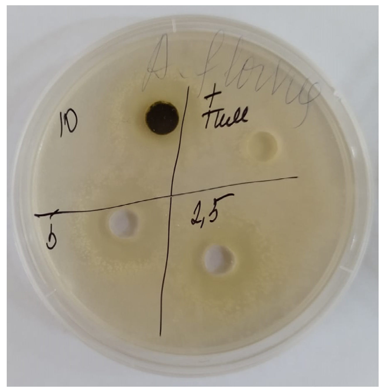
Figure 4.
HPLC spectrum of Zizíphora serpyllácea extract with AFB1.
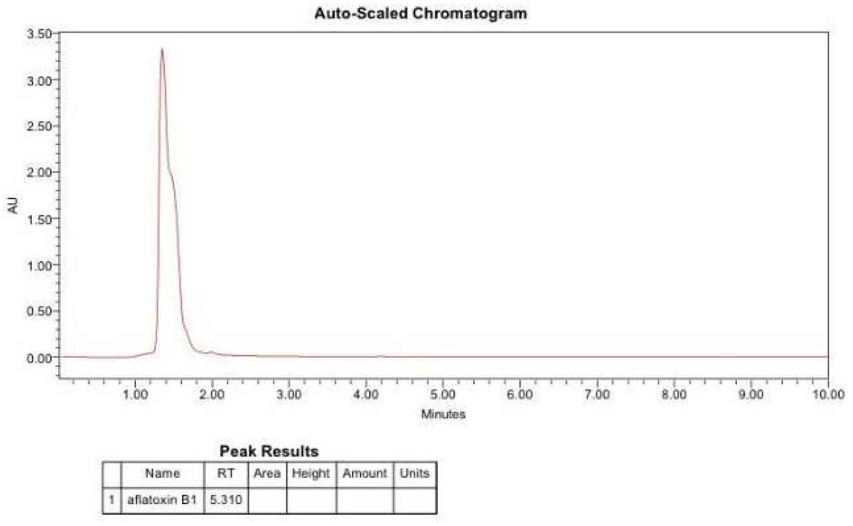

Table 1.
Cytotoxic activity of Ziziphara serpyllacea extract.
| Parallel | Amount of Nauplii in Monitoring | Amount of Nauplii in the Sample | % of Survived Nauplii in Monitoring | % of Survived Nauplii in the Sample | Case-Fatality Rate, А, %Survived | Neurotoxicity, %Died | |||
|---|---|---|---|---|---|---|---|---|---|
| Survived | Died | Survived | Died | par. | |||||
| Ziziphora serpyllácea in concentration of 10 mg/mL | |||||||||
| 1 | 20 | 1 | 20 | 2 | 0 | 95.4 | 85 | 15 | - |
| 2 | 21 | - | 20 | 2 | 0 | ||||
| 3 | 22 | 1 | 21 | 4 | 0 | ||||
| Avg. | 21 | 1 | 20 | 3 | 0 | ||||
| Ziziphora serpyllаcea in concentration of 5 mg/mL | |||||||||
| 1 | 23 | 1 | 26 | 1 | 0 | 95.4 | 87.5 | 29.1 | 12.5 |
| 2 | 20 | - | 25 | 2 | 0 | ||||
| 3 | 21 | 1 | 21 | 5 | 0 | ||||
| Avg. | 21 | 1 | 24 | 3 | 0 | ||||
| Ziziphora serpyllаcea in concentration 2.5 mg/mL | |||||||||
| 1 | 23 | 1 | 20 | 4 | 0 | 95.4 | 90.4 | 9.5 | 0 |
| 2 | 20 | - | 24 | 2 | 0 | ||||
| 3 | 21 | 1 | 20 | - | 0 | ||||
| Avg. | 21 | 1 | 21 | 2 | 0 | ||||
| Ziziphora serpyllаcea in concentration 1.0 mg/mL | |||||||||
| 1 | 23 | 1 | 25 | 2 | - | 95.4 | 95.6 | 4.3 | 0 |
| 2 | 20 | - | 22 | 0 | 0 | ||||
| 3 | 21 | 1 | 22 | 1 | 0 | ||||
| Avg. | 21 | 1 | 23 | 1 | 0 | ||||
Table 2.
Antimicrobial activity of extracts in concentrations of 5%, 10%, 15%, and 20%.
| Name | Antibacterial Activity, mm | Antifungal Activity, mm | ||||
|---|---|---|---|---|---|---|
| Sample code | % |
Staphylococcus aureus |
Sample Code | % |
Staphylococcus aureus |
Sample Code |
| Ziziphora serpyllacea | 5 | 11 ± 0.42 | 12 ± 0.4 | 14 ± 0.3 | 11 ± 0.51 | 12 ± 0.5 |
| 10 | 18 ± 0.55 | 17.5 ± 0.7 | 16 ± 0.38 | 15 ± 0.2 | 16 ± 0.5 | |
| 20 | 21 ± 0.54 | 20 ± 0.33 | 19.5 ± 0.4 | 19 ± 0.56 | 20 ± 0.6 | |
| Gentamycin | 24 ± 0.1 | 21 ± 0.2 | 26 ± 0.1 | - | ||
| Nystatin | - | - | - | 22 ± 0.1 | 21 ± 0.2 | |
Disclaimer/Publisher’s Note: The statements, opinions and data contained in all publications are solely those of the individual author(s) and contributor(s) and not of MDPI and/or the editor(s). MDPI and/or the editor(s) disclaim responsibility for any injury to people or property resulting from any ideas, methods, instructions or products referred to in the content. |
© 2023 by the authors. Licensee MDPI, Basel, Switzerland. This article is an open access article distributed under the terms and conditions of the Creative Commons Attribution (CC BY) license (http://creativecommons.org/licenses/by/4.0/).
Copyright: This open access article is published under a Creative Commons CC BY 4.0 license, which permit the free download, distribution, and reuse, provided that the author and preprint are cited in any reuse.


