Submitted:
27 September 2023
Posted:
28 September 2023
You are already at the latest version
Abstract
We investigate the structural correlation of non-covalent crown ether/H+/L-tryptophan (CR/TrpH+) host-guest complexes in the solution phase with those in the gas phase generated through electrospray ionization/mass spectrometry (ESI/MS) techniques. We perform quantum chemical calculations to determine their structures, relative Gibbs free energies, and infrared spectra. We compare the calculated infrared (IR) spectra with the IR multiphoton dissociation (IRMPD) spectra observed for the 18-Crown-6/TrpH+ complex by Polfer and co-workers [J. Phys. Chem. A 2013, 117, 1181−1188] for assigning the IR bands. We observe that the carboxyl group remains ‘naked’, lacking hydrogen bonding with the CR unit in the gas phase, and that this most stable conformer originates from the corresponding lowest Gibbs free energy structure in solution. Based on these findings, we propose that gas phase host-guest complexes directly correlate with those in solution, reinforcing the possibility of obtaining invaluable information about host-guest-solvent interactions in solution from the structure of the host-guest pair in the gas phase.
Keywords:
host-guest
; IRMPD
; crown ether
; tryptophan
1. Introduction
Determining the detailed molecular structures of host-guest complexes [1,2,3,4,5,6,7,8,9,10,11,12,13] and their interactions with solvent molecules in the solution phase can be a daunting task. While experimental techniques like NMR [14,15,16] or X-ray fluorescence [17,18,19] can provide insight into some aspects of host-guest complexes in solution, the unambiguous elucidation of interactions among multiple functional groups in the guest and host, as well as those with solvent molecules, remains challenging. The challenge is exemplified by the scarcity of experimental studies aimed at understanding the structural origins of chiral differentiation in a guest molecule by a chiral host in solution phase.
In contrast, there exists a powerful technique of electrospray ionization [20,21,22,23,24,25,26,27] (ESI)/mass spectrometry (MS)/infrared multiphoton dissociation [28,29,30,31,32,33,34,35,36,37,38,39,40,41,42] (IRMPD) spectroscopy, which is capable of identifying the structures of ionic host-guest complexes in the gas phase produced from solution phase. When combined with quantum chemical calculations, the IRMPD method has proven to be an efficient and accurate tool for elucidating the structures of gas-phase host-guest complexes. Consequently, the structural relationship between gas-phase and solution-phase host-guest complexes is a highly intriguing area of investigation.
The central question revolves around whether the structure of a gas-phase host-guest complex correlates with that in the solution phase. Two possibilities arise: If the host-guest complex has the opportunity to thermodynamically relax in the gas phase following its generation from the solution phase through ESI/MS techniques, then the structure in the gas phase might not be related to that in solution. Conversely, if the gas phase is a non-equilibrium environment that prevents thermal relaxation, the signature of host-guest interactions could be carried over and retained in the gas phase. This latter scenario appears to hold significant importance due to the potential to extract invaluable information about host-guest-solvent configurations in solution from the structures of host-guest complexes in the gas phase.
Therefore, if unambiguous evidence can be found to support the existence of structural connectivity between host-guest complexes in the two phases, ESI/MS/IRMPD would emerge as an exceptionally important and practical tool for examining host-guest interactions in solution based on observations in the gas phase.
In our previous study [43], we investigated permethylated β–cyclodextrin/protonated lysine (perm-CD/LysH+) host-guest complexes. This study demonstrated that the gas-phase structures of non-covalent complexes were derived from the corresponding conformers in the solution phase. As a result, we proposed that structural features (examined through IRMPD and quantum chemical calculations) of gas-phase host-guest complexes produced by the ESI/MS procedure could serve as highly accurate signatures of those in solution. The structural similarity between host-guest structures in the two phases clearly indicated the feasibility and practicality of this approach.
In this study, we focus on examining the L-TrpH+ guest complexed with the crown ether 18-Crown-6, both in the gas phase and in solution, to elucidate their structural connectivity. We begin by calculating the structure of the gas phase 18-Crown-6/TrpH+ complex, and subsequently compare its IR spectrum with the experimental IRMPD spectrum obtained by Polfer and coworkers [44]. We then investigate the structures of the CR/TrpH+ host-guest system in solution by ‘adding’ solvent (water) molecules to both the high Gibbs energy conformers and the most stable conformers in the gas phase. Our findings reveal that the most stable structure of CR/TrpH+ host-guest complex in solution closely resembles the lowest Gibbs free energy conformer of the CR/TrpH+ in the gas phase. This demonstrates that the host-guest complex in solution serves as the origin of the observed complex in the gas phase generated by the by ESI/MS procedure. This strong connection between the host–guest complexes in the gas phase and solution raises the possibility of utilizing gas-phase host-guest interactions to gain insights into those in solution.
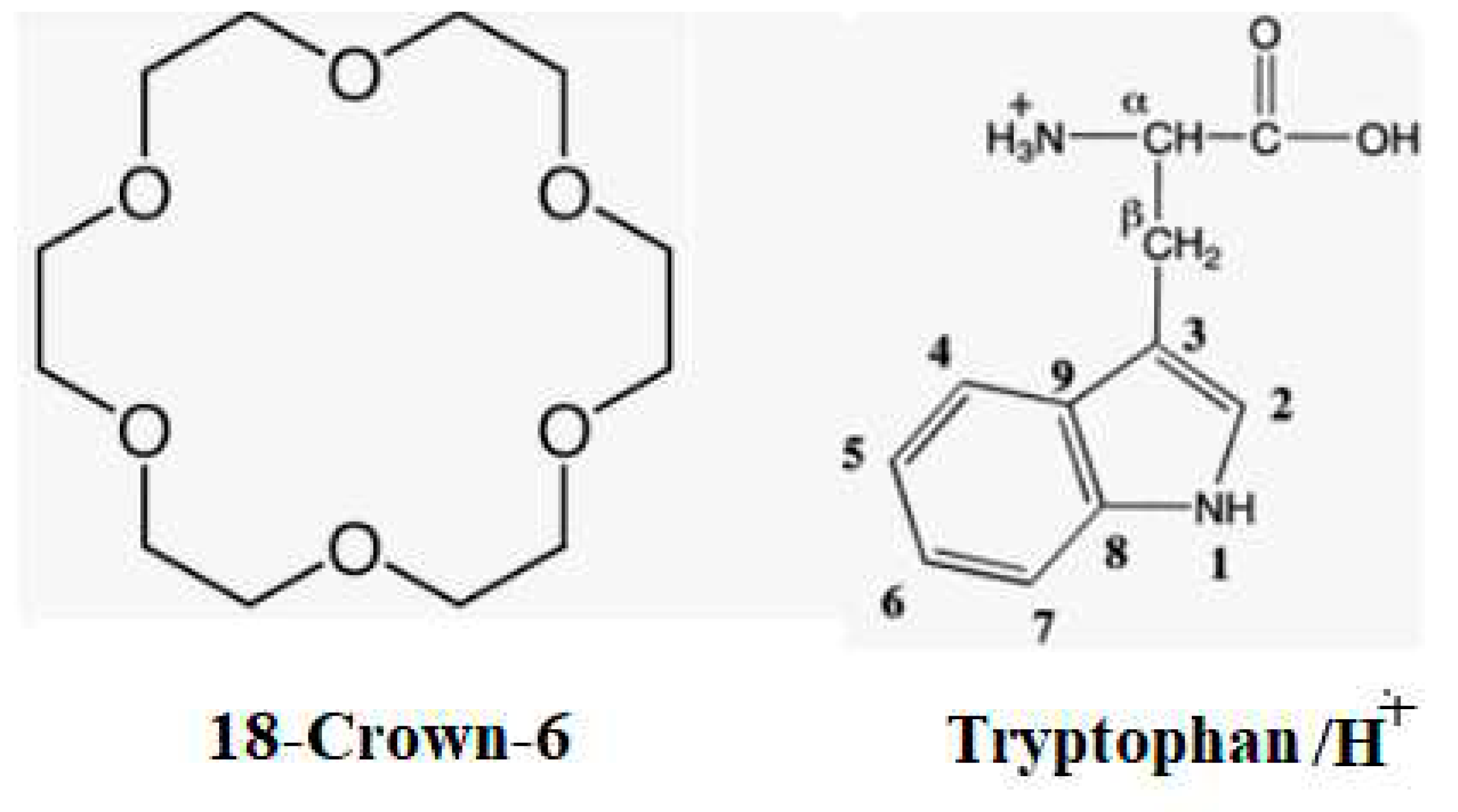
Chart 1.
Components of 18-Crown-6/TrpH+ complex.
2. Results and Discussions
2.1. Structures and Thermodynamic Stability of 18-Crown-6/TrpH+ Non-Covalent Complexes in the Gas Phase
Figure 1 presents the IRMPD spectrum obtained for the 18-Crown-6/TrpH+ complex by Polfer and co-workers [44]. Notably, two prominent high-frequency bands at 3510 and 3565 cm-1 were assigned [45] as the indole –NH- and –OH stretching modes, respectively. Intriguingly, these high frequency bands in the IRMPD spectrum for the 18-Crown-6/TrpH+ complex closely resemble the corresponding bands [44] (3500 and 3555 cm-1, respectively) observed for the isolated TrpH+ species (Figure S1, Supplementary Information). This congruence suggests that, in the gas phase, the indole –NH and –CO2H groups within the 18-Crown-6/TrpH+ complex remain ‘naked’ [46], devoid of any hydrogen bonding (H-bonding) interactions with the CR unit. This parallel can be drawn to our earlier investigations of permethylated cyclodextrin (perm-CD) complexes with amino acids (AAH+), namely Ala, Ile, and Lys, i.e., perm-CD/AAH+, where the carboxyl groups of protonated amino acids were similarly found to be free from interactions with the perm-CD.
Another noteworthy feature of the IRMPD spectrum for 18-Crown-6/TrpH+ is the significant redshift of the strong bands observed in the IRMPD spectrum of TrpH+ (at 3195 and 3340 cm-1), attributed to the –NH3+ stretching mode of the isolated TrpH+ [44]. Upon complexation with 18-Crown-6, these bands experience a redshift to the range of 3080–3140 cm-1. This shift unequivocally indicates the complexation of TrpH+ with the 18-Crown-6 unit through –NH3+.
Figure 2 displays the calculated structures of gas-phase 18-Crown-6/TrpH+ complexes. In all conformers, the ammonium side-chain is accommodated either completely or partially within the crown ether ring. Among these conformers, the lowest Gibbs free energy configurations, 18-Crown-6/TrpH+-G1 and 18-Crown-6/TrpH+-G2, exhibit ‘naked’ indole –NH- and –CO2H groups; other conformers of this type with higher relative Gibbs free energies >3 kcal/mol are given in Figure S1, Supplementary Information. Their structural disparities mainly involve the position and orientation of the indole moiety with respect to the rest of the complex. In contrast, conformers 18-Crown-6/TrpH+-G3 and 18-Crown-6/TrpH+-G4 reveal interactions between both the –NH3+ and –CO2H groups with the CR ring. These conformers, however, appear to suffer from increased strain due to the crowding of these functional groups, resulting in higher thermodynamic instability (relative Gibbs free energy >5.5 kcal/mol).
The calculated IR spectra for these conformers are presented in Figure 3. In the two conformers with the lowest Gibbs free energy (CR/TrpH+-G1 and CR/TrpH+-G2), the IR spectra clearly depict strong bands at 3486 and 3588 cm-1, confirming the presence of ‘naked’ indole –NH and –CO2H, consistent with the experimental IRMPD spectrum in Figure 1. These conformers also exhibit asymmetric and symmetric –NH3+ stretching frequencies at ~3070 and ~3105, 3175 cm-1, respectively. Notably, the IR spectra of CR/TrpH+-G1 and CR/TrpH+-G2 are nearly indistinguishable. Conversely, the IR spectra of the less stable conformers, CR/TrpH+-G3 and CR/TrpH+-G4, display significantly redshifted –OH stretching bands at ~3400 and ~3480 cm-1, in clear contradiction to the experimental observations for the gas phase CR/TrpH+ complex. These results strongly support the notion that the carboxyl group in the gas phase CR/TrpH+ complex remains isolated, devoid of interactions with the CR unit. Consequently, either CR/TrpH+-G1 or CR/TrpH+-G2 may reasonably account for the experimentally observed IRMPD spectrum in Figure 1. However, definitively assigning the observed IRMPD bands to either CR/TrpH+-G1 or CR/TrpH+-G2 based solely on calculated IR frequencies poses challenges.
Exploring the CR/TrpH+ host-guest pair in a solution-phase context may provide insights into the structure of the gas-phase CR/TrpH+ complex, as discussed further below. Specifically, it would be intriguing to investigate whether the more stable conformer, 18-Crown-6/TrpH+-G1, retains its stability in the solution phase or undergoes a thermodynamic ‘inversion’. This can be elucidated by calculating the structures of the 18-Crown-6/TrpH+ complex in solution and comparing their Gibbs free energies to ascertain the potential influence of solvent molecules.
In our previous work involving the perm-CD/AAH+ (AA = Ala, Ile, Lys) system, we determined (by IRMPD/DFT technique) that the experimentally identified gas-phase host-guest complexes with higher Gibbs free energies ( >17 kcal/mol) became the global minimum Gibbs free energy structures in the solution phase due to improved solvation [47,48,49,50]. These findings led us to propose that valuable insights into host-guest solvent interactions can be gleaned from gas-phase studies produced by the ESI/MS technique.
Consequently, a pertinent question arises regarding the present 18-Crown-6/TrpH+ system: Are the experimentally observed 18-Crown-6/TrpH+ complexes (18-Crown-6/TrpH+-G1, 18-Crown-6/TrpH+-G2) distinct from the most stable host-guest structure in the solution phase? Specifically, could the higher Gibbs free energy 18-Crown-6/TrpH+ complexes (18-Crown-6/TrpH+-3 or 18-Crown-6/TrpH+-4), where both the carboxyl and ammonium groups interact with the CR unit, become the most stable structures in the solution phase, influenced by solvent molecules? These intriguing questions warrant further exploration.
2.2. Structures of 18-Crown-6/TrpH+ Non-Covalent Complexes in Solution
To address the question concerning the structural connectivity of the thermodynamically most stable CR/TrpH+ complex in both the gas phase and in solution, it is essential to closely examine the influence of solvation. Since quantum chemical or simulation methods cannot feasibly account for an infinite number of solvent molecules, we have opted for the supramolecular/continuum approach in this study.
Herein, we consider solvent molecules (specifically, 5 H2O molecules) that directly interact with the functional groups (carboxyl and ammonium) in the first solvation shell as explicit molecules, while those in the second shell and beyond are treated as a dielectric continuum. The resulting CR/TrpH+ complexes in solution, denoted as CR/TrpH+-S1, CR/TrpH+-S2. and CR/TrpH+-S3, correspond to CR/TrpH+-G1, CR/TrpH+-S2, and CR/TrpH+-S3, in the gas-phase, respectively, as illustrated in Figure 4.
Interestingly, we find that the lowest Gibbs free energy gas-phase complexes, CR/TrpH+-G1, also represents the most stable configuration in an aqueous solution. Its Gibbs free energy is lower by 3.4 kcal/mol compared to CR/TrpH+-G2, aligning with the gas phase CR/TrpH+-G1. This difference in Gibbs free energies between CR/TrpH+-S1 and CR/TrpH+-S2 translates to a population ratio of approximately ~75:1 at 25 C, strongly indicating that the CR/TrpH+-S1 structure predominates in the solution phase. In contrast, the thermodynamically less favourable structure CR/TrpH+-G1, where both the ammonium and carboxyl groups bind to the CR ring, maintains its higher Gibbs free energy (4.7 kcal/mol higher than CR/TrpH+-S1) in the solution phase. These findings unequivocally suggest that in both the gas phase and solution, the carboxyl group prefers to establish stronger hydrogen bonding interactions with water molecules rather than with the CR unit.
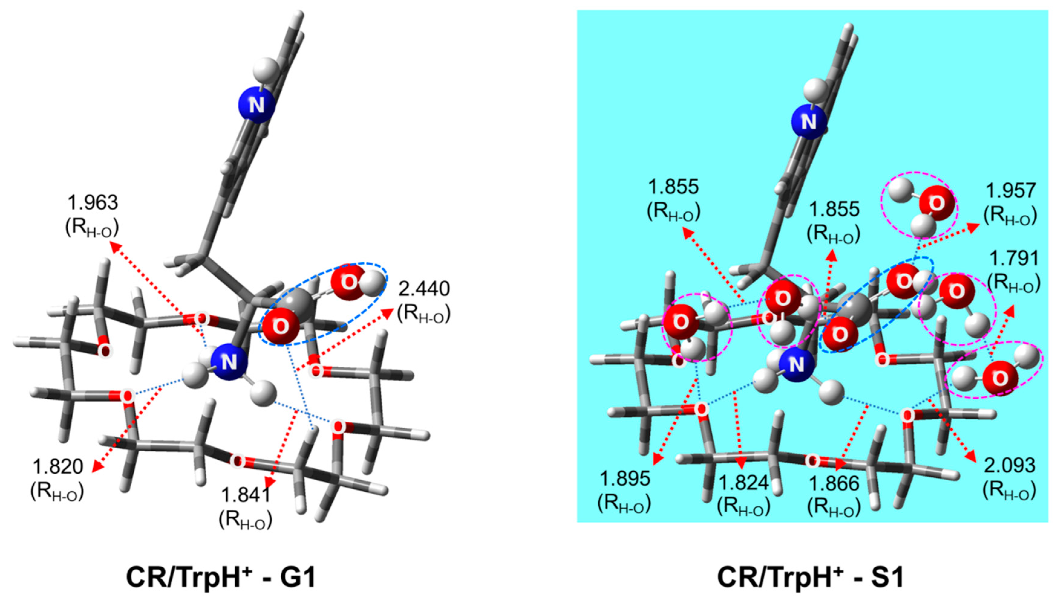
Thus, in contrast to the perm-CD/LysH+ system previously investigated [43], there is no evidence of thermodynamic inversion in the present study. However, it is reasonable to infer that the lowest Gibbs free energy gas-phase CR/TrpH+ complex, CR/TrpH+-G1, generated through the ESI/MS procedure, directly corresponds to the most stable solution-phase CR/TrpH+ complex, CR/TrpH+-S1. During the course of the ESI/MS experiments, the structural characteristics (with –CO2H positioned away from the CR unit) of the host-guest pair CR/TrpH+-S1 in solution are promptly transferred to the CR/TrpH+-G1 configuration, subsequently detected by the IRMPD technique. Consequently, the carboxyl group becomes “naked” without interacting with the host CR unit in the gas phase. If this proposition holds true, then the structure of the CR/TrpH+ complex, which yields the IRMPD spectrum shown in Figure 1 in the gas phase, can confidently be assigned to CR/TrpH+-G1. A detailed comparison of the structures of CR/TrpH+-G1 and CR/TrpH+-S1 is presented in Figure 5. The structure of the gas-phase conformer (CR/TrpH+- G1) remains nearly unchanged in comparison to that in the solution phase (CR/TrpH+- S1), with only minor adjustments of >0.11 Å in the distances between the ammonium hydrogen atoms and the CR oxygen atoms. This correlation between the gas-phase CR/TrpH+-G1 and the solution-phase structure CR/TrpH+-S1 underscores the potential for obtaining precise and invaluable insights into CR – TrpH - solvent interactions in solution on a molecular level through the identification of gas-phase host-guest pair structures using the IRMPD/DFT procedure, as previously demonstrated in our study of the perm-CD/LysH+ system [43].
3. Computational Details
All calculations were performed using the wB97X-D functional [51], which may treat weak interactions (hydrogen bonding included) very well, with 6-311G** and 6-31G* basis set implemented in Gaussian16 suite of programs [52]. For structures in solution, single point calculations were also conducted using the wB97X-D/6-311G* method at the wB97X-D/6-31G* optimized geometry. A least-square procedure with a scale factor of 0.9382 was used to optimize the fit of the calculated IR frequencies to experimental IRMPD spectrum. All structures were verified by checking all real vibrational frequencies. For the CR/TrpH+ complex in solution, the supramolecule/continuum approach was used, treating the solvent molecules directly interacting with –CO2H, ammonium groups in the first shell around the complex as explicit molecules and other numerous H2O molecules in the second shell and beyond as a water continuum by the SMD method [53].
In actual experiments, a mixture of water, methanol, and acetic acid (49:49:2 v/v/v) was used as solvent, but for simplicity, the system was modeled as the CR/TrpH+ complex in water. This assumption was based on two presumptions: first, methanol would preferentially evaporate as the ESI droplets become smaller due to its higher volatility, and second, H2O molecules would mostly constitute the first shell around the CR/TrpH+ complex because they would form stronger hydrogen bonding through two –OH groups per H2O molecule with the functional groups (ammonium, amino and carboxyl) than CH3OH would. Water molecules were added to the functional groups in the CR/H+/L-Trp complex, and the lowest Gibbs free energy configurations were searched at each step. These configurations were compared to those optimized from initial configurations by adding water molecules randomly, using the VMD (visual molecular dynamics) program.
4. Conclusions
In this study, we have provided insights into the structures of 18-Crown-6/H+/L-Trp complexes in both the gas phase and in solution, employing quantum chemical methods. Our primary objective centered on unraveling the structural connectivity between these complexes in these two distinct phases. Our findings unequivocally demonstrate that the most stable conformer in the solution phase precisely corresponds to the lowest Gibbs free energy gas-phase conformer observed in ESI/IRMPD experiments. This compelling alignment strongly supports the notion that the latter gas-phase conformer originates from the solution-phase complex.
We firmly believe that these results bolster the credibility of our proposed methodology for characterizing host-guest-solvent interactions in solution by leveraging gas-phase structures of host-guest complexes. This innovative approach holds great promise for advancing our understanding of host-guest systems. As such, we encourage further experimental investigations of these captivating host-guest systems, which we believe are a promising avenue for future research endeavors.
Supplementary Materials
The following supporting information can be downloaded at the website of this paper posted on Preprints.org. Structures of higher Gibbs free energy gas phase CR/TrpH+ complexes with naked –CO2H. Relative Gibbs free energy in kcal/mol. Cartesian Coordinates
Author Contributions
H. B. O. and S. L. devised the overall concept of the work. Y.-H. O. carried out the modelling of the system, DFT calculations and analyzed the structures and IR spectra of the crown ether/protonated Trp complexes. Y.H.O and S.L. wrote the manuscript.
Funding
This work was supported by grants from the National Research Foundation of Korea funded by the Ministry of Education (NRF-2019R1F1A1057609, 2018R1A6A1A03024940, 2021R1A2C2007397), and the KISTI Supercomputing Center. HBO is thankful to the research grant supported by Korea Environment Industry & Technology Institute (KEITI) through “Advanced Technology Development Project for Predicting and Preventing Chemical Accident” program, funded by Korea Ministry of Environment (MOE) (RS-2023-00219144).
Institutional Review Board Statement
Not applicable.
Informed Consent Statement
Not applicable.
Conflicts of Interest
The authors declare no conflict of interest.
Sample Availability
Samples of the compounds are available from the authors.
References
- Robert, F. How Far Can We Push Chemical Self-Assembly? Science (80-. ). 2005, 309, 95. [Google Scholar]
- Percec, V.; Dulcey, A.E.; Balagurusamy, V.S.K.; Miura, Y.; Smidrkal, J.; Peterca, M.; Nummelin, S.; Edlund, U.; Hudson, S.D.; Heiney, P.A. Self-Assembly of Amphiphilic Dendritic Dipeptides into Helical Pores. Nature 2004, 430, 764–768. [Google Scholar] [CrossRef] [PubMed]
- Davis, M.E.; Zuckerman, J.E.; Choi, C.H.J.; Seligson, D.; Tolcher, A.; Alabi, C.A.; Yen, Y.; Heidel, J.D.; Ribas, A. Evidence of RNAi in Humans from Systemically Administered SiRNA via Targeted Nanoparticles. Nature 2010, 464, 1067–1070. [Google Scholar] [CrossRef] [PubMed]
- Jiao, D.; Geng, J.; Loh, X.J.; Das, D.; Lee, T.; Scherman, O.A. Supramolecular Peptide Amphiphile Vesicles through Host–Guest Complexation. Angew. Chemie Int. Ed. 2012, 51, 9633–9637. [Google Scholar] [CrossRef]
- Qu, D.-H.; Wang, Q.-C.; Zhang, Q.-W.; Ma, X.; Tian, H. Photoresponsive Host–Guest Functional Systems. Chem. Rev. 2015, 115, 7543–7588. [Google Scholar] [CrossRef]
- Ma, X.; Zhao, Y. Biomedical Applications of Supramolecular Systems Based on Host–Guest Interactions. Chem. Rev. 2015, 115, 7794–7839. [Google Scholar] [CrossRef]
- Yang, H.; Yuan, B.; Zhang, X.; Scherman, O.A. Supramolecular Chemistry at Interfaces: Host–Guest Interactions for Fabricating Multifunctional Biointerfaces. Acc. Chem. Res. 2014, 47, 2106–2115. [Google Scholar] [CrossRef]
- Zhan, W.; Wei, T.; Yu, Q.; Chen, H. Fabrication of Supramolecular Bioactive Surfaces via β-Cyclodextrin-Based Host–Guest Interactions. ACS Appl. Mater. Interfaces 2018, 10, 36585–36601. [Google Scholar] [CrossRef]
- Kim, C.; Agasti, S.S.; Zhu, Z.; Isaacs, L.; Rotello, V.M. Recognition-Mediated Activation of Therapeutic Gold Nanoparticles inside Living Cells. Nat. Chem. 2010, 2, 962–966. [Google Scholar] [CrossRef]
- Ortega-Caballero, F.; Rousseau, C.; Christensen, B.; Petersen, T.E.; Bols, M. Remarkable Supramolecular Catalysis of Glycoside Hydrolysis by a Cyclodextrin Cyanohydrin. J. Am. Chem. Soc. 2005, 127, 3238–3239. [Google Scholar] [CrossRef]
- Shahgaldian, P.; Pieles, U. Cyclodextrin Derivatives as Chiral Supramolecular Receptors for Enantioselective Sensing. Sensors 2006, 6, 593–615. [Google Scholar] [CrossRef]
- Hu, Q.-D.; Tang, G.-P.; Chu, P.K. Cyclodextrin-Based Host–Guest Supramolecular Nanoparticles for Delivery: From Design to Applications. Acc. Chem. Res. 2014, 47, 2017–2025. [Google Scholar] [CrossRef] [PubMed]
- Miyata, K.; Nishiyama, N.; Kataoka, K. Rational Design of Smart Supramolecular Assemblies for Gene Delivery: Chemical Challenges in the Creation of Artificial Viruses. Chem. Soc. Rev. 2012, 41, 2562–2574. [Google Scholar] [CrossRef] [PubMed]
- Anguiano-Igea, S.; Otero-Espinar, F.J.; Vila-Jato, J.L.; Blanco-Méndez, J. Interaction of Clofibrate with Cyclodextrin in Solution: Phase Solubility, 1H NMR and Molecular Modelling Studies. Eur. J. Pharm. Sci. 1997, 5, 215–221. [Google Scholar] [CrossRef]
- Mathiron, D.; Iori, R.; Pilard, S.; Rajan, T.S.; Landy, D.; Mazzon, E.; Rollin, P.; Djedaïni-Pilard, F. A Combined Approach of NMR and Mass Spectrometry Techniques Applied to the α-Cyclodextrin/Moringin Complex for a Novel Bioactive Formulation. Molecules 2018, 23. [Google Scholar] [CrossRef]
- Sun, D.Z.; Li, L.; Qiu, X.M.; Liu, F.; Yin, B.L. Isothermal Titration Calorimetry and 1H NMR Studies on Host-Guest Interaction of Paeonol and Two of Its Isomers with β-Cyclodextrin. Int. J. Pharm. 2006, 316, 7–13. [Google Scholar] [CrossRef]
- Shamsutdinova, N.A.; Strelnik, I.D.; Musina, E.I.; Gerasimova, T.P.; Katsyuba, S.A.; Babaev, V.M.; Krivolapov, D.B.; Litvinov, I.A.; Mustafina, A.R.; Karasik, A.A.; et al. Host-Guest" Binding of a Luminescent Dinuclear Au(i) Complex Based on Cyclic Diphosphine with Organic Substrates as a Reason for Luminescence Tuneability. New J. Chem. 2016, 40, 9853–9861. [Google Scholar] [CrossRef]
- Incavo, J.A.; Dutta, P.K. Zeolite Host-Guest Interactions: Optical Spectroscopic Properties of Tris(Bipyridine)Ruthenium(II) in Zeolite Y Cages. J. Phys. Chem. 1990, 94, 3075–3081. [Google Scholar] [CrossRef]
- Mesu, J.G.; Visser, T.; Beale, A.M.; Soulimani, F.; Weckhuysen, B.M. Host-Guest Chemistry of Copper(II)-Histidine Complexes Encaged in Zeolite Y. Chem. - A Eur. J. 2006, 12, 7167–7177. [Google Scholar] [CrossRef]
- Fenn, J.B.; Mann, M.; Meng, C.K.; Wong, S.F.; Whitehouse, C.M. Electrospray Ionization for Mass Spectrometry of Large Biomolecules. Science (80-. ). 1989, 246, 64–71. [Google Scholar] [CrossRef]
- Pramanik, B.N.; Ganguly, A.K.; Gross, M.L. Applied Electrospray Mass Spectrometry: Practical Spectroscopy Series Volume 32; CRC Press, 2002; Vol. 32; ISBN 0203909275.
- Kebarle, P.; Verkerk, U.H. Electrospray: From Ions in Solution to Ions in the Gas Phase, What We Know Now. Mass Spectrom. Rev. 2009, 28, 898–917. [Google Scholar] [CrossRef] [PubMed]
- Konermann, L.; Ahadi, E.; Rodriguez, A.D.; Vahidi, S. Unraveling the Mechanism of Electrospray Ionization 2013.
- Monge, M.E.; Harris, G.A.; Dwivedi, P.; Fernández, F.M. Mass Spectrometry: Recent Advances in Direct Open Air Surface Sampling/Ionization. Chem. Rev. 2013, 113, 2269–2308. [Google Scholar] [CrossRef] [PubMed]
- Takats, Z.; Wiseman, J.M.; Gologan, B.; Cooks, R.G. Mass Spectrometry Sampling under Ambient Conditions with Desorption Electrospray Ionization. Science (80-. ). 2004, 306, 471–473. [Google Scholar] [CrossRef] [PubMed]
- Cescutti, P.; Garozzo, D.; Rizzo, R. Effect of Methylation of β-Cyclodextrin on the Formation of Inclusion Complexes with Aromatic Compounds. An Ionspray Mass Spectrometry Investigation. Carbohydr. Res. 1997, 302, 1–6. [Google Scholar] [CrossRef]
- Sun, W.; Cui, M.; Liu, S.; Song, F.; Elkin, Y.N. Electrospray Ionization Mass Spectrometry of Cyclodextrin Complexes with Amino Acids in Incubated Solutions and in Eluates of Gel Permeation Chromatography. Rapid Commun. mass Spectrom. 1998, 12, 2016–2022. [Google Scholar] [CrossRef]
- Oomens, J.; Sartakov, B.G.; Meijer, G.; Von Helden, G. Gas-Phase Infrared Multiple Photon Dissociation Spectroscopy of Mass-Selected Molecular Ions. Int. J. Mass Spectrom. 2006, 254, 1–19. [Google Scholar] [CrossRef]
- Bush, M.F.; O’Brien, J.T.; Prell, J.S.; Saykally, R.J.; Williams, E.R. Infrared Spectroscopy of Cationized Arginine in the Gas Phase: Direct Evidence for the Transition from Nonzwitterionic to Zwitterionic Structure. J. Am. Chem. Soc. 2007, 129, 1612–1622. [Google Scholar] [CrossRef]
- Scuderi, D.; Lepere, V.; Piani, G.; Bouchet, A.; Zehnacker-Rentien, A. Structural Characterization of the UV-Induced Fragmentation Products in an Ion Trap by Infrared Multiple Photon Dissociation Spectroscopy. J. Phys. Chem. Lett. 2014, 5, 56–61. [Google Scholar] [CrossRef]
- Sen, A.; Le Barbu-Debus, K.; Scuderi, D.; Zehnacker-Rentien, A. Mass Spectrometry Study and Infrared Spectroscopy of the Complex between Camphor and the Two Enantiomers of Protonated Alanine: The Role of Higher-energy Conformers in the Enantioselectivity of the Dissociation Rate Constants. Chirality 2013, 25, 436–443. [Google Scholar] [CrossRef]
- Burt, M.; Wilson, K.; Marta, R.; Hasan, M.; Hopkins, W.S.; McMahon, T. Assessing the Impact of Anion–π Effects on Phenylalanine Ion Structures Using IRMPD Spectroscopy. Phys. Chem. Chem. Phys. 2014, 16, 24223–24234. [Google Scholar] [CrossRef]
- Hurtado, P.; Gámez, F.; Hamad, S.; Martínez-Haya, B.; Steill, J.D.; Oomens, J. Crown Ether Complexes with H3O+ and NH 4+: Proton Localization and Proton Bridge Formation. J. Phys. Chem. A 2011, 115, 7275–7282. [Google Scholar] [CrossRef] [PubMed]
- McNary, C.P.; Nei, Y.W.; Maitre, P.; Rodgers, M.T.; Armentrout, P.B. Infrared Multiple Photon Dissociation Action Spectroscopy of Protonated Glycine, Histidine, Lysine, and Arginine Complexed with 18-Crown-6 Ether. Phys. Chem. Chem. Phys. 2019, 21, 12625–12639. [Google Scholar] [CrossRef] [PubMed]
- Polfer, N.C. Infrared Multiple Photon Dissociation Spectroscopy of Trapped Ions. Chem. Soc. Rev. 2011, 40, 2211–2221. [Google Scholar] [CrossRef] [PubMed]
- Azargun, M.; Fridgen, T.D. Guanine Tetrads: An IRMPD Spectroscopy, Energy Resolved SORI-CID, and Computational Study of M (9-Ethylguanine) 4+(M= Li, Na, K, Rb, Cs) in the Gas Phase. Phys. Chem. Chem. Phys. 2015, 17, 25778–25785. [Google Scholar] [CrossRef]
- Oh, H.; Breuker, K.; Sze, S.K.; Ge, Y.; Carpenter, B.K.; McLafferty, F.W. Secondary and Tertiary Structures of Gaseous Protein Ions Characterized by Electron Capture Dissociation Mass Spectrometry and Photofragment Spectroscopy. Proc. Natl. Acad. Sci. 2002, 99, 15863–15868. [Google Scholar] [CrossRef] [PubMed]
- Kou, M.; Oh, Y.-H.; Lee, S.; Kong, X. Distinguishing Gas Phase Lactose and Lactulose Complexed with Sodiated L-Arginine by IRMPD Spectroscopy and DFT Calculations. Phys. Chem. Chem. Phys. 2023. [Google Scholar] [CrossRef]
- Yao, H.; Steill, J.D.; Oomens, J.; Jockusch, R.A. Infrared Multiple Photon Dissociation Action Spectroscopy and Computational Studies of Mass-Selected Gas-Phase Fluorescein and 2′, 7′-Dichlorofluorescein Ions. J. Phys. Chem. A 2011, 115, 9739–9747. [Google Scholar] [CrossRef]
- Cheng, R.; Loire, E.; Fridgen, T.D. Hydrogen Bonding in Alkali Metal Cation-Bound i-Motif-like Dimers of 1-Methyl Cytosine: An IRMPD Spectroscopic and Computational Study. Phys. Chem. Chem. Phys. 2019, 21, 11103–11110. [Google Scholar] [CrossRef]
- Seo, J.; Hoffmann, W.; Warnke, S.; Bowers, M.T.; Pagel, K.; von Helden, G. Retention of Native Protein Structures in the Absence of Solvent: A Coupled Ion Mobility and Spectroscopic Study. Angew. Chemie - Int. Ed. 2016, 55, 14173–14176. [Google Scholar] [CrossRef]
- Bush, M.F.; Forbes, M.W.; Jockusch, R.A.; Oomens, J.; Polfer, N.C.; Saykally, R.J.; Williams, E.R. Infrared Spectroscopy of Cationized Lysine and ε-N-Methyllysine in the Gas Phase: Effects of Alkali-Metal Ion Size and Proton Affinity on Zwitterion Stability. J. Phys. Chem. A 2007, 111, 7753–7760. [Google Scholar] [CrossRef]
- Choi, H.; Oh, Y.H.; Park, S.; Lee, S.S.; Oh, H. Bin; Lee, S. Unveiling Host–Guest–Solvent Interactions in Solution by Identifying Highly Unstable Host–Guest Configurations in Thermal Non-Equilibrium Gas Phase. Sci. Rep. 2022, 12, 1–9. [Google Scholar] [CrossRef]
- Mino Jr, W.K.; Gulyuz, K.; Wang, D.; Stedwell, C.N.; Polfer, N.C. Gas-Phase Structure and Dissociation Chemistry of Protonated Tryptophan Elucidated by Infrared Multiple-Photon Dissociation Spectroscopy. J. Phys. Chem. Lett. 2011, 2, 299–304. [Google Scholar] [CrossRef]
- Stedwell, C.N.; Galindo, J.F.; Gulyuz, K.; Roitberg, A.E.; Polfer, N.C. Crown Complexation of Protonated Amino Acids: Influence on IRMPD Spectra. J. Phys. Chem. A 2013, 117, 1181–1188. [Google Scholar] [CrossRef] [PubMed]
- Linder, R.; Nispel, M.; Häber, T.; Kleinermanns, K. Gas-Phase FT-IR-Spectra of Natural Amino Acids. Chem. Phys. Lett. 2005, 409, 260–264. [Google Scholar] [CrossRef]
- Lee, S.S.; Park, S.; Hong, Y.; Lee, J.U.; Kim, J.H.; Yoon, D.; Kong, X.; Lee, S.; Oh, H. Bin Chiral Differentiation of D- and l-Alanine by Permethylated β-Cyclodextrin: IRMPD Spectroscopy and DFT Methods. Phys. Chem. Chem. Phys. 2017, 19, 14729–14737. [Google Scholar] [CrossRef]
- Lee, S.-S.; Lee, J.; Oh, J.H.; Park, S.; Hong, Y.; Min, B.K.; Lee, H.H.L.; Kim, H.I.; Kong, X.; Lee, S. Chiral Differentiation of D-and L-Isoleucine Using Permethylated β-Cyclodextrin: Infrared Multiple Photon Dissociation Spectroscopy, Ion-Mobility Mass Spectrometry, and DFT Calculations. Phys. Chem. Chem. Phys. 2018, 20, 30428–30436. [Google Scholar] [CrossRef]
- Lee, S.-S.; Park, S.; Kim, J.-Y.; Kim, H.-R.; Lee, S.; Oh, H. Bin Infrared Multiple Photon Dissociation Spectroscopy and Density Functional Theory (DFT) Studies of Protonated Permethylated β-Cyclodextrin–Water Non-Covalent Complexes. Phys. Chem. Chem. Phys. 2014, 16, 8376–8383. [Google Scholar] [CrossRef]
- Lee, J.U.; Lee, S.S.; Lee, S.; Oh, H. Bin Noncovalent Complexes of Cyclodextrin with Small Organic Molecules: Applications and Insights into Host–Guest Interactions in the Gas Phase and Condensed Phase. Molecules 2020, 25. [Google Scholar] [CrossRef]
- Chai, J. Da; Head-Gordon, M. Long-Range Corrected Hybrid Density Functionals with Damped Atom-Atom Dispersion Corrections. Phys. Chem. Chem. Phys. 2008, 10, 6615–6620. [Google Scholar] [CrossRef]
- Frisch, M.J.; Trucks, G.W.; Schlegel, H.B.; Scuseria, G.E.; Robb, M.A.; Cheeseman, J.R.; Scalmani, G.; Barone, V.; Petersson, G.A.; Nakatsuji, H. Gaussian 16, Gaussian. Inc., Wallingford CT 2016, 2016. [Google Scholar]
- Marenich, A. V.; Cramer, C.J.; Truhlar, D.G. Universal Solvation Model Based on Solute Electron Density and on a Continuum Model of the Solvent Defined by the Bulk Dielectric Constant and Atomic Surface Tensions. J. Phys. Chem. B 2009, 113, 6378–6396. [Google Scholar] [CrossRef] [PubMed]
Figure 1.
Experimental IRMPD spectra of CR/TrpH+ in the gas phase. Ref. pol_13.
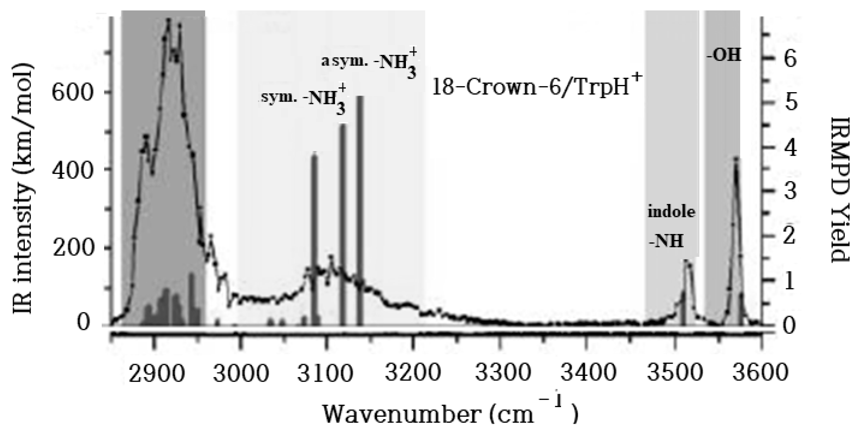
Figure 2.
Calculated structures of the gas phase CR/TrpH+ complexes. Relative Gibbs free energy in kcal/mol.
Figure 2.
Calculated structures of the gas phase CR/TrpH+ complexes. Relative Gibbs free energy in kcal/mol.
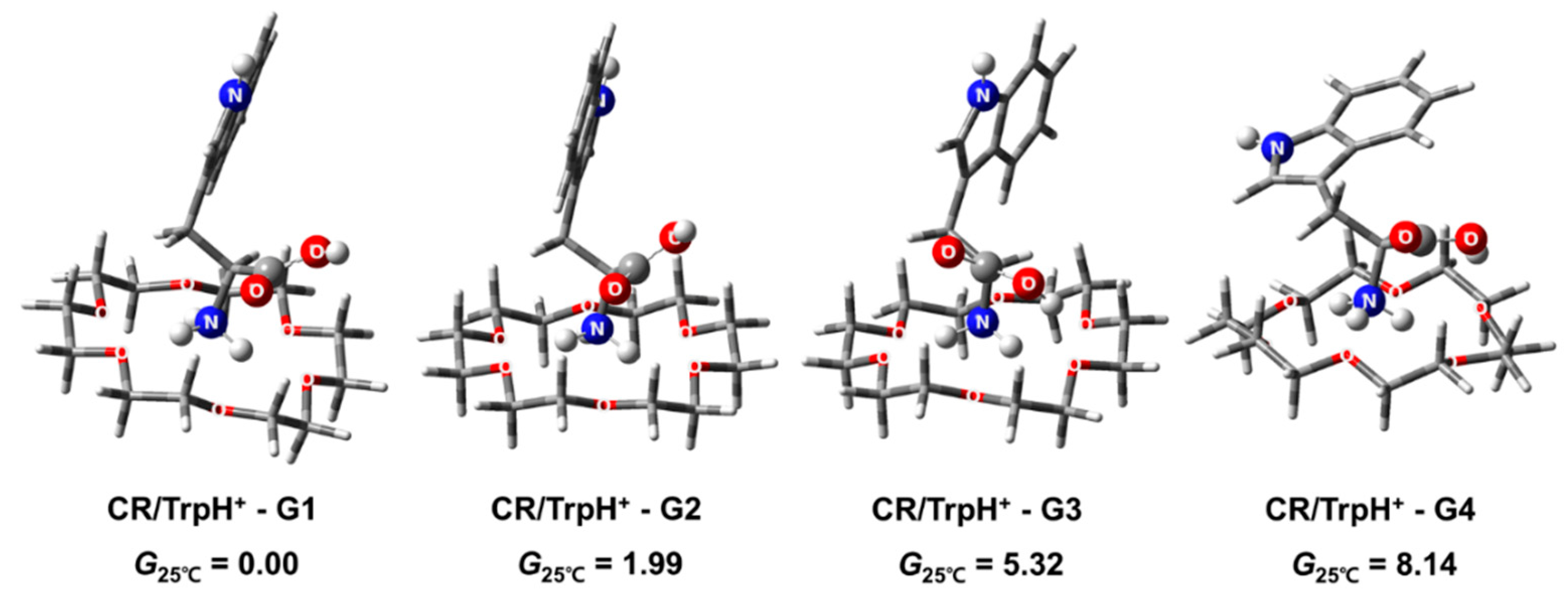
Figure 3.
Calculated infrared spectra and assignment of the stretch modes of gas phase CR/TrpH+ complexes.
Figure 3.
Calculated infrared spectra and assignment of the stretch modes of gas phase CR/TrpH+ complexes.
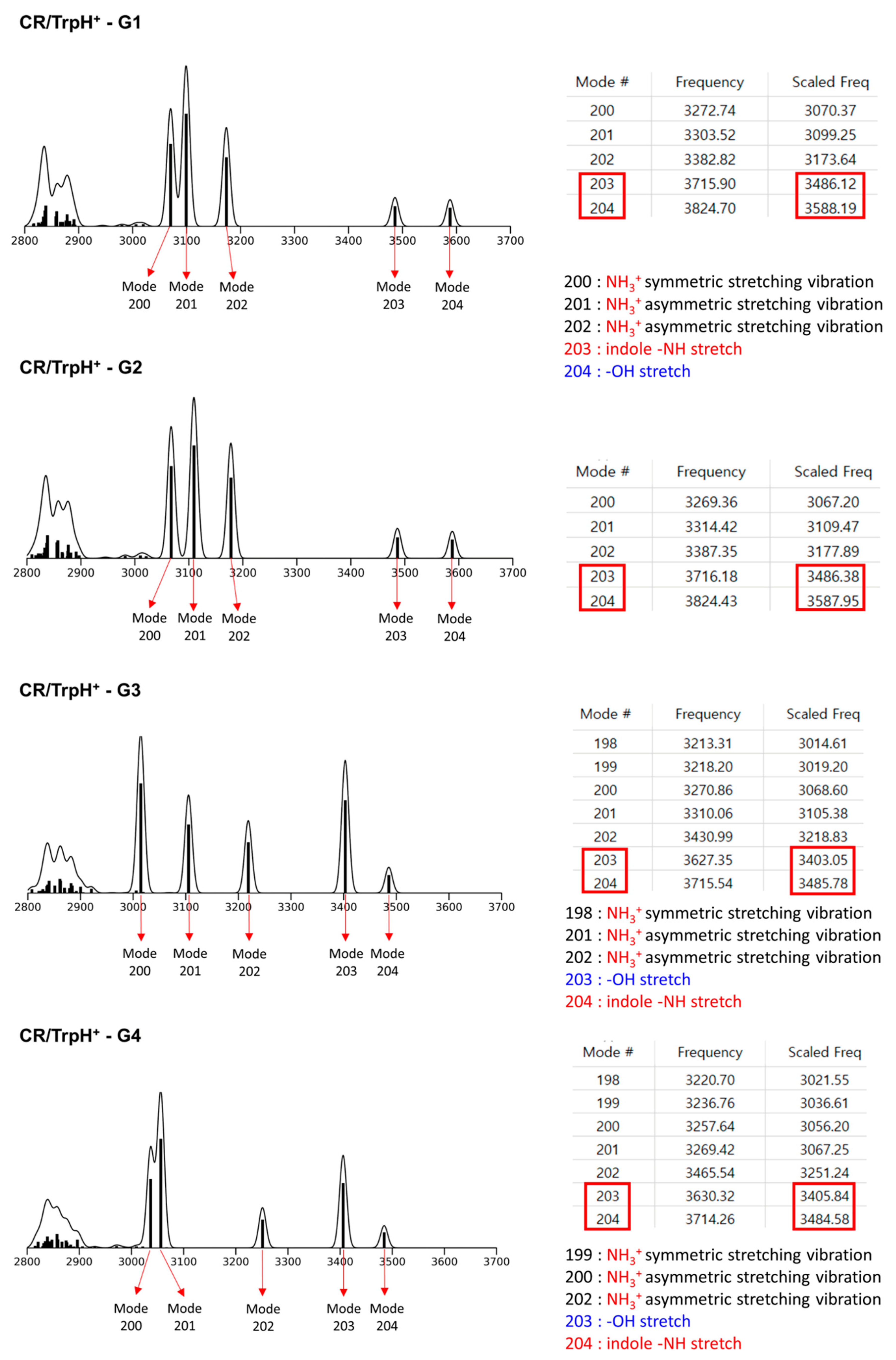
Figure 4.
Calculated structures of the 18-Crown-6/TrpH+ non-covalent complexes in solution corresponding to the gas phase conformers in Figure 2. Distance is in Å and relative Gibbs free energy in kcal/mol. Blue background represents solvent continuum.
Figure 4.
Calculated structures of the 18-Crown-6/TrpH+ non-covalent complexes in solution corresponding to the gas phase conformers in Figure 2. Distance is in Å and relative Gibbs free energy in kcal/mol. Blue background represents solvent continuum.
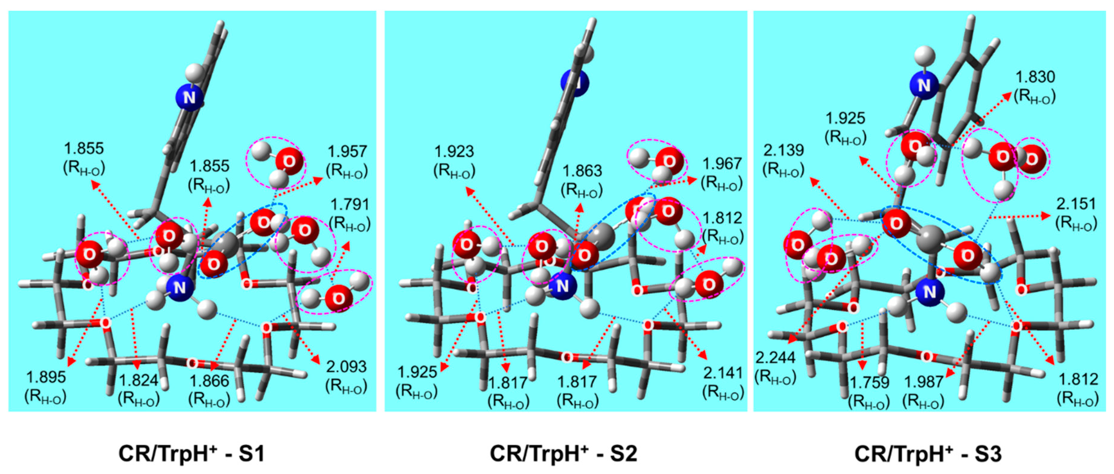
Figure 5.
Comparison of the structures of CR/TrpH+ complex in solution (18-Crown-6/TrpH+-S1) and in the gas phase (18-Crown-6/TrpH+-G1). Blue background represents solvent continuum.
Figure 5.
Comparison of the structures of CR/TrpH+ complex in solution (18-Crown-6/TrpH+-S1) and in the gas phase (18-Crown-6/TrpH+-G1). Blue background represents solvent continuum.
Disclaimer/Publisher’s Note: The statements, opinions and data contained in all publications are solely those of the individual author(s) and contributor(s) and not of MDPI and/or the editor(s). MDPI and/or the editor(s) disclaim responsibility for any injury to people or property resulting from any ideas, methods, instructions or products referred to in the content. |
© 2023 by the authors. Licensee MDPI, Basel, Switzerland. This article is an open access article distributed under the terms and conditions of the Creative Commons Attribution (CC BY) license (http://creativecommons.org/licenses/by/4.0/).
Copyright: This open access article is published under a Creative Commons CC BY 4.0 license, which permit the free download, distribution, and reuse, provided that the author and preprint are cited in any reuse.


