
Submitted:
23 March 2024
Posted:
25 March 2024
You are already at the latest version
Abstract
The objective of the this study was to reduce the benzo[a]pyrene-induced hepatotoxicity method in herbal medicine products and to give proof that neferine, daidzein, genistein possess antihepa-totoxic effects. B[a]P which classified as a group 1 carcinogen and metabolized to B[a]P-7,8-dihydrodiol-9,10-epoxide (BPDE) causes mutagenic DNA addition products. The reduc-tion of BPDE-DNA adduct formation by B[a]P-7,8-dihydrodiol-9,10-epoxide (BPDE) was derived by benzo[a]pyrene (B[a]P). In HepG2 cells, B[a]P exhibited toxicity and substance treatment of the cells with B[a]P with neferine in lotus and daidzein, genistein in soybean reduced the BPDE-DNA ad-ducts level. The level of B[a]P-metabolites in the substance treatment of the cells was presented that BPDE levels were reduced by neferine in lotus and daidzein, genisten in soybean. These results suggest that neferine in lotus and daidzein, genistein in soybean prevent B[a]P-induced hepato-toxicity for BPDE-DNA adduct formation.
Keywords:
herbal medicine products
; benzo[a]pyrene-induced in vitro toxicity
; B[a]P-7
; 8-dihydrodiol-9
; 10-epoxide (BPDE)
; benzo[a]pyrene (B[a]P)
; HepG2 cells
1. Introduction
Polycyclic aromatic hydrocarbons (PAHs) are potent and ubiquitous atmospheric contaminants [1]. Benzo[a]pyrene (B[a]P) is one of the high-molecular-weight PAHs. B[a]P is generated from the incomplete combustion of organic substances, such as some foods, tobacco smoke, coal tar, and automobile exhaust fumes [2]. The carcinogenic, mutagenic, and cytotoxic properties of B[a]P have been re-emphasized in recent years [3]. Inhalation of B[a]P causes various types of adverse health effects such as mutations and immunocompromised status. It damages to the cardiopulmonary and reproductive system [4]. Accordingly, the National Institute for Occupational Safety and Health (NIOSH) in USA nominated an occupational exposure limit of 100 μg/m3 for total PAHs in a workplace [5]. PAHs are highly lipophilic and, consequently, easily absorbed through the epithelial cells in the gastrointestinal tract, respiratory tract, and skin, with a low clearance rate and high residence in organs such as the brain, lung, and liver [6]. After inhalation, B[a]P was metabolized to B[a]P-7,8-dihydrodiol-9,10-epoxide (BPDE) and reactive oxygen species (ROS), which cause DNA adduct formation and oxidative DNA damage, leading to carcinogenesis [7]. In addition, PAHs also induce systemic effects, such as reproductive, cardiovascular, gastrointestinal, and developmental toxicity. B[a]P-induced in vitro toxicity has been associated with the synthesis of ROS in the liver [8]. In this respect, neferine in lotus seeds and daidzein and genistein in soybean, as representative active constituents of natural herbal medicines such as lotus seeds (Nelumbo nucifera) and soybeans (Glycine max). Neferine is a bisbenzylisoquinoline alkaloid isolated from lotus seed sample were selected for analysis in this study to evaluate their efficiency in reducing B[a]P-induced in vitro toxicity and has a wide range of pharmacological activities [9]. Neferine extracted from lotus seeds acted as a free radical scavenger, which inhibited lipid peroxide formation due to the hydroxyl groups present in its structure, and it enhanced the activity of superoxide dismutase, an antioxidant enzyme [10]. Lotus seeds are widely used in herbal medicines as a diuretic, as an antidote to poison, and to treat skin diseases, cancer, and tissue inflammation [11]. Embryos of lotus seeds are used in Chinese herbal medicines for the treatment of cardiovascular diseases, high fever, insomnia, and nervous disorders [12]. Poornima et al reported that neferine was correlated with the possible pathway to reduce the benzo[a]pyrene-induced in vitro toxicity in HepG2 cells [13]. Daidzein and genistein are the major isoflavones in soybeans and have been associated with beneficial effects on human health [14]. Soybeans were associated with a reduction of osteoporosis, cardiovascular disease, colon cancer, prostate cancer and breast cancer [15]. These effects were attributed to the anti-osteoporosis, anticarcinogenic, and antioxidant activities of daidzein and genistein in soybean [16]. Sarao et al. (2022) reported that soybean isoflavones, such as daidzein and genistein, correlated with the possible pathway to reduce the benzo[a]pyrene-induced in vitro toxicity in HepG2 cells [17]. However, the effects of neferine, daidzein, and genistein on the reduction of B[a]P-induced in vitro toxicity in HepG2 cells have not yet been reported. Considerable efforts have been made to understand the mechanism of in vitro toxicity induced by PAHs [18]. For this purpose, both biochemical and histopathological approaches have been employed [19]. According to previous studies, intraperitoneal and oral administration of PAHs changed liver weight caused preneoplastic hepatocytes as well as liver congestion, and induced the synthesis of hepatic enzymes [20]. Due to the toxicity and detrimental health effects of PAH exposure, researchers have investigated the effectiveness of dietary agents, such as polyphenols, terpenoids, flavonoids, and natural herbal medicines, for preventing the occurrence of various types of environment-induced diseases [21]. Therefore, the objective of this study was to evaluate the antioxidative effects of some active constituents in natural herbal medicines in inhibiting B[a]P-induced in vitro toxicity and to represent the inhibitory effect of neferine, daidzein, genistein on BPDE-DNA adduct formation by using enzyme-linked immunosorbent assay. Finally, this study can provide basic scientific data for safety management.
2. Results and Discussion
2.1. Reduction of B[a]P-Induced In Vitro Toxicity by Neferine, Daidzein, Genistein
B[a]P-induced cell death varies depending on the levels of BPDE−DNA adduct formation. Accordingly, we studied the potential preventive effects of neferine, daidzein, and genistein against BPDE formation associated with B[a]P-induced in vitro toxicity by using HepG2 cells. The effects of the natural products, at the concentrations of BaP tested (10 uM) with natural products (0 – 20 uM), are significant, but only moderate. HepG2 is a well-characterized hepatic cell line. The toxicities of B[a]P and neferine, daidzein, and genistein on HepG2 cells were evaluated by cell viability assays. The toxicities of B[a]P (Figure 1A) and neferine (Figure 1B), daidzein (Figure 1C), genistein (Figure 1D) on HepG2 cells were evaluated by using cell viability assays. Because neferine, daidzein, genistein were non-toxic (20 µM for 48 h), B[a]P induced cell death in a dose-dependent manner in comparison with treatment in Figure 1A-D. The protective effects of neferine, daidzein, genistein against B[a]P-induced in vitro toxicity, B[a]P was applied to HepG2 cells together with various concentrations of neferine, daidzein, genistein. Neferine, daidzein, genistein reduced B[a]P-induced in vitro toxicity and restored cell viability up to 90% in a dose-dependent manner. These expressed that neferine, daidzein, genistein have a protective effect against B[a]P-induced in vitro toxicity. Therefore, the potential preventive effect of neferine, daidzein, genistein against B[a]P-induced in vitro toxicity was evaluated in Figure 2, Figure 3 and Figure 4A. These formation was expressed by BPDE-DNA adduct enzyme-linked immunosorbent assay (ELISA) kit after treatment with B[a]P (10 µM), neferine, daidzein, genistein (20 µM), and B[a]P (10 µM) co-administered with neferine, daidzein, genistein (20 µM). It was resulted that B[a]P (10 µM) co-administered with neferine, daidzein, genistein (20 µM) treatment alone decreased the BPDE-DNA adduct level compared to the B[a]P treatment alone in Figure 2, Figure 3 and Figure 4B. These results expressed that neferine, daidzein, genistein exerts an antigenotoxic effect by reducing the formation of BPDE-DNA adducts. B[a]P was known to regulate various physiological signaling pathways, leading to various side effects related to cellular toxicity. In this regard, neferine has been reported to regulate various signaling pathways related to B[a]P-induced cellular toxicity. Specifically, neferine was reported to inhibit the B[a]P-induced NF-κB pathway [22,23,24]. NF-κB was one of the inflammatory signaling pathways that can induce inflammatory responses related to B[a]P, and it was believed that neferine reduces cellular toxicity by inhibiting the NF-κB signaling pathway. Additionally, neferine was reported to inhibit the B[a]P-induced MAPK pathway [25,26,27]. MAPK was one of the various signaling pathways related to cell survival, growth, inflammation, stress responses, and others. B[a]P was known to regulate the MAPK pathway to induce cellular toxicity, and it was believed that neferine reduces cellular toxicity by inhibiting the MAPK signaling pathway. Furthermore, neferine was reported to inhibit the B[a]P-induced PI3K-Akt pathway [28]. PI3K-Akt was one of the signaling pathways related to cell survival and growth. B[a]P was known to regulate the PI3K-Akt signaling pathway to induce cellular toxicity, and it was believed that neferine reduces cellular toxicity by inhibiting the PI3K-Akt pathway.
2.2. Reduction of Intracellular B[a]P Metabolites by Neferine, Daidzein, Genistein
B[a]P was metabolized to B[a]P-7,8-dihydrodiol and BPDE according to the procedure. BPDE caused in vitro toxicity by increasing mutagenic adducts. The quantity of B[a]P, B[a]P-7,8-dihydrodiol and BPDE in the treated with HepG2 cells were expressed by using a high performance liquid chromatography (HPLC) system. The calculated amount of BPDE rapidly increased in the cells were treated with B[a]P alone. As a result, B[a]P-7,8-dihydrodiol levels increased. In contrast, BPDE levels decreased and B[a]P, B[a]P with neferine, daidzein, genistein compared to the levels with B[a]P treatment alone in Figure 5, Figure 6 and Figure 7. Other similar studies express the well-characterized HepG2 cell to study the potential preventive effects of silymarin, quercetin and isorhamnetin on BPDE formation by reducing the cell toxicity induced by B[a]P. The toxicities of B[a]P and silymarin, quercetin and isorhamnetin on HepG2 cells were evaluated by using cell viability assays and the potential preventive effect of silymarin, quercetin and isorhamnetin against B[a]P-induced in vitro toxicity to confirm that silymarin, quercetin and isorhamnetin inhibits BPDE-DNA adduct formation compared to the effects of B[a]P alone and indicate that silymarin, quercetin and isorhamnetin attenuate B[a]P-induced in vitro toxicity by inhibiting BPDE-DNA adduct formation [29,30]. These results represented that neferine, daidzein, genistein reduced B[a]P-induced in vitro toxicity by the inhibition of BPDE-DNA adduct formation and excretion of BPDE. BPDE was one of the metabolites of B[a]P, which can form adducts with cellular DNA and molecules, leading to genetic mutations and cell damage. In particular, neferine has been found to be the most effective in reducing BPDE-DNA adduct levels which were believed to be due to its mechanisms such as antioxidant activity, modulation of metabolic enzymes, and enhancement of DNA repair. Neferine has been reported to have antioxidant activity, and this action was believed to inhibit the generation of BPDE-DNA adducts. Therefore, neferine has been reported to prevent DNA damage by reducing intracellular ROS (Radical Oxygen Species) and oxidative stress [31,32,33,34,35]. Additionally, B[a]P activates intracellular metabolic enzymes (CYP1A1 and CYP1B1) to produce BPDE, while neferine was reported to inhibit the expression of CYP1A1 and CYP1B1, thereby inhibiting the production of BPDE [26]. Furthermore, BPDE-DNA adducts cause DNA damage, and in order to repair it, cells need to activate DNA repair mechanisms. In this process, neferine was reported to prevent the formation of BPDE-DNA adducts and enhance DNA repair mechanisms, thus preventing DNA damage [36]. In this study, neferine was found to effectively inhibit the formation of intracellular B[a]P metabolites through the mechanisms mentioned above. However, further studies were needed to investigate the specific mechanisms by which neferine effectively inhibits the formation of intracellular B[a]P metabolites. In addition, studies have reported that daidzein and genistein also reduce intracellular B[a]P metabolites [37,38]. However, the results of this study showed that they were not as effective as neferine. Other similar studies suggested that silymarin, quercetin, and isorhamnetin promoted the metabolism, detoxification, and elimination of B[a]P, thereby increasing anti-genotoxic effects and protecting against B[a]P-induced cytotoxicity. These results represented that silymarin, quercetin and isorhamnetin reduced B[a]P-induced in vitro toxicity by the inhibition of BPDE-DNA adduct formation and excretion of BPDE [29,30].
2.3. Validation of Analytical Method for Neferine, Daidzein, Genistein Analysis
The HPLC chromatograms of neferine, daidzein and genistein standards, neferine, daidzein and genistein in a spiked sample were presented in Figure 8A−B, respectively.
Calibration curves were constructed for the neferine, daidzein and genistein at five concentrations (3, 5, 10, 20, and 40 μg/kg) by plotting concentration against the peak area. Regression analysis of each calibration curve yielded a slope of satisfactory linearity with values for the coefficient of determination (R2) > 0.99 over the tested concentration range. For the lotus and soybean matrices, the LOD was below 0.2 μg/kg (0.08−0.12 μg/kg) and the LOQ was below 0.6 μg/kg (0.24−0.36 μg/kg). The limit of detection (LOD), limit of quantification (LOQ) and linearity (R2) and calibration equations for validation of the neferine, daidzein and genistein matrices by using HPLC-UV analysis were presented in Table 1. The high linearity (R2 > 0.99) of our analytical method was similar to that reported for other spiked sample product matrices, including other lotus and soybean samples [12]. This indicates that the analytical method can accurately detect and quantify, and can be useful for detecting these substances in the medical industry. Generally, the lower the LOD and LOQ values, the higher the sensitivity of the analytical method, which allows for more sensitive detection. This helps to improve the accuracy and reliability of the analytical method. Therefore, it can be said that analytical results are more reliable when the LOD and LOQ values are lower. These same studies obtained values for LOD of 1.3−4.4 μg/mL and LOQ of 3.9−13.2 μg/mL, which were higher than the LOD and LOQ results of our validation tests [39]. It was noteworthy that the LOD and LOQ were similar between the lotus and soybean matrices.
The recovery values of the neferine, daidzein and genistein (Table 2) were expressed through the peak areas of the neferine, daidzein and genistein standards. Recovery values of neferine in lotus, daidzein and genistein in soybean samples ranged from 82.95% to 111.46%. Precision was performed at five different concentrations (3, 5, 10, 20, and 40 μg/kg) and mean values of the five different concentrations (3, 5, 10, 20, and 40 μg/kg) in intra, interday of precision of the neferine in lotus, daidzein and genistein in soybean were expressed in Table 3. The results of the intraday analysis (performed three times a day) and the interday analysis (carried out simultaneously on 3 days) showed a precision (%CV) of <15%. More specifically, the precision of neferine in lotus, daidzein and genistein in soybean analysis were in the ranges of 0.24−6.82% (intraday), and 0.65−4.67% (interday), respectively. Collectively, these data show the effectiveness of the proposed methodology for lotus and soybean matrices. The authors observed no substantial differences in sample concentration and recoveries between the two methods over the concentration range studied (3.0−40.0 μg/kg).
2.4. Comparison of Neferine, Daidzein and Genistein Concentrations
The concentrations of neferine daidzein and genistein were determined. These data were presented for 70 samples of the concentrations of neferine, daidzein and genistein matrices using HPLC-UV analysis. Neferine was 0.78±0.20 μg/kg, daidzein was 1.40±0.29 μg/kg and genistein was 1.01±0.27 μg/kg. Among the neferine, daidzein and genistein, the highest concentration of neferine (2.45±0.09 μg/kg), the highest concentration of daidzein (3.42±0.09 μg/kg) and the highest concentration of genistein (3.23±0.77 μg/kg). The occurrence of neferine, daidzein and genistein in herbal medicines was typically a consequence of the ubiquitous presence of these compounds in the environment, unintentional production during cooking or a manufacturing process. To increase the recovery of these HPLC-UV methods, it was important to select an appropriate isolation solvent with similar polarity to the neferine, daidzein and genistein. Most of the relevant studies have reported that methanol was a suitable isolation solvent because it has adequate polarity and yields a good recovery for most compounds [40]. In this study, we established, validated, and applied an HPLC-UV method for the evaluation of neferine, daidzein and genistein matrices in Korea. In the context of these limits, all of the lotus and soybean samples analyzed in the current study were recognized as having safe levels of neferine, daidzein and genistein. In the context of these limits, all of the lotus and soybean samples analyzed in this study were recognized as having safe levels of neferine, daidzein, and genistein, in agreement with related studies [39]. With few previously conducted studies on this topic, accurate quantitative analysis methods were required.
3. Materials and Methods
3.1. Chemicals and Materials
B[a]P, neferine, daidzein, genistein were acquired from Sigma-Aldrich Chemical (St. Louis, MO, USA). Trypsin-ethylenediaminetetraacetic acid (T-EDTA), penicillin/streptomycin, fetal bovine serum (FBS), sodium pyruvate and phosphate buffered saline (PBS), Minimum essential medium (MEM) were acquired from Gibco (Seoul, Korea). Dimethyl sulfoxide (DMSO) was acquired from VWR (Suwon, Korea). HPLC grade solvents (methanol and water) were purchased from J.T. Baker (USA). Trifluoroacetic acid (TFA) was purchased from DAEJUNG (Gyeonggi-do, Korea). Acetonitrile and ethyl acetate was obtained from Burdick & Jackson (Muskegon, MI, USA). EZ-CYTOX reagent was acquired from DOGEN (Daejeon, Korea). The lid (all plate type, 84.50 × 126.50 × 9.00) and immunoplate (96-well flat bottom cell culture plate) were acquired from SPL Life Sciences Co. Ltd. (Gyeonggi-do, Korea). 0.45 mm PTFE membrane filter paper (Chiyoda City, Japan) was acquired from Advantec Co., Ltd. A sterile 100 mm2 cell culture dish was acquired from SPL Life Sciences Co. Ltd. (Gyeonggi-do, Korea). QIAamp DNA Mini kit was acquired from Qiagen (Seoul, Korea). BPDE-DNA adduct enzyme-linked immunosorbent assay (ELISA) kit was acquired from Cell Biolabs (Seoul, Korea).
3.2. Sample Preparations for Isolation of Neferine
The 35 samples of lotus seeds (Nelumbo nucifera) of 20 g were produced in 2023 year in Gyeongsangbukdo area which was a province in eastern South Korea with the sample preparations for isolations of neferine with lotus seeds (Nelumbo nucifera) of 20 g which isolated in triplicate with 20 ml of 80% methanol by ultrasonication in 30 minutes with ultrasonic bath at room temperature. The isolated solution was obtained in portioning in n-hexane, CHCl3, ethyl acetate. Then this solution evaporated under 37°C to concentrate a volume of 0.5 L. Then, n-hexane, CHCl3, and ethyl acetate was added in 20 ml of the water solution. Ethyl acetate fractions were eluted once with water : methanol (1:4) with using SPE extraction method. The isolation solutions were filtered by using 0.45 mm PTFE membrane filter paper (Chiyoda City, Japan) [41].
3.3. Sample Preparations for Isolation of Daidzein, Genistein
The 35 samples of soybeans (Glycine max) of 20 g were produced in 2020 year in Gyeonggido area which was a province in eastern South Korea with the sample preparations for isolations of daidzein, genistein which kept frozen in a polyethylene bag until use. Before daidzein and genistein isolation, the soybean seeds were ground into a powder. For the isolation procedure, 10 mL of 80% methanol solution was added to a glass reactor and heated in a water bath for 20 min. Then, 10 g of the soybean powder was added. Daidzein and genistein were isolated for 60 min, followed by centrifugation at 500 rpm for 10 min. After centrifugation, 1.0 mL of the extract (supernatant) was mixed with 1.0 mL of solvent (80% methanol). The isolation solutions were filtered through a PTFE membrane filter paper (0.45 mm; Advantec Co., Ltd). This filtrate was diluted, and the powder of daidzein and genistein was prepared [42].
3.4. HepG2 Cells Culture and Treatment
HepG2 cells were obtained from the Cell Bank of the Type Culture Collection of the Chinese Academy of Sciences (Shanghai, China). They were cultured with 10% FBS in MEM, 1 mM sodium pyruvate, and 100 U/mL penicillin/streptomycin in a 100 mm2 cell culture dish with 5% CO2 at 37℃ in an incubator. Cells were incubated for 48 h with B[a]P, neferine, daidzein, and genistein in MEM, and the cell-free extracts were analyzed.
3.5. Cell Viability and Proliferation Assay
In order to assess the cytotoxicity of B[a]P, neferine, daidzein, and genistein on HepG2 cells, we evaluated the cell viability. HepG2 cells were seeded at 1 × 10⁴ cells/well in MEM in an immunoplate (96-well flat-bottom cell culture plate) with B[a]P, neferine, daidzein, and genistein at 0, 1, 2, 5, 10, and 20 µM concentrations for 48 h. After this procedure, EZ-Cytox reagent (100 µL) was added to each well. The cells were incubated for 2 h. Then, the absorbance at a wavelength of 450 nm was determined using a microplate reader (PerkinElmer Victor X4, PerkinElmer, Seoul, Korea). The cell viability was compared between the plates treated with B[a] P, neferine, daidzein, and genistein.
3.6. Cell Isolation
HepG2 cells were seeded at 5×10⁴ cells in a 100 mm2 dish and cultivated for 24 h for adherence. The medium was then replaced and treated with B[a]P, neferine, daidzein, and genistein for 48 h. Afterward, 1.0 mL of culture medium was extracted from the cell culture dish, transferred into a 1.5 mL EP tube, and centrifuged at 15,000 rpm for 30 min at 4°C. A 1.0 mL aliquot of the supernatant was put into a sample vial for HPLC-UV detection. Cell culture medium samples were stored at 4°C. The medium was eliminated from the dishes, and the dishes were cleaned three times with PBS preheated at 37°C to dispose the disturbance in removing any residual medium. To each dish, 700 μL of KH2PO4 pre-cooled to 4°C was added, and the cells were scraped from the dish with cell scrapers. The scraped cells from each culture dish were collected into a 1.5 mL EP tube, freeze-thawed three times to break cells, and then centrifuged at 15,000 rpm for 30 min at 4°C. After centrifugation, 500 μL of supernatant was put into a sample vial for HPLC-UV chromatographic analysis.
3.7. HPLC-UV Analysis for Method Validation
Stock solutions of neferine, daidzein, and genistein were prepared at five different concentrations (3, 5, 10, 20, and 40 μg/kg) by dissolving an adequate amount of the respective powders in 80% methanol. These stock solutions were stored in sample vials in the refrigerator for HPLC-UV analysis. The HPLC-UV analytical method was validated for linearity (coefficient of determination, R2), recovery (%), limit of detection (LOD), limit of quantification (LOQ), and precision (%) in lotus and soybean samples. A Dionex-C18 column (4.6 mm i.d. × 150 mm × 5 µm) was used for neferine analysis. A Hypersil-ODS C18 column (4.6 mm i.d. × 20 cm × 5 µm) was used for the analysis of daidzein and genistein. A mixture of methanol with 0.1% TFA in water, methanol, and acetonitrile was selected as the mobile phase. The neferine, daidzein, and genistein solutions were injected into the HPLC system, and then, according to the shape of the peaks and repeatability, the appropriate results of the mobile phase were expressed. The flow rate was 1.0 mL/min. The UV wavelength was set to 205 nm for the detection of neferine and 262 nm for the detection of daidzein and genistein [41,42].
3.8. BPDE-DNA Adduct Formation Analysis
HepG2 cells were seeded on an immunoplate (96-well flat-bottom cell culture plate) in MEM and treated with 10 µM B[a]P alone and cotreated with 10 µM B[a]P and 20 µM neferine, daidzein, and genistein, respectively, for 48 h. DNA was extracted from the cells using the QIAamp DNA Mini Kit (Qiagen). The BPDE−DNA adduct ELISA Kit (Cell Biolabs) was used for determining BPDE−DNA adduct formation, with the absorbance measured at 450 nm wavelength using a microplate reader (PerkinElmer Victor X4, PerkinElmer).
3.9. The Typical Intracellular Metabolites of B[a]P Were Measured by High Performance Liquid Chromatography (HPLC)
HepG2 cells were treated with 10 µM B[a]P alone and cotreated with 10 µM B[a]P and 20 µM neferine, daidzein, and genistein, respectively, in 100 mm2 cell culture dishes for 48 h and the cell-free extracts of the treated cels were mixed with ethyl acetate. After vacuum rotary evaporation to remove the solvent, a 50% acetonitrile and 0.1% acetic acid mixed solution was added to the residue. The mixture was injected (100 µL) into an HPLC instrument (Dionex U3000 HPLC, Thermo Fisher Scientific, Sunnyvale, CA, USA) equipped with a Kinetex C18 Plus column (4.6 mm i.d. × 250 mm × 5 µm; Phenomenex, Torrance, CA, USA). The flow rate was 1.2 mL/min at 30°C. The binary mobile phase consisted of 0.1% acetic acid in distilled water (solvent A) and 0.1% acetic acid with 50% acetonitrile in distilled water (solvent B). Chromatographic separation was conducted with the following gradient program: 50% B for 0−40 min. The retention time of BPDE, B[a]P-7,8-dihydrodiol, and B[a]P were compared with those of reference standards in the MRI Global Chemical Carcinogen Repository (Kansas City, MO, USA).
3.10. Statistical Analysis
All experiments were performed in triplicate. All data was expressed as mean ± standard deviation (SD). Significances differences between the treatment group (B[a]P with neferine, daidzein, genistein) and no-treatment group (B[a]P only) were determined by using one-way analysis of variance (ANOVA) in SPSS. Statistical significance was expressed at p < 0.05.
4. Conclusions
In conclusion, we showed that B[a]P detoxification prevents the formation of BPDE−DNA adducts by reducing intracellular B[a]P metabolites. In HepG2 cells, neferine, daidzein and genistein reduced the BPDE−DNA adduct formation caused by the reaction of BPDE derived by B[a]P with cellular DNA. These results suggest that neferine, daidzein and genistein prevent B[a]P-induced in vitro toxicity by inhibiting BPDE−DNA adduct formation. Based on these results, it was suggested that neferine, daidzein, genistein were effective in significantly reducing the BPDE-DNA adduct formation in that order. And HPLC-UV analytical methods were very well validated and calibrated. The HPLC-UV analytical methods were developed in this study allows the quantification of neferine, daidzein, genistein in matrices. We confirmed that the Nrf2 signaling pathway is mainly related to the inhibition of BPDE-DNA adduct formation by neferine, daidzein, genistein. These results suggest that neferine, daidzein, genistein has anti-genotoxicity properties against B[a]P through the inhibition of BPDE-DNA adduct formation in vitro and in vivo toxicity. The application of 32P-post-labeling to the study of the DNA-binding potential of non-radiolabeled BP metabolites, many of the latter, has greatly contributed to adding to the current knowledge of the metabolism and activation of this important benzo[a]pyrene-induced in vitro toxicity.
Author Contributions
Y.-Y.K.: Methodology, Visualization, Formal analysis, Writing?original draft preparation, reviewing and editing, H.-H.S.: Project administration, Writing—review and editing. Author was agreed to the published version of the manuscript.
Funding
This research was carried out with the support of “Cooperative Research Program for Agriculture Science and Technology Development (Project No. PJ01669005)” Rural Development Administration, Republic of Korea.
Institutional Review Board Statement
Not applicable.
Informed Consent Statement
Not applicable.
Data Availability Statement
The datasets generated for this study are available on request to the corresponding author.
Conflicts of Interest
The authors declare no conflict of interest.
References
- Phillips, D.H.; Venitt, S. DNA and protein adducts in human tissues resulting from exposure to tobacco smoke. Int J Cancer. 2012, 131, 2733–2753. [Google Scholar] [CrossRef] [PubMed]
- Baird, W.M.; Hooven, L.A.; Mahadevan, B. Carcinogenic polycyclic aromatic hydrocarbon-DNA adducts and mechanism of action. Environ Mol Mutagen. 2005, 45, 106–114. [Google Scholar] [CrossRef]
- Saunders, C.R.; Das, S.K.; Ramesh, A.; Shockley, D.C.; Mukherjee, S. Benzo[a]pyrene-induced acute neurotoxicity in the F-344 rat: role of oxidative stress. J Appl Toxicol. 2006, 26, 427–438. [Google Scholar] [CrossRef] [PubMed]
- Kim, K.H.; Jahan, S.A.; Kabir, E.; Brown, R.J. A review of airborne polycyclic aromatic hydrocarbons (PAHs) and their human health effects. Environ Int. 2013, 60, 71–80. [Google Scholar] [CrossRef] [PubMed]
- McCormick, S.; Snawder, J.E.; Chen, I.C.; Slone, J.; Calafat, A.M.; Wang, Y.; Meng, L.; Alexander-Scott, M.; Breitenstein, M.; Johnson, B.; Meadows, J.; Fairfield, E.C. Exposure assessment of polycyclic aromatic hydrocarbons in refined coal tar sealant applications. Int J Hyg Environ Health. 2022, 242, 113971. [Google Scholar] [CrossRef] [PubMed]
- Ramesh, A.; Inyang, F.; Hood, D.B.; Archibong, A.E.; Knuckles, M.E.; Nyanda, A.M. Metabolism, bioavailability, and toxicokinetics of benzo(alpha)pyrene in F-344 rats following oral administration. Exp Toxicol Pathol. 2001, 53, 275–290. [Google Scholar] [CrossRef] [PubMed]
- Li, F.; Xiang, B.; Jin, Y.; Li, C.; Ren, S.; Wu, Y.; Li, J.; Luo, Q. Hepatotoxic effects of inhalation exposure to polycyclic aromatic hydrocarbons on lipid metabolism of C57BL/6 mice. Environ Int. 2020, 134, 105000. [Google Scholar] [CrossRef] [PubMed]
- Jaeschke, H. Reactive oxygen and mechanisms of inflammatory liver injury. J Gastroenterol Hepatol. 2000, 15, 718–724. [Google Scholar] [CrossRef] [PubMed]
- Miller, K.P.; Ramos, K.S. Impact of cellular metabolism on the biological effects of benzo[a]pyrene and related hydrocarbons. Drug Metab Rev. 2001, 33, 1–35. [Google Scholar] [CrossRef]
- Zhao, L.; Wang, X.; Chang, Q.; Xu, J.; Huang, Y.; Guo, Q. Neferine, a bisbenzylisoquinline alkaloid attenuates bleomycin-induced pulmonary fibrosis. Eur. J. Pharmacol. 2010, 627, 304–312. [Google Scholar] [CrossRef]
- Mahbubur Rahman AHM. Traditional medicinal plants used in the treatment of different skin diseases of santals at abdullahpur village under akkelpur upazilla of joypurhat district, Bangladesh. Biomed. and Biotechnol. 2013, 1, 17–20. [Google Scholar]
- Chen, Y.; Fan, G.R.; Wu, H.L.; Wu, Y.T.; Mitchell, A. Separation, identification and rapid determination of liensine, isoliensinine and neferine from embryo of the seed of Nelumbo nucifera GAERTN by liquid chromatography coupled to diode array detector and tandem mass spectrometry. J. Pharm. Biomed. Anal. 2007, 43, 99–104. [Google Scholar] [CrossRef] [PubMed]
- Poornima, P.; Weng, C.F.; Padma, V.V. Neferine, an alkaloid from lotus seed embryo, inhibits human lung cancer cell growth by MAPK activation and cell cycle arrest. Biofactors. 2014, 40, 121–131. [Google Scholar] [CrossRef] [PubMed]
- Liggins, J.; Mulligan, A.; Runswick, S.; Bingham, S.A. Daidzein and genistein content of cereals. Eur. J. Clin Nutr. 2002, 56, 961–966. [Google Scholar] [CrossRef]
- Sirtori, C.R. Risks and benefits of soy phytoestrogens in cardiovascular diseases, cancer, climacteric symptoms and osteoporosis. Drug. Saf. 2001, 24, 665–682. [Google Scholar] [CrossRef] [PubMed]
- Adjakly, M.; Ngollo, M.; Boiteux, J.P.; Bignon, Y.J.; Guy, L.; Gallon, D.B. Genistein and daidzein: different molecular effects on prostate cancer. Anticancer Res. 2013, 33, 39–44. [Google Scholar] [PubMed]
- Sarao, L.; Kaur, S.; Malik, T.; Singh, A. Chapter 19 – Genistein and daidzein. Nutraceuticals and Health Care. 2022, 331–341. [Google Scholar] [CrossRef]
- Newman, D.J.; Cragg, G.M. Natural products as sources of new drugs over the last 25 years. J Nat Prod. 2007, 70, 461–477. [Google Scholar] [CrossRef]
- Ji, X.; Li, Y.; He, J.; Shah, W.; Xue, X.; Feng, G.; Zhang, H.; Gao, M. Depletion of mitochondrial enzyme system in liver, lung, brain, stomach and kidney induced by benzo[a]pyrene. Environ Toxicol Pharmacol. 2016, 43, 83–93. [Google Scholar] [CrossRef]
- Jin, Y.; Miao, W.; Lin, X.; Wu, T.; Shen, H.; Chen, S.; Li, Y.; Pan, Q.; Fu, Z. Sub-chronically exposing mice to a polycyclic aromatic hydrocarbon increases lipid accumulation in their livers. Environ Toxicol Pharmacol. 2014, 38, 353–363. [Google Scholar] [CrossRef]
- Mumtaz, M.M.; George, J.D.; Gold, K.W.; Cibulas, W.; DeRosa, C.T. ATSDR evaluation of health effects of chemicals. IV. Polycyclic aromatic hydrocarbons (PAHs): understanding a complex problem. Toxicol Ind Health. 1996, 12, 742–971. [Google Scholar] [CrossRef] [PubMed]
- Guan, G.; Han, H.; Yang, Y.; Jin, Y.; Wang, X.; Liu, X. Neferine prevented hyperglycemia-induced endothelial cell apoptosis through suppressing ROS/Akt/NF-κB signal. Endocrine. 2014, 47, 764–771. [Google Scholar] [CrossRef] [PubMed]
- Li, H.; Chen, W.; Chen, Y.; Zhou, Q.; Xiao, P.; Tang, R.; Xue, J. Neferine attenuates acute kidney injury by inhibiting NF-κB signaling and upregulating Klotho expression. Frontiers in Pharmacology. 2019, 10, 1197. [Google Scholar] [CrossRef] [PubMed]
- Wang, Y.K.; Li, W.Q.; Xia, S.; Guo, L.; Miao, Y.; Zhang, B.K. Metabolic activation of the toxic natural products from herbal and dietary supplements leading to toxicities. Frontiers in pharmacology. 2021, 12, 758468. [Google Scholar] [CrossRef] [PubMed]
- Eid, W.; Abdel-Rehim, W. Neferine enhances the antitumor effect of mitomycin-C in hela cells through the activation of p38-MAPK pathway. J. Cell. Biochem. 2017, 118, 3472–3479. [Google Scholar] [CrossRef] [PubMed]
- Wang, Y.; Wang, S.; Wang, R.; Li, S.; Yuan, Y. Neferine exerts antioxidant and anti-inflammatory effects on carbon tetrachloride-induced liver fibrosis by inhibiting the MAPK and NF-κB/IκBα pathways. Evid. Based Complementary Altern. Med. 2021, 2021, 1–12. [Google Scholar] [CrossRef]
- Guolan, D.; Lingli, W.; Wenyi, H.; Wei, Z.; Baowei, C.; Sen, B. Anti-inflammatory effects of neferine on LPS-induced human endothelium via MAPK, and NF-κβ pathways. Die Pharmazie. 2018, 73, 541–544. [Google Scholar] [CrossRef] [PubMed]
- Poornima, P.; Weng, C.F.; Padma, V.V. Neferine from Nelumbo nucifera induces autophagy through the inhibition of PI3K/Akt/mTOR pathway and ROS hyper generation in A549 cells. Food chem. 2013, 141, 3598–3605. [Google Scholar] [CrossRef]
- Jee, S.C.; Kim, M.; Sung, J.S. Modulatory effects of silymarin on benzo[a]pyrene-induced hepatotoxicity. Int. J. Mol. Sci. 2020, 21, 2369–2384. [Google Scholar] [CrossRef]
- Kim, M.; Jee, S.C.; Kim, K.S.; Kim, H.S.; Yu, K.N.; Sung, J.S. Quercetin and isorhamnetin attenuate benzo[a]pyrene-induced toxicity by modulating detoxification enzymes through the AhR and NRF2 signaling pathways. Antioxidants. 2021, 10, 787–801. [Google Scholar] [CrossRef]
- Jahan, N.; Chowdhury, A.; Li, T.; Xu, K.; Wei, F.; Wang, S. Neferine improves oxidative stress and apoptosis in benign prostate hyperplasia via Nrf2-ARE pathway. Redox Rep. 2021, 26, 1–9. [Google Scholar] [CrossRef] [PubMed]
- Jung, H.A.; Jin, S.E.; Choi, R.J.; Kim, D.H.; Kim, Y.S.; Ryu, J.H.; Choi, J.S. Anti-amnesic activity of neferine with antioxidant and anti-inflammatory capacities, as well as inhibition of ChEs and BACE1. Life Sciences. 2010, 87, 420–430. [Google Scholar] [CrossRef] [PubMed]
- Asokan, S.M.; Mariappan, R.; Muthusamy, S.; Velmurugan, B.K. Pharmacological benefits of neferine-A comprehensive review. Life Sciences. 2018, 199, 60–70. [Google Scholar] [CrossRef] [PubMed]
- Li, J.; Chou, H.; Li, L.; Li, H.; Cui, Z. Wound healing activity of neferine in experimental diabetic rats through the inhibition of inflammatory cytokines and nrf-2 pathway. Artif Cells Nanomed Biotehcnol. 2020, 48, 96–106. [Google Scholar] [CrossRef] [PubMed]
- Khan, A.; Bai, H.; Shu, M.; Chen, M.; Khan, A.; Bai, Z. Antioxidative and antiphotoaging activities of neferine upon UV-A irradiation in human dermal fibroblasts. Biosci. 2018, 38, BSR20181414. [Google Scholar] [CrossRef] [PubMed]
- Somasundram, B.; Manogaran, P.; Vasudevan, M.; Viswanadha, V.P. Chemosensitizing effect of neferine on cisplatin-resistant colorectal cancer: Identification of potential candidate genes and pathways through whole transcriptome profiling. Phytomedicine Plus. 2022, 2, 100299. [Google Scholar] [CrossRef]
- Borradaile, N.M.; Dreu, L.E.; Wilcox, L.J.; Edwards, J.Y.; Huff, M.W. Soya phytoestrogens, genistein and daidzein, decrease apolipoprotein B secretion from HepG2 cells through multiple mechanisms. Biochem J. 2002, 366, 531–539. [Google Scholar] [CrossRef]
- Lu, R.; Zheng, Z.; Yin, Y.; Jiang, Z. Effect of genistein on cholesterol metabolism-related genes in HepG2 cell. J. Food Sci. 2019, 84, 2330–2336. [Google Scholar] [CrossRef]
- Nan, G.; Gao, Y.; Guo, L.; Meng, X.; Yang, G. Solid-liquid extraction of daidzein and genistein from soybean: Kinetic modeling of influential factors. Prep. Biochem. Biotechnol. 2018, 48, 946–953. [Google Scholar] [CrossRef]
- Maštovská, K.; Lehotay, S.J. Evaluation of common organic solvents for gas chromatographic analysis and stability of multiclass pesticide residues. J. Chromatogr. A. 2004, 1040, 259–272. [Google Scholar] [CrossRef]
- Ryu, G.H.; Weon, J.B.; Yang, W.S.; Ma, C.J. Simultaneous determination of four compounds in a Nelumbo nucifera seed embryo by HPLC-DAD. J. Spectrosc. 2017, 1–6. [Google Scholar] [CrossRef]
- Arau´jo, J.M.A.; Silva, M.V.; Chaves, J.B.P. Supercritical fluid extraction of daidzein and genistein isoflavones from soybean hypocotyl after hydrolysis with endogenous ß-glucosidases. Food Chem. 2007, 105, 266–272. [Google Scholar] [CrossRef]
Figure 1.
HepG2 cell viability by evaluating cell viability assay treated with benzo[a]pyrene (B[a]P) (A) and neferine (B), daidzein (C), genistein (D) at 0, 1, 2, 5, 10, 20 µM concentrations for 48 h. All treatment group values were considerably different in comparison with the B[a]P (### p < 0.001, ## p < 0.01, # p < 0.05) and the controls (*** p < 0.001) in t-Test for Independent in SPSS.
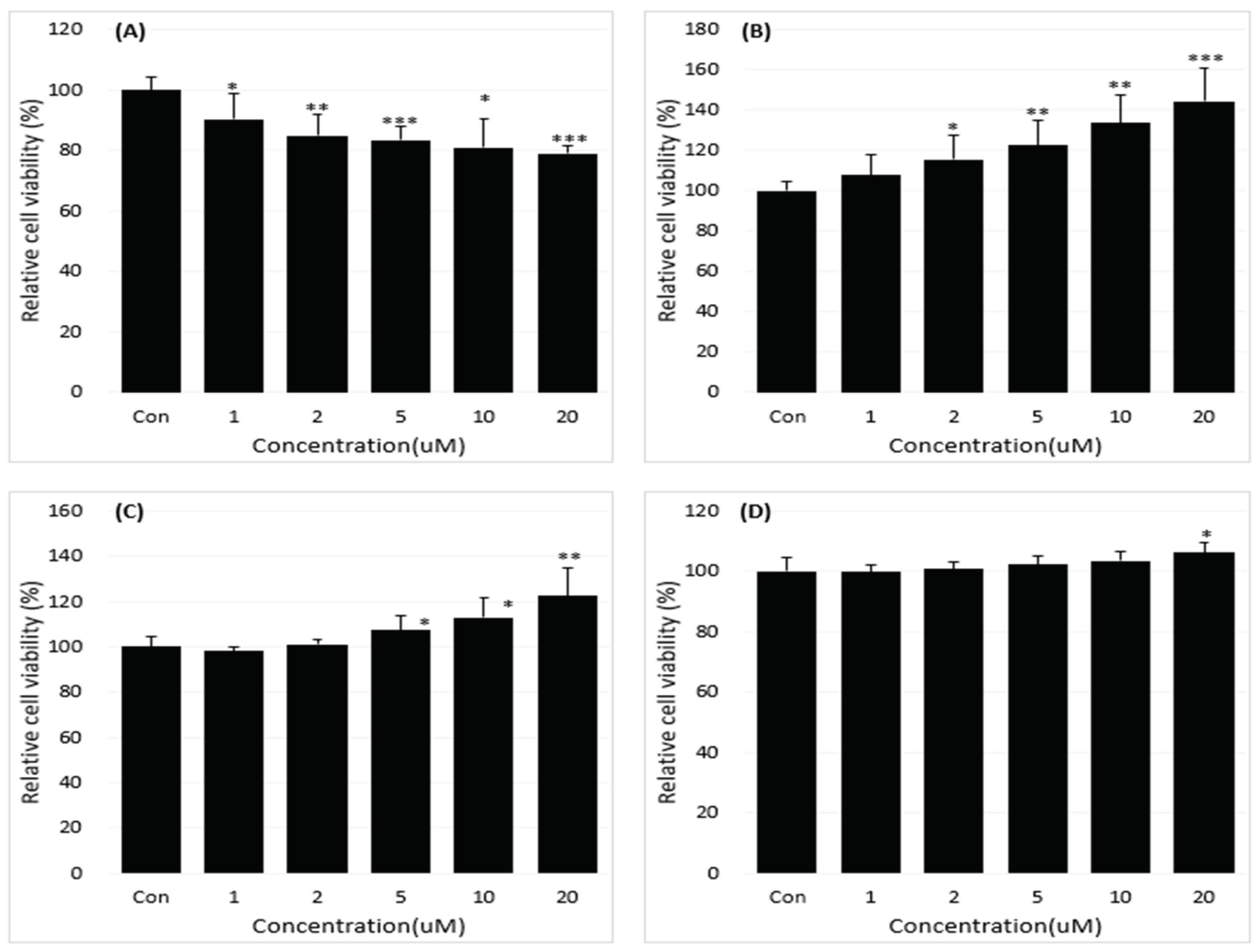
Figure 2.
Reduction of the B[a]P-induced in vitro toxicity in cells treated with 0, 1, 2, 5, 10, 20 µM concentrations of neferine for 48 h (A). The inhibitory effect of neferine on BPDE-DNA adduct formation by using enzyme-linked immunosorbent assay (ELISA) (B). All treatment group values were considerably different in comparison with the B[a]P (### p < 0.001, ## p < 0.01, # p < 0.05) and the controls (*** p < 0.001) in t-Test for Independent in SPSS.
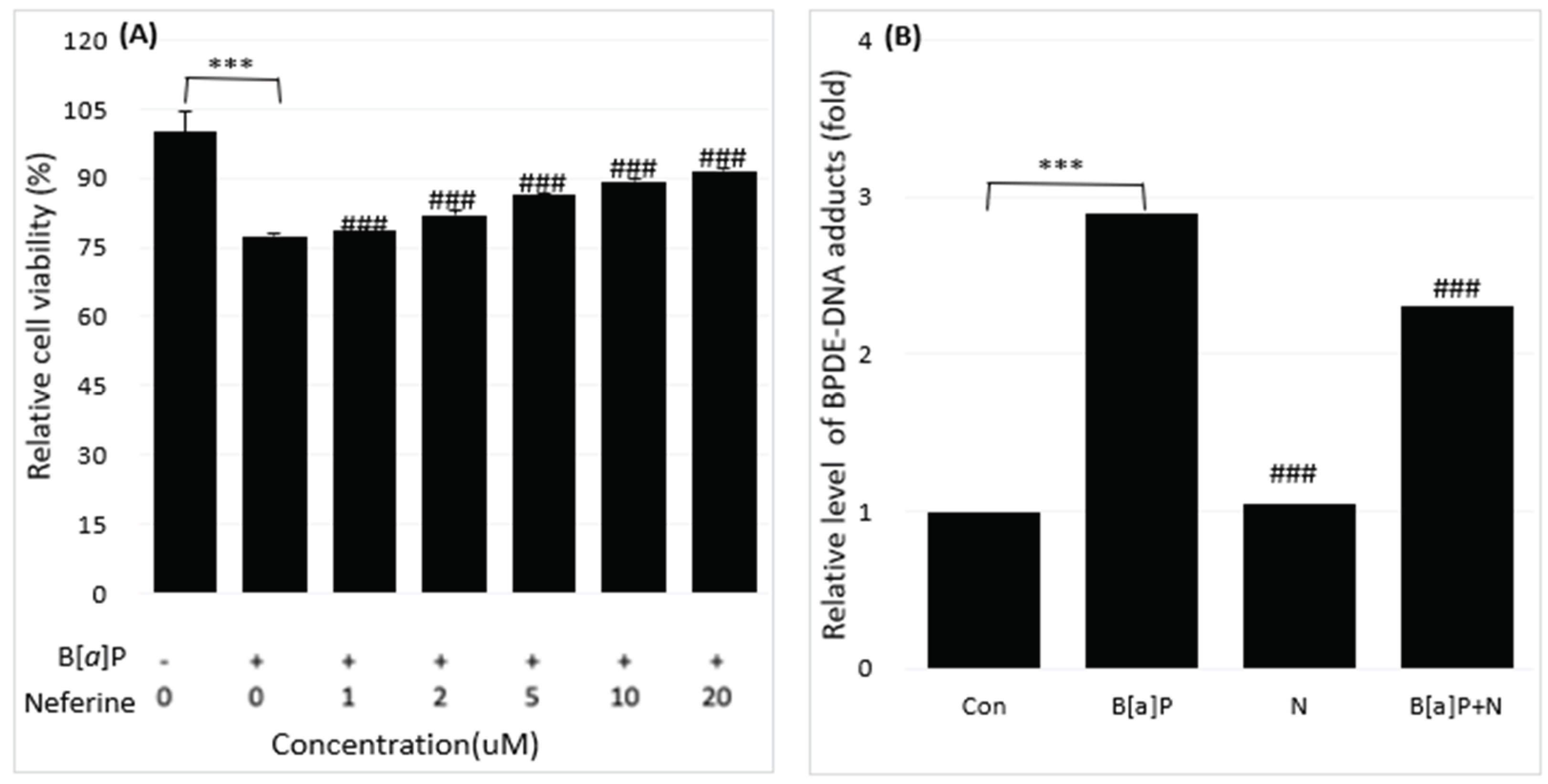
Figure 3.
Reduction of the B[a]P-induced in vitro toxicity in cells treated with 0, 1, 2, 5, 10, 20 µM concentrations of daidzein for 48 h (A). The inhibitory effect of daidzein on BPDE-DNA adduct formation by using enzyme-linked immunosorbent assay (ELISA) (B). All treatment group values were considerably different in comparison with the B[a]P (### p < 0.001, ## p < 0.01, # p < 0.05) and the controls (*** p < 0.001) in t-Test for Independent in SPSS.
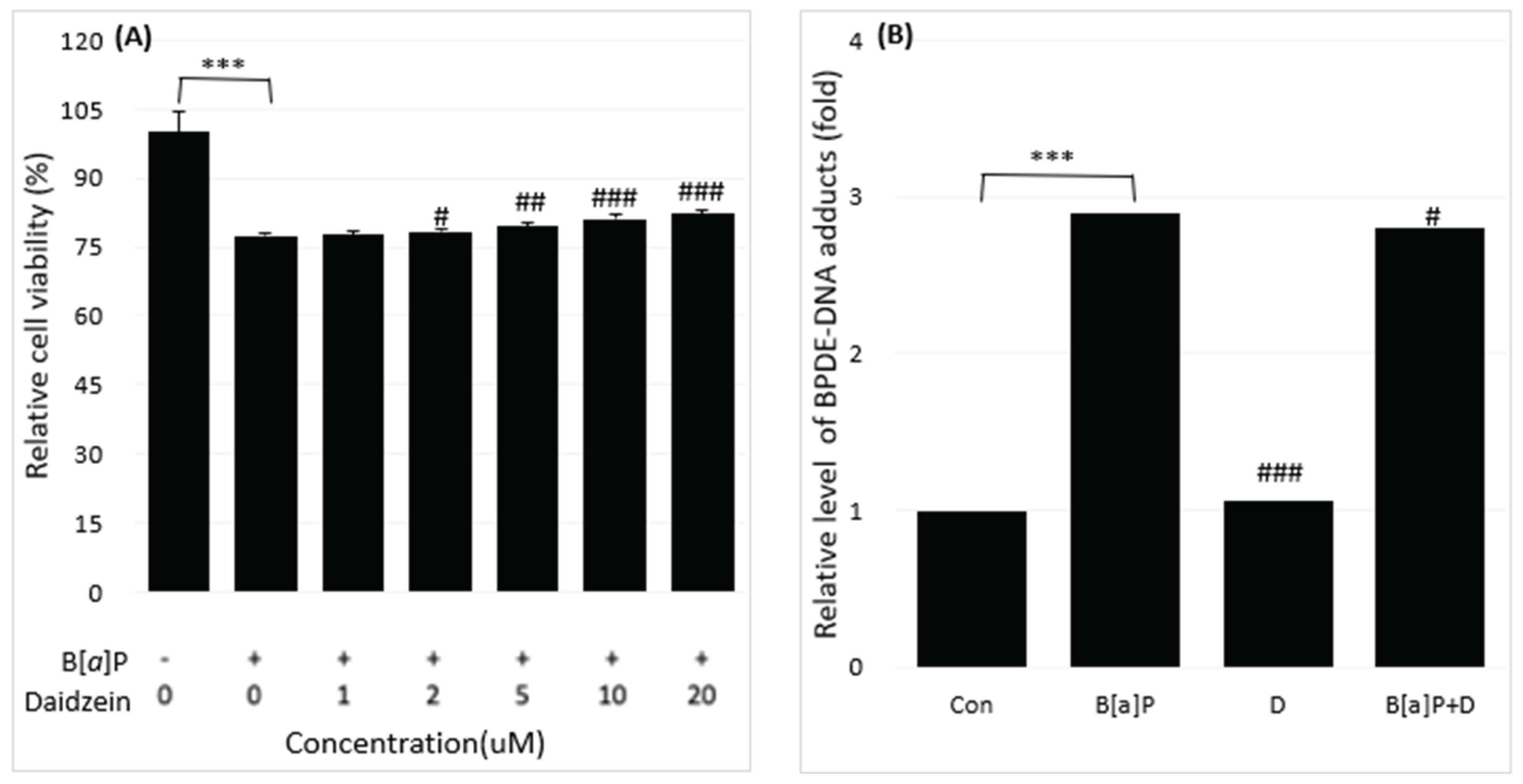
Figure 4.
Reduction of the B[a]P-induced in vitro toxicity in cells treated with 0, 1, 2, 5, 10, 20 µM concentrations of genistein for 48 h (A). The inhibitory effect of genistein on BPDE-DNA adduct formation by using enzyme-linked immunosorbent assay (ELISA) (B). All treatment group values were considerably different in comparison with the B[a]P (### p < 0.001, ## p < 0.01, # p < 0.05) and the controls (*** p < 0.001) in t-Test for Independent in SPSS.
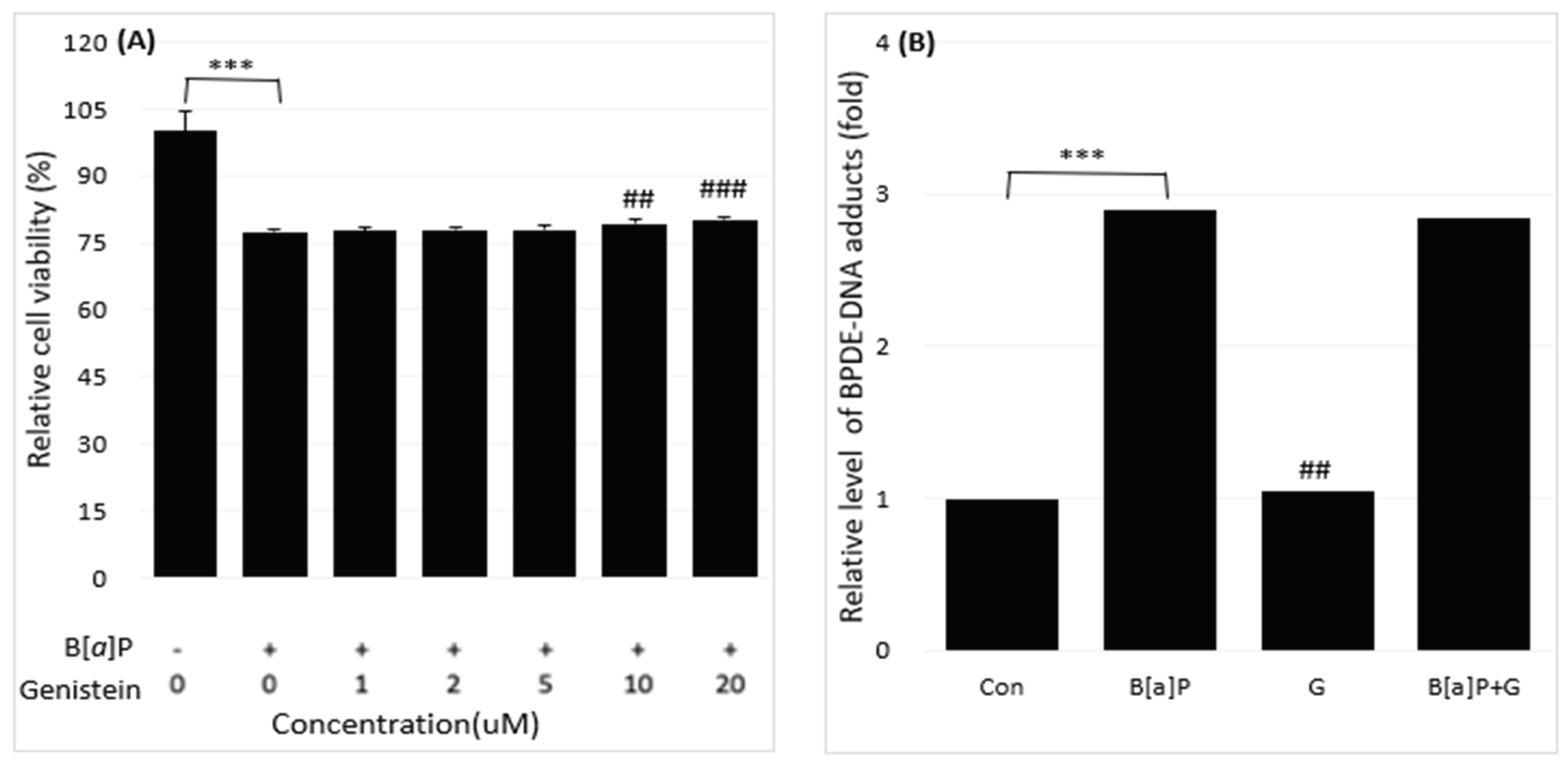
Figure 5.
The representative intracellular metabolites of B[a]P were measured by high performance liquid chromatography (HPLC). M3, BPDE; M2, B[a]P-7,8-dihydrodiol; M1, B[a]P that HepG2 cells were incubated with B[a]P (10 µM) and co-treated with neferine (20 µM) concentrations for 48 h.
Figure 5.
The representative intracellular metabolites of B[a]P were measured by high performance liquid chromatography (HPLC). M3, BPDE; M2, B[a]P-7,8-dihydrodiol; M1, B[a]P that HepG2 cells were incubated with B[a]P (10 µM) and co-treated with neferine (20 µM) concentrations for 48 h.
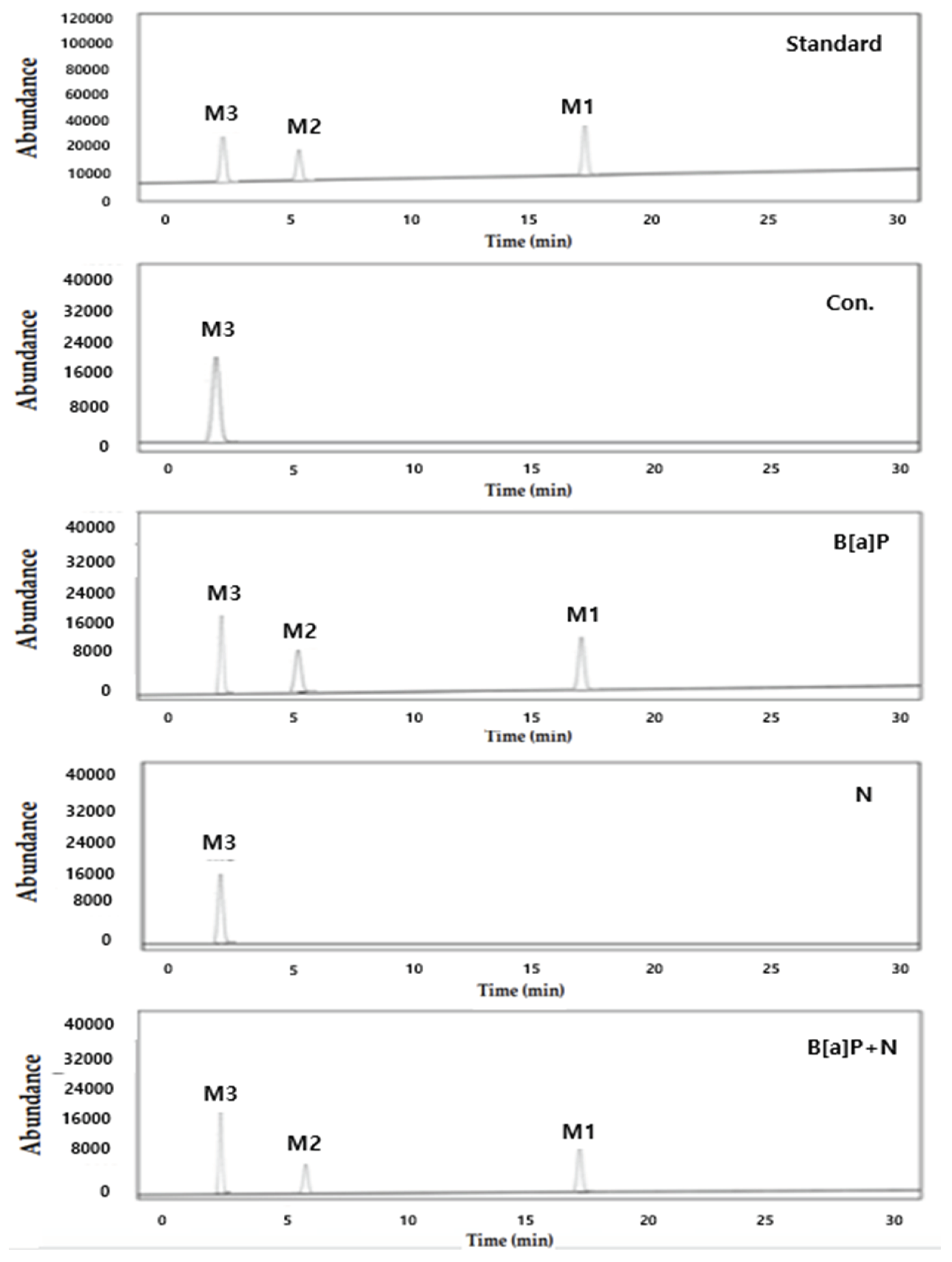
Figure 6.
The representative intracellular metabolites of B[a]P were measured by high performance liquid chromatography (HPLC). M3, BPDE; M2, B[a]P-7,8-dihydrodiol; M1, B[a]P that HepG2 cells were incubated with B[a]P (10 µM) and co-treated with daidzein (20 µM) concentrations for 48 h.
Figure 6.
The representative intracellular metabolites of B[a]P were measured by high performance liquid chromatography (HPLC). M3, BPDE; M2, B[a]P-7,8-dihydrodiol; M1, B[a]P that HepG2 cells were incubated with B[a]P (10 µM) and co-treated with daidzein (20 µM) concentrations for 48 h.
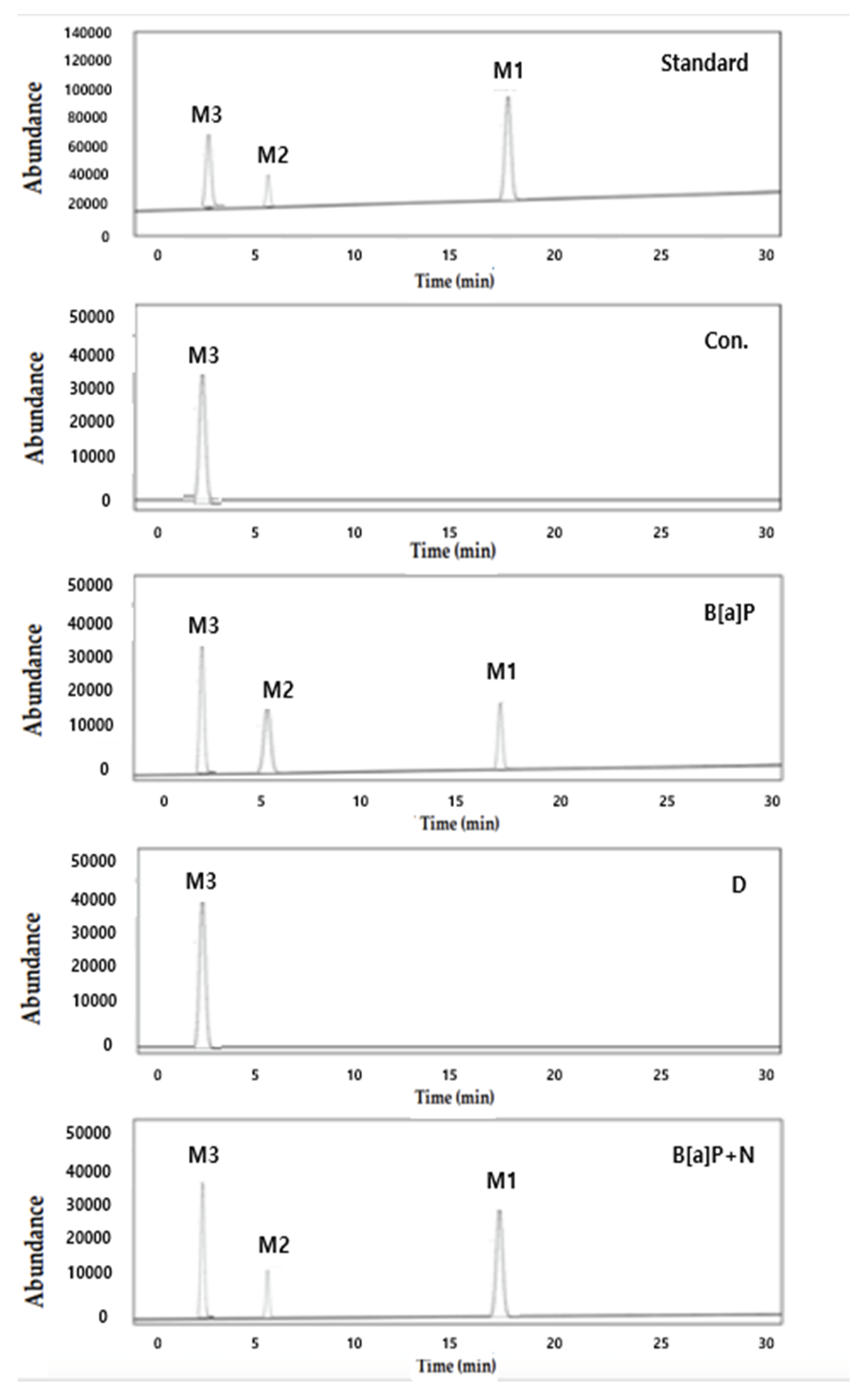
Figure 7.
The representative intracellular metabolites of B[a]P were measured by high performance liquid chromatography (HPLC). M3, BPDE; M2, B[a]P-7,8-dihydrodiol; M1, B[a]P that HepG2 cells were incubated with B[a]P (10 µM) and co-treated with genistein (20 µM) concentrations for 48 h.
Figure 7.
The representative intracellular metabolites of B[a]P were measured by high performance liquid chromatography (HPLC). M3, BPDE; M2, B[a]P-7,8-dihydrodiol; M1, B[a]P that HepG2 cells were incubated with B[a]P (10 µM) and co-treated with genistein (20 µM) concentrations for 48 h.
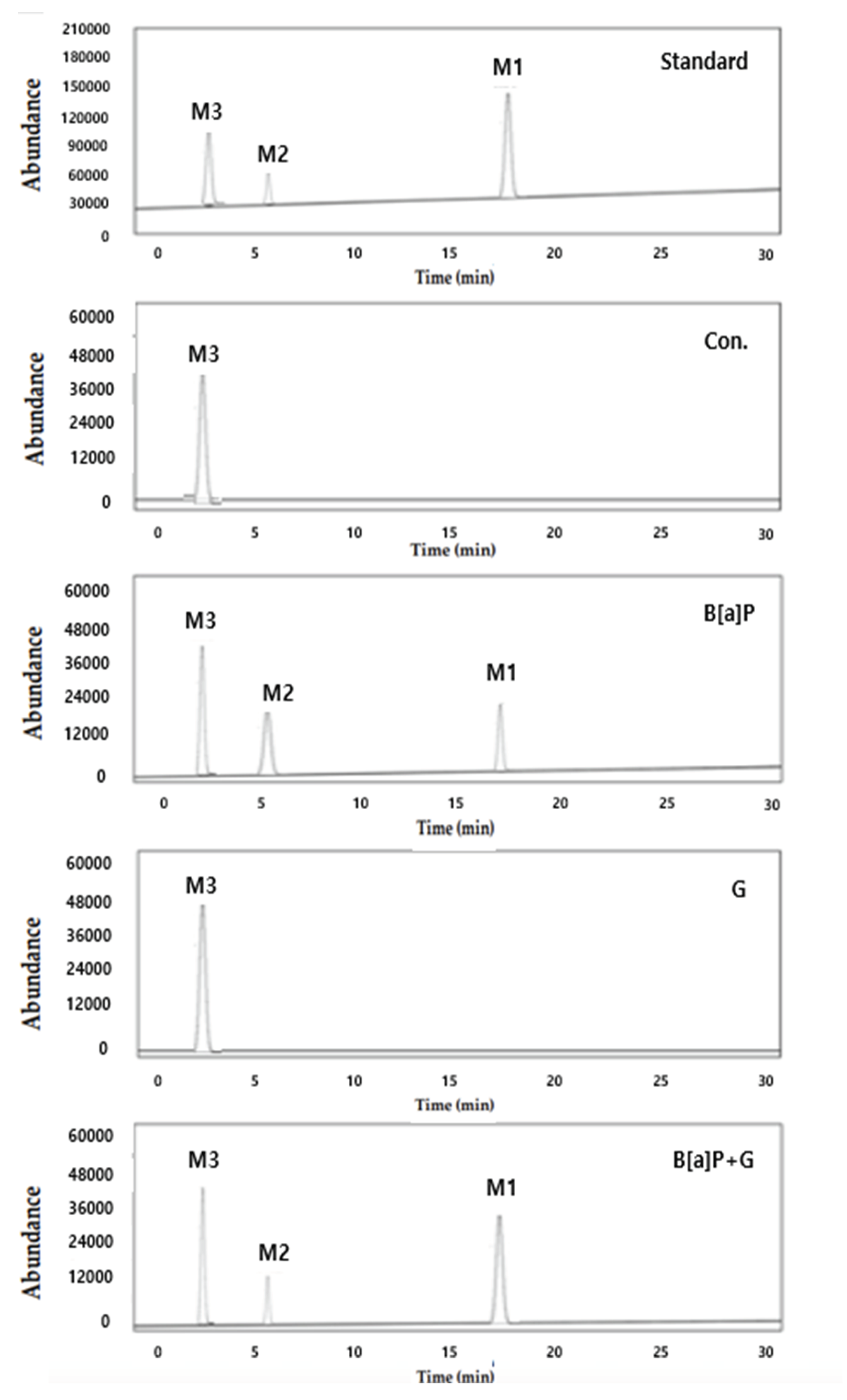
Figure 8.
The HPLC chromatograms of neferine, daidzein and genistein standards (A); neferine, daidzein and genistein in a spiked sample (B).
Figure 8.
The HPLC chromatograms of neferine, daidzein and genistein standards (A); neferine, daidzein and genistein in a spiked sample (B).
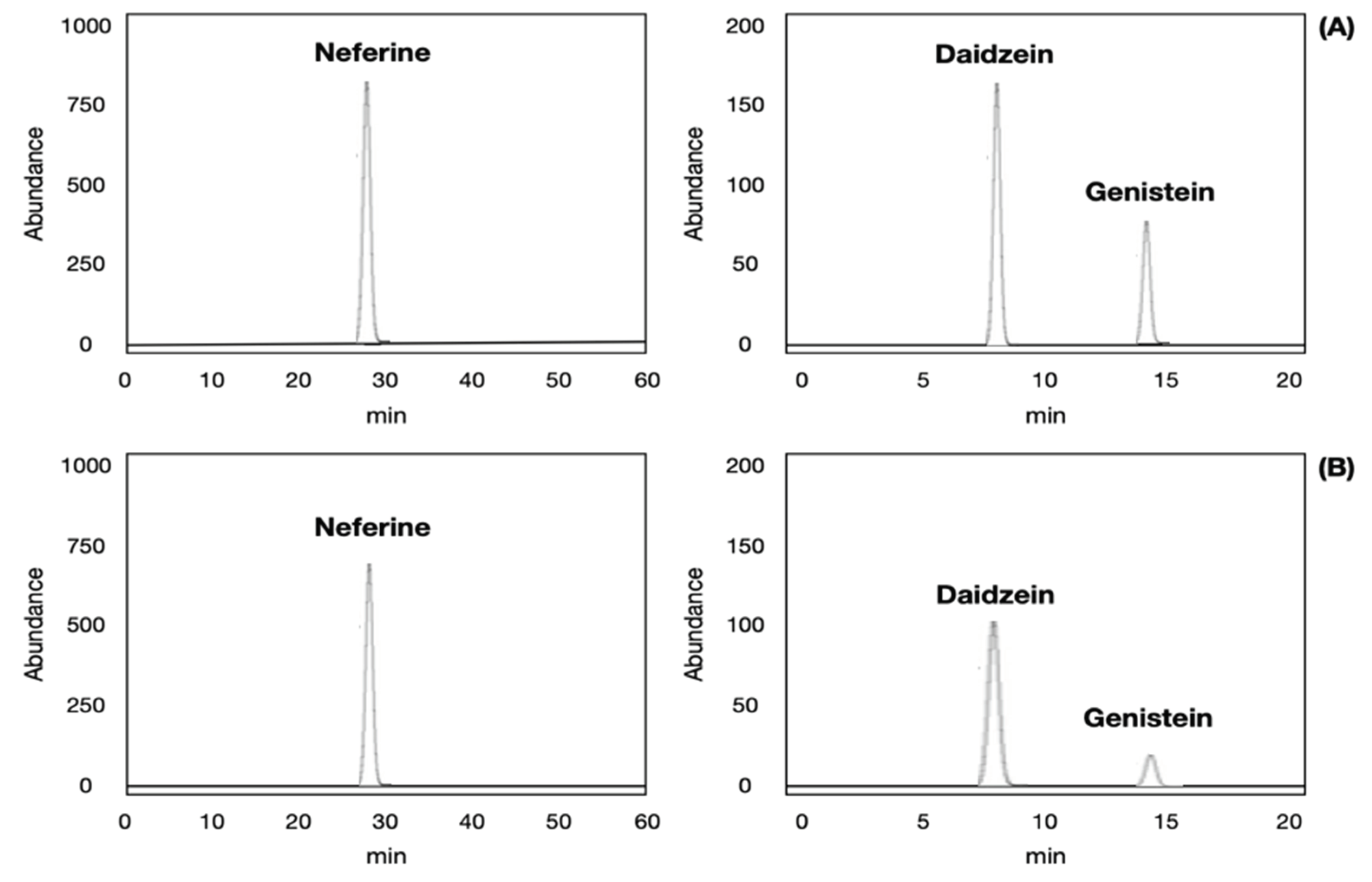

Table 1.
The limit of detection (LOD) (μg/kg), limit of quantification (LOQ) (μg/kg) and linearity (R2) and calibration equations for validation of the neferine, daidzein and genistein matrices using HPLC-UV analysis.
Table 1.
The limit of detection (LOD) (μg/kg), limit of quantification (LOQ) (μg/kg) and linearity (R2) and calibration equations for validation of the neferine, daidzein and genistein matrices using HPLC-UV analysis.
| Matrix type | Neferine, daidzein and genistein matrices | ||||
|---|---|---|---|---|---|
| LOD (μg/kg)1) |
LOQ (μg/kg)2) |
Linearity3) | Calibration equation | ||
| Lotus matrix (n = 3) |
Neferine | 0.12 | 0.36 | R2=0.9967 | y=0.0021x+0.007 |
| Soybean matrix (n = 3) |
Daidzein | 0.08 | 0.24 | R2=0.9958 | y=0.0012x-0.008 |
| Genistein | 0.09 | 0.27 | R2=0.9942 | y=0.0015x-0.005 | |
1) LOD is a signal-to-noise ratio (S/N) = 3.3. 2) LOQ is a signal-to-noise ratio (S/N) = 10. 3)Mathematical relationship between neferine, daidzein, genistein concentrations and result of chromatography analysis that can be represented as a straight line (n=3).
Table 2.
Recovery (%)1) for validation of the neferine, daidzein and genistein matrices using HPLC-UV analysis.
Table 2.
Recovery (%)1) for validation of the neferine, daidzein and genistein matrices using HPLC-UV analysis.
| Matrix type | Conc. | 3 (μg/kg) | 10 (μg/kg) | 40 (μg/kg) |
|---|---|---|---|---|
| Lotus matrix (n = 3) |
Neferine | 82.95±0.67 | 102.75±0.56 | 99.54±0.32 |
| Soybean matrix (n = 3) |
Daidzein | 93.02±2.45 | 111.46±5.67 | 86.72±0.11 |
| Genistein | 96.75±3.64 | 107.65±8.65 | 104.77±0.96 |
1) Recovery (%) was evaluated with 3,10, and 40 μg/kg concentration of spiked neferine in lotus and daidzein, genistein in soybean matrices and are shown as mean ± relative standard deviation (n=3).
Table 3.
Precision (%) for validation of the neferine, daidzein and genistein matrices using HPLC-UV analysis.
Table 3.
Precision (%) for validation of the neferine, daidzein and genistein matrices using HPLC-UV analysis.
| Matrix type | Conc. | Intraday (n=3) | Interday (n=3) |
|---|---|---|---|
| Precision (%)1) | Precision (%) | ||
| Lotus matrix (n = 3) |
Neferine | 0.24 | 0.65 |
| Soybean matrix (n = 3) |
Daidzein | 1.26 | 3.42 |
| Genistein | 6.82 | 4.67 |
1) Precision (%) was evaluated with CV (coefficient of variation, %) = (standard deviation/mean) × 100.
Disclaimer/Publisher’s Note: The statements, opinions and data contained in all publications are solely those of the individual author(s) and contributor(s) and not of MDPI and/or the editor(s). MDPI and/or the editor(s) disclaim responsibility for any injury to people or property resulting from any ideas, methods, instructions or products referred to in the content. |
© 2024 by the authors. Licensee MDPI, Basel, Switzerland. This article is an open access article distributed under the terms and conditions of the Creative Commons Attribution (CC BY) license (http://creativecommons.org/licenses/by/4.0/).
Copyright: This open access article is published under a Creative Commons CC BY 4.0 license, which permit the free download, distribution, and reuse, provided that the author and preprint are cited in any reuse.


