Submitted:
04 September 2023
Posted:
06 September 2023
You are already at the latest version
Abstract
: The cytotoxic effect of four extracts obtained from the wild plant and two from a culture of Ag-eratina pichinchensis calluses on cell lines: prostate carcinoma, cervical cancer, hepatocellular car-cinoma, human hepatoma, lung cancer and cellular keratinocytes was evaluated; the extracts were obtained with ethyl acetate and methanol, leaves and stems were used from the wild plant. The results of the cytotoxic evaluation showed that only the ethyl acetate extract obtained from the callus culture showed an effect on the cervical cancer cell line (HeLa) with an IC50 of 94.79 ± 2.0 µg/mL. The purification of this extract allowed obtaining a benzofuran type compound as the main product, which was identified as 2,3-dihydrobenzofuran. The cytotoxic evaluation of this compound showed an important effect against the HeLa cell line with an IC50 of 23.86 ± 2. .5µg/mL. These results contribute to the development of alternatives for the production of com-pounds for the treatment of cancer.
Keywords:
ytotoxic activity
; 2
; 3-dihydrobenzofuran
; cervical cancer
1. Introduction
Ageratina pichinchensis is a plants species endemic to the state of Morelos popularly known as “water sheet” and “axihuitl” mainly, it grows in the municipalities of Amatlán and Tepoztlán, where it is used for the treatment of gastric ulcers, healing, and diseases related to inflammatory events [1], it should be noted scientific phytochemical and pharmacological studies have validated its ethnomedical use [2,3,4,5,6,7], likewise, the establishment of its cultures of calluses and cells in suspension with the capacity to biosynthesize compounds structurally similar to those produced by the wild plant, which showed an important anti-inflammatory effect, has been reported. It should be noted that the compounds 3-epilupeol and a benzofuran were identified that had not been reported in the wild plant, whose production was improved in an airlift reactor 1.82 and 1.35 folds, respectively [8,9,10].
However, other biological properties have not been explored, such as those related to the cytotoxic effect, which has become relevant due to the impact in the health area in the search for molecules for the treatment of neoplastic diseases. The WHO estimates that cancer is the leading cause of death in the world and more than 18 million deaths were reported worldwide in 2022 alone, the most common being breast, lung, colon, rectum, and prostate [11,12], this has led to the search for and development of solutions from different approaches, given that the causes are diverse and diagnosis is also a challenge, which is why multiple research groups around the world have dedicated themselves to the task of searching for therapeutic compounds of synthetic and natural origin [13,14,15] to be applied in therapies or treatments that are already applied in order to improve their effectiveness; one of the common treatments in cancer patients are chemotherapies, which make use of molecules that act at different levels of the cell cycle, successfully saving lives or improving the quality of patients, despite the percentage of efficacy of chemotherapies, the search for therapeutic agents continues to be a necessary due to the high global demand [16,17,18].
In view of the above, medicinal plants are a prominent source of compounds with cytotoxic effects in different types of cancer, some of the molecules commonly used in chemotherapies are the tetracyclic diterpenoid compound taxol, which is isolated from the bark of species of the genus Taxus, and is commercially known as paclitaxel® and it has been impossible to develop the synthetic route due to the complexity of the molecule, which has led to its direct obtaining from the plant, with the disadvantage that trees require 15 years of age to be able to biosynthesize the compound [19,20]; on the other hand, the vinblastine and vincristine alkaloid-type compounds from Catharanthus roseus present yields below 0.001% and their cost ranges from one billion dollars per kilogram, but they are used for their efficacy in chemotherapies for patients with leukemia [21]; other potentially anticancer alkaloids are colchicine, vindesine, vinorelbine, podophyllotoxin, decotaxel, campotecin, curcumin, apigenin and vincamine, all of which have a common factor in their origin from plant species [22,23,24,25,26]. On the other hand, extracts of medicinal plants have also shown a cytotoxic effect where the compounds participate synergistically, as has occurred with the species Aristolochia baetica, Artemisia annua, Fagonia indica, among others [27,28]. However, there are numerous plant species around the world that have not yet been studied in Mexico alone, more than 300 endemic plants of different families and genera are used, of which only a proportion have been scientifically studied [29,30]. On the other hand, there are reports of cultures of calluses and cells in suspension of plant species whose main advantage is the production of bioactive compounds in a constant and controlled manner, which is a useful alternative because medicinal plants in wild conditions are affected by different seasonal, environmental, geographical conditions, etc. These factors are determinant in the production and yields of secondary metabolites and put medicinal plants at a disadvantage as a sufficient source for world needs in tumor pathologies. For this reason, this paper reports the study of wild plant extracts and callus cultures of the species A. pichinchensis whose scientific findings demonstrate its cytotoxic effect on the HeLa cell line, associating the chemical constituents with the activity observed in the extracts.
2. Results and Discussion
2.1. Cytotoxic evaluation of extracts
The cytotoxic activity of ethyl acetate and methanol extracts from callus cultures and wild Plants of A. pichinchensis was evaluated at different concentrations on cell lines: human prostate carcinoma (PC-3), cervical cancer (HeLa), hepatocellular carcinoma (Huh-7), human hepatoma (HepG2), breast tumor cells (MCF7), and keratinocyte cells (Hacat). The methanolic and ethyl acetate extracts of the wild plants and their callus cultures did not show a significant effect on the cell lines, while the ethyl acetate extract of the wild plant leaves had a slight effect on the HeLa cells and PC-3; however, the ethyl acetate extract of the callus cultures showed a greater effect on the cell lines (Table 1), with the HeLa cell line where this effect was more relevant.
A. pichinchensis, is widely used in traditional Mexican medicine. It is used to treat diseases caused by fungal and skin infections, wounds, as well as to relieve pain and treat gastric ulcers, and anti-inflammatory effects have been reported. Extracts from this plant have shown antifungal activity against Trichophyton mentagrophytes, T. rubrum and Candida albicans, and have shown therapeutic effectiveness in patients with vulvovaginal candidiasis [7,31].
It should be noted that the ethyl acetate extract of the callus culture contains compounds (1-11) (Fig.1), with compound 7 (2,3-dihydrobenzofuran) being a major one.
Figure 1.
Compounds identified in the ethyl acetate extract of A. pichinchensis callus.
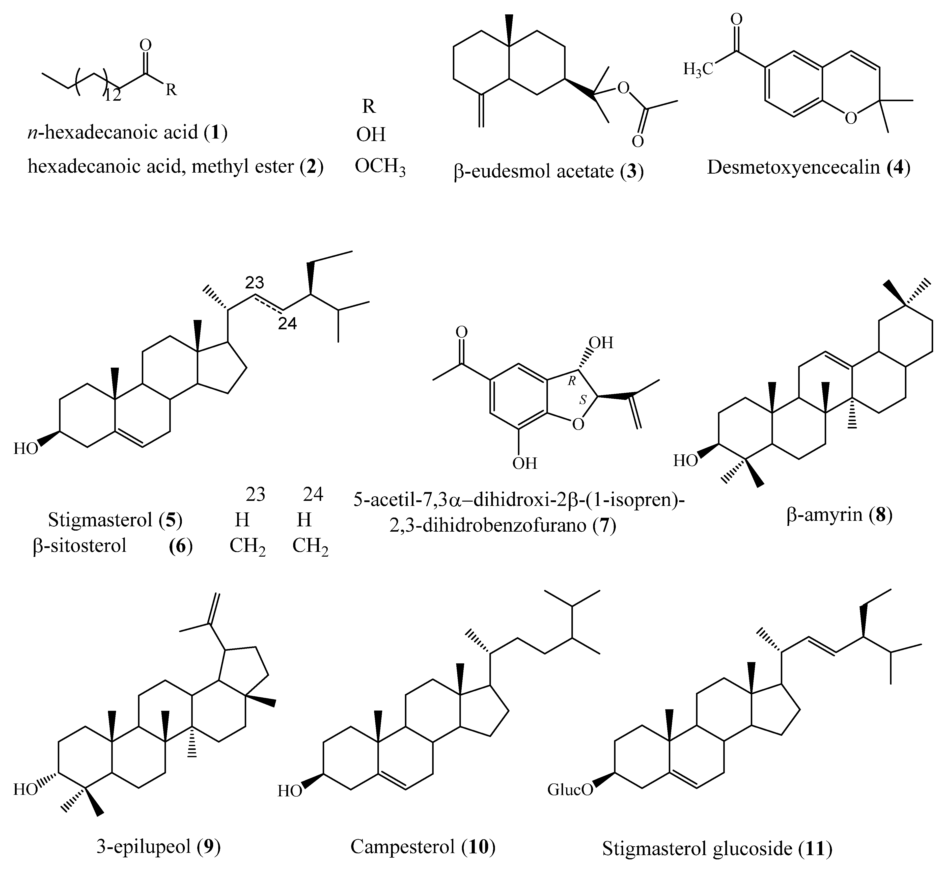
The compound campesterol (10) has been reported as an important phagocytosis suppressor and inhibitor of lipopolysaccharide in RAW 264 macrophage cells [32], in addition, extracts of different plant species with anti-inflammatory effect have revealed that the presence of the compound participates in the biological effect attributed to the extracts, such as in Cajanus cajan L. seeds, Ananas comosus leaves, Allium schoenoprasum L. leaves and in Opuntia ficus-indica seed oil [33,34,35,36] among other species.
Another compound identified in the extract is hexadecenoic acid (1), which has been shown to be an important inhibitor of the PLA2 enzyme that plays a role in inflammation of blood vessels and favors the development of atherosclerosis [37], it should be noted that it has been reported in other species, which stand out for their anti-inflammatory effect evaluated at the extract level, such as the species Chasmanthe aethiopica, Zostera japonica and Jatropha curcas [38,39,40]; likewise, the pentacyclic triterpene-type compound β-amyrin (8) has also exhibited anti-inflammatory effect induced in lipopolysaccharide and interferon-γ and even significantly reduces the expression of the levels of proinflammatory factors TNF-α, IL-1β, IL-6, PGE-2 and COX-2 [41,42], in addition, its cytotoxic effect was reported in the MCF-7 breast cancer cell line with IC50 15.5 µg/mL, and it has also shown a cytotoxic effect in colon, ovarian, cervical, lung, and breast cancer lines [43,44,45,46,47], and it has been reported in species from different families such as Alstonia boonei, Protium heptaphyllum and Gymnosporia montana [48,49,50,51] in addition, it has been attributed an antioxidant effect, as it has been in the species Myrcianthes pungens and Celastrus hindsii [52,53]; on the other hand, the compound 3-epilupeol (9) has revealed an important anti-inflammatory effect and the species of the genus Bursera are the main ones in producing it, whose extracts at the extract level reveal cytotoxic activity in most cell lines [54,55,56]; on the other hand, the stigmasterol glycoside compound has revealed a cytotoxic effect on kidney, breast and liver tumor cells [57,58,59,60], other reports argue its anti-inflammatory and antimicrobial effect antimicrobial [61,62,63]; finally, benzofuran compound has been reported as anti-inflammatory [10].
These scientific reports show the therapeutic effect attributed to the compounds and their respective plant species, therefore, the cytotoxic effect identified in the ethyl acetate extract of callus cultures is due to the presence of the compounds that act synergistically, significantly enhancing their effect, outstandingly, the extract biosynthesizes the compounds in a particular and constant way, which represents an advantage over wild plants whose production of compounds depends on environmental, geographical, and seasonal factors, among others, which is why calluses represent an important source of molecules with therapeutic effects as supported by numerous scientific reports.
Several studies indicate antiviral, antioxidant, antimicrobial, antitumor activities of dihydrofurans [64,65,66]. Considering this analyzed the effect of 2,3-dihydrobenzofuran on the inhibition of cell proliferation of the study cell lines. A clear effect on inhibition of HeLa cells is observed in Figure 2.
Figure 2.
Results of the cytotoxic evaluation of 2,3-dihydrobenzofuran (7) in cancer cells.
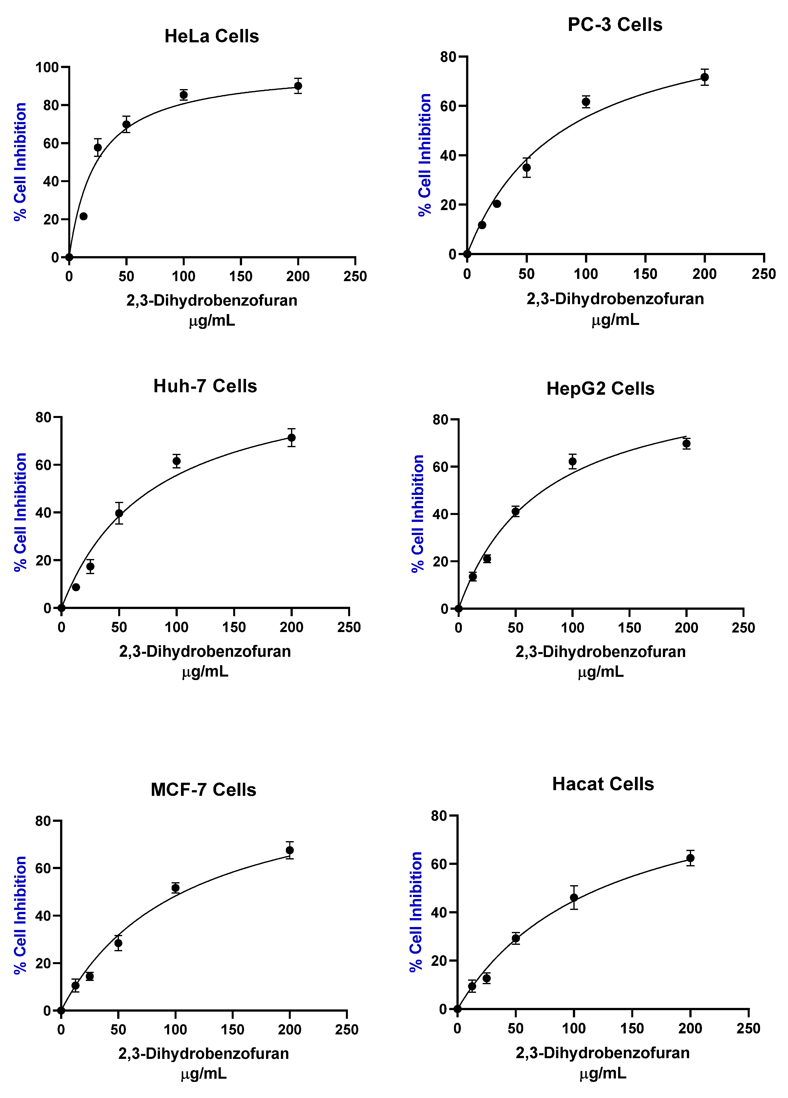
In addition, is notable that HeLa cervical cancer cells were the most sensitive to the treatment of 2,3-Dihydrobenzofuran (7) (Table 2) with a IC50 of 23.86 µg/mL 3.97 times lower than IC50 of ethyl acetate extract of callus cultures of A. pichinchensis. This may suggest that the cytotoxic activity is associated with the effect of this compound, also a selective for cancer cells was also observed. The selectivity index is commonly reported in the literature as a ratio of IC50 values calculated for healthy and cancer cells [67,68] with values greater than 1 indicating desirable selectivity against cancer cells. The selectivity index of 2,3-dihydrobenzofuran (7) was 5.17, 1.53, 1.54, 1.65, 1.15 for HeLa, PC-3, Huh-7, HepG2, MCF-7 cells respectively.

Table 2.
Cytotoxic activity of 2,3-dihydrobenzofuran (7) compound of A. pichinchensis.
| Cellular line | IC50 µg/mL |
|---|---|
| HeLa | 23.86 ± 2.5 |
| PC-3 | 80.3 ± 4.6 |
| Huh-7 | 79.9 ± 3.78 |
| HepG2 | 74.62 ± 2.02 |
| MCF7 | 107.2 ± 1.52 |
| Hacat | 123.5 ± 15.17 |
Previously, reported an important anti-inflammatory activity of 2,3-dihydrobenzofuran (7) [10], therefore, the results of our study reveal a relevant biological activity of the compound, with the advantage that its production is constant and controlled by the callus cultures of A. pichinchensis. This novel contribution shows that callus cultures represent a viable alternative for the production of bioactive compounds. It should be noted that the production of the compound has been improved in suspension cell cultures grown in flasks and airlift flasks [8,9].
3. Materials and Methods
3.1. Plant material from the wild plant
The plant species was collected in its natural habitat in the San Juan Tlacotenco neighborhood of the municipality of Tepoztlán in the state of Morelos, Mexico, and was identified by our research group in previously reported works and assigned the voucher number 39913. The specimen is under protection in the HUMO herbarium of the Autonomous University of the State of Morelos (UAEM), whose taxonomic identification was carried out by Biol. Gabriel Flores Franco [10].
3.2. Plant material from callus cultures
Calluses were previously established by our research group using leaf explants in Murashige and Skoog culture medium sterile (121 °C, 15 psi, for 15 min using an autoclave) supplemented with 30 g/L sucrose, 1 mg/L naphthaleneacetic acid, 0.1 mg/L kinetin, and 3 g/L phytagel [10]. Callus subcultures were carried out every 20 days and incubated at 25 ± 2 °C under photoperiod of 16 h with white fluorescent light (50 µmol / m2 s).
3.3. Obtaining organic extracts
In Tepoztlán Morelos, 1465 g of fresh A. pichinchensis plant were collected, the plant was separated into leaves (910.73 g) and stems (415.76 g), these were dried under the shade at room temperature, finally obtaining leaves (145.18 g) and stems (78.13 g) dried. On another hand, 570.42 g of fresh biomass obtained from a callus culture of A. pichinchensis previously established were harvest, 13.20 g of dry biomass were obtained. The dry plant material (leaves and stem) and dry biomass (callus culture) were ground and extracted by maceration (72 hours, at room temperature) first with ethyl acetate and later with methanol (in triplicate). The solvent was removed by distillation under reduced pressure, using a rotary evaporator, finally obtaining 3 ethyl acetate extracts; leaves (17.20 g), stems (7.31 g) and callus culture (0.924 g) and 3 with methanol; leaves (59.09 g), stems (28.47 g) and callus culture (2.346 g)
3.4. Antiproliferative assay
PC-3 (prostate), HeLa (cervical), MCF7 (breast) and Huh-7 and HepG2 (hepatocellular) human cancer cell lines were purchased from ATCC (Manassas, VA, USA). We also included immortalized human epidermal keratinocyte line (Hacat) as a control of non-cancerous cells [69]. PC-3 was grown in RPMI-1640 medium (Sigma Aldrich, St. Louis, MO, USA), while Huh-7, HepG2, HeLa and Hacat in DMEM medium (Invitrogen, Thermo Fisher Scientific, Waltham, MA, USA) supplemented with 10% SFB and 2 mM glutamine, all cultures were incubated at 37 °C in 5% CO2.
Cells (4 × 10³ cells/well) were seeding in 96-well plates. The cells were treated with the investigated samples (at 200, 100, 50, 25 and 12.5 µg/mL) and incubated at 37 °C in 5% CO2 for 48 h. Paclitaxel was used as positive control. For determining the number of viable cells in proliferation we used CellTiter 96® AQueous One Solution Cell Proliferation Assay kit (Promega, Madison, WI, USA), following the manufacturer’s instructions. Cell viability was determined by absorbance at 450 nm using an automated ELISA reader (Promega, Madison, WI, USA). Stock solutions of all compounds were prepared in DMSO at a maximum concentration of 0.5%. The experiments were conducted by triplicate in three independent experiments. Data were analyzed in Prism 8.0 statistical program (Graphpad Software Inc., La Jolla, CA, USA) and the Half maximal inhibitory concentration (IC50) were determined by regression analysis.
3.5. Purification of active compounds active ethyl acetate extract (Callus culture)
The chemical composition was determined by our research group in a previous study, which is why in this work we replicated the methodology and found reproducibility in the reported compounds [10], for which the technique of gas chromatography coupled with mass spectrometry was used that allowed the identification of the compounds in the present study, 2 mg of the extract were weighed and dissolved in 1 mL of chloroform to be analyzed by GC-MS (NIST 1.7a). The harvested calluses were dried in an oven at 40 °C, the dry biomass (14.76 g) was extracted three times with 100 ml of ethyl acetate by maceration; each extraction was every 72 hours at room temperature. The solvent was evaporated under reduced pressure and a yellow viscous extract (0.6859 g) was obtained. The ethyl acetate extract was fractionated on an open chromatographic column pre-packed with silica gel 18.76 g, (70-230 mesh; Merck) and eluted with a gradient system of n-hexane/ethyl acetate (90:10, 80:20, 70:30, 60:40, 50:50, 40:60, 30:70 and 00:100, v/v). 10 ml fractions were collected to obtain 45 fractions and monitored by TLC (ALUGRAM® SIL G/UV254 silica gel plates). The fractions that showed similarity in TLC were grouped obtaining 7 groups: GE-1 (1–16; 0.1802 mg), GE-2 (17–18; 15.6 mg), GE-3 (19–20; 78.3 mg), GE -4 (21–25; 40.8 mg), GE-5 (26–36; 81.6 mg), GE-6 (37–40; 70.6 mg), and GE-7 (41–45; 92.6 mg). The GE-1, GE-2 and GE-4 fractions were analyzed by GC-MS, this analysis indicated that the GE-1 fraction is made up of the mixture of n-hexadecanoic acid (1) and the ester of hexadecanoic acid (2), Fraction GE-2, by β-eudesmol acetate (3) and desmethoxyencecalin (4), purification of fraction GE-3 by column chromatography, using a gradient of n-hexane-ethyl acetate ( 100:00→70:30) provided 30 fractions. fractions 15–22 eluted with n-hexane-ethyl acetate (90:10) contained stigmasterol (5) and β-sitosterol (6), fractions 23-28 showed a single compound, identified by 1H and 13C NMR and by comparison with their values reported in the literature [10] as (2S,3R)-5-acetyl-7,3-dihydroxy-2-(1-isopropenyl)-2,3-dihydrobenzofuran (15.3 mg, 7). The GE-4 fraction is made up of α-amyrin (8), epilupeol (9) and campesterol (10). Compound (11) was identified in the GE-5 fraction by direct comparison with an authentic sample available in the laboratory.
4. Conclusions
For the first time, it is shown that the chemical content of the ethyl acetate extract from A. pichinchensis callus cultures exhibits an important cytotoxic effect on the HeLa cell line, whose activity is suggested to be attributed to 2,3-dihydrobenzofuran, which has also shown anti-inflammatory effects. These results contribute to the development of alternatives for the treatment of cervical cancer, which has become a health problem that causes many deaths worldwide, particularly, this finding opens the possibility of new applications of callus culture extracts. of A. pichinchensis since the extracts of the wild plant did not show the cytotoxic effect. This is because in vitro cultures produce the compounds of interest in a constant and controlled manner, while wild plants depend on environmental, geographical, seasonal, among others.
Author Contributions
Conceptualization, M.S.-R., F.C.-S. and J.N.S.‒C.; methodology, S.M.‒B., J.N.S.‒C. and J.G.E.‒G.; software, E.C.‒G. and F.C.-S.; validation, E.C.‒G., J.G.E.‒G. and F.C.-S.; formal analysis, V.D.‒V and A.B.‒A.; investigation, M.S.-R., J.G.E.‒G. and F.C.-S. ; resources, S.M.‒B. and E.C.‒G.; writing—original draft preparation, M.S.-R., F.C.-S. and J.N.S.‒C.; writing—review and editing, S.M.‒B., J.N.S.‒C., J.G.E.‒G., V.D.‒V., A.B.‒A., E.C.‒G. and S.M.‒B.; supervision, V.D.‒V., F.C.-S. and A.B.‒A.; funding acquisition, F.C.-S. and S.M.‒B.
Funding
This research received no external funding.
Institutional Review Board Statement
Not applicable.
Informed Consent Statement
Not applicable.
Data Availability Statement
Not applicable.
Acknowledgments
The authors thank Laboratorio Nacional de Estructura de Macromoléculas (CONACYT 279905) for the use of gas chromatography coupled with mass spectrometry and Maria Gregoria Medina Pintor (Centro de Investigaciones Químicas, UAEM) for skillful technical assistance in the analysis and GC/MS experiments.
Conflicts of Interest
The authors declare no conflict of interest.
References
- Argueta, A.; Cano, L.; Rodarte, M. Atlas de la Medicina Tradicional Mexicana, Tomo 1-3; Instituto Nacional Indigenista: Mexico, City, 1994, p. 1786.
- Romero-Cerecero, O.; Rojas, G.; Navarro, V.; Herrera-Arellano, A.; Zamilpa-Alvarez, A.; Tortoriello, J. Effectiveness and tolerability of a standardized extract from Ageratina pichinchensis on patients with tinea pedis: An explorative pilot study controlled with ketaconazole. Planta Medica 2006, 72, 1257-1261.
- Romero-Cerecero, O.; Román-Ramos, R.; Zamilpa, A.; Jiménez-Ferrer, J.E.; Rojas-Bribiesca, G.; Tortoriello, J. Clinical trial to compare the effectiveness of two concentrations of the Ageratina pichinchensis extract in the topical treatment of onychomycosis. J. Ethnopharmacol. 2009, 126, 74-78. [CrossRef]
- Romero-Cerecero, O.; Zamilpa, A.; González-Cortazar, M.; Alonso-Cortes, D.; Jiménez-Ferrer, E.; Aguilar-Santamaría, L.; Tortoriello, J. Pharmacological and chemical study to identify wound-healing active compounds in Ageratina pichinchensis. Planta Medica 2013, 79, 622-627. [CrossRef]
- Romero-Cerecero, O.; Zamilpa, A.; Jiménez-Ferrer, J.E.; Rojas-Bribiesca, G.; Román-Ramos, R.; Tortoriello, J. Double-blind clinical trial for evaluating the effectiveness and tolerability of Ageratina pichinchensis extract on patients with mild to moderate onychomycosis. A comparative study with ciclopirox. Planta Medica 2008, 78, 1430-1435. [CrossRef]
- Romero-Cerecero, O.; Zamilpa-Álvarez, A.; Ramos-Mora, A.; Alonso-Cortés, D.; Jiménez-Ferrer, J.E.; Huerta-Reyes, M.E.; Tortoriello, J. Effect on the wound healing process and in vitro cell proliferation by the medicinal mexican plant Ageratina pichinchensis. Planta Medica 2011, 77, 979-983. [CrossRef]
- Aguilar-Guadarrama B.; Navarro V.; León-Rivera I.; Ríos MY. Active compounds against tinea pedis dermatophytes from Ageratina pichinchensis var. bustamenta. Nat. Prod. Res. 2009, 23, 1559-1565. [CrossRef]
- Sánchez-Ramos, M.; Alvarez, L.; Romero-Estrada, A.; Bernabé-Antonio, A.; Marquina-Bahena, S.; Cruz-Sosa, F. Establishment of a cell suspension culture of Ageratina pichinchensis (Kunth) for the improved production of anti-inflammatory compounds. Plants 2020, 9, 1398. [CrossRef]
- Sánchez-Ramos, M.; Marquina-Bahena, S.; Alvarez, L.; Bernabé-Antonio, B.; Cabañas-García, E.; Román-Guerrero, A.; Cruz-Sosa, F. Obtaining 2,3-dihydrobenzofuran and 3-epilupeol from Ageratina pichinchensis (Kunth) R. King & Ho. Rob. Cell cultures grown in shake flasks under photoperiod and darkness, and its scale-up to an airlift bioreactor for enhanced production. Molecules 2023, 28, 578. [CrossRef]
- Sánchez-Ramos, M.; Marquina-Bahena, S.; Romero-Estrada, A.; Bernabé-Antonio, A.; Cruz-Sosa, F.; González-Christen, J.; Acevedo-Fernández, J.J.; Perea-Arango, I.; Alvarez, L. Establishment and phytochemical analysis of a callus culture from Ageratina pichinchensis (Asteraceae) and its anti-inflammatory activity. Molecules 2018, 23, 1258. [CrossRef]
- https://www.who.int/es/news/item/04-02-2020-who-outlines-steps-to-save-7-million-lives-from-cancer.
- Ferlay, J.; Colombet, M.; Soerjomataram, I.; Parkin, D.M.; Piñeros, M.; Znaor, A.; Bray, F. Cancer statistics for the year 2020: An overview. Int. J. Cancer 2021, 149, 778-798. [CrossRef]
- Rayan, A.; Raiyn, J.; Falah, M. Nature is the best source of anticancer drugs: Indexing natural products for their anticancer bioactivity. PLoS ONE 2017, 12, e0187925. [CrossRef]
- Khalifa, S.A.M.; Elias, N.; Farag, M.A.; Chen, L.; Saeed, A.; Hegazy, M.-E.F.; Moustafa, M.S.; El-Wahed, A.A.; Al-Mousawi, S.M.; Musharraf, S.G.; Chang, F.-R.; Iwasaki, A.; Suenaga, K.; Alajlani, M.; Göransson, U.; El-Seedi, H.R. Marine natural products: A source of novel anticancer drugs. Marine drugs 2019, 17, 491. [CrossRef]
- Yao, C.-L.; Zhang, J.-Q.; Li, J.-Y.; Wei, W.-L.; Wu, S.-F.; Guo, D.-A. Traditional chinese medicine (TCM) as a source of new anticancer drugs. Nat. Prod. Rep. 2020, 9, 1618-1633. [CrossRef]
- Obenauf, A.C. Mechanism-based combination therapies for metastatic cancer. Sci. Transl. Med. 2022, 14, eadd0887. [CrossRef]
- Ramakrishnan, R.; Gabrilovich, D.I. Mechanism of synergistic effect of chemotherapy and immunotherapy of cancer. Cancer Immunol Immun 2011, 60, 419-423. [CrossRef]
- Sharma, H.; Parihar, L.; Parihar, P. Review on cancer and anticancer properties of some medicinal plants. J. Med. Plant Res. 2011, 5, 1818-1835.
- Howat, S.; Park, B.; Oh, I.S.; Jin, Y.-W.; Lee, E.-K.; Loake, G.J. Paclitaxel: Biosynthesis, production and future prospects. N. Biotechnol. 2014, 31, 242-245. [CrossRef]
- Yan-Hua, Y.; Jia-Wang, M.; Xiao-Li, T. Research progress on the source, production, and anti-cancer mechanisms of paclitaxel. Chin. J. Nat. Med. 2020, 18, 890-897. [CrossRef]
- Kumar, A. Vincristine and vinblastine: A review. Int. J. Med. Pharm. Sci. 2016, 6, 23-30.
- Gezici, S.; Şekeroĝlu, N. Current perspectives in the application of medicinal plants against cancer: Novel therapeutic agents. Anti-cancer Agents Med. Chem. 2019, 19, 101-111.
- Tungmunnithum, D.; Thongboonyou, A.; Pholboon, A.; Yangsabai, A. Flavonoids and other compounds from medicinal plants for pharmaceutical and medical aspects: An overview. Medicines 2018, 5, 93. [CrossRef]
- Teiten, M.-H.; Gaascht, F.; Dicato, M.; Diederich, M. Anticancer bioactivity of compounds from medicinal plants used in European medieval traditions. Biochem. Pharmacol. 2013, BCP-11728. [CrossRef]
- Dhyani, P.; Quispe, C.; Sharma, E.; Bahukhandi, A.; Sati, P.; Attri, D.C.; Szopa, A.; Sharifi-Rad, J.; Docea, A.O.; Mardere, I.; Calina, D.; Cho, W.C. Anticancer potential of alkaloids: A key emphasis to colchicine, vinblastine, vincristine, vindesine, vinorelbine and vincamine. Cancer Cell Int. 2022, 22, 206. [CrossRef]
- Mondal, A.; Gandhi, A.; Fimognari, C.; Atanasov, A.G.; Bishayee, A. Alkaloids for cancer prevention and therapy: Current progress and future perspectives. Eur. J. Pharmacol. 2019, 858, 172472. [CrossRef]
- Abdalla, Y.O.A.; Subramaniam, B.; Nyamathulla, S.; Shamsuddin, N.; Arshad, N.M.; Mun, K.S.; Awang, K.; Nagoor, N.H. Natural products for cancer therapy: A review of their mechanism of actions and toxicity in the past decade. J. Trop. Med. 2022, 5794350. [CrossRef]
- Sharma, H.; Parihar, L.; Parihar, P. Review on cancer and anticancer properties of some medicinal plants. J. Med. Plant Res. 2011, 5, 1818-1835.
- Alonso-Castro, A.J.; Villarreal, M.L.; Salazar-Olivo, L.A.; Gomez-Sanchez, M.; Dominguez, F.; Garcia-Carranca, A. Mexican medicinal plants used for cancer treatment: Pharmacological, phytochemical and ethnobotanical studies. J. Ethnopharmacol. 2010, 133, 945-972. [CrossRef]
- Soto, K.M.; Pérez, B.J.J.; Mendoza, L.M.L.; Apátiga-Castro, M.; López-Romero, J.M.; Mendoza, S.; Manzano-Ramírez, A. Antioxidants in traditional Mexicans medicine and their applications as antitumor treatments. Pharmaceuticals 2023, 16, 482. [CrossRef]
- Romero-Cerecero, O.; Islas-Garduño, A.L, Zamilpa, A.; Tortoriello J. Effectiveness of Ageratina pichinchensis extract in patients with vulvovaginal candidiasis. A randomized, double-blind, and controlled pilot study. Phytotherapy Res. 2017, 31, 885-890. [CrossRef]
- Yuan, L.; Shen, M.; Jianhua, X.S.J. Phytosterols suppress phagocytosis and inhibit inflammatory mediators responses in RAW 264.7 macrophages and the correlation with their structure. Foods 2019, 8, 582. [CrossRef]
- Hassan, E.M.; Matloub, A.A.; Aboutabl, M.E.; Ibrahim, N.A.; Mohamed, S.M. Assessment of anti-inflammatory, antinociceptive, immunomodulatory, and antioxidant activities of Cajanus cajan L. seeds cultivated in Egypt and its phytochemical composition. Pharm. Biol. 2016, 8, 1380-1391. [CrossRef]
- Kargutkar, S.; Brijesh, S. Anti-inflammatory evaluation and characterization of leaf extract of Ananas comosus. Inflammopharmacology 2018, 26, 469-477. [CrossRef]
- Parvu, A.E.; Parvu, M.; Vlase, L.; Miclea, P.; Mot, A.C.; Silaghi-Dumitrescu, R. Anti-inflammatory effects of Allium schoenoprasum L. leaves. J. Physiol. Pharmacol. 2014, 2, 309-315.
- Koshak, A.E.; Adballah, H.M.; Esmat, A.; Rateb, M.E. Anti-inflammatory activity and chemical characterization of Opuntia ficus-indica seed oil cultivated in Saudi Arabia. Arab J Sci Eng 2020, 45, 4571-4578. [CrossRef]
- Arpana, V.; Dileep, K.V.; Mandal, P.K.; Karthe, P.; Sadasivan, C.; Haridas, M. Anti-inflammatory property of n-hexadecanoic acid: Structural evidence and kinetic assessment. Chem Biol Drug Des 2012, 80, 434-439. [CrossRef]
- Ayoub, I.M.; Korinek, M.; El-Shazly, M.; Wetterauer, B.; El-Beshbishy, H.A.; Hwang, T.-L.; Chen, B.-H.; Chang, F.-R.; Wink, M.; Singab, A.N.B.; Youseff, F.S. Anti-allergic, anti-inflammatory, and anti-hyperglycemic activity of Chasmanthe aethiopica leaf extract and its profiling using LC/MS and GLC/MS. Plants 2021, 10, 1118. [CrossRef]
- Hua, K.-F.; Hsu, H.-Y.; Su, Y.-C.; Lin, I.-F.; Yang, S.-S.; Chen, Y.-M.; Chao, L.K. Study on the antiinflammatory activity of methanol extract from seagrass Zostera japonica. J. Agric. Food Chem 2006, 54, 306-311. [CrossRef]
- Othman, A.R.; Abdullah, N.; Ahmad, S.; Ismail, I.S.; Zakaria, M.P. Elucidation of in-vitro anti-inflammatory bioactive compounds isolated from Jatropha curcas L. plant root. Complementary Altern Med 2015, 15, 11. [CrossRef]
- Askari, V.R.; Fereydouni, N.; Rahimi, V.B.; Askari, N.; Sahebkar, A.H.; Rahmanian-Devin, P.; Samzadeh-Kermani, A. β-amyrin, the cannabinoid receptors agonist, abrogates mice brain microglial cells inflammation induced by lipopolysaccharide/interferon-γ and regulates Mφ1/Mφ2 balances. Biomed. Pharmacother. 2018, 101, 438-446. [CrossRef]
- Krishnan, K.; Mathew, L.E.; Vijayalakshmi, N.R.; Helen, A. Anti-inflammatory potential of β-amyrin, a triterpenoid isolated from Costus igneus. Inflammopharmacol 2014, 22, 373-385. [CrossRef]
- Shan, L.Y.; Chuan, T.T.; Siow, P.T.; Awang, K.; Mohd, N.H; Azlan, N.M.; Kartini, A. Cytotoxic, antibacterial and antioxidant activity of triterpenoids from Kopsia singapurensis Ridl. J. Chem. Pharm. Res. 2014, 6, 815-822.
- Mishra, T.; Arya, R.K.; Meena, S.; Joshi, P.; Pal, M.; Meena, B.; Upreti, D.K.; Rana, T.S.; Datta, D. Isolation, characterization and anticancer potential of cytotoxic triterpenes from Betula utilis bark. PLoS ONE 2016, 11, e0159430. [CrossRef]
- Pinto, S.A.H.; Pinto, L.M.S.; Cunha, G.M.A.; Chaves, M.H.; Santos, F.A.; Rao, V.S. Anti-inflammatory effect of α, β-amyrin, a pentacyclic triterpene from Protium heptaphyllum in rat model acute periodontitis. Inflammopharmacology 2008, 16, 48-52. [CrossRef]
- Lima, E.M.; Nascimiento, A.M.; Lenz, D.; Scherer, R.; Meyrelles, S.S.; Boëchat, A.P.; Andrade, T.U.; Endringer, D.C. Triterpenes from the Protium heptaphyllum resin-chemical composition and cytotoxicity. Rev Bras Farmacogn. 2014, 24, 399-407. [CrossRef]
- Da Silva, T.B.C.; Costa, C.O.D.; Galvão, A.F.C.; Bomfim, L.M.; Rodrigues, A.C.B.C.; Mota, M.C.S.; Dantas, A.A.; dos Santos, T.R.; Soares, M.B.P.; Bezerra, D.P. Cytotoxic potential of selected medicinal plants in northeast Brazil. Complement Altern Med 2016, 16:199. [CrossRef]
- Okoye, N.N.; Ajaghaku, D.L.; Okeke, H.N.; Ilodigwe, E.E.; Nworu, C.S.; Okoye, F.B.C. Beta-amyrin and Alpha-amyrin acetate isolated from the stem bark of Alstonia boonei display profund anti-inflammatory activity. Pharm. Biol. 2014, 52, 1478-1486. [CrossRef]
- Melo, C.M.; Morais, T.C.; Tomé, A.R.; Brito, G.A.C.; Chaves, M.H.; Rao, V.S.; Santos, F.A. Anti-inflammatory effect of α, β-amyrin, a triterpene from Protium heptaphyllum, on cerulein-induced acute pancreatitis in mice. Inflammation Research 2011, 60, 673-681. [CrossRef]
- Nogueira, A.O.; Oliveira, Y.I.S.; Adjafre, B.L.; de Moraes, M.E.A.; Aragão, G.F. Pharmacological effects of the isomeric mixture of alpha and beta amyrin from Protium heptaphyllum: A literature review. Fundam. Clin. Pharmacol. 2019, 33, 4-12. [CrossRef]
- Ambati, G. G.; Yadav, K.; Maurya, R.; Kondepudi, K.K.; Bishnoi, M.; Jachak, S. M. Evaluation of the in vitro and in vivo anti-inflammatory activity of Gymnosporia montana (Roth). Benth leaves. J. Ethnopharmacol. 2022, 297, 115539. [CrossRef]
- Cardoso, B.K.; de Olivera, H.L.; Melo, U.Z.; Mariano, F.C.M.; de Araujo, A.C.C.F.; Gonçalves, J.E.; Laverde, Jr.A.; Barion, R.M.; Linde, G.A.; Cristiani, G.Z. Antioxidant activity of α, and β-amyrin isolated from Myrcianthes pungens leaves. Nat. Prod. Res. 2018, 12, 1777-1781. [CrossRef]
- Viet, T.D.; Xuan, T.D.; Anh, L.H. α-amyrin and β-amyrin isolated from Celastrus hindsii leaves and their antioxidant, anti-xanthine oxidase, and anti-tyrosinase potentials. Molecules 2021, 26, 7248. [CrossRef]
- Romero-Estrada, A.; Maldonado-Magaña, A.; González-Christen, J.; Marquina, B.S.; Garduño-Ramírez, M.L.; Rodríguez-López, V.; Alvarez, L. Anti-inflammatory and antioxidative effects of six pentacyclic triterpenes isolated from the Mexican copal resin of Bursera copallifera. Complement Altern Med 2016, 16, 422. [CrossRef]
- Blancas, J.; Abad-Fitz, I.; Beltrán-Rodríguez, L.; Cristians, S.; Rangel-Landa, S.; Casas, A.; Torres-García, I.; Sierra-Huelsz, J.A. Chemistry, biological activities, and uses of copal resin (Bursera spp.) in Mexico. In: Murthy, H.N. (eds.) Gums, Resins and Latexes of Plant Origin 2022, (pp. 1-14), Reference Series in Phytochemistry Springer, Cham. [CrossRef]
- Sánchez-Monroy, M.B.; León-Rivera, I.; Llanos-Romero, R.E.; García-Bores, A. M.; Guevara-Fefer, P. Cytotoxic activity and triterpenes content of nine Mexican species of Bursera. Nat. Prod. Res. 2021, 35, 4881-4885. [CrossRef]
- Akramov, D.Kh.; Mamadalieva, N.Z.; Porzel, A.; Hussain, H.; Dube, M.; Akhmedov, A.; Altyar, A.E.; Ashour, M.L.; Wessjohann, L.A. Sugar containing compounds and biological activities of Lagochilus setulosus. Molecules 2021, 6, 1755. [CrossRef]
- Maiyo, F.; Moodley, R.; Singh, M. Phytochemistry, cytotoxic and apoptosis studies of B-sitosteol-3-O-glucoside and B-amyrin from Prunus africana. Afr J Tradit Complement Altern Med. 2016, 13, 105-112. [CrossRef]
- Abdelhameed, R.F.; Ibrahim, A.K.; Yamada, K.; Ahmed, S.A. Cytotoxic and anti-inflammatory compounds from Red Sea Grass Thalassodendron ciliatum. Med Chem Res 2018, 27, 1238-1244. [CrossRef]
- Oyetunde-Joshua, F.; Moodley, R.; Cheniah, H.; Khan, R. Phytochemical and Biological studies of Helichrysum acutatum DC. A Multifaceted Journal in the field of Natural Products and Pharmacognosy 2022, 14, 603-609.
- Rashed, K.N.; Ćiriĉ, A.; Glamoĉlija, J.; Calhelha, R.C.; Ferreira, I.C.F.R.; Sokoviĉ, M. Identification of the bioactive constituents and the antibacterial, antifungal and cytotoxic activities of different fractions from Cestrum nocturnum L. Jordan J. Biol. Sci. 2018, 11, 273-279.
- Ezzat, M.I.; Ezzat, S.M.; El, D.K.S.; El, F.A.M. In vitro evaluation of cytotoxic activity of the ethanol extract and isolated compounds from the corms of Liatris spicata (L.) wild on HepG2. Nat. Prod. Res. 2017, 11, 1325-1328. [CrossRef]
- Corrêa, G.M.; Abreu, V.G.D.C.; Martins, D.A.D.A.; Aparecida, T.J.A.; Fontoura, H.D.S.; Carmona, C.D.; Piló-Veloso, D.; Alcântara, A.F.D.C. Anti-inflammatory and antimicrobial activities of steroids and triterpenes isolates from aerial parts of Justicia acumintissima (Acanthaceae). Int. J. Pharm. Pharm. Sci. 2014, 6, 75-81.
- Saleeb, M.; Mojica, S.; Eriksson, A.U.; Andersson, C.D.; Gylfe, A.; Elofsson M. Natural product inspired library synthesis - Identification of 2,3-diarylbenzofuran and 2,3-dihydrobenzofuran based inhibitors of Chlamydia trachomatis. Eur J Med Chem. 2018,143, 1077-1089. [CrossRef]
- Khanam H.; Shamsuzzaman. Bioactive Benzofuran derivatives: A review. Eur J Med Chem. 2015, 97, 483-504. [CrossRef]
- Naik, R.; Harmalkar, D.S.; Xu, X.; Jang, K.; Lee K. Bioactive benzofuran derivatives: Moracins A-Z in medicinal chemistry. Eur J Med Chem. 2015, 90, 379-393. [CrossRef]
- Kaminsky R.; Schmid C.; Brun R. An “in vitro selectivity index” for evaluation of cytotoxicity of antitrypanosomal compounds. In Vitro Toxicol. 1996, 9, 315–324.
- Badisa R.B.; Darling-Reed S.F.; Joseph P.; Cooperwood J.S.; Latinwo L.M.; Goodman C.B. Selective cytotoxic activities of two novel synthetic drugs on human breast carcinoma MCF-7 cells. Anticancer Res. 2009, 29, 2993–2996.
- Boukamp P.; Petrussevska R.T.; Breitkreutz D.; Hornung J.; Markham A.; Fusenig N.E. Normal keratinization in a spontaneously immortalized aneuploid human keratinocyte cell line. J Cell Biol. 1988, 3, 761-771. [CrossRef]
Table 1.
Cytotoxic activity of ethyl acetate and methanol extracts from callus cultures and of wild plants of A. pichinchensis.
Table 1.
Cytotoxic activity of ethyl acetate and methanol extracts from callus cultures and of wild plants of A. pichinchensis.
| Cellular Line | Wild Plant Leaves | Wild Plant Stems | Callus Cultures | ||||
|---|---|---|---|---|---|---|---|
| EAE (µg/mL) | ME (µg/mL) | EAE (µg/mL) | ME (µg/mL) | EAE (µg/mL) | ME (µg/mL) | Paclitaxel (nM)* |
|
| HeLa | 161±4.9 | >200 | >200 | >200 | 94.79±2.0 | 150.9±7.5 | 20±1.2 |
| PC-3 | 188.6±10.1 | >200 | >200 | >200 | 121.2±9.2 | 168.6±4.5 | 15±2 |
| Huh-7 | >200 | >200 | >200 | >200 | 132.8±8.5 | >200 | 25±3 |
| HepG2 | >200 | >200 | >200 | >200 | 122.9±2.5 | >200 | 10±1.5 |
| MCF-7 | >200 | >200 | >200 | >200 | 191.3±6.4 | >200 | 22±4 |
| Hacat | >200 | >200 | >200 | >200 | >200 | >200 | 114.7±15 |
EAE = Ethyl acetate extract; ME = methanol extract. * Positive control.
Disclaimer/Publisher’s Note: The statements, opinions and data contained in all publications are solely those of the individual author(s) and contributor(s) and not of MDPI and/or the editor(s). MDPI and/or the editor(s) disclaim responsibility for any injury to people or property resulting from any ideas, methods, instructions or products referred to in the content. |
© 2023 by the authors. Licensee MDPI, Basel, Switzerland. This article is an open access article distributed under the terms and conditions of the Creative Commons Attribution (CC BY) license (http://creativecommons.org/licenses/by/4.0/).
Copyright: This open access article is published under a Creative Commons CC BY 4.0 license, which permit the free download, distribution, and reuse, provided that the author and preprint are cited in any reuse.


