Submitted:
05 August 2023
Posted:
07 August 2023
You are already at the latest version
Abstract
Background: Despite advances in diabetes-related treatments, the effects of the disease have not yet been adequately reversed or prevented in patients. Therefore, there is an urgent need to develop more effective medication-assisted treatments in this field.
Methods: In this study, type 1 diabetes mice models was established using multiple low-dose alloxan, and the diabetic mice were treated with three doses of dimethyl fumarate (DMF) i.e low, medium, and high viz. 20, 40 and 80 mg/kg, respectively for a period of 21 days. Then, specific test were done to evaluate blood biochemical parameters, oxidative stress markers, inflammatory genes expression, and histopathological changes in the mice kidney and liver.
Results: The obtained results showed remarkably improved anti-diabetic, hepato-renal-protective, and oxidative stress indexes of DMF in alloxan-induced diabetic mice (p< 0.001). Treated mice with DMF demonstrated a noteworthy decrease in blood glucose levels when compared with diabetic group (p< 0.001). Diabetic liver and kidney tissues showed marked dilation of bile ducts, tubules, infiltration, and inflammation. On the contrary, the histological features of the treated mice with DMF improve as shown by normal size of glomerular capillaries along with decrease in less dilatation of ducts in comparison with diabetic mice. The real-time quantitative PCR results indicated that DMF injection decreased the alloxan-induced increase of significant elevations in mRNA levels of pro-inflammatory cytokines and adhesion molecules such as TNF-α, IL-6, and NF-κB levels in both kidney and liver tissues. Meanwhile, mice treated with DMF showed an increase in Sirt1 and Nrf2 expression in comparison to diabetic group.
Conclusion: In conclusion, it can be concluded that DMF treatment provides hepato-renal protective effects on alloxan-induced diabetic mice model by attenuating ROS inflammatory pathways.
Keywords: Diabetes, Dimethyl fumarate, Alloxan, Anti-inflammatory responses, Hepato-renal-protective effects.
Keywords:
diabetes
; dimethyl fumarate
; alloxan
; anti-inflammatory responses
; hepato-renal-protective effects
1. Introduction
With the development of urbanization, we see a growing increase in the rate of diabetes in developing countries [1]. Diabetes is a metabolic disorder of fuel balance. This disease is characterized by hyperglycemia and changes in fat metabolism as a result of the body's inability to produce enough insulin in response to excessive nutrition, inactivity, weight gain or secondary obesity, and insulin resistance [2]. Due to its rapid increase and general prevalence, this disease has caused destructive damage in many body organs, especially the liver and kidney [3]. However, the mechanisms of pathogenesis in the early stages of the diseases are not fully understood. The clear sign of these symptoms will be accompanied by an increase in the tissue dysfunction and indicators of inflammation [4,5]. Several studies have reported an association between liver abnormalities and diabetic nephropathy [3,5,6,7]. Interplay of long-term hyperglycemia, hyperlipidemia, and hyperinsulinemia cause multiple pathological responses such as generation of free radicals and excessive ROS and over activation of inflammatory cytokines. These incompatible changes lead to liver fibrosis, kidney nephropathy, and finally, changes in the structure and irreversible dysfunction of these two organs in the body [8]. Oxidative stress plays a major role in the pathogenesis of diabetic nephropathy and hepatocellular injury. Biomarkers of oxidative stress such as glutathione levels, superoxide dismutase activity, AGEs, NADPH oxidase activity, ROS, and MDA were reported to be altered in diabetic nephropathy and hepatocellular injury [9]. Enhancing hepato-renal antioxidant capacity and elimination of ROS is considered a promising strategy towards prevention and treatment of diabetic hepato-renal damage [10].
DMF show similar effects as that of MET in animal models through regulating the inflammatory pathway and oxidative markers amelioration with antioxidant properties. DMF, as a methyl ester of fumaric acid, is known to reduce cytokine and chemokine gene expression, and to increase anti-inflammatory responses. As a modulator of the Nrf2 pathway and NF-κB transmission, DMF reduces TNF and manifests its antioxidant and anti-inflammatory effects [11,12]. However, to date, hepato-renal-protective effects of DMF are not fully known and hence this forms the premise of our present study.
The present study was designed to investigate the possible protective effects of DMF alone and in combination with MET against diabetic mice kidney and liver dysfunction model, in addition to analyzing the role of inflammatory mediators, oxidative stress, and blood biochemical indicators.
2. Materials and methods
2.1. Chemicals
Alloxan was used to induce diabetic conditions in mice. MET was used as a drug control. Alloxan was purchased from Sigma Aldrich Chemical Co. (St. Louis, MO, USA). TRIzol reagent and MET bought from Ramopharmin pharmaceutical Co. (Tehran, Tran). DMF was obtained from Tocris Neuramin (Bristol, UK). The kits for estimating levels of blood glucose, albumin, and creatinine were purchased from Pars Azmoon Company (Tehran, Iran). All other chemicals were obtained from standard commercial suppliers. The chemicals used for conducting this research were premium analytical quality and the chemical solutions were prepared fresh each time well before use.
2.2. Animal treatments
Experiments were performed on 40 female mice (150 ± 10 g, 6-8 weeks old). The animals were divided randomly into eight groups with five animals each.
- Group I (GI)- Control group (normal saline).
- Group II (GII)- Alloxan (120 mg/kg/day) (Diabetic group).
- Group III (GIII)- Alloxan + DMF (20 mg/kg/day).
- Group IV (GIV)- Alloxan + DMF (40 mg/kg/day).
- Group V (GV)- Alloxan + DMF (80 mg/kg/day).
- Group VI (GVI)- Alloxan + MET (200 mg/kg/day).
- Group VII (GVII)- Only DMF (80 mg/kg/day).
The mice were maintained at 22°C under a 12-h light/dark cycle. Food and water were provided throughout the experiment period ad libitum throughout the experimental period. The animal experimentation protocols were conducted in accordance with the recommendations of the Mazandaran University of Medical Sciences Animal Ethical Committee (Code: IR.MAZUMS.4.REC.1401.11716).
2.3. Diabetes model and treatment methods
For inducing diabetes, alloxan (150 mg/kg as a 5% solution in normal saline) was injected in single administration intraperitonially to the animals [13]. The treatment was continued till 21 days; blood glucose level was measured on 21 days of post-treatment. The mice were included in the study only if they were diabetic and had blood glucose level above 250 mg/dl.
2.4. Biochemical analysis
Blood glucose, albumin, creatinine, urea, ALT, and AST were determined with the help of scientific kits available commercially.
2.5. Analysis of oxidative stress markers in kidney and liver homogenate
2.6. Histopathological examination
In briefly, the tissue samples from the livers and kidneys were fixed in 10% neutral buffered formalin solution (pH 7.4), dehydrated in gradual ethanol (70-100%), cleared in xylene, and embedded in paraffin. 5-μm sections were prepared and then routinely stained with hematoxylin and eosin (H&E) dyes [16]. Stained slides were microscopically analyzed using light microscopy.
2.7. Quantitative real-time RT-PCR
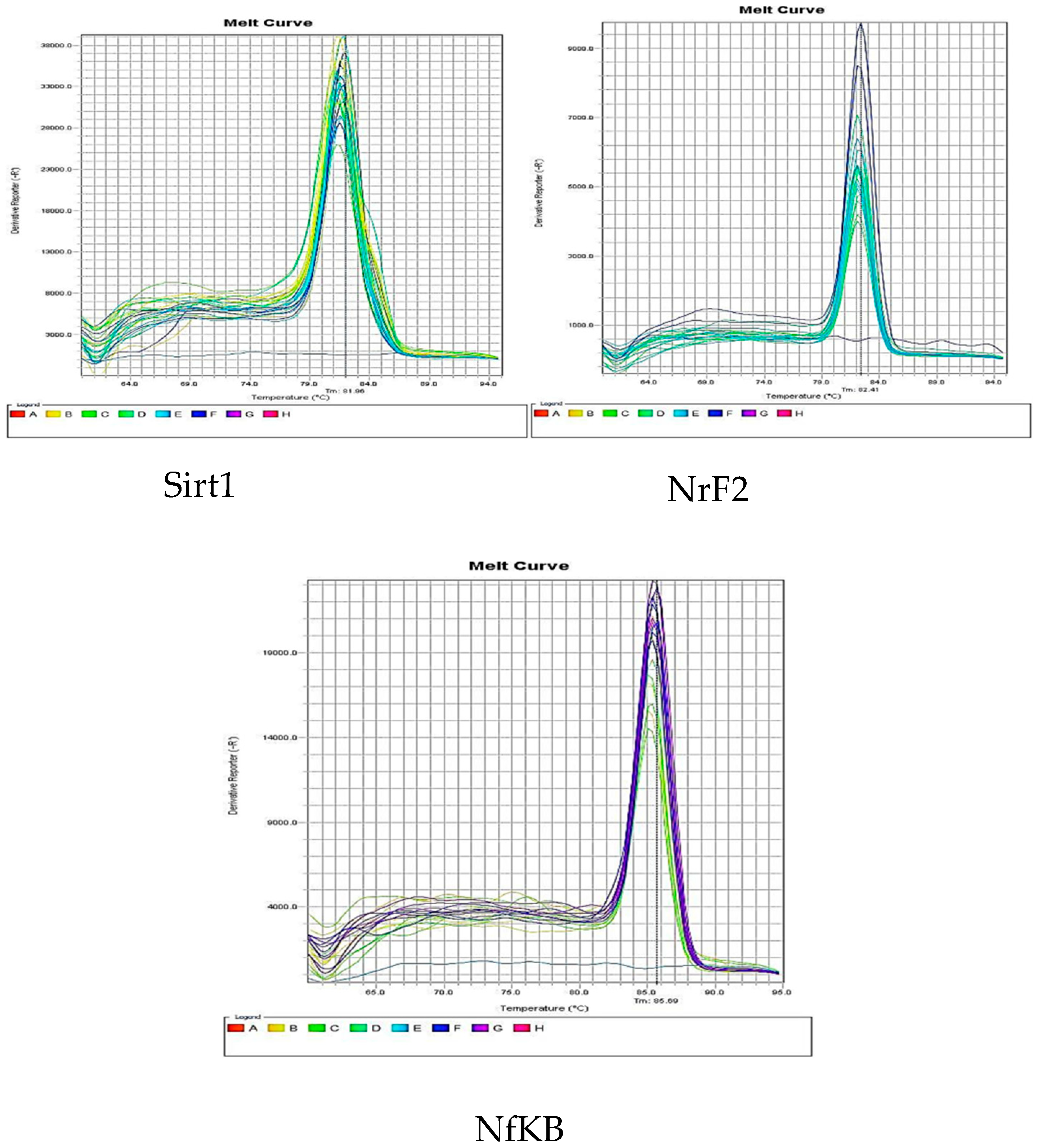
Total RNAs were extracted from tissues using TRIzol reagent (YTA, Iran) and treated with DNase I (Aminsan, Iran). One μg of each total RNA was reverse transcribed to cDNA using the first strand cDNA synthesis kit (YTA, Iran). Quantitative real-time PCR was performed to assess gene expression by the StepOnePlus™ Real-Time PCR System (ABI, USA) using qPCRBIO SyGreen Mix (PCR Biosystems, UK). The PCR parameters were as follows: initial denaturation (one cycle at 95°C for 2 minutes); 40 cycles of denaturation, annealing, and amplification (95°C for 5 seconds, 60-64°C for 30 seconds); and the melting curve (starting at 65°C and gradually increasing to 95°C). Gene expressions of TNF-α, IL-6, NF-κB, Sirt1, and Nrf2 were normalized to the levels of GAPDH, and expression differences were calculated according to the standard curve and efficiency (E) established for each primer set (2–ΔΔCT formula). Specific primers are listed in Table 1.
2.8. Statistical analysis
All the data generated from the research were presented as the mean ± standard deviation (SD). The values obtained were examined statistically by applying one way ANOVA technique to confirm statistical differences between the means of each group. Turkey-test was carried out to determine the significance of the difference of means. All graphs were plotted by using GraphPad Prism 5 software (GraphPad Software Inc., San Diego, CA, U.S.A.).
3. Results
3.1. Biochemical analysis
The important blood biochemical indicators of control and treated mice with DMF were investigated and the results summarized in Table 1.
3.1.1. Pharmacological intervention and their effects on levels of blood glucose
The glucose concentrations in diabetic group were estimated to be high post 220 mg/dL, i.p. injection of alloxan when compared against normal control group (P< 0.001). Alloxan-induced diabetic mice on treatment with DMF at different doses (20, 40, and 80 mg/kg/day) compared to diabetic control mice significantly decreased fasting concentrations of blood glucose with highest dose being the most effective (P< 0.001).
3.1.2. Pharmacological intervention and their effects on levels of blood urea nitrogen
Blood urea is an effective clinical diagnostic indicator for chronic and acute kidney disease [17]. Diabetic mice showed a significant rise in blood urea levels compared to control group (P< 0.01). DMF treatment was able to reverse the increase in blood urea level caused by diabetes in alloxan-treated mice in a dose dependent manner.
3.1.3. Pharmacological intervention and their effects on levels of blood creatinine
Blood creatinine levels were significantly elevated in diabetic mice. The excess level of blood creatinine is an indicator of impaired glomerular filtration rate which is a sign of renal injury [4]. DMF (20 mg/kg/day)-diabetic mice showed significantly diminished levels of blood creatinine (by 13.7%, P< 0.01 vs. diabetic group), the highest dose showed maximum therapeutic effects (Table 1).
3.1.4. Pharmacological intervention and their effects on AST and ALT levels
Blood ALT and AST levels were significantly elevated in diabetic mice (P< 0.001). DMF-diabetic mice showed significantly diminished levels of blood ALT and AST levels (~ 50%, P< 0.001 vs. untreated diabetic group).
3.1.5. Pharmacological intervention and their effects on levels of blood albumin
Diabetic kidney failure can be diagnosed by evaluating high levels of albumin in the urine sample and lower levels in the serum [17]. In the current study a noteworthy decrease in blood albumin levels was confirmed in diabetic mice as compared to normal control group (P< 0.001). Administering DMF (80 mg/kg/day + 200 mg/kg of MET) significantly increased albumin concentrations in blood of diabetic mice, highest dose showing best results.

Table 2.
Changes in blood biochemical parameters in different mice groups.
| Treatment groups | Glucose levels (mg/dL) |
BUN (mg/dL) |
CRT (mg/dL) |
AST (U/L) |
ALT (U/L) |
Albumin (g/dL) |
|---|---|---|---|---|---|---|
|
Group I Group II Group III Group IV Group V Group VI Group VII |
101.8±3/304 222.8±7.042a 182.5±6.608c 167±10.420d 153±8.406e 105.5±1.732b 108.3±4.573 |
55.2±9/311 79.2±5.02 a 73.0±11.27 64.8±14.65 60.6±6.731 c 40.2±5.495b 62.4±5.941 |
0.418±0/087 0.554±0.032 a 0.5±0.049 0.488±0.058 0.452±0.023 c 0.432±0.008 b 0.448±0.26 |
32.42±1/293 76.9±8.926 a 64.4±10.01 37.8±5.621 c 34.32±5.837d 34.04±3.182 b 26.36±4.268 |
39.3±4/57 87.2±3.982 a 66.35±2.87 45.85±4.219c 43.5±33.73d 35.3±5.899 b 38.65±7.887 |
4.06±0/439 2.94±0.403a 2.8±0.122 2.84±0.151 3.3±0.2 4.12±0.178b 4.3±0.204 |
The data were presented as mean ± SD, n = 7/group. aP <0.001 vs. control group. bP <0.001 vs. diabetic group.
3.2. Assessment of oxidative stress markers
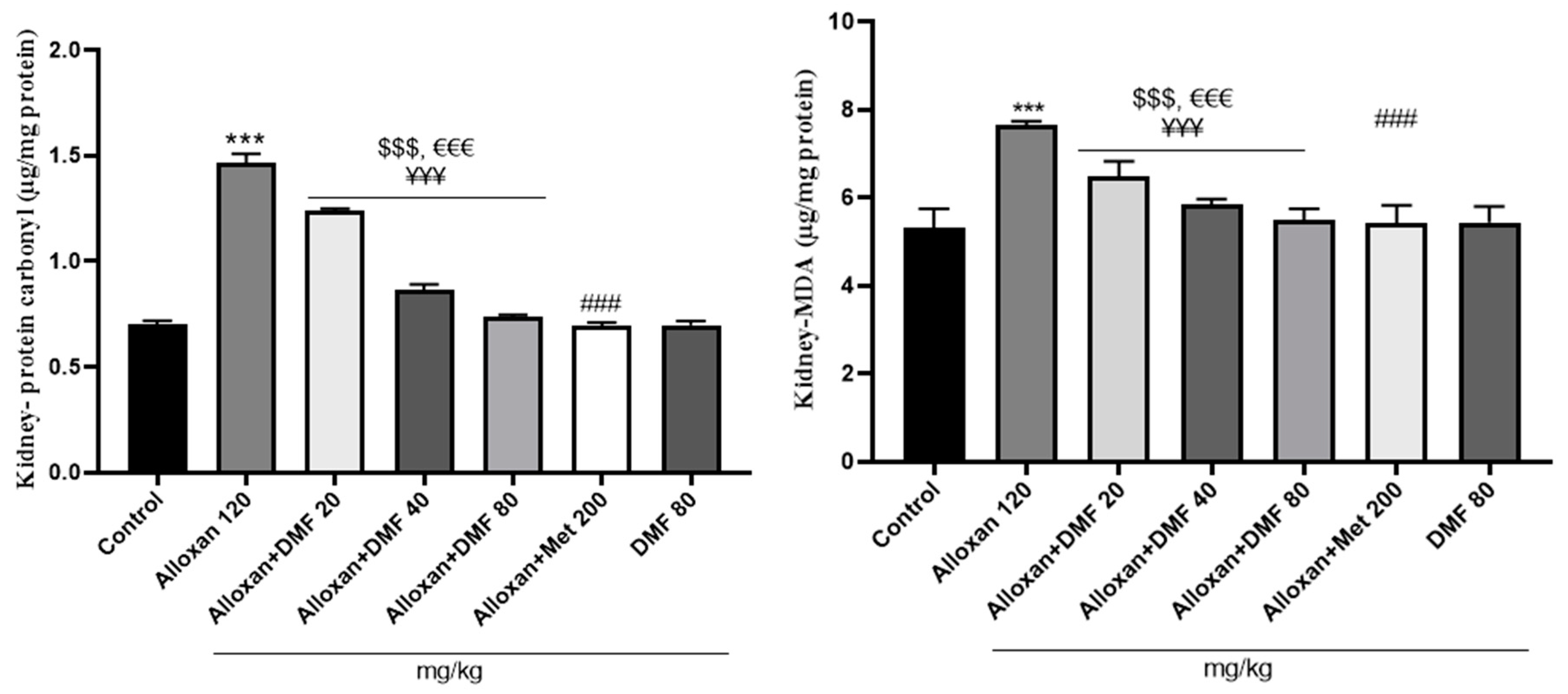
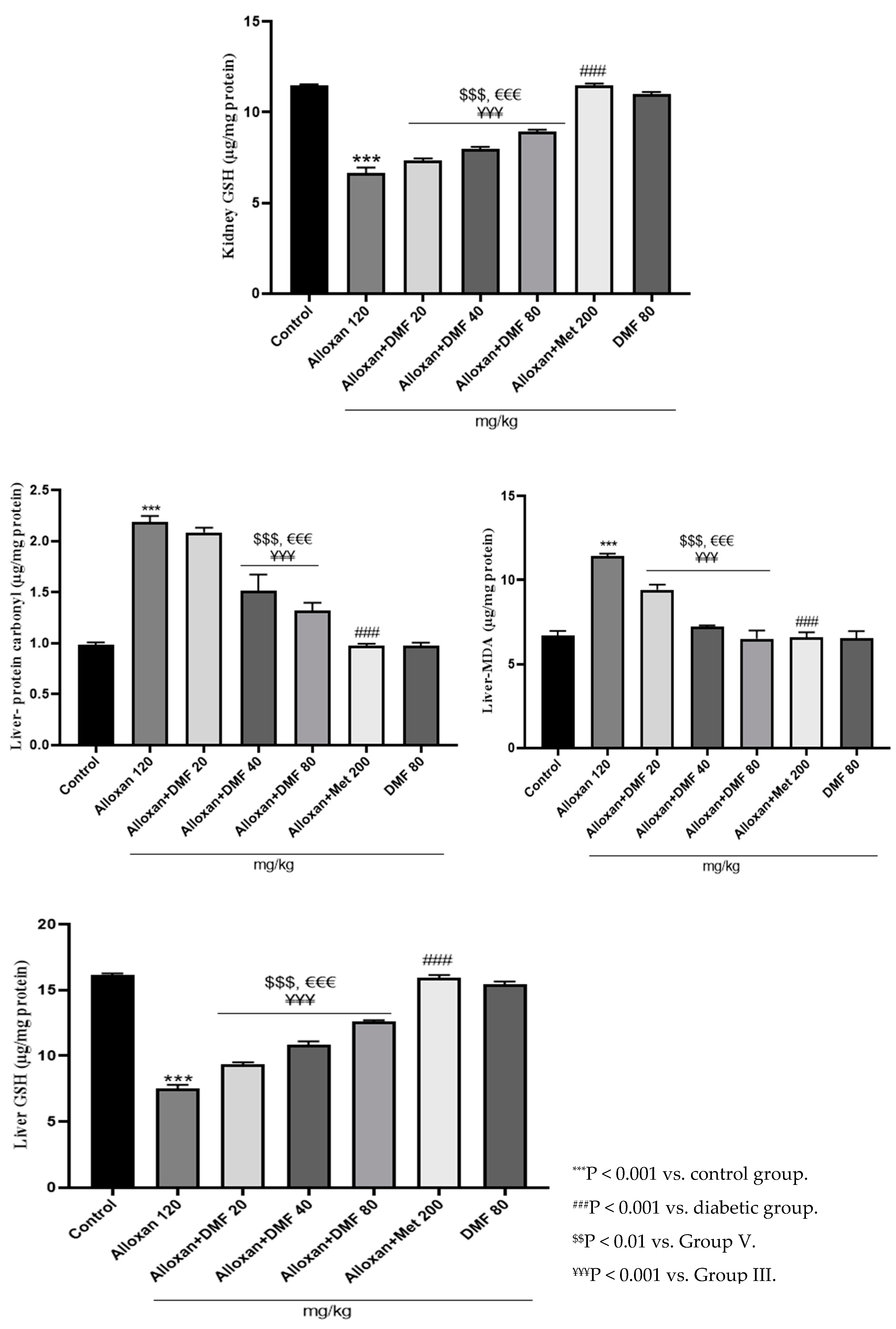
3.2.1. Pharmacological intervention and their effects on kidney and liver MDA content
The concentration of MDA in kidney and liver of diabetic mice increased post induction of alloxan when equated against control group indicating augmented oxidative stress (P< 0.001). Administration of DMF for 3 weeks at low (20 mg/kg/day), medium (40 mg/kg/day), and high (80 mg/kg/day) doses, p.o. significantly reduces the levels of hepato-renal MDA in a dose dependent manner.
3.2.2. Pharmacological intervention and their effects on kidney and liver protein carbonyl content
A subsequent growth in concentrations of protein carbonyl content was witnessed in alloxan- induced diabetic mice when equated against normal control group (P< 0.001). Administrating DMF for 21 days significantly amplified levels of protein carbonyl content in both liver and kidney in a dose-dependent manner.
3.2.3. Pharmacological intervention and their effects on kidney and liver GSH levels
A subsequent drop in concentrations of GSH levels was witnessed in alloxan-induced diabetic mice when equated against normal control group. Administrating DMF for 21 days at low, medium, and high doses significantly amplified levels of GSH levels in a dose-dependent manner.
Figure 1.
Pharmacological interventions and their effects on hepato-renal oxidative stress indexes.
3.3. Histopathological observations
3.3.1. Renal histopathological changes
Histopathological examination of renal tissue was undergone by using hematoxylin-eosin staining (Figure 2). The histopathology of the mice kidneys of group I (normal control group) showed normal histology architectures. It should be mentioned that high blood glucose levels could damage the kidney and hamper its filtration rate [4]. In diabetic conditions, the kidney grows large, and the balance of hydrostatic and colloid osmotic forces across the glomerular membrane in addition to the permeability and surface area gets impaired [18]. The histopathological changes in the renal tissue have been observed in all experimental groups apart from the control mice on the basis of the typical histological architecture of the normal renal parenchyma (Figure 2) as observed previously by Lone et al., (2020) [4]. As seen in the Figure 2, significant tubule interstitial changes including tubules dilation and degeneration, inflammation, and deformation were observed in treated mice with metformin after 21 days as compared with normal control groups. On the other hand, renal section of diabetic mice (120 mg/kg, i.p.) showed shrunken glomerular tufts, increase in Bowmans space and dilation of proximal, and distal convulated tubules with relatively higher number of mesangial cells. The renal section of diabetic mice post treatment with DMF for 21 consecutive days at medium and high doses (40 mg/kg/day and 80 mg/kg/day, p.o.) showed that the usual appearance and size of the glomerular capillaries were retained. The Bowman's capsule, proximal, and distal tubules also improve in size and thickness (Figure 2).
3.3.2. Hepatic histopathological changes
Photomicrograph of the control mice liver sections showed normal hepatic architectures with no inflammation which comprised, normal central vein, hepatic cords, hepatocytes and portal area contents (bile duct, hepatic artery, and vein). Liver sections taken from the alloxan-diabetic mice (GII) showed variable hepatic injuries such inflammation and enlargement of the bile ducts. However, no signs of fibrosis or fatty liver were observed in this group. Liver sections taken from the treated mice at low and medium doses with DMF showed less bile duct dilatation compared to other groups (Figure 3).
3.4. Effect of DMF on reno-hepato inflammatory genes expression
3.4.1. Renal levels of inflammatory genes expression
DMF-diabetic mice exhibited significant decreased of the levels of TNF-α, IL-6, and NF-kB expression (P < 0.001, Figure 4) in comparison to untreated diabetic animals. Meanwhile, the expression of sirt1 and Nrf2 genes in the treated mice with DMF (80 mg/kg body weight) and alloxan + MET (200 mg/kg/day) were significantly higher compared to the diabetic group (P < 0.001, Figure 4).
3.4.2. Hepatic levels of inflammatory genes expression
DMF treatment markedly reduced TNF-α, IL-6, and NF-kB, increased levels of Nrf2 and Sirt1 in mice, probably emphasizing its antioxidant potential reported previously (9) compared to diabetic values (P < 0.001, Figure 5).
Figure 5.
Effect of DMF on hepatic levels of inflammatory genes expression in diabetic mice.
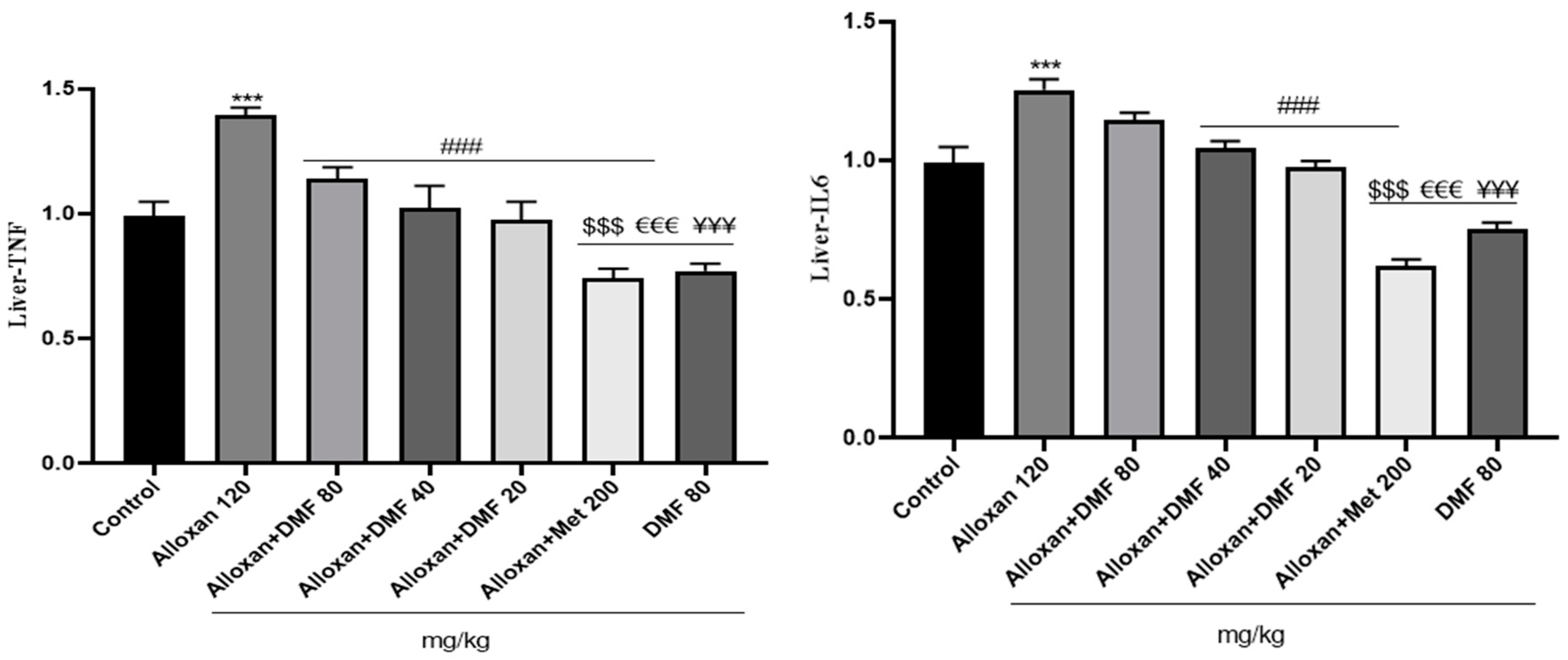
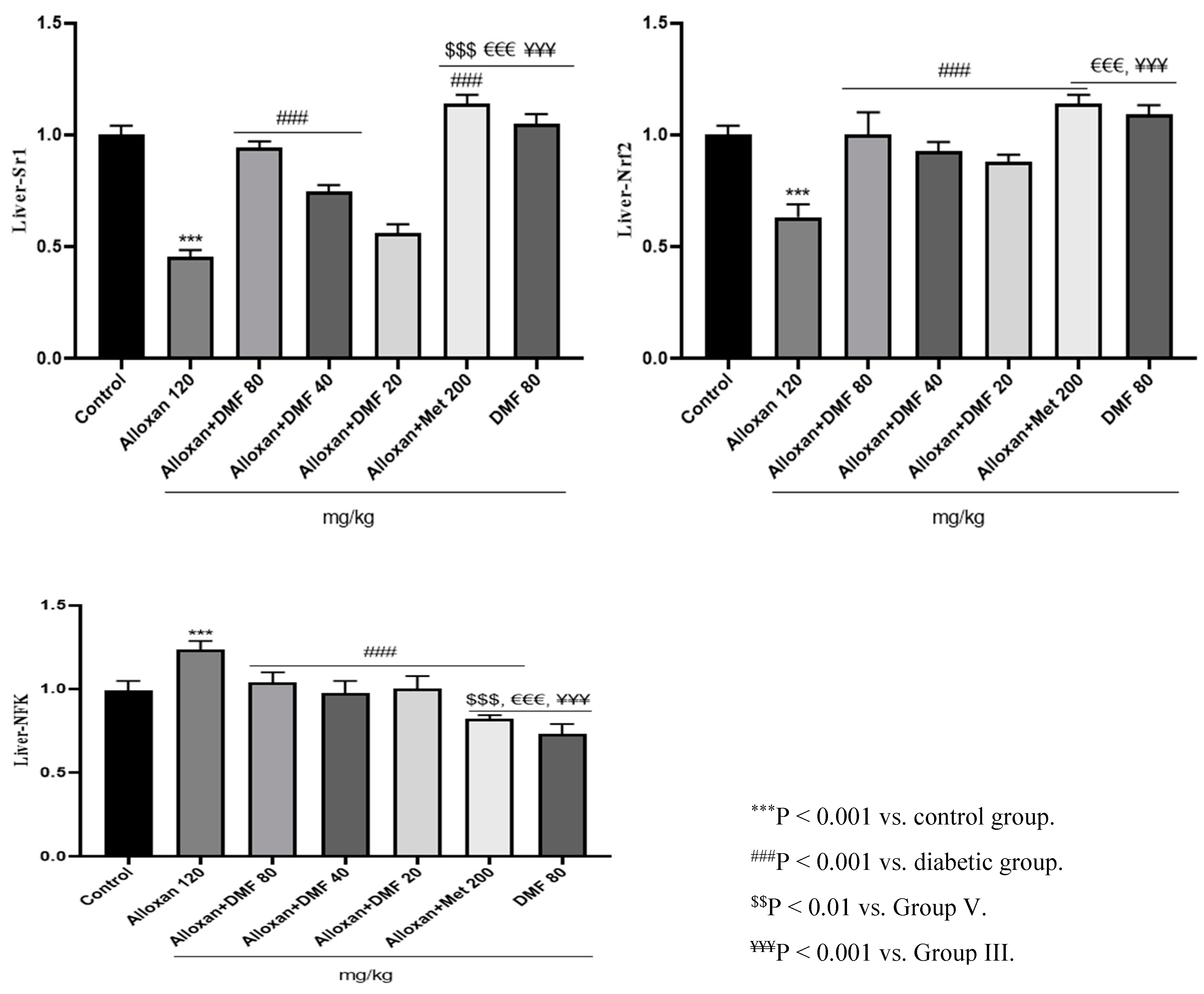
Figure 6.
Melting curves of different genes.

4. Discussion
Chronic diabetic hyperglycemia through several metabolic disorders advanced glycation end-products pathway and increased production of ROS leads to the onset and progression of adverse effects on major vital functions of the body's organs especially kidneys and livers [5,19]. In the recent years, an association between the progression of diabetes and the incidence of liver and kidney dysfunction, such as diabetic nephropathy, fatty liver disease, abnormal function of liver enzymes, and acute liver failure has been observed [19]. To date, there are many evidences of the effective role of inflammation and oxidative stress in the development of diabetic dysfunction in both liver and kidney [10]. This study investigates the tendency of hepato-renal protection by DMF in alloxan (as a highly cytotoxic glucose analogue)-induced diabetic mice model. The current results demonstrated that treated mice with alloxan become hyperglycemic and hepato-renal damage develops in mice was proven by increase in blood urea, creatinine, ALT, AST, MDA, protein carbonyl, and decrease of blood albumin and GSH levels. During diabetes, the activation of different signaling pathways by ROS causes liver and kidney damage. Produced ROS and oxidative stress process progression can facilitate hepato-renal inflammation and fibrosis generation; the last case can also contribute to further augmentation of ROS generation [9]. Also, the overproduction of ROS plays a critical role in the initiation and progression of diabetic kidney disease from the perspective of the renal inflammation, affecting renal structure and function [4].
DMF is a potent activator of Nrf2 used for the clinical treatment of multiple sclerosis, although the mechanism of action of DMF is not clearly understood [11,12]. DMF inhibits the production of pro-inflammatory cytokines and NF-κB signaling by inhibiting its nuclear translocation and also has unique antioxidant properties [20]. From this point of view, it can be considered a suitable combination to supplement the effectiveness of MET in improving diabetic injuries [21]. Despite its promising therapeutic effect on multiple sclerosis, the role of DMF in improving the diabetic liver and kidney dysfunction has not been determined.
Proinflammatory cytokines, such as TNF-α, NF-κB, IL6 expression is directly related to some diseases such as atherosclerosis, obesity, diabetes, and cancer. Collectively, these findings suggest that DMF probably exerts hepato-reno-protective effects on diabetic mice through attenuating the ROS-proinflammatory cytokines pathway. Interestingly, these data indicate that the combination of MET and DMF may have a synergistic effect in rescuing SIRT1 and Nrf2 activity while also being anti-inflammatory. Possible mechanisms of DMF in restoring liver-kidney function include suppressing activation of inflammatory genes expression through disruption of ROS-NF-kB-dependent mediators and pathways. Based on these observations, DMF may be a promising drug for the prevention of hepato-renal complications in diabetic patients [22].
In this line, Lee et al., (2009) concluded that SIRT1 expression protects β-cells against various toxic stresses such as oxidative stress and cytokines through NF-κB signaling suppression pathway [23]. This gene is able to directly interact with the insulin signaling pathway through various mechanisms. In other study, the overexpression of SIRT1 in transgenic mice led to the improvement of glucose tolerance in these animals due to the reduction of glucose output from the liver [24]. Metabolites such as free fatty acids and cytokines such as TNF-α during hyperglycemia cause excessive production of reactive oxygen species by mitochondria, which are the main source of ROS. Therefore, reduction of mitochondrial oxidative capacity can cause insulin resistance through oxidative stress. The expression of SIRT1 gene, in addition to the overexpression of antioxidant enzymes, with its effective role in the deacetylation process in the liver, leads to the reduction of liver damage [25].
Lone et al., (2020) investigated reno-protective potential of DMF in streptozotocin- induced diabetic nephropathy in rat models. Their results showed remarkably increased anti-diabetic, reno-protective, and hypolipidemic effects of DMF in streptozotocin- induced diabetic nephropathy in rats. Treated diabetic rats with DMF at low, medium, and high doses, respectively for 28 days positively decreased the level of blood glucose, regulated the levels of triglycerides cholesterol with enhancement of urine and serum parameters besides their antioxidant effect on kidney. In general, they concluded that DMF can be an advanced option in preventing diabetic nephropathy [4].
DMF counteracts both maladaptive indicators of oxidative stress and inflammation by the regulatory pathway of Nrf2 gene expression and activation of a series of downstream antioxidants. A review of studies in this field shows that upon response to oxidative stress and also possible others, DMF induce Nrf2 activation, will reduce the level of inflammation through ROS-NF-κB signaling pathways and the expression of pro-inflammatory cytokines in alloxan-induced diabetic liver and kidneys [20,26,27].
Hu et al., (2018) investigated the protective potential of DMF on diabetes-induced myocardial tissue injury, likely via activation of Nrf2 function. They found that diabetic animals treated with DMF exhibited blunted oxidative stress, inflammation, fibrosis and this correlated with Nrf2 activation type 1 diabetes mouse model. Their results showed that DMF could potentially thwart diabetes-induced myocardial tissue injury, likely via activation of Nrf2 function [10].
In other study, Amin et al., (2020) explored the potential mechanisms underlying the probable vasculoprotective effects of DMF on vascular complications in streptozotocin diabetic rats. Based on their observations, DMF attenuates vascular remodeling and functional alterations in streptozotocin-induced diabetic rats via several mechanisms, which mainly include suppression of NLRP3 inflammasome activation in diabetic aortas, possibly via impairing ROS-TXNIP and/or ROS-NF-κB pathways (9).
In our histopathology studies, multiple alterations were present in all experimental groups aside from the control mice. Our experimental results showing several systemic disturbances in liver and kidney cellular metabolism which alter its morphology in alloxan-induced diabetic mice model, in addition to several vascular and inflammatory changes. The alloxan-induced diabetic mice kidneys showed a series of degenerative changes up to necrosis, dilation of tubules, cell degeneration, and interstitial eosinophilic infiltration.
The alloxan-induced diabetic hepatic tissue exhibited variable hepatic injury as vacuolar degeneration, dilation of the tubules, the hyperplastic cells mixed with lymphocytic infiltration, congestion in the portal vein, and edema with the presence of newly formed nonfunctional bile ductulus. Our findings were in agreement with previously recorded by Lone et al., (2020) that analyzed the effect of DMF on renal histological changes [4]. Their results showed that renal section of diabetic rats post treatment with DMF for 28 consecutive days at 40 and 80 mg/kg/day had a usual appearance and size of the glomerular capillaries were retained.
Effect of DMF (25 mg/kg/day) on aortic histologic changes in streptozotocin-induced diabetic rats was evaluated by Amin et al., (2020). Histopathological analysis of diabetic aortas showed fibrous tissue proliferation in tunica media. They concluded that these structural alterations were markedly attenuated by DMF treatment and may be related to reduce aortic transforming growth factor beta 1 protein levels in the treated diabetic rats with DMF in comparison to untreated diabetic group [9].
5. Conclusions
Dimethyl fumarate treatment at 20, 40, and 80 mg/kg/day respectively for 21 days orally demonstrated a noteworthy decrease in blood glucose levels when compared with diabetic mice. A remarkable downfall in blood creatinine, ALT, AST, protein carbonyl, MDA, and blood urea was also witnessed. Under such conditions, the blood albumin concentrations and the GSH content increased significantly in a dose-dependent manner. The histological features improve as shown by normal size of glomerular capillaries along with less dilation of the bile ducts and the occurrence of inflammation when compared to alloxan-induced diabetic mice. The protective effects of DMF was completely dependent on the expression of the genes evaluated in this study, and the expression of genes such as Sirt1 and Nrf2 caused the effective protection of DMF on liver and kidney function compared to diabetic mice.
The findings of the present study may indicate that DMF improves the restoration of liver-kidney function in diabetic mice through several mechanisms such as decreasing cytokine and chemokine gene expression and increasing anti-inflammatory responses.
Our findings demonstrated that DMF alone or together with MET can be repurposed for future clinical use for the management of hepato-renal injuries and other complications of diabetes.
Funding
This work was supported by Grant from Mazandaran University of Medical Sciences with Grant number 11716.
Acknowledgments
The authors appreciate the financial and technical support of Department of Pharmacy, Mazandaran University of Medical Sciences. All authors contributed to the study conception and design. Material preparation, data collection and analysis were performed by Parisa Saberi-Hasanabadi, Dr. Ramin Ataee as supervisor and designer of the project, Fatemeh Shaki collaborated and leaded oxidative stress experiment, Mohammad Karami as toxicological consulter, Mohammad Ranaee as pathological experiments designer and director and Abouzar Bagheri as technical labs specialist collaborate in biochemical experiments All authors read and approved the final manuscript. Also it is declared that the abstract of this manuscript has been accepted for presentation in 2nd Edition of International Conference and Expo on Toxicology and Applied Pharmacology" (TOXICOLOGY 2023)”: https://toxicology-conferences.magnusgroup.org/.
Ethics approval
The animal experimentation protocols were conducted in accordance with the recommendations of the Mazandaran University of Medical Sciences Animal Ethical Committee (Code: IR.MAZUMS.4.REC.1401.11716).
Declaration of Conflicting Interests
The author(s) declare that there are no potential conflicts of interest with respect to the research, authorship, and/or publication of this article.
Abbreviations
AST: aspartate aminotransferase; ALT: alanine aminotransferase; BUN: blood urea nitrogen; CRT: creatinine; DMF: dimethyl fumarate; MDA: malondialdehyde; GSH: Glutathione; AGEs: Advanced glycation end-products. MET: metformin; NF-ĸB: Nuclear factor-κB; ROS: Reactive oxygen species; IL-6: Interleukin 6.
References
- Fei, Y.; He, Y.; Sun, L.; Chen, J.; Lou, Q.; Bao, L.; Cha, J. The study of diabetes prevalence and related risk factors in Fuyang, a Chinese county under rapid urbanization. Int J Diabetes Dev Ctrie. 2016, 36, 213-219. [CrossRef]
- Ye, W.; Ding, X.; Putnam, N.; Farej, R.; Singh, R.; Wang, D.; Kuo, S.; Kong, S.X.; Elliott, J.C.; Lott, J.; Herman, W.H. Development of clinical prediction models for renal and cardiovascular outcomes and mortality in patients with type 2 diabetes and chronic kidney disease using time-varying predictors. J. Diabetes Complicat. 2022,36(5), 108180. [CrossRef]
- Zhan, Y.T.; Zhang, C.; Li, L.; Bi, C.S.; Song, X.; Zhang, S.T. Non-alcoholic fatty liver disease is not related to the incidence of diabetic nephropathy in type 2 diabetes. Int. J. Mol. Sci. 2012, 13(11), 14698-14706. [CrossRef]
- Lone, A.; Behl, T.; Kumar, A.; Makkar, R.; Nijhawan, P.; Redhu, S.; Sharma, H.; Jaglan, D.; Goyal, A. Renoprotective potential of dimethyl fumarate in streptozotocin induced diabetic nephropathy in Wistar rats. Obes. Med. 2020, 18, 100237. [CrossRef]
- Oltean, S.; Coward, R.; Collino, M.; Baelde, H. Diabetic nephropathy: novel molecular mechanisms and therapeutic avenues. Biomed Res. Int. 2017. [CrossRef]
- Tao, Z.; Li, Y.; Cheng, B.; Zhou, T.; Gao, Y. Influence of nonalcoholic fatty liver disease on the occurrence and severity of chronic kidney disease. J. clin. transl. hepatol. 2020, 10(1), 164. [CrossRef]
- Adiga, U.S.; Malawadi, B.N. Association of diabetic nephropathy and liver disorders. J. Clin. Diagnostic Res. 2016, 10(10), BC05.
- Dubey, S.; Yadav, C.; Bajpeyee, A.; Singh, M.P. Effect of Pleurotus fossulatus aqueous extract on biochemical properties of liver and kidney in streptozotocin-induced diabetic rat. Diabetes Metab Syndr Obes. 2020, 3035-3046. [CrossRef]
- Amin, FM.; Abdelaziz, R.R.; Hamed, M.F.; Nader, M.A.; Shehatou, G.S. Dimethyl fumarate ameliorates diabetes-associated vascular complications through ROS-TXNIP-NLRP3 inflammasome pathway. Life Sci. 2020, 256, 117887. [CrossRef]
- Hu, X.; Rajesh, M.; Zhang, J.; Zhou, S.; Wang, S.; Sun, J.; Tan, Y.; Zheng, Y.; Cai, L. Protection by dimethyl fumarate against diabetic cardiomyopathy in type 1 diabetic mice likely via activation of nuclear factor erythroid-2 related factor 2. Toxicol. Lett. 2018, 287, 131-141. [CrossRef]
- Wrona, D, Majkutewicz, I, Świątek, G, Dunacka, J, Grembecka, B, Glac, W. Dimethyl fumarate as the peripheral blood inflammatory mediators inhibitor in prevention of streptozotocin-induced neuroinflammation in aged rats. J. Inflamm. Res. 2022, 33-52. [CrossRef]
- Li, Y.; Ma, F.; Li, H.; Song, Y.; Zhang, H.; Jiang, Z.; Wu, H. Dimethyl fumarate accelerates wound healing under diabetic conditions. J. Mol. Endocrinol. 2018, 61(4), 163-172. [CrossRef]
- Mostafavinia, A.; Amini, A.; Ghorishi, S.K.; Pouriran, R.; Bayat, M. The effects of dosage and the routes of administrations of streptozotocin and alloxan on induction rate of typel diabetes mellitus and mortality rate in rats. Lab. Anim. Res. 2016, 32, 160-165.
- Khan, H.A.; Abdelhalim, M.A.K.; Al-Ayed, M.S.; Alhomida, A.S. Effect of gold nanoparticles on glutathione and malondialdehyde levels in liver, lung and heart of rats. Saudi J. Biol. Sci. 2012, 19(4), 461-464. [CrossRef]
- Levine, RL. Carbonyl assay for determination of oxidatively modified proteins. Meth. Enzymol. 1994, 233, 246-257. [CrossRef]
- Bancroft, J.D.; Gamble, M. Theory and practice of histological techniques. Elsevier health sciences. J. Neuropathol. Exp. 2008, 67, 633.
- Seo, D.H.; Suh, Y.J.; Cho, Y.; Ahn, S.H.; Seo, S.; Hong, S.; Lee, Y.H.; Choi, Y.J.; Lee, E.; Kim, S.H. Advanced liver fibrosis is associated with chronic kidney disease in patients with type 2 diabetes mellitus and nonalcoholic fatty liver disease. Diabetes Metab J. 2022, 46(4), 630-639. [CrossRef]
- Jha JC, Banal C. Chow B.S, Cooper, ME, Jandeleit-Dahm, K. Diabetes and kidney disease: role of oxidative stress. Antioxid Redox Signal. 2016, 25(12), 657-684. [CrossRef]
- Targher, G.; Mantovani, A.; Pichiri, I.; Mingolla, L.; Cavalieri, V.; Mantovani, W.; Pancheri, S.; Trombetta, M.; Zoppini, G.; Chonchol, M.; Byrne CD. Nonalcoholic fatty liver disease is independently associated with an increased incidence of chronic kidney disease in patients with type 1 diabetes. Diabetes care. 2014, 37(6), 1729-1736. [CrossRef]
- Subba, R.; Ahmad, M.H.; Ghosh, B.; Mondal, A.C. Targeting NRF2 in Type 2 diabetes mellitus and depression: Efficacy of natural and synthetic compounds. Eur. J. Pharmacol. 2022, 925, 174993. [CrossRef]
- Trimeche, A.; Selmi, Y.; Slama, B.; Hazar, I.; Achour, A. Effect of protein restriction on renal function and nutritional status of type 1 diabetes at the stage of renal impairment. Tunis. Med. 2013, 91(2), 117-122.
- Laselva, O.; Allegretta, C.; Di Gioia, S.; Avolio, C.; Conese, M. Anti-Inflammatory and anti-oxidant effect of dimethyl fumarate in cystic fibrosis bronchial epithelial cells. Cells. 2021, 10(8), 2132. [CrossRef]
- Lee, J.H.; Song, M.Y.; Song, E.K.; Kim, E.K.; Moon, W.S.; Han, M.K.; Park, J.W.; Kwon, K.B.; Park, B.H. Overexpression of SIRT1 protects pancreatic β-cells against cytokine toxicity by suppressing the nuclear factor-κB signaling pathway. Diabetes. 2009, 58(2), 344-351.
- Banks, A.S.; Kon, N.; Knight, C.; Matsumoto, M.; Gutiérrez-Juárez, R.; Rossetti, L.; Gu, W.; Accili, D. SirT1 gain of function increases energy efficiency and prevents diabetes in mice. Cell Metab. 2008, 8(4), 333-341. [CrossRef]
- St-Pierre, J.; Drori S.; Uldry, M.; Silvaggi, J.M.; Rhee, J.; Jäger, S.; Handschin, C.; Zheng, K.; Lin, J.; Yang, W.; Simon, D.K. Suppression of reactive oxygen species and neurodegeneration by the PGC-1 transcriptional coactivators. Cell. 2006, 127(2), 397-408. [CrossRef]
- Lee, H.W.; Lee, J.S.; Kim, B.K.; Park, J.Y.; Ahn, S.H.; Kim, S.U. Evolution of liver fibrosis and steatosis markers in patients with type 2 diabetes after metformin treatment for 2 years. J. Diabetes Complicat. 2021, 35(1), 107747. [CrossRef]
- Nour, O.A.; Shehatou, G.S.; Rahim, M.A.; El-Awady, M.S.; Suddek, G.M. Antioxidant and anti-inflammatory effects of dimethyl fumarate in hypercholesterolemic rabbits. Egypt. j. basic appl. sci. 2017, 4(3), 153-159. [CrossRef]
Figure 2.
Pharmacological interventions and their effects on kidney histopathology (H&E staining 400×): GI). GI) Control group showing normal renal structure, GII) Dilation of tubules (purple arrow); Less tubules degeneration than to the second group (green arrow), GIII) Renal infiltration (green arrow), GIV) No specific changes were observed, GV) Less inflammation than to the previous groups (purple arrow); Less tubules degeneration than to the previous group (red arrow), GVI) Tubules dilation (yellow arrow); inflammation (red arrow), GVII) Only inflammation (red arrow).
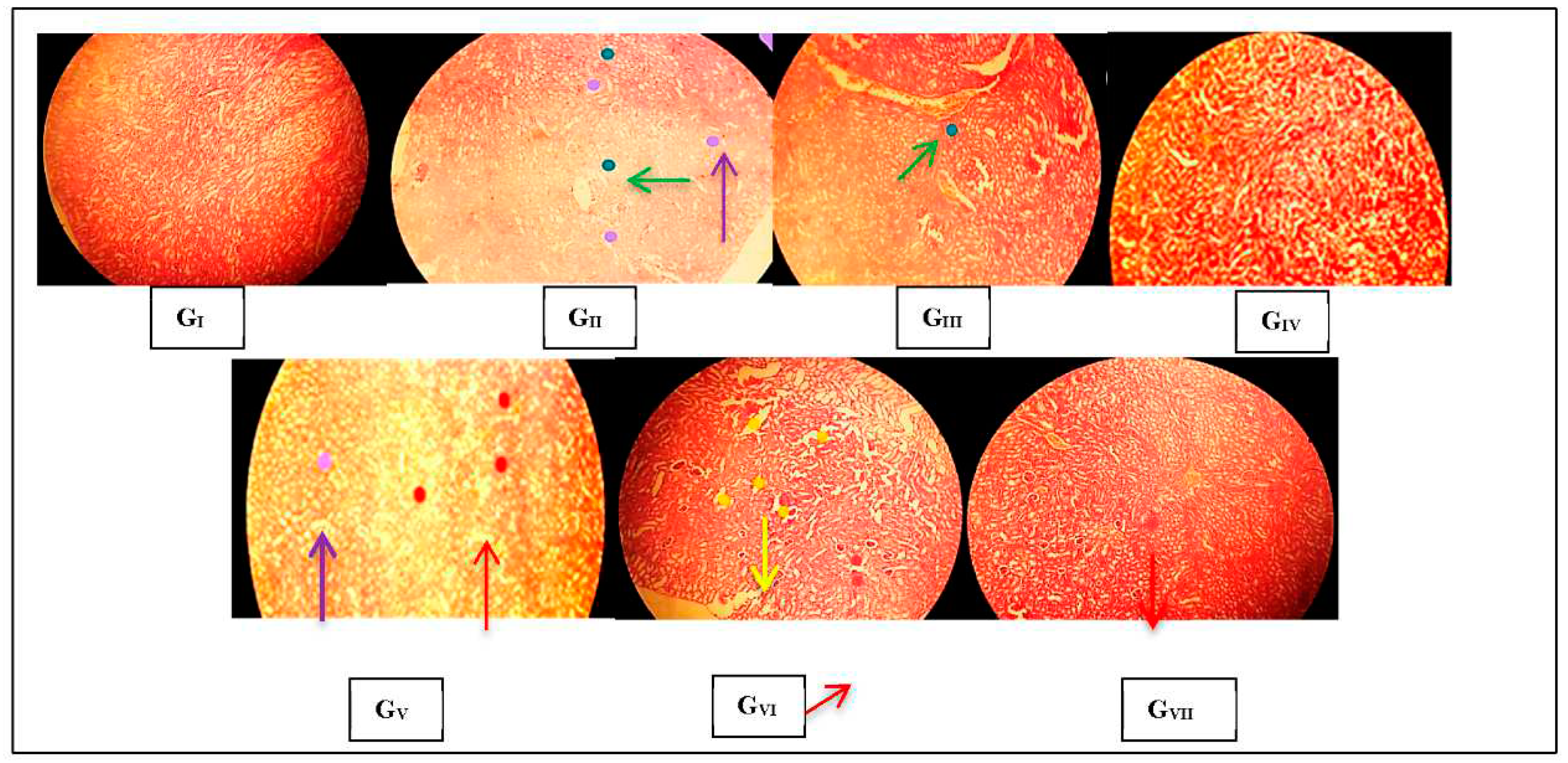
Figure 3.
Pharmacological interventions and their effects on liver histopathology (H&E staining 400×): GI) control group showing normal hepatic structure, GII) inflammation (purple arrow), GIII) Less inflammation than to the second group (yellow arrow), GIV) Less inflammation than to the third group (purple arrow), GVI) More inflammation than to the previous groups (purple arrow); GVI) Less involvement of the tubular structure (purple arrow); less inflammation (red arrow), GVII) More inflammation around the tubular structure (purple arrow); more tubular structure dilation (yellow arrow).
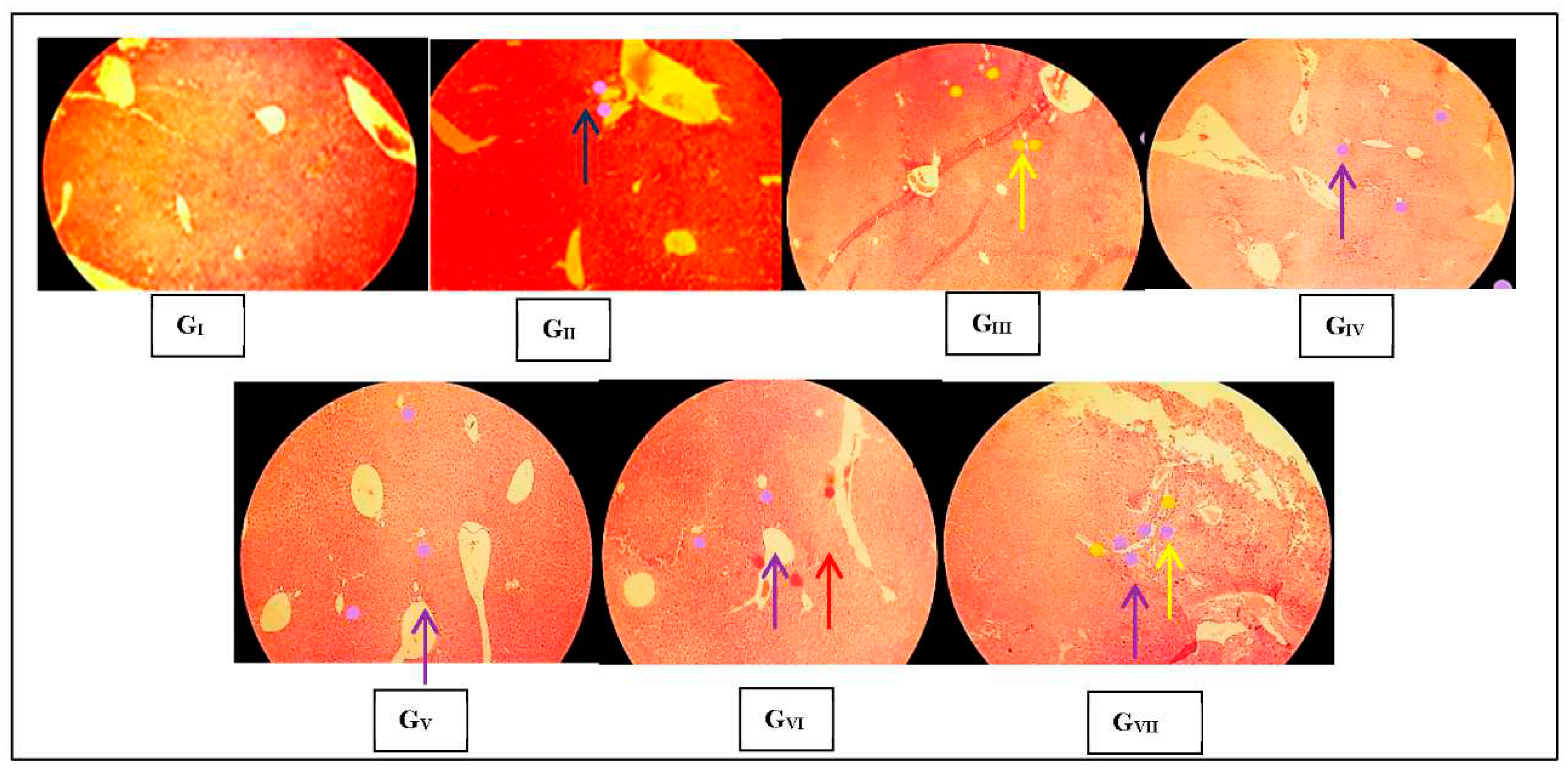
Figure 4.
Effect of DMF on renal levels of inflammatory genes expression in diabetic mice.
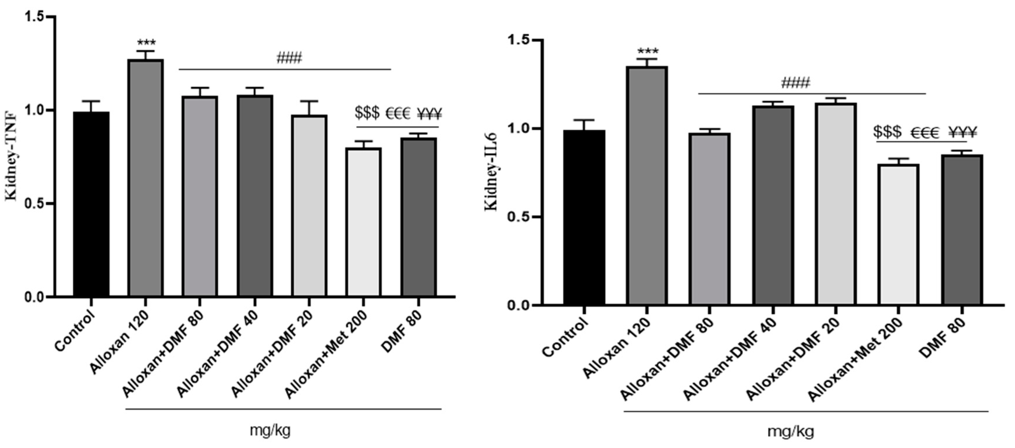
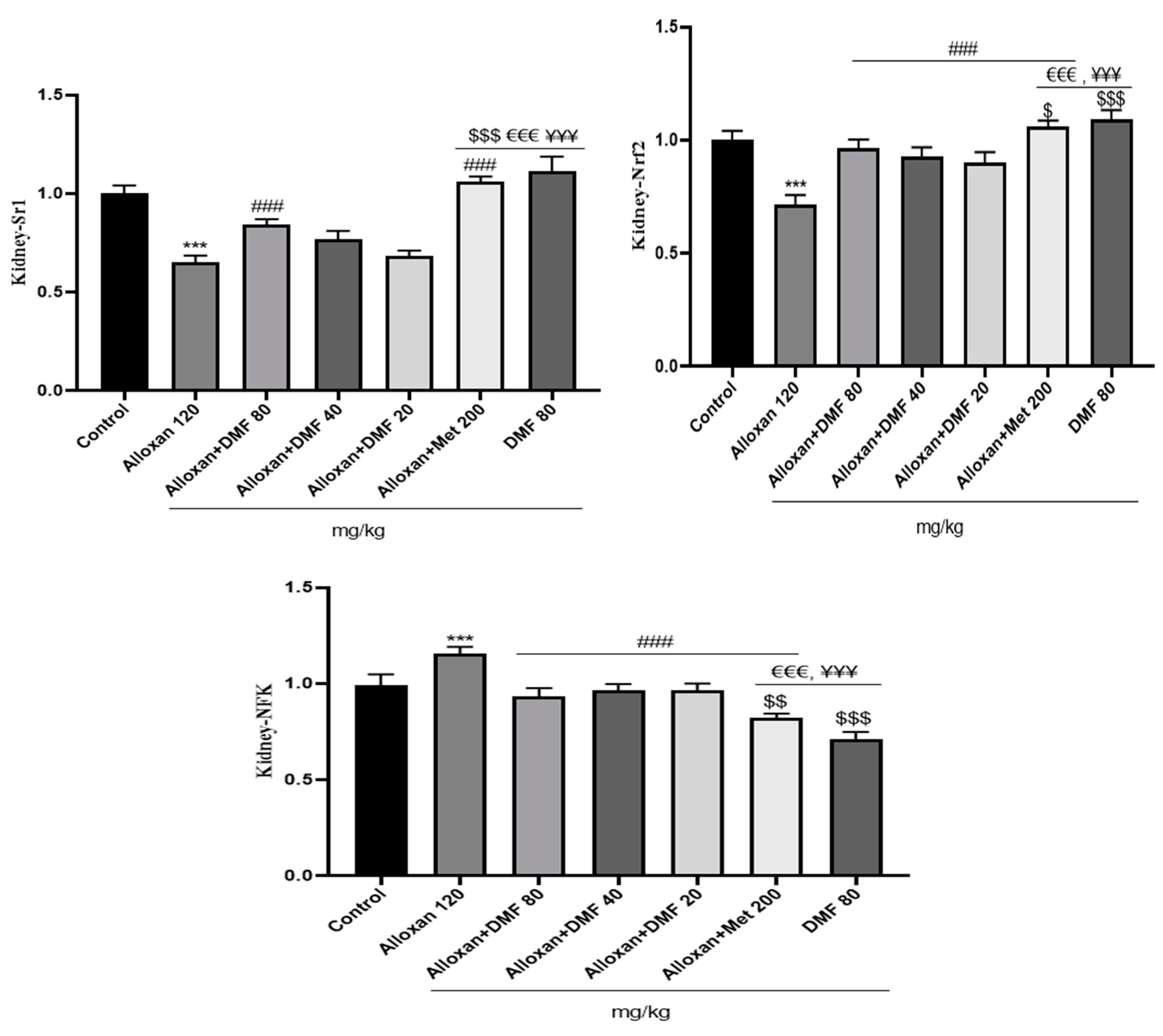
Table 1.
Sequences of forward and reverse primers used for real time quantitative PCR analyses.
| Primer | Sequence | |
|---|---|---|
| TNF-α | Forward Reverse |
AGGGTCTGGGCCATAGAACT CCACCACGCTCTTCTGTCTAC |
| IL-6 | Forward Reverse |
AGACTTCCATCCAGTTGCCT CATTTCCACGATTTCCCAGAGA´ |
| NF-κB | Forward Reverse |
AGCCACAGAGATGGAGGAGTTG GGATGTCAGCACCAGCCTTTAG |
| Sirt1 | Forward Reverse |
AGCTCCTTGGAGACTGCGAT´ ATGAAGAGGTGTTGGTGGCA |
| Nrf2 | Forward Reverse |
CACCATGGGAATGGACTTGGAGCTGCC CTAGTTTTTCTTAACATCTGGCTTCTTAC |
Disclaimer/Publisher’s Note: The statements, opinions and data contained in all publications are solely those of the individual author(s) and contributor(s) and not of MDPI and/or the editor(s). MDPI and/or the editor(s) disclaim responsibility for any injury to people or property resulting from any ideas, methods, instructions or products referred to in the content. |
© 2023 by the authors. Licensee MDPI, Basel, Switzerland. This article is an open access article distributed under the terms and conditions of the Creative Commons Attribution (CC BY) license (http://creativecommons.org/licenses/by/4.0/).
Copyright: This open access article is published under a Creative Commons CC BY 4.0 license, which permit the free download, distribution, and reuse, provided that the author and preprint are cited in any reuse.


