Submitted:
01 August 2023
Posted:
02 August 2023
You are already at the latest version
Abstract
The identification of an emerging pathogen in humans can remain difficult by conventional methods such as enrichment culture assays that remain highly selective, require appropriate medium and cannot avoid misidentifications, or serological tests that use surrogate antigens and are often hampered by the level of detectable antibodies. Although not originally designed for this purpose, the implementation of polymerase-chain-reaction (PCR) has resulted in an increasing number of diagnostic tests for many diseases. However, the design of specific molecular assays relies on the availability and reliability of published genetic sequences for the target pathogen as well as enough knowledge on the genetic diversity of species and/or variants giving rise to the same disease symptoms. Usually designed for clinical isolates, molecular tests are often not suitable for environmental samples in which the target DNA is mixed with a mixture of environmental DNA. A key challenge of such molecular assays is thus to ensure high specificity of the target genetic markers when focusing on clinical and environmental samples in order to follow the dynamics of disease transmission and emergence in humans. Here we focus on Buruli ulcer (BU), a human necrotizing skin disease mainly affecting tropical and subtropical areas, commonly admitted to be caused by Mycobacterium ulcerans worldwide although other mycolactone-producing mycobacteria and even mycobacterium species were found associated with BU or BU-like cases. By revisiting the literature, we show that many studies have used non-specific molecular markers (IS2404, IS2606, KR) to identify M. ulcerans from clinical and environmental samples and propose that all mycolactone-producing mycobacteria should be definitively considered as variants from the same group rather than different species. Importantly, we provide evidence that the diversity of mycolactone-producing mycobacteria variants as well as mycobacterium species potentially involved in BU or BU-like skin ulcerations might have been underestimated. We also suggest that the specific variants/species involved in each BU or BU-like case should be carefully identified during the diagnosis phase, either via the key of genetic identification proposed here or by broader metabarcoding approaches, in order to guide the medical community in the choice for the most appropriate antibiotic therapy.
Keywords:
buruli ulcer
; Mycobacterium ulcerans
; DNA
; cryptic biodiversity
; genetic diversity
; antibiotherapy
; diagnosis
Introduction
Emerging infectious diseases (EIDs) in humans are caused by a wide diversity of etiological agents, including viruses, bacteria, fungi or even parasites (Jones et al., 2008), which themselves exhibit complex inter- and intra-species genetic diversity. Interestingly, different pathogens, species of the same pathogen or even variants of the same species can cause diseases characterized by similar clinical manifestations in the early phase of infection. For example, several viral diseases can present flu-like symptoms, including fever, chills, headache, muscle, joint and back pain, nausea and vomiting (e.g. chikungunya, rift valley fever), sore throat and/or cough sometimes lead to pneumonia (e.g. influenza, nipah, SARS-CoV, MERS-CoV) (Dong et al., 2008). Several Rotavirus genotypes (RV G1, G2, G3, G4, P(4) and P(8) and Norovirus genogroups (NoV GI, GII, GIV) (which include several genotypes) present similar gastric illnesses in humans that cause symptoms such as vomiting, diarrhea and stomach pain (Van Trang et al., 2014). Leptospirosis, a global bacterial zoonosis, is also characterized – at least in tropical areas – by non-specific clinical symptoms in the initial phase, such as dengue-like or other arbovirus-like syndrome, making clinical diagnosis sometimes difficult and thus delaying antibiotic therapy (Le Turnier and Epelboin, 2019). Another illustration is the nosocomial yeast candidiasis caused in 78% of cases by the well-known Candida albicans, while some systemic cases can be caused by other (non albican) species (Rezazadeh et al., 2016). For diseases characterized by non-specific symptoms, it is inevitable to develop highly specific identification tools in order to i) identify the causative infectious agent(s) and thus support clinical diagnosis, ii) help to guide the choice of the most appropriate treatment, iii) eventually enable the development of vaccination strategies and iv) monitor transmission dynamics, pathogen circulation in the environment and disease emergence in humans.
The identification of an emerging pathogen by conventional methods is quite difficult and time-consuming. Enrichment culture methods are laborious, require the use of appropriate media and represent a selective constraint for pathogen growth. Furthemore, due to the high degree of phenotypic similarity between pathogenic species, as with Candida sp. (Rezazadeh et al., 2016), misidentifications are inevitable. Diagnosis by serology relies on the use of surrogate antigens and is often hampered by the difficulty that a detectable level of antibodies does not appear in the blood until several days after the onset of the disease, as for Rickettsia sp. (Guillemi et al., 2015). Second, culture-independent molecular tools offer - in theory - several advantages over conventional methods, including high sensitivity and specificity, speed, ease of standardization and automation (Dong et al., 2008; Radomski et al., 2013; Rezazadeh et al., 2016). Although not originally designed for this purpose, the implementation of polymerase-chain-reaction (PCR) has resulted in an increasing number of diagnostic tests for many diseases, as illustrated by the rapid identification of SARS-CoV, the discovery of a new hantavirus in North America or the detection of tick-borne human pathogenic bacteria (Dong et al., 2008). However, the design of specific molecular tests relies on the availability and reliability of published genetic sequences for the target pathogen, but also on the knowledge of the genetic diversity of species, subspecies or variants giving rise to the same symptoms and/or diseases. Another limitation is that molecular detection tests are usually designed from pure laboratory isolates and are often not suitable for the identification of pathogens from complex matrices such as environmental samples (i.e. soil, sediments, water, faeces, etc.). Indeed, an inherent difficulty in the identification of pathogens in the environment is the ability of PCR tests to distinguish the target species in a complex and highly diverse mixture of environmental DNA (eDNA) (Williamson et al., 2008). However, environmental samples provide important information on the dynamics of the pathogen and the source of human contamination (Guillemi et al., 2015). A key challenge is therefore to ensure high specificity of the target genetic markers in order to i) avoid non-specific amplifications of closely related species or variants of the same species that could potentially be present in the sample and ii) to allow the simultaneous screening of a wide range of samples (clinical, environmental) to follow the dynamics of disease transmission and human emergence.
Buruli ulcer (BU) is a human necrotizing skin disease that affects between 5000 and 6000 people annually across 33 countries worldwide, mainly located in tropical and subtropical areas (Combe et al., 2017). BU represents the third most common mycobacterial disease after tuberculosis and leprosy (Portaels et al., 1997) and is responsible for severe morbidity and mortality mainly in low-resources human populations living near contaminated water bodies (Combe et al., 2017). Clinical signs of the disease include painless nodules, plaques and oedema, followed by the development of skin ulcers that can lead to osteomyelitis and permanent disability if early detection and appropriate treatment are delayed or absent (Merritt et al., 2010; Giles-Vernick et al., 2015). BU and its infectious etiological agent Mycobacterium ulcerans (previously described as M. buruli) were first described by MacCallum, Buckle, Tolhurst & Sissons (1948) from 6 cases of skin ulceration occurring in Australia on the basis of histology, pathogenicity, culture and microscopy of M. ulcerans, but the first molecular tests targeting M. ulcerans DNA were developed years later (Portaels et al., 1996; Portaels et al., 1997; Ross et al., 1997). Since then, M. ulcerans is generally considered the only species responsible for BU worldwide and most studies have used three genetic markers to detect its DNA from human ulcerations, animal and environmental samples (IS2404, IS2606, KR). However, the genetic distinction between M. ulcerans and other mycolactone-producing mycobacteria (MPM), namely M. shinshuense, M. pseudoshottsii, M. liflandii, M. marinum (Yip et al., 2007; Tobias et al., 2013), based on these three commonly used molecular tests, remains impossible. Importantly, it has been shown that M. ulcerans and other MPM are genetically related enough to form a single species complex, all representing variants rather than different species (Demangel et al., 2009; Pidot et al., 2010; Doig et al., 2012; Tobias et al., 2013). More recently M. shinshuense, M. marinum and M. liflandii were found in BU-like cases diagnosed in Japan (Nakanaga et al., 2007; Ohtsuka et al., 2014), Côte d’Ivoire (Africa) (Nguetta et al., 2018) and French Guiana (South America) (Saad et al., 2019), respectively. Interestingly, in Côte d’Ivoire Nguetta et al. (2018) also found other mycobacteria, not belonging to the MPM complex, associated with BU-like cases, such as M. chelonae and M. smegmatis and M. gilvum was found associated with some BU-like cases in French Guiana (Combe et al., 2020). Taken together, these results suggest that i) all MPM should definitely be considered variants of the same MPM complex and ii) all MPM variants or even other mycobacteria species apparently not producing mycolactone could cause either the same BU disease or slightly different skin ulcerations all diagnosed as BU to date.
Here we first revisit the basis of MPM species designation, then the history of molecular identification tools development and assess in silico the specificity of the most commonly used genetic markers to target M. ulcerans DNA. We finally propose a step-by-step genetic identification key for MPM variants and discuss the potential implications of BU or BU-like skin lesions caused by other mycobacterial variants/species.
On the origin of Mycolactone-Producing Mycobacteria (MPM) species assignment
Mycolactone is a macrolide cytotoxin with immunosuppressive properties found in the extracellular matrix surrounding large clusters of M. ulcerans bacilli (George et al.1999). Until 2004 it was assumed that the synthesis of mycolactone was restricted to M. ulcerans and depended on the acquisition of a large circular virulence plasmid called pMUM and harboring three polyketide synthase (Pks) genes such as mlsA1 and mlsA2 (main lactone) and mlsB (fatty acid side chain) (Yip et al., 2007; Mve-Obiang et al., 2005). In addition, it has been proposed that plasmid acquisition may be the key event that enabled the recent emergence of M. ulcerans strains from a common M. marinum progenitor (Stinear et al., 2005). Interestingly, in 2004 a previously unknown mycobacterium named M. liflandii was isolated from the West African Xenopus tropicalis frogs that were housed in the laboratory with Xenopus laevis colonies to which the mycobacterium spread (Trott et al., 2004). This new mycobacterium appeared to harbor IS2404, IS2606 as well as a version of the pMUM plasmid and produced mycolactone E (Mve-Obiang et al., 2005). Sequence comparisons of hsp65, the 16s rRNA gene, the internal transcribed spacer (ITS) of the 16S-23S rRNA gene and fragments of the rpoB gene showed that M. liflandii shared >98% nucleotide identity with M. ulcerans and M. marinum (Trott et al., 2004). A year later in 2005, another mycobacterium was isolated during an epizootic of mycobacteriosis in striped bass (Morone saxatilis) in Chesapeake Bay, harboring IS2404, IS2606, a pMUM-like plasmid with mls genes, producing mycolatone F (Ranger et al., 2006) and sharing >98% nucleotide sequence identity with M. ulcerans, M. marinum and M. shottsii ; however it was given a new species name M. pseudoshottsii based on slightly different phenotypic traits such as photochromogenicity and lack of growth at 37°C (Rhodes et al., 2005). Similarly, Ucko et al. (2002) found a group of M. marinum strains isolated from diseased fish in Israel that were positive for mls genes and produced mycolactone F (Ranger et al., 2006). In Japan, the first case of M. shinshuense infection was described in 1980 (Tsukamura and Mikoshiba, 1982) and although showing some phenotypic and molecular variability it was closely related to M. ulcerans (Tsukamura et al., 1989), with the presence of multiple copies of IS2404, the pMUM plasmid and mycolactone production (Kim et al., 2005). Subsequently, all mycolactone-producing mycobacteria were collectively grouped into the mycolactone-producing mycobacteria (MPM) complex (Table 1), thus including M. ulcerans, M. shinshuense, M. liflandii, M. pseudoshottsii and M. marinum (with the exception of the non-mycolactone-producing strains of M. marinum). At that time, all MPM were considered different species based on phenotypic characteristics (e.g. colony morphology, growth rates) and limited genomic background (e.g. 16S rRNA, hsp65, rpoB). However, Pidot et al. (2010) stated that, based on the definition of prokaryotic species, all MPMs sharing >98% 16S rRNA identity, >70% DNA-DNA hybridization (DDH), harboring pMUM plasmids, containing IS2404 and producing mycolactone should be considered as variants of the same species complex. All MPMs harbor a mycolactone-producing virulence plasmid and multiple copies of IS2404 (Stinear et al. 2005, 2007). Portaels et al. (1996) found that M. shinshuense shared similar nucleotide modifications to M. ulcerans, including a G at position 1248 of the 16S rRNA that has been initially described as characteristic of M. ulcerans. Yip et al. (2007) conducted a DDH-based study to investigate the relationship between MPM and M. marinum and showed that all MPMs have a relative binding ratio (RBR) of 88%-100% when compared to African and Australian M. ulcerans strains, whereas they only have an RBR of 15%-60% when compared to non-mycolactone-producing M. marinum strains. In addition, analysis of large sequence polymorphisms showed a clear clustering of strains that were assigned different species names (Kazer et al., 2009). All these results converge to the same point and suggested years ago that all MPM represent genetic variability (variants) from the same MPM complex rather than different species, but also that these variants have a wider geographic distribution and host range than initially assessed (infecting fish, frogs and humans worldwide) (Nakanaga et al., 2007; Ohtsuka et al., 2014; Saad et al., 2019) as well as a broader zoonotic potential (Yip et al., 2007; Pidot et al., 2010). This was later confirmed by the association of M. shinshuense, M. marinum and M. liflandii with human skin ulcerations diagnosed as BU in Japan (Nakanaga et al., 2007; Ohtsuka et al., 2014), Côte d’Ivoire (Nguetta et al., 2018) and French Guiana (Saad et al., 2019). Based on the evidences found in the literature we suggest that all MPMs should definitely be considered as variants from the same complex. We will use this terminology later, in contrast with “M. ulcerans strains” which rather indicate different isolates from the same M. ulcerans variant.
History of BU diagnosis and M. ulcerans DNA PCR testing
BU was initially diagnosed by clinical observations of ulceration and confirmed by microscopic findings of acid-fast bacilli (AFB) after Ziehl-Neelsen staining (resistance to acid and/or ethanol decolorization, BAAR: bacillus acido-alcohol-resistant), a characteristic of bacteria belonging to the Genus Mycobacterium. The use of specific serological tests was not suitable due to cross-reactivity between mycobacterial antigens (Diaz et al., 2006). In addition, the M. ulcerans variant can be difficult to culture, especially from environmental samples, due to the long generation time of the pathogen (>48 hours) and proliferation by contaminants including other mycobacteria (Portaels et al., 1997; Stinear et al., 2000a; Demangel et al., 2009). As primary cultures usually become positive after several months of incubation and many clinical BU cases are reported as culture negative, the implementation of rapid molecular identification tools has been a major step in the diagnosis of BU (Portaels et al., 1997; Ross et al., 1997a). In addition, as BU infection has been associated with stagnant or slowly flowing waters worldwide (Hayman, 1991; Marston et al., 1995; Meyers, 1994), it was thought that these molecular detection tools would help understanding the environmental source of infection (Ross et al., 1997a). While the first molecular distinctions between the M. ulcerans variant and other MPM variants and mycobacteria species were performed by analyzing 16S rRNA, hsp65 and/or rpoB gene sequences, these were limited to small sequence fragments and were therefore insufficient to establish a strong phylogenetic relationship between species (Hofer et al., 1993; Jackson et al., 1995; Portaels et al., 1996). Portaels et al. (1997) and Ross et al. (1997a) were the first to develop PCR assays for the detection of the M. ulcerans variant. Although potential candidate genetic targets had been published, there were some problems: i) the genus-specific 65-kDa heat shock protein antigen had a high degree of homology between different mycobacterial species (Buchanan et al., 1987; Hance et al., 1989) and was therefore not a good target for specific amplifications (Ross et al., 1997a); ii) the 16S rRNA sequence showed only a single nucleotide divergence between M. ulcerans variant and M. marinum (Hofer et al., 1993) and it showed variability between different strains of the M. ulcerans variant (Portaels et al., 1996) and was thus unsuitable for developing a simple, sensitive and specific PCR-based test. Subsequently, Portaels et al. (1997) used a combination of nested PCR followed by oligonucleotide-specific capture plate hybridization (OSCPH) to “specifically” detect M. ulcerans variant. First, nested PCR of a partial sequence of the 16S rRNA gene (the 5’ side of the noncoding RNA-like strand) allowed the amplification of species belonging to the mycobacteria genus; then OSCPH allowed the identification of a specific species from the amplified DNA product. Unfortunately, it appeared that this PCR-OSCPH test was not specific to the M. ulcerans variant since it also amplified genetically related M. marinum strains (Portaels et al., 1997). These results indicate that other MPM variants or genetically similar mycobacterial species, which have not yet necessarily been identified or described genetically, could also be amplified by this method. In parallel, Ross et al. (1997a) searched for a new and more specific DNA sequence in the M. ulcerans variant genome and therefore screened a genomic library of DNA fragments after digestion with AluI and HaeIII. Southern blot hybridization using a M. ulcerans genomic probe revealed a band of approximately 1.1 kb that was highly reactive in AluI-digested DNA, which appeared to be repeated at least 50 times in the M. ulcerans variant genome and therefore was considered as an ideal target for PCR-based test (Ross et al., 1997a). This PCR test (primer pair MU1 and MU2, amplify between tandem copies of IS2404) was rapidly used for the diagnosis of BU human cases (Ross et al., 1997a) as well as for the detection of the M. ulcerans variant in the environment (Ross et al., 1997b). These authors showed that the length of the complete repeat element was 1,274 bp, flanked by 7-bp direct repeats with 12-bp terminal inverted repeats and a single large open reading frame potentially encoding a protein composed of 328 amino acids (Stinear et al., 1999). Their results suggested that this repeat element may constitute an insertion sequence (IS) element, which is commonly found in other mycobacteria and usually used as a template for PCR amplifications (Stinear et al., 1999). This repeat was named IS2404 (amplified with primer pairs MU5 and MU6). They also discovered another insertion element of 1,404 bp in length with 12-bp terminal inverted repeats, repeated between 30-40 times per genome, with a single open reading frame potentially encoding a protein of 445 amino acids that they named IS2606 (amplified with primer pairs MU7 and MU8) (Stinear et al., 1999). IS2404 and IS2606 appeared to be unrelated to and distinct from other known mycobacterial IS, such as IS1245 found in M. avium (Guerrero et al., 1995) and IS1512 found in M. gordonae (Picardeau et al., 1997). At that time, specificity tests of the IS2404 and IS2606 PCR assays showed that IS2404 was specific to the M. ulcerans variant, while IS2606 was also present in the genome of M. lentiflavum which is not phylogenetically related to M. ulcerans (Stinear et al., 1999). Since none of the other MPM variants were tested for the presence of IS2404 and IS2606 (mainly because they or their genome were unknown), it was assumed that “the probability of another organism carrying both elements was low” (Stinear et al., 1999). Therefore, the medical and scientific community working on BU assumed that detection of IS2404 and IS2606 sequences was specific enough to ensure the unique presence of the M. ulcerans variant in clinical and environmental samples. These IS-based PCR tests, particularly IS2404, have become the molecular basis for the detection of the M. ulcerans variant from which more recent studies have attempted to improve their specificity and sensitivity (Shinoda et al., 2016), notably through the implementation of the quantitative real-time PCR (qPCR) combined with the use of TaqMan probes that allow i) confirmation of the identity of a DNA amplification product and ii) simultaneous amplification of multiple DNA targets (Rondini et al., 2003; Fyfe et al., 2007) (see “Primers & Probes” in Supplementary Information). Indeed, Fyfe et al. (2007) later proposed the use of two multiplex real-time PCR assays targeting IS2404 and IS2606 in addition to the ketoreductase B domain (KR-B) coding sequence that is expected to be present in 15 copies in the mycolactone Pks genes mlsA1, mlsA2 and mlsB. The advantage of using such multiplex PCR-based assays was to avoid false positive results due to non-specific amplifications compared with the use of a single genetic target.
Lack of specificity of (q)PCR-based tests for M. ulcerans variant detection
Unfortunately, all these PCR-based tests have failed to specifically detect M. ulcerans variant. Indeed, Fyfe et al. (2007) showed that even their TaqMan multiplex-qPCR, which was intended to be highly sensitive and specific, resulted in positive DNA amplifications for several MPM variants, including 12 strains of M. ulcerans, M. liflanddii strain 128FXT, M. pseudoshottsii strain L15, and M. marinum strain DL045, strain CC240299 and strain DL240490. Since the molecular targets IS2404, IS2606 and KR-B were also present in the genome and/or plasmid of these mycobacteria, the authors proposed that the distinction between the M. ulcerans variant and other MPM variants could potentially be made on the basis of the difference between the number of IS2606 CT-values and the number of IS2404 CT-values. Indeed, while the M. ulcerans variant was expected to have a high copy number of IS2606 repeats, other MPM variants were found to have fewer or no IS2606 copies per genome (Fyfe et al., 2007). To validate these results, we performed an in silico genomic analysis to look for the presence and the copy number of IS2404, IS2606 and KR-B when using different (q)PCR based assays to detect the M. ulcerans variant. For this purpose, we collected a set of complete genomes and pMUM plasmids available for various variants and/or strains of MPMs. We included some M. marinum strains as controls and M. gilvum was also used as an outgroup since this mycobacterium was recently found to be associated with BU-like cases in French Guiana (South America) although it does not belong to the MPM complex (Combe et al., 2020). We were therefore interested in the potential presence of ISs and the KR-B domain in its genome and/or plasmids that could explain its association and pathogenicity in humans (See “Genomes & Plasmids” in Supplementary Information). As previously shown, we found that none of the IS2404 (q)PCR-based assays were specific to the M. ulcerans variant since IS2404 was present in multiple copies in the genomes and plasmids of MPM variants tested here, such as M. liflandii, M. pseudoshottsii, M. shinshuense and M. marinum DL24090 (Figure 1A,B). Furthermore, we observed that the number of copies of IS2404 per genome was variable depending on the primer pairs used (Figure 1A) while the copy number per plasmid was lower but more stable between the different molecular tests (Figure 1B). We found a higher copy number of IS2606 per genome in M. ulcerans strains while other MPM variants showed 1 or 0 copies per genome or per plasmid (Figure 1A,B). The KR-B domain was found in the plasmid of M. ulcerans strain Agy99 (15 copies), M. liflandii (14 copies) and M. shinshuense (15 copies) (Figure 2). However, it is well known that genetic sequences contained in bacterial plasmids, as is the case for the KR-B sequences of pMUM, can easily be transferred to other bacterial species by horizontal gene transfer (HGT) (Demangel et al., 2009) and therefore do not represent good candidates for species identification. Almost all M. marinum strains tested in silico harbored IS2404, IS2606 or KR-B, except M. marinum strain DL24090 that harbored IS2404 and IS2606 in its plasmid but lack the KR-B domain (then the distinction with other MPM variants relies only on the absence of KR amplifications). Interestingly, none of these mobile genetic elements were found in M. gilvum, raising the question of its mechanisms of pathogenicity in humans. This in silico results are also in agreement with the recent detection of M. gilvum in BU-like biopsies by metabarcoding when IS2404 and KR-B DNA amplifications and Lowenstein-Jensen (LJ) culture were all negative (Combe et al., 2020). All together these results suggest that we can definitively consider that all studies that focused on IS2404, IS2606 and/or KR-B DNA amplification to detect the M. ulcerans variant from patient or environmental samples could not distinguish with certainty between MPM variants, including M. ulcerans, M. shinshuense, M. liflandii and M. pseudoshottsii, neither for M. marinum (notably strain DL24090) for studies not targeting KR. Furthermore, our in silico test shows that M. ulcerans variant isolates have higher copies of IS2606 than other MPM variants. So, why have only few studies used the difference between the number of IS2606 CT-values and the number of IS2404 CT-values to discriminate the M. ulcerans variant from other MPM variants ? The answer maybe lie in several potential biaises : i) the IS2606 test is much less sensitive than the IS2404 test due to the presence of fewer target copies per genome (reliable detection limit of 9 copies of IS2606 vs 2 copies of IS2404 in the sample) (Fyfe et al., 2007); ii) the lack of knowledge of the number of ISs per genome for each MPM variant/strain present in the sample, which, as demonstrated here, is variable depending on the (q)PCR-based test used (Figure 1A); iii) the need to discard PCR enzyme inhibitors which is random from sample to sample. Overall, despite the obvious non-specificity of these (q)PCR-based tests, they remain the gold standard for detection of M. ulcerans variant from clinical and environmental samples worldwide. Importantly, most studies on BU have ignored – or at least underestimated – the possibility of other MPM variants as potential causative agents of BU disease, or the existence of very similar skin ulcerations all named BU to date.
Implementation of MIRU-VNTR to assess genotypic diversity
In order to improve our knowledge of the epidemiology of BU as well as to better identify the source of human infection, several authors have attempted to screen genotypic diversity among strains of the M. ulcerans variant of various origins: i) 16S rRNAs PCR-restriction profiling (AFLP) analyses revealed four different profiles of M. ulcerans strains such as African, South-East Asian, Mexican and South American strains (Chemlal et al., 2001a); ii) sequencing of the 3’ end of the 16S rRNA classified the M. ulcerans strains into five groups according to their geographic origin (Africa, Australia, Mexico, Asia, South America) (Portaels et al., 1996); iii) IS2404 restriction fragment length polymorphism (RFLP) fingerprinting divided the isolates into six groups corresponding to 6 geographical regions (Africa, Australia, Mexico, South America, Asia, Southeast Asia) (Chemlal et al., 2001b); iv) the 2426 PCR-based assay identified 9 distinct PCR profiles among M. ulcerans isolates that also correlated with their geographic origin (Stinear et al., 2000a). While these molecular tools were able to distinguish M. ulcerans strains from distinct geographical sources, they were not informative about intra-variant genetic variability within the same geographic area. Furthermore, they were all limited to the study of clinical samples or bacterial cultures since they differentiated M. ulcerans strains on the basis of multiband profiles that are not compatible with the non-specific (multi-band) amplifications usually found in the molecular screening of environmental samples (water, sediment, etc.). It is also important to note that the specificity of these methods has not been assessed by genotyping other MPM variants.
Variable number of tandem repeats (VNTR) typing is a PCR-based test that relies on the presence of short intergenic and polymorphic DNA regions (10- and 100-bp in length) repeated in variable copy number (tandem repeats, TRs) in the genome of monomorphic species (Stragier et al., 2005; Ablordey et al., 2005; Hilty et al., 2006). TRs have been identified in M. tuberculosis, M. bovis and M. avium and mycobacterial interspersed repeat units (MIRUs) and other VNTRs have been shown to be informative multilocus genotypic markers to differentiate subspecies (Supply et al., 1997; Bull et al., 2003). Stragier et al. (2005) were the first to investigate whether MIRU-VNTR typing could be suitable for differentiate and subtype M. ulcerans variant and M. marinum variant/strains. Although they found 7 polymorphic MIRU-VNTR loci in both genomes, their genotyping was only able to discriminate between M. ulcerans variant and M. marinum variant/strains from different geographical origins. These tests showed a lower discriminatory capability than the previously proposed PCR 2426 (Stinear et al., 2000a) and limited intra-variant genetic differentiation (Stragier et al., 2005). None of the MPM variants were tested, with the exception of M. ulcerans variant. Furthermore, this test required the combination of 4 MIRU-VNTR locus (MIRU locus 1, MIRU locus 5, MIRU locus 9, MIRU locus 33) to identify 7 distinct M. ulcerans profiles although MIRU1 could not be amplified for M. ulcerans isolates from Surinam and French Guiana, thus excluding this locus for further analysis of South American strains (see “MIRU-VNTR” in Supplementary Information). When we examine the presence and copy numbers of MIRU5 in the complete genomes of several MPM variants, we can observe that M. ulcerans and M. shinshuense variants share a similar pattern, while MIRU9 seems not to be useful to discriminate between MPM variants or M. marinum (Figure S1). At the same time, and since none of the MPM variant genomes were available, Ablordey et al. (2005) searched for TRs loci in closely related M. marinum genomes and found a total of 12/19 polymorphic loci also in M. ulcerans. They were able to identify 8 M. ulcerans variant genotypes, all of which clustered again by geographical origin. No intra-variants and/or intra-species variability could be identified. However, their results showed that the VNTR 18 and 19 loci showed the greatest variability, potentially distinguishing between M. ulcerans, M. shinshuense and M. pseudoshottsii variants (Figure S1). Although their results indicate that VNTR locus 6 and locus 14 could be used to discriminate between Surinamese and French Guiana isolates of M. ulcerans, we did not observe different profiles for VNTR6 among the tested isolates nor between French Guiana M. ulcerans and M. liflandii, M. pseudoshottsii or M. shinshuense variants (Figure S1). Interestingly, the VNTR ST1 combination with MIRU1 has been proposed to differentiate clinical isolates of M. ulcerans variant from Ghana into three VNTR allelic combinations (Hilty et al., 2006). Unfortunately, this type of VNTR combination (ST1-MIRU1) cannot be applied to all geographic areas as MIRU1 is not amplified by all M. ulcerans variants. Here we analyzed only one variant isolate from Ghana (MU.Agy99) but we have also found the presence and the same number of ST1 repeats into the genome of M. ulcerans variant isolate from Côte d’Ivoire (MU.CSURQ0185) and the Democratic Republic of Congo (MU.SGL03) as well as in the genomes of M. liflandii, M. pseudoshottsii and M. shinshuense variants (Figure S1). To further analyze genotypic variability, Hilty et al. (2007) identified an additional 34 VNTR loci in the complete M. ulcerans genome. This analysis allowed them to define subgroups (designated i-iv) consisting of several loci or a single locus, but they were not able to clearly discriminate between strains from the same geographic area. Furthermore, many of these VNTR loci are absent from the genomes of the MPM variants tested, while those that are present may not be sufficient to discriminate the variants (Figure S2). Finally, the VNTR SSR (minisatellite) showed heterogeneity reflecting the geographical origin of M. ulcerans isolates, while no other MPM variants were tested (Ablordey et al., 2007).
Many studies have used a combination of 4 VNTRs such as MIRU1, VNTR6, VNTR19 and ST1 to discriminate between M. ulcerans and other MPM variants in Ghana, not only from patient samples but also from environmental samples such as water, soil, biofilm and aquatic invertebrates (Williamson et al., 2008, 2014; Benbow et al., 2014; McIntosh et al., 2014; Narh et al., 2015). In Australia, patient isolates, aquatic plant biofilm, mosquitoes, water filtrates and also marsupial faeces were genotyped using a combination of 13 VNTRs, although the authors acknowledged that some loci (notably MIRU1 and MIRU33) generated non-specific amplifications and therefore false positives in all reactions tested and most notably for environmental samples (Lavender et al., 2008). Their results indicated that VNTR amplification was possible from clinical samples when at least ≥ 10 µL⁻¹ genomes were present in the sample whereas for environmental samples DNA concentrations had to reach a minimum of ≥ 100 µL⁻¹ genomes. Such a high concentration can be explained by the presence of enzyme inhibitors, DNA integrity and the presence of a huge amount of DNA from other organisms (Lavender et al., 2008). Therefore, few environmental samples from Victoria, Australia, contained sufficient amounts of DNA to be efficiently genotyped using VNTR amplifications and the authors recommended caution in interpreting these data due to the frequency of non-specific amplifications observed. In Côte d’Ivoire, the same combination of VNTRs was used to discriminate between isolates of M. ulcerans variant, M. marinum, M. chelonae and M. smegmatis (Nguetta et al., 2018) while in French Guiana a different combination of VNTRs (VNTR18, VNTR19, MIRU33, MIRU5, ST1, SSR) had to be used to genotype M. ulcerans variant isolates, revealing an uncommon high genetic diversity among M. ulcerans variant isolates of the same geographical origin (Reynaud et al., 2015). Using a combination of 6 MIRU-VNTR loci on tissue specimens or mycobacterial cultures, Stragier et al. (2007) defined specific VNTR profiles for all M. ulcerans variant isolates (0 repeat at MIRU locus 2; 1 repeat at MIRU locus 5) and a unique profile corresponding to M. liflandii variant. However, M. pseudoshottsii and M. marinum variants from Israel and Greece (belonging to the MPM complex) shared a common profile and M. shinshuense variant was not included in the analysis.
Although VNTR genotyping has some advantages, such as being inexpensive, being able to analyze multiple samples at the same time (multiplex PCR) and requiring little DNA load taken directly from a tissue sample, it has also been suggested that VNTR typing targets unstable repetitive elements of M. avium subsp. paratuberculosis genome that may be too unpredictable to draw accurate conclusions about genetic diversity and relationship between isolates (Ahlstrom et al., 2015). This tool still has major limitations in the study of BU epidemiology and M. ulcerans variant transmission pathways; firstly, the resolution of the method is cumulative and therefore requires combining multiple loci to define species- or variant-specific DNA profiles, leading to laborious genotype assignments. Secondly, some discrepancies in amplicon size can result from inaccuracies in the available genomes, leading to major problems in strain/variant identification (Ablordey et al., 2005). Thirdly, the VNTR combinations are not homogenized between studies, making it difficult to compare the genetic diversity between variants of M. ulcerans from different geographical areas. Fourth, the methodology assumes that only one mycobacterial species or MPM variant is present in the sample analyzed. However, and by contrast to previous considerations, other unidentified mycobacterial species (Mycobacteria sp.) have been found in biopsies of BU or BU-like from French Guiana and associated with the presence of the M. ulcerans variant and M. gilvum, also suggesting potential co-infections (Combe et al., 2020). Finally, the test may not be suitable for environmental samples in which several mycobacterial species and/or MPM variants could be present. In this case VNTR DNA amplifications could show multi-banded DNA profiles, also due to several non-specific amplifications, which would make it difficult or impossible to assign specific genotypes to a species or MPM variant. Furthermore, the consensus sequence resulting from the sequencing of the DNA amplifications would underestimate the true genetic diversity of the mycobacteria present in these samples. Therefore, we suggest that in order to obtain comparable VNTR profiles between future studies and thus comparable and reproducible strain genotyping it would be necessary to apply exactly the same VNTR analysis to all samples. With regards to this objective and based on previous results, we propose a universal identification key of species and/or MPM variants based on a hierarchical classification of copy number variations of the MIRU-VNTR loci (Figure 2). Whilst based on an in silico analysis, this key still has to be laboratory validated by using both clinical and more complex, and thus more challenging, environmental samples. Overall, we suggest that a combination of metabarcoding and NGS approaches should be considered in order to capture the true inter- and intra-species diversity in both clinical and environmental samples.
Implication for the diagnosis and treatment of BU
BU has a wide spectrum of clinical manifestations (Röltgen and Pluschke, 2019a), with extremely aggressive infections in some areas while others are indolent or rapidly healing (Portaels et al., 1996). These observations have raised the hypothesis that if, in a given area, M. ulcerans is genotypically homogeneous, then host factors such as immunity, metabolism and nutrition may play an important role in the history of infection (Portaels et al., 1996). However, while MPM variants were initially given different species names based on slightly different phenotypic characteristics, they all show great genotypic similarities making it clear that they represent rather variants of the same MPM complex. This means that a variety of MPM variants are capable of causing similar skin ulcerations all named BU, a scenario that could also explain the spectrum of clinical observations. This hypothesis is notably supported by the association of clinically diagnosed BU cases with M. liflandii or M. liflandii-like variants (Mve-Obiang et al., 2005; Saad et al., 2019) or M. marinum variants (Stragier et al., 2005). In these cases, IS2404 and KR-B PCR were both positive and while some studies reported negative cultures for M. liflandii variant (Mve-Obiang et al., 2005) others described positive cultures with phenotypic features similar to M. ulcerans variant such as slow growth (>6 weeks), yellowish coloration and granular appearance of the colonies (Saad et al., 2019). M. shinshuense variant infections are characterized by similar clinical presentations as M. ulcerans variant infections, although an aquatic exposure does not seem to be relevant in M. shinshuense variant transmission (Nakagana et al. 2011). So has BU been underestimated ? Since clinical diagnosis is usually based on careful examination of the ulceration and then confirmed either by IS2404 PCR amplification and/or direct microscopy and/or histopathology (including AFB staining, a characteristic shared by all mycobacteria; Dixit and Kotra, 2007) and/or culture, it seems that the answer is no. However, the genetic diversity of the mycobacterium capable of causing BU or BU-like disease has been underestimated. Another question remains: do these MPM variants represent the genetic diversity of M. ulcerans ? Some authors have suggested that the M. ulcerans variant has recently passed through an evolutionary bottleneck and adapted to a new niche environment (Demangel et al., 2009). Tobias et al. (2013) referred to “niche-adapted” mycobacteria, with M. liflandii and M. ulcerans variants representing thus different ecotypes. Interestingly, we found both variants in the same environmental habitats (sediments) in French Guiana as well as in BU biopsies (Saad et al., 2019), suggesting that these MPM variants share the same environments. Since BU is known to be caused by the M. ulcerans variant worldwide, we propose to consider that all other variants belonging to the MPM complex represent M. ulcerans’ variability. Nevertheless, we do not exclude that within this MPM complex M. ulcerans variant did not appear first but rather evolved from another ancestor variant. Notably, based on the number of ISs repeats (IS2404 and IS2606) and the number of pseudogenes present in the MPM genomes, we suggest that they could have evolved from an ancestor variant to adapt to the host they infect rather than to an ecological niche (Figure 1). For instance, M. ulcerans variant from French Guiana, which are genetically closer to and cluster with M. liflandii variant (named M. liflandii-like strain), could have evolved from the frog pathogen M. liflandii variant to infect humans (host-jump) (Saad et al., 2019). This scenario needs to be studied further, notably by focusing on the activity and dating of IS2404 and IS2606 transposable genetic elements, but remains an important step in our understanding of BU disease risk emergence.
Another important finding is the discovery of other mycobacterial species (not belonging to the MPM complex) associated with diagnosed BU cases. Stinear et al. (2020a) showed that out of 50 clinical samples diagnosed as BU, only 40 were positives for IS2404, raising the question about the etiology of the others. In Côte d’Ivoire, M. chelonae and M. smegmatis have been found in BU lesions (Nguetta et al., 2018) and more recently based on metabarcoding analysis of clinical samples, M. gilvum has been found associated with some BU cases in French Guiana (Combe et al., 2020). For the latter cases, AFB (BAAR) staining was negative, IS2404 and KR-B qPCR were both negative, culture on LJ solid medium was negative (Combe et al., 2020). As we have shown that M. gilvum does not harbor IS2404, IS2606 and KR-B in its genome and plasmids (Figure 1), this raises questions about its mechanisms of pathogenicity in humans. Taken together, these results raise the possibility that other mycobacterial species may have other types of virulence genes and immunosuppressive properties as the MPM variants that enable them to cause skin ulcerations similar to those caused by MPM variants. Furthermore, these results highlight the potential of metabarcoding studies, usually used for environmental investigations, in the search for the infectious agent responsible for human disease. These broader molecular screens could, for example, help the medical community target the most appropriate treatment and thus avoid late responses to inappropriate antibiotic therapy. For example, the World Health Organization (WHO) recommends a combination of rifampicin (10 mg/kg once daily) and clarithromycin (7.5 mg/kg twice daily) to treat BU (WHO, 2022). In Australia, although not proven effective, a combination of rifampicin (10 mg/kg once daily) and moxifloxacin or ciprofloxacin (400 mg once daily) is routinely used to treat patients (WHO, 2022). In Japan, a triple combination of rifampicin, levofloxacin and clarithromycin is used (Röltgen and Pluschke, 2019b) while in French Guiana, treatment consists of a combination of rifampicin, amikacin or clarithromycin (Röltgen and Pluschke, 2019c). Whilst all based on rifampicin, BU treatment also combines other antimicrobial molecules that are variable upon the region. Rifampicin inhibits bacterial DNA synthesis through the inhibition of the DNA-dependent RNA polymerase (Honore and Cole, 1993). However, some M. ulcerans variants previously showed resistance to rifampicin after monotherapy, suggesting that this mycobacterium has the ability to develop resistance against antimicrobial drugs commonly used to treat BU (Narh et al., 2014). Whilst rifampicin and rifabutin have been shown to be the most active drugs against M. marinum variant infections (Aubry et al., 2000) they are often treated with tritherapies including rifampicin, clarithromycin and ethambutol (Griffith, 2007; Johnson and Stout, 2015) thus slightly differing from M. ulcerans variant therapy. Amikacin, ciprofloxacin, kanamycin and lincomycin inhibited the growth of M. pseudoshottsii variant isolates (Matsumoto et al., 2022). In addition to be resistant to isoniazid, ethambutol and ethionamide, M. liflandii variant also exhibits resistance to rifampicin (rifampin) and clarithromycin (Nigou et al., 2003; Suykerbuyk et al., 2007), suggesting that the combination of these later two antibiotics may not be appropriate to treat BU cases caused by M. liflandii variants. In French Guiana, M. gilvum infections were cured either after erysipelas treatment (based on penicillin administration) or with a combination of rifampicin (600 mg daily) and clarithromycin (500 mg twice daily) (Combe et al., 2020) while this mycobacterium has been shown to be resistant to isoniazid, sodium aminosalicylate and rifampicin (Stanford and Gunthorpe, 1971). These findings suggest that common combinations of antibiotic molecules locally used to treat BU might be inappropriately used depending on the variant/species causing disease. We suggest that future studies on BU should carefully identify the exact causative infectious agent in all diagnosed cases in order to target the most efficient antibiotic therapy. Nevertheless, it is important to keep in mind that although M. marinum variant infections can be mistaken for BU, they usually present specific clinical syndromes such as verrucous plaques or sporotrichoid lesions which are not typical of M. ulcerans variant infections (Bonamonte et al. 2013). It is also noteworthy that even in lesions mimicking stricto sensu BU, M. marinum variant lesions do not present undermined edges, which are the most specific sign of M. ulcerans variant infections. Therefore, while we propose to include all the above mentioned MPM variants causing the same disease BU, it is necessary to bear in mind that some strains of M. marinum can also be responsible for a different nosological entity (the so-called “fish-tank disease”). Whilst M. pseudoshottsii and M. liflandii variant infections have been mainly reported in fish and frogs, respectively, these germs may be under-detected in human lesions and should be looked for in order to determine their pathogenicity, as is the case for other mycobacterium species.
Conclusions
Despite the extensive molecular evidence that all MPM represented variants rather than species and the association of other M. ulcerans variants and mycobacterium species with BU or BU-like cases, M. ulcerans variant remained the sole targeted infectious agent. Numerous studies have used non-specific molecular markers to identify this MPM variant from patient samples and environmental matrices, with the aim of locating the source of human infection and tracking transmission dynamics. However, empirical evidence strongly suggests that the diversity of etiological agents responsible for BU has been underestimated and that other mycobacterial species that apparently do not produce mycolactone toxin have similar pathogenic characteristics in humans. The mechanisms underlying their pathogenicity remain to be elucidated but deserve to be examined by the medical and scientific community. Furthermore, while the phylogeny of these mycobacteria has been mainly genome-wide based, we suggest that another way of interpreting their evolution should focus on the copy number of ISs as well as on the pseudogenes present in their genomes, potentially suggesting evolutionary dynamics based on host adaptation rather than niche adaptation since M. liflandii and M. ulcerans variants have been found in the same (clinical and environmental) settings. Finally, we recommend that the diversity of M. ulcerans variants and/or mycobacterium species associated with BU or BU-like skin ulcerations should be carefully studied using broader molecular approaches such as metagenomics even for clinical samples and should be considered in the choice of the antibiotic therapy.
Material & Methods
Literature search. We systematically searched for all peer-reviewed journal articles studying Buruli ulcer and/or M. ulcerans using the Web of Science and Google Scholar until 31 July 2020. We selected articles from the period 1996-2020 because the first PCR tests targeting M. ulcerans DNA were proposed from 1996 onwards. We excluded conference papers, preprints, PhD theses and papers that were not available online. We used the following keywords: (Buruli ulcer* OR Mycobacterium ulcerans*) AND (Mycobacterium ulcerans DNA OR detection) AND (PCR IS2404 OR PCR IS2606 OR PCR KR OR qPCR OR VNTR). We obtained a dataset containing 175 studies of which 89 were relevant in that they focused on M. ulcerans DNA detection or genetic characterization using molecular tests. We decided to exclude nested-PCR amplifications from our analysis due to their low use in the literature and their high potential to generate non-specific amplifications. Also, studies focusing on loop-mediated isothermal amplification (LAMP) were not included (Ablordey et al., 2012; Njiru et al., 2012). See “Studies” in Supplementary Information.
In silico evaluation of M. ulcerans detection primers. Thirty-one primer sets from nine previous reference studies were screened against M. ulcerans genome and plasmid sequences to assess the efficiency of M. ulcerans detection methods. The Primer-Blast (Ye et al., 2012) and Primer3Plus (Untergasser et al., 2007) tools were used for this purpose. The complete genome sequence of M. ulcerans Agy99 (NC_008611.1) and the complete sequence of the Agy99 plasmid pMUM001 (NC_005916.1) were used as the database for the screening. Only primers with 100% alignment coverage and specific amplification (expected position and size) were considered effective.
VNTR-based identification key. Ten VNTR loci have been tested to construct the identification key. First, only 8 VNTRs (see “MIRU-VNTR” in the Supplementary Information) amplified by the primers validated in the in silico evaluation step were retained. Then, two additional VNTRs (MIRU33 and VNTR18) for which the VNTR sequences are available and aligned against the Agy99 genome were added to the set.
The VNTR sequences were extracted from the Agy99 genome based on primers positions using an in-house extraction script. Next, the VNTRs sequences were aligned against a MPM genome variants database consisting of nine variants plus two outgroups, M. marinum and M. gilvum (Figure S1), using blastn implemented in BLAST+ suite (Camacho et al., 2009). To count the number of VNTRs in each genome, only matches with at least 99% alignment coverage were counted.
The identification key was constructed with the VNTRs loci identified as the most discriminating for each variant. First, the loci were classified hierarchically according to the highest copy number variation (CNV) per locus. Then, the best combination of VNTR loci discriminating all variants is selected to finally classify the variants according to their CNV profile on the selected VNTRs.
Copy number variation of IS2404, IS2606 and KR. The target sequences for IS2404, IS2606 and KR amplification were extracted from plasmid pMUM001 according to primer positions using an in-house extracting script. Next, the sequences were aligned against the MPM genomes and plasmids database consisting of nine variants plus two outgroups, M. marinum and M. gilvum (Figure S1), using blastn implemented in the BLAST+ suite (Camacho et al., 2009). To count the number of copy number variations in each genome and plasmid, only matches with at least 99% alignment coverage were counted.
Supplementary Materials
The following supporting information can be downloaded at the website of this paper posted on Preprints.org.
Author Contributions
MC: EC, RG conceptualized the review. MC and DB conducted an online research to find the related publications and performed literature reviews. MC wrote this review. EC, RG, RB revised and edited the draft manuscript. All authors approved the manuscript.
Funding
This work was funded by the Agence Nationale de la Recherche (ANR-17-CE35-0006-01 PRIME, http://www.agence-nationale-recherche.fr/). EC received a postdoctoral fellow from the French National Research Institute for Development (IRD).
Conflicts of Interest
The authors declare no conflict of interest.
References
- Ablordey, A., Amissah, D.A., Aboagye, I.F., Hatano, B., Yamazaki, T., Sata, T., et al. (2012). Detection of Mycobacterium ulcerans by the loop mediated isothermal amplification method. PLoS Neglect. Trop. Dis 6, e1590. [CrossRef]
- Ablordey, A., Fonteyne, P., Stragier, P., Vandamme, P., and Portaels, F. (2007). Identification of a new variable number tandem repeat locus in Mycobacterium ulcerans for potential strain discrimination among African isolates. Clin. Microbiol. Infect. 13, 734–736. [CrossRef]
- Ablordey, A., Swings, J., Hubans, C., Chemlal, K., Locht, C., Portaels, F., et al. (2005). Multilocus Variable-Number Tandem Repeat Typing of Mycobacterium ulcerans. J. Clin. Microbiol. 43, 1546–1551. [CrossRef]
- Ahlstrom, C., Barkeman, H.W., Stevenson, K., Zadoks, R.N., Biek, R., Kao, R., et al. (2015). Limitations of variable number of tandem repeat typing identified through whole genome sequencing of Mycobacterium avium subsp. paratuberculosis on a national and herd level. BMC Genomics 16, 161. [CrossRef]
- Aubry, A., Jarlier, V., Escolano, S., Truffot-Pernot, C., and Cambau, E. (2000). Antibiotic susceptibility pattern of Mycobacterium marinum. Antimicrob. Agents Chemo. 44, 3133-3136. [CrossRef]
- Benbow, E., Kimbirauskas, M., McIntosh, R., Williamson, M.D., Quaye, H., Boakye, C., et al. (2014). Aquatic Macroinvertebrate Assemblages of Ghana, West Africa: Understanding the Ecology of a Neglected Tropical Disease. EcoHealth 11, 168–183. [CrossRef]
- Bonamonte, D., De Vito, D., Vestita, M., Delvecchio, S., Ranieri, L. D., Santantonio, et al. (2013). Aquarium-borne Mycobacterium marinum skin infection. Report of 15 cases and review of the literature. Europ. J. Dermatol. 23, 510–516.
- Buchanan, T.M.H, Nomaguchi, H., Anderson, D.C., Young, R.A., Gillis, T.P., Britton, W.J., et al. (1987). Characterization of antibofy-reactive epitopes on the 65-kilodalton protein of Mycobacterium leprae. Infect. Immun. 55, 1000–1003. [CrossRef]
- Bull, T.J, Sidi-Boumedine, K., McMinn, E.J., Stevenson, K., Pickup, R., and Hermon-Taylor, J. (2003). Mycobacterial interspersed repetitive units (MIRU) differentiate Mycobacterium avium subspecies paratuberculosis from other species of the Mycobacterium avium complex. Cell Probes 17, 157–164. [CrossRef]
- Camacho, C., Coulouris, G., Avagyan, V., Ma, N., Papadopoulos, J., Bealer, K. et al. (2009). BLAST+: architecture and applications. BMC bioinformatics 10, 1–9. [CrossRef]
- Chemlal, K., Huys, G., Fonteyne, P-A., Vincent, V., Lopez, G., Rigouts, J., et al. (2001a). Evaluation of PCR-restriction profile analysis and IS2404 restriction fragment length polymorphisms and amplified fragment length polymorphisms fingerprinting for identification and typing of Mycobacterium ulcerans and M. marinum. J. Clin. Microbiol. 39, 3272–3278. [CrossRef]
- Chemlal, K., De Ridder, K., Fonteyne, P-A., Meyers, W.M., Swings, J., and Portaels, F. (2001b). The use of IS2404 restriction fragment length polymorphisms suggests the diversity of Mycobacterium ulcerans from different geographical areas. Am. J. Trop. Med. Hyg. 64, 270–273.
- Combe, M., Couppié, P., Blaizot, R., Valentini, A., and Gozlan, R. E. (2020). Are all Buruli ulcers caused by Mycobacterium ulcerans? Br. J. Dermatol. 183, 968–970. [CrossRef]
- Combe, M., Velvin, C. J., Morris, A., Garchitorena, A., Carolan, K., Sanhueza, D., et al. (2017). Global and local environmental changes as drivers of Buruli ulcer emergence. Emerg. Microbes Infect. 6. [CrossRef]
- Demangel, C., Stinear, T. P., and Cole, S. T. (2009). Buruli ulcer: reductive evolution enhances pathogenicity of Mycobacterium ulcerans. Nat. Rev. Microbiol. 7, 50–60. [CrossRef]
- Diaz, D., Döbeli, H., Yeboah-Manu, D., Mensah-Quaino, E., Friedlein, A., Soder, N., et al. (2006). Use of the immunodominant 18-kiloDalton small heat shock protein as a serological marker for exposure to Mycobacterium ulcerans. Clin. Vaccine Immunol. 13, 1314–11321. [CrossRef]
- Dixit, P. and Kotra, L.P. (2007). Diseases caused by acid-fast bacteria. Eds S.J. Enna, D.B. Bylund,xPharm: the comprehensive pharmacology reference. Elsevier, pp. 1-5. [CrossRef]
- Doig, K. D., Holt, K. E., Fyfe, J. A. M., Lavender, C. J., Eddyani, M., Portaels, F., et al. (2012). On the origin of Mycobacterium ulcerans, the causative agent of Buruli ulcer. BMC Genomics 13. [CrossRef]
- Dong, J., Olano, J. P., McBride, J. W., and Walker, D. H. (2008). Emerging pathogens: Challenges and successes of molecular diagnostics. J. Mol. Diagnostics 10, 185–197. [CrossRef]
- Fyfe, J. A. M., Lavender, C. J., Johnson, P. D. R., Globan, M., Sievers, A., Azuolas, J., et al. (2007). Development and application of two multiplex real-time PCR assays for the detection of Mycobacterium ulcerans in clinical and environmental samples. Appl. Environ. Microbiol. 73, 4733–4740. [CrossRef]
- George, K.M. (1999). Mycolactone: a polyketide toxin from Mycobacterium ulcerans required for virulence. Science 283, 854-857. [CrossRef]
- Giles-Vernick, T., Owona-Ntsama, J., Landier, J., and Eyangoh, S. (2015). The puzzle of Buruli ulcer transmission, ethno-ecological history and the end of “love” in the Akonolinga district, Cameroon. Soc. Sci. Med. 129, 20–27. [CrossRef]
- Griffith, D.E. (2007). on behalf of the ATS Mycobacterial Diseases Subcommittee: An official ATS/IDSA statement: diagnosis, treatment, and prevention of nontuberculous mycobacterial diseases. Am. J. Respir. Crit. Care Med. 175, 367–415.
- Guerrero, C., Bernasconi, C., Burki, D., Bodmer, T., and Talenti, A. (1995). A novel insertion element from Mycobacteriul avium, IS1245, is a specific target for analysis of strain relatedness. J. Clin. Microbiol. 33, 304–307. [CrossRef]
- Guillemi, E. C., Tomassone, L., and Farber, M. D. (2015). Tick-borne Rickettsiales: Molecular tools for the study of an emergent group of pathogens. J. Microbiol. Methods 119, 87–97. [CrossRef]
- Hance, A.J., Grandchamp, B., Lévy-Frébault, V., Lecossier, D., Rauzier, J., Bocart, D., and Gicquel, B. (1989). Detection and identification of mycobacteria by amplification pf mycobacterial DNA. Molec. Microbiol. 3, 843–849. [CrossRef]
- Hayman, J. (1991). Postulated epidemiology of Mycobacterium ulcerans infection. Int. J. Epidemiol. 20, 1093–1098. [CrossRef]
- Hilty, M., Käser, M., Zinsstag, J., Stinear, T., and Pluschke, G. (2007). Analysis of the Mycobacterium ulcerans genome sequence reveals new loci for variable number tandem repeats (VNTR) typing. Microbiology 153, 1483–1487. [CrossRef]
- Hilty, M., Yeboah-Manu, D., Boakye, D., Mensah-Quainoo, E., Rondini, S., Schelling, E., et al. (2006). Genetic Diversity in Mycobacterium ulcerans Isolates from Ghana Revealed by a Newly Identified Locus Containing a Variable Number of Tandem Repeats. J. Bacteriol. 188, 1462–1465. [CrossRef]
- Hofer, M., Hirschel, B., Kirschner, P., Beghetti, M., Kaelin, A., Siegrist, C-.A., et al. (1993). Brief report: dissaminated osteomyelitis from Mycobacterium ulcerans after snakebite. N. Engl. J. Med. 328, 1007–1009. [CrossRef]
- Jackson, K., Edwards, R., Leslie, D.E., and Hayman, J. (1995). Molecular method for typing Mycobacterium ulcerans. J. Clin. Microbiol. 33, 2250–2253. [CrossRef]
- Johnson, M.G., and Stout, J.E. (2015). Twenty-eight cases of Mycobacterium marinum infection: retrospective case series and literature review. Infection 43, 655–662 (2015). [CrossRef]
- Jones, K., Patel, N., Levy, M., Storeygard, A., Balk, D., Gittleman, J., et al. (2008). Global trends in emerging infectious diseases. Nature 451, 990–993. [CrossRef]
- Kim, H., Kim, S. H., Shim, T. S., Kim, M. N., Bai, G. H., Park, Y. G., et al. (2005). Differentiation of Mycobacterium species by analysis of the heat-shock protein 65 gene (hsp65). Int. J. Syst. Evol. Microbiol. 55, 1649–1656. [CrossRef]
- Lavender, C. J., Stinear, T. P., Johnson, P. D. R., Azuolas, J., Benbow, M. E., Wallace, J. R., et al. (2008). Evaluation of VNTR typing for the identification of Mycobacterium ulcerans in environmental samples from Victoria, Australia. FEMS Microbiol. Lett. 287, 250–255. [CrossRef]
- Le Turnier, P., and Epelboin, L. (2019). Mise au point sur la leptospirose. La Revue de Médecine Interne 40, 306–312. [CrossRef]
- MacCULLUM, P. (1948). A new mycobacterial infection in man; clinical aspects. J. Pathol. Bacteriol. 60, 93–102.
- Marston, B.J., Diallo, M.O., Horsburgh, C.R., Diomande, I., Saki, M.Z., Kanga, J.M., et al. (1995). Emergence of Buruli ulcer disease in the Daola region of Côte d’Ivoire. Am. J. Tropl. Med. Hyg. 52, 219–224. [CrossRef]
- Matsumoto, M., Machida, Y., Kanemaru, M., Yamamoto, M., Sano, M. and Kato, G. (2022). Infection with Mycobacterium pseudoshottsii in cultured yellowtail Seriola quinqueradiata in Owase Bay, Japan. Fish Pathol. 57, 35-40. [CrossRef]
- McIntosh, M., Williamson, H., Benbow, E., Kimbirauskas, R., Quaye, C., Boakye, D., et al. (2014).Associations between Mycobacterium ulcerans and aquatic plant communities of West Africa: implications for Buruli ulcer disease. EcoHealth 11, 184-196. [CrossRef]
- Merritt, R. W., Walker, E. D., Small, P. L. C., Wallace, J. R., Johnson, P. D. R., Benbow, M. E., et al. (2010). Ecology and Transmission of Buruli Ulcer Disease: A Systematic Review. PLoS Negl. Trop. Dis. 4, e911. [CrossRef]
- Meyers, W.M. (1994). Mycobacterial infections of the skin. In G. Seifert (ed.) Trop. Dermatol. Springer-Verlag, Heidelberg, Germany.
- Mve-Obiang, A., Lee, R. E., Umstot, E. S., Trott, K. A., Grammer, T. C., Parker, J. M., et al. (2005). A newly discovered mycobacterial pathogen isolated from laboratory colonies of Xenopus species with lethal infections produces a novel form of mycolactone, the Mycobacterium ulcerans macrolide toxin. Infect. Immun. 73, 3307–3312. [CrossRef]
- Narh, C. A., Mosi, L., Quaye, C., Dassi, C., Konan, D. O., Tay, S. C. K., et al. (2015). Source Tracking Mycobacterium ulcerans Infections in the Ashanti Region, Ghana. PLoS Negl. Trop. Dis. 9, 1–18. [CrossRef]
- Narh, C.A., Mosi, L., Quaye, C., Tay, S.C.K., Bonfoh, B., and de Souza, D.K. (2014). Genotyping Tools for Mycobacterium ulcerans Drawbacks and Future Prospects. Mycobact. Dis. 04. [CrossRef]
- Nakanaga, K., Hoshino, Y., Yotsu, R. R., Makino, M., and Ishii, N. (2011). Nineteen cases of Buruli ulcer diagnosed in Japan from 1980 to 2010. J. clin. microbiol. 49, 3829–3836. [CrossRef]
- Nakanaga, K., Ishii, N., Suzuki, K., Tanigawa, K., Goto, M., Okabe, T., et al. (2007). “Mycobacterium ulcerans subsp. shinshuense” isolated from a skin ulcer lesion: Identification based on 16S rRNA gene sequencing. J. Clin. Microbiol. 45, 3840–3843. [CrossRef]
- Nguetta, A., Coulibaly, N.D., Kouamé-Elogne, N.C., Acquah, K.J.R., Amon, A.C., Kouamé, K., et al. (2018). Phenotypic and Genotypic Characterization of Mycobacteria Isolates from Buruli Ulcer Suspected Patients Reveals the Involvement of Several Mycobacteria in Chronic Skin Lesions. Am. J. Microbiol. Res. 6, 79–87. [CrossRef]
- Nigou, J., Gilleron, M., and Puzo, G. (2003). Lipoarabinomannans: from structure to biosynthesis. Biochimie 85, 153-166. [CrossRef]
- Njiru, Z.K., Yeboah-Manu, D., Stinear, T.P., and Fyfe, J.A.M. (2012). Rapid and sensitive detection of Mycobacterium ulcerans by use of a loop-mediated isothermal amplification test. J. Clin. Microbiol. 50, 1737-1741. [CrossRef]
- Ohtsuka, M., Kikuchi, N., Yamamoto, T., Suzutani, T., Nakanaga, K., Suzuki, K., et al. (2014). Buruli ulcer caused by Mycobacterium ulcerans subsp shinshuense. JAMA Dermatol. 150, 64–67. [CrossRef]
- Picardeau, M., Bull, T.J., Vincent, V. (1997). Identification and characterization of IS-like elements in Mycobacterium gordonae. FEMS Microbiol Lett. 154, 95–102. [CrossRef]
- Pidot, S. J., Asiedu, K., Käser, M., Fyfe, J. a M., and Stinear, T. P. (2010). Mycobacterium ulcerans and other Mycolactone-producing mycobacteria should be considered a single species. PLoS Negl. Trop. Dis. 4, 6–8. [CrossRef]
- Portaels, F., Aguiar, J., Fissette, K., Fonteyne, P.A., de Beenhouwer, H., de Rijk, P., et al. (1997). Direct detection and identification of Mycobacterium ulcerans in clinical speciemns by PCR and oligonucleotide-specific capture plate hybridization. J. Clin. Microbiol. 35, 1097-1100.
- Portaels, F., Fonteyne, P. A., De Beenhouwer, H., De Rijk, P., Guédénon, A., Hayman, J., et al. (1996). Variability in 3′ end of 16S rRNA sequence of Mycobacterium ulcerans is related to geographic origin of isolates. J. Clin. Microbiol. 34, 962–965. [CrossRef]
- Radomski, N., Kreitmann, L., Mcintosh, F., and Behr, M. A. (2013). The Critical Role of DNA Extraction for Detection of Mycobacteria in Tissues. PLoS ONE 8, 1–8. [CrossRef]
- Ranger, B. S., Mahrous, E. A., Mosi, L., Adusumilli, S., Lee, R. E., Colorni, A., et al. (2006). Globally distributed mycobacterial fish pathogens produce a novel plasmid-encoded toxic macrolide, mycolactone F. Infect. Immun. 74, 6037–6045. [CrossRef]
- Reynaud, Y., Millet, J., Couvin, D., Rastogi, N., and Brown, C. (2015). Heterogeneity among Mycobacterium ulcerans from French Guiana Revealed by Multilocus Variable Number Tandem Repeat Analysis ( MLVA ). 1–9. [CrossRef]
- Rezazadeh, E., Moazeni, maryam, and Sabokbar, A. (2016). Use of cost effective and rapid molecular tools for identification of Candida species, opportunistic pathogens. Curr. Med. Mycol. 2, 1–4. [CrossRef]
- Rhodes, M. W., Kator, H., McNabb, A., Deshayes, C., Reyrat, J. M., Brown-Elliott, B. A., et al. (2005). Mycobacterium pseudoshottsii sp. nov., a slowly growing chromogenic species isolated from Chesapeake Bay striped bass (Morone saxatilis). Int. J. Syst. Evol. Microbiol. 55, 1139–1147. [CrossRef]
- Rondini, S., Mensah-Quainoo, E., Troll, H., Bodmer, T., and Pluschke, G. (2003). Development and application of real-time PCR assay for quantification of Mycobacterium ulcerans DNA. J. Clin. Microbiol. 41, 4231–4237. [CrossRef]
- Ross, B. C., Johnson, P. D. R., Oppedisano, F., Marino, L., Sievers, a., Stinear, T., et al. (1997a). Detection of Mycobacterium ulcerans in environmental samples during an outbreak of ulcerative disease. Appl. Environ. Microbiol. 63, 4135–4138.
- Ross, B. C., Marino, L., Oppedisano, F., Edwards, R., Robins-Browne, R. M., and Johnson, P. D. R. (1997b). Development of a PCR assay for rapid diagnosis of Mycobacterium ulcerans infection. J. Clin. Microbiol. 35, 1696–1700. [CrossRef]
- Saad, J., Combe, M., Hammoudi, N., Couppié, P., Blaizot, R., Jedir, F. et al. (2019). Whole-genome sequence of Mycobacterium ulcerans CSURP7741, a French Guianan clinical isolate. Microbiol. Resour. Announc. 8, e00215-19. [CrossRef]
- Shinoda, N., Nakamura, H., and Watanabe, M. (2016). Detection of Mycobacterium ulcerans by real-time PCR with improved primers. Trop. Med. Health 44. [CrossRef]
- Stanford, J.L. and Gunthorpe, W.J. (1971). A study of some fast-growing scotochromogenic mycobacteria including species descritpions of Mycobacterium gilvum (new species) and Mycobacterium duvalii (new species). Br. J. Exp. Pathol. 52, 627-673.
- Stinear, T. P., Davies, J. K., Jenkin, G. A., Hayman, J. A., Oppedisano, F., and Johnson, P. D. R. (2000b). Identification of Mycobacterium ulcerans in the environment from regions in southeast Australia in which it is endemic with sequence capture-PCR. Appl. Environ. Microbiol. 66, 3206–3213. [CrossRef]
- Stinear, T., Davies, J. K., Jenkin, G. A., Portaels, F., Ross, B. C., Oppedisano, F., et al. (2000a). A simple PCR method for rapid genotype analysis of Mycobacterium ulcerans. J. Clin. Microbiol. 38, 1482–1487. [CrossRef]
- Stinear, T.P., Pryor, M.J., Porter, J.L., and Cole, S.T. (2005). Functional analysis and annotation of the virulence plasmid pMUM001 from Mycobacterium ulcerans. Microbiol. 151, 683-692. [CrossRef]
- Stinear, T., Ross, B. C., Davies, J. K., Marino, L., Robins-Browne, R. M., Oppedisano, F., et al. (1999). Identification and characterization of IS2404 and IS2606: Two distinct repeated sequences for detection of Mycobacterium ulcerans by PCR. J. Clin. Microbiol. 37, 1018–1023. [CrossRef]
- Stinear, T.P., Seemann, T., Pidot, S., Frigui, W., Reysset, G., Garnier, T., et al. (2007). Reductive evolution and niche adaptation inferred from the genome of Mycobacterium ulcerans, the causative agent of Buruli ulcer. Genome Research 17, 192-200. [CrossRef]
- Stragier, P., Ablordey, A., Durnez, L., and Portaels, F. (2007). VNTR analysis differentiates Mycobacterium ulcerans and IS2404 positive mycobacteria. Syst. Appl. Microbiol. 30, 525–530. [CrossRef]
- Stragier, P., Ablordey, A., and Meyers, W. M. (2005). Using Mycobacterial Interspersed Repetitive Units. 187, 1639–1647. [CrossRef]
- Supply, P., Magdalena, J., Himpens, S., and Locht, C. (1997). Identification of novel intergenic repetitive units in a mycobacterial two-component system operon. Mol. Microbiol. 26, 991–1003. [CrossRef]
- Suykerbuyk, P., Vleminckx, K., Pasmans, F., Stragier, P., Ablordey, A., Tran, H.T., et al. (2007). Mycobacterium liflandii infection in European colony of Silurana tropicalis. Emerg. Infec. Dis. 13, 743-746.
- Tobias, N. J., Doig, K. D., Medema, M. H., Chen, H., Haring, V., Moore, R., et al. (2013). Complete genome sequence of the frog pathogen Mycobacterium ulcerans ecovar Liflandii. J. Bacteriol. 195, 556–564. [CrossRef]
- Trott, K. A., Stacy, B. A., Lifland, B. D., Diggs, H. E., Harland, R. M., Khokha, M. K., et al. (2004). Characterization of a Mycobacterium ulcerans -like infection in a colony of African tropical clawed frogs (Xenopus tropicalis). Comp. Med. 54, 309–317.
- Tsukamura, M., Kaneda, K., Imaeda, T., and Mikoshiba, H. (1989). A taxonomic study on mycobacterium which caused a skin ulcer in a Japanese girl and resembled Mycobacterium ulcerans. Kekkaku 64, 691–697.
- Tsukamura, M., and Mikoshiba, H. (1982). A New Mycobacterium which Caused Skin Infection. Microbiol. Immunol. 26, 951–955. [CrossRef]
- Ucko, M., Colorni, A., Kvitt, H., Diamant, A., Zlotkin, A., and Knibb, W. R. (2002). Strain variation in Mycobacterium marinum fish isolates. Appl. Environ. Microbiol. 68, 5281–5287. [CrossRef]
- Untergasser, A., Nijveen, H., Rao, X., Bisseling, T., Geurts, R., & Leunissen, J.A. (2007). Primer3Plus, an enhanced web interface to Primer3. Nucleic acids research, 35, W71-W74. [CrossRef]
- Van Trang, N., ThiBich Vu, H., ThiHong Le, N., Huang, P. Jiang, X., and Duc Anh, D. (2014). Association between norovirus and rotavirus infection and histo-blood group antigen types in Vietnamese children. J. Clin. Microbiol. 52, 1366–1374. [CrossRef]
- Williamson, H. R., Benbow, M. E., Nguyen, K. D., Beachboard, D. C., Kimbirauskas, R. K., McIntosh, M. D., et al. (2008). Distribution of Mycobacterium ulcerans in Buruli ulcer endemic and non-endemic aquatic sites in Ghana. PLoS Negl. Trop. Dis. 2. [CrossRef]
- Williamson, H., Phillips, R., Sarfo, S., Wansbrough-Jones, M., and Small, P. (2014). Genetic diversity of PCR-positive, culture-negative and culture-positive Mycobacterium ulcerans isolated from buruli ulcer patients in Ghana. PLoS ONE 9: e88007. [CrossRef]
- Yip, M. J., Porter, J. L., Fyfe, J. A. M., Lavender, C. J., Portaels, F., Rhodes, M., et al. (2007). Evolution of Mycobacterium ulcerans and other mycolactone-producing mycobacteria from a common Mycobacterium marinum progenitor. J. Bacteriol. 189, 2021–2029. [CrossRef]
- Ye, J., Coulouris, G., Zaretskaya, I., Cutcutache, I., Rozen, S., and Madden, T. (2012). Primer-BLAST: A tool to design target-specific primers for polymerase chain reaction. BMC Bioinformatics 13:134. [CrossRef]
Figure 1.
Presence and copy numbers of genetic elements IS2404, IS2606 and KR-B in M. ulcerans strains and other MPM variants. M. marinum strains were selected as controls and M. gilvum was included as an outgroup but also as a mycobacterium not belonging to the MPM complex although it has recently been found in BU cases in French Guiana (Combe et al. 2020). A: Mycobacteria selected according to the availability of their complete genomes. B: Mycobacteria selected according to the availability of their plasmid. The most commonly used (q)PCR-based tests for M. ulcerans were selected from the list established in this study (See “Primers & Probes” in Supplementary Information). The presence of each molecular target in the genomes is indicated by the color gradient while copy numbers are shown for each strain/variant according to each molecular assay. Red stars indicate mycolactone-producing mycobacteria.
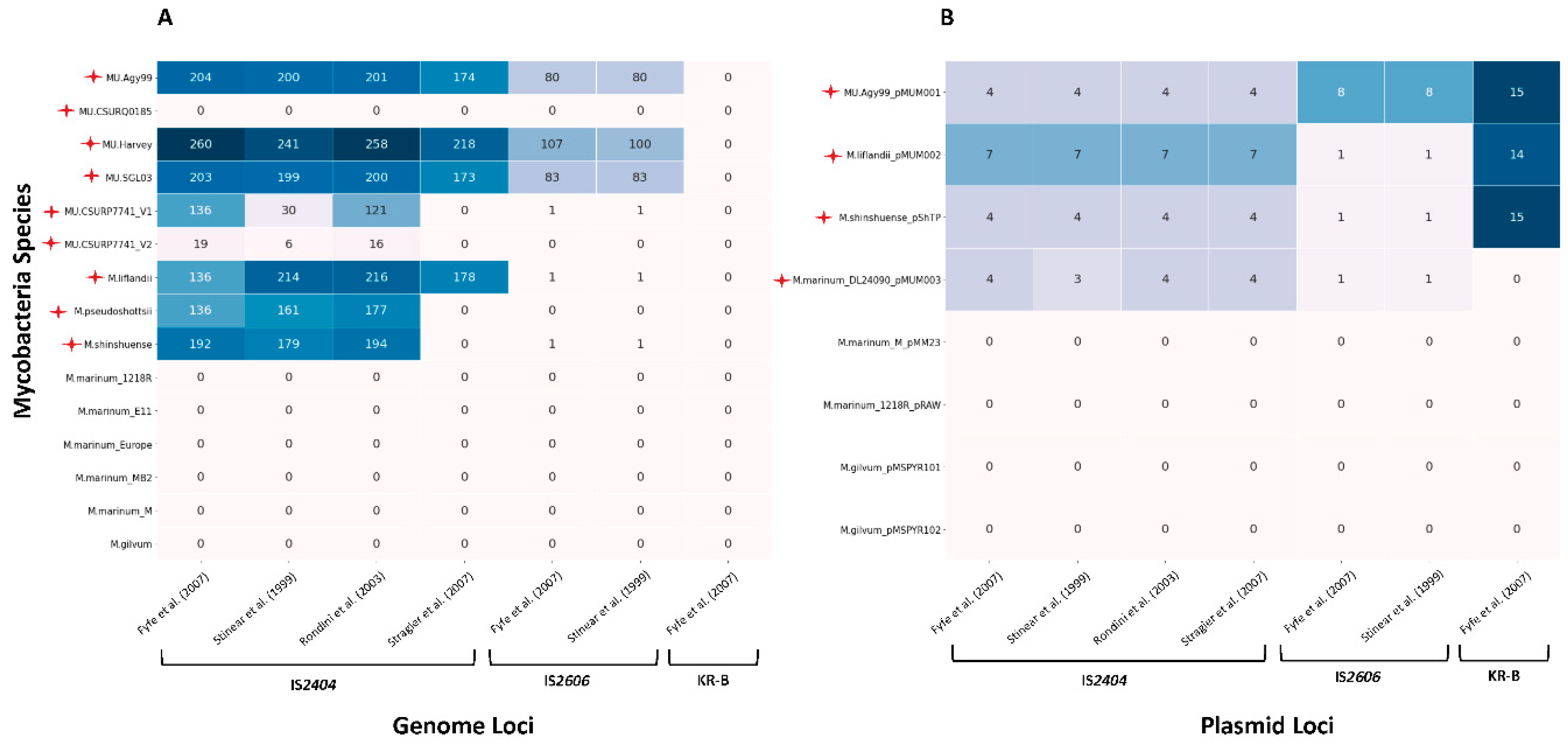
Figure 2.
Universal key for identifying variants of MPM, M. marinum and M. gilvum based on a combination of previously published MIRU-VNTR loci. This key was established on the basis of the availability of complete genomes, for strains isolated from different geographical areas and allows the genetic distinction between MPM variants. M. marinum strains were included as controls (not belonging to the MPM complex) and M. gilvum was added as an outgroup (not belonging to the MPM complex) although found in BU cases in French Guiana (Combe et al. 2020). The clustering process proposed here is based on the MIRU-VNTR loci DNA amplification and TRs copy number variations for VNTR19 (0; 1; 2; 3; 4 or 9), followed by MIRU5 (1; 2; 3; 4), followed by MIRU33, and so on.


Table 1.
Summary of the first isolation and phenotypic description of MPM. Adapted from the publication of Pidot et al. (2010). NA means not indicated.
Table 1.
Summary of the first isolation and phenotypic description of MPM. Adapted from the publication of Pidot et al. (2010). NA means not indicated.
| MPM | First isolation | Origin | Host | Colony coloration | Growth rate | Growth temperature | Mycolactone | Source |
|---|---|---|---|---|---|---|---|---|
| M. ulcerans | 1948 | Australia | Human | Greenish or brownish yellow | Slow growth (>4 weeks incubation) | 30°C-33°C | A/B | MacCallum, Buckle, Tolhurst and Sissons (1948) |
|
M. shinshuense (strain 753) |
1980 | Japan | Human | Yellowish, pigmentation in dark | Slow growth (>3 weeks incubation) | 28°C | A/B | Tsukamura and Mikoshiba (1982) |
|
M. marinum (strain CC240299) |
2002 | Israel | Koi (Cyprinus carpio) | NA | NA | NA | F | Ucko et al. (2002) |
|
M. marinum (strain DL240490) |
2002 | Red Sea | European sea bass (Dicentrarchus labrax) | NA | NA | NA | F | Ucko et al. (2002) |
|
M. marinum (strain DL045) |
2002 | Mediterranean Sea | European sea bass (Dicentrarchus labrax) | NA | NA | NA | F | Ucko et al. (2002) |
|
M. liflandii (strain 128FXT) |
2004 | US | African tropical clawed frogs (Xenopus tropicalis) | Buff-colored, non-pigmented | Slow growth (>4 weeks incubation) | 28°C | E | Trott et al. (2004) |
| M. pseudoshottsii (strain L15) | 2005 | US | Striped bass (Morone saxatilis) | Pale-yellow to gold, non-pigmented | Slow growth (>4 weeks incubation) |
23°C | F | Rhodes et al. (2005) |
Disclaimer/Publisher’s Note: The statements, opinions and data contained in all publications are solely those of the individual author(s) and contributor(s) and not of MDPI and/or the editor(s). MDPI and/or the editor(s) disclaim responsibility for any injury to people or property resulting from any ideas, methods, instructions or products referred to in the content. |
© 2023 by the authors. Licensee MDPI, Basel, Switzerland. This article is an open access article distributed under the terms and conditions of the Creative Commons Attribution (CC BY) license (http://creativecommons.org/licenses/by/4.0/).
Copyright: This open access article is published under a Creative Commons CC BY 4.0 license, which permit the free download, distribution, and reuse, provided that the author and preprint are cited in any reuse.


