Submitted:
21 July 2023
Posted:
24 July 2023
You are already at the latest version
Abstract
Analytical chemistry applied to medical and diagnostic analysis, recently focused on the development of cost-effective biosensors able to monitor the health status or to assess the level of specific biomarkers that can be indicative for several diseases. The improvement of technologies relating to the possibility of non-invasive sampling of biological fluids, as well as sensors for the detection of analytical signals and the computational capabilities of the systems routinely employed in everyday life (e.g. smartphones, computers, etc.), make the complete integration of self-standing analytical devices more accessible. This review aims to discuss the biosensors that have been proposed in the last 5 years focusing on two principal detecting approaches, optical and electrochemical, which have been employed for quantifying different kind of target analytes reaching detection limits below the clinical sample levels required. These detection principles applied to Point-of-care (POC) devices, have been extensively reported in literature and even the limited examples found on the market are based on these strategies. This work will show the latest innovations considering the integration of optical and electrochemical detection with the most commonly reported analytical platforms for POC applications such as paper-based or wearable and implantable devices.
Keywords:
Optical detection
; Point-of-care
; Lab-On-Chip
; Microfluidic device
; Paper-based device
; Biosensor
; Electrochemical detection
1. Introduction
New technologies for the development of portable and self-standing devices for rapid diagnosis of health disorder are emerging every day and they are linked to the advances in different areas including chemistry, bioengineering, physics and medicine. Indeed, the latest generation biosensors benefited from an interdisciplinary approach that has allowed the development of sophisticated and innovative analytical systems that could potentially improve human health, particularly in the developing world. In this context, the Point-of-care testing (POCT) approach represents a set of diagnostics that can allow the rapid detection of diseases leading to a prompt execution of the necessary treatments. The International Organization for Standardization (ISO) defines POCT as ‘testing that is performed near or at the site of a patient with the result leading to possible change in the care of the patient’ [1]. Recently, POCT is used across a variety of settings, including general practice, nursing homes, pharmacies, outpatient and off-site clinics and in-home patient care [2]. The immediate goal of POCT is to use the information it can provide to directly influence the timely and proper care of patients [3] and to enlarge the accessibility to clinical-diagnostics regardless of existing medical and laboratory infrastructure [3,4,5,6]. Several POCT devices were proposed for different applications, such as detection of bacteria [7,8,9], viruses [10,11,12,13], biomarkers related to specific disease or health conditions [14,15,16,17]. Their increased availability is partly due to technological advances that have made them more robust, easy-to-use and cost-effective, including advances in smartphone based technologies, lab-on-a-chip platforms, novel assay formats, e.g. automated assays and fully integrated assays (which include all the required reagents and equipment), and advances in the long term storage of reagents needed for POCT [4]. Indeed, these devices are based on biosensors able to combine in a unique platform bioanalytical assays and transduction systems for the acquisition and elaboration of analytical signals (Figure 1). The integrated platform should provide all the analytical steps, from the pretreatment of the sample, to analysis and data processing in order to obtain a qualitative and/or quantitative information. In light of these considerations, it is important that the optimization of the analytical assay, which should be based on the use of small volumes of sample and reagents and on simple procedures, is accompanied by the development of portable and easy-to-use detectors.
As it concerns bioanalytical assays, the most widespread are those based on immunoassays. Indeed, the high specificity and selectivity make them ideal candidates for detecting low traces of target analytes even in complex matrices and small volumes. In the same way, enzymatic assays already well established in laboratory practice, have been extensively proposed for POCT platforms. Several devices actually spread on the market for clinic-diagnostic purposes are based on these biorecognition reaction principles [18]. Recently also biosensors based on DNA or aptamers probes as well as whole cell biosensors are gaining interest among the scientific community. These innovative systems are very promising taking advantage of their implementation with new nanomaterials, but until now their use is limited to academic literature and are not well established in the market [19,20]. Detection techniques combined with these bioassays and exploited for developing POCT devices are generally based on optical and electrochemical principles [18,21,22,23,24]. Optical biosensors can exploit different phenomena, depending on the tracer employed in the bioanalytical assay. One of the most widespread approach well known for routinary applications is based on the use of metal nanoparticle (MNPs) which are characterized by strong surface plasmon resonance (SPR) bands, which frequency depends on the size and shape of the nanoparticle and on the nature of the metal together with the composition of the surrounding medium [25]. This phenomenon is widely exploited by commercially available Lateral Flow Immunoassay (LFIA) which are well known for their application in pregnancy test and more recently for SARS-COV-2 diagnosis. LFIA is one of the most successful colorimetric assay because of the advantages of simplicity, rapidity, cost-effectiveness and no requirement of equipment or technical expertise for operation. It is based on immunoassays in which the sample and a suitable labeled probe flow by capillary forces along a porous membrane that contains immobilized reagents with molecular recognition properties. These are placed in specific areas of the membrane, that are usually defines as Test and Control lines, where the former gives information about the target analyte, while the latter ensures the correct functioning of the test. The occurring of immunoreactions leads to the development of detectable bands with naked eyes in correspondence of the Test and Control lines, due to the accumulation of the label in such zones which allow to obtain qualitative information about the presence or the absence of the target analyte [26]. Otherwise, chemiluminescence (CL) is another optical technique alternative to colorimetric approach and it is triggered by a chemical reaction in which an atom or molecule passes from the excited state to the steady state, releasing photons as a by-product of the reaction [27]. CL-based biosensors usually rely on the use of enzymatic tracers that are able to catalyze CL reaction in presence of a suitable substrate. The measurement of the produced photons can be related to the concentration of the target analyte enabling quantitative analysis, but it implies the use of an external detector able to detect luminescent signals. Nowadays, there is a wide choice of miniaturized and compact CL detector that can be implemented into portable and easy-to-use analytical devices, since the main requisite is the ability to collect as much light as possible for achieving the highest detectability [28]. Besides the traditional photomultiplier, silicon p-i-n photodiode have been extensively proposed since they guarantee small size, low noise together with fast response time [29,30,31]. In additional, if it is necessary to obtain spatial information, charge coupled device (CCD) [32,33] and complementary metal-oxide semiconductor (CMOS) can be exploited for developing compact imaging systems. In particular, the use of CMOS is attracting great attention since this technology is integrated into smartphone photocamera enabling the smartphone to be used as a portable CL detector [34,35,36]. In the context of CL detection, recently also thermochemiluminescent (TCL) approach was proposed in which light emission is triggered by heating [37]. Therefore it is required the same instrumentation for acquiring photon emission with the addition of an heating system to induce the luminescent phaenomenon. As optical tranducing system, also fluorescence (FL) has been extensively proposed for developing clinical-diagnostic biosensors thanks to its high sensitivity. In this case, however, the analytical device must be integrated with both the luminescent detector and a light source for exciting the fluorophore used as label for the bioassay [38].
On the other hand, electrochemical transducers usually are based on the measurement of the current developed during a redox reaction (amperometric and potentiometric methods) or of the change in surface conductivity and impedance (conductometric and impedimetric methods) [39]. Amperometric-based transducers are one of the most frequently used and in this case the measured current results from the oxidation and reduction reactions of electroactive species. Amperometric measurements are performed by exploiting the passage of the current of the working electrode at the constant amplitude voltage against the reference electrode through the sample keeping the potential unchanged. If the current is measured applying controlled variations of the potential, the method is called voltammetry. Among the most widespread amperometric sensor, the glucose meter is one of the major applied for routinary analysis for people who suffer from diabetes mellitus or hypoglycemia. The assay is started by depositing a drop of blood on test strips equipped with an electrode containing glucose oxidase or dehydrogenase. Glucose present in the sample is oxidized by the enzyme which is regenerated by the presence of a mediator reagent generating an electric current. The total charge passing through the electrode is proportional to the concentration of glucose in blood that has reacted with the enzyme. Also potentiometric-based biosensors focus on redox processes but in this case it is measured the potential difference between working and reference electrode by a voltammeter. Impedimetric methods rely on the measurements of electrical changes that occur after a biorecognition reaction on the surface of a modified electrode. In particular, electrochemical impedance spectroscopy (EIS) is widely applied in developing biosensors based on the interaction between the biological probe and the target analyte that are selectively adsorbed on a modified electrode surface [40]. These adsorption cause changes in the electron transfer kinetics between a redox species present in the solution and the electrode. By monitoring the charge transfer resistance (Rct) it is possible to quantitatively determine the amount of target analyte bound at the electrode surface. Finally, also conductimetry can be exploited for developing biosensors. An ohmmeter is used for measuring the change in conductance between two metal electrodes at a certain distance from each other. Since an alternating current is applied to the electrode, the change of ionic content causes a conductance that can be measured. Generally, the change of ionic strength occurs as a result of an enzymatic reaction in the system, so this technique is employed for developing enzyme-based biosensors.
To combine the advantages of electrochemical and optical detection, electrochemiluminescence (ECL) has been propose which is very wide employed for analytical routinary applications in laboratory settings. ECL is a luminescent phenomenon triggered by an electrochemical stimulus on a specific molecular system [41,42]. Compared with FL and CL, ECL provides high temporal and spatial control of light emission, low background, high sensitivity, wide dynamic range, and rapid measurement [43,44], while with respect to other electrochemical techniques it is less sensitive to electrical interferences and can exploit simplified detector, since photon emission is measured instead of current [45]. Thanks to these advantages, ECL is particularly interesting for the development of portable biosensing devices [46,47].
This review aims to do the state-of-the-art about the development of POC biosensors for clinical-diagnosis purposes based on optical and electrochemical detection. The review will focus on the major latest achievements in this field showing the advantages and the weakness of the two different approaches as well as the possibilities for future development with a critical comparison with the widespread and commercialized systems that are already available.
2. Lab-on-chip based biosensors
The expression Lab-on-chip (LOC) refers to a technology based on the integration of fluidic, biosensing probes and measuring components (electrodes or optical apparatus) onto the same chip [48,49,50,51,52,53].
As it concerns electrochemical biosensors, the methods require a set of electrodes (working, reference and counter electrodes), their respective connectors, a potentiostat for regulating the electric processes and a laptop (or similar) to analyze the data [54]. Computer and potentiostat have been already replaced by portable and handheld wireless all-in-one devices which are suitable for POCT approach [55,56,57,58,59,60,61]. Following this trend, also electrodes are moving to easy-to-use and low-cost platforms such screen-printed electrodes (SPEs) that are developed by depositing or pasting a combination of layers onto a wide variety of flat substrates [62]. Electrochemical techniques combined with SPEs have proved to be an ideal candidate for replacing conventional benchtop equipment with affordable, rapid and portable devices thus offering versatility in terms of electrode design, materials and compatibility with different applications [63]. For developing biosensors, the immobilization of the recognition element onto the electrode surface is generally required. The immobilization processes on SPEs include different approaches such as adsorption, covalent coupling, entrapment or cross-linking for obtaining the formation of a self-assembled monolayer of biospecific probes [64,65,66,67]. Hu et al. proposed an electrochemical immunochip based on MoS2/ poly (diallyldimethylammonium chloride) (PDDA) hybrid film produced via layer-by-layer self-assembly method [68]. An electrochemical impedance detection platform was developed for the detection of alpha-fetoprotein (AFP) by integrating a three-electrode system in the micro-channel, which introduces MoS2/PDDA film modified with antibodies specific for recognition of AFP as the working electrode, Ag/AgCl wire as the reference electrode and indium tin oxide (ITO) as the counter electrode. This approach showed a linear range from 0.1 ng/mL to 10 ng/mL and a detection limit of 0.033 ng/mL. Timilsin et al. described a multi-electrode sensor chips suitable for detecting several analytes in complex biological fluids based on sandwich enzyme-linked immunosorbent assay (ELISA) method [69] (Figure 2a). Functionalized electrodes coated with immobilized capture antibodies were exposed to samples containing the antigen and a secondary biotinylated detection antibody. Upon incubation, the specific antigen is sandwiched between the capture and biotinylated detection antibodies. Finally, streptavidin poly-horseradish peroxidase (spHRP) was added in order to bind the biotinylated detection antibody and the measurment step was performed exploiting the TMB substrate which results in the production of an insoluble electroconductive product that locally precipitates at the reaction site and that can be electrochemically read using cyclic voltammetry. A dual sensor for simultaneous detection of free and total prostate specific antigen was developed on SPE surfaces for detecting human chorionic gonadotropin hormone [70]. The authors described an electrochemical-based immunosensing method, gold-linked immune-assay based on the direct electrochemical activity of Au nanoparticles (NPs) on SPEs. A primary antibody was immobilized on an electrode surface, and a non competitive immunoassay was carried out. Then a secondary antibody conjugated with Au NPs was employed and upon application of approximately 1.2 V, the Au NPs are pre-oxidized from Au0 to Au3+. A differential pulse voltammetry (DPV) from 0 V to 1.0 V immediately after the pre-oxidation step reduces the oxidised NPs back to Au0. The reduction process is detected at approximately 0.48 V.
Electrochemical LOC devices has been exhaustively proposed also for the detection of viruses [71,72,73,74,75,76] and recently they were applied for diagnosing SARS-CoV-2 [76]. Seo et al., functionalized graphene with SARS-CoV-2 spike antibody and the detection of the virus was performed both in the transport medium and clinical sample with a LOD of 1 fg/ml [77]. Zhao et al. [78], proposed the use of calixarene functionalized graphene oxide for detecting RNA of SARS-CoV-2 without nucleic acid amplification by exploiting a portable electrochemical smartphone. The LOD of the clinical specimen was 200 copies/mL and only two copies (10 μL) of SARS-CoV-2 were required for starting the assay. Azahar et al. [79] reported about the development of 3D nanoprinted electrodes, coated by nanoflakes of reduced-graphene-oxide (rGO) on which specific viral antigens were immobilized (Figure 2b). The electrodes were then integrated with a microfluidic device and used in a standard electrochemical cell. The antibodies present in the sample selectively binded the antigens, changing the impedance of the electrical circuit which was detected by impedance spectroscopy using a smartphone-based user interface. Furthermore the sensor can be regenerated within a minute by introducing a low-pH solution suitable for eluting the antibodies from the antigens, allowing successive sensing of test samples using the same sensor.
As for the electrochemical detection, also optical detection requires additional components that has to be integrated into the LOC device. Indeed, while electrochemistry needs the presence of electrodes and potentiostat, optical equipment comprises detector suitable for quantifying the colorimetric, CL or FL signals (in the last case also an excitation source is required for exciting the fluorescent specie). In an optics of integration with LOC devices, recently several detectors characterized by an adequate sensitivity combined with portability have been proposed leading to the development of ultrasensitive POCT assays [28,80]. For example, optical biosensors were designed exploiting new generation of (thermally cooled) back illuminated (BI) CCD [32] and smartphone BI-CMOS camera [36] or thin film photosensors, such as single photon avalanche photodiodes [81] or amorphous silicon (a-Si:H) photosensors [82]. Fluorescent detection has been widely proposed for the development of LOC devices. Dinter et al. [83] developed an adaptable platform to detect biomarkers using a microfluidic technology (Figure 2c). They were able to distinguish fluorescently labeled biomarkers exploiting an immunoassay based on physically and spectroscopically different functionalized microbeads. This multiplexing approach allowed to quantify four cardiovascular disease biomarkers using a commercial video imaging detector. Also Chang et al. [84] proposed a FL immunoassay developing a 3D microfluidic chip composed by a nichel mesh that acted as bead trap comprised between two layers sealed together by an adhesive of the PET sheet. Photonic crystal beads (PCBs) immobilized with capture antibodies are introduced into the microfluidic chip by injection and trapped into the grids of the nickel mesh. FL immunoassays were performed into the microfluidic chip and the signals were acquired exploiting a FL microscope equipped with a CCD. The PCBs were encoded by their colors coming from the photonic structure and the analysis of the target analytes (human immunoglobulin G, carcinoembryonic antigen and alpha fetoprotein) were performed by the identifying PCB colors and quantifying the FL intensity. Another work based on FL detection was proposed by Yuan et al. [85] The authors presented a bead-based assay for the detection of one of the four serotypes of dengue virus non-structural protein (DENV-NS1) as well as its cognate human IgG. In this system, the FL microspheres containing the classification fluorophore and detection fluorophore are imaged through a microfluidic chip using an infinity-corrected microscope system. To capture dengue virus non-structural protein (DENV-NS1), the magnetic beads with higher dye concentration were coupled with the anti-DENV-NS1 specific antibody, while the beads with lower dye concentration were loaded with DENV-NS1 to capture anti-DENV-NS1 antibodies. The coupled beads were then introduced into microfluidic channels for analysis using a laser and portable imaging setup These examples show a difficult integration between the LOC device and the detector for the measurement of the analytical signals. Indeed, the detection system is generally bench-top instrumentation applied for acquiring FL signals that occur into a microfluidic chip. The implementation of LOC device is better achieved in the field of CL. Dei et al. [86] developed a small-sized instrument for CL detection and signal analysis. They proposed a pump-free microfluidic chip, in which flow is self-initialized by capillary pumping and continued by imbibition of a filter paper. Microfluidic units in teardrop shape ensure that flow passes through the reaction areas at a reduced flux to facilitate the association between antigen and antibody and speeds up after the reaction areas. By spotting different antibodies into the reaction area, four types of biomarkers can be measured simultaneously in one microfluidic chip. The system was validated by testing four biomarkers of colorectal cancer (carcinoembryonic antigen, alpha-fetoprotein, carbohydrate antigen 125 and carbohydrate antigen 19-9) using plasma samples from patients. Sharafeldin et al. [87] used a system of micropumps and an Arduino microcontroller to create an automated 3D-printed device that lysed cells and performed a sandwich immunoassay for four metastasis biomarkers. The chip was composed by a detection chamber in which the inner walls coated with chitosan hydrogel film were functionalized with capture antibodies (Figure 2d). Non-competitive immunoassays where performed and the CL signals were acquired a CCD camera. In addition to FL and CL, also bioluminescence (BL) can be exploited for the development of LOC devices. Mirasoli et al. [88] described an integrated lab-on-chip, in for the amplification of viral DNA composed of a disposable 10-μL polydimethylsiloxane reaction chamber coupled to a glass substrate that hosts a thin-film metallic resistive heater and (a-Si:H) photosensors which act as light detector. A loop-mediated isothermal amplification (LAMP) technique was developed for specifically amplifying parvovirus B19 DNA and it was coupled with Bioluminescent Assay in Real Time (BART) technology to provide real-time detection of target DNA.
As an alternative, the ECL approach was employed by Calabria et al [89] for developing a 3D-printed miniaturized biosensor for glucose detection. The system employed a two-electrode configuration and it relied on the glucose oxidation catalysed by glucose oxidase. The developed hydrogen peroxide was detected by the addition of luminol and the ECL signal was acquired employing a smartphone.
As emerged from the reported works, the development of LOC devices has required in recent years a great effort mainly addressed to the resolution of problems such as the implementation of bioassay on microfluidic platform and the increase in the sensitivity of analytical methods (e.g. use of nanomaterials, innovative tracers, signal amplification). From this point of view, LOC biosensors have significantly improved their potential, reaching performances that are comparable with those obtained using laboratory settings. To date the most critical aspect remains the complete integration of the microfluidic device with the signal detector. Indeed, many works report the development of miniaturized and easy-to-use microfluidic platforms, but the detection step is very often performed with bench top instrumentation. From this point of view, the use of the smartphone (both for electrochemical and optical detection) represents a very important possibility to make these devices more usable even by non-specialized personnel. Instead, limited to the optical detection, the use of low-cost photosensors is also a significant step forward in achieving full integration of the analytical device.
3. Paper-based diagnostic devices
Paper-based devices have represented a real revolution in do-it-yourself diagnostics and most of the biosensors currently on the market for POCT applications are based on this platform. In addition to accessibility, affordability, and ease of disposal compared to traditional materials used in microfluidics, paper is also suitable for being processed in many different ways including printing, coating, cutting and lamination. This peculiarity offers a great versatility and the opportunity to design different devices that potentially can be mass produced with low cost [90,91]. By changing properties such as hydrophobicity, conductivity, porosity and reactivity (modifying the chemical structure) paper could be adapted to the specific need [92]. Furthermore, the use of paper as sensing element allows to simplify the required instrumentation since capillary force can drive fluid without an external pump.
Indeed, the most successfully commercialized devices with both electrochemical and optical detection are based on the use of paper as the analytical platform. Most glucometers, for example, are based on the use of disposable strips on which the reagents necessary for performing the test are immobilized. The detection is carried out by adding a drop of blood on the strip and then inserting it into the portable detector. Since the introduction of the first prototype of glucometer, many electrochemical biosensors were proposed in literature and recently some new nanomaterials were proposed for increasing the analytical performances [93,94,95,96,97,98,99,100,101].
Similarly, also the most common optical detection tests commercially available for POCT (pregnancy tests and COVID-19 tests) are based on LFIA technique. Traditionally, LFIA are employed for qualitative analyses, which involved the user’s conclusion about the presence or absence of a specific target analyte in the sample based on visual evaluation of the colored bands formed upon the strip. Recently this technique was implemented in order to obtain a quantitative output thanks to the combination with different detection principles [102]. Indeed, over the past decade, several affordable and compact detector were designed for integrating the LFIA strip with portable and easy-to-use system [103,104,105]. Even if the inclusion of instrumental measurement can complicate the analytical procedure, the possibility of obtaining quantitative informations for several biomarkers without the need of an equipped laboratory is attracting great attention [106].
One of the applications on which more publications based on colorimetric LFIA have been reported is about the detection of the Sars-CoV-2. Indeed, from 2020 several analytical devices have been released in commerce for the diagnosis of this disease. Literature focuses on the possibility to improve the detectability of these systems. Lee et al. [107] developed a colorimetric LFIA exploiting a specific linker for immobilizing antibodies on the cellulose membrane in an oriented manner. The proposed system allowed to increase the sensitivity with a detection limit of 5 × 10⁴ copies/mL. Another approach to improve the performances of the method relied the combination of different detection principle. For example a colorimetric and FL dual-functional LFIA biosensor was developed for the sensitive detection of spike 1 (S1) protein of SARS-CoV-2 by Han et al. [108]. The dual approach allowed to combine a rapid visual detection for a screening of suspected SARS-CoV-2 infection on sites while the FL signal allowed the quantitative detection of virus infection at the early stage. The authors coated a single-layer shell formed by mixing 20 nm Au NP and quantum dots (QDs) on SiO2 core to produce a dual immune label that simultaneously produced strong colorimetric and fluorescence signals. The detection limits of detecting S1 protein via colorimetric and fluorescence functions of the biosensor were 1 and 0.033 ng/mL, respectively. Also Roda et al. [109] proposed a dual optical/CL format of LFIA immunosensor for IgA detection in serum and saliva exploiting a recombinant nucleocapsid antigen which specifically captured SARS-CoV-2 antibodies in patient specimens. As detection system, a smartphone-camera-based device measures the colour signal provided by nanogold-labelled anti-human IgA while for the CL measurements a CCD was employed for acquiring the light signal resulting from the reaction of the HRP-labelled anti-human IgA with a H2O2/luminol/enhancers substrate. The use of the smartphone has been widely proposed in the field of optical detection associated with the LFIA technique [110,111,112]. Li et al [113] designed a QD LFIA for the FL detection of specific IgG for SARS-CoV-2 in human serum or whole blood samples. In this case the smartphone was employed as interface for elaborating the FL signals, indeed the detector was a portable fluorescence strip reader connected via WIFI to a smartphone platform. Mahmoud et al [114] exploited the smartphone directly as a FL detector, by developing a 3D printed smartphone imager with a built in UV-LED light source. Interleukin-6 and thrombin were measured by using FL green and red QDs as labels for the two target analytes, respectively. Through separation of RGB-channels, the acquired images can be processed to simultaneously quantify two analytes on the same test line enabling the optical multiplexingapproach. Also Rong et al. [115] developed a smartphone-based FL-LFIA platform for the detection of Zika virus nonstructural protein 1 (ZIKV NS1) exploiting QD microspheres as FL probes. A device to be integrated with the smartphone comprising optical and electrical components was designed and 3D-printed (Figure 3a). The implementation of CL detection with smartphone camera has been also investigated. Chabi et al [116] reported a LFIA platform employing phage-based CL reporters, combined with smartphone detection and applied to the diagnosis of SARS-CoV-2. Exploiting the same approach Ren et al. [117] trying to increase the sensitivity of the system, proposed a CL-LFIA based on the synthesis of the Au NP–antibody–HRP–polyethylene glycol conjugate for detecting cardiac troponin I (Figure 3b). Due to the presence of polyethylene glycol that allowed a controlled orientation of the immunocoplex, the accuracy of the LFIA was improved significantly allowing a detection limit of 10 pg mL–1. In the context of CL detection, Calabria et al. [29] employed an origami paper-based analytical device (μPAD) format for allowing the preloading of all the reagents in the dried form on the paper substrate. The analytical protocols were started by injecting a buffer solution in the μPAD, and the device was designed to be hosted in the AstroBio CubeSat (ABCS) nanosatellite. This biosensor represented the first step to develop an innovative technology to conduct research in space, implementing different kind of bioassays on the same platform to verify their compatibility with the effect of deep space conditions. An example of paper-based device was also proposed combined with TCL detection. Roda et al. [118] reported a TCL vertical flow immunoassay (VFIA) in which a one-step test was performed for detecting valproic acid. The VFIA sensor was composed by several layers layers functionalized with reagents stored in a stable form and by by 3D printing, accessories were produced to turn a smartphone into a biosensing device that provides a power source for the heat shock required to trigger the TCL reaction and a sensitive camera for measuring emitted photons.
As it concerns the electrochemical detection applied on paper-based devices, the electrode fabrication is a fundamental step for their development [119,120]. The first example was reported by Henry et al [121] who proposed carbon SPE onto filter paper. From this pioneering work, several devices have been presented, based on different electrode fabrication techniques [122]. Also LFIA has been extensively combined to electrochemical detection by implementing the nitrocellulose membrane with SPE. The main difference with the optical based LFIA is that in the electrochemical approach the membrane presents only the test line equipped with the SPE. The labels selected are generally molecules that undergoes an oxidation–reduction reaction on the electrode surface, or enzyme which can promote the oxidation–reduction reaction of the pre-existing reactant, resulting in an electrical current, voltage and other electrical changes. A portable potentiometer is then employed in order to measure the signal through the electrode and perform quantitative analysis [123]. Deenin et al [124] reported about the development of a biosensor for diagnosing COVID-19 based on an test strip integrated with a SPE (Figure 3c). The ferrocene carboxylic acid was used as tracer for SARS-CoV-2 antibody that binds the SARS-CoV-2 antigen in the sample before flowing on the strip. On the test line in correspondence of the electrode, angiotensin-converting enzyme 2 (ACE2) receptor was immobilized for capturing the immunocomplex formed in the presence of the target analyte. The electrochemical signal was measured with a smartphone interface achieving a detection limit of 2.98 pg/mL. Sinawang et al. [125] presented a work for the quantification of Dengue NS1 protein in human serum based on an electroactive immunonanoparticles that bind to the target biomarker and subsequently move along toward the biofunctionalized gold SPE to generate an amperometric signal measured by a potentiostat (Figure 3d). The gold SPE worked simultaneously as a signal transducer and a solid-state support for a sandwich ELISA-like immunoassay. Srisomwat et al [126] proposed an automated automated electrochemical LFIA device for the quantitative detection of the hepatitis B virus (HBV). The novelty is related to the use of a time-delayed microfluidic strategy fabricated on paper, which allowed a sequenced solution transfer. The device consisted of a straight nondelayed channel, a zigzag delayed channel, and a detection zone. In this configuration, the high-performance pyrrolidinyl peptide nucleic acid (acpcPNA) probe was immobilized on the test line of the test strip equipped with an electrode, while the Au3+ for gold metallization was deposited at the delayed channel zone. When the sample containing the HBV DNA was injected, the solution flowed across the nondelayed channel and hybridize with the acpcPNA probe at the test line. Meanwhile, the solution flowing through the delayed channel was retarded by the wax-printed barriers and gradually merged with the preceding flow from the nondelayed channel. Taking advantage of this differential flow behavior, DNA hybridization occured first, followed by Au3+ binding to the captured DNA through electrostatic interactions. The two steps of the assay proceed automatically and sequentially following the sample application and the buffer flow without the requirement of additional steps.
Also LFIA was combined with ECL detection. Tris(2,2′-bipyridyl)ruthenium (II) [Ru(bpy)32+], a photoactive species with optical properties, is frequently used in ECL-based sensing platforms due to its good solubility in water, high electrochemical stability, and capability for repeated regeneration in ECL reactions with tripropylamine (TPA). Hong et al. [127] proposed an ECL-LFIA for detecting troponin using Ru(bpy)32+-loaded mesoporous silica nanoparticles (RMSNs). Antibody-immobilized RMSNs were used for developing a non competitive LFIA and the detection was performed using a CCD camera.
The widespread diffusion of paper-based devices in commerce and in our everyday life demonstrates how this approach is functional and effective when combined with both electrochemical and optical detection. Certainly, in the future, qualitative detection technologies based on colorimetric detection could be replaced by more advanced quantitative systems which could give additional information and could therefore be applied to different types of target analytes including specific biomarkers for diagnosis or monitoring of several diseases. This will lead in an increasing interest in the research filed in improving the implementation of detectors on entirely portable platforms and increasing the sensitivity of the analytical methods involved.
4. Wearable technologies
Wearable and implantable technologies have gained great interest since the introduction of smartphones and mobile devices, thanks to the possibility of providing a monitoring of performance and state of health [128,129,130,131,132] and enabling measurement of clinically-relevant parameters in real time [133,134]. The first efforts in this field were mainly addressed on physical sensors for measuring parameters such as mobility, steps, calories burned or heart rate. With the implementation of new technologies, the demand for wearable devices changed requiring instrumentation suitable for performing non-invasive analysis on several biomarkers (such as metabolites, hormones, bacteria) in biofluids, like tears, saliva, urine, sweat and interstitial fluids. [135] Following this trend, researchers have proposed several examples of wearable biosensors mainly based on electrochemical and optical detection combined with the most innovative nanomaterials technologies and the use of flexible and advanced materials.
Electrochemical biosensors were the earliest platform that was exploited in the wearable field as they can benefit from easy readout and data processing [136,137]. Lei et al proposed a stretchable, wearable, and modular multifunctional biosensor, implementing enzymatic assays on a MXene/Prussian blue (Ti3C2Tx/PB) electrodes for the detection of glucose, lactate and pH levels in sweat [138]. The patch-type wearable device consisted of three modules (a sweat sampling layer, a sensor layer and a cover layer) and it could be connected to an electrochemical analyzer in order to measure a current signal. Also Wang et al [139] measured glucose level in sweat using a gold electrode deposited on the surface of PET through a procedure that avoid expensive and complex fabrication processes. Zhao et al [140] reported a fully integrated self-powered smartwatch for noninvasive and continuous monitoring of glucose levels. The system integrated several components: amorphous silicon (a-Si) photovoltaic cells that can be stored in rechargeable flexible Zn-MnO2 batteries providing sufficient power for the operation of the whole system, an electrochemical sensor for measuring glucose in sweat, a controlling module consisted in a printed circuit board (PCB) and a display to allow a direct and real-time monitoring. Lin et al [141] developed a flexible electrode system based on graphene oxide nanosheets for the impedance measurement of lactate in sweat exploiting an enzymatic assay involving lactate oxidase. Exploiting the same enzymatic approach for the detection of lactate, also Wang et al [142] proposed a stretchable gold-fiber-based electrodes which were included in textiles in a planar configuration. A different approach was used by Zhang et al [143] who exploited molecular imprinted polymers (MIP) as recognition probe. They generated a film of MIP on Ag nanowires which coated carbon working electrode. MIP allowed to obtain a selective and sensitive detection of lactate by measuring differential pulse voltammetry response. Fiore et al [144] reported about an immunosensor paper-based microfludic device for monitoring cortisol in sweat (Figure 4a). Using wax printing technology and laser-cutter technique they designed a microfluidic pattern that allowed to perform a competitve immunoassay in which antibodies for the recognition of cortisol were immobilized on magnetic beads and captured on a reaction zone. The competition between cortisol in the sample and cortisol conjugated to tracer (acetylcholinesterase enzyme) allowed the formation of the analytical signal by folding the pad pre-loaded with the proper enzymatic substrate. Combining this system with Near-Field Communication wireless module it was possible to monitor the signal using a smartphone. The same research group also developed a flexible device for monitoring the pH in sweat by depositing an iridium oxide film onto graphite working electrode and combinig it with an integrated circuit board allows for data acquisition [145].
As well as electrochemical detection, also optical detection has been proposed for its application on wearable biosensors [146,147,148,149,150,151] (Figure 4b). In this case some biomarkers have intrinsic optical properties like absorption and emission spectra that allows to selectively detect these species [152]. Among the applications of colorimetric approach, several works reported the use of acid-base indicators to measure the pH allowing a rapid and reversible detection [153,154,155,156,157]. Generally, these methods are based on naked-eye evaluation of the color formation which allows to work with a very simple equipment. If a quantitative analysis is required, the use of smartphone camera as detector is the most employed solution. A multiplex colorimetric assay for detecting creatinine, pH and urea was developed by Rogers et al [158]. The sensor was composed by different areas on which specific reagents for the detection of different target analytes have been immobilized. The colorimetric readout was performed using a smartphone that allowed the quantification exploiting the RGB analysis system. Optical detection based on FL principle has been proposed since the sensitivity offered by FL is higher respect to colorimetric methods. However, FL implies a more complex equipment and therefore analytical procedure. Xu et al. [159] proposed a wearable Cl− monitoring platform which combined a flexible cotton piece and two fluorescent materials (lanthanide metal–organic frameworks (MOFs) acting as test and control signals respectively) by simple ultrasonic loading. The two lanthanide-based fluorescent materials provide high color purity and accurate measurements. By designing Cl− and fluorescence signals for a logic circuit, a codec device was produced, exploiting a smartphone as detection platform.
The reported examples focus on the non-invasive analysis of sweat or saliva but these kind of samples can be not sufficient for determining some biomarkers. For this reason the minimally invasive sampling of intestitial fluid has been proposed since in comparison with the other peripheral biofluids, it contains several physiological information and exhibits a close correlation with blood samples due to the transcapillary exchange between blood and cells [160,161]. Recently, microneedles have gained great success due to the micron size and minimal invasion. [162,163] and they have been widely exploited for sampling interstitial fluid for the detection of several target analytes [164,165,166,167,168,169] exploiting different principle of extraction (negative pressure, capillary action, swelling force, ionophoretic). One of the most important applications for mincroneedle devices is the integration in biosensors for the monitoring of glucose. Generally, the electrochemical glucose sensors are based on a reaction that occurs at the working electrode involving glucose [170,171] and it can be classified as enzymatic [172] or non-enzymatic [173,174]. In literature several devices have been proposed based on enzymatic reaction between glucose and glucose oxidase and exploiting oxygen as redox mediator (first generation of glucose monitoring devices) [175]. In these applications, selectively permeable membranes were used to confined glucose oxidase on the surface of electrodes and to prevent electroactive interference [176,177,178,179,180,181,182]. The second generation of glucose sensors employed artificial mediators to avoid the oxygen dependence, and is also widely integrated on microneedles based device for glucose monitoring [183,184,185,186,187,188,189,190,191] (Figure 4c). The possibility to exploit non enzymatic reaction allows to overcome limitations due to the use of enzyme and to obtain advantages such as good stability, reproducibility, and free from oxygen limitation. In this case, generally the glucose sensors are based on the direct electrochemical oxidation of nanomaterials, such as metals (Pt, Au, Ni, Cu, etc.), metal-oxides (NiO, CuO, etc.), metal sulfides, metal-organic framework, metal azolate frame work and carbon materials [192,193]. Several works has been published about the development of microneedle-based monitoring of glucose exploiting these mechanisms [194,195,196].
The technology of minimally invasive sampling using microneedles for glucose monitoring in interstitial fluid has been extensively proposed also in combination with optical detection. The colorimetric approach is the one that gained much more attention since colorimetric signals can be directly evaluated by naked eyes, or measured by a smartphone camera, and finally elaborated through an RGB processing for obtaining a quantitative analysis. In this context, the most exploited mechanism is that involving the use of glucose oxidase to generate hydrogen peroxide which can be used for a subsequent reaction with a chromogenic agent. He et al [197] proposed a Polyvinyl alcohol/ Chitosan hydrogel integrated with microneedles suitable for the detection of electrolyte ions, glucose, lactate, and proteins even if the analysis procedure needs a substantial simplification in order to be compatible with the POCT approach. Paper-based platform have been applied also in combination with these technologies and several biosensors were reported in literature [198,199,200,201]. Besides the production of color on paper located upon microneedles, different colors can be directly produced on the microneedles patch or even on the skin surface. As an example, Gu et al. [202] developed a transdermal colorimetric microneedles patch consisted of two layers: glucose oxide embedded bottom needle layer, and calcium phosphate encapsulated HRP and TMB in the upper layer. The back of the patch turned to blue as a consequence of the reaction in presence of glucose and the quantification can be performed by scanning the color intensity with a miniaturized scanner. Yang et al. [203] exploited a microneedles patch made of glucose oxide-conjugated MnO2/graphene oxide nanozymes and Gelatin methacryloyl. When the interstitial fluid diffused into the patch the presence of glucose induced the production of H2O2, which bind to MnO2 and promoted the oxidation of TMB on the patch, causing a color change. However, many of the colorimetric system relies on the use of enzyme and chromogenic substrates, which can cause problems including enzyme denaturation, color quenching, byproduct toxicity, and non-reusability. As an alternative always exploiting optical detection, FL has been explored for these applications. Zhao et al [204] proposed an integrated microneedles device with photonic crystal barcodes to enable the multiplex detection of tumor necrosis factor-alpha (TNF-), interleukin-1β (IL-1), and IL-6 . A FL conjugated antibody immobilized onto microneedles was used to detect these biomarkers and the quantification was performed by reading the FL signal of the barcodes (Figure 4d). Also Wang et al. [205], coated the surface of microneedles with antibodies linked to FL plasmonic fluorophore for the detection of biomarker. Zheng et al [206] reported about a reagentless biosensor based on FL labeled aptamer probe for the on-needle detection of target analytes (glucose, adenosine triphosphate, l-tyrosinamide, and thrombin). By employing a strand displacement, the aptamer-conjugated to fluorophore were bound to DNA competitor strand marked with a quencher specie. Sang et al [207] developed a biosensors based on microneedles array that enabled the emission of a FL signal using a glucose responsive FL monomer. The monomer has two moieties, one for the recognition of glucose and the other one conjugated to anthracene for the FL emission.
Even if the efforts in this field are increasing, the FL signal is affected by light intensity, probe stability and variation due to human tissue. Furthermore, complex procedures and the need of optical instruments limit their diffusion for routinary applications.
Up to now, the wearable biosensors based on the use of microneedles available on the market are those for the monitoring of glucose and they are all based on electrochemical detection. Several companies such as Medtronic (Dublin, Ireland), Dexcom (San Diego California, US) and Abbott (Chicago, Illinois, US) have proposed their devices [208] and they are gaining a great success thanks to the easy-of-use, the relatively low-cost and advantages in terms of real-time monitoring of the health status. With the improvements in technologies for the optical detection as it concerns the integration with the biosensing element, portability and reproducibility of the results, in the future also this detection principle will be more explored and exploited for the development of biosensors suitable for the market.
5. Conclusions
In this review are described the advances in the development of POC devices for clinical-medical applications based on optical and electrochemical detection. The most significant efforts have been made in the study of new nanomaterials to employed like platforms for bioanalytical assays, innovative strategies for the amplification of analytical signals, the integration within self-standing devices capable of performing the analysis outside centralized laboratories. In many cases, results have been reported that are comparable with those obtained with bench top instruments that make use of specialized personnel. Despite these enormous steps forward, the most common biosensors in everyday life continue to be the glucometer with predominantly electrochemical detection and the pregnancy or COVID-19 tests mostly based on optical detection. Given the high interest raised from this research field, we will certainly observe an increase in their diffusion and use in the coming years. One of the most significant advances in this sense is represented by wearable devices which have already attracted attention from researchers and which will probably find wide use also for routine applications.
Author Contributions
Conceptualization M.Z, M.M, M.G; writing-original draft preparation, S.R.S.P., A.E., E.L., A.P.; writing—review and editing, D.C., M.Z., supervision M.Z, M.M, M.G. All authors have read and agreed to the published version of the manuscript.
Funding
This research was funded by the Italian Ministry of University and Research: PRIN2017 project “Cutting edge analytical chemistry methodologies and bio-tools to boost precision medicine in hormone-related diseases”, Prot. 2017Y2PAB8; and PRIN2017 project “Development of novel DNA-based analytical platforms for the rapid, point-of-use quantification of multiple hidden allergens in food samples”, Prot. 2017YER72K.
Institutional Review Board Statement
Not applicable.
Data Availability Statement
Not applicable.
Conflicts of Interest
The authors declare no conflict of interest.
References
- ISO. ISO 22870:2016 Point-of-care testing (POCT) - Requirements for quality and competence. 2016.
- Larsson, A.; Greig-Pylypczuk, R.; Huisman, A. ; The state of point-of-care testing: A European perspective. Ups J Med Sci 2015, 120, 1–10. [Google Scholar] [CrossRef] [PubMed]
- Kozel, T.R.; Burnham-Marusich, A.R. Point-of-Care Testing for Infectious Diseases: Past, Present, and Future. J Clin Microbiol 2017, 55, 2313. [Google Scholar] [CrossRef]
- Vashist, S.K.; Luppa, P.B.; Yeo, L.Y.; Ozcan, A.; Luong, J.H.T. Emerging Technologies for Next-Generation Point-of-Care Testing. Trends Biotechnol 2015, 33, 692–705. [Google Scholar] [CrossRef] [PubMed]
- Chen, H.; Liu, K.; Li, Z.; Wang, P. Point of care testing for infectious diseases. Clin Chim Acta. 2019, 493, 138–147. [Google Scholar] [CrossRef] [PubMed]
- ECDC. Public health guidance on screening and vaccination for infectious diseases in newly arrived migrants within the EU/EEA. 2018.
- Reali, S.; Najib, E.Y.; Balázs, K.E.T.; Tan, A.C.H.; Váradi, L.; Hibbs, D.E.; Groundwater, P.W. Novel diagnostics for point-of-care bacterial detection and identification. RSC advance 2019, 9, 21486–21497. [Google Scholar] [CrossRef]
- Gopal, A.; Yan, L.; Kashif, S.; Munshi, T.; Roy, V.A.; Voelcker, N.H.; Chen, X. Biosensors and Point-of-Care Devices for Bacterial Detection: Rapid Diagnostics Informing Antibiotic Therapy. Advanc Health Mat 2022, 11, 2101546. [Google Scholar] [CrossRef]
- Nath, P.; Kabir, A.; Khoubafarin Doust, S.; Kreais, Z.J.; Ray, A. Detection of bacterial and viral pathogens using photonic point-of-care devices. Diagnostics 2020, 10, 841. [Google Scholar] [CrossRef]
- Zhang, Z.; Ma, P.; Ahmed, R.; Wang, J.; Akin, D.; Soto, F.; Demirci, U. Advanced point-of-care testing technologies for human acute respiratory virus detection. Advanc Mat 2022, 34, 2103646. [Google Scholar] [CrossRef]
- Xiao, M.; Tian, F.; Liu, X.; Zhou, Q.; Pan, J.; Luo, Z.; Yi, C. Virus Detection: From State-of-the-Art Laboratories to Smartphone-Based Point-of-Care Testing. Advanc Sci 2022, 9, 2105904. [Google Scholar] [CrossRef]
- Zhao, V.X.T.; Wong, T.I.; Zheng, X.T.; Tan, Y.N.; Zhou, X. Colorimetric biosensors for point-of-care virus detections. Mat sci energy technol 2020, 3, 237–249. [Google Scholar] [CrossRef]
- Lu, S.; Lin, S.; Zhang, H.; Liang, L.; Shen, S. Methods of respiratory virus detection: Advances towards point-of-care for early intervention. Micromachines 2021, 12, 697. [Google Scholar] [CrossRef] [PubMed]
- Sohrabi, H.; Bolandi, N.; Hemmati, A.; Eyvazi, S.; Ghasemzadeh, S.; Baradaran, B.; Mokhtarzadeh, A. State-of-the-art cancer biomarker detection by portable (Bio) sensing technology: A critical review. Microchem J 2022, 107248. [Google Scholar] [CrossRef]
- Suntornsuk, W.; Suntornsuk, L. Recent applications of paper-based point-of-care devices for biomarker detection. Electrophoresis 2020, 41, 287–305. [Google Scholar] [CrossRef]
- Mahmoudi, T.; de la Guardia, M.; Baradaran, B. Lateral flow assays towards point-of-care cancer detection: A review of current progress and future trends. TrAC Trends Anal Chem 2020, 125, 115842. [Google Scholar] [CrossRef]
- Ouyang, M.; Tu, D.; Tong, L.; Sarwar, M.; Bhimaraj, A.; Li, C.; Di Carlo, D. A review of biosensor technologies for blood biomarkers toward monitoring cardiovascular diseases at the point-of-care. Biosens Bioelectron 2021, 171, 112621. [Google Scholar] [CrossRef] [PubMed]
- Sachdeva, S.; Davis, R.W.; Saha, A.K. Microfluidic point-of-care testing: Commercial landscape and future directions. Frontiers Bioengineering Biotechnol 2021, 8, 602659. [Google Scholar] [CrossRef] [PubMed]
- Kaur, H.; Bruno, J.G.; Kumar, A.; Sharma, T.K. Aptamers in the therapeutics and diagnostics pipelines. Theranostics. 2018, 8, 4016. [Google Scholar] [CrossRef]
- Gui, Q.; Lawson, T.; Shan, S.; Yan, L.; Liu, Y. The application of whole cell-based biosensors for use in environmental analysis and in medical diagnostics. Sensors 2017, 17, 1623. [Google Scholar] [CrossRef]
- Mitchell, K.R.; Esene, J.E.; Woolley, A.T. Advances in multiplex electrical and optical detection of biomarkers using microfluidic devices. Anal Bioanal Chem 2022, 414, 167–180. [Google Scholar] [CrossRef]
- Shi, Y.; Li, Z.; Liu, P.Y.; Nguyen, B.T.T.; Wu, W.; Zhao, Q.; Liu, A.Q. On-Chip Optical Detection of Viruses: A Review. Adv Photon Res 2021, 2, 2000150. [Google Scholar] [CrossRef]
- Sezgintürk, M.K. Introduction to commercial biosensors. In Commercial Biosensors and Their Applications; Sezgintürk, M. K., Ed.; Elsevier: Netherlands, 2020; pp. 1–28. [Google Scholar]
- Haleem, A.; Javaid, M.; Singh, R.P.; Suman, R.; Rab, S. Biosensors applications in medical field: A brief review. Sensors Int. 2021, 2, 100100. [Google Scholar] [CrossRef]
- Anfossi, L.; Di Nardo, F.; Russo, A.; Cavalera, S.; Giovannoli, C.; Spano, G.; Baggiani, C. Silver and gold nanoparticles as multi-chromatic lateral flow assay probes for the detection of food allergens. Anal bioanal chem 2019, 411, 1905–1913. [Google Scholar] [CrossRef] [PubMed]
- Di Nardo, F.; Baggiani, C.; Giovannoli, C.; Spano, G.; Anfossi, L. Multicolor immunochromatographic strip test based on gold nanoparticles for the determination of aflatoxin B1 and fumonisins. Microchim Acta 2017, 184, 1295–1304. [Google Scholar] [CrossRef]
- Roda, A.; Mirasoli, M.; Michelini, E.; Di Fusco, M.; Zangheri, M.; Cevenini, L.; Simoni, P. Progress in chemical luminescence-based biosensors: A critical review. Biosens Bioelectron 2016, 76, 164–179. [Google Scholar] [CrossRef]
- Calabretta, M.M.; Lopreside, A.; Montali, L.; Zangheri, M.; Evangelisti, L.; D’Elia, M.; Michelini, E. Portable light detectors for bioluminescence biosensing applications: A comprehensive review from the analytical chemist’s perspective. Anal Chim Acta 2022, 339583. [Google Scholar] [CrossRef]
- Calabria, D.; Trozzi, I.; Lazzarini, E.; Pace, A.; Zangheri, M.; Iannascoli, L.; Mirasoli, M. AstroBio-CubeSat: A lab-in-space for chemiluminescence-based astrobiology experiments. Biosens Bioelectron 2023, 226, 115110. [Google Scholar] [CrossRef] [PubMed]
- Zangheri, M.; Mirasoli, M.; Nascetti, A.; Caputo, D.; Bonvicini, F.; Gallinella, G.; Roda, A. Microfluidic cartridge with integrated array of amorphous silicon photosensors for chemiluminescence detection of viral DNA. Sens bio-sens res 2016, 7, 127–132. [Google Scholar] [CrossRef]
- Mirasoli, M.; Bonvicini, F.; Lovecchio, N.; Petrucci, G.; Zangheri, M.; Calabria, D.; Nascetti, A. On-chip LAMP-BART reaction for viral DNA real-time bioluminescence detection. Sens Actuat B Chem 2018, 262, 1024–1033. [Google Scholar] [CrossRef]
- Lazzarini, E.; Pace, A.; Trozzi, I.; Zangheri, M.; Guardigli, M.; Calabria, D.; Mirasoli, M. An Origami Paper-Based Biosensor for Allergen Detection by Chemiluminescence Immunoassay on Magnetic Microbeads. Biosensors 2022, 12, 825. [Google Scholar] [CrossRef]
- Sciutto, G.; Zangheri, M.; Anfossi, L.; Guardigli, M.; Prati, S.; Mirasoli, M.; Roda, A. Miniaturized biosensors to preserve and monitor cultural heritage: From medical to conservation diagnosis. Angew Chem 2018, 130, 7507–7511. [Google Scholar] [CrossRef]
- Zangheri, M.; Di Nardo, F.; Calabria, D.; Marchegiani, E.; Anfossi, L.; Guardigli, M.; Roda, A. Smartphone biosensor for point-of-need chemiluminescence detection of ochratoxin A in wine and coffee. Anal Chim Acta 2021, 1163, 338515. [Google Scholar] [CrossRef] [PubMed]
- Roda, A.; Calabretta, M.M.; Calabria, D.; Caliceti, C.; Cevenini, L.; Lopreside, A.; Zangheri, M. Smartphone-based biosensors. Past, Present and Future Challenges of Biosensors and Bioanalytical Tools in Analytical Chemistry: A Tribute to Professor Marco Mascini. Compr. Anal. Chem 2017, 77, 237–286. [Google Scholar]
- Calabria, D.; Zangheri, M.; Trozzi, I.; Lazzarini, E.; Pace, A.; Mirasoli, M.; Guardigli, M. Smartphone-based chemiluminescent origami µPAD for the rapid assessment of glucose blood levels. Biosensors 2021, 11, 381. [Google Scholar] [CrossRef]
- Di Fusco, M.; Quintavalla, A.; Lombardo, M.; Guardigli, M.; Mirasoli, M.; Trombini, C.; Roda, A. Organically modified silica nanoparticles doped with new acridine-1, 2-dioxetane analogues as thermochemiluminescence reagentless labels for ultrasensitive immunoassays. Anal. Bioanal. Chem 2015, 407, 1567–1576. [Google Scholar] [CrossRef] [PubMed]
- Roda, A.; Michelini, E.; Zangheri, M.; Di Fusco, M.; Calabria, D.; Simoni, P. Smartphone-based biosensors: A critical review and perspectives. TrAC Trends Anal Chem 2016, 79, 317–325. [Google Scholar] [CrossRef]
- Bahadır, E.B.; Sezgintürk, M.K. Electrochemical biosensors for hormone analyses. Biosens Bioelectron 2015, 68, 62–71. [Google Scholar] [CrossRef]
- Chakraborty, A.; Tibarewala, D.N.; Barui, A. Impedance-based biosensors, in: Bioelectronics and Medical Devices, 2019, pp. 97e122.
- Richter, M.M. Electrochemiluminescence (ecl). Chem Rev 2004, 104, 3003–3036. [Google Scholar] [CrossRef]
- Miao, W. Electrogenerated chemiluminescence and its biorelated applications. Chem rev 2008, 108, 2506–2553. [Google Scholar] [CrossRef]
- Du, F.; Chen, Y.; Meng, C.; Lou, B.; Zhang, W.; Xu, G. Recent advances in electrochemiluminescence immunoassay based on multiple-signal strategy. Current Opin Electrochem 2021, 28, 100725. [Google Scholar] [CrossRef]
- Nasrollahpour, H.; Khalilzadeh, B.; Naseri, A.; Sillanpaa, M.; Chia, C.H. Homogeneous electrochemiluminescence in the sensors game: What have we learned from past experiments? Anal Chem 2021, 94, 349–365. [Google Scholar] [CrossRef]
- Ma, X., Gao; Xu, G. Rational design of electrochemiluminescent devices. Accounts of Chemical Research 2021, 54, 2936–2945. [Google Scholar] [CrossRef] [PubMed]
- Nikolaou, P.; Sciuto, E.L.; Zanut, A.; Petralia, S.; Valenti, G.; Paolucci, F.; Conoci, S. Ultrasensitive PCR-Free detection of whole virus genome by electrochemiluminescence. Biosens Bioelectron 2022, 209, 114165. [Google Scholar] [CrossRef] [PubMed]
- Yoo, S.M.; Jeon, Y.M.; Heo, S.Y. Electrochemiluminescence Systems for the Detection of Biomarkers: Strategical and Technological Advances. Biosensors 2022, 12, 738. [Google Scholar] [CrossRef] [PubMed]
- Nascetti, A.; Mirasoli, M.; Marchegiani, E.; Zangheri, M.; Costantini, F.; Porchetta, A.; Roda, A. Integrated chemiluminescence-based lab-on-chip for detection of life markers in extraterrestrial environments. Biosens Bioelectron 2019, 123, 195–203. [Google Scholar] [CrossRef] [PubMed]
- Zhang, T., Chakrabarty. Microelectrofluidic systems: Modeling and simulation; CRC Press: Florida, US, 2018. [Google Scholar]
- Reyes, D.R.; Iossifidis, D.; Auroux, P.A.; Manz, P.A. Micro total analysis systems: 1. Introduction, theory, and technology. Anal. Chem. 2002, 74, 2623–2636. [Google Scholar] [CrossRef]
- Auroux, A.; Reyes, D.R.; Iossifidis, D.; Manz, P.A. Micro total analysis systems: 2. Analytical standard operations and applications. Anal. Chem. 2002, 74, 2637–2652. [Google Scholar] [CrossRef]
- West, J.; Becker, M.; Tombrink, S.; Manz, A. Micro total analysis systems: Latest achievements. Anal. Chem. 2008, 80, 4403–4419. [Google Scholar] [CrossRef]
- Giannitsis, A.T. Microfabrication of biomedical lab-on-chip devices. A review. Est J Eng 2011, 17, 109. [Google Scholar] [CrossRef]
- Zhang, W.; Wang, R.; Luo, F.; Wang, P.; Lin, Z. Miniaturized electrochemical sensors and their point-of-care applications. Chinese Chem Let 2020, 31, 589–600. [Google Scholar] [CrossRef]
- Ainla, A.; Mousavi, M.P.; Tsaloglou, M.N.; Redston, J.; Bell, J.G.; Fernández-Abedul, M.T.; Whitesides, G.M. Open-source potentiostat for wireless electrochemical detection with smartphones. Anal chem 2018, 90, 6240–6246. [Google Scholar] [CrossRef]
- Dryden, M.D.; Wheeler, A.R. DStat: A versatile, open-source potentiostat for electroanalysis and integration. PLoS ONE 2015, 10, e0140349. [Google Scholar] [CrossRef] [PubMed]
- Rowe, A.A.; Bonham, A.J.; White, R.J.; Zimmer, M.P.; Yadgar, R.J.; Hobza, T.M.; Plaxco, K.W. CheapStat: An open-source,“Do-It-Yourself” potentiostat for analytical and educational applications. PLoS ONE 2011, 6, e23783. [Google Scholar] [CrossRef] [PubMed]
- Dobbelaere, T.; Vereecken, P.M.; Detavernier, C. A USB-controlled potentiostat/galvanostat for thin-film battery characterization. HardwareX 2017, 2, 34–49. [Google Scholar] [CrossRef]
- Lopin, P.; Lopin, K.V. PSoC-Stat: A single chip open source potentiostat based on a Programmable System on a Chip. PLoS ONE 2018, 13, e0201353. [Google Scholar] [CrossRef] [PubMed]
- Beach, R.D.; Conlan, R.W.; Godwin, M.C.; Moussy, F. Towards a miniature implantable in vivo telemetry monitoring system dynamically configurable as a potentiostat or galvanostat for two-and three-electrode biosensors. IEEE T INSTRUM MEAS 2005, 54, 61–72. [Google Scholar] [CrossRef]
- Ferrari, A.G.M.; Rowley-Neale, S.J.; Banks, C.E. Screen-printed electrodes: Transitioning the laboratory in-to-the field. Talanta Open 2021, 3, 100032. [Google Scholar] [CrossRef]
- Ferrari, A.G.M.; Amor-Gutiérrez, O.; Costa-Rama, E.; Fernández-Abedul, M.T. Batch injection electroanalysis with stainless-steel pins as electrodes in single and multiplexed configurations. Sens Actuat B Chem 2017, 253, 1207–1213. [Google Scholar] [CrossRef]
- Foster, C.W.; Kadara, R.O.; Banks, C.E. Screen-printing electrochemical architectures; Springer: Berlin, Germany, 2016. [Google Scholar]
- Giacomelli, C.E.; Vermeer, A.W.P.; Norde, W. Adsorption of immunoglobulin G on core-shell latex particles precoated with chaps. J. Colloid Interf. Sci. 2000, 231, 283–288. [Google Scholar] [CrossRef]
- Charelier, R.C.; Gengenbach, T.R.; Griesser, H.J.; Brigham-Burke, M.; O’Shannessy, D.J. A general method to recondition and reuse BIAcore sensor chips fouled with covalently immobilized protein/peptide. Anal. Biochem. 1995, 229, 112–118. [Google Scholar] [CrossRef]
- Charles, P.T.; Goldman, E.R.; Rangasammy, J.G.; Schauer, C.L.; Chen, M.S.; Taitt, C.R. Fabrication and characterization of 3D hydrogel microarrays to measure antigenicity and antibody functionality for biosensor application. Biosens. Bioelectron. 2004, 20, 753–764. [Google Scholar] [CrossRef]
- Palma, R.; Borghs, G.; Declerck, P.; Goddeeris, B. Comparison of random and oriented immobilization of antibody fragments on mixed self-assembled monolayers. J. Immunol. Methods 2006, 312, 167–181. [Google Scholar]
- Hu, T.; Zhang, M.; Wang, Z.; Chen, K.; Li, X.; Ni, Z. Layer-by-layer self-assembly of MoS2/PDDA hybrid film in microfluidic chips for ultrasensitive electrochemical immunosensing of alpha-fetoprotein. Microchem J. 2020, 158, 105209. [Google Scholar] [CrossRef]
- Timilsina, S.S.; Ramasamy, M.; Durr, N.; Ahmad, R.; Jolly, P.; Ingber, D.E. Biofabrication of Multiplexed Electrochemical Immunosensors for Simultaneous Detection of Clinical Biomarkers in Complex Fluids. Advanc Health Mat 2022, 11, 2200589. [Google Scholar] [CrossRef] [PubMed]
- Escamilla-Gómez, V.; Hernández-Santos, D.; González-García, M.B.; Pingarrón-Carrazón, J.M.; Costa-García, A. Simultaneous detection of free and total prostate specific antigen on a screen-printed electrochemical dual sensor. Biosens Bioelectron. 2009, 24, 2678–2683. [Google Scholar] [CrossRef]
- Singh, R.; Hong, S.; Jang, J. Label-free detection of influenza viruses using a reduced Graphene oxide-based electrochemical immunosensor integrated with a microfluidic platform, Sci. Rep. 2017, 7, 4277. [Google Scholar]
- Roberts, A.; Chauhan, N.; Islam, S.; Mahari, S.; Ghawri, B.; Gandham, R.K.; Gandhi, S. Graphene functionalized field-effect transistors for ultrasensitive detection of Japanese encephalitis and Avian influenza virus. Sci Rep. 2020, 10, 14546. [Google Scholar] [CrossRef]
- Ono, T., Oe. Glycan-functionalized graphene-FETs toward selective detection of human-infectious avian influenza virus. Japanese Journal of Applied Physics 2017, 56, 030302. [Google Scholar] [CrossRef]
- Maity, A.; Sui, X.; Jin, B.; Pu, H.; Bottum, K.J.; Huang, X.; Chen, J. Resonance-frequency modulation for rapid, point-of-care Ebola-Glycoprotein diagnosis with a graphene-based field-effect biotransistor. Anal chem. 2018, 90, 14230–14238. [Google Scholar] [CrossRef]
- Jin, X.; Zhang, H.; Li, Y.T.; Xiao, M.M.; Zhang, Z.L.; Pang, D.W.; Zhang, G.J. A field effect transistor modified with reduced graphene oxide for immunodetection of Ebola virus. Microchim Acta. 2019, 186, 1–9. [Google Scholar] [CrossRef]
- Sengupta, J.; Adhikari, A.; Hussain, C.M. Graphene-based analytical lab-on-chip devices for detection of viruses: A review. Carbon Trends 2021, 4, 100072. [Google Scholar] [CrossRef]
- Seo, G.; Lee, G.; Kim, M.J.; Baek, S.H.; Choi, M.; Ku, K.B.; Kim, S.I. Rapid detection of COVID-19 causative virus (SARS-CoV-2) in human nasopharyngeal swab specimens using field-effect transistor-based biosensor. ACS nano 2020, 14, 5135–5142. [Google Scholar] [CrossRef]
- Zhao, H.; Liu, F.; Xie, W.; Zhou, T.C.; OuYang, J.; Jin, L.; Li, C.P. Ultrasensitive supersandwich-type electrochemical sensor for SARS-CoV-2 from the infected COVID-19 patients using a smartphone. Sens Actuat B Chem 2021, 327, 128899. [Google Scholar] [CrossRef] [PubMed]
- Ali, M.A.; Hu, C.; Jahan, S.; Yuan, B.; Saleh, M.S.; Ju, E.; Panat, R. Sensing of COVID-19 antibodies in seconds via aerosol jet nanoprinted reduced-graphene-oxide-coated 3D electrodes. Adv Mat. 2021, 33, 2006647. [Google Scholar] [CrossRef] [PubMed]
- Roda, A.; Arduini, F.; Mirasoli, M.; Zangheri, M.; Fabiani, L.; Colozza, N.; Moscone, D. A challenge in biosensors: Is it better to measure a photon or an electron for ultrasensitive detection? Biosens Bioelectron. 2020, 155, 112093. [Google Scholar] [CrossRef] [PubMed]
- Iinuma, M., Kadoya; Kuroda, A. Photon counting system for high-sensitivity detection of bioluminescence at optical fiber end. Bioluminescence: Methods and Protocols 2016, 299–310. [Google Scholar]
- Calabria, D.; Trozzi, I.; Lazzarini, E.; Pace, A.; Zangheri, M.; Iannascoli, L.; Mirasoli, M. AstroBio-CubeSat: A lab-in-space for chemiluminescence-based astrobiology experiments. Biosens Bioelectron 2023, 226, 115110. [Google Scholar] [CrossRef]
- Dinter, F.; Burdukiewicz, M.; Schierack, P.; Lehmann, W.; Nestler, J.; Dame, G.; Rödiger, S. Simultaneous detection and quantification of DNA and protein biomarkers in spectrum of cardiovascular diseases in a microfluidic microbead chip. Anal bioanal chem 2019, 411, 7725–7735. [Google Scholar] [CrossRef]
- Chang, N.; Zhai, J.; Liu, B.; Zhou, J.; Zeng, Z.; Zhao, X. Low cost 3D microfluidic chips for multiplex protein detection based on photonic crystal beads. Lab Chip 2018, 18, 3638–3644. [Google Scholar] [CrossRef]
- Yuan, X.; Garg, S.; De Haan, K.; Fellouse, F.A.; Gopalsamy, A.; Tykvart, J.; Aitchison, J.S. Bead-based multiplex detection of dengue biomarkers in a portable imaging device. Biomed Opt Express 2020, 11, 6154–6167. [Google Scholar] [CrossRef]
- Dai, B.; Yin, C.; Wu, J.; Li, W.; Zheng, L.; Lin, F.; Zhuang, S. A flux-adaptable pump-free microfluidics-based self-contained platform for multiplex cancer biomarker detection. Lab Chip 2021, 21, 143–153. [Google Scholar] [CrossRef]
- Sharafeldin, M.; Chen, T.; Ozkaya, G.U.; Choudhary, D.; Molinolo, A.A.; Gutkind, J.S.; Rusling, J.F. Detecting cancer metastasis and accompanying protein biomarkers at single cell levels using a 3D-printed microfluidic immunoarray. Biosens Bioelectron. 2021, 171, 112681. [Google Scholar] [CrossRef] [PubMed]
- Mirasoli, M.; Bonvicini, F.; Lovecchio, N.; Petrucci, G.; Zangheri, M.; Calabria, D.; Nascetti, A. On-chip LAMP-BART reaction for viral DNA real-time bioluminescence detection. Sens Actuat B Chem 2018, 262, 1024–1033. [Google Scholar] [CrossRef]
- Calabria, D.; Lazzarini, E.; Pace, A.; Trozzi, I.; Zangheri, M.; Cinti, S.; Mirasoli, M. Smartphone-based 3D-printed electrochemiluminescence enzyme biosensor for reagentless glucose quantification in real matrices. Biosens Bioelectron 2023, 227, 115146. [Google Scholar] [CrossRef] [PubMed]
- Noviana, E.; McCord, C.P.; Clark, K.M.; Jang, I.; Henry, C.S. Electrochemical paper-based devices: Sensing approaches and progress toward practical applications. Lab Chip 2020, 20, 9–34. [Google Scholar] [CrossRef] [PubMed]
- He, Y.; Wu, Y.; Fu, J.Z.; Wu, W.B. Fabrication of paper-based microfluidic analysis devices: A review. Rsc Advan 2015, 5, 78109–78127. [Google Scholar] [CrossRef]
- Lee, V.B.C.; Mohd-Naim, N.F.; Tamiya, E.; Ahmed, M.U. Trends in paper-based electrochemical biosensors: From design to application. Anal Sci 2018, 34, 7–18. [Google Scholar] [CrossRef]
- Tran, V.K.; Ko, E.; Geng, Y.; Kim, M.K.; Jin, G.H.; Son, S.E.; Seong, G.H. Micro-patterning of single-walled carbon nanotubes and its surface modification with gold nanoparticles for electrochemical paper-based non-enzymatic glucose sensor. J Electroanal Chem. 2018, 826, 29–37. [Google Scholar] [CrossRef]
- Punjiya, M.; Moon, C.H.; Matharu, Z.; Nejad, H.R.; Sonkusale, S. A three-dimensional electrochemical paper-based analytical device for low-cost diagnostics. Analyst 2018, 143, 1059–1064. [Google Scholar] [CrossRef]
- Amor-Gutiérrez, O.; Costa-Rama, E.; Fernández-Abedul, M.T. Sampling and multiplexing in lab-on-paper bioelectroanalytical devices for glucose determination. Biosens Bioelectron 2019, 135, 64–70. [Google Scholar] [CrossRef]
- He, X.; Chang, S.J.; Settu, K.; Chen, C.J.; Liu, J.T. An anti-HCT-interference glucose sensor based on a fiber paper-based screen-printed carbon electrode. Sens Actua B Chem. 2019, 297, 126763. [Google Scholar] [CrossRef]
- Mohammadifar, M.; Tahernia, M.; Choi, S. An equipment-free, paper-based electrochemical sensor for visual monitoring of glucose levels in urine. SLAS TECHNOL Transl Life Sci Inn 2019, 24, 499–505. [Google Scholar] [CrossRef]
- Calabria, D.; Pace, A.; Lazzarini, E.; Trozzi, I.; Zangheri, M.; Guardigli, M.; Mirasoli, M. Smartphone-Based Chemiluminescence Glucose Biosensor Employing a Peroxidase-Mimicking, Guanosine-Based Self-Assembled Hydrogel. Biosensors 2023, 13, 650. [Google Scholar] [CrossRef]
- Cao, Q.; Liang, B.; Yu, C.; Fang, L.; Tu, T.; Wei, J.; Ye, X. High accuracy determination of multi metabolite by an origami-based coulometric electrochemical biosensor. J Electroanal Chem 2020, 873, 114358. [Google Scholar] [CrossRef]
- Rafatmah, E.; Hemmateenejad, B. Dendrite gold nanostructures electrodeposited on paper fibers: Application to electrochemical non-enzymatic determination of glucose. Sens Actuat B: Chem 2020, 304, 127335. [Google Scholar] [CrossRef]
- Pesaran, S.; Rafatmah, E.; Hemmateenejad, B. An all-in-one solid state thin-layer potentiometric sensor and biosensor based on three-dimensional origami paper microfluidics. Biosensors 2021, 11, 44. [Google Scholar] [CrossRef] [PubMed]
- Urusov, A.E.; Zherdev, A.V.; Dzantiev, B.B. Towards lateral flow quantitative assays: Detection approaches. Biosensors 2019, 9, 89. [Google Scholar] [CrossRef]
- Urusov, A.E.; Jerdev, A.V.; Starovoitova, T.A.; Vengerov, Y.Y.; Dzantiev, B.B. The device registration of immune chromatographic express-tests. Klinicheskaia Laboratornaia Diagnostika 2016, 61, 173–179. [Google Scholar]
- Zangheri, M.; Mirasoli, M.; Guardigli, M.; Di Nardo, F.; Anfossi, L.; Baggiani, C.; Roda, A. Chemiluminescence-based biosensor for monitoring astronauts’ health status during space missions: Results from the International Space Station. Biosens Bioelectron 2019, 129, 260–268. [Google Scholar] [CrossRef]
- Yang, J.; Wang, K.; Xu, H.; Yan, W.; Jin, Q.; Cui, D. Detection platforms for point-of-care testing based on colorimetric, luminescent and magnetic assays: A review. Talanta 2019, 202, 96–110. [Google Scholar] [CrossRef]
- Park, J. Lateral Flow Immunoassay Reader Technologies for Quantitative Point-of-Care Testing. Sensors 2022, 22, 7398. [Google Scholar] [CrossRef]
- Lee, A.S.; Kim, S.M.; Kim, K.R.; Park, C.; Lee, D.G.; Heo, H.R.; Kim, C.S. A colorimetric lateral flow immunoassay based on oriented antibody immobilization for sensitive detection of SARS-CoV-2. Sens Actuat B: Chem 2023, 379, 133245. [Google Scholar] [CrossRef] [PubMed]
- Han, H.; Wang, C.; Yang, X.; Zheng, S.; Cheng, X.; Liu, Z.; Xiao, R. Rapid field determination of SARS-CoV-2 by a colorimetric and fluorescent dual-functional lateral flow immunoassay biosensor. Sens Actuat B: Chem 2022, 351, 130897. [Google Scholar] [CrossRef] [PubMed]
- Roda, A.; Cavalera, S.; Di Nardo, F.; Calabria, D.; Rosati, S.; Simoni, P.; Anfossi, L. Dual lateral flow optical/chemiluminescence immunosensors for the rapid detection of salivary and serum IgA in patients with COVID-19 disease. Biosens Bioelectron 2021, 172, 112765. [Google Scholar] [CrossRef]
- Li, H.; Ying, Y.; Cao, Z.; Liu, G.; Wang, J. Research progress on rapid detection technology based on smartphone and lateral flow immunoassay. Chinese J Anal Chem 2022, 50, 1–11. [Google Scholar]
- Pohanka, M. Point-of-care diagnoses and assays based on lateral flow test. Int J Anal Chem 2021, 1–9. [Google Scholar] [CrossRef]
- Calabria, D.; Calabretta, M.M.; Zangheri, M.; Marchegiani, E.; Trozzi, I.; Guardigli, M.; Mirasoli, M. Recent advancements in enzyme-based lateral flow immunoassays. Sensors 2021, 21, 3358. [Google Scholar] [CrossRef]
- Li, J.; Liu, B.; Tang, X.; Wu, Z.; Lu, J.; Liang, C.; Li, C. Development of a smartphone-based quantum dot lateral flow immunoassay strip for ultrasensitive detection of anti-SARS-CoV-2 IgG and neutralizing antibodies. Int J Infect Dis 2022, 121, 58–65. [Google Scholar] [CrossRef]
- Mahmoud, M.; Ruppert, C.; Rentschler, S.; Laufer, S.; Deigner, H.P. Combining aptamers and antibodies: Lateral flow quantification for thrombin and interleukin-6 with smartphone readout. Sens Actuat B Chem. 2021, 333, 129246. [Google Scholar] [CrossRef]
- Rong, Z.; Wang, Q.; Sun, N.; Jia, X.; Wang, K.; Xiao, R.; Wang, S. Smartphone-based fluorescent lateral flow immunoassay platform for highly sensitive point-of-care detection of Zika virus nonstructural protein 1. Anal chim acta 2019, 1055, 140–147. [Google Scholar] [CrossRef]
- Chabi, M.; Vu, B.; Brosamer, K.; Smith, M.; Chavan, D.; Conrad, J.C.; Kourentzi, K. Smartphone-read phage lateral flow assay for point-of-care detection of infection. Analyst 2023, 148, 839–848. [Google Scholar] [CrossRef]
- Ren, Z.; Xu, L.; Yang, L.; Cui, Y. Minimizing Cross-Reactivity for the Chemiluminescent Lateral Flow Immunoassay of Cardiac Troponin I Based on PEGylation of Gold Nanoparticles. Anal Chem 2023, 95, 6646–6654. [Google Scholar] [CrossRef] [PubMed]
- Roda, A.; Zangheri, M.; Calabria, D.; Mirasoli, M.; Caliceti, C.; Quintavalla, A.; Simoni, P. A simple smartphone-based thermochemiluminescent immunosensor for valproic acid detection using 1, 2-dioxetane analogue-doped nanoparticles as a label. Sens Actuat B Chem 2019, 279, 327–333. [Google Scholar] [CrossRef]
- Mazurkiewicz, W.; Podrażka, M.; Jarosińska, E.; Kappalakandy Valapil, K.; Wiloch, M.; Jönsson-Niedziółka, M.; Witkowska Nery, E. Paper-Based Electrochemical Sensors and How to Make Them (Work). ChemElectroChem 2020, 7, 2939–2956. [Google Scholar] [CrossRef]
- Arduini, F. Electrochemical paper-based devices: When the simple replacement of the support to print ecodesigned electrodes radically improves the features of the electrochemical devices. Curr Opin Electrochem 2022, 101090. [Google Scholar] [CrossRef]
- Dungchai, W.; Chailapakul, O.; Henry, C.S. Electrochemical detection for paper-based microfluidics. Anal. Chem 2009, 81, 5821–5826. [Google Scholar] [CrossRef] [PubMed]
- Noviana, E.; McCord, C.P.; Clark, K.M.; Jang, I.; Henry, C.S. Electrochemical paper-based devices: Sensing approaches and progress toward practical applications. Lab on a Chip 2020, 20, 9–34. [Google Scholar] [CrossRef]
- Cheng, J.; Yang, G.; Guo, J.; Liu, S.; Guo, J. Integrated electrochemical lateral flow immunoassays (eLFIAs): Recent advances. Analyst 2022, 147, 554–570. [Google Scholar] [CrossRef]
- Deenin, W.; Yakoh, A.; Pimpitak, U.; Pasomsub, E.; Rengpipat, S.; Crespo, G.A.; Chaiyo, S. Electrochemical lateral-flow device for rapid COVID-19 antigen-diagnostic testing. Bioelectrochem 2023, 152, 108438. [Google Scholar] [CrossRef]
- Sinawang, P.D.; Fajs, L.; Elouarzaki, K.; Nugraha, J.; Marks, R.S. TEMPO-based immuno-lateral flow quantitative detection of dengue NS1 protein. Sens Actuat B Chem 2018, 259, 354–363. [Google Scholar] [CrossRef]
- Srisomwat, C.; Yakoh, A.; Chuaypen, N.; Tangkijvanich, P.; Vilaivan, T.; Chailapakul, O. Amplification-free DNA sensor for the one-step detection of the hepatitis B virus using an automated paper-based lateral flow electrochemical device. Anal chem 2020, 93, 2879–2887. [Google Scholar] [CrossRef]
- Hong, D.; Jo, E.J.; Kim, K.; Song, M. B.; Kim, M.G. Ru (bpy)32+-loaded mesoporous silica nanoparticles as electrochemiluminescent probes of a lateral flow immunosensor for highly sensitive and quantitative detection of troponin I. Small 2020, 16, 2004535. [Google Scholar] [CrossRef] [PubMed]
- Bandodkar, A.J.; Jeerapan, I.; Wang, J. Wearable chemical sensors: Present challenges and future prospects. ACS Sens. 2016, 1, 464–482. [Google Scholar] [CrossRef]
- Heikenfeld, J.; Jajack, A.; Rogers, J.; Gutruf, P.; Tian, L.; Pan, T.; Wang, J. Wearable sensors: Modalities, challenges, and prospects. Lab on a Chip. 2018, 18, 217–248. [Google Scholar] [CrossRef] [PubMed]
- Liu, Y.; Pharr, M.; Salvatore, G.A. Lab-on-skin: A review of flexible and stretchable electronics for wearable health monitoring. ACS Nano 2017, 11, 9614–9635. [Google Scholar] [CrossRef]
- Amjadi, M.; Kyung, K.U.; Park, I.; Sitti, M. Stretchable, skin-mountable, and wearable strain sensors and their potential applications: A review. Adv. Funct. Mater. 2016, 26, 1678–1698. [Google Scholar] [CrossRef]
- Bariya, M.; Nyein, H.Y.Y.; Javey, A. Wearable sweat sensors. Nat. Electron. 2018, 1, 160–171. [Google Scholar] [CrossRef]
- Roda, A.; Mirasoli, M.; Guardigli, M.; Zangheri, M.; Caliceti, C.; Calabria, D.; Simoni, P. Advanced biosensors for monitoring astronauts’ health during long-duration space missions. Biosens Bioelectron 2018, 111, 18–26. [Google Scholar] [CrossRef]
- Calabretta, M.M.; Zangheri, M.; Lopreside, A.; Marchegiani, E.; Montali, L.; Simoni, P.; Roda, A. Precision medicine, bioanalytics and nanomaterials: Toward a new generation of personalized portable diagnostics. Analyst 2020, 145, 2841–2853. [Google Scholar] [CrossRef]
- Kim, J.; Campbell, A.S.; de Ávila, B.E.F.; Wang, J. Wearable biosensors for healthcare monitoring. Nat biotechnol 2019, 37, 389–406. [Google Scholar] [CrossRef]
- Grieshaber, D.; MacKenzie, R.; Vörös, J.; Reimhult, E. Electrochemical biosensors-sensor principles and architectures. Sensors 2008, 8, 1400–1458. [Google Scholar] [CrossRef]
- Pillai, S.; Upadhyay, A.; Sayson, D.; Nguyen, B.H.; Tran, S.D. Advances in medical wearable biosensors: Design, fabrication and materials strategies in healthcare monitoring. Molecules 2021, 27, 165. [Google Scholar] [CrossRef] [PubMed]
- Lei, Y.; Zhao, W.; Zhang, Y.; Jiang, Q.; He, J.H.; Baeumner, A.J.; Alshareef, H.N. A MXene-based wearable biosensor system for high-performance in vitro perspiration analysis. Small 2019, 15, 1901190. [Google Scholar] [CrossRef] [PubMed]
- Wang, Y.; Wang, X.; Lu, W.; Yuan, Q.; Zheng, Y.; Yao, B. A thin film polyethylene terephthalate (PET) electrochemical sensor for detection of glucose in sweat. Talanta 2019, 198, 86–92. [Google Scholar] [CrossRef] [PubMed]
- Zhao, J.; Lin, Y.; Wu, J.; Nyein, H.Y.Y.; Bariya, M.; Tai, L.C.; Javey, A. A fully integrated and self-powered smartwatch for continuous sweat glucose monitoring. ACS sensors 2019, 4, 1925–1933. [Google Scholar] [CrossRef]
- Lin, K.C.; Muthukumar, S.; Prasad, S. Flex-GO (Flexible graphene oxide) sensor for electrochemical monitoring lactate in low-volume passive perspired human sweat. Talanta 2020, 214, 120810. [Google Scholar] [CrossRef]
- Wang, R.; Zhai, Q.; An, T.; Gong, S.; Cheng, W. Stretchable gold fiber-based wearable textile electrochemical biosensor for lactate monitoring in sweat. Talanta 2021, 222, 121484. [Google Scholar] [CrossRef] [PubMed]
- Zhang, Q.; Jiang, D.; Xu, C.; Ge, Y.; Liu, X.; Wei, Q.; Wang, Y. Wearable electrochemical biosensor based on molecularly imprinted Ag nanowires for noninvasive monitoring lactate in human sweat. Sens Actuat B Chem 2020, 320, 128325. [Google Scholar] [CrossRef]
- Fiore, L.; Mazzaracchio, V.; Serani, A.; Fabiani, G.; Fabiani, L.; Volpe, G.; Arduini, F. Microfluidic paper-based wearable electrochemical biosensor for reliable cortisol detection in sweat. Sens Actuat B: Chemical 2023, 379, 133258. [Google Scholar] [CrossRef]
- Mazzaracchio, V.; Fiore, L.; Nappi, S.; Marrocco, G.; Arduini, F. Medium-distance affordable, flexible and wireless epidermal sensor for pH monitoring in sweat. Talanta 2021, 222, 121502. [Google Scholar] [CrossRef]
- Promphet, N.; Hinestroza, J.P.; Rattanawaleedirojn, P.; Soatthiyanon, N.; Siralertmukul, K.; Potiyaraj, P.; Rodthongkum, N. Cotton thread-based wearable sensor for non-invasive simultaneous diagnosis of diabetes and kidney failure. Sens Actuat B: Chem. 2020, 321, 128549. [Google Scholar] [CrossRef]
- Bandodkar, A.J.; Gutruf, P.; Choi, J.; Lee, K.; Sekine, Y.; Reeder, J.T.; Rogers, J.A. Battery-free, skin-interfaced microfluidic/electronic systems for simultaneous electrochemical, colorimetric, and volumetric analysis of sweat. Sci advan. 2019, 5, eaav3294. [Google Scholar] [CrossRef]
- Sekine, Y.; Kim, S.B.; Zhang, Y.; Bandodkar, A.J.; Xu, S.; Choi, J.; Rogers, J.A. A fluorometric skin-interfaced microfluidic device and smartphone imaging module for in situ quantitative analysis of sweat chemistry. Lab on Chip 2018, 18, 2178–2186. [Google Scholar] [CrossRef]
- Xu, X.Y.; Yan, B. A fluorescent wearable platform for sweat Cl− analysis and logic smart-device fabrication based on color adjustable lanthanide MOFs. J Mat Chem C 2018, 6, 1863–1869. [Google Scholar] [CrossRef]
- Choi, J.; Bandodkar, A.J.; Reeder, J.T.; Ray, T.R.; Turnquist, A.; Kim, S.B.; Rogers, J.A. Soft, skin-integrated multifunctional microfluidic systems for accurate colorimetric analysis of sweat biomarkers and temperature. ACS sensors 2019, 4, 379–388. [Google Scholar] [CrossRef] [PubMed]
- Han, X.Y.; Chen, Z.H.; Zeng, J.Z.; Fan, Q.X.; Fang, Z.Q.; Shi, G.; Zhang, M. Inorganic–organic hybrid tongue-mimic for time-resolved luminescent noninvasive pattern and chiral recognition of thiols in biofluids toward healthcare monitoring. ACS applied mat inter 2018, 10, 31725–31734. [Google Scholar] [CrossRef]
- Promphet, N.; Ummartyotin, S.; Ngeontae, W.; Puthongkham, P.; Rodthongkum, N. Non-invasive wearable chemical sensors in real-life applications. Anal Chim Acta 2021, 1179, 338643. [Google Scholar] [CrossRef] [PubMed]
- Soni, A.; Jha, S.K. Smartphone based non-invasive salivary glucose biosensor. Anal chim acta 2017, 996, 54–63. [Google Scholar] [CrossRef] [PubMed]
- Soni, A.; Surana, R.K.; Jha, S.K. Smartphone based optical biosensor for the detection of urea in saliva. Sens Actuat B: Chem 2018, 269, 346–353. [Google Scholar] [CrossRef]
- Promphet, N.; Hinestroza, J.P.; Rattanawaleedirojn, P.; Soatthiyanon, N.; Siralertmukul, K.; Potiyaraj, P.; Rodthongkum, N. Cotton thread-based wearable sensor for non-invasive simultaneous diagnosis of diabetes and kidney failure. Sens Actuat B Chem 2020, 321, 128549. [Google Scholar] [CrossRef]
- Promphet, N.; Rattanawaleedirojn, P.; Siralertmukul, K.; Soatthiyanon, N.; Potiyaraj, P.; Thanawattano, C.; Rodthongkum, N. Non-invasive textile based colorimetric sensor for the simultaneous detection of sweat pH and lactate. Talanta 2019, 192, 424–430. [Google Scholar] [CrossRef]
- Siripongpreda, T.; Siralertmukul, K.; Rodthongkum, N. Colorimetric sensor and LDI-MS detection of biogenic amines in food spoilage based on porous PLA and graphene oxide. Food chem. 2020, 329, 127165. [Google Scholar] [CrossRef] [PubMed]
- Zhang, Y.; Guo, H.; Kim, S.B.; Wu, Y.; Ostojich, D.; Park, S.H.; Rogers, J.A. Passive sweat collection and colorimetric analysis of biomarkers relevant to kidney disorders using a soft microfluidic system. Lab on Chip 2019, 19, 1545–1555. [Google Scholar] [CrossRef] [PubMed]
- Xu, X.Y.; Yan, B. A fluorescent wearable platform for sweat Cl− analysis and logic smart-device fabrication based on color adjustable lanthanide MOFs. J Mat Chem C 2018, 6, 1863–1869. [Google Scholar] [CrossRef]
- Miller, P.R.; Taylor, R.M.; Tran, B.Q.; Boyd, G.; Glaros, T.; Chavez, V.H.; Polsky, R. Extraction and biomolecular analysis of dermal interstitial fluid collected with hollow microneedles. Comm biol. 2018, 1, 173. [Google Scholar] [CrossRef] [PubMed]
- Wang, Y.; Wu, Y.; Lei, Y. Microneedle-based glucose monitoring: A review from sampling methods to wearable biosensors. Biomat Sci 2023. [Google Scholar] [CrossRef] [PubMed]
- Ali, M.; Namjoshi, S.; Benson, H.A.E.; Mohammed, Y.; Kumeria, T. Dissolvable polymer microneedles for drug delivery and diagnostics. J. Control. Release 2022, 347, 561–589. [Google Scholar] [CrossRef]
- Wang, J.; Lu, Z.; Cai, R.; Zheng, H.; Yu, J.; Zhang, Y.; Gu, Z. Microneedle-based transdermal detection and sensing devices. Lab Chip 2023, 23, 869–887. [Google Scholar] [CrossRef]
- Blicharz, T.M.; Gong, P.; Bunner, B.M.; Chu, L.L.; Leonard, K.M.; Wakefield, J.A.; Williams, R.E.; Dadgar, M.; Tagliabue, C.A.; El Khaja, R.; Marlin, S.L.; Haghgooie, R.; Davis, S.P.; Chickering, D. E.; Bernstein, H. Microneedle-based device for the one-step painless collection of capillary blood samples, Nat. Biomed. Eng. 2018, 2, 151–157. [Google Scholar]
- Kim, D.; Cao, Y.; Mariappan, D.; Bono Jr, M.S.; Hart, A.J.; Marelli, B. A microneedle technology for sampling and sensing bacteria in the food supply chain. Adv Funct Mat 2021, 31, 2005370. [Google Scholar] [CrossRef]
- Tehrani, F.; Teymourian, H.; Wuerstle, B.; Kavner, J.; Patel, R.; Furmidge, A.; Wang, J. An integrated wearable microneedle array for the continuous monitoring of multiple biomarkers in interstitial fluid. Nat Biomed Engineer 2022, 6, 1214–1224. [Google Scholar] [CrossRef]
- Paul, R.; Saville, A.C.; Hansel, J.C.; Ye, Y.; Ball, C.; Williams, A.; Wei, Q. Extraction of plant DNA by microneedle patch for rapid detection of plant diseases. ACS nano 2019, 13, 6540–6549. [Google Scholar] [CrossRef] [PubMed]
- Lee, H.; Choi, T.K.; Lee, Y.B.; Cho, H.R.; Ghaffari, R.; Wang, L.; Kim, D.H. A graphene-based electrochemical device with thermoresponsive microneedles for diabetes monitoring and therapy. Nat nanotechnol 2016, 11, 566–572. [Google Scholar] [CrossRef] [PubMed]
- Yu, J.; Wang, J.; Zhang, Y.; Chen, G.; Mao, W.; Ye, Y.; Gu, Z. Glucose-responsive insulin patch for the regulation of blood glucose in mice and minipigs. Nat biomed engineer 2020, 4, 499–506. [Google Scholar] [CrossRef] [PubMed]
- Teymourian, H.; Barfidokht, A.; Wang, J. Electrochemical glucose sensors in diabetes management: An updated review (2010–2020). Chem Soc Rev 2020, 49, 7671–7709. [Google Scholar] [CrossRef]
- Wang, L.; Xie, S.; Wang, Z.; Liu, F.; Yang, Y.; Tang, C.; Peng, H. Functionalized helical fibre bundles of carbon nanotubes as electrochemical sensors for long-term in vivo monitoring of multiple disease biomarkers. Nat Biomed Engineer. 2020, 4, 159–171. [Google Scholar] [CrossRef]
- Lee, H.; Hong, Y.J.; Baik, S.; Hyeon, T.; Kim, D.H. Enzyme-based glucose sensor: From invasive to wearable device. Adv health mat 2018, 7, 1701150. [Google Scholar] [CrossRef]
- Tian, K.; Prestgard, M.; Tiwari, A. A review of recent advances in nonenzymatic glucose sensors. Mat Sci Engineer C 2014, 41, 100–118. [Google Scholar] [CrossRef]
- Adeel, M.; Rahman, M.M.; Caligiuri, I.; Canzonieri, V.; Rizzolio, F.; Daniele, S. Recent advances of electrochemical and optical enzyme-free glucose sensors operating at physiological conditions. Biosens Bioelectron 2020, 165, 112331. [Google Scholar] [CrossRef]
- Manasa, G.; Mascarenhas, R.J.; Shetti, N.P.; Malode, S.J.; Mishra, A.; Basu, S.; Aminabhavi, T.M. Skin patchable sensor surveillance for continuous glucose monitoring. ACS Applied Bio Mat 2022, 5, 945–970. [Google Scholar] [CrossRef]
- Sharma, S.; Huang, Z.; Rogers, M.; Boutelle, M.; Cass, A.E. Evaluation of a minimally invasive glucose biosensor for continuous tissue monitoring. Anal bioanal chem 2016, 408, 8427–8435. [Google Scholar] [CrossRef]
- Windmiller, J.R.; Valdés-Ramírez, G.; Zhou, N.; Zhou, M.; Miller, P.R.; Jin, C.; Wang, J. Bicomponent microneedle array biosensor for minimally-invasive glutamate monitoring. Electroanal 2011, 23, 2302–2309. [Google Scholar] [CrossRef]
- Chen, D.; Wang, C.; Chen, W.; Chen, Y.; Zhang, J.X. PVDF-Nafion nanomembranes coated microneedles for in vivo transcutaneous implantable glucose sensing. Biosens Bioelectron. 2015, 74, 1047–1052. [Google Scholar] [CrossRef] [PubMed]
- Zhang, B.L.; Yang, Y.; Zhao, Z.Q.; Guo, X.D. A gold nanoparticles deposited polymer microneedle enzymatic biosensor for glucose sensing. Electrochim Acta 2020, 358, 136917. [Google Scholar] [CrossRef]
- Gao, J.; Huang, W.; Chen, Z.; Yi, C.; Jiang, L. Simultaneous detection of glucose, uric acid and cholesterol using flexible microneedle electrode array-based biosensor and multi-channel portable electrochemical analyzer. Sens Actuat B: Chem. 2019, 287, 102–110. [Google Scholar] [CrossRef]
- Liu, Y.; Yu, Q.; Luo, X.; Yang, L.; Cui, Y. Continuous monitoring of diabetes with an integrated microneedle biosensing device through 3D printing. Microsyst nanoeng 2021, 7, 75. [Google Scholar] [CrossRef]
- Teymourian, H.; Moonla, C.; Tehrani, F.; Vargas, E.; Aghavali, R.; Barfidokht, A.; Wang, J. Microneedle-based detection of ketone bodies along with glucose and lactate: Toward real-time continuous interstitial fluid monitoring of diabetic ketosis and ketoacidosis. Anal chem 2019, 92, 2291–2300. [Google Scholar] [CrossRef]
- Trzebinski, J.; Sharma, S.; Moniz, A.R.B.; Michelakis, K.; Zhang, Y.; Cass, A.E. Microfluidic device to investigate factors affecting performance in biosensors designed for transdermal applications. Lab on Chip 2012, 12, 348–352. [Google Scholar] [CrossRef]
- Valdés-Ramírez, G.; Li, Y.C.; Kim, J.; Jia, W.; Bandodkar, A.J.; Nuñez-Flores, R.; Wang, J. Microneedle-based self-powered glucose sensor. Electrochem Comm 2014, 47, 58–62. [Google Scholar] [CrossRef]
- Caliò, A.; Dardano, P.; Di Palma, V.; Bevilacqua, M.F.; Di Matteo, A.; Iuele, H.; De Stefano, L. Polymeric microneedles based enzymatic electrodes for electrochemical biosensing of glucose and lactic acid. Sens Actuat B Chem 2016, 236, 343–349. [Google Scholar] [CrossRef]
- Kim, K.B.; Choi, H.; Jung, H.J.; Oh, Y.J.; Cho, C.H.; Min, J.H.; Cha, H.J. Mussel-inspired enzyme immobilization and dual real-time compensation algorithms for durable and accurate continuous glucose monitoring. Biosens Bioelectron 2019, 143, 111622. [Google Scholar] [CrossRef]
- Dervisevic, M.; Alba, M.; Yan, L.; Senel, M.; Gengenbach, T.R.; Prieto-Simon, B.; Voelcker, N.H. Transdermal electrochemical monitoring of glucose via high-density silicon microneedle array patch. Adv Func Mat 2022, 32, 2009850. [Google Scholar] [CrossRef]
- Bollella, P.; Sharma, S.; Cass, A.E.; Tasca, F.; Antiochia, R. Minimally invasive glucose monitoring using a highly porous gold microneedles-based biosensor: Characterization and application in artificial interstitial fluid. Catalysts 2019, 9, 580. [Google Scholar] [CrossRef]
- Bollella, P.; Sharma, S.; Cass, A.E.G.; Antiochia, R. Minimally-invasive microneedle-based biosensor array for simultaneous lactate and glucose monitoring in artificial interstitial fluid. Electroanal 2019, 31, 374–382. [Google Scholar] [CrossRef]
- Kim, K.B.; Lee, W.C.; Cho, C.H.; Park, D.S.; Cho, S.J.; Shim, Y.B. Continuous glucose monitoring using a microneedle array sensor coupled with a wireless signal transmitter. Sens Actuat B Chem 2019, 281, 14–21. [Google Scholar] [CrossRef]
- Invernale, M.A.; Tang, B.C.; York, R.L.; Le, L.; Hou, D.Y.; Anderson, D.G. Microneedle electrodes toward an amperometric glucose-sensing smart patch. Adv health mat 2014, 3, 338–342. [Google Scholar] [CrossRef] [PubMed]
- Dong, Q.; Ryu, H.; Lei, Y. Metal oxide based non-enzymatic electrochemical sensors for glucose detection. Electrochim Acta 2021, 370, 137744. [Google Scholar] [CrossRef]
- Rasheed, T.; Rizwan, K. Metal-organic frameworks based hybrid nanocomposites as state-of–the-art analytical tools for electrochemical sensing applications. Biosens Bioelectron 2022, 199, 113867. [Google Scholar] [CrossRef] [PubMed]
- Yoon, Y.; Lee, G.S.; Yoo, K.; Lee, J.B. Fabrication of a microneedle/CNT hierarchical micro/nano surface electrochemical sensor and its in-vitro glucose sensing characterization. Sensors 2013, 13, 16672–16681. [Google Scholar] [CrossRef]
- Lee, S.J.; Yoon, H.S.; Xuan, X.; Park, J.Y.; Paik, S.J.; Allen, M.G. A patch type non-enzymatic biosensor based on 3D SUS micro-needle electrode array for minimally invasive continuous glucose monitoring. Sens Actuat B: Chem 2016, 222, 1144–1151. [Google Scholar] [CrossRef]
- Chinnadayyala, S.R.; Park, I.; Cho, S. Nonenzymatic determination of glucose at near neutral pH values based on the use of nafion and platinum black coated microneedle electrode array. Microchim Acta 2018, 185, 1–8. [Google Scholar] [CrossRef]
- He, R.; Niu, Y.; Li, Z.; Li, A.; Yang, H.; Xu, F.; Li, F. A hydrogel microneedle patch for point-of-care testing based on skin interstitial fluid. Adv Health Mat 2020, 9, 1901201. [Google Scholar] [CrossRef] [PubMed]
- Li, C.G.; Joung, H.A.; Noh, H.; Song, M.B.; Kim, M.G.; Jung, H. One-touch-activated blood multidiagnostic system using a minimally invasive hollow microneedle integrated with a paper-based sensor. Lab on Chip 2015, 15, 3286–3292. [Google Scholar] [CrossRef] [PubMed]
- Nicholas, D.; Logan, K.A.; Sheng, Y.; Gao, J.; Farrell, S.; Dixon, D.; Callan, J.F. Rapid paper based colorimetric detection of glucose using a hollow microneedle device. Int j pharma 2018, 47, 244–249. [Google Scholar] [CrossRef] [PubMed]
- Lee, H.; Bonfante, G.; Sasaki, Y.; Takama, N.; Minami, T.; Kim, B. Porous microneedles on a paper for screening test of prediabetes. Med Devices Sensors 2020, 3, e10109. [Google Scholar] [CrossRef]
- Zhu, D.D.; Duong, P.K.; Cheah, R.H.; Liu, X.Y.; Wong, J.R.; Wang, W.J.; Chen, P. Colorimetric microneedle patches for multiplexed transdermal detection of metabolites. Biosens Bioelectron 2022, 212, 114412. [Google Scholar] [CrossRef]
- Wang, Z.; Li, H.; Wang, J.; Chen, Z.; Chen, G.; Wen, D.; Chan, A.; Gu, Z. Transdermal colorimetric patch for hyperglycemia sensing in diabetic mice. Biomat 2020, 237, 119782. [Google Scholar] [CrossRef]
- Hsu, W.-L.; Huang, C.-Y.; Hsu, Y.-P.; Hwang, T.-L.; Chang, S.-H.; Wang, H.-Y. J.; Feng, L.-Y.; Tzou, S.-J.; Wei, K.-C.; Yang, H.-W. On-skin glucose-biosensing and on-demand insulin-zinc hexamers delivery using microneedles for syringe-free diabetes management, Chem. Eng. J. 2020, 398, 125536. [Google Scholar]
- Zhang, X.X.; Chen, G.P.; Bian, F.K.; Cai, L. J.; Zhao, Y.J. Encoded microneedle arrays for detection of skin interstitial fluid biomarkers, Adv. Mater. 2019, 31, 1902825. [Google Scholar]
- Wang, Z.; Luan, J.; Seth, A.; Liu, L.; You, M.; Gupta, P.; Rathi, P.; Wang, Y.; Cao, S.; Jiang, Q.; Zhang, X.; Gupta, R.; Zhou, Q.; Morrissey, J. J; . Scheller, E.L.; Rudra, J. S.; Singamaneni, S. Microneedle patch for the ultrasensitive quantification of protein biomarkers in interstitial fluid, Nat. Biomed. Eng. 2021, 5, 64–76. [Google Scholar]
- Zheng, H.; GhavamiNejad, A.; GhavamiNejad, P.; Samarikhalaj, M.; Giacca, A.; Poudineh, M. Hydrogel microneedle-assisted assay integrating aptamer probes and fluorescence detection for reagentless biomarker quantification. ACS Sens 2022, 7, 2387–2399. [Google Scholar] [CrossRef]
- Sang, M.; Cho, M.; Lim, S.; Min, I.S.; Han, Y.; Lee, C.; Shin, J.; Yoon, K.; Yeo, W.-H.; Lee, T.; Won, S.M.; Jung, Y.; Heo, Y.J.; Yu, K.J. Fluorescent-based biodegradable microneedle sensor array for tether-free continuous glucose monitoring with smartphone application, Sci. Adv. 2023, 9, 1765. [Google Scholar]
- Zou, Y., Chu; Guo, J., Liu. Minimally invasive electrochemical continuous glucose monitoring sensors: Recent progress and perspective. Biosens Bioelectron 2023, 115103. [Google Scholar] [CrossRef] [PubMed]
Figure 1.
Scheme of the POCT devices considering the target analytes of interest, the biorecognition reactions and the transduction systems that are most widely employed and can be selected depending on the needed performances and instrumental requirements of the analytical method.
Figure 1.
Scheme of the POCT devices considering the target analytes of interest, the biorecognition reactions and the transduction systems that are most widely employed and can be selected depending on the needed performances and instrumental requirements of the analytical method.
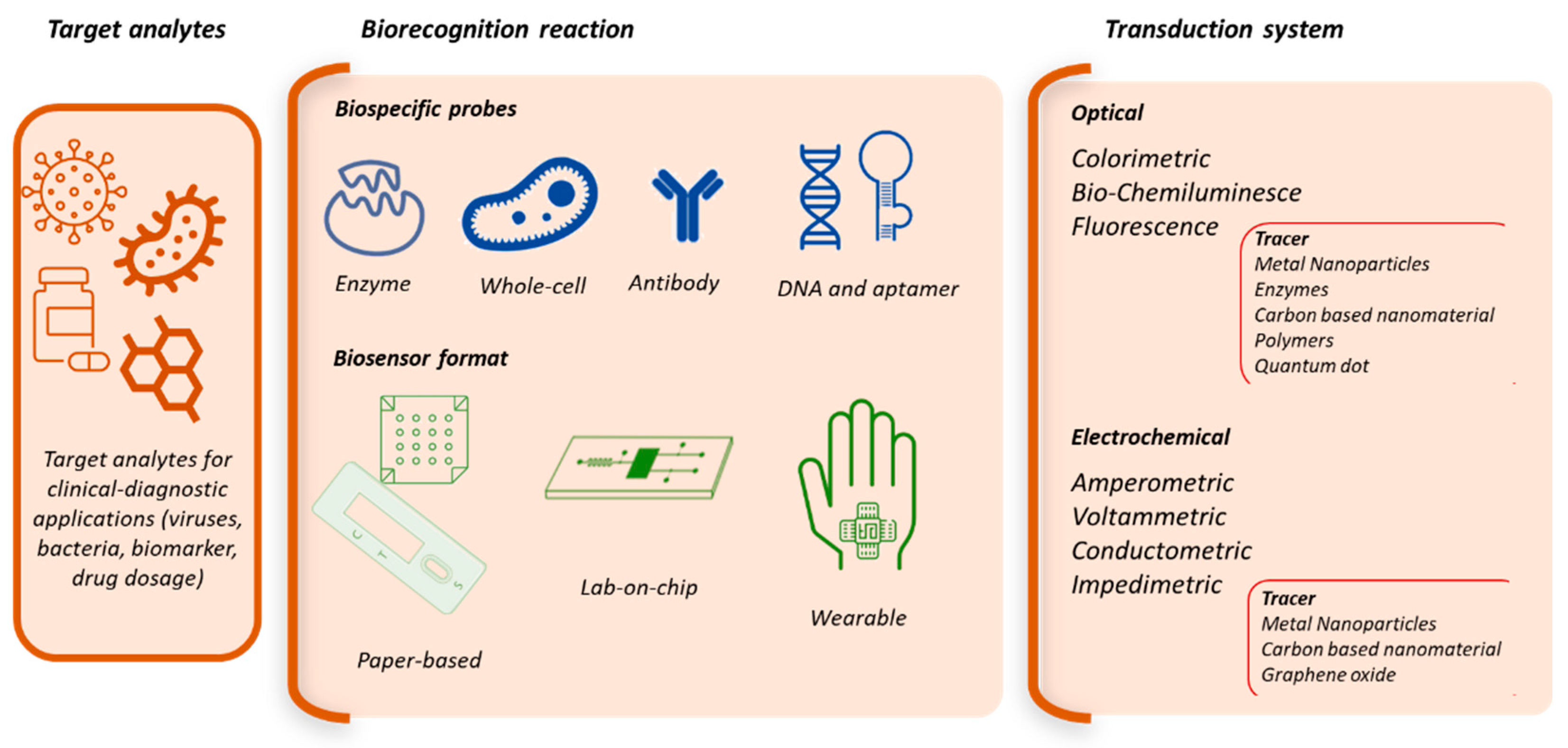
Figure 2.
a) Scheme of working principle of the analytical device: a) 3D schematic of sandwich immunoassay on biosensor coated with antifouling nanocomposite. a. Electrochemical sensor with gold electrodes b. Immobilization of capture antibody and saturation with BSA c. Incubation of sample mixed with biotinylated detection antibodies. d. Addition of poly-HRP streptavidin. e) Addition of TMB followed by precipitation over the gold electrode f) Results of the cyclic voltammetry. Reprinted with permission from Ref [69]; b) The electrochemical analyticaldevice interfaced with a portable potentiostat connected to a smartphone via a USB-C connection to measure the signal. Reprinted with permission from Ref [79]; c) Microbeads immobilized in microfluidic chip and reagent reservoirs filled with assay reagents. Pumps inject reagents during the assay. Measurements were performed using VideoScan technology (Bioimage informatics). Reprinted with permission from Ref [83]; d) Device design comprising a microfluidic chip with 5 inlets connected to peristaltic micropumps, sample and rectangular prism reagent chambers with capacity of 80 ± 5 μL, and 8 cylindrical detection chambers with 8 ± 1 μL capacity each.. A microfluidic chip is designed to house sample and reagents and deliver them sequentially to detection compartment. The assay protocol is based on poly-HRP and ultra-bright femto-luminol to produce CL that is measured using a CCD camera. Reprinted with permission from Ref [87].
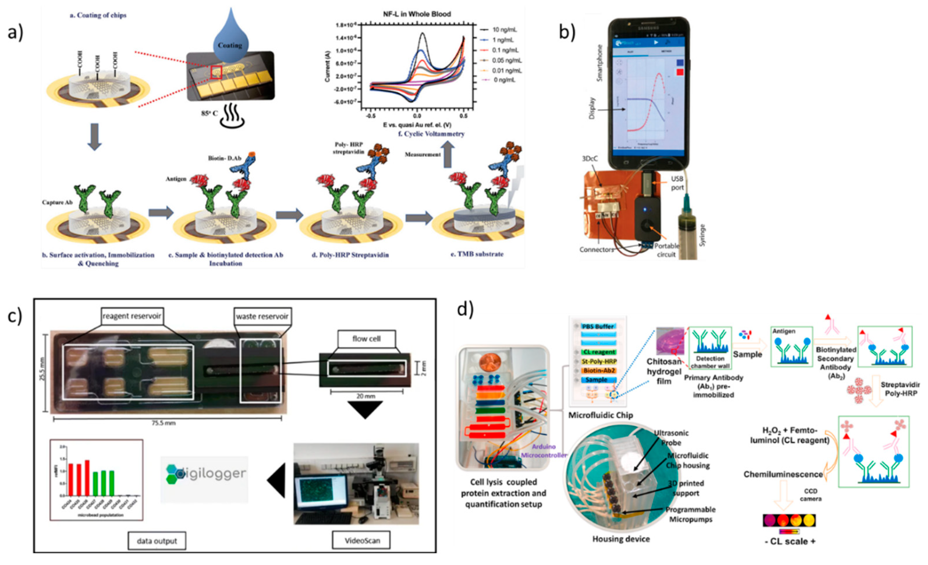
Figure 3.
a) Scheme of the design and application of the smartphone-based fluorescent LFIA platform. a. Internal structure of 3D smartphone-based imaging device. b: Photo of the developed fluorescent LFIA reader. c. Schematic of the fluorescent LFIA for the detection of ZIKV NS1. d. Images of the test strips in the presence (left) and absence (right) of ZIKV NS1. Reprinted with permission from Ref [115] b) LFIA based on the PEGylation of AuNPs. a. Preparation of AuNP-Ab-HRP-PEG conjugates based on the PEG-modified AuNPs. b. Scheme of CL-LFIA in which the AuNP-Ab-HRP-PEG conjugates are applied for the detection. c. Light signal detection with a camera of a smartphone d. PEGylation of AuNPs for improving the performance of CL-LFIA biosensor. Reprinted with permission from [117] Copyright {2023} American Chemical Society.” c) Schematic illustration of the E-test strip testing: a. SARS-CoV-2 binding with the ACE2 cellular receptor. b. Schematic of the E-test strip in which the SARS-CoV-2 spike antigen is detected using the ACE2 WE test zone on the nitrocellulose membrane. Reprinted with permission from Ref [124] d) a. Electrolateral flow immunosensor (ELLI) b. ELLI’s assay based on modified PEG-stabilized and TEMPO-tagged AuNPs for an amperometric point-of-care detection of dengue NS1 protein. Reprinted with permission from Ref [125].
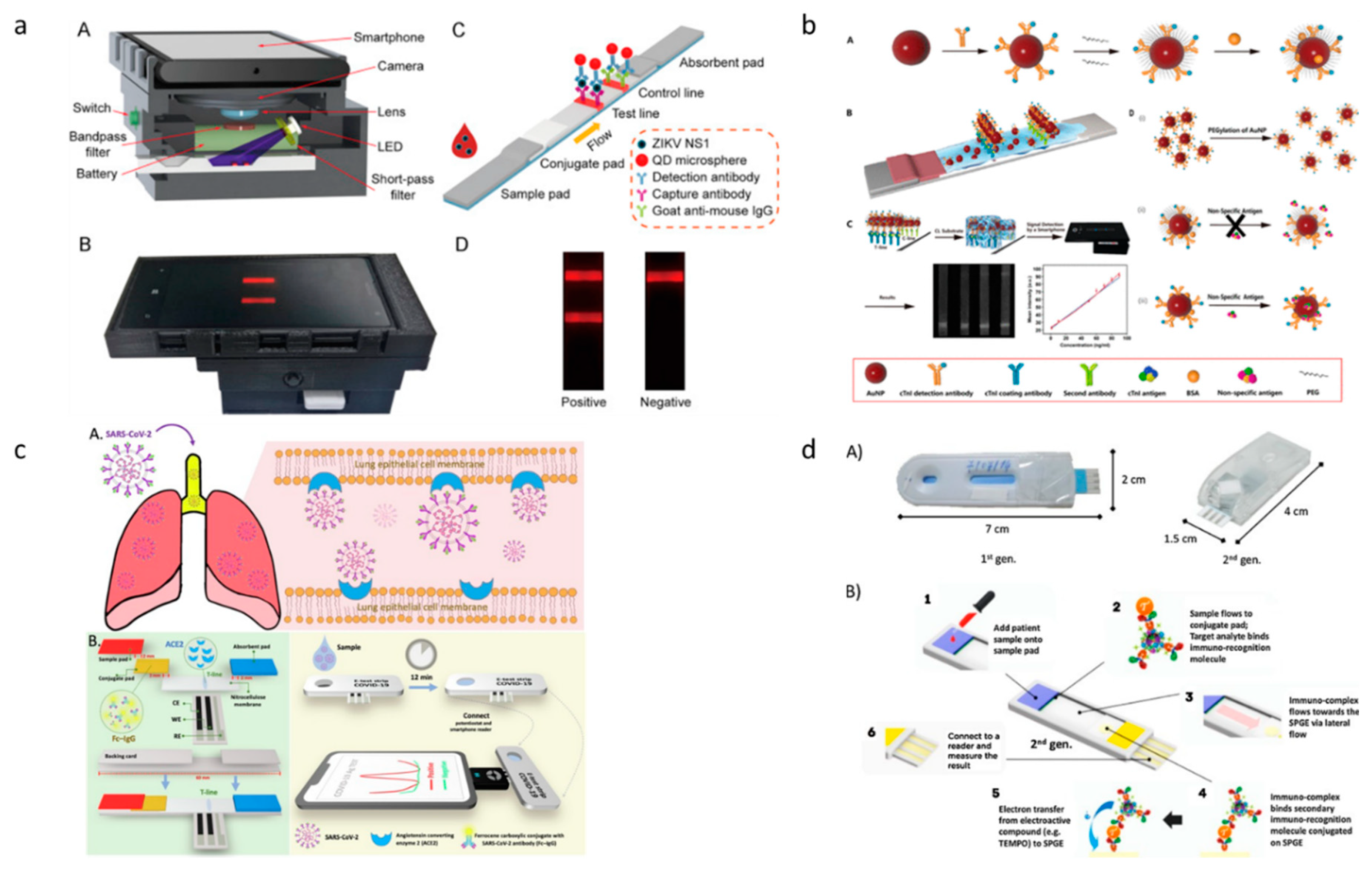
Figure 4.
a) Illustration of cortisol during physical activity. a. sampling process and b. measuring signal and transmitting data to a smartphone via NFC. Reprinted with Permission from Ref [144]; b) Scheme of the microfluidic device for colorimetric analysis of sweat a. Optical images microfluidic devices for colorimetric analysis of sweat on the skin (top) and under mechanical deformation with bending (bottom left) and twisting (bottom right). b. Top view of microfluidic channels filled with blue-dyed water. c. Exploded scheme of a device and its interface with the skin. d. Procedure for collecting sweat samples and color analysis of digital images of the device. Reprinted with permission from Ref [150] Copyright 2019 American Chemical Society; c) Schematic illustration of the high-density silicon MNA electrode preparation. a. Si-MNA and 3D printed holder, b. Si-MNA substrate attached to the 3D printed holder, c. sputter deposition of a thin film of Au, d. insulating ink application, and e. three-electrode sensing patch with Au-Si-MNA as working and counter electrodes, and reference electrode with Au-Si-MNA coated with AgCl ink. f. Schematic of the three-electrode MNA patch penetrating the skin and interfacing the epidermis and superficial dermis. g. Schematic illustration of the modification of the working electrode. Reproduced with permission from Ref [187]; d) The generation and application of the encoded MNs. a) Schematic illustration of the fabrication of the encoded MNs by micromolding. b) Schematic illustration of the application of the MNs in ISF detection. Reproduced with permission from Ref [204].
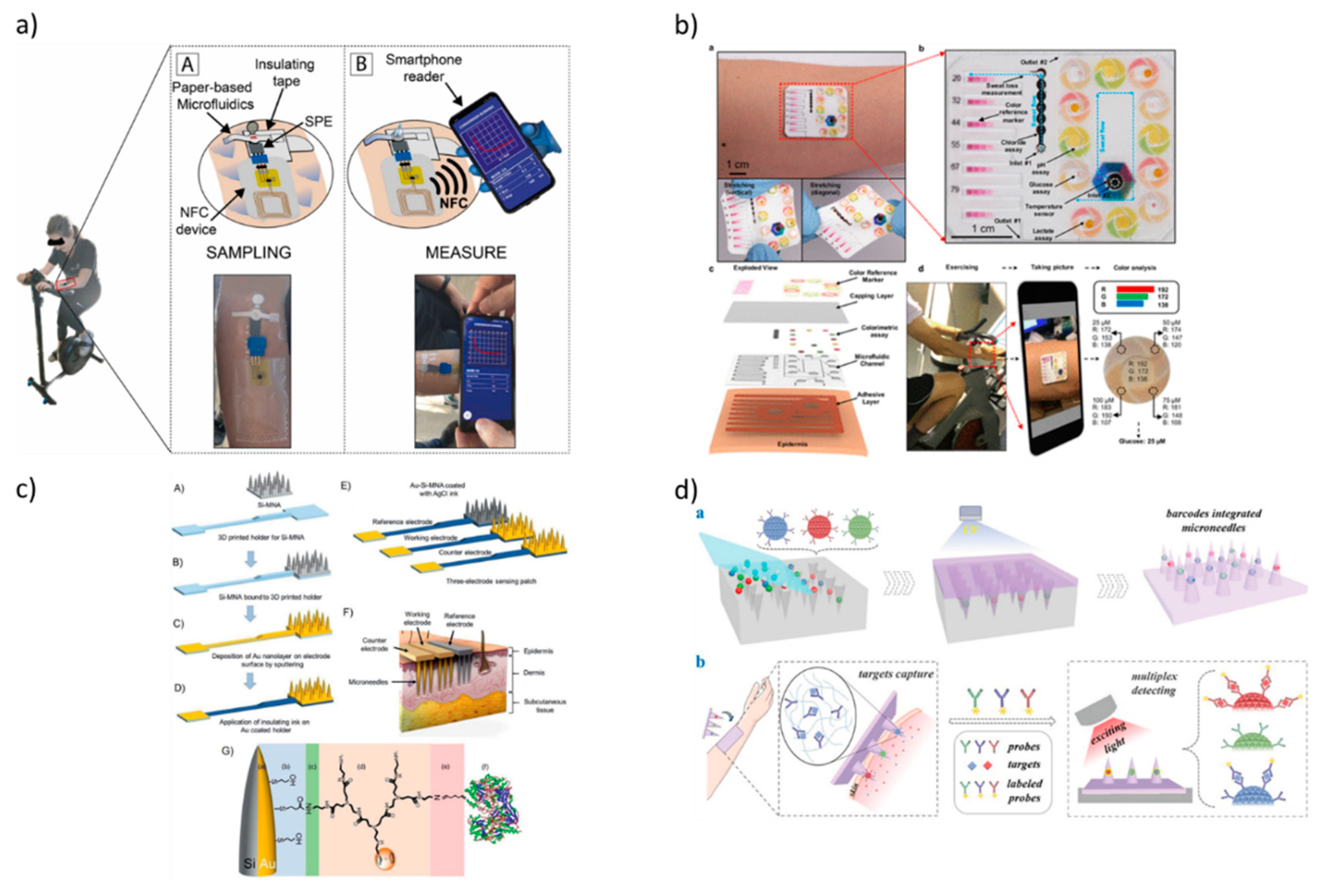
Disclaimer/Publisher’s Note: The statements, opinions and data contained in all publications are solely those of the individual author(s) and contributor(s) and not of MDPI and/or the editor(s). MDPI and/or the editor(s) disclaim responsibility for any injury to people or property resulting from any ideas, methods, instructions or products referred to in the content. |
© 2023 by the authors. Licensee MDPI, Basel, Switzerland. This article is an open access article distributed under the terms and conditions of the Creative Commons Attribution (CC BY) license (http://creativecommons.org/licenses/by/4.0/).
Copyright: This open access article is published under a Creative Commons CC BY 4.0 license, which permit the free download, distribution, and reuse, provided that the author and preprint are cited in any reuse.


