Submitted:
10 July 2023
Posted:
12 July 2023
You are already at the latest version
Abstract
Inhibition of quorum sensing (QS)-dependent biofilm formation and virulence is extensively investigated as a novel approach to combat bacterial pathogens by impeding their ability to cause antibiotic-tolerant diseases. Such strategies are considered a promising alternative to conventional antibacterial therapy since interrupting the central cell-to-cell communication system imposes less selective pressure on the target pathogen. In this study, novel hybrid compounds composed of anthranilic acids substituted with halogens at different positions of the phenyl ring, and 4-(2-aminoethyl/4-aminobuthyl)amino-7-chloroquinoline linked via 1,3,4-oxadiazole were synthesized using standard procedures. Their anti-QS activities were evaluated using Chromobacterium violaceum ATCC31532 (anti-biofilm and bactericidal activities) and Pseudomonas aeruginosa PAO1 (anti-biofilm/-virulence activities). The results showed that compounds 15–19 and 23 inhibited the production of violacein, the QS-inducible pigment, in C. violaceum to a similar extent as the model QS inhibitor - quercetin (83.5–90%). Compound 15 exhibited the strongest effect on P. aeruginosa in the anti-biofilm screening, reducing its ability to form biofilm by almost 50%, while compounds 16 and 19 were able to reduce the biofilm formation by approximately 30%. However, compound 23 did not demonstrate significant anti-biofilm activity and only inhibited pyocyanin production in P. aeruginosa. In conclusion, this study suggests that 1,3,4-oxadiazoles 15, 16, and 19 are the most promising compounds for future research as novel QS inhibitors against Gram-negative bacteria equipped with different QS signaling pathways.
Keywords:
4-amino-7-chloroquinoline
; 1
; 3
; 4-oxadiazole
; anthranilic acid
; synthesis
; quorum sensing
; anti-biofilm
; P. aeruginosa
; PQS
1. Introduction
Bacterial pathogens use different strategies to cause disease and overcome obstacles faced in human hosts, including also strategies that allow increased viability in response to the use of traditional antibiotics. While efficiently killing the target bacteria, antibiotics promote selective pressure on the bacterial cell, thereby leading to the development of resistant species and complicating the successful treatment of infections caused by those bacteria [1]. Based on the data collected from 204 countries worldwide, an estimated 4.95 million deaths were associated with bacterial antimicrobial resistance (AMR) in 2019, including 1.27 million deaths directly attributable to bacterial AMR [2]. The decline in antibacterial drug discovery and development together with the rise of AMR represents one of the leading public health threats of the 21st century [3].
Quorum sensing (QS) is a mechanism of bacterial cell-to-cell communication and gene expression coordination and an important strategy performed by bacteria to enhance their virulence and promote host damage. It relies on signaling molecules called autoinducers (AI) [4] to activate community behaviors such as biofilm formation, which is a significant mechanism contributing to bacterial tolerance against antibiotics. Biofilm formation is also a key virulence factor enabling bacterial pathogens to establish chronic infections and promoting the development of antimicrobial resistance (AMR). In view of these findings, inhibition of QS represents a highly promising approach for anti-bacterial and anti-biofilm therapies, as it is expected to exert less selective pressure on bacterial pathogens compared to conventional antibiotics [5].
Molecular hybridization, i.e., covalent linking of two or more bioactive scaffolds to form a single molecule with improved properties, was used to design the title compounds. The advantages of such compounds include increased efficacy, improved pharmacokinetics, and reduced risk of resistance development, side effects, and drug-drug interactions. In recent years, our group has successfully implemented this approach to obtain a harmine-based antimalarial compound library [6,7,8,9,10,11]. The concept of hybridization has already been used by other research groups in the subject area [12,13,14].
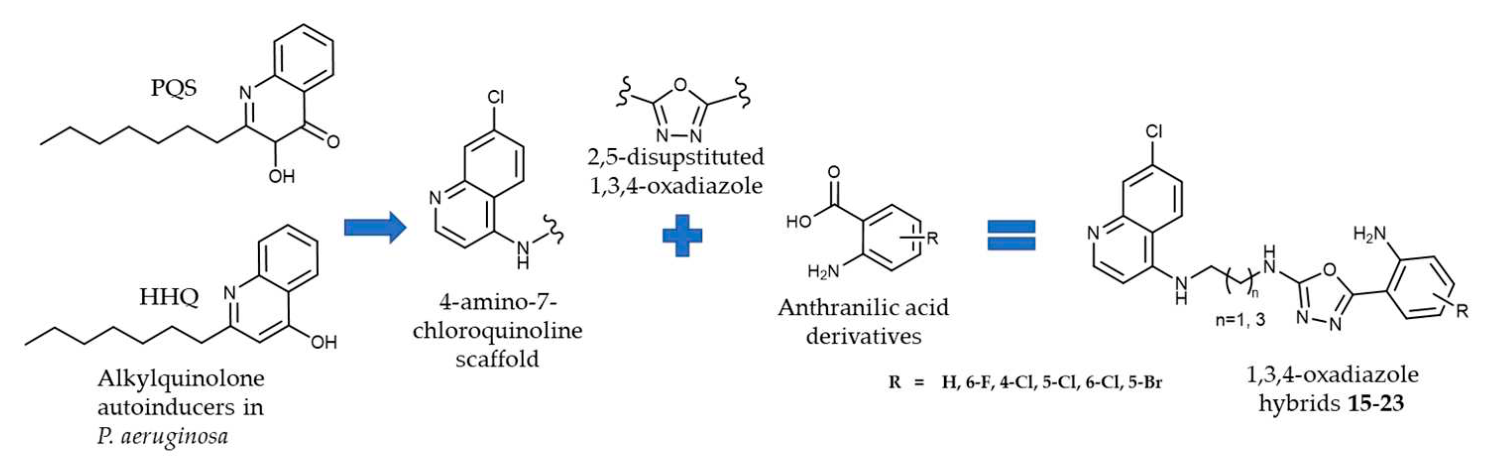
Several classes of quinoline derivatives were investigated as potential agents against QS and biofilm formation due to their structural similarity to Pseudomonas alkylquinolone AIs (e.g., 2-heptyl-3-hydroxy-4(1H)-quinolone, PQS, and its precursor 2-heptyl-4(1H)-quinolone, HHQ). Among these studies, the 4-amino-7-chloroquinoline scaffold emerged as a promising candidate for developing anti-QS and anti-biofilm agents targeting multidrug-resistant bacteria [15,16,17,18,19]. Additionally, anthranilic acid serves as a precursor in PQS biosynthesis [20], and halogenated anthranilic acids and their derivatives have been reported as inhibitors of PQS biosynthesis [21,22,23,24,25]. Thus, interrupting the PQS biosynthesis could lead to the discovery of novel anti-infective agents based on anti-virulence strategies.
Continuing our work on QS inhibitors [26], we synthesized a series of hybrid molecules incorporating the quinoline and anthranilic acid scaffolds (Figure 1). These compounds were screened for their anti-QS, antibacterial and antivirulence activities using Gram-negative Chromobacterium violaceum and Pseudomonas aeruginosa as the known biofilm models using a VioI/R (resembling LuxI/R-type QS) and several LuxI/R- dependent QS systems, respectively [27,28,29].
2. Results and Discussion
2.1. Chemistry
In this study, we present the synthesis of novel hybrid compounds 15–23, derived from anthranilic acids and 4-amino-7-chloroquinoline-based amines, linked via 1,3,4-oxadiazole ring, as well as the synthesis of their precursors. 1,3,4-Oxadiazole is a 5-membered heterocyclic ring, composed of one oxygen, two carbons, and two nitrogen atoms. It was selected as a linker due to the variety of favorable physicochemical and biological properties. It is often used as a bioisostere of carbonyl-containing functional groups, such as amides, esters, and carbamates. In addition, 1,3,4-oxadiazole-containing compounds have demonstrated increased aqueous solubility, decreased hERG inhibition, and improved metabolic stability when compared to 1,2,4-oxadiazole ones [30,31,32].
The structural variety of the title compounds was achieved by changing the length of the alkyl chain attached to the 4-amino-7-chloroquinoline scaffold (2 or 4 carbon atoms) and the position and the type of the halogen atom attached to the phenyl ring of the anthranilic acid scaffold (F, Cl or Br).
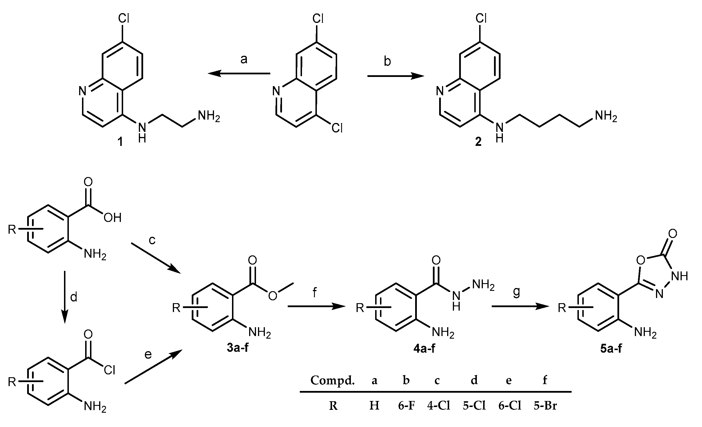
The compounds were obtained by standard synthetic procedures. The synthesis was divided into two parts. First, 4-amino-7-chloroquinoline- (1 and 2) and anthranilic acid-based (5a–f) building blocks were obtained, as outlined in Scheme 1.
The 4-amino-7-chloroquinoline building blocks bearing terminal amino group (1,2) were prepared according to the previously published procedure, in the reaction of 4,7-dichloroquinoline and ethylenediamine or 1,4-diaminobuthane, respectively [33,34]. The anthranilic acid building blocks in the form of the corresponding 1,3,4-oxadiazol-2-ones (series 5) were obtained in multiple reaction steps. First, anthranilic acid esters (series 3) were either purchased (3a–c) or obtained from the corresponding anthranilic acids (3d–f). Hydrazides 4 were obtained from the anthranilic acid esters 3 and hydrazine hydrate. 3-H-1,3,4-oxadiazole-2-ones (series 5) were obtained from the corresponding hydrazides 4 in the presence of 1,1-carbonyldiimidazole (CDI) following the previously published procedure [35,36]. The key reaction step was a nucleophilic attack of the primary amino group (1 or 2) on the carbonyl of the 3-H-1,3,4-oxadiazole-2-ones 5, leading to the ring opening and merging of the two building blocks in the form of the corresponding acylsemicarbazides 6–14. The reaction was conducted in ethanol at 100 °C in a sealed vial. In the case of acylsemicarbazides 7, 10, and 13, we proceeded to the next reaction step without the purification of the product due to the low yields. The final reaction step presents intramolecular cyclization of the acylsemicarbazides 6–14. Under the dehydration conditions (utilizing triphenylphosphine, carbon tetrachloride, and triethylamine) the desired 1,3,4-oxadiazoles 15–24 were obtained (Scheme 2).
The reagents used in the cyclization reaction include triphenylphosphine, carbon tetrachloride, and triethylamine. Dichloromethane was used as a solvent, which proved to be suitable due to its dielectric constant that affects the reaction rate [37]. Triethylamine was also added to the reaction mixture as it promotes intra-molecular cyclization by deprotonating the hydroxyl group of the enol form of acylsemicarbazide [38,39].
The final reaction step progressed well, and the reaction yields were slightly higher for the butan-1,4-diamine 1,3,4-oxadiazoles (20–23), ranging from 41 to 73 %, as opposed to the ethan-1,2-diamine 1,3,4-oxadiazoles (15–19) (yields from 9 to 31 %). The lowest reactivity was observed in the case of the 5-Br derivative.
2.2. Anti-QS and bactericidal activity against the QS-reporter strain
Gram-negative C. violaceum ATCC 31532 was used as a reporter strain to indicate compounds with anti-QS and bactericidal activities. In this biofilm-forming pathogen, activation of QS induces the expression of genes contributing to biofilm formation and synthesis of a deep-purple violacein. This pigment, excreted out of the cells, acts as an indicator of induced QS in C. violaceum and its changes could be quantitatively monitored in 96-well microtiter plate format [40,41]. Since acylsemicarbazides and 1,3,4-oxadiazoles prepared in this study, are built from anthranilic acid derivatives and 4-amino-7-chloroquinoline, we decided to screen both types of compounds for their anti-QS and bactericidal activity against the C. violaceum reporter. Due to the low reaction yields for the acylsemicarbazides series 6-14, only 3 compounds (8, 12, and 14) were tested. Quercetin was used as the positive control due to its known anti-QS effect and azithromycin as the positive control for bactericidal activity (cell viability) [40,41]. Inhibitory activity was first tested at 400 μM concentrations (Table 1). Six out of twelve tested compounds (15–19 and 23) inhibited violacein production in C. violaceum almost to the same extent as quercetin (indicated as red arrows in Figure 2a, 83.5–90 %). Interestingly, all ethan-1,2,diamine 1,3,4-oxadiazoles (15-19), and one butan-1,4-diamine 1,3,4-oxadiazole derivative (23) were active. Additionally, two compounds (17 and 18) also exerted a strong bactericidal effect on the reporter strain (Figure 2b, more than 70 %) which was measured as the ability of viable and metabolically active cells to reduce resazurin to fluorescent resorufin [42]. At the same time, acylsemicarbazides 8, 12, and 14 showed only weak or no effect on the violacein production as well as on the viability of the reporter strain. It is worth mentioning that in the acylsemicarbazide series, similarly to the 1,3,4-oxadiazoles, the ethan-1,2,diamine compound 8 was more potent when compared to the butan-1,4-diamine compounds 12 and 14.
The most promising compounds (15, 16, 19, and 23) were selected for dose-response analyses at 400, 200, or 100 and 40 μM concentrations. The results indicated that each compound with concentrations up to 100 μM reduced the violacein production by more than 50 % compared to the control cells with DMSO, while the viability of the reporter under the same conditions was only marginally affected (Figure 3)
For compound 19, IC50 of 63.15 μM (95 % CI 47.62–83.75 μM) was measured. Compound 19 was one of the most active compounds in anti-QS screening since it reduced the violacein production in C. violaceum by more than 85 % at 400 μM concentration. The detected bactericidal effect under the same conditions was moderate (47 %, Table 1). At a concentration (100 μM), this compound was able to reduce the QS activity by more than 50 % (Figure 3a), while no effect on cell viability was observed (Figure 3b).
2.3. Effect of selected compounds on biofilm and pyocyanin production in Pseudomonas aeruginosa PAO1
P. aeruginosa is a Gram-negative pathogen conferring extremely difficult-to-treat infections due to its excellent ability to colonize a variety of medical materials (urinary catheters, implants, contact lenses, etc.) as well as to form resilient biofilms in the cystic fibrosis lung environment [43]. Widely studied P. aeruginosa PAO1 strain has been shown to consist of four interconnected systems (IQS, PQS, las, rhl) to coordinate biofilm-formation and virulence-related traits (e.g., elastase, rhamnolipids, and pyocyanin) [44], and is considered a standard model to study QS-dependent biofilm formation within this species [45]. Agents that would prevent the biofilm formation, or disrupt them, could be useful in the treatment of chronic and recurring infections, such as bacterial diseases, including the clinically relevant P. aeruginosa, whose biofilm shows higher tolerance to antibiotics and host’s immune cells [46,47]. In view of these findings, we tested the most promising compounds against P. aeruginosa PAO1. We determined the MIC and MBC of compounds 15, 16, 19, and 23 that showed the best anti-QS activity on the C. violaceum reporter strain, with minimal influence on bacterial growth (Figure 3). We also included compound 18 which showed very strong anti-QS activity but also had a somewhat stronger bactericidal effect on the C. violaceum reporter strain. For P. aeruginosa PAO1, the MIC of all five compounds was 1600 μM and the MBC was 3200 μM. We tested the anti-QS activity of the selected compounds (15, 16, 18, 19, and 23) only at the subinhibitory 100 μM concentration because at that concentration these compounds showed the optimal anti-QS activity vs. bactericidal effect on the C. violaceum reporter strain. Furthermore, 100 μM concentration was substantially below the MIC and MBC values determined for P. aeruginosa PAO1. In P. aeruginosa, the PQS system is involved in the regulation of biofilm formation and the production of virulence factor pyocyanin. Therefore, we wanted to assess whether the selected compounds interfere with these processes. We first incubated these compounds with bacteria to see if their presence will inhibit the formation of biofilm (results presented in Figure 4). The results revealed that all compounds reduced the formation of biofilm, with compound 19 seeming to be the most effective when compared to the control cells with DMSO (over 60% inhibition compared to non-treated cells, as shown in the BFI column of Table 2). However, we also observed that compound 19 showed the strongest inhibition of cell growth, compared to other tested compounds (column GI in Table 2). We, therefore, calculated the biofilm index that includes both the biofilm mass and the cell count (Table 2). We found out that compound 15 showed the most potent effect on P. aeruginosa and reduced its ability to form biofilm by almost 50%, followed by compounds 16 and 19 (approximately 30 % reduction), compound 18 (approximately 20 % reduction), and least effective compound (less than 10 % reduction).
In the next experiment, we allowed P. aeruginosa to form biofilm for 24 hours and then treated established biofilm with selected compounds (Figure 5, Table 2 – column BE). Compound 16 was most effective and reduced the biofilm mass by 43%, then compound 19 with 35 % reduction, followed by compound 18 (almost 30% reduction) and compound 15 (almost 25 % reduction), while compound 23 was again the least effective with approximately 11 % reduction. Considering that compound 15 showed minimal inhibition of cell growth in the assay evaluating biofilm formation (less than 10 %, Table 2 - GI) we can assume that here the reduction in biofilm mass resulted from the interference with the exopolysaccharide, rather than from killing the cells within the biofilm. On the other hand, compounds 16, 18, and 19 showed more prominent inhibition of cell growth (in the range of 30–45 %, Table 2 - GI) and, therefore, appear to have a combined effect both on the biofilm matrix and cells within the biofilm.
We also examined the effect of the compounds on the production of pyocyanin, a QS-controlled metabolite produced by P. aeruginosa. Again, the 100 μM concentration was selected, since it showed the strongest anti-QS activity without the prominent influence on the growth of the C. violaceum (Figure 3), and it was significantly below the MIC and MBC values for P. aeruginosa. Figure 6 and Table 2 (column PI) show that compound 23 was the most effective, reducing pyocyanin production by over 70 %. Compounds 15 and 19 similarly decreased the level of pyocyanin by approximately 40 %, while compounds 16 and 18 showed a decrease close to 20 %.
3. Materials and Methods
3.1. Chemistry
3.1.1. General information
Melting points of compounds 9 and 15–19 were determined by differential scanning calorimetry (DSC). Thermograms were recorded using DSC 822e (Mettler Toledo), and analyzed by STARe software (V16.20b) (Mettler-Toledo). Melting points of compounds 8, 11, 12, 14, and 20-23 were determined on a Stuart Melting Point Apparatus (Barloworld Scientific, UK) in open capillaries and were uncorrected. FTIR-ATR spectra were recorded using a Fourier-Transform Infrared Attenuated Total Reflection UATR Two spectrometer (PerkinElmer, Waltham, MA, USA) in the range from 450 to 4000 cm–1. 1H and 13C NMR spectra were recorded on a Bruker Avance III 600, Bruker Avance DRX500, Bruker Avance AV400, and Bruker Avance DPX300 spectrometers operating at 300 or 400 MHz for the 1H and 75, 101, or 151 MHz for the 13C nuclei, BrukerDPX 300 MHz, Bruker AV400 MHz and Bruker DRX 500 MHz (Bruker, Billerica, MA, USA). 1H and 13C NMR spectra were recorded on Bruker Avance III 600, Bruker Avance DRX500, Bruker Avance AV400, and Bruker Avance DPX300 spectrometers (Billerica, MA, USA), Chemical shifts are reported in parts per million (ppm) using tetramethylsilane (TMS) as a reference in the 1H and DMSO residual peak as a reference in the 13C spectra (39.52 ppm). Data for 1H NMR are described as follows: chemical shift (δ in ppm), multiplicity (s: singlet; d: doublet; t: triplet; q: quartet; m: multiplet; bs: broad signal), integration, coupling constant J (Hz). Samples were measured in DMSO-d6 solutions at 20 °C in 5 mm NMR tubes. Mass spectra were recorded on Agilent 1200 Series HPLC coupled with Agilent 6410 Triple Quad (Agilent Technologies, St. Clara, CA, USA). LCMS data were acquired on a Waters Acquity UPLC instrument using a Waters Acquity UPLC C18, (2.1x50 mm, 1.7um) column, water/MeCN gradient, and 0.1% (v/v) formic acid as an acidic modifier or 0.05% (v/v) NH4OH as a basic modifier. The column eluent was analyzed using a Waters SQ mass spectrometer with ESI scanning in both positive and negative ion modes from 100 to 2000 Da and reaction conversions expressed as percentage (where applied) by using a UV detector at 254 nm.
All compounds were routinely checked by TLC with silica gel 60F-254 glass plates (Merck, Germany) using DCM/MeOH 7.5:2.5, 7:3, 8.5:1.5, 8:2, 9.5:0.5, 9:1, cyclohexane/EtOAc/MeOH 3:1:0.5 and 1:1:0.5 as the solvent system. Spots were visualized by UV light (λ = 254 nm; 365 nm) and iodine vapor. Column chromatography was performed on silica gel 0.063–0.200 mm (Sigma-Aldrich, USA) with the same eluents used for TLC. Flash chromatography was performed on Interchem Puriflash XS 520 Plus, Interchim Puriflash SiHC (12–25 g; 15 μm) columns. All chemicals and solvents were of analytical grade and purchased from commercial sources.
Dry toluene was obtained by the following procedure: toluene was extracted with water and dried over anhydrated calcium chloride, distilled and stored over elemental sodium. Dry DCM: DCM was extracted with water and dried over anhydrated calcium chloride and distilled. Dry DMF was stored over activated molecular sieves.
3.1.1. Synthesis of 4-amino-7-chloroquinoline intermediates 1 and 2
N1-(7-chloroquinolin-4-yl)butane-1,4-diamine (1)
Compound 1 was prepared according to the published procedure [34]. A mixture of 4,7-dichloroquinoline (5.00 g, 25.3 mmol) and 1,2-diaminoethane (13.50 g, 227.2 mmol) was gradually heated to 80 °C for 1 h while stirring. The reaction mixture was then heated to 120 °C and stirred for an additional 6 h and poured on iced water and stirred for 18 h. 5.2 g (93 %) of the white solid 1 was filtered off, washed with water, and vacuum-dried.
1H NMR (DMSO-d6, δ ppm, J/Hz) δ 8.38 (d, J = 4.83 Hz, 1H), 8.28 (d, J = 9.05 Hz, 1H), 7.78 (d, J = 3,02 Hz, 1H), 7.43 (d, J = 8.99 Hz, 1H), 7.29–7.20 (bs, 1H), 6.48 (d, J = 5.99 Hz, 1H), 3.25 (q, J = 5.99 Hz, 2H), 2.82 (t, J = 6.74 Hz, 2H).
N1-(7-chloroquinolin-4-yl)butane-1,4-diamine (2)
Compound 2 was prepared according to the published procedure [33]. A mixture of 4,7-dichloroquinoline (0.400 g, 2.02 mmol) and 1,4-diaminobutane (1.78 g, 20,2 mmol) was stirred under microwave irradiation (300 W) at 95 °C for 1 h. The reaction mixture was then diluted with dichloro-methane (100 mL), extracted with 5 % NaOH (3 × 100 mL), and washed with water (1 × 100 mL). The organic layer was dried over anhydrous sodium sulfate, filtered, and evaporated under reduced pressure. The crude product was triturated with diethyl ether to give 0.428 g (86 %) of white solid 2.
1H NMR (DMSO-d6, δ ppm, J/Hz) δ 8.39 (d, J = 5.4 Hz, 1H), 8.27 (d, J = 9.0 Hz, 1H), 7.78 (d, J = 2,2 Hz, 1H), 7.43 (dd, J = 9.0, 2.2 Hz, 1H), 7.39 (s, 1H), 6.46 (d, J = 5.5 Hz, 1H), 3.26 (dd, J = 12.0, 6.9 Hz, 2H), 2.59 (t, J = 6.9 Hz, 2H), 1.69 (dt, J = 14.8, 7.4 Hz, 2H), 1.46 (dt, J = 14.0, 6.9 Hz, 2H).
3.1.2. Synthesis of methyl anthranilates 3 and hydrazides 4.
Methyl anthranilates 3a–f were either purchased from commercial sources (3a–c) or prepared from the corresponding anthranilic acid (3d–f) according to the previously published procedures [35,36].
Hydrazides 4a–f were obtained in the reaction of the corresponding methyl anthranilate (3a–f) and hydrazine hydrate according to the previously published procedure [35].
3.1.3. General procedure for the synthesis of 5-(2-aminophenyl)-l,3,4-oxadiazole-2(3H)-ones 5a–f
The compounds 5a–f were obtained according to slightly modified procedures [35].
1,1’-carbonyldiimidazole (0.254 g, 1.6 mmol) was added to a solution of the corresponding anthranilic acid hydrazide 4a–f (1.4 mmol) in dry DMF (1 mL). The reaction mixture was stirred for 4-18 hours at room temperature, followed by the addition of ethyl acetate (30 mL) and extraction with brine (3 × 30 mL). The organic layer was dried over anhydrous sodium sulfate, filtered, and evaporated under reduced pressure. The crude product was used in successive reaction steps with (5d) or without (5a–c, 5e, 5f) trituration with diethyl ether.
5-(2-aminophenyl)-1,3,4-oxadiazol-2(3H)-one (5a)
Hydrazide 4a: 0.212 g; 1H NMR (DMSO-d6): δ 12.70–12.27 (bs, 1H), 7.44 (dd, 1H, J = 8.0, 1.6 Hz), 7.21 (dt, 1H, J = 7.2, 1.6 Hz), 6.84 (dd, 1H, J = 8.4, 0.8 Hz), 6.64 (t, 1H, J = 7.2, 1.2 Hz),6.37–6.29 (s, 2H).
5-(2-amino-6-fluorophenyl)-1,3,4-oxadiazol-2(3H)-one (5b)
Hydrazide 4b: 0.236 g.
5-(2-amino-4-chlorophenyl)-1,3,4-oxadiazol-2(3H)-one (5c)
Hydrazide 4c: 0.260 g.
5-(2-amino-5-chlorophenyl)-1,3,4-oxadiazol-2(3H)-one (5d)
Hydrazide 4d: 0.260 g; yield: 0.177 g (60 %); IR (ATR, ν/cm-1) 3465, 3364, 3170, 1734, 1628, 1562, 1488, 1310, 1257, 1161, 1042, 813, 717; 1H NMR (DMSO-d6): δ 12.63 (bs, 1H), 7.39 (d, 1H, J = 2.6 Hz), 7.25 (dd, 1H, J = 8.9, 2.6 Hz), 6.88 (d, 1H, J = 8.9 Hz), 6.45 (s, 2H).
5-(2-amino-6-chlorophenyl)-1,3,4-oxadiazol-2(3H)-one (5e)
Hydrazide 4e: 0.260 g.
5-(2-amino-5-bromophenyl)-1,3,4-oxadiazol-2(3H)-one (5f)
Hydrazide 4f: 0.322 g.
3.1.4. General procedure for the synthesis of acylsemicarbazides (6–10)
N1-(7-chloroquinolin-4-yl)athane-1,2-diamine 1 (0.213 g, 0,961 mmol) was added to a suspension of an appropriate 5-(2-aminophenyl)-l,3,4-oxadiazole-2(3H)-one 5 (0.915 mmol) in 5-10 mL of anhydrous EtOH. The reaction mixture was refluxed at 100 °C for 18-72 hours and cooled to room temperature. The crude product was filtered off and washed with EtOH and vacuum-dried (6–9) or purified using flash chromatography (10) (eluent DCM/MeOH 80:20).
2-(2-aminobenzoyl)-N-(2-((7-chloroquinolin-4-yl)amino)ethyl)hydrazine-1-carboxamide (6)
Oxadiazol-2-one 5a: 0.162 g; reaction time: 48 h; yield 0.179 g (49 %); mp nd; 1H NMR (DMSO-d6): δ 9.93-9.72 (bs, 1H), 8.40 (d, 1H, J = 5.2 Hz), 8.16 (d, 1H, J = 8.8 Hz), 8.05 – 7.92 (bs, 1H), 7.79 (d, 1H, J = 2.8 Hz), 7.60 (d, 1H, J = 8.4 Hz), 7.43 (dd, 1H, J = 10.4, 2.4 Hz), 7.48–7.36 (m, 1H), 7.18 (dt, 1H, J = 8.4, 1.2 Hz), 6.86–6.75 (bs, 1H), 6.72 (d, 1H, J = 6.00 Hz), 6.57 (d, 1H, J = 5.6 Hz), 6.51 (t, 1H, J = 7.6 Hz), 6.44–6.32 (bs, 2H), 3.40–3.25 (m, 4H) ppm; 13C NMR (DMSO-d6): δ ): δ 168.9, 159.3, 152.0, 150.1, 150.0, 149.0, 133.4, 132.2, 128.4, 127.5, 124.1, 123.8, 117.3, 116.3, 114.4, 112.4, 98.6, 43.4, 37.8 ppm; ESI-MS: m/z 399.14 (M+1)+.
2-(2-amino-6-fluorobenzoyl)-N-(2-((7-chloroquinolin-4-yl)amino)ethyl)hydrazine-1-carboxamide (7)
Oxadiazol-2-one 5b: 0.179 g, reaction time: 18 h; the structure of the product was indirectly confirmed in the successive reaction step.
2-(2-amino-4-chlorobenzoyl)-N-(2-((7-chloroquinolin-4-yl)amino)ethyl)hydrazine-1-carboxamide (8)
Oxadiazol-2-one 5c: 0.194 g; reaction time: 72 h; yield: 0.177 g (45 %); mp 222.5–224 °C; IR (ATR, ν/cm-1) 3344, 1661, 1630, 1613, 1587, 1539, 1449, 1370, 1255, 1235, 1146, 1081, 941, 877, 807; 1H NMR (DMSO-d6): δ 10.00–9.87 (bs, 1H), 8.41 (d, 1H, J = 5.4 Hz), 8.16 (d, 1H, J = 8.4 Hz), 8.02 (s, 1H), 7.79 (d, 1H, J = 2.4 Hz), 7.61 (d, 1H, J = 9,0 Hz), 7.44 (dd, 1H, J = 9.6, 2.4 Hz), 7.41 (t, 1H, J = 4.8 Hz), 6.88-6.80 (bs, 1H), 6.79 (d, 1H, J = 2.4 Hz), 6.73-6.65 (bs, 2H), 6.56 (d, 1H, J = 5.4 Hz), 6.54 (dd, 1H, J = 9.0, 1.8 Hz), 3.38-3.27 (m, 4H) ppm; 13C NMR (DMSO-d6): δ 168.2, 159.2, 152.0, 151.2, 150.0, 149.0, 136.7, 133.3, 130.4, 127.5, 124.1, 123.8, 117.3, 115.0, 114.1, 111.2, 98.6, 43.3, 37.8 ppm; ESI-MS: m/z 432.97 (M+1)+.
2-(2-amino-6-chlorobenzoyl)-N-(2-((7-chloroquinolin-4-yl)amino)ethyl)hydrazine-1-carboxamide (9)
Oxadiazol-2-one 5e: 0.194 g; reaction time: 18 h; yield: 0.192 g (49 %); mp 213 °C; IR (ATR, ν/cm-1) 3231, 1687, 1629, 1603, 1582, 1540, 1457, 1428, 1319, 1250, 1146, 907, 877, 811, 763; 1H NMR (DMSO-d6): δ 10.08–9.85 (bs, 1H), 8.56 (s, 1H), 8.41 (d, 1H, J = 6.0 Hz), 8.18 (d, 1H, J = 9.0 Hz), 7.79 (d, 1H, J = 2.0 Hz), 7.46 (dd, 1H, J = 4.5, 2.0 Hz), 7.40 (t, 1H, J = 5.0 Hz), 7.05 (t, 1H, J = 7.5 Hz), 6.63 (d, 1H, J = 9.0 Hz), 6.63 (s, 1H), 6.56 (d, 1H, J = 5.0 Hz), 6.54 (d, 1H, J = 6.5 Hz), 5.99 (s, 2H), 3.46-3.41 (m, 4H) ppm; 13C NMR (DMSO-d6): δ 165.9, 159.2, 152.0, 150.0, 149.0, 147.9, 133.4, 130.6, 130.5, 127.5, 124.2, 123.9, 119.1, 117.3, 115.1, 113.2, 98.7, 43.3, 38.1 ppm; ESI-MS: m/z 433.1 (M+1)+.
2-(2-amino-5-bromobenzoyl)-N-(2-((7-chloroquinolin-4-yl)amino)ethyl)hydrazine-1-carboxamide (10)
Oxadiazol-2-one 5f: 0.234 g; reaction time: 18 h; the structure of the product was indirectly confirmed in the successive reaction step.
3.1.5. General procedure for the synthesis of acylsemicarbazides (11-14)
N1-(7-chloroquinolin-4-yl)butane-1,4-diamine 2 (0.240 g, 0,961 mmol) was added to a suspension of an appropriate 5-(2-aminophenyl)-l,3,4-oxadiazole-2(3H)-one 5 (0.915 mmol) in 5 mL of anhydrous EtOH. The reaction mixture was refluxed at 100 °C for 24–48 hours and cooled at room temperature, the organic solvent was evaporated and the crude product was purified using column chromatography (DCM/MeOH 75:25).
2-(2-aminobenzoyl)-N-(4-((7-chloroquinolin-4-yl)amino)butyl)hydrazine-1-carboxamide (11)
Oxadiazol-2-one 5a: 0.162 g; yield: 0.155 g (40%); mp 184–185 °C; IR (ATR, ν/cm-1) 3328, 2934, 1645, 1613, 1580, 1532, 1450, 1368, 1251, 1137, 852, 806, 748; 1H NMR (DMSO-d6): δ 9.78 (s, 1H), 8.38 (d, 1H, J = 5,4 Hz), 8.28 (d, 1H, J = 9.0 Hz), 7.77 (d, 1H, J = 2.2 Hz), 7.69 (s, 1H), 7.58 (d, 1H, J = 7.9 Hz), 7.43 (dd, 1H, J = 9.0, 2.3 Hz), 7.34 (t, 1H, J = 5.4 Hz), 7.20 – 7.13 (m, 1H), 6.70 (dd, 1H, J = 8.4, 1.2 Hz), 6.52 – 6.46 (m, 3H), 6.41 (s, 2H), 3.28 (q, 2H, J = 7,1 Hz), 3.10 (q, 2H, J = 6.6 Hz), 1.66 (q, 2H, J = 7.3 Hz), 1.53 (q, 2H, J = 7.1 Hz) ppm; 13C NMR (DMSO-d6): δ 168.9, 158.7, 151.9, 150.1, 149.8, 149.0, 133.3, 132.1, 128.0, 127.4, 124.1, 124.0, 117.4, 116.2, 114.4, 112.7, 98.7, 42.2, 38.9, 27.6, 25.1 ppm; ESI-MS: m/z 427.1 (M+1)+.
2-(2-amino-4-chlorobenzoyl)-N-(4-((7-chloroquinolin-4-yl)amino)butyl)hydrazine-1-carboxamide (12)
Oxadiazol-2-one 5c: 0.194 g; yield 0.086 g (21 %); mp 174–175 °C; IR (ATR, ν/cm-1) 3306, 2956, 2869, 1612, 1578, 1482, 1430, 1368, 1322, 1253, 1207, 1141, 1078, 920, 847, 809, 766, 643; 1H NMR (DMSO-d6): δ 9.86 (s, 1H), 8.39 (d, 1H, J = 5,4 Hz), 8.29 (d, 1H, J = 9.1 Hz), 7.78 (d, 1H, J = 2.2 Hz), 7.72 (s, 1H), 7.58 (d, 1H, J = 8.5 Hz), 7.44 (dd, 1H, J = 9.0, 2.3 Hz), 7.42 (t, 1H, J = 5.3 Hz), 6.77 (d, 1H, J = 2.2 Hz), 6.66 (s, 2H), 6.52 (dd, 2H, J = 8.5, 2.2 Hz), 6.49 (d, 1H, J = 5.5 Hz), 3.29 (q, 2H, J = 7,1 Hz), 3.09 (q, 2H, J = 6.6 Hz), 1.69 – 1.62 (m, 2H), 1.56 – 1.49 (m, 2H) ppm; 13C NMR (DMSO-d6): δ 168.1, 158.6, 151.5, 151.1, 150.3, 148.6, 136.6, 133.5, 130.3, 127.1, 124.2, 124.1, 117.4, 115.0, 114.1, 111.5, 98.7, 42.2, 38.9, 27.6, 25.1 ppm; ESI-MS: m/z 461.1 (M+1)+.
2-(2-amino-5-chlorobenzoyl)-N-(4-((7-chloroquinolin-4-yl)amino)butyl)hydrazine-1-carboxamide (13)
Oxadiazol-2-one 5d: 0.194 g; the structure of the compounds was indirectly confirmed in the next reaction step.
2-(2-amino-6-chlorobenzoyl)-N-(4-((7-chloroquinolin-4-yl)amino)butyl)hydrazine-1-carboxamide (14)
Oxadiazol-2-one 5e: 0.194 g; yield 0.085 g (22 %); mp 144–155.5 °C; IR (ATR, ν/cm-1) 3345, 3229, 2934, 1651, 1601, 1579, 1532, 1469, 1450, 1368, 1332, 1279, 1250, 1206, 1137, 1081, 1041, 903, 852, 805, 644; NMR (DMSO-d6): δ 9.92 (s, 1H), 8.39 (d, 1H, J = 5,4 Hz), 8.28 (d, 1H, J = 9.1 Hz), 8.17 (s, 1H), 7.77 (d, 1H, J = 2.2 Hz), 7.42 (dd, 1H, J = 9.0, 2.3 Hz), 7.32 (t, 1H, J = 5.4 Hz), 7.04 (t, 1H, J = 8.1 Hz), 6.60 (dd, 1H, J = 8.3, 1.0 Hz), 6.54 (dd, 2H, J = 7.8, 0.9 Hz), 6.49 (d, 1H, J = 5.5 Hz), 6.35 (t, 1H, J = 5.8 Hz), 5.93 (s, 2H), 3.29 (q, 2H, J = 7,1 Hz), 3.13 (q, 2H, J = 6.6 Hz), 1.72 – 1.64 (m, 2H), 1.59 – 1.51 (m, 2H) ppm; 13C NMR (DMSO-d6): δ 166.0, 158.6, 152.0, 150.1, 149.1, 147.9, 133.3, 130.6, 127.5, 124.1, 124.0, 119.1, 117.5, 115.1, 113.2, 98.7, 42.1, 39.1, 27.5, 25.1 ppm; ESI-MS: m/z 461.1 (M+1)+.
3.1.6. General procedure for the synthesis of oxadiazoles (15-19)
Triphenylphosphine (0,168 g, 0,639 mmol for 15,17-19 or 0.223 g, 0.852 mmol for 16), TEA (0.238 mL, 0.172 g, 1.704 mmol) and carbon tetrachloride (0,091 mL, 0,105 g, 0,682 mmol) were added to the solution of an appropriate acylsemicarbazide (6–10) (0.426 mmol) in dry dichloromethane (4 mL). The reaction mixture was refluxed 18 - 72 h at 46 °C. The reaction mixture was cooled to r.t. and 25 mL of dichloromethane was added. The organic layer was extracted with water (3 × 20 mL), dried over anhydrous sodium sulfate, filtered, and evaporated under reduced pressure. The crude product was purified using flash chromatography (Interchim 12 g, 15 μm) and trituration with MeOH (15-18) or diisopropyl ether (19).
N’-(5-(2-aminophenyl)-1,3,4-oxadiazol-2-yl)-N-(7-chloroquinolin-4-yl)ethane-1,2-diamine (15)
Acylsemicarbazide 6: 0.170 g; additional 0.056 g (0.213 mmol) of triphenylphosphine was added to the reaction mixture and refluxed at 46 °C for another 72 h; eluent: DCM/MeOH 9:1; yield 0.050 g (31 %); mp 219 °C; IR (ATR, ν/cm-1) 3350, 1619, 1581, 1548, 1494, 1375, 1332, 1146, 1024, 806, 738; 1H NMR (DMSO-d6): δ 8.43 (d, 1H, J = 6.0 Hz), 8.22 (d, 1H, J = 9.6 Hz), 7.92 (t, 1H, J = 5.4 Hz), 7.79 (d, 1H, J = 4.2 Hz), 7.52 (t, 1H, J = 5.4 Hz), 7.45 (dd, 1H, J = 4.2Hz, 2.4 Hz), 7.38 (dd, 1H, J = 7.5, 1.2 Hz), 7.16 (dt, 1H, J = 7.8, 1.8 Hz), 6.83 (d, 1H, J = 4.2 Hz), 6.64 (d, 1H, J = 3.0 Hz), 6.60 (t, 1H, J = 7.2 Hz), 6.58 – 6.52 (bs, 2H), 3.55 (m, 4H) ppm; 13C NMR (DMSO-d6): δ 162.5, 158.7, 152.3, 150.6, 149.5, 147.2, 134.0, 131.4, 127.9, 126.6, 124.6, 124.5, 117.9, 115.9, 115.8, 105.6, 99.1, 41.9, 41.3 ppm; ESI-MS: m/z 380.93 (M+1)+.
N’-(5-(2-amino-6-fluorophenyl)-1,3,4-oxadiazol-2-yl)-N-(7-chloroquinolin-4-yl)ethane-1,2-diamine (16)
Acylsemicarbazide 7: 0.178 g; eluent: DCM/MeOH 0-20 %; yield 0.031 g (18 %); mp 191 °C; IR (ATR, ν/cm-1) 3460, 3304, 1641, 1628, 1579, 1540, 1453, 1483, 1378, 1233, 1076, 1037, 998, 914, 867, 842, 810, 760; 1H NMR (DMSO-d6): δ 8.46 (d, 1H, J = 10.8 Hz), 8.30 (d, 1H, J = 9.6 Hz), 8.04 (t, 1H, J = 7.2 Hz), 8.03 – 7.93 (bs, 1H), 7.83 (d, 1H, J = 2.4 Hz), 7.53 (dd, 1H, J = 8.4, 1.8 Hz), 7.15 (q, 1H, J = 7.2 Hz), 6.85 (s, 2H), 6.71 (d, 1H, J = 5.4 Hz), 6.60 (d, 1H, J = 8.4 Hz), 6.44 – 6.40 (m, 1H), 3.63 (q, 2H), 3.53 (q, 2H) ppm; 13C NMR (DMSO-d6): δ 163.1, 161.2, 159.5, 151.9, 150.2, 149.4, 135.0, 132.0 (d, J = 46.2 Hz), 125.9, 125.3, 124.9, 117.5, 111.7, 101.9 (d, J = 87.6 Hz), 99.1, 95.1 (d, J = 60.0 Hz), 42.0, 41.3 ppm; ESI-MS: m/z 399.16 (M+1)+.
N’-(5-(2-amino-4-chlorophenyl)-1,3,4-oxadiazol-2-yl)-N-(7-chloroquinolin-4-yl)ethane-1,2-diamine (17)
Acylsemicarbazide 8: 0.185 g; eluent: DCM/MeOH 0–20 %; yield 0.044 g (25 %); mp 208 °C; IR (ATR, ν/cm-1) 3489, 3363, 1641, 1619, 1579, 1537, 1488, 1446, 1331, 1265, 1145, 1026, 844, 815, 787, 770, 737; 1H NMR (DMSO-d6): δ 8.42 (d, 1H, J = 5.4 Hz), 8.20 (d, 1H, J = 9.6 Hz), 7.97 (t, 1H, J = 5.4 Hz), 7.78 (d, 1H, J = 1.8 Hz), 7.46 (m, 1H), 7.45 (dd, 1H, J = 9.6, 2.4 Hz), 7.34 (d, 1H, J = 9.0 Hz), 6.89 (d, 1H, J = 2.4 Hz), 6.83–6.78 (bs, 2H), 6.62 (m, 2H), 3.54 (m, 4H) ppm; 13C NMR (DMSO-d6): δ 162.6, 157.9, 152.4, 150.5, 148.6, 148.2, 135.7, 133.9, 128.2, 128.0, 124.6, 124.5, 118.0, 115.6, 114.8, 104.6, 99.1, 41.8, 41.2 ppm; ESI-MS: m/z 414.89 (M+1)+
N’-(5-(2-amino-6-chlorophenyl)-1,3,4-oxadiazol-2-yl)-N-(7-chloroquinolin-4-yl)ethane-1,2-diamine (18)
Acylsemicarbazide 9: 0.185 g; reaction time: 72 h; eluent: DCM/MeOH 0-17 %; yield 0.041 g (23 %); mp 210 °C; IR (ATR, ν/cm-1) 3311, 1627, 1611, 1578, 1453, 1428, 1331, 1281, 1140, 1019, 907, 852, 778, 725; 1H NMR (DMSO-d6): δ 8.68–8.58 (bs, 1H), 8.50 (d, 1H, J = 6.6 Hz), 8.41 (d, 1H, J = 8.4 Hz), 8.01 (t, 1H, J = 5.4 Hz), 7.89 (s, 1H), 7.62 (d, 1H, J = 9.0 Hz), 7.14 (t, 1H, J = 7.8 Hz), 6.82 (d, 1H, J = 7.2 Hz), 6.75 (d, 1H, J = 9.0 Hz), 6.66 (d, 1H, J = 8.4 Hz), 6.22–6.12 (bs, 2H), 3.70 (q, 2H, J = 3.6 Hz), 3.55 (q, 2H, J = 3.5 Hz) ppm; 13C NMR (DMSO-d6): δ 163.4, 154.4, 151.7, 150.7, 149.8, 148.8, 136.5, 133.5, 132.7, 131.8, 127.1, 124.2, 124.1, 117.4, 116.5, 114.1, 105.7, 98.6, 41.4, 40.9 ppm; ESI-MS: m/z 415.12 (M+1)+
N’-(5-(2-amino-5-bromophenyl)-1,3,4-oxadiazol-2-yl)-N-(7-chloroquinolin-4-yl)ethane-1,2-diamine (19)
Acylsemicarbazide 10: 0.204 g; an additional 0.056 g (0.213 mmol) of triphenylphosphine was added to the reaction mixture and refluxed at 46 °C for another 72 h; flash chromatography eluent: DCM/MeOH 0-15%; yield 0.018 g (9 %); mp 213 °C; IR (ATR, ν/cm-1) 3456, 3304, 1627, 1604, 1579, 1541, 1452, 1333, 1282, 1231, 1075, 1039, 866, 841, 810, 779; 1H NMR (DMSO-d6): δ 8.46 (d, 1H, J =6.6 Hz), 8.26 (d, 1H, J = 9.6 Hz), 8.00 (t, 1H, J = 6.0 Hz), 7.82 (d, 1H, J = 2.4 Hz), 7.86–7.73 (m, 1H), 7.49 (dd, 1H, J = 8.4, 2.4 Hz), 7.47 (d, 1H, J = 3.6 Hz), 7.29 (dd, 1H, J =9.0, 2.4 Hz), 6.82 (d, 1H, J = 9.6 Hz), 6.75–6.70 (bs, 2H), 6.69 (d, 1H, J = 5.4 Hz), 3.60–3.55 (m, 4H) ppm; ESI-MS: m/z 461.0 (M+1)+
3.1.7. General procedure for the synthesis of oxadiazoles (20–23)
PPh3 (0.168 g, 0.639 mmol), TEA (0.238 mL, 0.172 g, 1.704 mmol), and carbon tetrachloride (0.091 mL, 0.105 g, 0.682 mmol) were added to the solution of an appropriate acylsemicarbazide (11–14) (0.426 mmol) in dry dichloromethane (4 mL). The reaction mixture was stirred for 18 h at 46 °C. The reaction mixture was cooled to r.t. and 30 mL of dichloromethane was added. The organic layer was extracted with water (3 × 30 mL) and evaporated under reduced pressure. The crude product was purified using column chromatography and trituration with diethyl ether.
N1-(5-(2-aminophenyl)-1,3,4-oxadiazol-2-yl)-N4-(7-chloroquinolin-4-yl)butane-1,4-diamine (20)
Acylsemicarbazide 11: 0.182 g; column chromatography eluent: DCM/MeOH 85:15; yield 0.128 g (74 %); mp 127.0–128.0 °C; IR (ATR, ν/cm-1) 3646, 3448, 3319, 3195, 2948, 1636, 1585, 1610, 1547, 1494, 1453, 1367, 1332, 1268, 1199, 1147, 1025, 897, 870, 814, 764, 739, 645; 1H NMR (DMSO-d6): δ 8.40 (d, 1H, J = 5.5 Hz), 8.30 (d, 1H, J = 9.1 Hz), 7.81 – 7.75 (m, 2H), 7.51 – 7.42 (m, 3H), 7.19 – 7.14 (m, 1H), 6.84 (dd, 1H, J = 8.3, 1.1 Hz), 6.62 (t, 1H, J = 8.1 Hz), 6.57 (s, 2H), 6.52 (d, 1H, J = 5,6 Hz), 3.36 – 3.28 (m, 4H), 1.78 – 1.69 (m, 4H) ppm; 13C NMR (DMSO-d6): δ 162.1, 158.0, 151.2, 150.5, 148.3, 146.7, 133.7, 130.8, 126.8, 126.1, 124.2, 117.3, 115.4, 115.3, 105.2, 98.7, 42.3, 42.1, 26.5, 25.1 ppm; ESI-MS: m/z 409.0 (M+1)+.
N1-(5-(2-amino-4-chlorophenyl)-1,3,4-oxadiazol-2-yl)-N4-(7-chloroquinolin-4-yl)butane-1,4-diamine (21)
Acylsemicarbazide 12: 0.196 g; column chromatography eluent: DCM/MeOH 85:15; yield 0.115 g (62 %); mp 123.0–124.5°C; IR (ATR, ν/cm-1) 3454, 3319, 2949, 1644, 1610, 1585, 1546, 1489, 1426, 1369, 1350, 1335, 1262, 1201, 1141, 1085, 1029, 901, 864, 844, 816, 791, 764; 1H NMR (DMSO-d6): δ 8.40 (d, 1H, J = 5.5 Hz), 8.30 (d, 1H, J = 9.1 Hz), 7.83 (t, 1H, J = 5.6 Hz), 7.79 (d, 1H, J = 2.1 Hz), 7.50 (t, 1H, J = 4.9 Hz), 7.46 (dd, 1H, J = 9.0, 2.1 Hz), 7.41 (d, 1H, J = 8.5 Hz), 6.90 (d, 1H, J = 2.0 Hz), 6.81 (s, 2H), 6.64 (dd, 1H, J = 8.5, 2.0 Hz), 6.53 (d, 1H, J = 5.6 Hz), 3.80 – 3.08 (m, 4H), 1.94 – 1.51 (m, 4H) ppm; 13C NMR (DMSO-d6): δ 162.6, 157.7, 151.6, 151.0, 148.6, 148.2, 135.6, 134.2, 128.2, 127.1, 124.7, 117.7, 115.6, 114.8, 104.7, 99.1, 42.8, 42.6, 26.9, 25.5 ppm; ESI-MS: m/z 443.1 (M+1)+.
N1-(5-(2-amino-5-chlorophenyl)-1,3,4-oxadiazol-2-yl)-N4-(7-chloroquinolin-4-yl)butane-1,4-diamine (22)
Acylsemicarbazide 13: 0.196 g; column chromatography eluent: DCM/MeOH 85:15; yield 0.104 g (55 %); mp 125 °C; IR (ATR, ν/cm-1) 3395, 3097, 2947, 2869, 1672, 1613, 1581, 1547, 1492, 1471, 1450, 1357, 1322, 1254, 1212, 1138, 1058, 873, 721; 1H NMR (DMSO-d6): δ 8.39 (d, 1H, J = 5.5 Hz), 8.29 (d, 1H, J = 9.0 Hz), 7.84 (t, 1H, J = 5.7 Hz), 7.78 (d, 1H, J = 2.3 Hz), 7.48 – 7.44 (m, 2H), 7.39 (d, 1H, J = 2.5 Hz), 7.20 (dd, 1H, J = 8.8, 2.5 Hz), 6.87 (d, 1H, J = 8.8 Hz), 6.71 (s, 2H), 6.52 (d, 1H, J = 5.5 Hz), 3.40 – 3.31 (m, 4H), 1.77 – 1.70 (m, 4H) ppm; 13C NMR (DMSO-d6): δ 162.3, 156.9, 151.3, 150.4, 148.4, 145.5, 133.7, 130.5, 126.9, 124.9, 124.2, 124.2, 118.4, 117.3, 117.2, 106.3, 98.7, 42.3, 42.1, 26.5, 25.1 ppm; ESI-MS: m/z 443.1 (M+1)+.
N1-(5-(2-amino-6-chlorophenyl)-1,3,4-oxadiazol-2-yl)-N4-(7-chloroquinolin-4-yl)butane-1,4-diamine (23)
Acylsemicarbazide 14: 0.196 g; column chromatography eluent: DCM/MeOH 75:25; yield 0.077 g (41 %); mp 133.0–134.0 °C; IR (ATR, ν/cm-1) 3278, 2945, 2866, 1645, 1610, 1584, 1465, 1357, 1334, 1282, 1233, 1140, 1017, 900, 855, 811, 774, 726; 1H NMR (DMSO-d6): δ 8.41 (d, 1H, J = 5.7 Hz), 8.35 (d, 1H, J = 9.1 Hz), 7.81 (dd, 2H, J = 10.3, 3.9 Hz), 7.74 (s, 1H), 7.50 (dd, 1H, J = 9.0, 2.2 Hz), 7.14 (t, 1H, J = 8.1 Hz), 6.77 (dd, 1H, J = 7.8, 0.7 Hz), 6.56 (d, 1H, J = 5.8 Hz), 6.20 (s, 2H), 3.46 – 3.23 (m, 4H), 1.80 – 1.68 (m, 4H) ppm; 13C NMR (DMSO-d6): δ 162.9, 154.7, 151.5, 150.7, 150.2, 147.6, 134.6, 133.1, 132.2, 126.4, 125.0, 124.9, 117.6, 117.0, 114.5, 106.3, 99.1, 42.8, 42.7, 26.9, 25.6 ppm; ESI-MS: m/z 443.0 (M+1)+.
3.2. Anti-QS and bactericidal activity screening
The Chromobacterum violaceum ATCC31532 (ATCC; Wesel, Germany) was used as the indicator reporter strain to screen the title compounds for anti-QS/-biofilm or bactericidal activities [19,40,48]. Shortly, the reporter strain grown overnight at 27 °C on Luria-Bertani agar (Fischer Scientific, Leicestershire, UK) was suspended in PDYT (0.5% peptone, 0.3 % D-glucose, 0.25 % yeast extract, 0.05 % L-tryptophan, m/v) to achieve OD600 = 0.02. Then, 200 µL of the obtained cell suspension with 2 % DMSO (control) or with the indicated compounds dissolved in DMSO at varying concentrations (400, 200, 100, and 40 μM) were added into the wells in two parallel 96-well plates (Tissue Culture Treated, polystyrene, flat-bottom, Becton Dickinson). Quercetin [49] and azithromycin (Sigma-Aldrich) at 400 μM dissolved in DMSO were used as positive controls for QS inhibition and cell viability (bactericidal agent), respectively. The plates were incubated at 27 °C under aerobic conditions (200 rpm) for 22 hours. Resazurin, a redox-sensitive dye that is reduced to fluorescent resorufin only by viable cells, was added at 200 μM per well in the first 96-well plate to assess the bactericidal effects of the compounds [50,51]. The 96-well plates, with/without the resazurin, were shaken for an additional 30 min (pm) in the dark and then centrifuged (4,000 rpm, for 20 min, 20 °C) to pellet insoluble violacein and cells. Resorufin containing supernatants (100 µL) were transferred into a new plate and the produced/remaining fluorescence was recorded with a PerkinElmer Victor3 multilabel microtiter plate reader using an excitation/emission wavelengths of 550/590 nm. Supernatants from the 96-well plate without resazurin were removed and the pelleted violacein was dissolved in 96 % (v/v) ethanol. The 96-well plate was centrifuged (4,000 rpm for 20 min at 20 °C) and the supernatants with soluble violacein (100 µL) were transferred into a new 96-well plate. Changes in the violacein yields were monitored at 595 nm using the PerkinElmer Vic-tor3 reader. Both the anti-QS and bactericidal screening experiments were repeated twice with at least three technical replicates in each plate. Statistical parameters (Z´, S/N, and S/B) [52,53] for each assay were calculated to confirm the assay performance and high quality of the obtained results. Potency (half inhibitory concentrations, IC50) calculation was conducted using the GraphPad Prism version 8 (GraphPad Software Inc., San Diego, CA, USA).
3.3. Determination of Minimum Inhibitory Concentration (MIC) and Minimum Bactericidal Concentration (MBC)
Minimum inhibitory concentration (MIC) and minimum bactericidal concentration (MBC) for Pseudomonas aeruginosa PAO1 (Leibnitz Institute DSMZ 1707, Braunschweig, Germany) were determined for compounds 15, 16, 18, 19, and 23 (initially dissolved in DMSO) by the broth dilution method [54]. For MIC assessment P. aeruginosa PAO1 was grown overnight in Luria-Bertani (LB) medium, diluted with LB medium to 105 CFU/mL, and added to the 96-well plate with serially diluted compounds (from 3200 to 100 μM). After incubation at 37 °C for 24 h, the MIC value was determined as the minimum compound concentration that prevented visible growth. The MBC was evaluated by transferring 10 µL from the wells showing no bacterial growth after the MIC assay on the LB agar plate. After 24 h of incubation at 37 °C, the MBC was determined as the lowest concentration of the compound that showed no growth on LB agar plates. Three independent experiments with three technical replicates per experiment were performed for both the MIC and MBC assay.
3.4. Anti-biofilm assay
The effect of compounds on biofilm formation and on the eradication of preformed biofilm was studied on Pseudomonas aeruginosa PAO1 (Leibnitz Institute DSMZ 1707, Braunschweig, Germany). We performed three independent experiments with 8 technical replicates per experiment.
For the inhibition of biofilm formation, P. aeruginosa PAO1 was grown overnight in Luria-Bertani (LB) medium and diluted with LB medium to the optical density of 0.5 McFarland. To initiate QS-dependent biofilm formation, we performed an additional 1:100 dilution step to obtain the optical density of approximately 0.01 at 600 nm. In this second step bacterial culture was diluted with 1 % DMSO in the M63 medium with 0.4 % Arg (control) or with the M63 medium with 0.4 % Arg containing indicated compounds (15, 16, 18, 19, and 23; initially dissolved in DMSO) at 100 μM concentration and 100 μL was transferred to the 96 well microtiter plate with U-bottom in 8 technical replicates. Plates were incubated aerobically at 37℃ for 24 h. Biofilm was detected as described in O'Toole G. A., 2011 [55]. Bacterial growth was measured at A570 (Wallac Victor 2 1420, Perkin Elmer), planktonic cells were removed, and biofilm was stained with 125 μL of 0.1 % crystal violet for 15 minutes. The microtiter plate was rinsed three times with H2O, dried and biofilm was extracted using 150 μL of 30 % acetic acid. 125 μL of solubilized crystal violet was transferred to the new flat-bottom microtiter plate and absorbance was measured at 540 nm (Wallac Victor 2 1420, Perkin Elmer). Biofilm index was calculated as a ratio (A540/A570)x100. To study the effect of selected compounds (15, 16, 18, 19, and 23) on already formed biofilm, P. aeruginosa PAO1 was grown overnight in LB medium and diluted with LB medium to the optical density of 0.5 McFarland. Bacterial culture was then diluted 1:100 in M63 medium with 0.4 % Arg and 100 μL was transferred to the 96-well microtiter plate with a U-bottom and incubated aerobically at 37℃ for 24 h to form biofilm. The bacterial suspension was removed, and wells were washed twice with M63 medium with 0.4 % Arg. 100 μL of 1 % DMSO in the M63 medium with 0.4 % Arg (control) or 100 μM indicated compounds in the M63 medium with 0.4 % Arg was added to each well in 8 technical replicates. Plates were further incubated at 37 ℃ for 24 h and biofilm was stained as described above.
3.5. Effect on pyocyanin production
P. aeruginosa PAO1 cells were grown overnight in LB medium, adjusted to 0.5 McFarland with LB medium, and diluted 1:100 in 5 mL LB medium containing either 1 % DMSO (control) or 100 μM compounds 15, 16, 18, 19, and 23. The culture was grown aerobically for 24 h at 37 ℃ and bacterial growth was measured at A570. Pyocyanin was extracted as described in Essar et al., 1990 [56]. Cells were pelleted and pyocyanin was extracted from the supernatant with 3 mL of chloroform, centrifuged for 10 minutes at 5,000 rpm (Thermo Jouan BR4i) and the chloroform layer was reextracted with 1 mL of 0.2 M HCl. Absorbance was measured at 520 nm and the results were expressed as a ratio (A520/A570)x100 to normalize pyocyanin values to cell growth. We performed three independent experiments with 2 technical replicates per experiment.
3.6. Statistical analysis
In anti-biofilm assays and the test on the pyocyanin production the data are presented as mean ± SD for the indicated number of independent experiments. Significance (p < 0.05) was determined using a one-way analysis of variance (ANOVA).
4. Conclusions
As the prevalence of antimicrobial resistance increases, there is an urgent need for new innovative therapeutic approaches. With this objective in mind, we have chosen to tackle the central cell-to-cell communication system (QS) and QS-dependent biofilm formation of Gram-negative pathogens by applying a molecular hybridization approach. We successfully synthesized and characterized a series of nine novel 1,3,4-oxadiazole hybrid compounds, incorporating 4-(2-aminoethyl/4-aminobuthyl)amino-7-chloro- quinoline and anthranilic acid scaffolds. Subsequently, their anti-QS and antibiofilm activities were evaluated using C. violaceum and P. aeruginosa as the biofilm models, representing Gram-negative bacteria using different QS signaling pathways to coordinate virulence and biofilm formation. Among the tested compounds, ethan-1,2-diamine 1,3,4-oxadiazoles 15-19 significantly reduced violacein production of the C. violaceum reporter strain, while only one butan-1,4-diamine 1,3,4-oxadiazole (23) exhibited comparable activity. Furthermore, we examined the effects of these compounds on biofilm formation, disruption of pre-formed biofilms, and pyocyanin production using P. aeruginosa PAO1 as the model. Once again, ethan-1,2-diamine 1,3,4-oxadiazoles showed significant anti-biofilm activity and inhibitory effect on pyocyanin production, while compound 23 was most effective only in blocking the production of this virulence factor. In our future studies, we will focus on expanding the chemical library around the most active compounds to establish a reliable SAR and to obtain more potent compounds.
Supplementary Materials
The following supporting information can be downloaded at the website of this paper posted on Preprints.org.
Author Contributions
Conceptualization, I.P.; methodology, I.P., T.P., A.K, M.B., I.D., K.S, P.V, G.M.-V., writing—original draft preparation, K.S., T.P., I.P., G.M.-V. and Z.R.; writing—review and editing, I.P.; supervision, I.P. and T.P.; funding acquisition, I.P., T.P., K.S., P.V. and Z.R. All authors have read and agreed to the published version of the manuscript.
Funding
This work was supported by the project FarmInova (KK.01.1.1.02.0021) funded by the European Regional Development Fund, University of Zagreb, and Tampere Tuberculosis Foundation (support for 2020-21).
Institutional Review Board Statement
Not applicable.
Informed Consent Statement
Not applicable.
Data Availability Statement
The data presented in this study are available in this article and in the Supplementary Materials.
Conflicts of Interest
The authors declare no conflict of interest. The funders had no role in the design of the study; in the collection, analyses, or interpretation of data; in the writing of the manuscript; or in the decision to publish the results.
References
- Rasko, D.; Sperandio, V. Anti-virulence strategies to combat bacteria-mediated disease. Nat Rev Drug Discov 2010, 9, 117–128. [Google Scholar] [CrossRef] [PubMed]
- Antimicrobial Resistance Collaborators. Global burden of bacterial antimicrobial resistance in 2019: a systematic analysis. Lancet 2022, 399, 625–655. [Google Scholar] [CrossRef]
- Boyd, N.K.; Teng, C.; Frei, C.R. Brief Overview of Approaches and Challenges in New Antibiotic Development: A Focus On Drug Repurposing. Front Cell Infect Microbiol 2021, 11, 684515. [Google Scholar] [CrossRef] [PubMed]
- Ilangovan, A.; Fletcher, M.; Rampioni, G.; Pustelny, C.; Rumbaugh, K.; Heeb, S.; Cámara, M.; Truman, A.; Chhabra, S.R.; Emsley, J.; Williams, P. Structural Basis for Native Agonist and Synthetic Inhibitor Recognition by the Pseudomonas aeruginosa Quorum Sensing Regulator PqsR (MvfR). PLOS Pathogens 2013, 9, e1003508. [Google Scholar] [CrossRef] [PubMed]
- Dickey, S.; Cheung, G.; Otto, M. Different drugs for bad bugs: antivirulence strategies in the age of antibiotic resistance. Nat Rev Drug Discov 2017, 16, 457–471. [Google Scholar] [CrossRef] [PubMed]
- Perković, I.; Raić-Malić, S.; Fontinha, D.; Prudêncio, M.; Pessanha de Carvalho, L.; Held, J.; Tandarić, T.; Vianello, R.; Zorc, B.; Rajić, Z. Harmicines − harmine and cinnamic acid hybrids as novel antiplasmodial hits. Eur J Med Chem 2019, 187, 111927. [Google Scholar] [CrossRef]
- Marinović, M.; Perković, I.; Fontinha, D.; Prudêncio, M.; Held, J.; de Carvalho, L.P.; Tandarić, T.; Vianello, R.; Zorc, B.; Rajić, Z. Novel Harmicines with Improved Potency against Plasmodium. Molecules 2020, 25, 4376. [Google Scholar] [CrossRef]
- Marinović, M.; Poje, G.; Perković, I.; Fontinha, D.; Prudêncio, M.; Held, J.; de Carvalho, L.P.; Tandarić, T.; Vianello, R.; Rajić, Z. Further investigation of harmicines as novel antiplasmodial agents: Synthesis, structure-activity relationship and insight into the mechanism of action. Eur J Med Chem 2021, 224, 113687. [Google Scholar] [CrossRef]
- Poje, G.; de Carvalho, L.P.; Held, J.; Moita, D.; Prudêncio, M.; Perković, I.; Tandarić, T.; Vianello, R.; Rajić, Z. Design and synthesis of harmiquins, harmine and chloroquine hybrids as potent antiplasmodial agents. Eur J Med Chem 2022, 238, 114408. [Google Scholar] [CrossRef]
- Poje, G.; Marinović, M.; Pavić, K.; Mioč, M.; Kralj, M.; de Carvalho, L.P.; Held, J.; Perković, I.; Rajić, Z. Harmicens, Novel Harmine and Ferrocene Hybrids: Design, Synthesis and Biological Activity. Int J Mol Sci 2022, 23, 9315. [Google Scholar] [CrossRef]
- Pavić, K.; Beus, M.; Poje, G.; Uzelac, L.; Kralj, M.; Rajić, Z. Synthesis and Biological Evaluation of Harmirins, Novel Harmine–Coumarin Hybrids as Potential Anticancer Agents. Molecules 2021, 26, 6490. [Google Scholar] [CrossRef] [PubMed]
- Kutty, S.K.; Barraud, N.; Ho, K.K.K.; Iskander, G.M.; Griffith, R.; Rice, S.A.; Bhadbhade, M.; Willcox, M.D.P.; Black, D.S.; Kumar, N. Hybrids of acylated homoserine lactone and nitric oxide donors as inhibitors of quorum sensing and virulence factors in Pseudomonas aeruginosa. Org Biomol Chem 2015, 13, 9850–9861. [Google Scholar] [CrossRef] [PubMed]
- Rogers, S.A.; Lindsey, E.A.; Whitehead, D.C.; Mullikin, T.; Melander, C. Synthesis and biological evaluation of 2-aminoimidazole/carbamate hybrid anti-biofilm and anti-microbial agents. Bioorg Med Chem Lett 2011, 21, 1257–1260. [Google Scholar] [CrossRef] [PubMed]
- Minvielle, M.J.; Bunders, C.A.; Melander, C. Indole/triazole conjugates are selective inhibitors and inducers of bacterial biofilms. MedChemComm 2013, 4, 916–919. [Google Scholar] [CrossRef]
- Aleksić, I.; Šegan, S.; Andrić, F.; Zlatović, M.; Moric, I.; Opsenica, D.M.; Senerovic, L. Long-Chain 4-Aminoquinolines as Quorum Sensing Inhibitors in Serratia marcescens and Pseudomonas aeruginosa. ACS Chem Biol 2017, 12, 1425–1434. [Google Scholar] [CrossRef]
- Lu, C.; Kirsch, B.; Zimmer, C.; de Jong, J.C.; Henn, C.; Maurer, C.K.; Müsken, M.; Häussler, S.; Steinbach, A.; Hartmann, R.W. Discovery of antagonists of PqsR, a key player in 2-alkyl-4-quinolone-dependent quorum sensing in Pseudomonas aeruginosa. Chem Biol 2012, 19, 381–390. [Google Scholar] [CrossRef]
- Lu, C.; Kirsch, B.; Maurer, C.K.; de Jong, J.C.; Braunshausen, A.; Steinbach, A.; Hartmann, R.W. Optimization of anti-virulence PqsR antagonists regarding aqueous solubility and biological properties resulting in new insights in structure–activity relationships. Eur J Med Chem 2014, 79, 173–183. [Google Scholar] [CrossRef]
- Huang, X.H.; She, M.T.; Zhang, Y.H.; Liu, Y.F.; Zhong, D.X.; Zhang, Y.H.; Zheng, J.X.; Sun, N.; Wong, W.L.; Lu, Y.J. Novel quinoline-based derivatives as the PqsR inhibitor against Pseudomonas aeruginosa PAO1. J Appl Microbiol. 2022, 133, 2167–2181. [Google Scholar] [CrossRef]
- Beus, M.; Savijoki, K.; Patel, J.Z.; Yli-Kauhaluoma, J.; Fallarero, A.; Zorc, B. Chloroquine fumardiamides as novel quorum sensing inhibitors. Biorg Med Chem Lett 2020, 30, 127336. [Google Scholar] [CrossRef]
- Witzgall, F.; Ewert, W.; Blankenfeldt, W. Structures of the N-Terminal Domain of PqsA in Complex with Anthraniloyl- and 6-Fluoroanthraniloyl-AMP: Substrate Activation in Pseudomonas Quinolone Signal (PQS) Biosynthesis. ChemBioChem 2017, 18, 2045–2055. [Google Scholar] [CrossRef]
- Ji, C.; Sharma, I.; Pratihar, D.; Hudson, L.L.; Maura, D.; Guney, T.; Rahme, L.G.; Pesci, E.C.; Coleman, J.P.; Tan, D.S. Designed Small-Molecule Inhibitors of the Anthranilyl-CoA Synthetase PqsA Block Quinolone Biosynthesis in Pseudomonas aeruginosa. ACS Chem Biol 2016, 11, 3061–3067. [Google Scholar] [CrossRef]
- Lesic, B.; Lépine, F.; Déziel, E.; Zhang, J.; Zhang, Q.; Padfield, K.; Castonguay, M.H.; Milot, S.; Stachel, S.; Tzika, A.A.; Tompkins, R.G.; Rahme, L.G. Inhibitors of pathogen intercellular signals as selective anti-infective compounds. PLoS Pathog 2007, 3, 1229–1239. [Google Scholar] [CrossRef] [PubMed]
- Schütz, C.; Empting, M. Targeting the Pseudomonas quinolone signal quorum sensing system for the discovery of novel anti-infective pathoblockers. Beilstein J Org Chem 2018, 15, 2627–2645. [Google Scholar] [CrossRef] [PubMed]
- Kalia, V.C. Quorum sensing inhibitors: an overview. Biotechnol Adv 2013, 31, 224–245. [Google Scholar] [CrossRef] [PubMed]
- Calfee, M.W.; Coleman, J.P.; Pesci, E.C. Interference with Pseudomonas quinolone signal synthesis inhibits virulence factor expression by Pseudomonas aeruginosa. Proc Natl Acad Sci U S A 2001, 98, 11633–11637. [Google Scholar] [CrossRef]
- Beus, M.; Persoons, L.; Daelemans, D.; Schols, D.; Savijoki, K.; Varmanen, P.; Yli-Kauhaluoma, J.; Pavić, K.; Zorc, B. Anthranilamides with quinoline and β-carboline scaffolds: design, synthesis, and biological activity. Mol Divers 2022, 26, 2595–2612. [Google Scholar] [CrossRef]
- Miranda, S.W.; Asfahl, K.L.; Dandekar, A.A.; Greenberg, E.P. Pseudomonas aeruginosa Quorum Sensing. Adv Exp Med Biol 2022, 1386, 95–115. [Google Scholar] [CrossRef]
- McClean, K.H.; Winson, M.K.; Fish, L.; Taylor, A.; Chhabra, S.R.; Camara, M.; Daykin, M.; Lamb, J.H.; Swift, S.; Bycroft, B.W.; Stewart, G.S.A.B.; Williams, P. Quorum sensing and Chromobacterium violaceum: expression of violacein production and inhibition for the detection of N-acyl homoserine lactones. Microbiology 1997, 143, 3703–3711. [Google Scholar] [CrossRef]
- Stauff, D.L.; Bassler, B.L. Quorum sensing in Chromobacterium violaceum: DNA recognition and gene regulation by the CviR receptor. J Bacteriol. 2011, 193, 3871–3878. [Google Scholar] [CrossRef]
- Desai, N.; Monapara, J.; Jethawa, A.; Khedkar, V.; Shingate, B. Oxadiazole: A highly versatile scaffold in drug discovery. Arch Pharm (Weinheim) 2022, 355, e2200123. [Google Scholar] [CrossRef]
- Boström, J.; Hogner, A.; Llinàs, A.; Wellner, E.; Plowright, A.T. Oxadiazoles in medicinal chemistry. J Med Chem 2012, 55, 1817–1830. [Google Scholar] [CrossRef]
- Sun, S.; Jia, Q.; Zhang, Z. Applications of amide isosteres in medicinal chemistry. Bioorg Med Chem Lett 2019, 29, 2535–2550. [Google Scholar] [CrossRef] [PubMed]
- Pavić, K.; Rajić, Z.; Mlinarić, Z.; Uzelac, L.; Kralj, M.; Zorc, B. Chloroquine Urea Derivatives: Synthesis and Antitumor Activity in Vitro. Acta Pharm 2018, 68, 471–483. [Google Scholar] [CrossRef] [PubMed]
- Meunier, B.; Robert, A.; Dechy-Cabaret, O.; Benoit-Vical, F. Dual Molecules Containing a Peroxide Derivative, Synthesis and Therapeutic Applications thereof. U. S. Pat. 20040038957A1, 26 Feb 2004. U. S. Pat 20040038957A1, 26 Feb 2004. [Google Scholar]
- El-Azzounyl, A.A.; Maklad, Y.A.; Bartsch, H.; Zaghary, W.A.; Ibrahim, W.M.; Mohamed, M.S. Synthesis and Pharmacological Evaluation of Fenamate Analogues: 1,3,4-Oxadiazol-2-ones and 1,3,4-Oxadiazole-2-thiones. Sci Pharm 2003, 71, 331–356. [Google Scholar] [CrossRef]
- Davidson, J.S. The preparation of 5-(2-aminophenyl)-1,3,4-oxadiazole-2(3H)-one and its rearrangement to 3-amino-2,4(1H,3H)-quinazolinedione. Monatsh Chem 1984, 115, 565–571. [Google Scholar] [CrossRef]
- Appel, R. Tertiary Phosphane/Tetrachloromethane, a Versatile Reagent for Chlorination, Dehydration, and P-N Linkage. Angew Chem int Ed Engl 1975, 14, 801–811. [Google Scholar] [CrossRef]
- Dumčiūtė, J.; Martynaitis, V.; Holzer, W.; Mangelinckx, S.; De Kimpe, N.; Šačkus, A. Synthesis and ring transformations of 1-amino-1,2,3,9a-tetrahydroimidazo[1,2-a]indol-2(9H)-ones. Tetrahedron 2006, 62, 3309–3319. [Google Scholar] [CrossRef]
- Ilangovan, A.; Saravanakumar, S.; Umesh, S. T3P as an efficient cyclodehydration reagent for the one-pot synthesis of 2-amino-1,3,4-oxadiazoles. J Chem Sci 2015, 127, 797–801. [Google Scholar] [CrossRef]
- Skogman, M.E.; Kanerva, S.; Manner, S.; Vuorela, P.M.; Fallarero, A. Flavones as quorum sensing inhibitors identified by a newly optimized screening platform using Chromobacterium violaceum as reporter bacteria. Molecules 2016, 21, 1211. [Google Scholar] [CrossRef]
- Manner, S.; Fallarero, A. Screening of natural product derivatives identifies two structurally related flavonoids as potent quorum sensing inhibitors against Gram-negative bacteria. Int J Mol Sci 2018, 19, 1346. [Google Scholar] [CrossRef]
- O'Brien, J.; Wilson, I.; Orton, T.; Pognan, F. Investigation of the Alamar Blue (resazurin) fluorescent dye for the assessment of mammalian cell cytotoxicity. Eur J Biochem. 2000, 267, 5421–5426. [Google Scholar] [CrossRef]
- Oluyombo, O.; Penfold, C.N.; Diggle, S.P. Competition in Biofilms between Cystic Fibrosis Isolates of Pseudomonas aeruginosa Is Shaped by R-Pyocins. mBio 2019, 10, e01828–18. [Google Scholar] [CrossRef]
- Lee, J.; Zhang, L. The hierarchy quorum sensing network in Pseudomonas aeruginosa. Protein Cell 2015, 6, 26–41. [Google Scholar] [CrossRef]
- Hall, C.V.; Mah, T.-F. Molecular mechanisms of biofilm-based antibiotic resistance and tolerance in pathogenic bacteria. FEMS Microbiol 2017, 41, 276–301. [Google Scholar] [CrossRef]
- Koo, H.; Allan, R.N.; Howlin, R.P.; Stoodley, P.; Hall-Stoodley, L. Targeting microbial biofilms: current and prospective therapeutic strategies. Nat Rev Microbiol 2017, 15, 740–755. [Google Scholar] [CrossRef] [PubMed]
- Ciofu, O.; Moser, C.; Jensen, P.Ø. Tolerance and resistance of microbial biofilms. Nat Rev Microbiol 2022, 20, 621–635. [Google Scholar] [CrossRef]
- McClean, K.H.; Winson, M.K.; Fish, L.; Taylor, A.; Chhabra, S.R.; Camara, M.; Daykin, M.; Lamb, J.H.; Swift, S.; Bycroft, B.W.; Stewart, G.S.; Williams, P. Quorum sensing and Chromobacterium violaceum: exploitation of violacein production and inhibition for the detection of N-acylhomoserine lactones. Microbiology 1997, 143, 3703–3711. [Google Scholar] [CrossRef] [PubMed]
- Gopu, V.; Meena, C.K.; Shetty, P.H. Quercetin Influences Quorum Sensing in Food Borne Bacteria: In-Vitro and In-Silico Evidence. PLoS One 2015, 6, e0134684. [Google Scholar] [CrossRef] [PubMed]
- Sandberg, M.E.; Schellmann, D.; Brunhofer, G.; Erker, T.; Busygin, I.; Leino, R.; Vuorela, P.M.; Fallarero, A. Pros and cons of using resazurin staining for quantification of viable Staphylococcus aureus biofilms in a screening assay. J Microbiol Methods 2009, 78, 104–106. [Google Scholar] [CrossRef]
- Guerin, T.F.; Mondido, M.; McClenn, B.; Peasley, B. Application of resazurin for estimating abundance of contaminant-degrading micro-organisms. Lett Appl Microbiol 2001, 32, 340–345. [Google Scholar] [CrossRef]
- Zhang, J.H.; Chung, T.D.; Oldenburg, K.R. A simple statistical parameter for use in evaluation and validation of high throughput screening assays. J Biomol Screen 1999, 4, 67–73. [Google Scholar] [CrossRef] [PubMed]
- Bollini, S.; Herbst, J.J.; Gaughan, G.T.; Verdoorn, T.A.; Ditta, J.; Dubowchik, G.M.; Vinitsky, A.J. High-throughput fluorescence polarization method for identification of FKBP12 ligands. Biomol Screen 2002, 7, 526–530. [Google Scholar] [CrossRef] [PubMed]
- Wiegand, I.; Hilpert, K.; Hancock, R.E. Agar and broth dilution methods to determine the minimal inhibitory concentration (MIC) of antimicrobial substances. Nat Protoc 2008, 3, 163–175. [Google Scholar] [CrossRef]
- O'Toole, G.A. Microtiter Dish Biofilm Formation Assay. J Vis Exp 2011, 47, 2437. [Google Scholar] [CrossRef]
- Essar, D.W.; Eberly, L.; Hadero, A.; Crawford, I.P. Identification and characterization of genes for a second anthranilate synthase in Pseudomonas aeruginosa: interchangeability of the two anthranilate synthases and evolutionary implications. J Bacteriol 1990, 172, 884–900. [Google Scholar] [CrossRef]
Figure 1.
Novel 1,3,4-oxadiazoles as quorum sensing inhibitors.
Scheme 1.
Synthesis of 4-amino-7-chloroquinoline and anthranilic acid intermediates 1,2 and 5. Reagents and conditions: (a) ethylenediamine, 120 °C; (b) 1,4-butylenediamine, 95 °C, MW; (c) SOCl2, MeOH, 60 °C; (d) SOCl2, dry toluene, 120 °C; (e) MeOH, 0 °C; (f) N2H4/H2O, 105 °C; (g) CDI, DMF, rt.
Scheme 1.
Synthesis of 4-amino-7-chloroquinoline and anthranilic acid intermediates 1,2 and 5. Reagents and conditions: (a) ethylenediamine, 120 °C; (b) 1,4-butylenediamine, 95 °C, MW; (c) SOCl2, MeOH, 60 °C; (d) SOCl2, dry toluene, 120 °C; (e) MeOH, 0 °C; (f) N2H4/H2O, 105 °C; (g) CDI, DMF, rt.
Scheme 2.
Synthesis of 2,5-disubstituted 1,3,4-oxadiazoles 15–19 and 20–24. Reagents and conditions: (a) EtOH, 100 °C; (b) PPh3, CCl4, TEA, DCM, 46 °C.
Scheme 2.
Synthesis of 2,5-disubstituted 1,3,4-oxadiazoles 15–19 and 20–24. Reagents and conditions: (a) EtOH, 100 °C; (b) PPh3, CCl4, TEA, DCM, 46 °C.
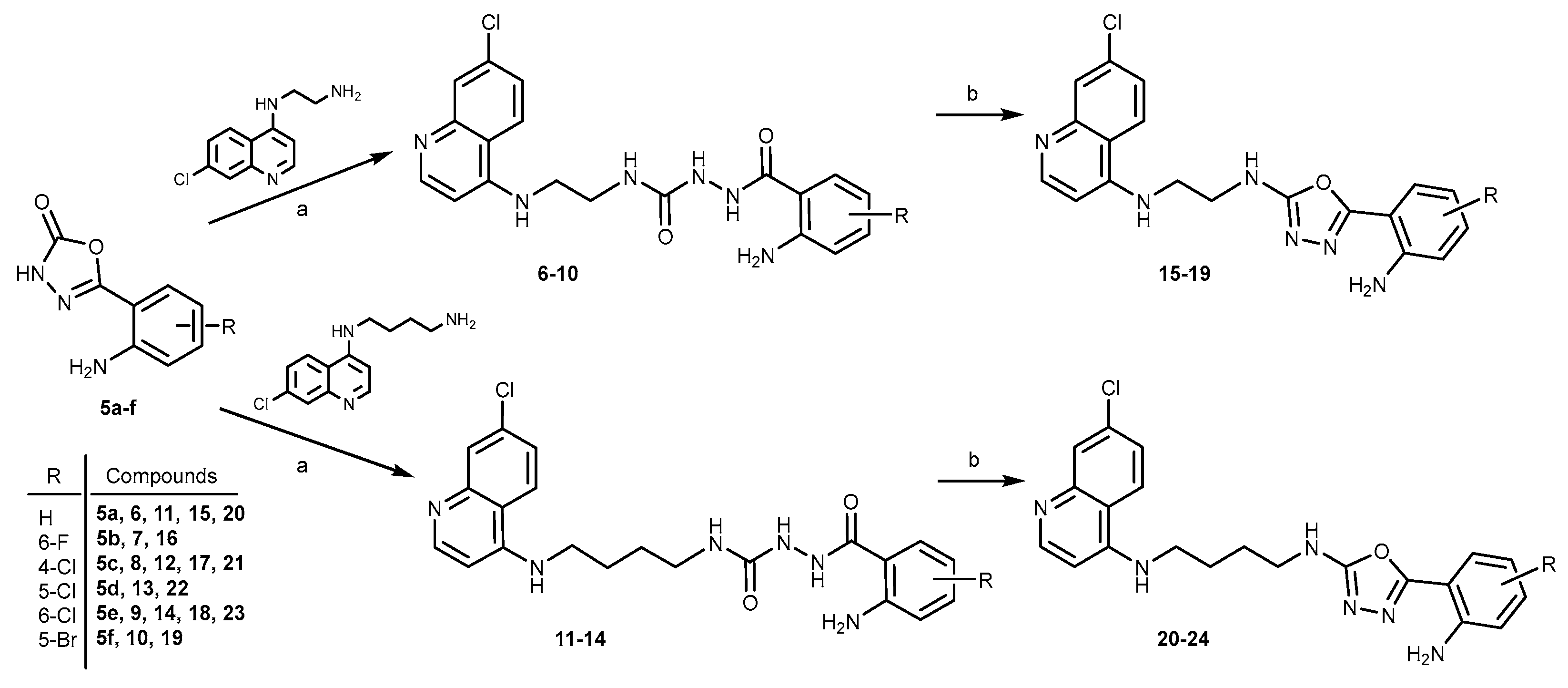
Figure 2.
Comparison of violacein production (a) and cell viability (b) of C. violaceum ATCC 31523 cultures with and without the tested compounds. Medium – culture medium with 2 % DMSO; Cells+DMSO – C. violaceum cells with 2 % DMSO; Q – quercetin; AZ – azithromycin (both at 400 μM); (c) F267 and F2896 – previously identified flavonols with demonstrated anti-QS activity [40,41]. Red arrows in a) indicate the most active anti-QS compounds and blue arrows in b) indicate the compounds with the most pronounced bactericidal activity.
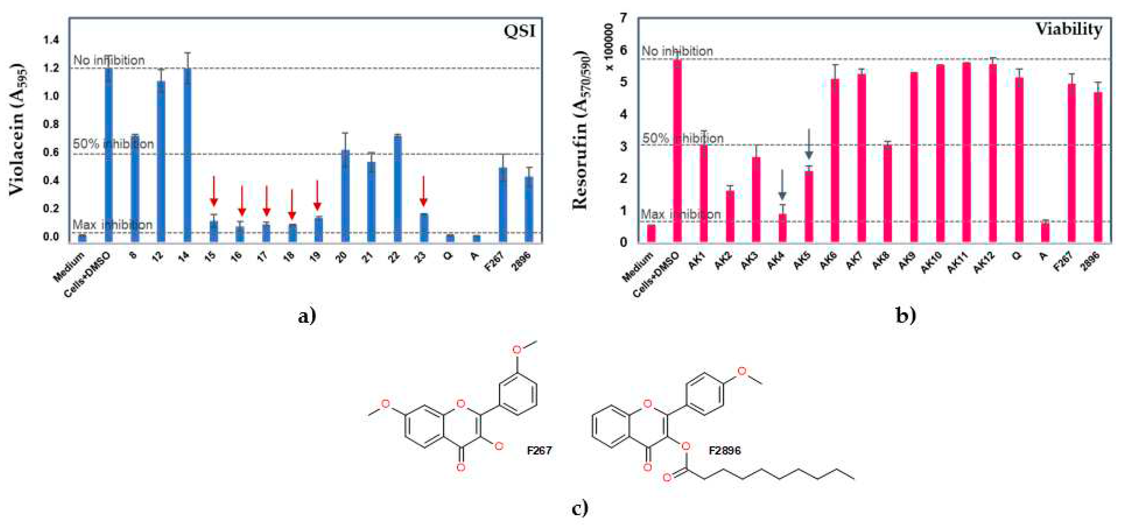
Figure 3.
Dose-response analyses with the most promising anti-QS compounds (15, 16, 19, and 23) at 400, 200, 100, and 40 µM concentrations of violacein production (a) and cell viability (b) of C. violaceum ATCC 31523 cultures with and without the tested compounds. Medium – culture medium with 2 % DMSO; Cells+DMSO – C. violaceum cells with 2 % DMSO; Q – quercetin; A – azithromycin (both at 400 μM).
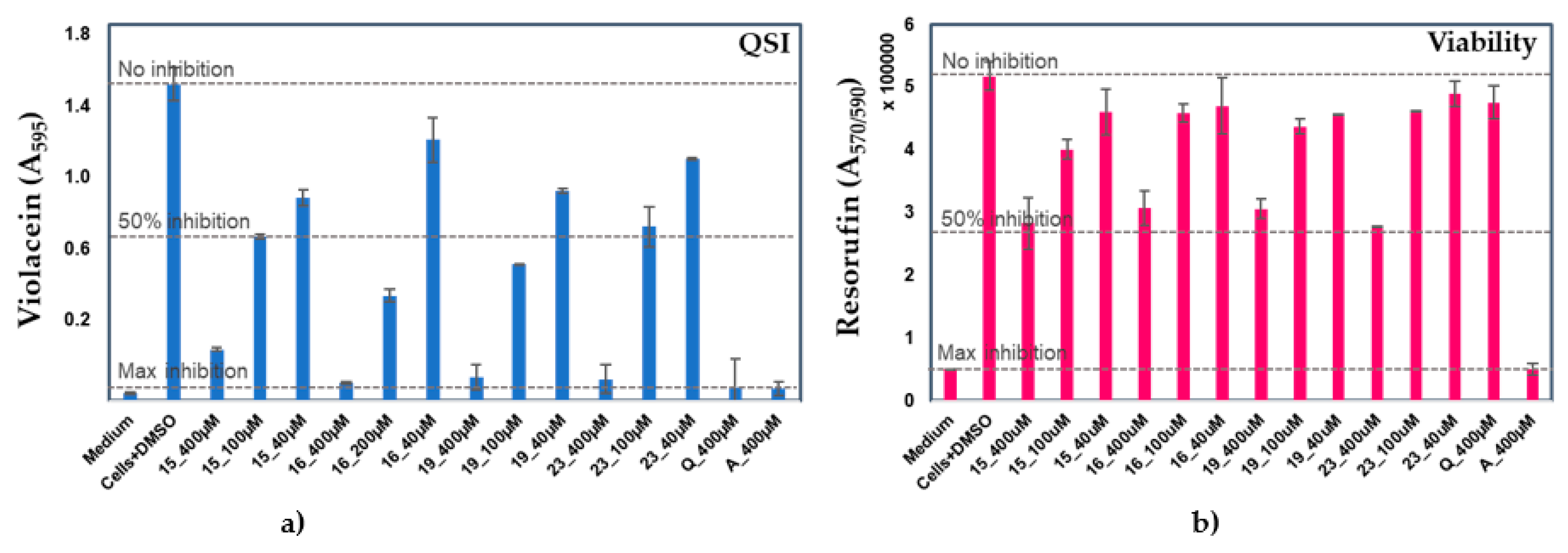
Figure 4.
Effect of compounds 15, 16, 18, 19, and 23 at 100 μM concentrations on the biofilm formation of P. aeruginosa PAO1 compared to control cells treated with 1 % DMSO.
Figure 4.
Effect of compounds 15, 16, 18, 19, and 23 at 100 μM concentrations on the biofilm formation of P. aeruginosa PAO1 compared to control cells treated with 1 % DMSO.
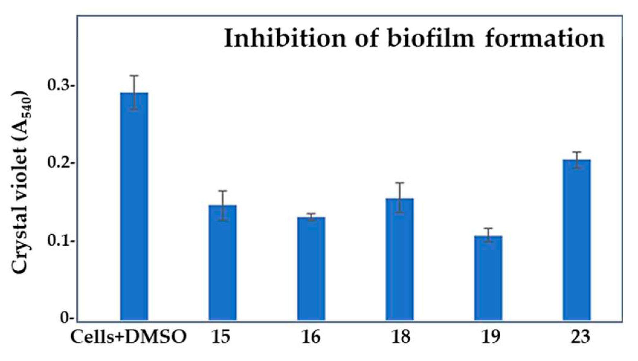
Figure 5.
Effect of compounds 15, 16, 18, 19, and 23 in 100 μM concentrations on the established biofilm of P. aeruginosa PAO1 compared to control cells treated with 1% DMSO.
Figure 5.
Effect of compounds 15, 16, 18, 19, and 23 in 100 μM concentrations on the established biofilm of P. aeruginosa PAO1 compared to control cells treated with 1% DMSO.
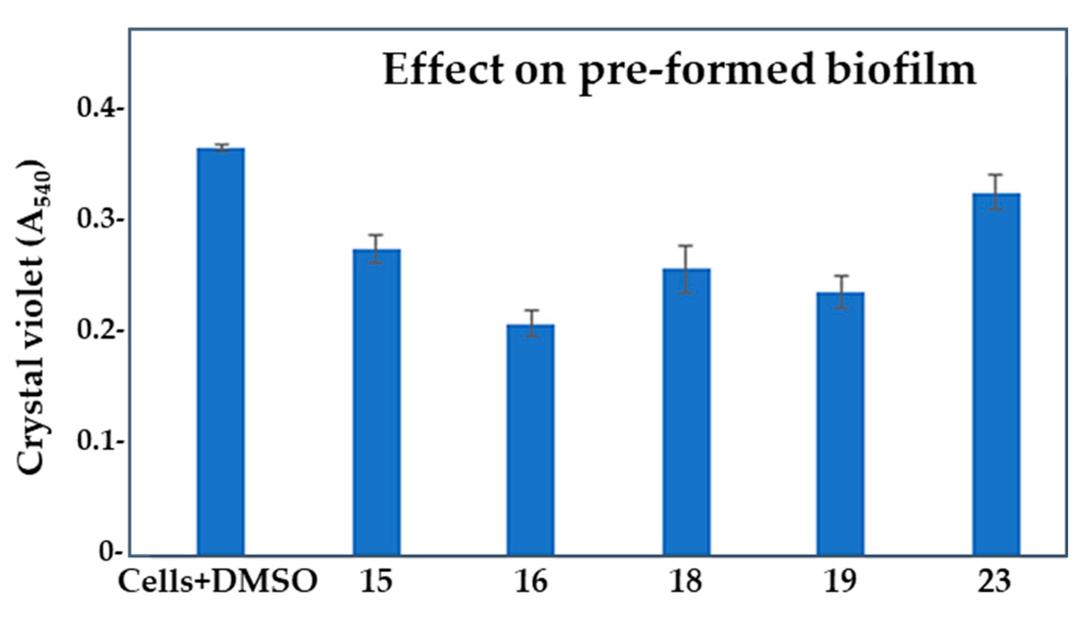
Figure 6.
Effect of compounds 15, 16, 18, 19, and 23 at 100 μM concentrations on the production of pyocyanin by P. aeruginosa PAO1 compared to control cells treated with 1 % DMSO. Results are expressed as a ratio (A520/A570)x100 to normalize pyocyanin absorbance to the cell count.
Figure 6.
Effect of compounds 15, 16, 18, 19, and 23 at 100 μM concentrations on the production of pyocyanin by P. aeruginosa PAO1 compared to control cells treated with 1 % DMSO. Results are expressed as a ratio (A520/A570)x100 to normalize pyocyanin absorbance to the cell count.
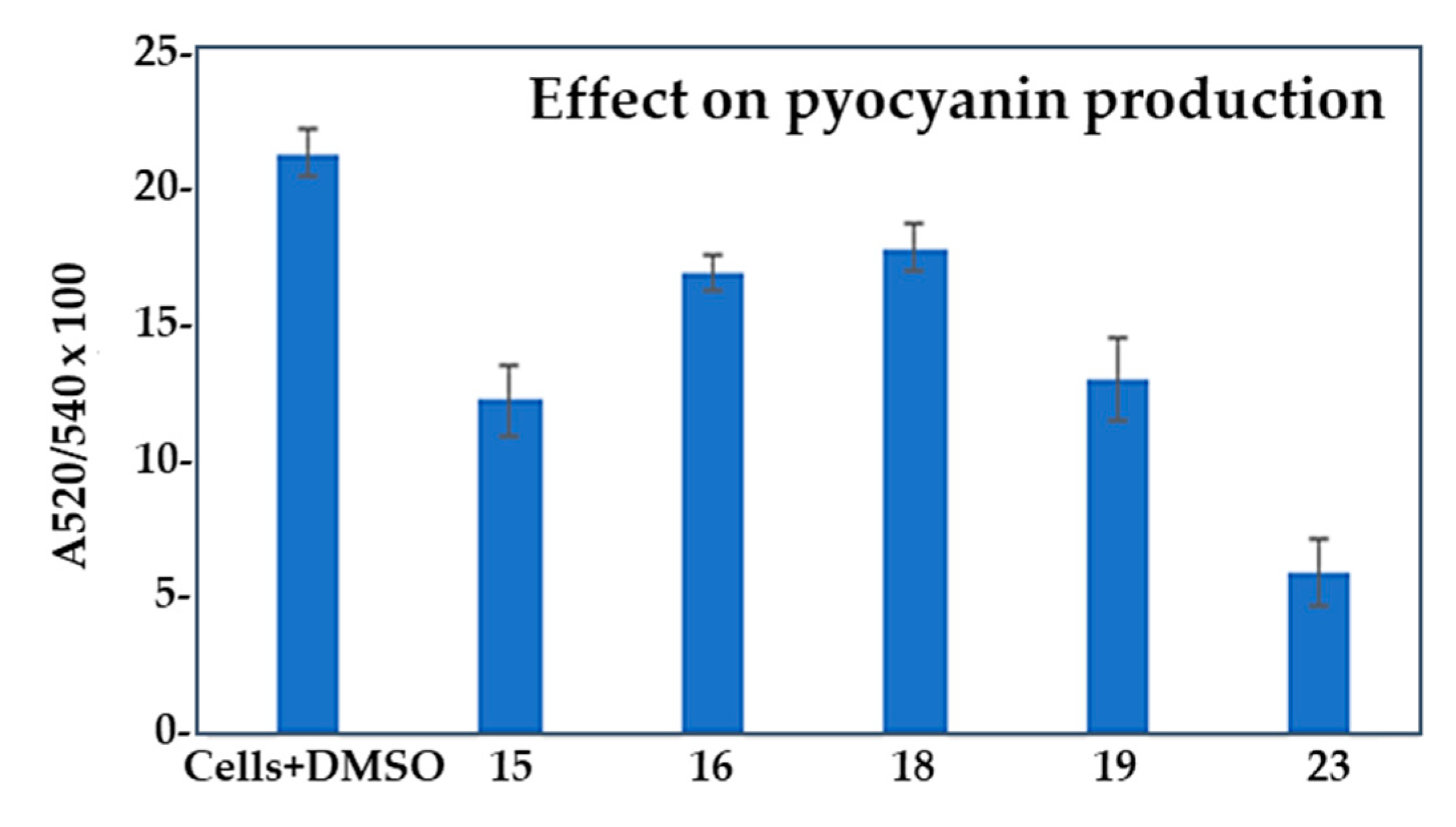

Table 1.
Quorum sensing inhibition and bactericidal effect of acylsemicarbazide (8, 12 and 14) and 1,3,4-oxadiazole (15–23) derivatives at 400 μM concentrations on C. violaceum strain (ATCC 31532).
Table 1.
Quorum sensing inhibition and bactericidal effect of acylsemicarbazide (8, 12 and 14) and 1,3,4-oxadiazole (15–23) derivatives at 400 μM concentrations on C. violaceum strain (ATCC 31532).
| Compd. | QSI (%) | Bactericidal effect (%) |
|---|---|---|
| 8 | 38.6 ± 0.9 | 7.2 ± 0.0 |
| 12 | ne | 2.9 ± 0.1 |
| 14 | ne | 1.8 ± 0.2 |
| 15 | 87.4 ± 3.6 | 52.9 ± 6.3 |
| 16 | 90.5 ± 2.8 | 60.6 ± 2.5 |
| 17 | 89.6 ± 0.4 | 84.0 ± 4.8 |
| 18 | 89.3 ± 0.8 | 71.4 ± 2.8 |
| 19 | 85.8 ± 0.9 | 46.0 ± 7.1 |
| 20 | 46.5 ± 9.7 | 10.6 ± 7.8 |
| 21 | 53.7 ± 5.5 | 7.6 ± 2.7 |
| 22 | 38.6 ± 0.7 | 2.6 ± 3.6 |
| 23 | 83.5 ± 0.03 | 46.9 ± 2.1 |
| Q | 95.8 ± 0.4 | 9.8 ± 4.7 |
| AZ | 95.9 ± 0.2 | 89.1 ± 1.5 |
| F267 | 56.8 ± 7.7 | 13.1 ± 5.3 |
| F2896 | 62.1 ± 5.6 | 17.8 ± 5.6 |
Table 2.
Parameters of cell growth inhibition, inhibition of biofilm formation, biofilm eradication, pyocyanin inhibition, and biofilm index of P. aeruginosa PAO1 cells treated with 100 μM compounds compared to control cells treated with 1% DMSO.
Table 2.
Parameters of cell growth inhibition, inhibition of biofilm formation, biofilm eradication, pyocyanin inhibition, and biofilm index of P. aeruginosa PAO1 cells treated with 100 μM compounds compared to control cells treated with 1% DMSO.
| Compd. | GI (%) | BFI (%) | Biofilm index | BE (%) | PI (%) |
|---|---|---|---|---|---|
| Ø | 60.25 | ||||
| 15 | 8.13 ± 2.76 | 48.6 ± 1.81 | 33.72 | 24.86± 1.13 | 42.34 ± 1.37 |
| 16 | 31.85 ± 1.64 | 53.72 ± 0.43 | 40.9 | 43.08 ± 2.15 | 20.45 ± 0.69 |
| 18 | 28.34 ± 2.57 | 45.26 ± 1.88 | 46.02 | 29.74 ± 1.47 | 16.02 ± 0.87 |
| 19 | 43.7 ± 2.24 | 61.57 ± 0.84 | 41.13 | 35.36 ± 1.62 | 38.87 ± 1.57 |
| 23 | 23.38 ± 3.03 | 29.08 ± 1.03 | 55.75 | 10.89 ± 1.29 | 72.02 ± 1.25 |
Ø - 1 % DMSO; GI – growth inhibition (A570); BFI – biofilm formation inhibition (A540); Biofilm index – (A540/A570)x100; BE – biofilm eradication (A540) ; PI – pyocyanin inhibition – (A520/A570)x100.
Disclaimer/Publisher’s Note: The statements, opinions and data contained in all publications are solely those of the individual author(s) and contributor(s) and not of MDPI and/or the editor(s). MDPI and/or the editor(s) disclaim responsibility for any injury to people or property resulting from any ideas, methods, instructions or products referred to in the content. |
© 2023 by the authors. Licensee MDPI, Basel, Switzerland. This article is an open access article distributed under the terms and conditions of the Creative Commons Attribution (CC BY) license (http://creativecommons.org/licenses/by/4.0/).
Copyright: This open access article is published under a Creative Commons CC BY 4.0 license, which permit the free download, distribution, and reuse, provided that the author and preprint are cited in any reuse.


