Submitted:
05 July 2023
Posted:
06 July 2023
You are already at the latest version
Abstract
Brainstem auditory evoked potentials (BAEPs) testing is very important when microvascular decompression (MVD) is performed with hemifacial spasm (HFS). The reason for this is that the vestibulocochlear nerve is located right next to the facial nerve, so the vestibulocochlear nerve may be affected by manipulation by surgery. The BAEPs test method for detecting vestibulocochlear nerve damage during surgery has been developed a lot and is helping a lot during surgery. In most HFS patients with normal vestibulocochlear nerve, the degree of vestibulocochlear nerve damage caused by surgery is reflected very well in the BAEPs test waveforms. Therefore, a real time test is the best way to minimize the damage to the vestibulocochlear nerve. The purpose of this study was to review the most recently published BAEPs test used in MVD surgery and to find out in detail the relationship between the vestibulocochlear nerve damage and the BAEPs waveforms.
Keywords:
Brainstem auditory evoked potentials
; microvascular decompression
; hemifacial spasm
; vestibulocochlear nerve damage
1. Introduction
After obtaining BAEPs waveforms with click sound stimulation in the human head for the first time [1], In the 1980s, with the development of equipment software from analog systems to digital systems, more sophisticated BAEPs waveforms were obtained [2]. Thanks to the dedication of Moller AR and Jannetta PJ, we are now testing BAEPs during surgery [3,4]. A lot of time has passed since then, and many studies have been published.
In 2003, M P Sindou mentioned the importance of intraoperative neurophysiological monitoring and, in particular, analyzed in detail the change in wave V latency in the BAEPs waveform. In this study, for intraoperative brainstem auditory evoked potential changes and warning values to prevent hearing loss in a prospective study in a consecutive series of 84 patients, if the latency of wave V is delayed by 0.4ms, it is a "watching" signal, and if it is delayed by 0.6ms, it is a "warning" of Real risk and "critical" warning if it is extended by 1.0ms, it is classified into each stage as "critical" warning [5]. And he described in detail the meaning of wave I loss as well as wave V loss when hearing loss occurred after surgery [6].
Hatayama was not associated with postoperative hearing loss when the latency period of the wave V was extended, and all patients who showed a decrease in amplitude more than 40% of the wave V were accompanied by prolongation of the latency and it was reported to be highly related to postoperative hearing loss [7]
As a slightly different opinion from the above, studies have also been published titled Amplitude is more important than latency. These papers are a little association with the latency of wave V and hearing loss but the reduction of amplitude by more than 50% was highly related to hearing loss [8].
The reason why there are various warning criteria is that each surgeon has a different experience of postoperative results, and also because the BAEPs test method is slightly different for each surgeon. The American Clinical Neurophysiology Society reported that V latency longer than 1ms or 10% increase in latency and/or with a 50% reduction in amplitude as a warning criterion [9]. And the BAEPs test suggested that the stimulation rate be 8-10Hz and the averaging time be tested 1000-4000 times [10].
Despite the clear proposals on suggest to test BAEPs as above, in some hospitals, the stimulation rate is 31.1 Hz and the average time is 1000-4000 tests [11], another hospital has a stimulation rate of 17.5 Hz and an average time of 256 (at least) tests [12]. This reason may be a test method learned to reduce hearing loss from many surgical experiences.
Previously, postoperative hearing loss has been reported to be in the range of 7.7-20% when surgery was performed without intraoperative BAEPs monitoring [13]. Nowadays, with intraoperative BAEPs monitoring, the average postoperative hearing loss is 3.4% incidence rates are shown [14].
We present findings on the meaning of BAEPs waveform changes and the latest test of method that can further reduce postoperative hearing loss by monitoring BAEPs during surgery.
2. Methods
2-1. BAEPs test in Routine
Short-latency auditory evoked potentials use click sound stimulation to maximize activation from the vestibulocochlear nerve to the brainstem, and the units of stimulation are dB nHL (normal hearing level) or dB SPL (sound pressure level).
The stimulus intensity should be set at a level to produce clear BAEPs but not cause eardrum damage. In the case of testing with SPL, the threshold is first measured. The intensity is increased sequentially from 0 dB, and the test is conducted with a value obtained by adding 60 dB to the intensity at which the sound begins to be heard. A maximum click intensity of 120 dB SPL or 90 dB nHL is commonly utilized. We do not test at higher than these levels of intensity. To mask crossover responses, white noise at 60 dB SPL or 30–35 dB HL is applied to the contralateral ear. Clicks can be of two polarities, either condensation or rarefaction, depending on the initial movement of the diaphragm of the transducer.
We try to obtain a pure waveform by removing artifacts from the patient under examination by using an 8 – 10 Hz stimulation rate, where the amplitude of the waveform is observed the largest, and averaging time is 1000 - 4000 times for a long period of time [15].
To explain in more detail, the reason for testing at 10 Hz is that when testing with a faster stimulation rate of 30 Hz or 70 Hz, there is a phenomenon in which the amplitude of wave V is measured small and the inter-peak latency from wave I to wave V because it extends further. That is, short-latency auditory evoked potentials, which measure the function of a person's vestibulocochlear nerve to the brainstem, are the best waveforms when tested at 10 Hz [16]. In particular, if the patient moves or coughs during the test, it affects the averaging of the BAEPs waveform under test, so at least 1,000 averaging times are required to average sufficiently to exclude these effects.
2-2. BAEPs test in INM
2-2-1. Real-time BAEPs
2-2-1-1. Stimulation rate and averaging time
Generally, in an awakening state, many of these impure waveforms are absorbed in averaging, so you have to continue averaging over 1000 times for a long time until the waveform stabilizes to obtain a waveform with good reproducibility as the patient's unique waveform. Since the BAEPs test performed during surgery is performed under general anesthesia, there are no artifacts mixed with the patient's minute movements during the BAEPs test. Therefore, a very pure waveform is obtained. Stimulation rates up to 100 are possible with most medical devices. In other words, it is possible to test BAEPs that can enter 100 stimuli per second.
If the BAEPs test is performed intraoperatively in the same way as for the Routine test, a stimulation rate of 10 Hz for an average of 1000 trials requires approximately 100 seconds to obtain BAEP. This relative increase in duration may limit the prevention of postoperative hearing loss with the development of an INM machine with a high signal-to-noise ratio, the disintegration of the waveform amplitude that occurs with a higher stimulation rate is significantly improved. According to this study, no significant difference was found in the BAEPs waves obtained by measuring the stimulation rate faster than 40 Hz and measuring it at 10 Hz and it is possible to obtain a reliable waveform with fewer trials. it is reliable waveforms at 43.9 Hz/sec in an average of 400 trials [17].
Therefore, BAEPs that test within 10 seconds with a combination of an averaging time of 400 and a stimulation rate of 40-50 Hz are possible. In this study, these test methods are called real-time BAEP.
2-2-2. Warning criteria
When analyzing the relationship between the change of the BAEP waveform tested by the real-time BAEP method during surgery and the hearing loss after surgery, we could apply the known V latency longer than 1ms or 10% increase in latency and / or with a 50% reduction in amplitude as a warning criterion as it is.
However, it was possible to distinguish the change in wave V in detail. first of all, it was found that when a weak influence from surgery occurred, a change in latency occurred, and when it was affected more strongly, a change in amplitude occurred. And it was found that the latency of wave V was gradually extended to 1ms continuously, while the amplitude rapidly decreased by 50% within 10 seconds. Therefore, in most cases, the change in latency occurs first and then the change in amplitude occurs later. In some cases, if a serious effect is received within a few seconds, latency does not prolong, and the amplitude of wave V suddenly decreases by more than 50% and loss occurs [18].
In a study that observed changes in BAEP waveforms tested by the real-time method during surgery on 606 patients who visited the hospital for hemifacial spasm and underwent MVD surgery, they were divided into six more subdivided groups as follows [19]. Looking at Group 4, when latency was prolonged by 2ms and amplitude was reduced by more than 50%, 36 patients accounted for 5.9% of the total, and one patient had postoperative deaf. In group 5, amplitude loss occurred and did not recover, 5 patients, 0.83% of the total, and all of them had deaf after surgery. Looking at group 6, amplitude loss occurred and recovered, and there were 23 patients with transient loss, accounting for 3.8% of the total, and no total deaf patients after surgery.
Therefore, it can be seen that the permanent loss of wave V in the BAEPs wave is closely related to the postoperative hearing loss. Also, when latency 2ms prolongation occurs, amplitude decrease often occurs, and when latency 2ms prolongation occurs, amplitude loss occurs suddenly in some cases. Therefore, if latency is extended by 2ms, you should be aware that it can change to a very serious state (Table 1).
In real-time BAEP tests, latency changes were always observed first, followed by amplitude changes. In addition, latency change was observed slowly and continuously, and amplitude change suddenly decreased by more than 50% within 10 seconds. When BAEPs are tested quickly in real-time, it is sometimes observed that wave V latency is extended up to 3ms. With such a quick BAEP test, vestibulocochlear nerve damage is quickly reflected in the BAEP waveform, so we can minimize vestibulocochlear nerve damage with surgical manipulation. As a result, a lot of transient loss tended to be observed in the real-time BAEP method, and in most cases of transient loss, hearing was normal after surgery.
So, the relationship between the amplitude and latency of BAEPs wave V could be summarized in the table below (Figure 2).
2-2-2-1. Prewarning sign
Until now, only absolute latency was observed when measuring the change in latency of BAEPs wave V. However, when the BAEPs wave V latency was extended beyond 1ms, it was classified into two patterns. This is the case when the Wave V latency is extended in Wave I (group A) and when the Wave V latency is extended in Wave III (group B). During surgery, the cochlear nerve is stretched under the influence of surgical manipulation. The reason why Gr A occurs is that as the nerves stretch in the direction of the brainstem, the cochlear part is affected, causing expansion in wave I and the reason why Gr B occurs is thought to be that the area around the brainstem is directly affected and expansion occurs in wave III [20] (Figure 3).
When analyzing the maximal change of wave V after observing the prewarning changes in BAEPs of significant differences between the two groups. Analyzing the percentage within each group, the case where the wave V latency was extended by more than 1ms was observed more in Gr B with 34.6% in Gr A and 50.6% in Gr B. However, transient loss was observed more in Gr A with 29.6% for Gr A and 20.0% for Gr B.
When analyzing the final change of wave V after observing the prewarning changes in BAEPs of significant differences between the two groups. when the intraoperative BAEPs wave V latency was extended by more than 1ms, 88.9% of patients recovered to less than 1ms before the end of surgery in Gr A, in 9.9% of patients, the operation was completed with a condition that was longer than 1ms. In Gr B, 65.6% of patients recovered well within 1ms immediately before the end of surgery, in 28.1% of patients, the operation was terminated with a prolongation of more than 1ms. In particular, BAEPs wave loss was observed in 1 patient (1.2%) in Gr A and 10 patients (6.3%) in Gr B.
The two patterns showed different postoperative patterns. In Wave I, there was no postoperative hearing loss in patients with both temporary and permanent loss of BAEP in the case of dilation. However, when expansion occurred in Wave III, 3 out of 10 patients with permanent loss of BAEP developed total hearing loss and 2 out of 32 patients with temporary loss of BAEP developed partial hearing loss.
In summary, if the BAEPs Wave V Latency is extended by more than 1ms, the recovery of the waveform can be better when the extension (Gr A) occurs in Wave I than when the extension (Gr B) occurs in Wave III. It can be seen that when extension (Gr B) occurs in Wave III, it is a serious situation in which there is a lot of waveform loss and the hearing loss after surgery also increases (Table 2).
Previously, there was a study that evaluated the changes in the amplitude and latency of BAEPs wave III during MVD and the association with postoperative HL. According to this study, intraoperative wave III and wave V were compared. When hearing loss was evaluated after surgery in cases where wave III was maintained and only wave V was lost or both wave III and wave V were lost, it was said that the amplitude change of wave III had a greater variation in all steps of surgery [21]. The conclusions of this study and the prewarning sign study are considered to be the same.
2-2-2-2. Significance of wave I
Loss of BAEPs waveform is closely related to postoperative hearing loss. The change in the BAEPs waveform in the main procedure during surgery is reflected in the waveform immediately when the cochlear nerve is damaged due to traumatic mechanical damage. In addition, since it takes time to affect the cochlear nerve due to poor blood circulation, it takes some time before the effects of vascular circulation damage are reflected in BAEPs [22]. For this reason, changes in the BAEPs waveform are observed very quickly in the case of traumatic mechanical damage. However, in the case of vascular circulation damage, changes may occur after 10 minutes.
More importantly, in the case of traumatic mechanical damage, the existing warning criteria of 1ms delayed and 50% reduction of wave V are applied. In the case of vascular circulation damage, the waveform disappears suddenly and without warning sign.
When traumatic mechanical damage is severely affected, BAEPs wave loss occurs, but wave I is always present. Wave I is present in the BAEPs waveform of patients with hearing disturbance caused by brain tumor, and latency delayed or decreased in amplitude of the remaining waveforms, wave III or V, is similar to that observed.
That is, the presence of wave I in the BAEPs waveform means that the sound transmission ability to the cochlear is normal, and that there is a problem in the process from the cochlear nerve to the brainstem. In fact, when looking at the postoperative hearing status of patients in which wave I is present and all other waveforms are lost, most cases show only hearing loss or partial HL (Figure 4).
However, in the case of vascular circulation damage, a waveform without wave V is suddenly observed without extension or decrease of wave V, and all waveforms are lost without even wave I. That is, it means that the vestibular cochlear system was affected as a whole. In this way, patients who have observed BAEPs wave all loss and have not recovered and have completed surgery have very serious and diverse symptoms after surgery. HL is naturally accompanied by tinnitus, dizziness, and hoarseness (Table 3).
We must observe very importantly whether wave I exists when BAEPs wave loss occurs and do not neglect the BAEPs test just because the main procedure has been completed during surgery. We always have to continue testing until the end of surgery.
Previously in the study, wave I amplitude decrease was considered a warning-signal of cochlear dysfunction. And it was hypothesized that AICA and labyrinth arterial flow reduction and an association due to manipulation of these vessels [6]. The conclusions of this study and the Significance of wave I study are considered to be the same
3. Discussion
3-1. Patterns of BAEPs wave change during Microvascular Decompression for Hemifacial Spasm
3-1-1. Latency and amplitude of BAEPs wave V
When BAEPs are inspected by real-time method, a waveform is formed within 10 seconds. Therefore, the change of the waveform can be observed every 10 seconds, and the change of the waveform can be detected very precisely and precisely. In particular, the change in latency and amplitude of wave V of BAEPs is observed in a very different pattern, and the change in wave V latency is prolonged slowly and continuously. However, the wave V amplitude rapidly decreases by 50% within 10 seconds, and in some cases, wave loss occurs suddenly.
Looking at the process of forming a waveform while measuring BAEPs, the waveform is shaped like a skeleton before 300 averaging times. The shape of the waveform formed in this way does not change significantly even if the averaging time is continued up to 1000 times, and the shape is maintained. In the case of testing with a stimulation rate of 10Hz and an averaging time of 1000, the latency of wave V, which gradually and continuously changes, is reflected in the test waveform. However, the amplitude of wave V, which rapidly decreases within 10 seconds, is reflected very little because hundreds of normal averaged waveforms already exist.
Therefore, if the BAEPs waveform is tested in a way that cannot be observed within 10 seconds, the slowly progressive wave V latency may be discernable, but the rapidly changing amplitude will be difficult to detect [19].
3-1-2. Time-dependent classification of BAEPs wave changes
The change of the waveform can be divided into the case of rapid change and the case of showing slow change, and can be classified into three times concepts [19].
Phase I: When the operation is stable, there is no change in the amplitude of the V wave, and only the wave V latency is observed little by little in less than 1ms.
In general, the change in the wave V latency is slowly and continuously extended for several minutes to several tens of minutes when the dura is opened, even if there is no particular abnormality. After the main procedure, the original waveform will gradually recover.
This stage corresponds to almost all situations of surgery except for the main procedure, and is the period observed for the longest time.
Phase II: This is a state in which changes in both latency and amplitude of wave V are observed. When affected by a brain retractor or manipulation during this procedure, an extended wave V latency of more than 1ms is observed. But, wave V amplitude is observed to decrease by more than 50% of wave V amplitude within 10 seconds. When wave V is extended for more than 1ms, the probability of decreasing the amplitude of wave V is greatly increased.
This stage occurs in the main procedure and occurs during decompression of the offending vessels and facial nerves.
Phase III: When the brain retractor is used excessively in main procedure, if the vestibulocochlear nerve is greatly affected, BAEP wave V loss occurs very quickly and suddenly.
This step occurs when the offending site is deep or excessive retraction is used in the main procedure. It often happens in as little as 10 seconds, so be very careful.
In general, more serious damage occurs when all waveforms suddenly disappear after no waveform change than when wave V disappears after being delayed by 1ms and reduced by 50% amplitude.
3-1-3. Classification of BAEPs wave and cochlear nerve damage
In the case of Phase I, where only the extension of wave V latency is observed very slowly and continuously, it accounts for more than half of all surgeries, and this phenomenon is observed even with only dura open. Waveform changes due to traumatic mechanical damage are observed in all phases of Phase I, II, and III, and can be divided into four stages, Mild, Moderate, Severe, and Critical damage, depending on the degree of direct damage to the vestibulocochlear nerve [19] [Table 4].
Mild damage: This is the case where no significant change in the waveform is observed, in which the latency of wave V is extended to less than 1ms or the amplitude is decreased by less than 50%. in this case, waves I, III, and V are clearly observed. The state in which the latency of wave V is extended by more than 1ms and the amplitude is reduced to less than 50% also applies. Corresponds to phase I stage.
Moderate damage: This is a more advanced damage stage from mild damage, in which the latency of wave V is extended by more than 1ms and the amplitude is reduced by more than 50%. It is important that the amplitude is reduced by more than 50%. If the effect is not stopped by surgery, most of them will decrease by more than 80% or progress to wave V loss. Corresponds to phase I stage.
Severe damage can be classified into several types.
Severe damage-1: The wave V delay is greater than 2ms and the wave V amplitude is reduced by more than 80%. When the wave V latency is delayed by more than 2ms, the V wave amplitude often decreases rapidly. Therefore, even if the latency is delayed by more than 2ms without the amplitude change of wave V, the surgeon must be notified. Corresponds to phase II stage.
Severe damage-2: This is a case where the Phase II suddenly occurs without going through the mild, moderate nerve damage of Phase I. This is when BAEPs wave abrupt change occurs. When vestibulocochlear nerve focal damage is severe, the amplitude suddenly decreases without changing the latency of wave V. Corresponds to phase II stage.
Severe damage-3: This is a change in the waveform due to vascular circulation damage. As with phase I and II, the reproducibility of the waveform suddenly disappears without any change in the latency or amplitude of wave V.
As if artifacts are mixed, the waveform wobbles like a dance, and then all wave loss occurs. In other words, it refers to the state of the waving wave like dancing without observing the I, III, and V waveforms of the BAEPs wave.
If there is a change in the morphology of the BAEPs waveform, it is because the vascular circulation is disturbed. In this case, a vasodilator such as papaverin should be administered to facilitate the vascular circulation of the surrounding blood vessels. Otherwise, if left unattended, all wave loss will be observed where wave I is not observed. And after the operation, the patient will not only complain of overall disorders of the vestibular system, but also complain of hearing impairment. Corresponds to phase II stage.
Critical damage: BAEPs wave loss state. It is divided into traumatic mechanical damage in which wave I is observed and vascular circulation damage in which all wave loss is observed without wave I. BAEPs wave loss can cause hearing loss after surgery. In particular, there are many cases that occur suddenly, such as severe damage-2 of phase II. Therefore, it is important to observe the BAEPs test continuously during surgery. Corresponds to phase III stage.
In most cases, a change in the wave V latency of the waveform is observed, followed by an observation of a decrease in the wave V amplitude sequentially. A much more dangerous situation is when there is no change in the waveform and then a sudden change in latency or amplitude is observed.
4. Conclusion
Postoperative hearing loss due to injury of vestibulocochlear nerve is a common complication during MVD for HFS. This complexity could be greatly reduced with the advent of BAEP's intraoperative neurophysiological monitoring.
Advances in BAEPs testing have provided the Neurosurgeon with greater insight during MVD surgery and may prevent many vestibulocochlear nerve damages.
Future studies on developing novel monitoring techniques and the modification and optimization of existing BAEPs methodologies will continue to improve clinical outcomes in hearing change following MVD for HFS.
Funding
This research did not receive any specific grant from funding agencies in public, commercial, or not-for-profit sectors.
Author Contributions
Conceptualization: Sang-Ku Park, Hyun Seok Lee, Kyung Rae Cho, Kwan Park. Methodology: Sang-Ku Park, Hyun Suk Lee. Supervision: Kwan Park. Validation: Kwan Park. Visualization: Sang-Ku Park, Kyung Rae Cho. Writing - original draft: Sang-Ku Park, Kyung Rae Cho. Writing - review & editing: all authors.
Data Availability Statement
All data generated or analyzed during this study are included in this published article.
Conflicts of Interest
The authors declare no conflict of interest.
Abbreviations
BAEPs: brainstem auditory evoked potentials; MVD: microvascular decompression; HFS: hemifacial spasm; nHL: normal hearing level; SPL: sound pressure level; HL: hearing loss.
References
- Jewett, D.L.; Williston, J.S. Auditory-evoked far fields averaged from the scalp of humans. Brain 1971, 94, 681–696. [Google Scholar] [CrossRef] [PubMed]
- Doyle, D.J.; Hyde, M.L. Analogue and Digital Filtering of Auditory Brainstem Responses. Scand. Audiol. 1981, 10, 81–89. [Google Scholar] [CrossRef] [PubMed]
- Møller, A.R.; Jannetta, P.J. Comparison Between Intracranially Recorded Potentials from the Human Auditory Nerve and Scalp Recorded Auditory Brainstem Responses (ABR). Scand. Audiol. 1982, 11, 33–40. [Google Scholar] [CrossRef] [PubMed]
- Moller AR, Jannetta PJ. Neural generators of auditory evoked potentials. In: Jacobson JT, editor. The auditory brainstem response. SanDiego: College-Hill Press, 1985, 13-31.
- Polo, G.; Fischer, C.; Sindou, M.P.; Marneffe, V. Brainstem Auditory Evoked Potential Monitoring during Microvascular Decompression for Hemifacial Spasm: Intraoperative Brainstem Auditory Evoked Potential Changes and Warning Values to Prevent Hearing Loss—Prospective Study in a Consecutive Series of 84 Patients. Neurosurgery 2004, 54, 97–106. [Google Scholar] [CrossRef] [PubMed]
- Sindou, M.P. Microvascular decompression for primary hemifacial spasm. Importance of intraoperative neurophysiological monitoring. Acta Neurochir. 2005, 147, 1019–1026. [Google Scholar] [CrossRef] [PubMed]
- Hatayama, T.; Møller, A.R. Intraoperatve recordings in MVD operations. Acta Neurochir (Wien) 1998, 140, 681–687. [Google Scholar] [CrossRef] [PubMed]
- Legatt, A.D. Mechanisms of intra operative brainstem auditory evoked potential changes. J Clin Neurophysiol. 2002, 19, 396–408. [Google Scholar] [CrossRef]
- Martin, W.H.; Stecker, M.M. ASNM position statement: intraoperative monitoring of auditory evoked potentials. J Clin Monit Comput. 2008, 22, 75–85. [Google Scholar] [CrossRef] [PubMed]
- American Clinical Neurophysiology Society. Guideline 9C: Guidelines on short-latency auditory evoked potentials. J Clin Neurophysiol. 2006, 23, 157–167. [Google Scholar] [CrossRef] [PubMed]
- James, M.L.; Husain, A.M. Brainstem auditory evoked potential monitoring: When is change in wave V significant? Neurology 2005, 65, 1551–1555. [Google Scholar] [CrossRef] [PubMed]
- Thirumala, P.D.; Carnovale, G.; Habeych, M.E.; Crammond, D.J.; Balzer, J.R. Diagnostic accuracy of brainstem auditory evoked potentials during microvascular decompression. Neurology 2014, 83, 1747–1752. [Google Scholar] [CrossRef] [PubMed]
- Polo, G.; Fischer, C.; Sindou, M.P.; Marneffe, V. Brain auditory evoked potentialmonitoring during microvascular decompression for hemifacial spasm: intra-operative brainstem auditory evoked potential changes and warning values to prevent hearing loss—prospective study in a consecutive series of 84 patients. Neurosurgery 2004, 54, 97–106. [Google Scholar] [PubMed]
- Sprenghers, L.; Lemmens, R.; van Loon, J. Usefulness of intraoperative monitoring in microvascular decompression for hemifacial spasm: a systematic review and meta-analysis. Br. J. Neurosurg. 2022, 36, 346–357. [Google Scholar] [CrossRef] [PubMed]
- American Clinical Neurophysiology Society. Guideline 9C: Guidelines on short-latency auditory evoked potentials. J Clin Neurophysiol. 2006. [Google Scholar]
- Chiappa, K.; Gladstone, K.J.; Young, R.R. Brainstem auditory evoked responses. Studies of waveform variation in 50 normal human subjects. Arch Neurol 1979, 36, 81–87. [Google Scholar] [CrossRef] [PubMed]
- Joo, B.-E.; Park, S.-K.; Cho, K.-R.; Kong, D.-S.; Seo, D.-W.; Park, K. Real-time intraoperative monitoring of brainstem auditory evoked potentials during microvascular decompression for hemifacial spasm. J. Neurosurg. 2016, 125, 1061–1067. [Google Scholar] [CrossRef] [PubMed]
- Park, S.-K.; Joo, B.-E.; Lee, S.; Lee, J.-A.; Hwang, J.-H.; Kong, D.-S.; Seo, D.-W.; Park, K.; Lee, H.-T. The critical warning sign of real-time brainstem auditory evoked potentials during microvascular decompression for hemifacial spasm. Clin. Neurophysiol. 2018, 129, 1097–1102. [Google Scholar] [CrossRef] [PubMed]
- Park, S.-K.; Joo, B.-E.; Park, K. Intraoperative Neurophysiological Monitoring in Hemifacial Spasm. A Practical Guide. 2021. ISBN 978-981-16-1326-5 ISBN 978-981-16-1327-2 (eBook). [CrossRef]
- Park, S.-K.; Joo, B.-E.; Kwon, J.; Kim, M.; Lee, S.; Lee, J.-A.; Park, K. A prewarning sign for hearing loss by brainstem auditory evoked potentials during microvascular decompression surgery for hemifacial spasm. Clin. Neurophysiol. 2020, 132, 358–364. [Google Scholar] [CrossRef] [PubMed]
- Thirumala, P.D.; Krishnaiah, B.; Crammond, D.J.; Habeych, M.E.; Balzer, J.R. Analysis of Wave III of Brain stem Auditory Evoked Potential Waveforms during Microvascular Decompression of Cranial Nerve VII for Hemifacial Spasm. J. Clin. Neurophysiol. 2014, 31, 127–132. [Google Scholar] [CrossRef] [PubMed]
- Joo, B.-E.; Park, S.-K.; Lee, M.H.; Lee, S.; Lee, J.-A.; Park, K. Significance of wave I loss of brainstem auditory evoked potentials during microvascular decompression surgery for hemifacial spasm. Clin. Neurophysiol. 2020, 131, 809–815. [Google Scholar] [CrossRef] [PubMed]
Figure 1.
Without any special abnormality, latency is observed to be prolonged slowly and steadily. In some cases, even if the waveform is lost, it can be recovered by taking a quick action.
Figure 1.
Without any special abnormality, latency is observed to be prolonged slowly and steadily. In some cases, even if the waveform is lost, it can be recovered by taking a quick action.
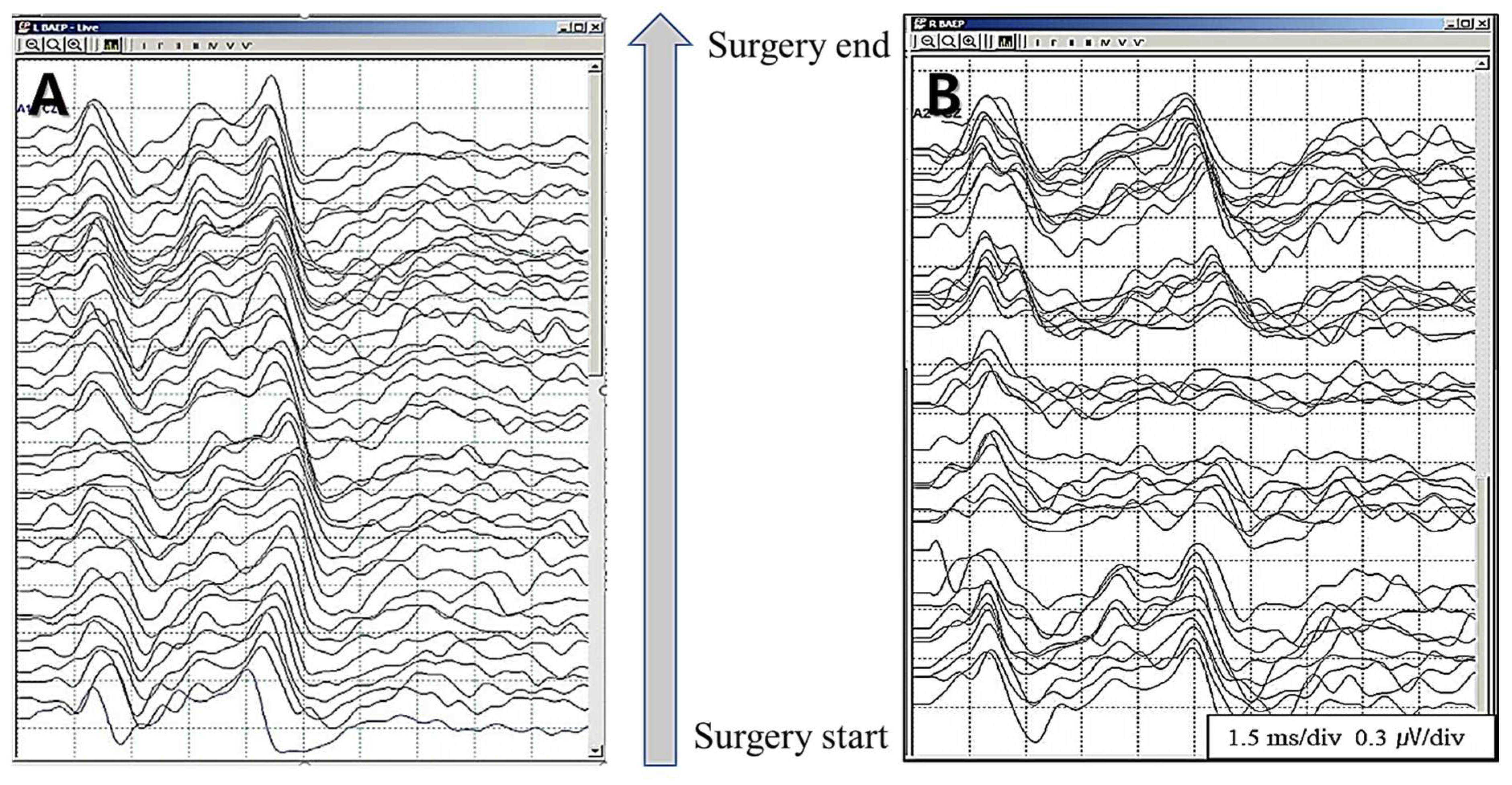
Figure 2.
Correlation of amplitude and latency in Warning Criteria, when using real-time BAEPs. [Intraoperative Neurophysiological Monitoring in Hemifacial Spasm. A Practical Guide. 2021].
Figure 2.
Correlation of amplitude and latency in Warning Criteria, when using real-time BAEPs. [Intraoperative Neurophysiological Monitoring in Hemifacial Spasm. A Practical Guide. 2021].
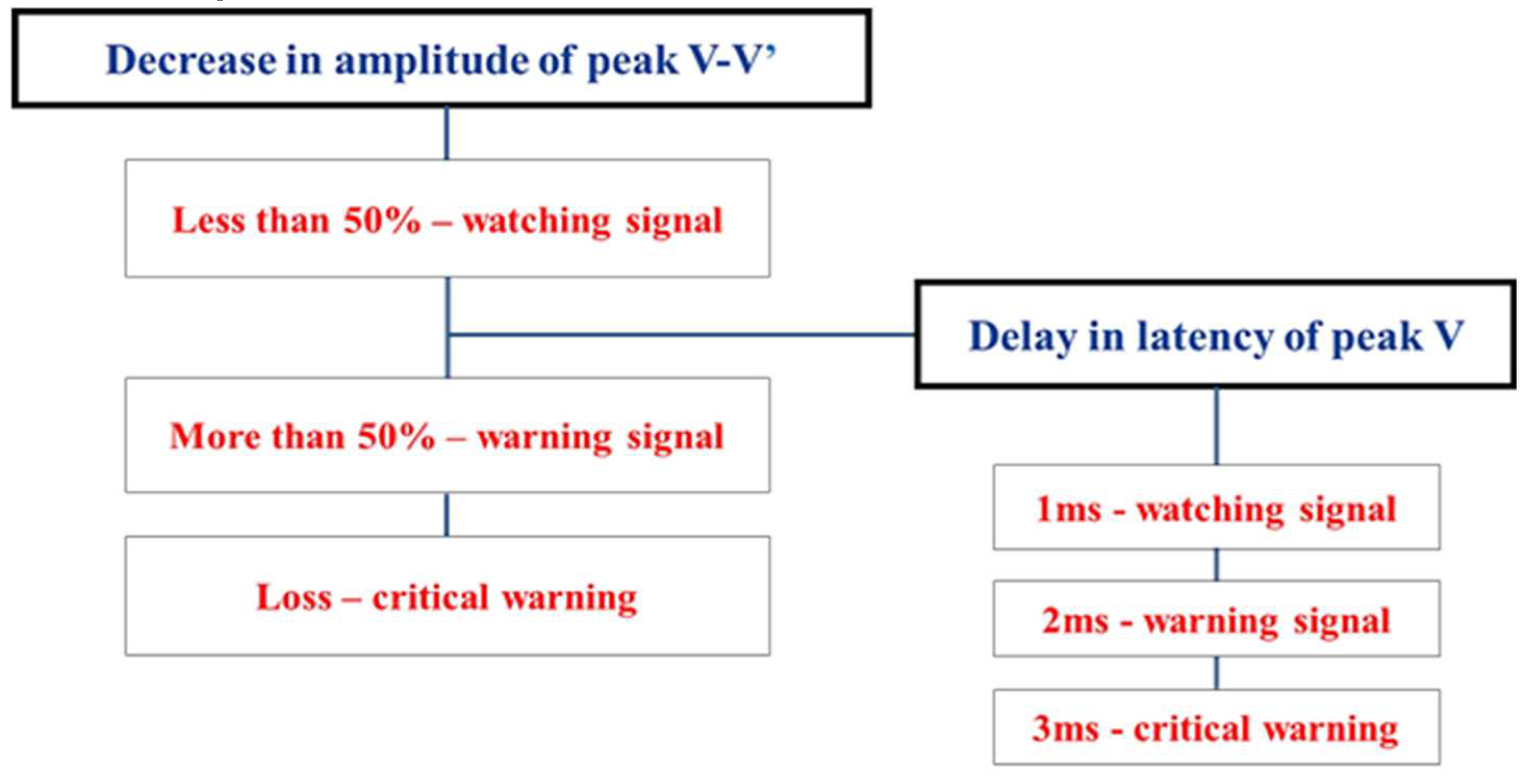
Figure 3.
Differences in consecutive brainstem auditory evoked potentials between the study groups.
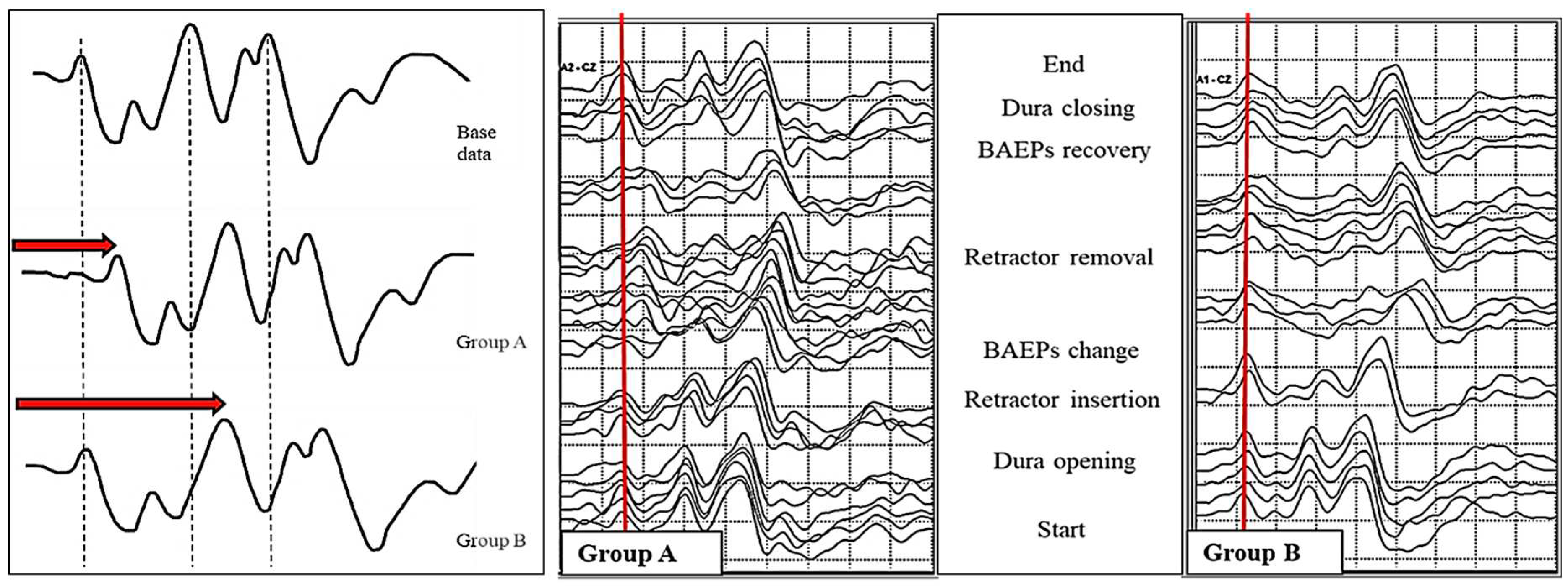
Figure 4.
Example of brainstem auditory evoked potentials (BAEPs) according to wave I persistence/absent in patients with wave V loss during microvascular decompression surgery for hemifacial spasm.
Figure 4.
Example of brainstem auditory evoked potentials (BAEPs) according to wave I persistence/absent in patients with wave V loss during microvascular decompression surgery for hemifacial spasm.
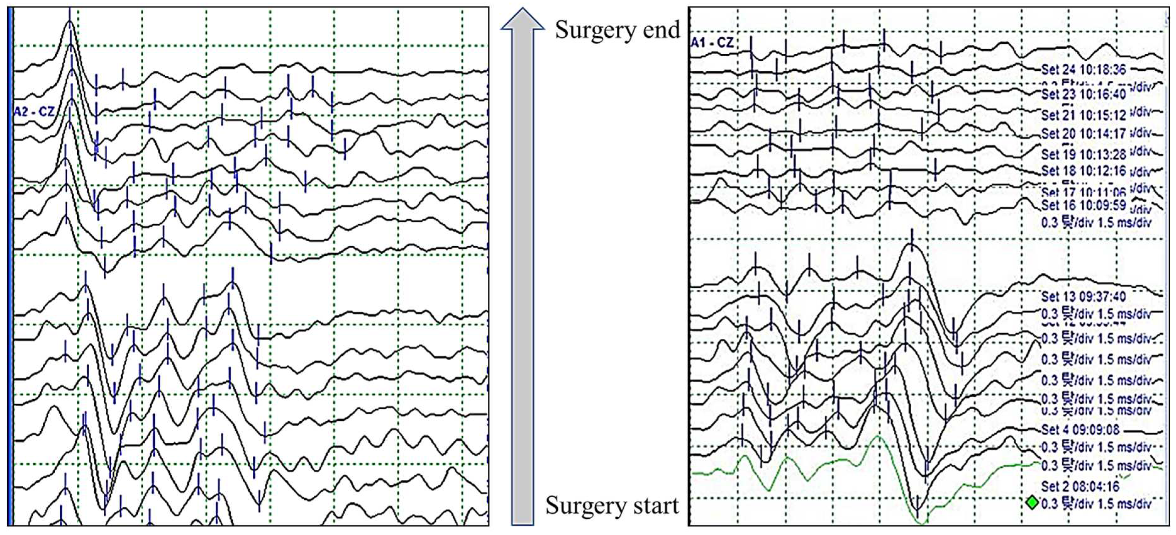

Table 1.
BAEPs groups of change patterns.
| Group | Latency | Amplitude | N = 463 | Post OP HL |
|---|---|---|---|---|
| 1 | < 1ms | < 50% | 346 | 0 |
| 2 | > 2ms | < 50% | 38 | 0 |
| 3 | < 1ms | > 50% | 15 | 0 |
| 4 | > 2ms | > 50% | 36 | 1 |
| 5 | Permanent loss | 5 | 5 | |
| 6 | Transient loss | 23 | 0 | |
* BAEPs wave V standard. [Intraoperative Neurophysiological Monitoring in Hemifacial Spasm. A Practical Guide. 2021].
Table 2.
Proportion of patients according to the final change of wave V between the study groups.
| The final change of wave V | Group A | Group B | p-value |
|---|---|---|---|
| Latency prolongation (<1 ms) with amplitude decrement (<50%) |
72 (88.9%) | 105 (65.6%) | <0.001 |
| Only latency prolongation (1 ms) | 8 (9.9%) | 45 (28.1%) | 0.001 |
| Only amplitude decrement (50%) | 0 | 0 | ns |
| Latency prolongation (1 ms) with amplitude decrement (50%) |
0 | 0 | ns |
| Wave V loss | 1 (1.2%) | 10 (6.3%) | 0.078 |
| Total | 81 | 160 |
[published by Clinical Neurophysiology 132 (2021) 358–364].
Table 3.
The proportion of postoperative complications according to persistence of wave I among the patients showing wave V loss.
Table 3.
The proportion of postoperative complications according to persistence of wave I among the patients showing wave V loss.
| w/i persistence of wave I |
w/o persistence of wave I |
P value | |
|---|---|---|---|
| Patients, n | 24 | 12 | |
| Hearing loss, n (%) | 2 (8.33%) | 6 (50.00%) | 0.009 |
|
Subtype of Hearing loss, n (Low : High : Total) |
2 : 0 : 0 | 0 : 0 : 6 | |
| Dizziness, n (%) | 0 | 5 (41.67%) | 0.002 |
| Tinnitus, n (%) | 0 | 3 (25.00%) | 0.031 |
| Diplopia, n (%) | 0 | 1 (8.34%) | 0.333 |
| Hoarseness, n (%) | 0 | 1 (8.34%) | 0.333 |
w/i = with; w/o = without; HL = Hearing loss; Low = low-frequency hearing loss, High = high-frequency hearing loss; Total = total hearing loss. In this analysis, the level of significance was set to p<0.05, and significant p values are highlighted with bold. [published by Clinical Neurophysiology 131 (2020) 809–815].
Table 4.
The relationship between the phase and the degree of nerve damage is explained by the change of the waveforms.
Table 4.
The relationship between the phase and the degree of nerve damage is explained by the change of the waveforms.
| Damage degree | Latency change | Amplitude change | ||
|---|---|---|---|---|
| Phase I | Mild | < 1ms | < 50% | |
|
Phase II |
Moderate | > 1ms | > 50% | |
|
Severe |
1 | > 2ms | > 80% | |
| 2 | No change | > 50% (abrupt change) | ||
| 3 | No reproducibility | |||
|
Phase III |
Critical |
1 2 |
Loss - traumatic mechanical damage Loss – vascular circulation damage |
|
[Intraoperative Neurophysiological Monitoring in Hemifacial Spasm. A Practical Guide. 2021].
Disclaimer/Publisher’s Note: The statements, opinions and data contained in all publications are solely those of the individual author(s) and contributor(s) and not of MDPI and/or the editor(s). MDPI and/or the editor(s) disclaim responsibility for any injury to people or property resulting from any ideas, methods, instructions or products referred to in the content. |
© 2023 by the authors. Licensee MDPI, Basel, Switzerland. This article is an open access article distributed under the terms and conditions of the Creative Commons Attribution (CC BY) license (http://creativecommons.org/licenses/by/4.0/).
Copyright: This open access article is published under a Creative Commons CC BY 4.0 license, which permit the free download, distribution, and reuse, provided that the author and preprint are cited in any reuse.


