
Submitted:
15 June 2023
Posted:
16 June 2023
You are already at the latest version
Abstract
The current paper presents a case of a 33-year-old female with an uncommon localization of a leiomyoma in the oral cavity - the anterior palatal fibromucosa and the incisive papilla. The patient referred to the Oro-Maxillo-Facial Surgery Clinic of the Emergency City Hospital Timisoara, Romania accusing a slight discomfort in the act of mastication and the occurrence and persistence of a diastema between the upper central incisors, due to the presence of a tumoral mass located in the anterior palatal mucosa, between the upper central incisors, without any changes of the subjacent bone structure in the anterior hard palate visible on the cone beam computed tomography image (CBCT). The tumor was removed by a surgical excisional biopsy and a histopathological examination was performed using morphological Hematoxylin–Eosin (HE) staining and additional immunohistochemical (IHC) reactions, in order to confirm the diagnosis. On microscopic examination, there were found bundles of spindle cells with eosinophilic cytoplasm and vesicular nuclei, with finely granular chromatin. The immunohistochemically staining found positive results for smooth muscle actin (SMA) and desmin and negative result for vimentin. The treatment of choice for leiomyoma of the oral cavity is surgical excision with clear margins, followed by periodical clinical monitoring.
Keywords:
oral pathology
; leiomyoma
; incisive papilla
; palatal fibromucosa
; surgical excision
; immunohistochemical staining
1. Introduction
Leiomyoma is a benign tumor of the smooth muscle, mostly found in the uterus, gastrointestinal tract, skin and subcutaneous tissues, with a rare incidence in the pathology of the oral cavity [1,2,3,4]. Oral leiomyomas present themselves as progressive, slow growing, asymptomatic masses [1,2,3,5,6], mostly found in the lingual interstitium, labial mucosa, fibromucosa of the hard palate or buccal mucosa [1,5,7].The scarcity of smooth muscle tissue in the structures of the oral cavity region, can be an explanation for the low occurrence of the leiomyomas in this anatomical region [1,6].
They may debut at any age, in both sexes, but are usually discovered when the dimensions get visible and palpable. The certain diagnosis is determined by histological examination performed after special staining that can confirm the smooth muscle origin [5]. The treatment for this type of tumor is the surgical removal [1,2,3,4,5,6,7,8,9]. When it comes to the recurrence of the tumor, it is mainly unexpected [1,2,3,4,5,7,8,9,10], however, periodical clinical monitoring is highly recommended.
In this article we present a case of a 33-year-old female with an uncommon localization of a leiomyoma – incisive papilla and anterior palatal fibromucosa of the oral cavity, anamnestic with a debut eight years prior.
2. Materials and Methods
The current paper presents a case of leiomyoma in the oral cavity, with a rare and uncommon localization: the anterior palatal fibromucosa and involvement of the incisive papilla, diagnosed in Oro-Maxillo-Facial Surgery Clinic within the Timisoara Municipal Emergency Clinical Hospital, in March 2021.
For obtaining the certain diagnosis, a surgical excisional biopsy procedure was performed. The collected tissue, fixed in 10% (v/w) neutral buffered formalin, was sent to the Department of Pathology of Timisoara Emergency City Hospital, for the histopathological examination. The harvested specimen was embedded in paraffin. Four micrometers thick serial sections were prepared for the diagnosis, using morphological Hematoxylin–Eosin (HE) staining. To complete the diagnosis, additional immunohistochemical (IHC) reactions were used. All the data regarding the antibodies used for IHC reactions are centralized in Table 1. The panel of antibodies (Anti-Desmin Antibody, Anti-SMA Antibody, Anti-Vimentin Antibody and Anti-Ki-67 Antibody) and the reagents utilized for immunohistochemistry were acquired from Novocastra™, respectively Bond™, Leica Biosystems.
3. Case Report
A 33 years old female patient, without significant conditions nor diseases throughout her life (bronchitis in childhood, Helicobacter Pillory infection treated with medication) referred to the Oro-Maxillo-Facial Surgery Clinic of the Emergency City Hospital Timisoara, Romania accusing a slight discomfort in the act of mastication and the occurrence and persistence of a diastema between the upper central incisors, due to the presence of a tumoral mass located in the anterior hard palate, with the involvement of the incisive papilla and extended between the upper central incisors. Anamnestic, the patient establishes the occurrence of the lesion approximately 8 years prior, with a continuous, slow, progressive growth.
The tumor had a shape of a droplet, well delimited, with a dimension of approximately 1,5 cm (sagittal plane) / 1 cm (transverse plane), with a nodular lower pole located at the median palatal fibromucosa, extended between the bilateral palatal rugae. It had a pink tint, smooth surface, soft, resilient, depressible consistency. The upper pole of the tumoral apex was located interdentally between the superior central incisors (1.1/8-2.1/9). On the buccal side of the alveolar crest, the lesion was in contact with the crestal insertion of a hypertrophic upper labial frenulum, having a reddish color and a slightly firmer consistency, apparently fixed to the underlying bone (Figure 1).
The clinical examination also revealed a dento-alveolar incongruence with the presence of a maxillary interincisal diastema of 2mm and a slight distal tipping of the right upper central incisor, with delicate coverage, that might be associated with the presence of the mentioned tumor. Furthermore, a median buccal gingivo-mucosal scar is present between the two upper central incisors 1.1/8 -2.1/9, the patient affirming a previous surgical intervention for a frenoplasty of the upper labial frenulum.
The cone beam computed tomography image (CBCT) does not reveal any changes of the subjacent bone structure in the anterior hard palate, suggesting the sole involvement of the soft tissue (Figure 2).
Under local anesthesia, a classical incision was performed that circumscribed the tumoral lesion at the level of the palatal mucosa, as well as the interincisive alveolar ridge. The tumor was detached from the underlying bone and removed. Hemostasis was achieved by monopolar electrocautery. The postoperative defect was closed by marginal-marginal suture at the posterior palatal level by placing non-absorbable suture threads 4.0 separately and a vertical mattress.The residual gingival defect was protected by a periodontal dressing, for the purpose of “per secundam” healing.
On microscopic examination of Hematoxylin–Eosin (HE) stained slides, there were observed fascicles of spindle-shaped cells arranged in unequal bundles, with large eosinophilic cytoplasm and vesicular, monotonous, blunt-ended nuclei, with finely granular chromatin (Figure 3). Immunohistochemically (IHC) intense and diffuse positive reactions were found for desmin (Figure 4), as well as for smooth muscle actin (SMA) (Figure 5). On the other hand, there were immunohistochemically negative tumor cells for vimentin staining, with positive internal control at the level of the vascular component (Figure 6). Ki67 cell proliferation index was 3% (Figure 7). The final diagnosis was of leiomyoma.
4. Discussion
Leiomyoma is a benign smooth muscle tumor, that can develop in any site [1,3,9]. The most frequent area is represented by the female genital tract (95%), followed by skin (3%) and gastrointestinal tract (1,5%). Even if it is a relatively common tumor, in the oral cavity, leiomyoma is encountered not that often [2,3,5,6,7,10]. Less than 1% out of the total number of leiomyomas arise in the head and neck region, while only 0,065% occur in the oral cavity [1,4,9,11].Meanwhile, leiomyoma represents less than 1% of all tumors of the oral cavity [2].
Benign tumors of the smooth muscle are rarely present in the oral cavity and are usually non-aggressive [2,6]. “
The earliest report of an oral leiomyoma was by Blanc in 1884 [10]. The sporadic presence in oral cavity, indistinguishable clinical appearance and nonetheless, variable histopathological images can often lead to misdiagnosis, with numerous differential diagnosis [1,2,10]. Da Silva et al. demonstrated that this benign tumor accounted for only 0,9% of 790 oral soft tissue neoplasms [12].
Due to the scarcity of smooth muscle in the structures of the oral cavity [1,10], the origin of leiomyoma in this area is limited to these possible areas: tunica media of the blood vessels, ductus lingualis, circumvallate papilla [3,10] or heterotopic embryonaltissue [5,10].
Oral cavity leiomyoma may debut at any age, but most of the authors reported that this pathology is usually present in adults, with the greatest incidence in 4th and 5th decade of life [1,3,5,6,10].
Regarding gender distribution of oral leiomyoma, there isn’t a common opinion shared by the authors. Some of them claimed that there is a male predominance of oral leiomyoma [3,5,10,11,14], others argued that in female patients are more frequent [1,6], while other authors stated similar distribution in both sexes [2,9,10].
We report a case of 33 years old female patient in the moment of diagnosis, affirming the presence of the lesion for 8 years.
The most common sites of occurrence of leiomyoma in the oral cavity are: lingual, labial, hard or soft palate and jugal mucosa [2,8]; cases were outlined also in other less frequent locations, such as the retro molar trigon, floor of the mouth, gingiva or submandibular region [3,5]. Additionally, cases with intraosseous localization of leiomyoma with the involvement of the jaws, mainly in the mandible bone, were also reported in literature [2,6,13].
In our female patient, the site of the leiomyoma is uncommon: the midline fibromucosa of the anterior hard palate, involving the incisive papilla, extended on the alveolar ridge between the upper central incisors.
There is a general agreement between the authors regarding the most common clinical characteristics of the leiomyoma, affecting oral cavity structures, especially the ones involving the hard palate: small (<3cm), solitary, slow growing nodular mass, firm to the touch [1,2,3,5,6,9,10,12]. For the majority of the reported cases of oral cavity leiomyoma, the color of the lesion had a pinkish, reddish, bluish, grayish or purplish tint [2,8,9,14], depending on their depth and vascularity [5], with the surface that resembles the texture of the normal neighboring mucosa [6,10].
In our case, the aspect of the tumor was a solitary, droplet-shaped with dimensions of 1,5/1 cm, localized in the anterior palatal fibromucosa, involving the incisive papilla, with a smooth surface and a pink tint, of soft, resilient, depressible consistency.
Generally, leiomyoma of the oral cavity is characterized as an asymptomatic lesion [1,3,5,6,7] but in some cases, as the tumor evolves, some symptoms may occur, such as: pain, teeth mobility, toothache or even difficulty in chewing or deglutition [1,3,5,6,8,10].
In our case, the patient did not present any symptoms other than a slight discomfort in the act of mastication and the occurrence and persistence of a diastema between the upper central incisors, where the anterior apex of tumoral lesion in discussion here, was inserted interdentally. For this dento-alveolar incongruence, the patient sought dental treatment prior and a frenoplasty procedure was performed. The persistence of the diastema was the main reason for which the patient was referred for surgical treatment, regarding an “enlarged incisive papilla inserted on the alveolar crest”. The misdiagnosis, followed by repeated medical procedures that attempted to treat the complication, not the cause, were the reason for the 8 years delay between the occurrence of the lesion and the time of surgical excision, with a definitive histopathological diagnosis of the tumor. This outcome, in our opinion, might be explained up to a point, by the lack of pain or other important symptoms, the slow-growing characteristic of the leiomyoma, the quasinormal aspect of the tumoral surface, characteristics that can lead to difficulties in the clinical diagnosis, postponed radical treatment and final histopathological diagnosis.
While the clinical appearance of oral leiomyioma is unspecific [1,2,3,5], the final diagnosis is established by histopathological examination, with specific immunohistochemical staining for smooth muscle origin [1,2,3,5].
From the clinical point of view, the differential diagnosis of oral leiomyoma is very difficult and should include other benign tumors (fibroma, neurofibroma, lipoma, etc.), salivary gland tumors (mucocele, pleomorphic adenoma), vascular tumors (lymphangioma, hemangioma) even tumors of the periodontium, but the upmost importance is to differentiate leiomyoma from the malignant counterpart, leiomyosarcoma [1,4,5,10].
The histopatological diagnosis can sometimes encounter difficulties in differentiating leiomyoma from other tumors, for example: schwannoma (neurilemmoa), neurofibroma ,myofibroma, myopericytoma/haemangiopericytoma, solitary fibrous tumors, benign fibrous histiocytoma, spindle cell pleomorphic adenoma or well differentiate/low-grade leiomyosarcoma [2,4,10]. Leiomyosarcoma is composed of interlacing spindle-shaped cells fascicles with blunt-ended nuclei and mild or severe atypia [2]. The well differentiated leiomyosarcoma is a very similar lesion to leiomyoma, except the nuclei are more hyperchromatic and the mitotic activity is prominent. The high Ki67 proliferation index (>10% nuclear staining) is necessary for separating benign from malignant tumors. The final diagnosis can be established only by immunohistochemical staining that identify smooth muscle components [10].
There is a general consensus concerning the treatment of the oral cavity leiomyoma, that the surgical excision with clear margins and periodical monitorization for observing eventual recurrences, is the most successful therapeutical attitude [1,2,3,4,5,6,7,8,9]. In case of complete resection of the tumor, the recurrences rates are very low, followed by a favorable postoperative prognostic [1,2,3,4,5,7,8,9].
For our patient, the complete resection of the tumor was performed, using classical incision with a cold scalpel, with a good postoperative outcome, without any relapse at the 12-monthsfollow-up.
5. Conclusions
The paper presented a case of leiomyoma of the oral cavity with an uncommon localization: the midline fibromucosa of the anterior hard palate, involving the incisive papilla, extended on the alveolar ridge between the upper central incisors. Because of its unspecific clinical appearance, and nonetheless, rarity in the oral cavity, establishing a diagnosis can be a laborious task. The final diagnosis depends upon histopathological evaluation with immunohistochemical staining for smooth muscle origin. The treatment of choice is surgical excision with clear margins, followed by periodical clinical monitoring. All things considered, reporting of oral leiomyomas cases is meaningful for growing of the literature, overcoming histopathological challenges and improving surgical experiences.
Author Contributions
Conceptualization, Rakitovan M.; methodology, Baderca F.; software, Nicoara A.; validation, Rakitovan M., Nicoara A.,Closca R.M.,Balica N.C., Baderca F.; formal analysis, Closca R.M.; investigation, Rakitovan M.; resources, Baderca F., Closca R.M.; data curation, Nicoara A.; writing—original draft preparation, Rakitovan M.; writing—review and editing, Nicoara A.; visualization, Stefanescu E.H.; supervision, Baderca F., Balica N.C.;project administration, Rakitovan M. All authors have read and agreed to the published version of the manuscript.
Funding
This research received no external funding.
Institutional Review Board Statement
The study was conducted in accordance with the Declaration of Helsinki andapproved by the ethical committee of the institution (CECS No 11214/26.04.2023 )
Informed Consent Statement
Informed consent was obtained from the subject involved in the study.
Acknowledgments
We acknowledge the support given by our colleagues and especially by Conf. Univ. Dr. Ianes Emilia. Thank you for always being by our side.
Conflicts of Interest
The authors declare no conflict of interest.
References
- Gianluca S.; Marini R.; Tonoli F.; Cristalli M.P. Leiomyoma of oral cavity: case report and literature review. Ann Stomatol (Roma)2011, Jan;2(1-2):9-12. Epub 2011 Jul 18. PMID: 22238716; PMCID: PMC3254389, pp.9–12.
- de Araújo, G.R.; Costa, S.F.D.S.; Mesquita, R.A.; Gomez, R.S.; Dos Santos, J.N.; Pontes, H.A.R.; de Andrade, B.A.B.; Romañach, M.J.; Agostini, M.; Vargas, P.A.; de Cáceres, C.V.B.L.; Santos-Silva, A.R.; Ribeiro, A.C.P.; Brandão, T.B.; Tomasi, R.A.; Ferreyra, R.S.; de Almeida, O.P.; Fonseca, F.P. Leiomyoma and Leiomyosarcoma (Primary and Metastatic) of the Oral and Maxillofacial Region: A Clinicopathological and Immunohistochemical Study of 27 Cases. Head Neck Pathol.2022, Mar;16(1):294-303. Epub 2021 Jun 9. PMID: 34106410; PMCID: PMC9018928, pp. 294–303. [CrossRef]
- Luaces Rey, R.; Lorenzo Franco, F.; Gómez Oliveira, G.; PatiñoSeijas, B.; Guitián, D.; López-CedrúnCembranos, J.L. Oral leiomyoma in retromolartrigone. A case report. Med Oral Patol Oral Cir Bucal.2007, Jan 1;12(1):E53-5. PMID: 17195829, pp. E53-5.
- Nguyen A.P.;Frydrych A.M. Oral Leiomyoma in an Adult Male: A Case Report. Open Dent J.2017, Oct 24;11:520-526. PMID: 29238412; PMCID: PMC5712643. pp.520-526. [CrossRef]
- Kaur G.; Gondal R. Oral leiomyoma. J Oral MaxillofacPathol. 2011,Sep;15(3):361-2. PMID: 22144848; PMCID: PMC3227272, pp. 361–362. [CrossRef]
- GaitanCepeda L.A.; Quezada Rivera D.; Tenorio Rocha F.; Leyva Huerta E.R.; Mendez Sánchez E.R. Vascular leiomyoma of the oral cavity. Clinical, histopathological and immunohistochemical characteristics. Presentation of five cases and review of the literature. Med Oral Patol Oral Cir Bucal.2008,Aug 1;13(8):E483-8. PMID: 18667980, pp. E483-8.
- Gueiros L.A.; Romañach M.J.; Pires-Soubhia A.M.; Pires F.R.; Paes-de-Almeida O.; Vargas P.A. Angioleiomyoma affecting the lips: report of 3 cases and review of the literature. Med Oral Patol Oral Cir Bucal.2011,Jul 1;16(4):e482-7. PMID: 20526260, pp. E482-7. [CrossRef]
- Veeresh M.; Sudhakara M.; Girish G.; Naik C. Leiomyoma: A rare tumor in the head and neck and oral cavity: Report of 3 cases with review. J Oral MaxillofacPathol.2013,May;17(2):281-7. PMID: 24250094; PMCID: PMC3830242, pp. 281–287. [CrossRef]
- González Sánchez M.A.; Colorado Bonnin M.; BeriniAytés L.; Gay Escoda C. Leiomyoma of the hard palate: a case report. Med Oral Patol Oral Cir Bucal.2007, May 1;12(3):E221-4. PMID: 17468719. pp. E221-4. [PubMed]
- Kam-Wing Leung; Daniel Yong-Kie Wong; Wing-Yin Li. Oral leiomyoma: Case report. Journal of Oral and Maxillofacial Surgery, Volume 48, Issue 7, 1990, Pages 735-738, ISSN 0278-2391, pp. 735-738.
- Aitken-Saavedra J.; da Silva K.D.; Gomes A.P.; Vasconcelos A.C.; Etges A.; Nóbrega T.G.; Tarquinio S.B. Clinicopathologic and immunohistochemical characterization of 14 cases of angioleiomyomas in oral cavity. Med Oral Patol Oral Cir Bucal.2018, Sep 1;23(5):e564-e568. PMID: 30148476; PMCID: PMC6167101, pp. E564-8. [CrossRef]
- da Silva L.A.B.; Monroy E.A.C.; Serpa M.S.; de Souza L.B. Oral benign neoplasms: A retrospective study of 790 patients over a 14-year period. ActaOtorrinolaringolEsp (Engl Ed).2019,May-Jun;70(3):158-164. English, Spanish. Epub 2018 Nov 27. PMID: 30497661, pp. 158-164. [CrossRef]
- Liang H.; Frederiksen N.L.; Binnie W.H.; Cheng Y.S. Intraosseous oral leiomyoma: systematic review and report of one case. DentomaxillofacRadiol.2003,Sep;32(5):285-90. PMID: 14709601, pp. 285-90. [CrossRef]
- Brooks J.K.; Nikitakis N.G.; Goodman N.J.; Levy B.A. Clinicopathologic characterization of oral angioleiomyomas. Oral Surg Oral Med Oral Pathol Oral RadiolEndod.2002, Aug;94(2):221-7. PMID: 12221390. pp.P221-227. [CrossRef] [PubMed]
Figure 1.
Intraorally aspect: (a) Aspect of the tumoral lesion at the level of the incisive papilla and anterior palatal fibomucosa; (b) Aspect of the tumoral lesion (buccal view), interincisal diastema and the gingivo-mucosal scar.
Figure 1.
Intraorally aspect: (a) Aspect of the tumoral lesion at the level of the incisive papilla and anterior palatal fibomucosa; (b) Aspect of the tumoral lesion (buccal view), interincisal diastema and the gingivo-mucosal scar.
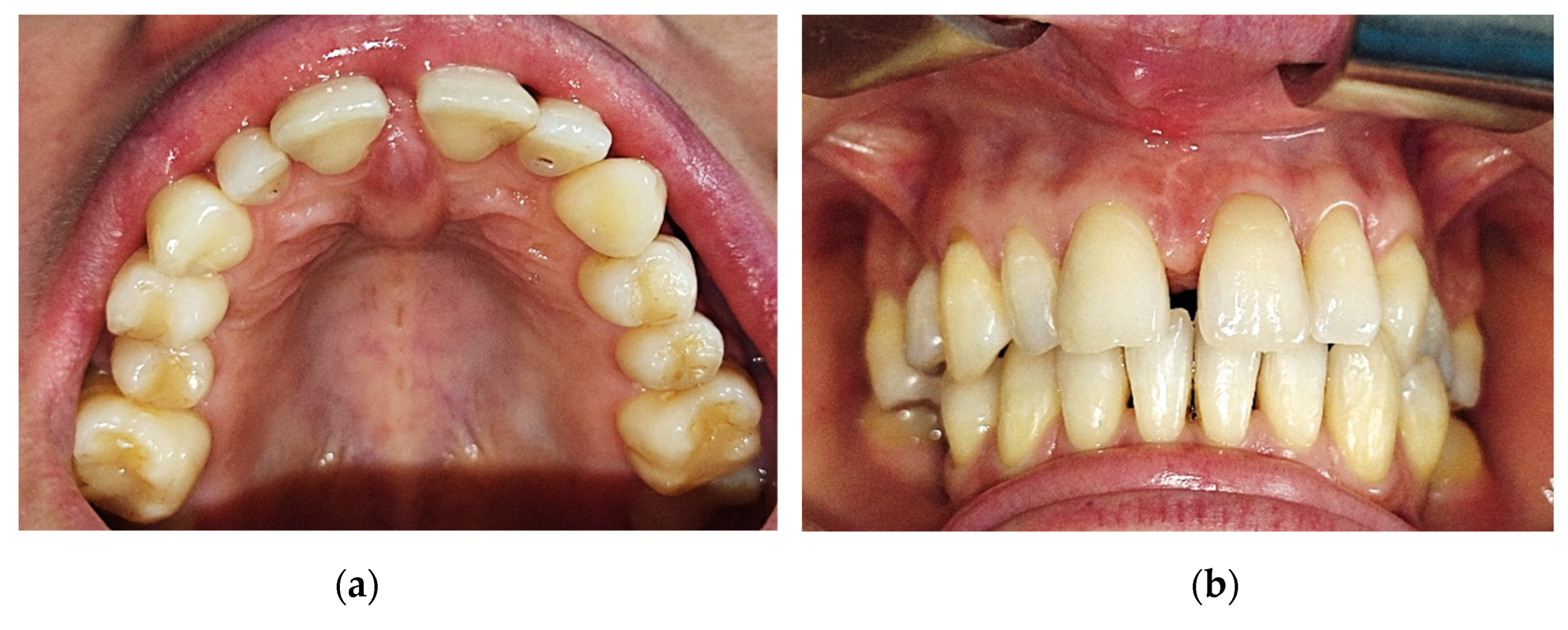
Figure 2.
Cone Beam Computed Tomography shows no underlying bone structure alteration.
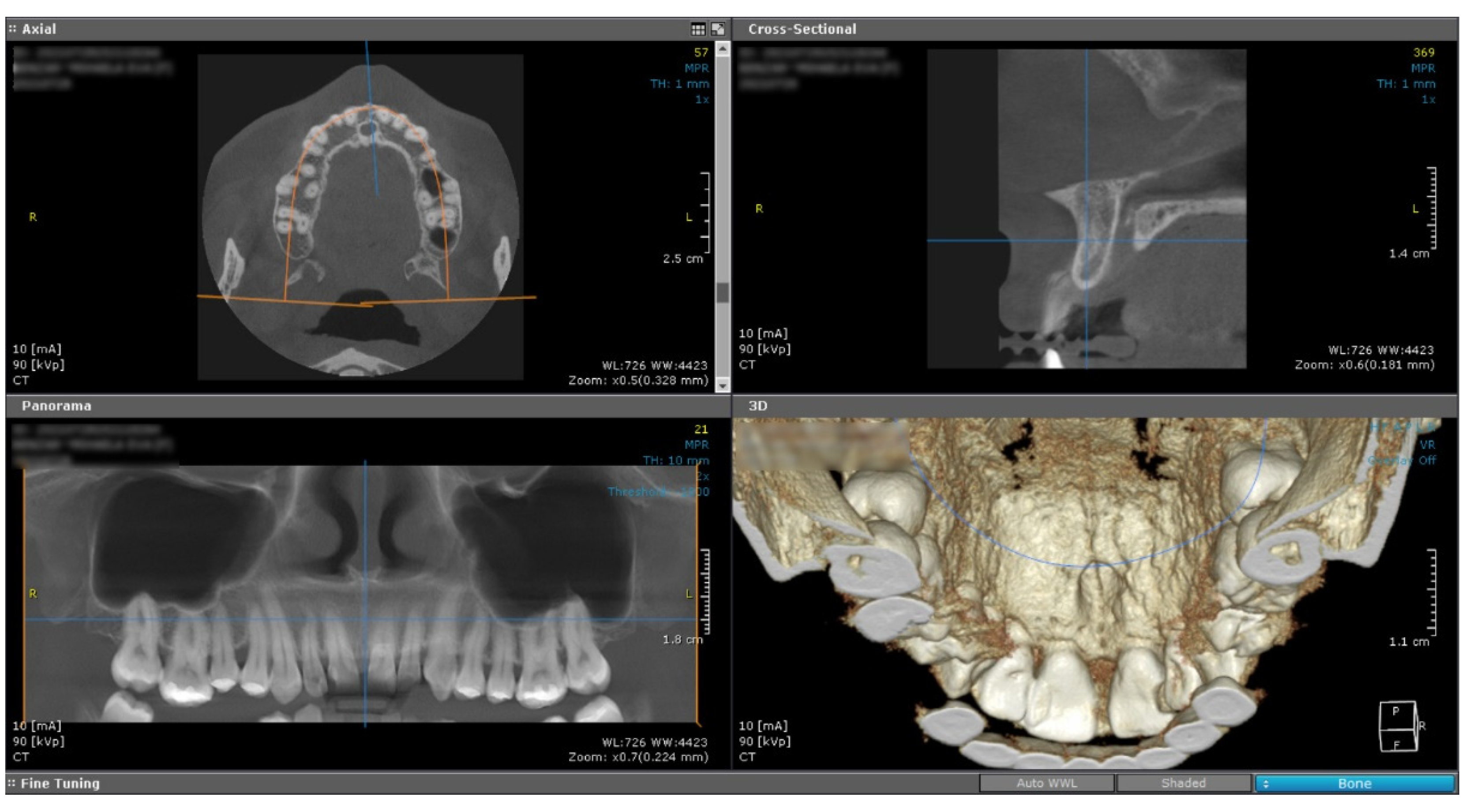
Figure 3.
Microscopic image using Hematoxylin–Eosin (HE) staining: (a) Tumor proliferation of the mucosa with spindle-shaped cells arranged in unequal bundles, HE staining, ob. 5x; (b) Fusiform tumor cells in a fasciculate pattern, with large, eosinophilic cytoplasm and elongated monotonous blunt-ended nuclei, HE staining, ob. 20x; (c) Fascicles of tumor cells arranged among acinar structures in the lamina propria of the mucosa, HE staining, ob. 5x.
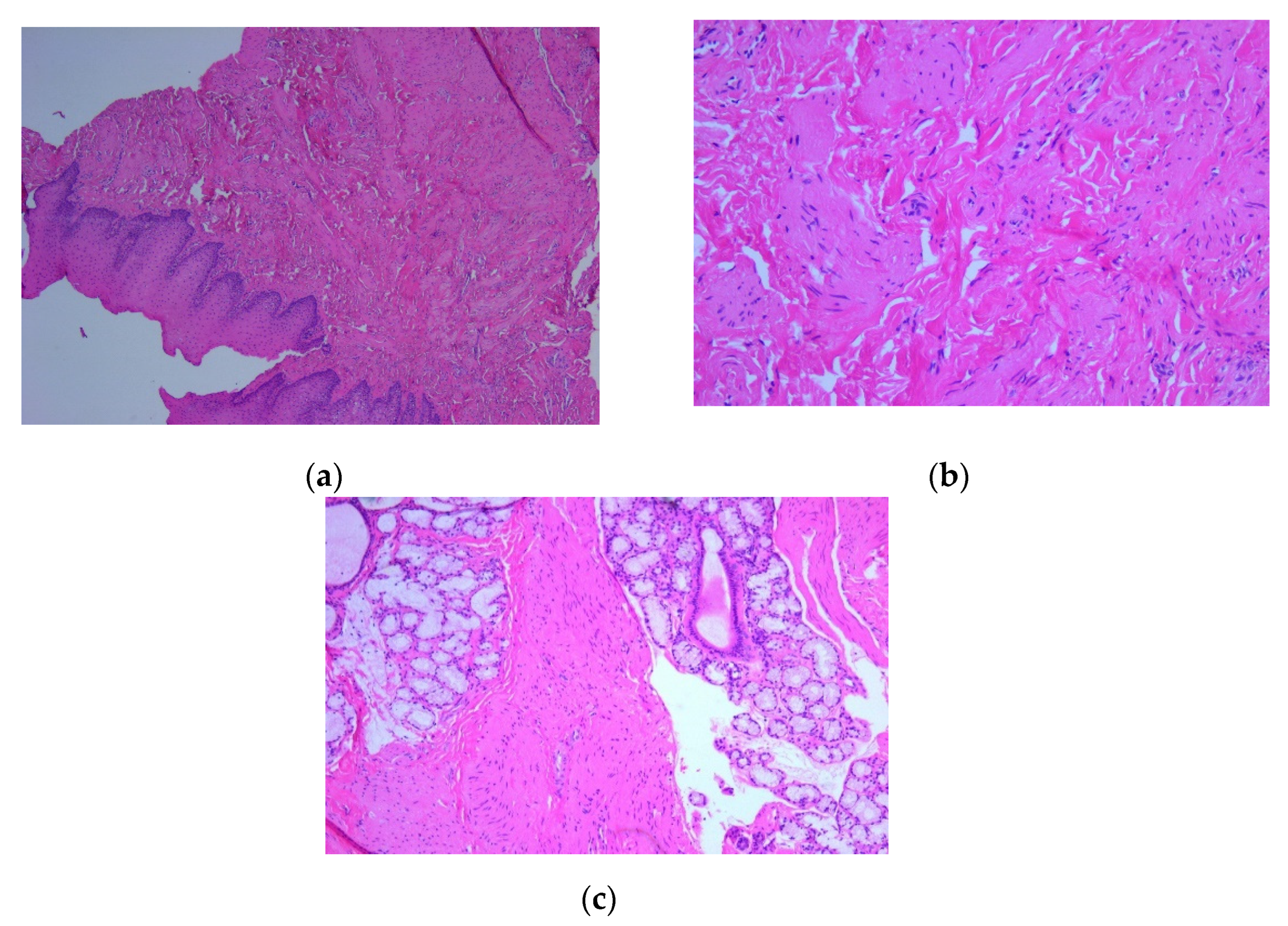
Figure 4.
Microscopic image using immunohistochemical reactions for desmin: (a) Tumor cells with diffuse and intense reaction IHC positive for desmin, Anti-Desmin Antibody, ob. 5x; (b) Intense and diffuse positive reaction for desmin, immunolabelling of the cytoplasm, Anti-Desmin Antibody, ob. 20x.
Figure 4.
Microscopic image using immunohistochemical reactions for desmin: (a) Tumor cells with diffuse and intense reaction IHC positive for desmin, Anti-Desmin Antibody, ob. 5x; (b) Intense and diffuse positive reaction for desmin, immunolabelling of the cytoplasm, Anti-Desmin Antibody, ob. 20x.
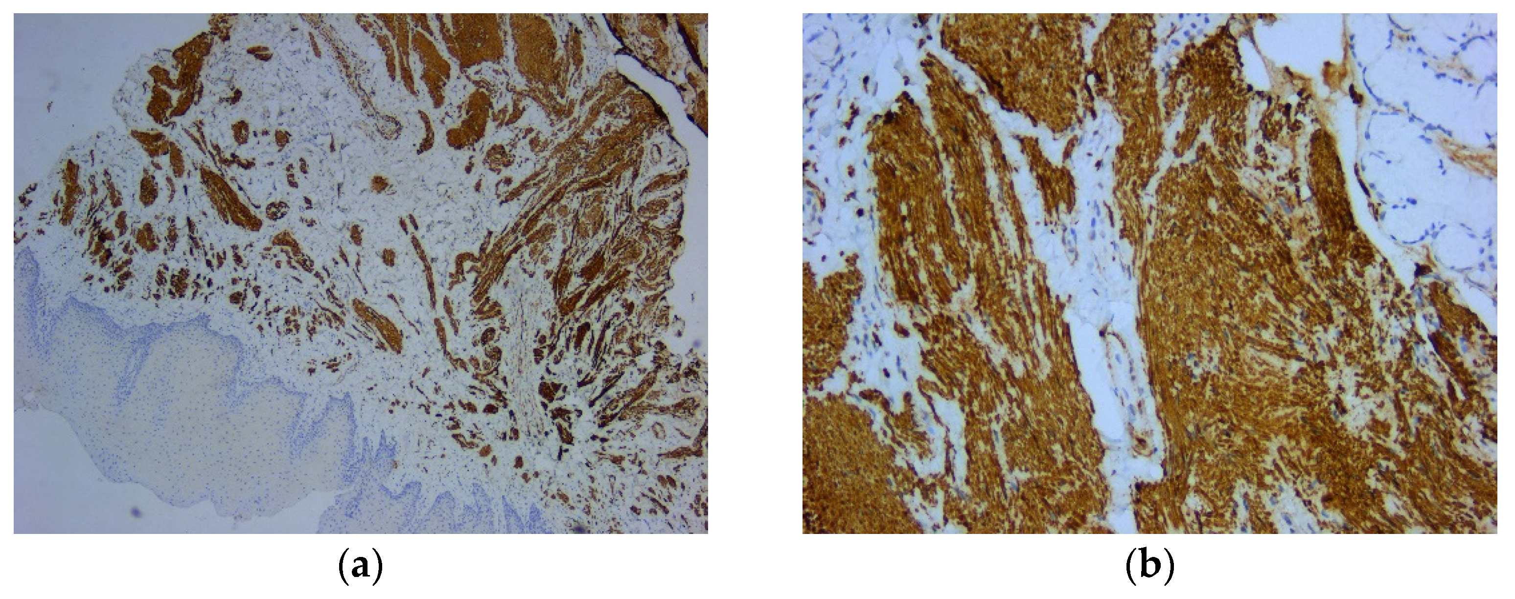
Figure 5.
Microscopic image using immunohistochemical reactions for smooth muscle actin: (a) Positive intense and diffuse reaction for smooth muscle actin, Anti-SMA Antibody, ob. 5x; (b) Positive intense and diffuse reaction for smooth muscle actin, immunolabelling of the cytoplasm, Anti-SMA Antibody, ob. 20x.
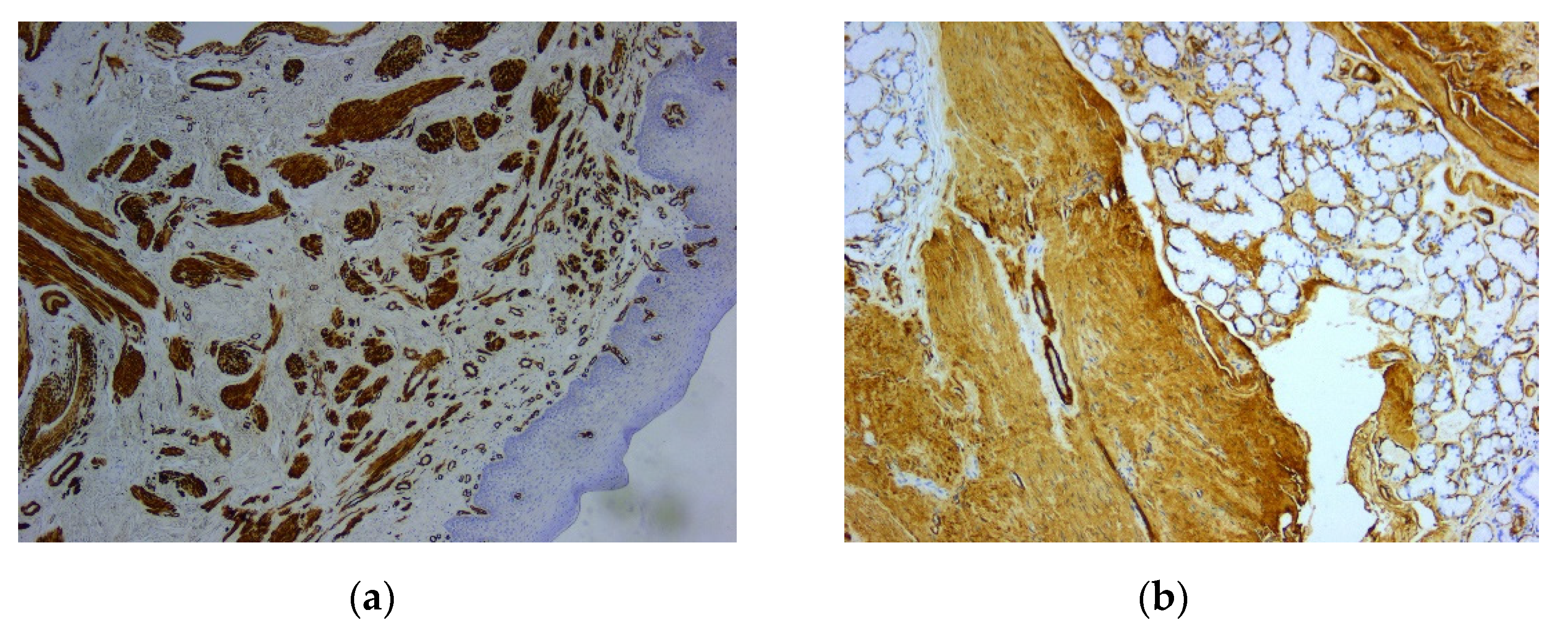
Figure 6.
Microscopic image using immunohistochemical reactions for vimentin: (a) Negative IHC reaction for vimentin, with positive control, Anti-Vimentin Antibody, ob. 5x; (b) Immunohistochemically negative tumor cells for vimentin, positive internal control at the level of the vascular component, Anti-Vimentin Antibody, ob. 20x.
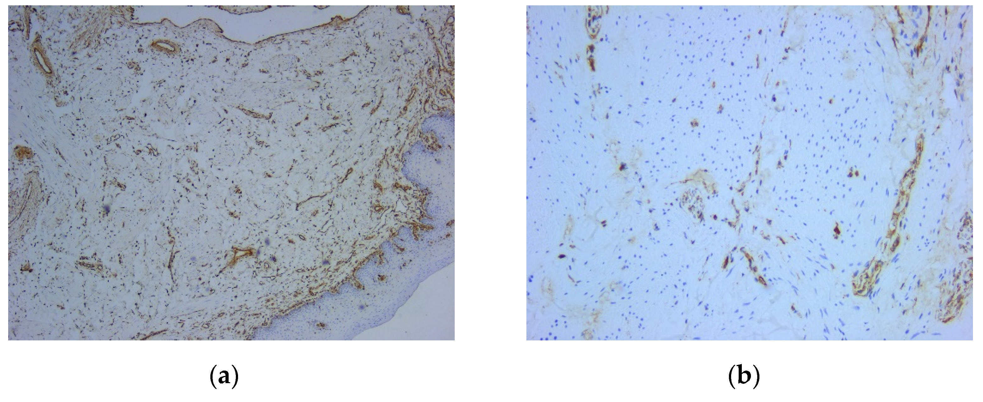
Figure 7.
Microscopic image using immunohistochemical reactions for Ki67: IHC reaction for Ki67 (positive internal control at the level of the basal layer of the covering epithelium), Anti-Ki-67 Antibody, ob. 5x.
Figure 7.
Microscopic image using immunohistochemical reactions for Ki67: IHC reaction for Ki67 (positive internal control at the level of the basal layer of the covering epithelium), Anti-Ki-67 Antibody, ob. 5x.
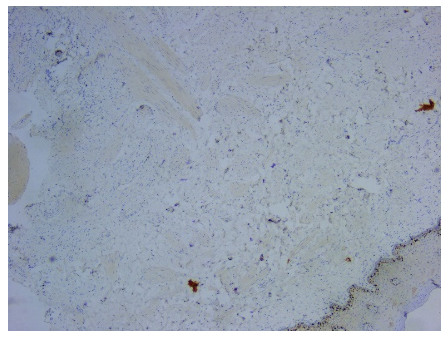

Table 1.
Data related to the antibodies used for immunohistochemical reactions.
| Antibody | Substrate | Dilution | Clone |
|---|---|---|---|
| Smooth Muscle Actin (SMA) | Monoclonal Mouse | Ready-To-Use | asm-1 |
| Desmin | Monoclonal Mouse | 1:200 for 30 minutes at 25°C | DE-R-11 |
| Vimentin | Monoclonal Mouse | 1:800 for 30 minutes at 25°C | V9 |
| Ki67 | Monoclonal Mouse | Ready-To-Use | MM1 |
Disclaimer/Publisher’s Note: The statements, opinions and data contained in all publications are solely those of the individual author(s) and contributor(s) and not of MDPI and/or the editor(s). MDPI and/or the editor(s) disclaim responsibility for any injury to people or property resulting from any ideas, methods, instructions or products referred to in the content. |
© 2023 by the authors. Licensee MDPI, Basel, Switzerland. This article is an open access article distributed under the terms and conditions of the Creative Commons Attribution (CC BY) license (http://creativecommons.org/licenses/by/4.0/).
Copyright: This open access article is published under a Creative Commons CC BY 4.0 license, which permit the free download, distribution, and reuse, provided that the author and preprint are cited in any reuse.


