Submitted:
14 June 2023
Posted:
14 June 2023
Read the latest preprint version here
Abstract
Autism Spectrum Disorder (ASD) is characterized by varying degrees of difficulty in social interaction and communication. These deficits are often associated with gastrointestinal symptoms, indicating alterations in both intestinal microbiota composition and metabolic activities. The intestinal microbiota influences the function and development of the nervous system. In individuals with ASD, there is an increase in bacterial genera such as Clostridium, as well as species involved in the synthesis of branched-chain amino acids (BCAA) like Prevotella copri. Conversely, decreased amounts of Akkermansia muciniphila, and Bifidobacterium spp. are observed. Epigallocatechin gallate (EGCG) is one of the polyphenols with the greatest beneficial activity on microbial growth and its consumption is associated with reduced psychological distress. Therefore, the objective of this review is to analyze how EGCG and its metabolites can improve the microbial dysbiosis present in ASD and its impact on the pathology. The analysis reveals that EGCG inhibits the growth of pathogenic bacteria like Clostridium perfringens and Clostridium difficile. Moreover, it increases the abundance of Bifidobacterium spp. and Akkermansia spp. As a result, EGCG demonstrates efficacy in increasing the production of metabolites involved in maintaining epithelial integrity and improving brain function. This identifies EGCG as highly promising for complementary treatment in ASD.
Keywords:
Autism Spectrum Disorder (ASD)
; microbiota
; epigallocatechin-3-gallate (EGCG)
; inflammation
; oxidative stress
1. Introduction
ASD is a highly heterogeneous and complex disorder, characterized by two major groups of core symptoms: persistent deficits in social communication and interaction, and restricted and repetitive patterns of behavior, interests, or activities [1,2,3,4,5]. Current statistics indicate that the disorder affects approximately 1 in every 160 children worldwide [6].
Characteristic deficits are often associated with a range of gastrointestinal symptoms such as abdominal pain, diarrhea, or constipation [7,8]. Several studies have found that both the composition of the intestinal microbiota and metabolic activities may be altered in individuals with the disorder [9,10]. Therefore, several authors concur that disruptions in the microbiota and intestinal microbiome, i.e., the collection of microorganisms present in the human gastrointestinal tract and their respective genomes [3,11], could trigger many of the gastrointestinal issues experienced by children with ASD, as well as exacerbate some of the core symptoms of the disorder [12,13].
Microbial colonization begins in infancy through the acquisition of maternal microbiota during vaginal delivery [3]. Subsequently, beneficial microorganisms feed on breast milk, which has a high content of oligosaccharides [14]. Similarly, the composition of the microbiota in the early years may be subject to alterations influenced by the delivery method, hygiene habits and feeding practices and routines. Among these factors, formula feeding has a particularly significant impact [3].
It has been shown that the intestinal microbiota affects the function and development of the immune, metabolic and nervous systems. Regarding the immune and metabolic systems, intestinal microbiota and its metabolites impact host physiology by regulating the function of the intestinal barrier, redox and mitochondrial metabolism, and mucosal inflammatory response through the regulation of intestinal lymphocytes that provide resistance against potential pathogens [15,16,17,18,19,20]. On the other hand, intestinal microbiota can influence neurochemistry, function, gene expression and the development of the central nervous system (CNS) through the gut-brain axis, which represents a bidirectional link between the cognitive and emotional functions of the CNS and peripheral intestinal function [15,21,22,23]. It is worth noting that the genus Bifidobacterium can metabolize gamma-aminobutyric acid (GABA), Lactobacillus spp. can metabolize acetylcholine, Bacillus spp. and Serratia spp., dopamine, and Escherichia spp. and Saccharomyces spp., noradrenaline. All these neurotransmitters are essential for the proper functioning of the nervous system as they can enter circulation and directly affect neural processes throughout the body, including the brain [3,24]. Additionally, intestinal proinflammatory cytokines can have an impact on the brain because they make the blood-brain barrier (BBB) more permeable, allowing peripheral immune cells to enter the brain and stimulating brain cells to produce additional proinflammatory mediators [25,26].
All of this has led to the hypothesis that the development of symptoms related to ASD may be influenced by the disturbance of the gut-brain-microbiota axis caused by changes in the intestinal microbiota.
2. Alterations in the intestinal microbiota in ASD
The Firmicutes (40-60%) and Bacteroidetes (20-40%) are the two major phyla of bacteria in the healthy human intestinal flora, followed by Proteobacteria, Actinomycetes, Clostridium spp., and Verruciformis [3,27]. However, in ASD, increased levels of bacterial genera such as Clostridium, Desulfovibrio and Ruminococcus have been observed [15,28], along with species that synthesize BCAA such as Bacteroides vulgatus and Prevotella copri. On the other hand, lower quantities of Bacteroides fragilis, Akkermansia muciniphila, Bifidobacterium spp. and Enterococcus spp. have been found [29,30]. Additionally, the ratio of the Escherichia/Shigella genera is altered, as Shigella spp. is present at higher levels, while Escherichia coli is decreased [31].
It should be noted that, despite the consensus regarding these alterations, it is important to consider that not all studies quantifying the microbiota in ASD analyze the same species or genera, leading to some heterogeneity in the results, and various studies emphasize the relationship between the composition of the human intestinal microbiota and the individual's diet, which can lead to variations in composition depending on the region where the analyses were conducted [32]. In fact, a study in the United States highlighted that children with ASD had lower levels of Bifidobacterium spp. and Prevotella spp., but higher levels of Lactobacillus spp., compared to healthy children [33]. Another study in China described that children with ASD had higher levels of the Actinobacteria and Proteobacteria phyla than the control group [34].
Furthermore, Ho et al. (2020) indicated that although the results of different studies are not entirely consistent, individuals with ASD generally exhibit a decrease in the percentage of the phylum Bacteroidetes or no significant difference compared to the control group of healthy individuals [35]. This could be due to the fact that within the phylum Bacteroidetes there are bacteria of the genus Bacteroides spp. that are increased, such as B. vulgatus, while others are decreased, such as B. fragilis, potentially leading to a compensatory effect that results in an unclear profile [30].
The genus Prevotella presents a more variable pattern. In the aforementioned review, the authors noted a lower relative abundance of Prevotella spp. in children diagnosed with ASD compared to the control group. However, multiple studies indicate an elevated prevalence of Prevotella spp. in children with ASD [15,30,31,35] and this variability could be due to dietary factors. Filippo et al. (2010) compared the composition of Prevotella spp. in the microbiota of children following European and African diets and observed a significant increase in Prevotella spp. in the microbiota of African children, likely due to their consumption of a grain-rich diet [36].
Finally, to establish a correlation between gastrointestinal issues and behavioral problems with the microbiota, the high growth rates of Clostridium histolyticum, Clostridium difficile, Clostridium perfringens and Sutterella spp., the alteration in the Escherichia/Shigella genera ratio, and the decreased Bacteroidetes/Firmicutes phylum ratio have been identified as factors associated with gastrointestinal problems. Furthermore, the relative abundance of Desulfovibrio spp., Clostridium spp. and Bacteroides vulgatus has been linked to behavioral disorders [37].
3. EGCG as an alternative for ASD
Currently, ASD lacks a medical cure, and a deeper understanding of molecular pathogenic mechanisms are necessary to propose therapeutic alternatives to current medications, which are primarily used for treating autism-related symptoms such as anxiety, hyperactivity, obsessive-compulsive behavior or gastrointestinal disorders [38]. Given the previously mentioned disruptions in the microbiota, it is crucial to focus current research on exploring the connection between the microbiota-gut-brain axis to modify the course of the disease. Polyphenols, as bioactive dietary compounds, emerge as a highly promising option. The interest in these molecules stems from their ability to act both in the intestine, where they reach the highest concentrations in the human body after oral administration, counteracting intestinal inflammation and modulating the intestinal microbiota and its metabolites, as well as in the brain, where they have neuroprotective effects upon penetrating the BBB [39].
3.1. Possible role of EGCG in the intestinal microbiota of patients with ASD
Compounds capable of targeting the intestine and modulating the intestinal microbiota, while reducing intestinal inflammation, have been shown to influence memory, cognition, mood and behavior, thereby contributing to the prevention and treatment of various brain disorders [25]. Among these compounds, polyphenols appear to be particularly relevant. Numerous authors have proposed their use in the treatment of neurological disorders due to their stimulating effect on specific intestinal bacteria that utilize and metabolize phenolic compounds [40,41].
Polyphenols are the most ubiquitous phytochemicals in the human diet, with a total daily intake of approximately 1 g [42]. These phenolic compounds, as secondary metabolites in plants, primarily protect them against various aggressions or infections caused by bacteria, insects or viruses [40,41]. These protective properties in plants are expected to be beneficial for humans as well. Polyphenolic compounds found in green and black tea are the most potent inhibitors of microbial growth. These compounds include epigallocatechin gallate (EGCG), epicatechin gallate, epigallocatechin, catechin gallate, epicatechin and catechin [43]. They have shown significant efficacy in inhibiting the growth of pathogens such as Helicobacter pylori [44], Staphylococcus aureus, Escherichia coli O157:H7 [45,46], Salmonella typhimurium DT104, Listeria monocytogenes, methicillin-resistant Staphylococcus aureus [47,48] and Pseudomonas aeruginosa [49]. They have also been effective against viruses like HIV and Epstein-Barr [50,51]. Among all the polyphenols, several studies highlighted by Duda-Chodak et al. (2015) emphasize the significant role of EGCG [52]. EGCG is the primary catechin in green tea, but it is also found in lower concentrations in certain plant-based foods, such as apples, peaches, kiwis, blackberries, pears and nuts like pistachios, hazelnuts and walnuts [53]. Its consumption is linked to reduced psychological distress [54], possibly by inhibiting the growth of specific pathogenic bacteria such as Clostridium perfringens, Clostridium difficile [55], Bacteroides ovatus, and enterobacteria (Salmonella spp., Escherichia coli, Yersinia pestis, Klebsiella spp., Shigella spp. and Eggerthella spp.) [46,56,57,58]. Furthermore, EGCG increases the abundance of Bifidobacterium spp. and bacteria of the Bacteroides genus (Bacteroides uniformis, Bacteroides stercoris, Bacteroides thetaiotaomicron and Bacteroides cellulosilyticus), as well as Prevotella spp., Lachnoclostridium spp. and Akkermansia spp. [56,57,58,59].
A substantial portion of the vital microorganisms present in the fecal microbiota of breastfed infants is comprised of species belonging to the Bifidobacterium genus [60]. Bifidobacterium spp. has psychobiotic effects that contribute to reduce anxiety (specifically, strains B. longum and B. breve), stress and other depressive behaviors [61,62]. This bacterial genus is among the first to colonize the intestines of newborns, and a dysbiosis of Bifidobacterium spp. may impact infant neurodevelopment [62]. In this context, the increase of Bifidobacterium spp. through the administration of EGCG correlates with an improvement in the intestinal microbiota. Previous studies conducted on animal models have demonstrated that a diet enriched with EGCG has the potential to elevate populations of Bifidobacterium spp. [63].
Bacteria within the Bacteroides genus play a crucial role in the breakdown of complex molecules, particularly carbohydrates, and confer advantages to their hosts by impeding the colonization of potential pathogens in the digestive tract [64,65]. It has been seen that there are bacteria capable of modulating the immune system. For instance, B. fragilis has been shown to restore the balance of T-cell populations in mice affected by ASD [66], while B. uniformis enhances the production of anti-inflammatory cytokines and improve metabolic and immune dysfunction [56]. Moreover, this genus can improve both social behaviors and physiological abnormalities in individuals affected by ASD [67], indicating that an increase in Bacteroides abundance caused by EGCG could enhance the quality of life in patients. Additionally, this polyphenol can reduce B. ovatus, the main intestinal commensal responsible for a systemic antibody response in inflammatory bowel disease [68]. These findings suggest that even within the same genus, EGCG may exert different effects on bacterial species.
Prevotella spp. are commensal bacteria in the human microbiota, and their function is to degrade plant polysaccharides and participate in the synthesis of vitamin B1 [69]. Within the Prevotella genus, Prevotella copri can be found. This species has been associated with improved glucose and insulin tolerance and is commonly found in individuals who follow a fiber-rich diet, suggesting a strong connection between the effects of this bacterium and dietary patterns [70,71,72,73]. Consequently, both the bacteria within the Prevotella genus and their metabolic activity may hold significant relevance to ASD, which will be further elaborated.
In relation to the Lachnoclostridium genus, it has been seen to possess anti-inflammatory properties and contribute to the maintenance of intestinal balance by producing butyric acid [74]. This metabolite aids in the elimination of intestinal gases and its absence is associated with irritable bowel syndrome (IBS) [75]. Therefore, the increase of Lachnoclostridium spp. mediated by EGCG can be seen as a positive impact on the microbiota of ASD patients.
EGCG modulates the composition of the intestinal microbiota and its metabolites, enriching the population of short-chain fatty acid (SCFA)-producing bacteria such as Akkermansia spp. This leads to an increased production of these metabolites. [76,77]. Therefore, EGCG promotes an anti-inflammatory and antioxidant state in the intestine. [76]. Specifically, Akkermansia spp. utilizes intestinal epithelial mucin as a source of energy. By degrading mucin, it releases nutrients such as monosaccharides, amino acids and SCFAs, which are used by other bacteria in the microbiota, stimulating their metabolic functions [78]. Akkermansia spp. is involved in the regulation of glucose metabolism and adipose tissue homeostasis, therefore, the fact that EGCG increases the levels of Akkermansia muciniphila improves intestinal dysbiosis and enhances barrier integrity [58,79,80,81].
Among the bacteria reduced by EGCG, Clostridia stand out as a group of Gram-positive bacilli that, when in excess, can lead to infection in the large intestine [28]. Furthermore, butyrate or butyric acid, along with other SCFAs derived from the fermentation of dietary fiber, are noteworthy compounds produced by Clostridia [82]. Butyrate is an essential metabolite in the human colon as it is the preferred energy source for colonic epithelial cells. It contributes to maintaining intestinal barrier functions and has immunomodulatory and anti-inflammatory properties [83]. However, it promotes intestinal permeability, which may allow the passage of toxic substances into the bloodstream, potentially leading to inflammation [82]. In individuals with ASD, both the absolute and relative abundance of these bacteria are increased. Thus, the inhibitory capacity of EGCG on this group of bacteria, which can decrease the levels of Clostridium spp., as seen in rats, is beneficial [63]. This activity may be particularly relevant for Clostridium perfringens, as its impact on the symptomatology of the disease, including gastrointestinal issues, is significant. Both the bacterium itself and the gene that produces its toxin (CPB2) have been associated with gastrointestinal complications in ASD and are correlated with disease severity. Moreover, the isolated species from children with ASD show greater antibiotic resistance compared to healthy children [84]. On the other hand, the increase of Clostridium difficile also has a notably negative impact on ASD, and effective treatments against this bacterium using oral vancomycin have shown improvements in behavior and communication [85,86]. Therefore, the reduction of this strain mediated by EGCG could be beneficial for the clinical presentation of children affected by ASD.
Another group that decreases after treatment with EGCG is enterobacteria. Their presence in the body is common, but uncontrolled growth of these bacteria can lead to infections. Certain species have been linked to specific pathologies. Specifically, Salmonella typhi is responsible for typhoid fever, Shigella dysenteriae is the causative agent of bacillary dysentery and causes infantile gastroenteritis, and Yersinia pestis causes plague [87].
Lastly, it is important to highlight at this stage that specific bacteria increased by EGCG, such as Lactobacillus acidophilus, Bifidobacterium longum, Akkermansia muciniphila and Prevotella ruminicola, restore the balance of the intestinal microbiota when added to the diet, thereby reducing oxidative stress in the intestine and the brain [88].
3.2. Outlook on the anti-inflammatory and antioxidant activity of EGCG in autism. Neuroprotective role
Dysregulation of the immune system leading to inflammation and increased oxidative stress has been recognized as a significant contributing factor to ASD [89], which is directly linked to dysbiosis. The presence of multiple comorbid inflammatory conditions associated with immune dysregulation is closely related to the emergence and progression of the key clinical features observed in children with ASD [90,91,92]. Cytokines constitute a group of small proteins that act as immunomodulatory and endocrine messengers involved in the regulation of inflammation and neurological development [93,94]. Specifically, there is substantial evidence of altered cytokine levels in children with ASD, which are not observed in typically developing children [95]. Particularly noteworthy are the alterations identified in the levels of interferon-α (IFN-α), interleukin-7 (IL-7), IFN-γ-inducible protein-10 and IL-8, with the latter appearing to be particularly relevant to the pathogenesis of the disorder [96]. Additionally, levels of IL-1β and IL-10 produced by peripheral blood monocytes, along with the IL-1β/IL-10 ratio and changes in miRNA expression, seem to be relevant in relation to innate immunity [97,98]. All these alterations are closely associated with social behavioral impairments and characteristic cognitive development in children with ASD [99].
Regarding oxidative stress, it appears to have a direct link to the intestinal microbiota, especially in children with ASD. The microbial fermentation of dietary fibers and resistant starch in the intestinal tract leads to the generation of SCFAs [100]. Notably, propionate has been associated with metabolic disturbances in the gastrointestinal system and neuroinflammation, which may elucidate the higher prevalence of gastrointestinal issues, including dysbiosis, observed in children with ASD [101]. Consequently, the response to oxidative stress, closely intertwined with dysbiosis, plays a crucial role in the neuroinflammatory response, thus representing a significant pathogenic factor in the development of ASD [102,103,104,105,106]. It has been observed that there are evident differences in antioxidant capacity and the concentration of various oxidative stress-related metabolites between children with autism and healthy children [107]. Autistic children are more susceptible to oxidative stress, primarily due to an imbalance in intracellular and extracellular glutathione (GSH) levels and a decrease in glutathione storage capacity [108]. Moreover, this increased production of free radicals has been linked to the activity of amyloid precursor proteins (APP) in various human disorders, including neurodevelopmental disorders, with autism being a prominent example [109,110,111].
At the cerebral level, polyphenols promote neurogenesis and plasticity, elucidating their role as antioxidants that have demonstrated their potential in alleviating autistic symptoms. Additionally, they can reduce intestinal inflammation, leading to a significant decrease in inflammatory cascades such as nuclear factor kappa B (NF-kB), activator protein-1 (AP-1) and Janus kinase-signal transducer and activator of transcription (JAK/STAT). Furthermore, they modulate inflammatory markers including nitric oxide (NO), tumor necrosis factor-alpha (TNF-α), ionized calcium-binding adapter molecule 1 (Iba1), prostaglandin E2 (PGE2), inducible nitric oxide synthase (iNOS) and cyclooxygenase-2 (COX-2). Moreover, polyphenols mitigate the production of reactive oxygen species (ROS) and regulate the synthesis of SCFAs. Consequently, they facilitate bidirectional communication between the gut and the brain [39]. All this evidence supports a recent study where rat pups with autistic traits showed a reversal of autistic behaviors following treatment with a polyphenol-probiotic complex, which was accompanied by modulation of the biochemical alterations in IL-6, TNF-α, BDNF, 5-HT, AchE, and the granular layer [112].
Therefore, when analyzing the activity of various polyphenols in inflammation and oxidative stress, particularly EGCG has demonstrated high efficacy in regulating APP levels [113]. This could partly explain its significant effectiveness in mitigating inflammation and oxidative stress. One contributing factor is its ability to cross the BBB, which accounts for its remarkable efficacy in protecting cultured cortical neurons against oxidative stress-induced cell death. Specifically, the cerebral accessibility of polyphenols such as quercetin, cyanidin-3-glucoside (C3G), and EGCG in relation to their neuroprotective capacity was evaluated. Primary cortical neuron cultures were exposed to oxidative stress in the absence and presence of the selected compounds, and both neuroprotection and cerebral accessibility through the BBB were assessed. It was seen that the human BBB model was rapidly crossed by EGCG, followed by slower permeation by C3G, while quercetin did not cross the BBB in any case. Hence, among the different polyphenols, EGCG stands out as a particularly promising neuroprotector [114].
Regarding the cytokines most involved in ASD, as already mentioned, the role of IL-8 possibly stands out. It has been seen that the administration of EGC reduces its levels through the inhibition of intracellular Ca2+ levels and the activation of ERK1/2 and NF-kappaB pathways Furthermore, in the colon, EGCG is capable of inhibiting the production of IL-8 cytokines by regulating genes involved in inflammatory pathways. This suggests that EGCG could be useful for the prevention and/or mitigation of colonic disorders present in autism [115].
Finally, it should be noted that elevated levels of reactive oxygen species (ROS), specifically hydrogen peroxide (H2 O2) and superoxide (O2-), are reported in the autistic intestine as a cause of damage to the epithelial tissue [116,117,118], and EGCG negatively regulates the inflammatory response in inflamed intestinal epithelial cells, largely through a post-transcriptional regulatory mechanism [119].
3.3. Role of EGCG in the metabolic activity associated with dysbiosis in ASD
Regarding the metabolites that can be modified by the action of EGCG, it should be mentioned that this polyphenol has beneficial effects by regulating the production of SCFAs [39]. This fact may be particularly relevant given the prevalent role of these metabolites in the bidirectional communication between the gut and the brain. Agarwala et al. (2021) highlight the significance of succinate and butyrate, which are particularly important and found to be altered in individuals with autism due to microbial activity [120]. Concerning succinate, it is known that an imbalanced ratio of succinate production/consumption can lead to intestinal disturbances, which can affect the gut-brain axis to varying degrees [121]. Altered levels of succinate have been observed to result in disrupted calcium homeostasis and dysregulated metabolic functions in autistic individuals [38].
Regarding butyrate, it has been seen that children with ASD have lower levels of butyrate, as well as a decrease in the abundance of taxa producing this metabolite [122]. Butyrate is particularly relevant in ASD as its production mainly derives from the gut microbiome. Butyrate displays potent anti-inflammatory activity that contributes to gut health, regulates intestinal homeostasis and BBB permeability, and modulates the expression of neurotransmitter genes [123,124]. Furthermore, it positively modulates the impaired mitochondrial function in ASD, characterized by reduced activity of complex IV in the electron transport chain (ETC) [125], including improvements in oxidative phosphorylation and beta-oxidation. These observations have underscored the significant neuroprotective role of butyrate in children with ASD [126]. In fact, it has recently been seen that maternal treatment with butyrate in the BTBR mouse model of ASD rescues social and partially repetitive behavior deficits in the offspring [127]. Thus, proper butyrate production at adequate levels is crucial, and this depends on the correct development of the gut bacterial community. Oral treatment with EGCG has been shown to increase both the abundance of Akkermansia muciniphila and its butyrate production [76], potentially reversing the adverse effects resulting from altered butyrate levels in ASD.
On the other hand, among the altered fecal metabolite profiles in children with ASD (not present in neurotypical children), there is an increase in p-cresol, caprate, aspartate, and a reduction in GABA, nicotinate, glutamine, and thymine [128]. Among all these metabolites, p-cresol is a uremic toxin produced by certain strains of Clostridium spp., which has negative biological effects and appears to adversely affect the homeostasis of colonic epithelial cells in children with ASD. When present in excess, p-cresol induces DNA damage in vitro and negatively affects the integrity of colonic epithelial cells [129]. In fact, EGCG may have beneficial effects by reversing the activity of this metabolite. In a recent study, it has been seen that an EGCG-enriched diet reduces plasma and urinary concentrations of p-cresol in mice by suppressing the abundance of Firmicutes at the phylum level and Clostridia at the order level [130].
In conclusion, EGCG has an impact on both the intestinal microbiota and variables directly related to the microbiota such as metabolites, inflammation and oxidative stress, which could provide benefits for patients with ASD (Figure 1).
It is also interesting to highlight the activity of metabolites that are derived from the polyphenol after its interaction with the intestinal microbiota. Specifically, in rats, the metabolic pathway of EGCG leads to gallic acid (GA) and epigallocatechin (EGC) [131]. EGC is degraded by colonic bacteria to yield microbial ring-fission metabolites, specifically converting to 5-(3',5'-dihydroxyphenyl)-γ-valerolactone (EGC-M5). Then, the bacteria Adlercreutzia equolifaciens MT4s-5 and Flavonifractor plautii MT42 are capable of degrading EGC. The bacterium Adlercreutzia equolifaciens MT4s-5 catalyzes the conversion of EGC to 1-(3,4,5-trihydroxyphenyl)-3-(2,4,6-trihydroxyphenyl)propan-2-ol (EGC-M1), and subsequently, Flavonifractor plautii MT42 converts the propan-2-ol to 5-(3,4,5-trihydroxyphenyl)-γ-valerolactone (EGC-M5) and 4-hydroxy-5-(3,4,5-trihydroxyphenyl)valeric acid (EGC-M4). Similarly, EGC-M5 is produced from 1-(3,5-dihydroxyphenyl)-3-(2,4,6-trihydroxyphenyl)propan-2-ol (EGC-M3), which is formed from EGC by Adlercreutzia equolifaciens MT4s-5 in the presence of hydrogen [120]. A large portion of the formed EGC-M5 is absorbed and undergoes glucuronidation in the intestinal mucosa and/or liver to form EGC-M5 glucuronide (EGC-M5-GlcUA), which is distributed to various tissues through the bloodstream and ultimately excreted in urine [132] (Figure 2).
The microbial ring-fission metabolites of EGCG are found in plasma in both free and conjugated forms [132], and in vitro studies have shown that they may reach the brain parenchyma through the BBB, promoting neuritogenesis and potentially exerting a relevant activity against the degenerative processes of neurodegenerative diseases. Specifically, when assessing the penetration capacity of these metabolites through the BBB, it has been seen that GA has higher permeability than EGCG and EGC, possibly due to its smaller molecular size [133]. Moreover, comparing only the ring-fission metabolites derived from ECG, EGC-M5 demonstrated greater permeability than its conjugate EGC-M5-GlcUA [131]. Some of these ring-fission metabolites also exhibit anti-inflammatory activity [134]. In particular, EGC-M5 has been found to have immunomodulatory properties, as it enhances the activity of CD4+ T cells and the cytotoxic activity of natural killer cells in BALB/c mice [135]. Therefore, it seems evident that the ring-fission metabolites derived from the intestinal microflora of catechins demonstrate a protective capacity against various diseases, including neurodegenerative disorders.
However, it is crucial to understand the metabolic process and bioavailability of green tea catechins and EGCG, particularly in assessing their biological activity and comprehending their beneficial effects on human health. EGCG presents significantly lower bioavailability compared to other components of catechins [136,137]. Therefore, when assessing the activity of EGCG and its metabolites, it is essential to emphasize that their applications are greatly limited due to their low solubility, bioavailability and stability. The use of liposomal formulations may serve as an effective strategy for their administration in autism. Liposomal delivery aims to improve the poor stability of polyphenols against temperature, light, pH and oxygen [28,29], as well as their low permeability across intestinal membranes, which results in only a small proportion of these compounds remaining available for absorption in the human body after ingestion [29,30]. Both stability and oral bioavailability are enhanced through liposomal encapsulation, as it provides protection against degradation when passing through the gastrointestinal tract [31]. Moreover, nanotechnology can promote controlled release of the polyphenol and modulating the interaction between polyphenols and the microbiota, which also represents an intriguing approach [31], and it has been seen that EGCG has significantly improved stability when formulated as dual-drug loaded PEGylated PLGA nanoparticles (EGCG/AA NPs). Following oral administration in mice, EGCG accumulated in all major organs, including the brain, leading to increased synapse formation and reduction of neuroinflammation in Alzheimer's disease [138]. Furthermore, concerning its metabolites and their administration using nanotechnology, Abbasalipour H. et al. (2022) studied the neuroprotective effects of gallic acid on oxidative stress-induced cognitive impairment and the expression of the Nrf2/Keap1 gene in an autism model. Gallic acid-loaded nanophytosomes (GNP) were administered, and the results revealed improvements in learning and memory deficits by reducing oxidative stress, enhancing antioxidant enzyme activity and modulating the Keap1/Nrf2 gene expression [139].
4. Conclusions
ASD is a complex disorder characterized by inflammatory processes, high oxidative stress and gastrointestinal disturbances. These issues may be attributed to changes in the intestinal microbiome, resulting in microbial dysbiosis in children with ASD. Polyphenols have demonstrated sufficient antioxidant and anti-inflammatory capacity to counteract ASD symptoms. Their role in dysbiosis has also been studied, revealing their ability to regulate and improve the percentages of beneficial microbial species, thereby enhancing the pathology of ASD and promoting gut-brain axis function, consequently improving brain function. In this regard, EGCG appears particularly effective in improving microbial species and increasing the production of metabolites involved in maintaining epithelial integrity. It also demonstrates effectiveness in reducing inflammation directly linked to ASD by influencing specific pro-inflammatory cytokine levels and reducing oxidative stress. Furthermore, it can modulate the altered metabolic production in ASD by decreasing toxic metabolites such as p-cresol. All of these factors have an impact on brain function, which could provide significant benefits for children with ASD. However, it should be noted that these polyphenols have limitations in terms of intestinal assimilation and bioavailability. Hence, when considering the advantages of nanotechnology in overcoming these limitations, it is advisable to administer them in liposomal form as a complementary treatment for ASD.
Consequently, this review identifies EGCG liposomes as a potential adjunctive therapy for children with ASD, aiming to improve their quality of life and alleviate their symptoms.
Author Contributions
Conceptualization, J.E.d.l.R.O. and A.B.C.-S.; Writing—Original Draft Preparation, J.E.d.l.R.O., A.B.C.-S, M.B., B.P.; and C.E.S.-S.; C.M.; P.S.-B.; G.C.; R.B.-B. and M.M. writing—review and editing, J.E.d.l.R.O., A.B.C.-S and M.B. All authors have read and agreed to the published version of the manuscript.
Funding
This research received no external funding, but has been supported by the Foundation of Catholic University of Valencia San Vicente Mártir.
Data Availability Statement
Not applicable due to data privacy.
Conflicts of Interest
The authors declare no conflict of interest.
References
- Taylor, M. J.; Rosenqvist, M. A.; Larsson, H.; Gillberg, C.; D’Onofrio, B. M.; Lichtenstein, P.; Lundström, S. Etiology of Autism Spectrum Disorders and Autistic Traits Over Time. JAMA Psychiatry 2020, 77 (9), 936–943. https://doi.org/10.1001/JAMAPSYCHIATRY.2020.0680. [CrossRef]
- American Psychiatric Association. Diagnostic and Statistical Manual of Mental Disorders. Am. Psychiatr. Assoc. 2013.
- Hughes, H. K.; Rose, D.; Ashwood, P. The Gut Microbiota and Dysbiosis in Autism Spectrum Disorders. Curr. Neurol. Neurosci. Reports 2018 1811 2018, 18 (11), 1–15. https://doi.org/10.1007/S11910-018-0887-6. [CrossRef]
- Baio, J.; Wiggins, L.; Christensen, D. L.; Maenner, M. J.; Daniels, J.; Warren, Z.; Kurzius-Spencer, M.; Zahorodny, W.; Rosenberg, C. R.; White, T.; Durkin, M. S.; Imm, P.; Nikolaou, L.; Yeargin-Allsopp, M.; Lee, L. C.; Harrington, R.; Lopez, M.; Fitzgerald, R. T.; Hewitt, A.; Pettygrove, S.; Constantino, J. N.; Vehorn, A.; Shenouda, J.; Hall-Lande, J.; Naarden Braun, K. Van; Dowling, N. F. Prevalence of Autism Spectrum Disorder among Children Aged 8 Years - Autism and Developmental Disabilities Monitoring Network, 11 Sites, United States, 2014. MMWR Surveill. Summ. 2018, 67 (6), 1–23. https://doi.org/10.15585/MMWR.SS6706A1. [CrossRef]
- Grimaldi, R.; Gibson, G. R.; Vulevic, J.; Giallourou, N.; Castro-Mejía, J. L.; Hansen, L. H.; Leigh Gibson, E.; Nielsen, D. S.; Costabile, A. A Prebiotic Intervention Study in Children with Autism Spectrum Disorders (ASDs). Microbiome 2018, 6 (1), 1–13. https://doi.org/10.1186/S40168-018-0523-3/TABLES/2. [CrossRef]
- World Health Organization. Autism. https://www.who.int/news-room/fact-sheets/detail/autism-spectrum-disorders (accessed 2023-02-26).
- Li, Q.; Zhou, J. M. The Microbiota–Gut–Brain Axis and Its Potential Therapeutic Role in Autism Spectrum Disorder. Neuroscience 2016, 324, 131–139. https://doi.org/10.1016/J.NEUROSCIENCE.2016.03.013. [CrossRef]
- Yang, J.; Fu, X.; Liao, X.; Li, Y. Effects of Gut Microbial-Based Treatments on Gut Microbiota, Behavioral Symptoms, and Gastrointestinal Symptoms in Children with Autism Spectrum Disorder: A Systematic Review. Psychiatry Res. 2020, 293. https://doi.org/10.1016/J.PSYCHRES.2020.113471. [CrossRef]
- de Theije, C. G. M.; Wopereis, H.; Ramadan, M.; van Eijndthoven, T.; Lambert, J.; Knol, J.; Garssen, J.; Kraneveld, A. D.; Oozeer, R. Altered Gut Microbiota and Activity in a Murine Model of Autism Spectrum Disorders. Brain. Behav. Immun. 2014, 37, 197–206. https://doi.org/10.1016/J.BBI.2013.12.005. [CrossRef]
- Strati, F.; Cavalieri, D.; Albanese, D.; De Felice, C.; Donati, C.; Hayek, J.; Jousson, O.; Leoncini, S.; Renzi, D.; Calabrò, A.; De Filippo, C. New Evidences on the Altered Gut Microbiota in Autism Spectrum Disorders. Microbiome 2017, 5 (1), 1–11. https://doi.org/10.1186/S40168-017-0242-1/FIGURES/5. [CrossRef]
- Huttenhower, C.; Gevers, D.; Knight, R.; Abubucker, S.; Badger, J. H.; Chinwalla, A. T.; Creasy, H. H.; Earl, A. M.; Fitzgerald, M. G.; Fulton, R. S.; Giglio, M. G.; Hallsworth-Pepin, K.; Lobos, E. A.; Madupu, R.; Magrini, V.; Martin, J. C.; Mitreva, M.; Muzny, D. M.; Sodergren, E. J.; Versalovic, J.; Wollam, A. M.; Worley, K. C.; Wortman, J. R.; Young, S. K.; Zeng, Q.; Aagaard, K. M.; Abolude, O. O.; Allen-Vercoe, E.; Alm, E. J.; Alvarado, L.; Andersen, G. L.; Anderson, S.; Appelbaum, E.; Arachchi, H. M.; Armitage, G.; Arze, C. A.; Ayvaz, T.; Baker, C. C.; Begg, L.; Belachew, T.; Bhonagiri, V.; Bihan, M.; Blaser, M. J.; Bloom, T.; Bonazzi, V.; Paul Brooks, J.; Buck, G. A.; Buhay, C. J.; Busam, D. A.; Campbell, J. L.; Canon, S. R.; Cantarel, B. L.; Chain, P. S. G.; Chen, I. M. A.; Chen, L.; Chhibba, S.; Chu, K.; Ciulla, D. M.; Clemente, J. C.; Clifton, S. W.; Conlan, S.; Crabtree, J.; Cutting, M. A.; Davidovics, N. J.; Davis, C. C.; Desantis, T. Z.; Deal, C.; Delehaunty, K. D.; Dewhirst, F. E.; Deych, E.; Ding, Y.; Dooling, D. J.; Dugan, S. P.; Michael Dunne, W.; Scott Durkin, A.; Edgar, R. C.; Erlich, R. L.; Farmer, C. N.; Farrell, R. M.; Faust, K.; Feldgarden, M.; Felix, V. M.; Fisher, S.; Fodor, A. A.; Forney, L. J.; Foster, L.; Di Francesco, V.; Friedman, J.; Friedrich, D. C.; Fronick, C. C.; Fulton, L. L.; Gao, H.; Garcia, N.; Giannoukos, G.; Giblin, C.; Giovanni, M. Y.; Goldberg, J. M.; Goll, J.; Gonzalez, A.; Griggs, A.; Gujja, S.; Kinder Haake, S.; Haas, B. J.; Hamilton, H. A.; Harris, E. L.; Hepburn, T. A.; Herter, B.; Hoffmann, D. E.; Holder, M. E.; Howarth, C.; Huang, K. H.; Huse, S. M.; Izard, J.; Jansson, J. K.; Jiang, H.; Jordan, C.; Joshi, V.; Katancik, J. A.; Keitel, W. A.; Kelley, S. T.; Kells, C.; King, N. B.; Knights, D.; Kong, H. H.; Koren, O.; Koren, S.; Kota, K. C.; Kovar, C. L.; Kyrpides, N. C.; La Rosa, P. S.; Lee, S. L.; Lemon, K. P.; Lennon, N.; Lewis, C. M.; Lewis, L.; Ley, R. E.; Li, K.; Liolios, K.; Liu, B.; Liu, Y.; Lo, C. C.; Lozupone, C. A.; Dwayne Lunsford, R.; Madden, T.; Mahurkar, A. A.; Mannon, P. J.; Mardis, E. R.; Markowitz, V. M.; Mavromatis, K.; McCorrison, J. M.; McDonald, D.; McEwen, J.; McGuire, A. L.; McInnes, P.; Mehta, T.; Mihindukulasuriya, K. A.; Miller, J. R.; Minx, P. J.; Newsham, I.; Nusbaum, C.; Oglaughlin, M.; Orvis, J.; Pagani, I.; Palaniappan, K.; Patel, S. M.; Pearson, M.; Peterson, J.; Podar, M.; Pohl, C.; Pollard, K. S.; Pop, M.; Priest, M. E.; Proctor, L. M.; Qin, X.; Raes, J.; Ravel, J.; Reid, J. G.; Rho, M.; Rhodes, R.; Riehle, K. P.; Rivera, M. C.; Rodriguez-Mueller, B.; Rogers, Y. H.; Ross, M. C.; Russ, C.; Sanka, R. K.; Sankar, P.; Fah Sathirapongsasuti, J.; Schloss, J. A.; Schloss, P. D.; Schmidt, T. M.; Scholz, M.; Schriml, L.; Schubert, A. M.; Segata, N.; Segre, J. A.; Shannon, W. D.; Sharp, R. R.; Sharpton, T. J.; Shenoy, N.; Sheth, N. U.; Simone, G. A.; Singh, I.; Smillie, C. S.; Sobel, J. D.; Sommer, D. D.; Spicer, P.; Sutton, G. G.; Sykes, S. M.; Tabbaa, D. G.; Thiagarajan, M.; Tomlinson, C. M.; Torralba, M.; Treangen, T. J.; Truty, R. M.; Vishnivetskaya, T. A.; Walker, J.; Wang, L.; Wang, Z.; Ward, D. V.; Warren, W.; Watson, M. A.; Wellington, C.; Wetterstrand, K. A.; White, J. R.; Wilczek-Boney, K.; Wu, Y.; Wylie, K. M.; Wylie, T.; Yandava, C.; Ye, L.; Ye, Y.; Yooseph, S.; Youmans, B. P.; Zhang, L.; Zhou, Y.; Zhu, Y.; Zoloth, L.; Zucker, J. D.; Birren, B. W.; Gibbs, R. A.; Highlander, S. K.; Methé, B. A.; Nelson, K. E.; Petrosino, J. F.; Weinstock, G. M.; Wilson, R. K.; White, O. Structure, Function and Diversity of the Healthy Human Microbiome. Nature 2012, 486 (7402), 207–214. https://doi.org/10.1038/NATURE11234. [CrossRef]
- Rinninella, E.; Raoul, P.; Cintoni, M.; Franceschi, F.; Miggiano, G. A. D.; Gasbarrini, A.; Mele, M. C. What Is the Healthy Gut Microbiota Composition? A Changing Ecosystem across Age, Environment, Diet, and Diseases. Microorganisms 2019, 7 (1), 14. https://doi.org/10.3390/MICROORGANISMS7010014. [CrossRef]
- Iovene, M. R.; Bombace, F.; Maresca, R.; Sapone, A.; Iardino, P.; Picardi, A.; Marotta, R.; Schiraldi, C.; Siniscalco, D.; Serra, N.; de Magistris, L.; Bravaccio, C. Intestinal Dysbiosis and Yeast Isolation in Stool of Subjects with Autism Spectrum Disorders. Mycopathologia 2017, 182 (3–4), 349–363. https://doi.org/10.1007/S11046-016-0068-6/METRICS. [CrossRef]
- Yadav, M.; Kapoor, A.; Verma, A.; Ambatipudi, K. Functional Significance of Different Milk Constituents in Modulating the Gut Microbiome and Infant Health. J. Agric. Food Chem. 2022, 70 (13), 3929–3947. https://doi.org/10.1021/ACS.JAFC.2C00335/ASSET/IMAGES/MEDIUM/JF2C00335_0006.GIF. [CrossRef]
- Ding, H.; Yi, X.; Zhang, X.; Wang, H.; Liu, H.; Mou, W. W. Imbalance in the Gut Microbiota of Children With Autism Spectrum Disorders. Front. Cell. Infect. Microbiol. 2021, 11, 496. https://doi.org/10.3389/FCIMB.2021.572752/BIBTEX. [CrossRef]
- Heijtz, R. D.; Wang, S.; Anuar, F.; Qian, Y.; Björkholm, B.; Samuelsson, A.; Hibberd, M. L.; Forssberg, H.; Pettersson, S. Normal Gut Microbiota Modulates Brain Development and Behavior. Proc. Natl. Acad. Sci. U. S. A. 2011, 108 (7), 3047–3052. https://doi.org/10.1073/PNAS.1010529108. [CrossRef]
- Hooper, L. V.; Littman, D. R.; Macpherson, A. J. Interactions between the Microbiota and the Immune System. Science (80-. ). 2012, 336 (6086), 1268–1273. https://doi.org/10.1126/SCIENCE.1223490. [CrossRef]
- Frye, R. E.; Melnyk, S.; Macfabe, D. F. Unique Acyl-Carnitine Profiles Are Potential Biomarkers for Acquired Mitochondrial Disease in Autism Spectrum Disorder. Transl. Psychiatry 2013, 3. https://doi.org/10.1038/TP.2012.143. [CrossRef]
- Slattery, J.; Macfabe, D. F.; Frye, R. E. The Significance of the Enteric Microbiome on the Development of Childhood Disease: A Review of Prebiotic and Probiotic Therapies in Disorders of Childhood. Clin. Med. Insights Pediatr. 2016, 10, CMPed.S38338. https://doi.org/10.4137/CMPED.S38338. [CrossRef]
- Zhang, L.; Xu, Y.; Li, H.; Li, B.; Duan, G.; Zhu, C. The Role of Probiotics in Children with Autism Spectrum Disorders: A Study Protocol for a Randomised Controlled Trial. PLoS One 2022, 17 (2). https://doi.org/10.1371/JOURNAL.PONE.0263109. [CrossRef]
- Grenham, S.; Clarke, G.; Cryan, J. F.; Dinan, T. G. Brain-Gut-Microbe Communication in Health and Disease. Front. Physiol. 2011, 2 DEC. https://doi.org/10.3389/FPHYS.2011.00094. [CrossRef]
- Jiang, C.; Li, G.; Huang, P.; Liu, Z.; Zhao, B. The Gut Microbiota and Alzheimer’s Disease. J. Alzheimer’s Dis. 2017, 58 (1), 1–15. https://doi.org/10.3233/JAD-161141. [CrossRef]
- Carabotti, M.; Scirocco, A.; Maselli, M. A.; Severi, C. The Gut-Brain Axis: Interactions between Enteric Microbiota, Central and Enteric Nervous Systems. Ann. Gastroenterol. 2015, 28 (2), 203–209.
- Barrett, E.; Ross, R. P.; O’Toole, P. W.; Fitzgerald, G. F.; Stanton, C. γ-Aminobutyric Acid Production by Culturable Bacteria from the Human Intestine. J. Appl. Microbiol. 2012, 113 (2), 411–417. https://doi.org/10.1111/J.1365-2672.2012.05344.X. [CrossRef]
- Serra, D.; Almeida, L. M.; Dinis, T. C. P. Polyphenols in the Management of Brain Disorders: Modulation of the Microbiota-Gut-Brain Axis. Adv. Food Nutr. Res. 2020, 91, 1–27. https://doi.org/10.1016/BS.AFNR.2019.08.001. [CrossRef]
- Powell, N.; Walker, M. M.; Talley, N. J. The Mucosal Immune System: Master Regulator of Bidirectional Gut–Brain Communications. Nat. Rev. Gastroenterol. Hepatol. 2017 143 2017, 14 (3), 143–159. https://doi.org/10.1038/nrgastro.2016.191. [CrossRef]
- Sun, Q.; Cheng, L.; Zhang, X.; Wu, Z.; Weng, P. The Interaction between Tea Polyphenols and Host Intestinal Microorganisms: An Effective Way to Prevent Psychiatric Disorders. Food Funct. 2021, 12 (3), 952–962. https://doi.org/10.1039/D0FO02791J. [CrossRef]
- Argou-Cardozo, I.; Zeidán-Chuliá, F. Clostridium Bacteria and Autism Spectrum Conditions: A Systematic Review and Hypothetical Contribution of Environmental Glyphosate Levels. Med. Sci. 2018, 6 (2), 29. https://doi.org/10.3390/MEDSCI6020029. [CrossRef]
- Xu, M.; Xu, X.; Li, J.; Li, F. Association between Gut Microbiota and Autism Spectrum Disorder: A Systematic Review and Meta-Analysis. Front. Psychiatry 2019, 10 (JULY). https://doi.org/10.3389/FPSYT.2019.00473. [CrossRef]
- Zou, R.; Xu, F.; Wang, Y.; Duan, M.; Guo, M.; Zhang, Q.; Zhao, H.; Zheng, H. Changes in the Gut Microbiota of Children with Autism Spectrum Disorder. Autism Res. 2020, 13 (9), 1614–1625. https://doi.org/10.1002/AUR.2358. [CrossRef]
- De Angelis, M.; Piccolo, M.; Vannini, L.; Siragusa, S.; De Giacomo, A.; Serrazzanetti, D. I.; Cristofori, F.; Guerzoni, M. E.; Gobbetti, M.; Francavilla, R. Fecal Microbiota and Metabolome of Children with Autism and Pervasive Developmental Disorder Not Otherwise Specified. PLoS One 2013, 8 (10). https://doi.org/10.1371/JOURNAL.PONE.0076993. [CrossRef]
- Andreo-Martínez, P.; García-Martínez, N.; Sánchez-Samper, E. P.; Martínez-González, A. E. An Approach to Gut Microbiota Profile in Children with Autism Spectrum Disorder. Environ. Microbiol. Rep. 2020, 12 (2), 115–135. https://doi.org/10.1111/1758-2229.12810. [CrossRef]
- Tomova, A.; Husarova, V.; Lakatosova, S.; Bakos, J.; Vlkova, B.; Babinska, K.; Ostatnikova, D. Gastrointestinal Microbiota in Children with Autism in Slovakia. Physiol. Behav. 2015, 138, 179–187. https://doi.org/10.1016/J.PHYSBEH.2014.10.033. [CrossRef]
- Xie, X.; Li, L.; Wu, X.; Hou, F.; Chen, Y.; Shi, L.; Liu, Q.; Zhu, K.; Jiang, Q.; Feng, Y.; Xiao, P.; Zhang, J.; Gong, J.; Song, R. Alteration of the Fecal Microbiota in Chinese Children with Autism Spectrum Disorder. Autism Res. 2022, 15 (6), 996–1007. https://doi.org/10.1002/AUR.2718. [CrossRef]
- Ho, L. K. H.; Tong, V. J. W.; Syn, N.; Nagarajan, N.; Tham, E. H.; Tay, S. K.; Shorey, S.; Tambyah, P. A.; Law, E. C. N. Gut Microbiota Changes in Children with Autism Spectrum Disorder: A Systematic Review. Gut Pathog. 2020, 12 (1), 1–18. https://doi.org/10.1186/S13099-020-0346-1/TABLES/5. [CrossRef]
- De Filippo, C.; Cavalieri, D.; Di Paola, M.; Ramazzotti, M.; Poullet, J. B.; Massart, S.; Collini, S.; Pieraccini, G.; Lionetti, P. Impact of Diet in Shaping Gut Microbiota Revealed by a Comparative Study in Children from Europe and Rural Africa. Proc. Natl. Acad. Sci. U. S. A. 2010, 107 (33), 14691–14696. https://doi.org/10.1073/PNAS.1005963107. [CrossRef]
- Nogay, N. H.; Nahikian-Nelms, M. Can We Reduce Autism-Related Gastrointestinal and Behavior Problems by Gut Microbiota Based Dietary Modulation? A Review. Nutr. Neurosci. 2021, 24 (5), 327–338. https://doi.org/10.1080/1028415X.2019.1630894. [CrossRef]
- Cheng, N.; Rho, J. M.; Masino, S. A. Metabolic Dysfunction Underlying Autism Spectrum Disorder and Potential Treatment Approaches. Front. Mol. Neurosci. 2017, 10, 34. https://doi.org/10.3389/FNMOL.2017.00034/BIBTEX. [CrossRef]
- Serra, D.; Almeida, L. M.; Dinis, T. C. P. Polyphenols as Food Bioactive Compounds in the Context of Autism Spectrum Disorders: A Critical Mini-Review. Neurosci. Biobehav. Rev. 2019, 102, 290–298. https://doi.org/10.1016/j.neubiorev.2019.05.010. [CrossRef]
- Pérez-Burillo, S.; Navajas-Porras, B.; López-Maldonado, A.; Hinojosa-Nogueira, D.; Pastoriza, S.; Rufián-Henares, J. Á. Green Tea and Its Relation to Human Gut Microbiome. Molecules 2021, 26 (13). https://doi.org/10.3390/MOLECULES26133907. [CrossRef]
- Liu, Z.; Bruins, M. E.; Ni, L.; Vincken, J. P. Green and Black Tea Phenolics: Bioavailability, Transformation by Colonic Microbiota, and Modulation of Colonic Microbiota. J. Agric. Food Chem. 2018, 66 (32), 8469–8477. https://doi.org/10.1021/acs.jafc.8b02233. [CrossRef]
- Francisco A .; Tomás-Barberán; González-Sarrías, A.; García-Villalba, R. Dietary Polyphenols: Metabolism and Health Effects; Wiley-Blackwell: Murcia, 2020.
- Peterson, J.; Dwyer, J.; Bhagwat, S.; Haytowitz, D.; Holden, J.; Eldridge, A. L.; Beecher, G.; Aladesanmi, J. Major Flavonoids in Dry Tea. J. Food Compos. Anal. 2005, 18 (6), 487–501. https://doi.org/10.1016/J.JFCA.2004.05.006. [CrossRef]
- Ankolekar, C.; Johnson, D.; Pinto, M. D. S.; Johnson, K.; Labbe, R.; Shetty, K. Inhibitory Potential of Tea Polyphenolics and Influence of Extraction Time against Helicobacter Pylori and Lack of Inhibition of Beneficial Lactic Acid Bacteria. J. Med. Food 2011, 14 (11), 1321–1329. https://doi.org/10.1089/JMF.2010.0237. [CrossRef]
- Shin, J. S.; Chung, H. S. Antibacterial Activities of Phenolic Components from Camellia Sinensis L. on Pathogenic Microorganisms. Prev. Nutr. Food Sci. 2007, 12 (3), 135–140. https://doi.org/10.3746/JFN.2007.12.3.135. [CrossRef]
- Nakayama, M.; Shigemune, N.; Tsugukuni, T.; Jun, H.; Matsushita, T.; Mekada, Y.; Kurahachi, M.; Miyamoto, T. Mechanism of the Combined Anti-Bacterial Effect of Green Tea Extract and NaCl against Staphylococcus Aureus and Escherichia Coli O157:H7. Food Control 2012, 25 (1), 225–232. https://doi.org/10.1016/J.FOODCONT.2011.10.021. [CrossRef]
- Kohda, C.; Yanagawa, Y.; Shimamura, T. Epigallocatechin Gallate Inhibits Intracellular Survival of Listeria Monocytogenes in Macrophages. Biochem. Biophys. Res. Commun. 2008, 365 (2), 310–315. https://doi.org/10.1016/J.BBRC.2007.10.190. [CrossRef]
- Si, W.; Gong, J.; Tsao, R.; Kalab, M.; Yang, R.; Yin, Y. Bioassay-Guided Purification and Identification of Antimicrobial Components in Chinese Green Tea Extract. J. Chromatogr. A 2006, 1125 (2), 204–210. https://doi.org/10.1016/J.CHROMA.2006.05.061. [CrossRef]
- Bancirova, M. Comparison of the Antioxidant Capacity and the Antimicrobial Activity of Black and Green Tea. Food Res. Int. 2010, 43 (5), 1379–1382. https://doi.org/10.1016/J.FOODRES.2010.04.020. [CrossRef]
- Liu, S.; Lu, H.; Zhao, Q.; He, Y.; Niu, J.; Debnath, A. K.; Wu, S.; Jiang, S. Theaflavin Derivatives in Black Tea and Catechin Derivatives in Green Tea Inhibit HIV-1 Entry by Targeting Gp41. Biochim. Biophys. Acta 2005, 1723 (1–3), 270–281. https://doi.org/10.1016/J.BBAGEN.2005.02.012. [CrossRef]
- Chen, Y. L.; Tsai, H. L.; Peng, C. W. EGCG Debilitates the Persistence of EBV Latency by Reducing the DNA Binding Potency of Nuclear Antigen 1. Biochem. Biophys. Res. Commun. 2012, 417 (3), 1093–1099. https://doi.org/10.1016/J.BBRC.2011.12.104. [CrossRef]
- Duda-Chodak, A.; Tarko, T.; Satora, P.; Sroka, P. Interaction of Dietary Compounds, Especially Polyphenols, with the Intestinal Microbiota: A Review. Eur. J. Nutr. 2015, 54 (3), 325–341. https://doi.org/10.1007/S00394-015-0852-Y. [CrossRef]
- Nagle, D. G.; Ferreira, D.; Zhou, Y. D. Epigallocatechin-3-Gallate (EGCG): Chemical and Biomedical Perspectives. Phytochemistry 2006, 67 (17), 1849–1855. https://doi.org/10.1016/J.PHYTOCHEM.2006.06.020. [CrossRef]
- Scholey, A.; Downey, L. A.; Ciorciari, J.; Pipingas, A.; Nolidin, K.; Finn, M.; Wines, M.; Catchlove, S.; Terrens, A.; Barlow, E.; Gordon, L.; Stough, C. Acute Neurocognitive Effects of Epigallocatechin Gallate (EGCG). Appetite 2012, 58 (2), 767–770. https://doi.org/10.1016/J.APPET.2011.11.016. [CrossRef]
- Lee, H. C.; Jenner, A. M.; Low, C. S.; Lee, Y. K. Effect of Tea Phenolics and Their Aromatic Fecal Bacterial Metabolites on Intestinal Microbiota. Res. Microbiol. 2006, 157 (9), 876–884. https://doi.org/10.1016/J.RESMIC.2006.07.004. [CrossRef]
- Liu, Z.; De Bruijn, W. J. C.; Bruins, M. E.; Vincken, J. P. Reciprocal Interactions between Epigallocatechin-3-Gallate (EGCG) and Human Gut Microbiota in Vitro. J. Agric. Food Chem. 2020, 68 (36), 9804–9815. https://doi.org/10.1021/ACS.JAFC.0C03587/ASSET/IMAGES/LARGE/JF0C03587_0011.JPEG. [CrossRef]
- Xu, Y.; Xie, M.; Xue, J.; Xiang, L.; Li, Y.; Xiao, J.; Xiao, G.; Wang, H. L. EGCG Ameliorates Neuronal and Behavioral Defects by Remodeling Gut Microbiota and TotM Expression in Drosophila Models of Parkinson’s Disease. FASEB J. 2020, 34 (4), 5931–5950. https://doi.org/10.1096/FJ.201903125RR. [CrossRef]
- Ushiroda, C.; Naito, Y.; Takagi, T.; Uchiyama, K.; Mizushima, K.; Higashimura, Y.; Yasukawa, Z.; Okubo, T.; Inoue, R.; Honda, A.; Matsuzaki, Y.; Itoh, Y. Green Tea Polyphenol (Epigallocatechin-3-Gallate) Improves Gut Dysbiosis and Serum Bile Acids Dysregulation in High-Fat Diet-Fed Mice. J. Clin. Biochem. Nutr. 2019, 65 (1), 34–46. https://doi.org/10.3164/JCBN.18-116. [CrossRef]
- Qu, Y.; Wu, Y.; Cheng, W.; Wang, D.; Zeng, L.; Wang, Y.; Li, T.; Zhang, L.; Yang, J.; Sun, L.; Ai, J. Amelioration of Cognitive Impairment Using Epigallocatechin-3-Gallate in Ovariectomized Mice Fed a High-Fat Diet Involves Remodeling with Prevotella and Bifidobacteriales. Front. Pharmacol. 2023, 13. https://doi.org/10.3389/FPHAR.2022.1079313. [CrossRef]
- Mastromarino, P.; Capobianco, D.; Campagna, G.; Laforgia, N.; Drimaco, P.; Dileone, A.; Baldassarre, M. E. Correlation between Lactoferrin and Beneficial Microbiota in Breast Milk and Infant’s Feces. Biometals 2014, 27 (5), 1077–1086. https://doi.org/10.1007/S10534-014-9762-3. [CrossRef]
- Savignac, H. M.; Kiely, B.; Dinan, T. G.; Cryan, J. F. Bifidobacteria Exert Strain-Specific Effects on Stress-Related Behavior and Physiology in BALB/c Mice. Neurogastroenterol. Motil. 2014, 26 (11), 1615–1627. https://doi.org/10.1111/NMO.12427. [CrossRef]
- Andreo-Martínez, P.; Martínez-Gonzlez, A. E. Una Propuesta de Probiótico Basada En El Bifidobacterium Para Autismo. Rev. Española Nutr. Humana y Dietética 2022, 26 (Supl. 1). https://doi.org/10.14306/renhyd.26.S1.1429. [CrossRef]
- Unno, T.; Sakuma, M.; Mitsuhashi, S. Effect of Dietary Supplementation of (-)-Epigallocatechin Gallate on Gut Microbiota and Biomarkers of Colonic Fermentation in Rats. J. Nutr. Sci. Vitaminol. (Tokyo). 2014, 60 (3), 213–219. https://doi.org/10.3177/JNSV.60.213. [CrossRef]
- Tarek El-Banna; El-Aziz, A. A.; EL-Mahdy, N.; Samy, Y. Sherris Medical Microbiology, 4th Editio.; McGraw-Hill, 2004.
- Pérez de Rozas Ruiz de Gauna, A. M.; Badiola Sáiz, I.; Castellà Gómez, G.; Universitat Autònoma de Barcelona. Departament de Sanitat i d’Anatomia Animals.; Centre de Recerca de Sanitat Animal.; Institut de Recerca i Tecnologia Agroalimentàries. Utilización de Cepas de Bacteroides Spp. Como Probiótico En Conejos. TDX (Tesis Dr. en Xarxa) 2014.
- Jiang, C. C.; Lin, L. S.; Long, S.; Ke, X. Y.; Fukunaga, K.; Lu, Y. M.; Han, F. Signalling Pathways in Autism Spectrum Disorder: Mechanisms and Therapeutic Implications. Signal Transduct. Target. Ther. 2022, 7 (1). https://doi.org/10.1038/S41392-022-01081-0. [CrossRef]
- Xu, X. J.; Lang, J. D.; Yang, J.; Long, B.; Liu, X. D.; Zeng, X. F.; Tian, G.; You, X. Differences of Gut Microbiota and Behavioral Symptoms between Two Subgroups of Autistic Children Based on ΓδT Cells-Derived IFN-γ Levels: A Preliminary Study. Front. Immunol. 2023, 14, 1100816. https://doi.org/10.3389/FIMMU.2023.1100816/FULL. [CrossRef]
- Saitoh, S.; Noda, S.; Aiba, Y.; Takagi, A.; Sakamoto, M.; Benno, Y.; Koga, Y. Bacteroides Ovatus as the Predominant Commensal Intestinal Microbe Causing a Systemic Antibody Response in Inflammatory Bowel Disease. Clin. Diagn. Lab. Immunol. 2002, 9 (1), 54–59. https://doi.org/10.1128/CDLI.9.1.54-59.2002/ASSET/31DDD9DA-5B8D-48DB-B7EC-B7A51912F2F0/ASSETS/GRAPHIC/CD0120142004.JPEG. [CrossRef]
- Srikantha, P.; Hasan Mohajeri, M. The Possible Role of the Microbiota-Gut-Brain-Axis in Autism Spectrum Disorder. Int. J. Mol. Sci. 2019, 20 (9). https://doi.org/10.3390/IJMS20092115. [CrossRef]
- Tett, A.; Huang, K. D.; Asnicar, F.; Fehlner-Peach, H.; Pasolli, E.; Karcher, N.; Armanini, F.; Manghi, P.; Bonham, K.; Zolfo, M.; De Filippis, F.; Magnabosco, C.; Bonneau, R.; Lusingu, J.; Amuasi, J.; Reinhard, K.; Rattei, T.; Boulund, F.; Engstrand, L.; Zink, A.; Collado, M. C.; Littman, D. R.; Eibach, D.; Ercolini, D.; Rota-Stabelli, O.; Huttenhower, C.; Maixner, F.; Segata, N. The Prevotella Copri Complex Comprises Four Distinct Clades Underrepresented in Westernized Populations. Cell Host Microbe 2019, 26 (5), 666-679.e7. https://doi.org/10.1016/J.CHOM.2019.08.018/ATTACHMENT/EA844E49-40CE-4DF3-B25D-3B09A250022A/MMC6.XLSX. [CrossRef]
- O’Grady, J.; O’Connor, E. M.; Shanahan, F. Review Article: Dietary Fibre in the Era of Microbiome Science. Aliment. Pharmacol. Ther. 2019, 49 (5), 506–515. https://doi.org/10.1111/APT.15129. [CrossRef]
- Matusheski, N. V.; Caffrey, A.; Christensen, L.; Mezgec, S.; Surendran, S.; Hjorth, M. F.; McNulty, H.; Pentieva, K.; Roager, H. M.; Seljak, B. K.; Vimaleswaran, K. S.; Remmers, M.; Péter, S. Diets, Nutrients, Genes and the Microbiome: Recent Advances in Personalised Nutrition. Br. J. Nutr. 2021, 126 (10), 1489–1497. https://doi.org/10.1017/S0007114521000374. [CrossRef]
- Ortega-Santos, C. P.; Whisner, C. M. The Key to Successful Weight Loss on a High-Fiber Diet May Be in Gut Microbiome Prevotella Abundance. J. Nutr. 2019, 149 (12), 2083–2084. https://doi.org/10.1093/JN/NXZ248. [CrossRef]
- Dandachi, I.; Anani, H.; Hadjadj, L.; Brahimi, S.; Lagier, J. C.; Daoud, Z.; Rolain, J. M. Genome Analysis of Lachnoclostridium Phocaeense Isolated from a Patient after Kidney Transplantation in Marseille. New Microbes New Infect. 2021, 41, 100863. https://doi.org/10.1016/J.NMNI.2021.100863. [CrossRef]
- McNabney, S. M.; Henagan, T. M. Short Chain Fatty Acids in the Colon and Peripheral Tissues: A Focus on Butyrate, Colon Cancer, Obesity and Insulin Resistance. Nutrients 2017, 9 (12). https://doi.org/10.3390/NU9121348. [CrossRef]
- Wu, Z.; Huang, S.; Li, T.; Li, N.; Han, D.; Zhang, B.; Xu, Z. Z.; Zhang, S.; Pang, J.; Wang, S.; Zhang, G.; Zhao, J.; Wang, J. Gut Microbiota from Green Tea Polyphenol-Dosed Mice Improves Intestinal Epithelial Homeostasis and Ameliorates Experimental Colitis. Microbiome 2021, 9 (1), 1–22. https://doi.org/10.1186/S40168-021-01115-9/FIGURES/11. [CrossRef]
- Wu, Z.; Huang, S.; Li, T.; Li, N.; Han, D.; Zhang, S.; Pang, J.; Wang, S.; Zhang, G.; Wang, J. Oral, but Not Rectal Delivery of Epigallocatechin-3-Gallate Alleviates Colitis by Regulating the Gut Microbiota, Oxidative Stress, Inflammation, and Barrier Integrity. Res. Sq. 2020. https://doi.org/10.21203/rs.3.rs-52274/v1. [CrossRef]
- Amaral Montesino, C.; Abrego Sánchez, A.; Díaz Granados, M. A.; González Ponce, R.; Salinas Flores, A.; Rojas García, O. C.; Amaral Montesino, C.; Abrego Sánchez, A.; Díaz Granados, M. A.; González Ponce, R.; Salinas Flores, A.; Rojas García, O. C. Akkermansia Muciniphila, Una Ventana de Investigación Para La Regulación Del Metabolismo y Enfermedades Relacionadas. Nutr. Hosp. 2021, 38 (3), 675–676. https://doi.org/10.20960/NH.03598. [CrossRef]
- Chang, Y.; Yang, Y.; Xu, N.; Mu, H.; Zhang, H.; Duan, J. Improved Viability of Akkermansia Muciniphila by Encapsulation in Spray Dried Succinate-Grafted Alginate Doped with Epigallocatechin-3-Gallate. Int. J. Biol. Macromol. 2020, 159, 373–382. https://doi.org/10.1016/J.IJBIOMAC.2020.05.055. [CrossRef]
- Sheng, L.; Jena, P. K.; Hui-Xin, L.; Hu, Y.; Nagar, N.; Bronner, D. N.; Settles, M. L.; Bäumler, A. J.; Wan, Y. J. Y. Obesity Treatment by Epigallocatechin-3-Gallate−regulated Bile Acid Signaling and Its Enriched Akkermansia Muciniphila. FASEB J. 2018, 32 (12), 6371–6384. https://doi.org/10.1096/FJ.201800370R. [CrossRef]
- Liu, X.; Zhao, K.; Jing, N.; Zhao, Y.; Yang, X. EGCG Regulates Fatty Acid Metabolism of High-Fat Diet-Fed Mice in Association with Enrichment of Gut Akkermansia Muciniphila. J. Funct. Foods 2020, 75, 104261. https://doi.org/10.1016/J.JFF.2020.104261. [CrossRef]
- Manrique, D.; Elie, V. Ácidos Grasos de Cadena Corta (Ácido Butírico) y Patologías Intestinales. Nutr. Hosp. 2017, 34, 58–61. https://doi.org/10.20960/nh.1573. [CrossRef]
- Rivière, A.; Selak, M.; Lantin, D.; Leroy, F.; De Vuyst, L. Bifidobacteria and Butyrate-Producing Colon Bacteria: Importance and Strategies for Their Stimulation in the Human Gut. Front. Microbiol. 2016, 7 (JUN), 979. https://doi.org/10.3389/FMICB.2016.00979/BIBTEX. [CrossRef]
- Alshammari, M. K.; AlKhulaifi, M. M.; Al Farraj, D. A.; Somily, A. M.; Albarrag, A. M. Incidence of Clostridium Perfringens and Its Toxin Genes in the Gut of Children with Autism Spectrum Disorder. Anaerobe 2020, 61, 102114. https://doi.org/10.1016/J.ANAEROBE.2019.102114. [CrossRef]
- Liu, J.; Gao, Z.; Liu, C.; Liu, T.; Gao, J.; Cai, Y.; Fan, X. Alteration of Gut Microbiota: New Strategy for Treating Autism Spectrum Disorder. Front. Cell Dev. Biol. 2022, 10, 192. https://doi.org/10.3389/FCELL.2022.792490/BIBTEX. [CrossRef]
- Sandler, R. H.; Finegold, S. M.; Bolte, E. R.; Buchanan, C. P.; Maxwell, A. P.; Väisänen, M. L.; Nelson, M. N.; Wexler, H. M. Short-Term Benefit From Oral Vancomycin Treatment of Regressive-Onset Autism. https://doi.org/10.1177/088307380001500701 2000, 15 (7), 429–435. https://doi.org/10.1177/088307380001500701. [CrossRef]
- Enterobacterias - EcuRed. https://www.ecured.cu/Enterobacterias (accessed 2022-10-11).
- Mohammad, F. K.; Palukuri, M. V.; Shivakumar, S.; Rengaswamy, R.; Sahoo, S. A Computational Framework for Studying Gut-Brain Axis in Autism Spectrum Disorder. Front. Physiol. 2022, 13. https://doi.org/10.3389/FPHYS.2022.760753. [CrossRef]
- Rossignol, D. A.; Frye, R. E. Mitochondrial Dysfunction in Autism Spectrum Disorders: A Systematic Review and Meta-Analysis. Mol. Psychiatry 2012, 17 (3), 290–314. https://doi.org/10.1038/MP.2010.136. [CrossRef]
- Matelski, L.; Van de Water, J. Risk Factors in Autism: Thinking Outside the Brain. J. Autoimmun. 2016, 67, 1–7. https://doi.org/10.1016/J.JAUT.2015.11.003. [CrossRef]
- McAllister, A. K. Immune Contributions to Cause and Effect in Autism Spectrum Disorder. Biol. Psychiatry 2017, 81 (5), 380–382. https://doi.org/10.1016/J.BIOPSYCH.2016.12.024. [CrossRef]
- Meltzer, A.; Van De Water, J. The Role of the Immune System in Autism Spectrum Disorder. Neuropsychopharmacology 2017, 42 (1), 284–298. https://doi.org/10.1038/NPP.2016.158. [CrossRef]
- Bielekova, B.; Komori, M.; Xu, Q.; Reich, D. S.; Wu, T. Cerebrospinal Fluid IL-12p40, CXCL13 and IL-8 as a Combinatorial Biomarker of Active Intrathecal Inflammation. PLoS One 2012, 7 (11), e48370. https://doi.org/10.1371/JOURNAL.PONE.0048370. [CrossRef]
- Gumusoglu, S. B.; Fine, R. S.; Murray, S. J.; Bittle, J. L.; Stevens, H. E. The Role of IL-6 in Neurodevelopment after Prenatal Stress. Brain. Behav. Immun. 2017, 65, 274–283. https://doi.org/10.1016/J.BBI.2017.05.015. [CrossRef]
- Leviton, A.; Joseph, R. M.; Allred, E. N.; Fichorova, R. N.; O’Shea, T. M.; Kuban, K. K. C.; Dammann, O. The Risk of Neurodevelopmental Disorders at Age 10 Years Associated with Blood Concentrations of Interleukins 4 and 10 during the First Postnatal Month of Children Born Extremely Preterm. Cytokine 2018, 110, 181–188. https://doi.org/10.1016/J.CYTO.2018.05.004. [CrossRef]
- Shen, Y.; Li, Y.; Shi, L.; Liu, M.; Wu, R.; Xia, K.; Zhang, F.; Ou, J.; Zhao, J. Autism Spectrum Disorder and Severe Social Impairment Associated with Elevated Plasma Interleukin-8. Pediatr. Res. 2021, 89 (3), 591–597. https://doi.org/10.1038/S41390-020-0910-X. [CrossRef]
- Jyonouchi, H.; Geng, L.; Davidow, A. L. Cytokine Profiles by Peripheral Blood Monocytes Are Associated with Changes in Behavioral Symptoms Following Immune Insults in a Subset of ASD Subjects: An Inflammatory Subtype? J. Neuroinflammation 2014, 11 (1), 1–13. https://doi.org/10.1186/S12974-014-0187-2/FIGURES/5. [CrossRef]
- Jyonouchi, H.; Geng, L.; Streck, D. L.; Dermody, J. J.; Toruner, G. A. MicroRNA Expression Changes in Association with Changes in Interleukin-1ß/Interleukin10 Ratios Produced by Monocytes in Autism Spectrum Disorders: Their Association with Neuropsychiatric Symptoms and Comorbid Conditions (Observational Study). J. Neuroinflammation 2017, 14 (1), 1–14. https://doi.org/10.1186/S12974-017-1003-6/TABLES/8. [CrossRef]
- Masi, A.; Quintana, D. S.; Glozier, N.; Lloyd, A. R.; Hickie, I. B.; Guastella, A. J. Cytokine Aberrations in Autism Spectrum Disorder: A Systematic Review and Meta-Analysis. Mol. Psychiatry 2015, 20 (4), 440–446. https://doi.org/10.1038/MP.2014.59. [CrossRef]
- Hu, J.; Lin, S.; Zheng, B.; Cheung, P. C. K. Short-Chain Fatty Acids in Control of Energy Metabolism. Crit. Rev. Food Sci. Nutr. 2018, 58 (8), 1243–1249. https://doi.org/10.1080/10408398.2016.1245650. [CrossRef]
- Codina-Solà, M.; Rodríguez-Santiago, B.; Homs, A.; Santoyo, J.; Rigau, M.; Aznar-Laín, G.; Del Campo, M.; Gener, B.; Gabau, E.; Botella, M. P.; Gutiérrez-Arumí, A.; Antiñolo, G.; Pérez-Jurado, L. A.; Cuscó, I. Integrated Analysis of Whole-Exome Sequencing and Transcriptome Profiling in Males with Autism Spectrum Disorders. Mol. Autism 2015, 6 (1), 1–16. https://doi.org/10.1186/S13229-015-0017-0/FIGURES/3. [CrossRef]
- Bjørklund, G.; Meguid, N. A.; El-Bana, M. A.; Tinkov, A. A.; Saad, K.; Dadar, M.; Hemimi, M.; Skalny, A. V.; Hosnedlová, B.; Kizek, R.; Osredkar, J.; Urbina, M. A.; Fabjan, T.; El-Houfey, A. A.; Kałużna-Czaplińska, J.; Gątarek, P.; Chirumbolo, S. Oxidative Stress in Autism Spectrum Disorder. Mol. Neurobiol. 2020, 57 (5), 2314–2332. https://doi.org/10.1007/S12035-019-01742-2. [CrossRef]
- Nadeem, A.; Ahmad, S. F.; Attia, S. M.; AL-Ayadhi, L. Y.; Al-Harbi, N. O.; Bakheet, S. A. Dysregulated Enzymatic Antioxidant Network in Peripheral Neutrophils and Monocytes in Children with Autism. Prog. Neuropsychopharmacol. Biol. Psychiatry 2019, 88, 352–359. https://doi.org/10.1016/J.PNPBP.2018.08.020. [CrossRef]
- Messina, A.; Monda, V.; Sessa, F.; Valenzano, A.; Salerno, M.; Bitetti, I.; Precenzano, F.; Marotta, R.; Lavano, F.; Lavano, S. M.; Salerno, M.; Maltese, A.; Roccella, M.; Parisi, L.; Ferrentino, R. I.; Tripi, G.; Gallai, B.; Cibelli, G.; Monda, M.; Messina, G.; Carotenuto, M. Sympathetic, Metabolic Adaptations, and Oxidative Stress in Autism Spectrum Disorders: How Far from Physiology? Front. Physiol. 2018, 9 (MAR), 261. https://doi.org/10.3389/FPHYS.2018.00261/BIBTEX. [CrossRef]
- Fransen, M.; Nordgren, M.; Wang, B.; Apanasets, O. Role of Peroxisomes in ROS/RNS-Metabolism: Implications for Human Disease. Biochim. Biophys. Acta 2012, 1822 (9), 1363–1373. https://doi.org/10.1016/J.BBADIS.2011.12.001. [CrossRef]
- Cipolla, C. M.; Lodhi, I. J. Peroxisomal Dysfunction in Age-Related Diseases. Trends Endocrinol. Metab. 2017, 28 (4), 297–308. https://doi.org/10.1016/J.TEM.2016.12.003. [CrossRef]
- James, S. J.; Melnyk, S.; Jernigan, S.; Cleves, M. A.; Halsted, C. H.; Wong, D. H.; Cutler, P.; Bock, K.; Boris, M.; Bradstreet, J. J.; Baker, S. M.; Gaylor, D. W. Metabolic Endophenotype and Related Genotypes Are Associated with Oxidative Stress in Children with Autism. Am. J. Med. Genet. Part B Neuropsychiatr. Genet. 2006, 141B (8), 947–956. https://doi.org/10.1002/AJMG.B.30366. [CrossRef]
- Rose, S.; Bennuri, S. C.; Wynne, R.; Melnyk, S.; James, S. J.; Frye, R. E. Mitochondrial and Redox Abnormalities in Autism Lymphoblastoid Cells: A Sibling Control Study. FASEB J. 2017, 31 (3), 904–909. https://doi.org/10.1096/FJ.201601004R. [CrossRef]
- Ray, B.; Long, J. M.; Sokol, D. K.; Lahiri, D. K. Increased Secreted Amyloid Precursor Protein-α (SAPPα) in Severe Autism: Proposal of a Specific, Anabolic Pathway and Putative Biomarker. PLoS One 2011, 6 (6). https://doi.org/10.1371/JOURNAL.PONE.0020405. [CrossRef]
- Sokol, D. K.; Maloney, B.; Long, J. M.; Ray, B.; Lahiri, D. K. Autism, Alzheimer Disease, and Fragile X: APP, FMRP, and MGluR5 Are Molecular Links. Neurology 2011, 76 (15), 1344–1352. https://doi.org/10.1212/WNL.0B013E3182166DC7. [CrossRef]
- Lahiri, D. K.; Sokol, D. K.; Erickson, C.; Ray, B.; Ho, C. Y.; Maloney, B. Autism as Early Neurodevelopmental Disorder: Evidence for an SAPPα-Mediated Anabolic Pathway. Front. Cell. Neurosci. 2013, 7, 94. https://doi.org/10.3389/FNCEL.2013.00094. [CrossRef]
- Sunand, K.; Mohan, G. K.; Bakshi, V. Synergetic Potential of Combination Probiotic Complex with Phytopharmaceuticals in Valproic Acid Induced Autism: Prenatal Model. Int. J. Appl. Pharm. Sci. Res. 2021, 6 (03), 33–43. https://doi.org/10.21477/IJAPSR.6.3.02. [CrossRef]
- Avramovich-Tirosh, Y.; Reznichenko, L.; Amit, T.; Zheng, H.; Fridkin, M.; Weinreb, O.; Mandel, S.; Youdim, M. Neurorescue Activity, APP Regulation and Amyloid-Beta Peptide Reduction by Novel Multi-Functional Brain Permeable Iron- Chelating- Antioxidants, M-30 and Green Tea Polyphenol, EGCG. Curr. Alzheimer Res. 2007, 4 (4), 403–411. https://doi.org/10.2174/156720507781788927. [CrossRef]
- Pogačnik, L.; Pirc, K.; Palmela, I.; Skrt, M.; Kwang, K. S.; Brites, D.; Brito, M. A.; Ulrih, N. P.; Silva, R. F. M. Potential for Brain Accessibility and Analysis of Stability of Selected Flavonoids in Relation to Neuroprotection in Vitro. Brain Res. 2016, 1651, 17–26. https://doi.org/10.1016/J.BRAINRES.2016.09.020. [CrossRef]
- Porath, D.; Riegger, C.; Drewe, J.; Schwager, J. Epigallocatechin-3-Gallate Impairs Chemokine Production in Human Colon Epithelial Cell Lines. J. Pharmacol. Exp. Ther. 2005, 315 (3), 1172–1180. https://doi.org/10.1124/JPET.105.090167. [CrossRef]
- Frye, R. E.; Rossignol, D. A. Mitochondrial Dysfunction Can Connect the Diverse Medical Symptoms Associated with Autism Spectrum Disorders. Pediatr. Res. 2011, 69 (5 Pt 2). https://doi.org/10.1203/PDR.0B013E318212F16B. [CrossRef]
- Ming, X.; Stein, T. P.; Barnes, V.; Rhodes, N.; Guo, L. Metabolic Perturbance in Autism Spectrum Disorders: A Metabolomics Study. J. Proteome Res. 2012, 11 (12), 5856–5862. https://doi.org/10.1021/PR300910N. [CrossRef]
- Frye, R. E.; Rose, S.; Slattery, J.; MacFabe, D. F. Gastrointestinal Dysfunction in Autism Spectrum Disorder: The Role of the Mitochondria and the Enteric Microbiome. Microb. Ecol. Health Dis. 2015, 26 (0). https://doi.org/10.3402/MEHD.V26.27458. [CrossRef]
- Sergent, T.; Piront, N.; Meurice, J.; Toussaint, O.; Schneider, Y. J. Anti-Inflammatory Effects of Dietary Phenolic Compounds in an in Vitro Model of Inflamed Human Intestinal Epithelium. Chem. Biol. Interact. 2010, 188 (3), 659–667. https://doi.org/10.1016/J.CBI.2010.08.007. [CrossRef]
- Agarwala, S.; Naik, B.; Ramachandra, N. B. Mucosa-Associated Specific Bacterial Species Disrupt the Intestinal Epithelial Barrier in the Autism Phenome. Brain, Behav. Immun. - Heal. 2021, 15, 100269. https://doi.org/10.1016/J.BBIH.2021.100269. [CrossRef]
- DeSantis, T. Z.; Hugenholtz, P.; Larsen, N.; Rojas, M.; Brodie, E. L.; Keller, K.; Huber, T.; Dalevi, D.; Hu, P.; Andersen, G. L. Greengenes, a Chimera-Checked 16S RRNA Gene Database and Workbench Compatible with ARB. Appl. Environ. Microbiol. 2006, 72 (7), 5069–5072. https://doi.org/10.1128/AEM.03006-05. [CrossRef]
- Liu, S.; Li, E.; Sun, Z.; Fu, D.; Duan, G.; Jiang, M.; Yu, Y.; Mei, L.; Yang, P.; Tang, Y.; Zheng, P. Altered Gut Microbiota and Short Chain Fatty Acids in Chinese Children with Autism Spectrum Disorder. Sci. Reports 2019 91 2019, 9 (1), 1–9. https://doi.org/10.1038/s41598-018-36430-z. [CrossRef]
- Zhang, M.; Ma, W.; Zhang, J.; He, Y.; Wang, J. Analysis of Gut Microbiota Profiles and Microbe-Disease Associations in Children with Autism Spectrum Disorders in China. Sci. Rep. 2018, 8 (1). https://doi.org/10.1038/S41598-018-32219-2. [CrossRef]
- Brown, C. T.; Davis-Richardson, A. G.; Giongo, A.; Gano, K. A.; Crabb, D. B.; Mukherjee, N.; Casella, G.; Drew, J. C.; Ilonen, J.; Knip, M.; Hyöty, H.; Veijola, R.; Simell, T.; Simell, O.; Neu, J.; Wasserfall, C. H.; Schatz, D.; Atkinson, M. A.; Triplett, E. W. Gut Microbiome Metagenomics Analysis Suggests a Functional Model for the Development of Autoimmunity for Type 1 Diabetes. PLoS One 2011, 6 (10). https://doi.org/10.1371/JOURNAL.PONE.0025792. [CrossRef]
- Rose, S.; Bennuri, S. C.; Murray, K. F.; Buie, T.; Winter, H.; Frye, R. E. Mitochondrial Dysfunction in the Gastrointestinal Mucosa of Children with Autism: A Blinded Case-Control Study. PLoS One 2017, 12 (10). https://doi.org/10.1371/JOURNAL.PONE.0186377. [CrossRef]
- Rose, S.; Bennuri, S. C.; Davis, J. E.; Wynne, R.; Slattery, J. C.; Tippett, M.; Delhey, L.; Melnyk, S.; Kahler, S. G.; MacFabe, D. F.; Frye, R. E. Butyrate Enhances Mitochondrial Function during Oxidative Stress in Cell Lines from Boys with Autism. Transl. Psychiatry 2018, 8 (1). https://doi.org/10.1038/S41398-017-0089-Z. [CrossRef]
- Cristiano, C.; Hoxha, E.; Lippiello, P.; Balbo, I.; Russo, R.; Tempia, F.; Miniaci, M. C. Maternal Treatment with Sodium Butyrate Reduces the Development of Autism-like Traits in Mice Offspring. Biomed. Pharmacother. 2022, 156, 113870. https://doi.org/10.1016/J.BIOPHA.2022.113870. [CrossRef]
- Kang, D. W.; Ilhan, Z. E.; Isern, N. G.; Hoyt, D. W.; Howsmon, D. P.; Shaffer, M.; Lozupone, C. A.; Hahn, J.; Adams, J. B.; Krajmalnik-Brown, R. Differences in Fecal Microbial Metabolites and Microbiota of Children with Autism Spectrum Disorders. Anaerobe 2018, 49, 121–131. https://doi.org/10.1016/J.ANAEROBE.2017.12.007. [CrossRef]
- Andriamihaja, M.; Lan, A.; Beaumont, M.; Audebert, M.; Wong, X.; Yamada, K.; Yin, Y.; Tomé, D.; Carrasco-Pozo, C.; Gotteland, M.; Kong, X.; Blachier, F. The Deleterious Metabolic and Genotoxic Effects of the Bacterial Metabolite P-Cresol on Colonic Epithelial Cells. Free Radic. Biol. Med. 2015, 85, 219–227. https://doi.org/10.1016/J.FREERADBIOMED.2015.04.004. [CrossRef]
- Unno, T.; Ichitani, M. Epigallocatechin-3-Gallate Decreases Plasma and Urinary Levels of p-Cresol by Modulating Gut Microbiota in Mice. ACS Omega 2022, 7 (44), 40034–40041. https://doi.org/10.1021/ACSOMEGA.2C04731/SUPPL_FILE/AO2C04731_SI_001.PDF. [CrossRef]
- Unno, K.; Pervin, M.; Nakagawa, A.; Iguchi, K.; Hara, A.; Takagaki, A.; Nanjo, F.; Minami, A.; Nakamura, Y. Blood–Brain Barrier Permeability of Green Tea Catechin Metabolites and Their Neuritogenic Activity in Human Neuroblastoma SH-SY5Y Cells. Mol. Nutr. Food Res. 2017, 61 (12), 1700294. https://doi.org/10.1002/MNFR.201700294. [CrossRef]
- Kohri, T.; Matsumoto, N.; Yamakawa, M.; Suzuki, M.; Nanjo, F.; Hara, Y.; Oku, N. Metabolic Fate of (−)-[4-3H]Epigallocatechin Gallate in Rats after Oral Administration. Am. Chem. Soc. 2001, 49, 4102–4112. https://doi.org/10.1021/jf001491+. [CrossRef]
- Pervin, M.; Unno, K.; Takagaki, A.; Isemura, M.; Nakamura, Y. Function of Green Tea Catechins in the Brain: Epigallocatechin Gallate and Its Metabolites. Int. J. Mol. Sci. 2019, 20 (15). https://doi.org/10.3390/IJMS20153630. [CrossRef]
- Lambert, J. D.; Rice, J. E.; Hong, J.; Hou, Z.; Yang, C. S. Synthesis and Biological Activity of the Tea Catechin Metabolites, M4 and M6 and Their Methoxy-Derivatives. Bioorg. Med. Chem. Lett. 2005, 15 (4), 873–876. https://doi.org/10.1016/J.BMCL.2004.12.070. [CrossRef]
- Kim, Y. H.; Won, Y. S.; Yang, X.; Kumazoe, M.; Yamashita, S.; Hara, A.; Takagaki, A.; Goto, K.; Nanjo, F.; Tachibana, H. Green Tea Catechin Metabolites Exert Immunoregulatory Effects on CD4(+) T Cell and Natural Killer Cell Activities. J. Agric. Food Chem. 2016, 64 (18), 3591–3597. https://doi.org/10.1021/ACS.JAFC.6B01115. [CrossRef]
- Chen, L.; Lee, M.-J.; Li, H.; Yang, C. S. Absorption, Distribution, and Elimination of Tea Polyphenols in Rats. Drug Metab. Dispos. 1997, 25 (9).
- Kim, S.; Lee, M. J.; Hong, J.; Li, C.; Smith, T. J.; Yang, G. Y.; Seril, D. N.; Yang, C. S. Plasma and Tissue Levels of Tea Catechins in Rats and Mice during Chronic Consumption of Green Tea Polyphenols. Nutr. Cancer 2000, 37 (1), 41–48. https://doi.org/10.1207/S15327914NC3701_5. [CrossRef]
- Cano, A.; Ettcheto, M.; Chang, J. H.; Barroso, E.; Espina, M.; Kühne, B. A.; Barenys, M.; Auladell, C.; Folch, J.; Souto, E. B.; Camins, A.; Turowski, P.; García, M. L. Dual-Drug Loaded Nanoparticles of Epigallocatechin-3-Gallate (EGCG)/Ascorbic Acid Enhance Therapeutic Efficacy of EGCG in a APPswe/PS1dE9 Alzheimer’s Disease Mice Model. J. Control. Release 2019, 301, 62–75. https://doi.org/10.1016/J.JCONREL.2019.03.010. [CrossRef]
- Abbasalipour, H.; Hajizadeh Moghaddam, A.; Ranjbar, M. Sumac and Gallic Acid-Loaded Nanophytosomes Ameliorate Hippocampal Oxidative Stress via Regulation of Nrf2/Keap1 Pathway in Autistic Rats. J. Biochem. Mol. Toxicol. 2022, 36 (6). https://doi.org/10.1002/JBT.23035. [CrossRef]
Figure 1.
EGCG impact in patients with ASD. Alterations in the intestinal microbiota in ASD and its implications on the left; EGCG impact on both the intestinal microbiota and variables directly related to the microbiota such as metabolites, inflammation and oxidative stress on the right.
Figure 1.
EGCG impact in patients with ASD. Alterations in the intestinal microbiota in ASD and its implications on the left; EGCG impact on both the intestinal microbiota and variables directly related to the microbiota such as metabolites, inflammation and oxidative stress on the right.
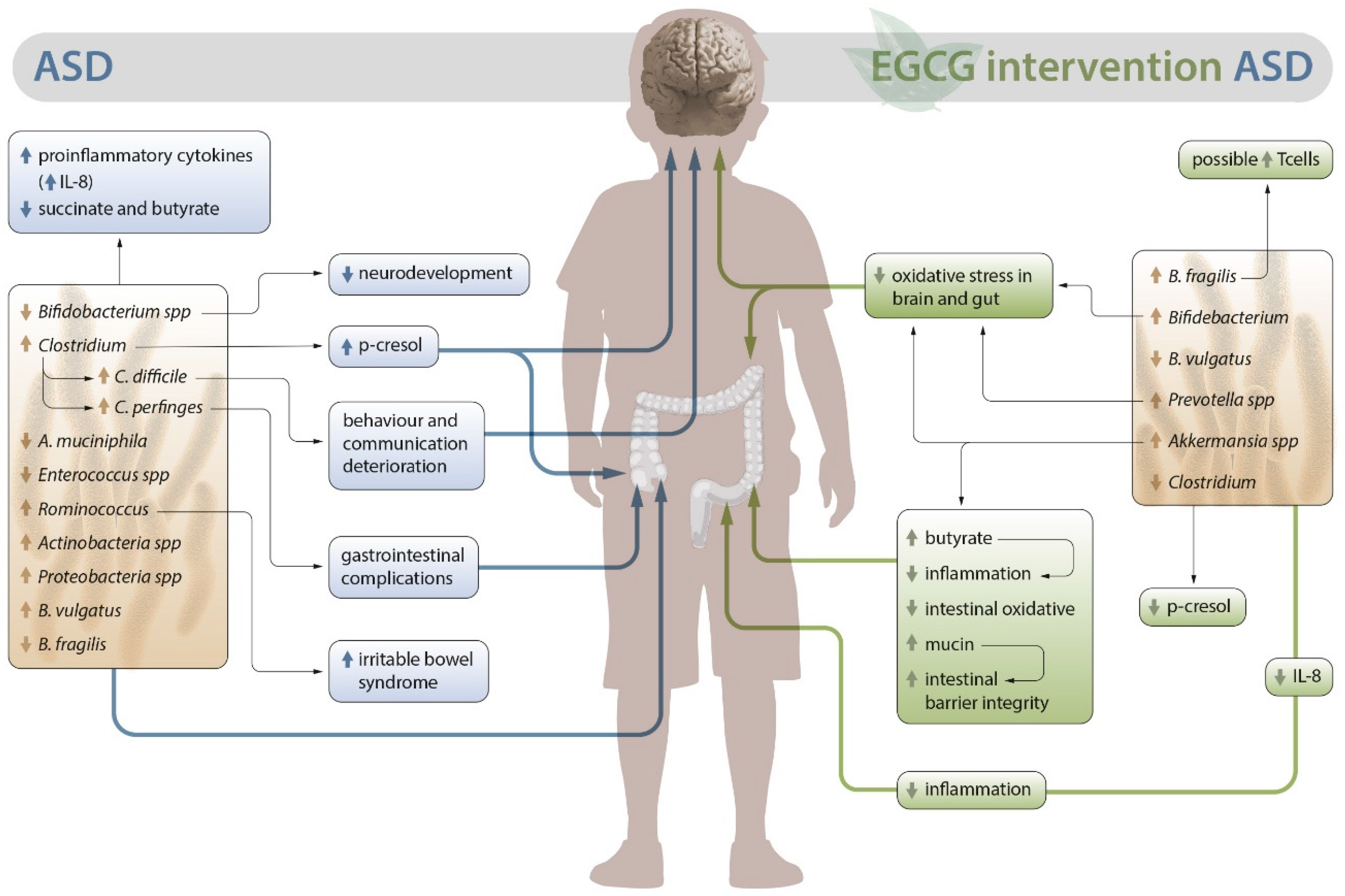
Figure 2.
Mechanism of EGCG metabolite generation in the intestinal microbiota. Reciprocal interactions between EGCG and human gut microbiota in vitro [56].
Figure 2.
Mechanism of EGCG metabolite generation in the intestinal microbiota. Reciprocal interactions between EGCG and human gut microbiota in vitro [56].
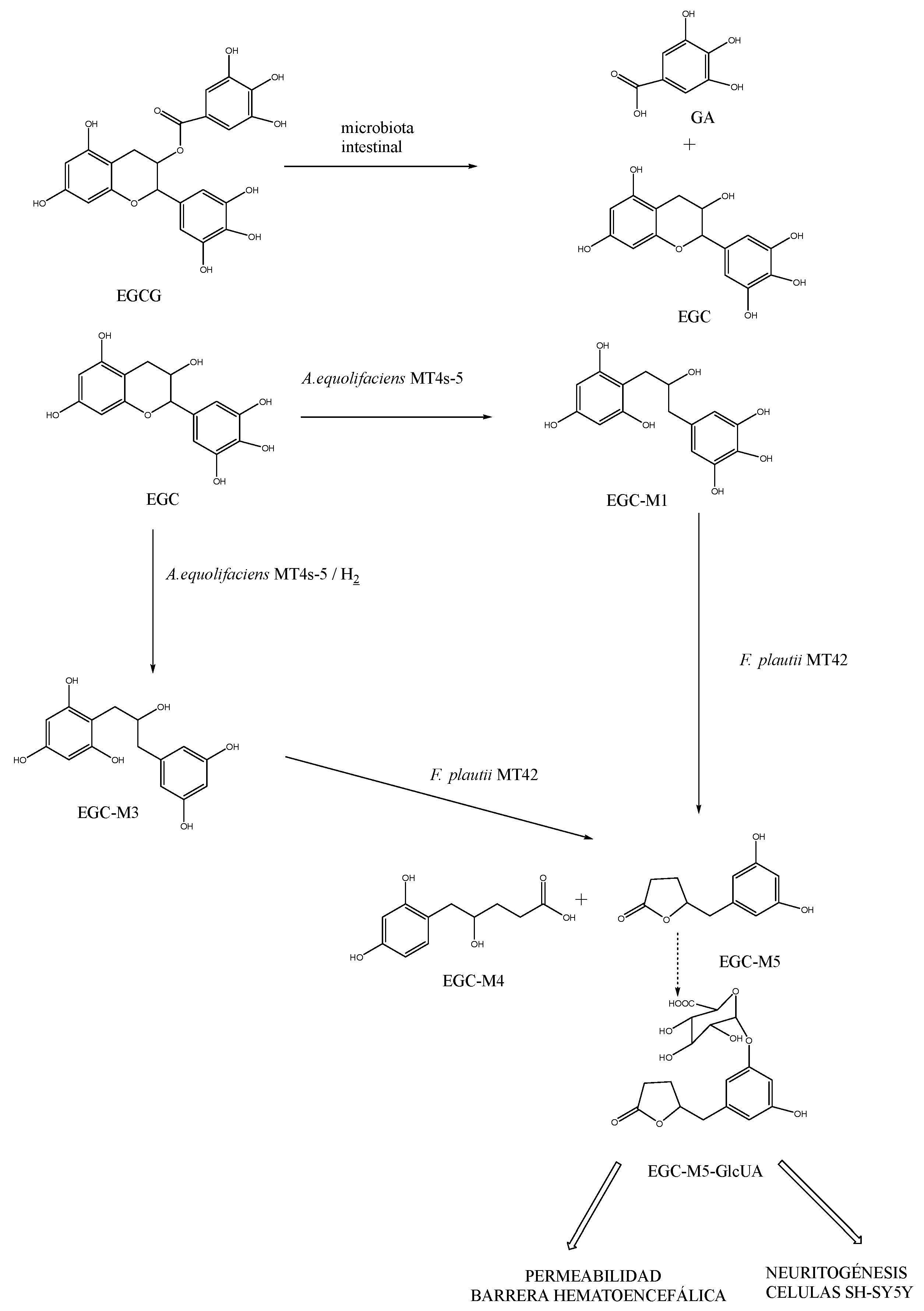
Disclaimer/Publisher’s Note: The statements, opinions and data contained in all publications are solely those of the individual author(s) and contributor(s) and not of MDPI and/or the editor(s). MDPI and/or the editor(s) disclaim responsibility for any injury to people or property resulting from any ideas, methods, instructions or products referred to in the content. |
© 2023 by the authors. Licensee MDPI, Basel, Switzerland. This article is an open access article distributed under the terms and conditions of the Creative Commons Attribution (CC BY) license (http://creativecommons.org/licenses/by/4.0/).
Copyright: This open access article is published under a Creative Commons CC BY 4.0 license, which permit the free download, distribution, and reuse, provided that the author and preprint are cited in any reuse.


