
Submitted:
12 June 2023
Posted:
13 June 2023
You are already at the latest version
Abstract
The influence of preoperative examination of oral squamous cell carcinoma (OSCC) mucosal surrounding by VELscope (Visually Enhanced Lesion Scope) on the radicality of surgical treatment of OSCC was the goal of this study.It is the first analysis of its kind with a total number of 122 patients suffering from OSCC with homogenous distribution of different tumor stages randomized into a study and a control group and enrolled in our study after meeting the inclusion criteria. The preoperative checkup by VELscope accompanied by the marking of the range of a loss of fluorescence in the study group was performed before the surgery. We have developed a unique mucosal tattoo marking technique for this purpose. The pathohistological results after surgical treatment, i.e. the margin status, were then compared. We achieved the pathological free margin (pFM) in 55 patients, the pathological close margin (pCM) in 6 cases and we encountered no case of the pathological positive margin (pPM) in the mucosal layer in the study group. In comparison, the control group results revealed pPM in 7 cases, pCM in 14 cases and pFM in 40 of all cases in the mucosal layer. This study proved that the preoperative autofluorescence evaluation of a tumor mucosal surrounding can enhance the ability to reach the pFM resection.
Keywords:
autofluorescence
; oral squamous cell carcinoma
; margin status
1. Introduction
Oral squamous cell carcinoma is a serious and relatively frequent disease of the oral cavity and, unfortunately, also belongs to the most common malignancies in the orofacial region [1]. The estimated age-standardized rate incidence in Europe is 16.9, in the European Union 17.0, followed by the mortality of 7.1 and 6.7 respectively per 100 000 for the year 2020 [2]. The situation in the Czech Republic is comparable to the EU average (Chart 1). The surgical treatment, especially in the early stage of the disease, has the best curative results [3]. The radicality of the procedure is crucial for the prognosis and the treatment success [4,5,6,7,8,9,10,11]. It is essential to determine the area of a tissue affected by tumor cells from the healthy one during the surgery to ensure the radicality.
New effective examination and imaging techniques are being developed that allow the surgeon to better visualize the boundaries of the primary tumor before and during the surgery more precisely [12,13,14,15,16,17,18,19,20,21,22,23,24]. With the help of these techniques, it is possible, among other things, to have a positive impact on margin surveillance. An ideal investigative technique for this purpose should meet certain characteristics. High sensitivity and specificity while maintaining complete non-invasiveness should be the essence. Furthermore, it is important that this examination is feasible intraoperatively, achieves stable results, is reproducible, quick and simple, as well as economically sustainable and applicable in a wide range of practice. Optical methods fulfill most of these properties, but so far, they themselves are burdened with a number of shortcomings [25]. We assumed that the use of the selected optical examination method, despite its shortcomings, would bring improvement in the treatment results of our patients suffering from OSCC in our study. For further investigation, the direct autofluorescence method was chosen.
Direct autofluorescence is a technique used for screening or better determination of potentially malignant changes of oral mucosa. In 2006, thanks to extensive research efforts, the VELscope system was registered in Canada and also certified by the FDA in the United States. Similar to some other systems working on the principle of natural autofluorescence (Identafi 3000, Sapphire Plus Lesion Detection), the VELScope device uses a non-invasive method of examination using a handpiece emitting bright blue light (400 - 460 nm), enabling direct visualization of the oral mucosa in real time. This illumination leads to the excitation of endogenous mucosal and submucosal fluorophores [26], which emit a green light, that could be registered through the semipermeable handpiece filter. The visible loss of physiologic fluorescence signifies dysplastic changes of the epithelium, but could be seen also in hyperemia, traumatization, hyperkeratosis and other benign changes, that lower the specificity [27,28,29,30]. We evaluate the hypothesis, that the use of direct autofluorescence (VELscope system tumor mucosal surrounding examination) is enhancing the ability to reach the pathologically free margin (pFM) in our study.
In order to ensure global comparability and to clearly declare what evaluation criteria were used, it is necessary to properly define the quality of the resection margin. Despite the prevailing belief that leaving part of the tumor cells in the patient's body is the most common reason for OSCC treatment failure, there is still no clear definition of an adequate resection margin [31,32]. Resection margins are mostly classified as either positive (pPM), that means tumor "cut- through", close (pCM), or negative (pFM), with different definitions of the healthy tissue rim range [33]. The distance between the tumor border and surgical margin to gain the pFM vary in some studies [34] [11]. According to International Collaboration on Cancer Reporting (ICCR) the definition of a resection margin >5 mm is clear and 1-5 mm is close and <1 mm is positive [11]. Similar to this definition is a statement of the National Comprehensive Cancer Network (NCCN), where pFM is 5 mm or more from the invasive tumor front, pCM is defined as a distance from the invasive tumor front to the resected margin that is less than 5 mm and a pPM means a carcinoma in situ or an invasive carcinoma at the margin of resection [35]. Some role in this uncertainty plays probably shrinkage of the histological specimen. There are some studies to address margin shrinkage in patients with head and neck cancer, which was on the order of 20 % to 25 % [36,37]. In accordance with established international practice, the ICCR model was used to assess the condition of surgical margins.
2. Materials and Methods
This pilot retrospective randomized study was conducted at the Department of Stomatology, University Hospital Pilsen, Faculty of Medicine in Pilsen, Charles University, Pilsen, Czech Republic in cooperation with the Sikl´s Department of Pathology, Faculty of Medicine in Pilsen, Charles University, Pilsen, Czech Republic. For better objectivity the whole study design, data processing and the results emerged from the study were discussed with the European front specialists from the University Clinic of Oral and Maxillofacial Surgery, Medical University of Innsbruck, Innsbruck, Austria. We collected two groups (a study group and a random control group) comprising a total number of 122 patients cured in the period of the years 2016 – 2022 at our department. The inclusion criteria of the study group were as follows: age over 18 years, histologically verified oral squamous cell carcinoma with no sign of inflammation or traumatization of the surrounding mucosa and no previous surgery (except for a small primary biopsy to confirm the diagnosis), radiotherapy or chemotherapy for head and neck cancer, signed informed consent, indication for primary surgical treatment and tumor localization at oral anatomical sites that can be directly visualized using both white light and fluorescence visualization device (this includes ICD-10 site codes: C02.0-C06.9)[38] and VELScope examination before surgery. Other parameters taken into account were tumor site, sex, age, TNM classification and grade according to the Union for International Cancer Control (UICC). The inclusion criteria of the control group were the same except for the VELscope examination. Simple randomization was used for the distribution of the patients into mentioned groups. All of the patients underwent standard preoperative examination including staging based on clinical examination and imaging (ultrasonography, computed tomography, magnetic resonance or hybrid positron emission tomography). The patients signed a detailed informed consent form, with the privacy policy agreement. The design of this study was approved by the Committee of Ethics in Research of Department of Stomatology, University Hospital Pilsen, Faculty of Medicine in Pilsen, Charles University under the code 333/2020. This study was conducted according to the Declaration of Helsinki.
Preoperative evaluation of tumor margins with the help of VELscope device (model No. V1, LED Dental, Inc., 201-15047 Marina Driva White Rock, BC V4B IC5, Canada) was then provided by an experienced surgeon trained and calibrated with the VELscope system in each patient of the study group. It is recommended to provide this examination in a dark room to avoid other illumination interference and to gain the best contrast of examined field. During this procedure, a field of loss of autofluorescence was marked by permanent (tattoo) or transient (gentian violet - directly before surgery) staining, or marked by monopolar electrocoagulation device directly at the beginning of the surgery in general anesthesia (marking modality was chosen according to the patient compliance and surgeons’ preference in each case), and the discrepancy between the mark and day-light visible tumor boundary was measured (Figure 1).
In an attempt to shorten the surgical time and thus also the time of general anesthesia for the patient, we developed a technique for permanent tattoo marking, which can be done even a few days before the operation. An insulin syringe was filled with a small amount of the conventional tattoo dye and a little superficial mucosal scratch by the syringe needle was directly stained by the chosen dye. For instant specimen orientation and better cooperation with the pathologist different colors for each specimen site were used (Figure 2). The procedure was provided under topical anesthesia (lidocaine spray 10 g / 100 ml) for higher comfort for the patient. The interference between the marking and the histological margin examination was avoided by the rim of tissue excised behind the marks, as descripted subsequently. None of the patients had a peri- or post procedural painful perception, we noticed no health complications or negative tumor site affection by this procedure.
Cold steel or a gentle high-frequency electro surgery excision at least 3 mm behind the marks in the study group was performed (in the control group, the excision was provided according to the international conventional recommendation at least 10 mm behind the naked-eye visible tumor boundary) and the specimen was sent to the dedicated pathologist for histological examination at the Sikl´s Department of Pathology. After hematoxylin-eosin staining and immunohistochemical examination, typing, grading, perineural and intravascular invasion and the tumor to margin distance were evaluated. The histological outcome, VELscope findings and other data were processed by a professional statistician. The main emphasis was placed on evaluating the increase in the extent of visibility of tumor changes when comparing white light versus VELscope and, above all, on the differences in the quality of the resection margin of both groups from a histological point of view. As mentioned before, the ICCR model was used for evaluating the status of surgical margins.
3. Results
3.1. Groups characteristics
Gender distribution was 40 males and 21 females in the study group and 36 males and 25 females in the control group suffering from OSCC (Table 1).
The mean age in the study group was 65.3 years with a range from 37 to 90 years of age and 64.3 years with a range from 34 to 88 years in the control group respectively (Chart 2).
The most affected site of the oral cavity was the tongue in both groups (Table 2).
The stage and grade status of all tumors were evaluated and the situation in both groups is presented in appropriate tables (Table 3, 4).
Statistical analysis did not reveal any significant difference between the two groups in terms of the features described above.
3.2. Treatment outcomes comparison
- The loss of physiologic fluorescence resulted in a resection enlargement of 4.68 mm in average (1 – 12 mm) compared to the polychromatic light and the tactile tumor borders assessment.
- The histological examination revealed no case of pPM (n = 0; 0 %), six cases of pCM (n = 6; 9.84 %) and fifty-five specimens were pFM (n = 55; 90.16 %) in the mucosal margins in the study group. The situation in the control group was as follows: pPM (n = 7; 11.48 %), pCM (n = 14; 22.95 %), pFM (n = 40; 65.57 %) (Chart 3). Although the autofluorescence technique has no influence on the deep surgical margins, we present the deep margin situation for comprehensiveness – in the study group we encountered pPM in eight cases (n = 8; 13.11 %), pCM in ten cases (n = 10; 16.39 %) and pFM in forty-three cases (n = 43; 70.49 %), in the control group situation is as follows: pPM (9; 14.75 %), pCM (6; 9.84 %), pFM (46; 75.41 %) (Chart 4).
3.3. Observed effect of autofluorescence assistance
- As expected, our study proved a statistically significant difference between the two groups in the mucosal layer only (see table 5). There was no significant difference in deep margins. Despite of the larger resection in the study group, there was no significant increase in postoperative morbidity with regard to either swallowing or speech. No patient experienced major complications in the postoperative period and no patient was discharged with a feeding tube or tracheostomy. The overall treatment outcome in terms of surgical tumor eradication was significantly better in the study group compared to the control group.

Table 5.
Comparison of mucosal margin status in both groups.
| Margin status | Frequency – amount / percentil | Total | |
| Study group | Control group | ||
| MpFM | 55 / 90.16 | 40 / 65.57 | 54 |
| MpCM | 6 / 9.84 | 14 / 22.95 | 56 |
| MpPM | 0 / 0.00 | 7 / 11.48 | 12 |
| Total | 61 | 61 | 122 |
4. Discussion
A multimodal treatment with an accent on radical surgical tumor excision has the best curative results [39,40]. Early detection, prompt staging and individual setting of the required treatment plan can lower the risk of curative failure, recurrence and enormous impairment of the quality of life of the patient [4]. New examination techniques, as well as the modern surgical approaches, came into the clinical practice thanks to the scientific progress [41]. One of the modern techniques described in our study – the mucosal autofluorescence – has the potential to help clinicians to early detect a mucosal malignity in the oral cavity and in addition to determine the tumor boundaries more precisely [42,43,44,45]. The surgeon’s main goal in the surgical treatment of OSCC is to achieve the R0 resection, which means ideally the pFM [40]. The presence of residual tumor cells is convicted to be the most important prognostic factor. The high number of studies comparing local recurrence rate with margin status in OSCC found a strong correlation, although the absolute number of local recurrences and the criteria used to define positive margins vary significantly among the particular studies [46,47,48]. Some studies surprisingly failed to demonstrate any correlation between the recurrence and margin status [49]. It is still not clear, how wide the distance between surgical margin and tumor should be. It is crucial to obtain adequate surgical margins of the tumor, however, the surgeon must find a balance between the radicality of the operation and the effort to preserve the function of the orofacial system and thus the quality of life and limit the cosmetic disability [3,50,51,52,53,54,55,56]. Many scientific publications deal with the problem of positive resection margins, unfortunately there are currently no globally valid guidelines. The setting of a minimum safety margin is discussed in a number of studies and the range varies between 10 – 2 mm, the median recommended clinical distance for the resection of OSCC is 5 mm, although a clear basis for this distance is currently lacking. Mainly because of the specimen shrinkage and “invisible” carcinoma cells spread, the excised rim of macroscopically unchanged mucosa should be more than the mentioned pFM distance. It is recommended to keep at least 10 mm distance from the tumor to avoid impairing the histological margin status during the surgery [57]. The intraoperative evaluation of resection margins can be made by several methods. The use of frozen sections (FS) used to be the gold standard, it was accepted worldwide but, as underlined by Yahalom et al. [58], the procedure is not standardized. The issue of discussion is also the site where the FS should be taken if it is the resection bed or the specimen margin [59]. The other problem is the risk of incorrect identifying of the area to be re-excised in the event of positive margins [60] and time consumption during surgery. For this reason, the endeavor of finding a more favorable method for intraoperative determination of adequate resection margins is very topical and research is very dynamic in this regard. There are some more methods, that have been suggested for better achieving pFM in mucosal layer OSCC surgical treatment or oral mucosa malignity detection and we present a list of some of them for comparison below:
Contact endoscopy - allows an in vivo microscopic examination of upper aerodigestive tract mucosa with a rigid endoscope. It is a noninvasive technique and provides information on a microscopic diagnosis and lesion margins. A sensitivity of 80 %, a specificity of 100 % and an accuracy of 93 % for contact endoscopy in the diagnosis of malignancy is reported [61,62,63].
Narrow band imaging is a video endoscopic system for the examination of mucous membranes. Thanks to narrow band filters, only two specific bands of visible light, which are typical for the absorption peak of hemoglobin, are allowed to pass through. The observed wavelengths increase the visibility of microvascular abnormalities that could be related to preneoplastic and neoplastic mucosal changes [64,65,66,67,68].
Staining with Lugol’s iodine solution or toluidine blue is one of the methods, that can reduce the number of positive margins by pointing up the tumor margins [69,70,71,72,73]. Vital staining utilizes the enhanced affinity of some dyes to certain cell structures present in dysplastic or malignant cells, making them more apparent. The widest use has the toluidine blue in the oral cavity mucosa. This basic metachromatic stain has an affinity toward DNA and RNA and demonstrates an invasive malignancy, carcinoma in situ (CIS) and dysplasia by staining abnormal tissues blue [73,74].
Using touch imprint cytology, the properties of the cells located on the surface of the resection margin can be evaluated intraoperatively. It is a fast, simple, cheap and relatively accurate examination technique [75].
Optical coherence tomography is another technique for distinguishing between positive and negative surgical margins [76,77]
Some publications mention a molecular definition of surgical margins, from protein markers to DNA-based techniques, that can evaluate the margins at the subcellular level and can explain, for example, the local recurrence in optical microscope pFM [78,79,80]. Various monoclonal antibodies and other ligands are already being clinically used or researched in this area, which have the ability to mark the structures of tumor cells in a certain way and make them visible intraoperatively, for example by means of fluorescence [12,13,17,18].
The last-mentioned technique is a tissue reflectance. The ViziLite device, which uses reflectance is adapted for use in the oral cavity. The principle of this method is, that the abnormal cells reflect the light (high nucleus - cytoplasm ratio, keratinization excess, hyperparakeratinization), while normal cells absorb the light and are depicted in a bluish color [30].
We verified the potential of autofluorescence to detect the dysplastic and malignant mucosal changes in our study. The limitations of this technique and the methodology of our study is that the detection capability stays only on the superficial mucosal layer. In our study, we identified some possible sources of bias that need to be taken into account. A frequent weakness of clinical studies is that the compared groups are not identical, but statistically very similar, which is also the case in our study. Another problem is the burden of the natural autofluorescence method with a relatively low specificity despite a very high sensitivity. We partially eliminated this weakness by the fact that the histological nature of the disease was known and further by using the set inclusion parameters. According to published studies, the other presented optical examination techniques also achieve promising results, but there is a lack of scientific works relevantly comparing individual methods with each other. Further research is needed in this area. The biggest limitation of autofluorescence in the way of use we are investigating is the limitation to the mucosal layer. In this regard, it would be appropriate to combine the method with another similarly non-burdensome and simple examination technique that detects deep tumor margins. Such a technique could be, for example, the adjuvant perioperative use of ultrasonography, which also achieves very good results [19,20,21]. In this sphere, we consider further research to be extremely interesting, as the potential of both methods could be exponentially enhanced. On the other hand, we developed a technique of preoperative marking of the pathological mucosal changes by a permanent tattoo, which can help the surgeon to set the resection line more precisely. We gain a better specimen orientation, which leads to surgical treatment success enhancement, more precise orientation for better pathologist communication and better identification of risk field in case of reexcision need by this technique.
The potential of this technique for better detection of mucosal malignancy is well known and have been multiple published. The role of autofluorescence in setting the margins into the R0 region or even better in achieving the pFM is being worldwide researched. The results of some studies are promising, but it is hard to compare the results because of its nonhomogenity. Some presented works are limited to the early stage of OSCC only, or do not depict the resection margin changes clearly [44,45,81,82,83]. The further investigation in this field is needed to assess the potential of this technique and to create a recommendation for the standardized use of autofluorescence in OSCC surgery in the sense of a guideline.
5. Conclusions
Our findings support the hypothesis of improving the surgery outcomes in the study group, we have discovered the statistically significant difference between the study and the control group. We proved the efficacy of mucosal autofluorescence in setting the sufficient mucosal surgical margins in our study. If combined with the permanent tattoo marking of the pathological changes visible under VELscope as described above, this technique can enhance the surgeon´s ability to successfully treat the patient with OSCC even more.
Supplementary Materials
The following supporting information can be downloaded at the website of this paper posted on Preprints.org.
Author Contributions
Conceptualization, P.P, K.A. and H.L.; methodology, P.P., K.A. and H.L.; software, G.J. and P.A.; validation, L.J.; formal analysis, F.M.; investigation, P.P. and M.C.; resources, P.P., P.K. and G.J.; data curation, M.O.; writing—original draft preparation, P.P.; writing—review and editing, K.A. and H.L.; visualization, F.M.; supervision, P.P., K.A. and H.L.; project administration, P.P. and M.C.; funding acquisition, P.P. All authors have read and agreed to the published version of the manuscript.
Funding
This study was supported by the grant of Ministry of Health of the Czech Republic – Conceptual Development of Research Organization (Faculty Hospital in Pilsen – FNPI, 00669806). Scholarship Aktion Österreich-Tschechien, AÖCZ-Semesterstpendien, Referenfe number MPC-2022-05031, financed by Federal Ministry of Education, Science and Research (BMBWF), awarding organisation OeAD – Austria´s Agency for Education and Internationalisation; Hugo Obwegeser Travel Scholarship, European Association for Cranio-Maxillo-Facial Surgery; Charles University Mobility Fund.
Institutional Review Board Statement
The study was conducted in accordance with the Declaration of Helsinki and approved by the Committee of Ethics in Research of Department of Stomatology, University Hospital Pilsen, Faculty of Medicine in Pilsen, Charles University under the code 333/2020, date of approval 30.6.2020.
Informed Consent Statement
Informed consent was obtained from all subjects involved in the study. Written informed consent has been obtained from the patients to publish this paper.
Data Availability Statement
The data presented in this study are available on request from the corresponding author. The data are not publicly available due to institutional committee of ethics statement.
Acknowledgments
The author would like to thank Ing. Stanislav Kormunda for the statistical processing of research data.
Conflicts of Interest
The authors declare no conflict of interest. The funders had no role in the design of the study; in the collection, analyses, or interpretation of data; in the writing of the manuscript; or in the decision.
References
- Cariati, P.; Pampin Ozan, D.; Gonzalez Corcóles, C.; Tursun, R.; Peña Barreño, M.; Ferrari, S.; Arroyo Rodriguez, S. Clinical Behavior of T1–T2 Squamous Cell Carcinoma of the Oral Cavity. J. Cranio-Maxillofacial Surg. 2020, 48, 1152–1157. [Google Scholar] [CrossRef]
- Dyba, T.; Randi, G.; Bray, F.; Martos, C.; Giusti, F.; Nicholson, N.; Gavin, A.; Flego, M.; Neamtiu, L.; Dimitrova, N.; et al. The European Cancer Burden in 2020: Incidence and Mortality Estimates for 40 Countries and 25 Major Cancers. Eur. J. Cancer 2021, 157, 308–347. [Google Scholar] [CrossRef]
- Bschorer, M.; Schneider, D.; Goppold, K.; Sperling, J.; Schön, G.; Bschorer, R. Quality of Life and Survival Rate after Primary Surgical Treatment of Oral Squamous Cell Carcinoma: A Retrospective Study with 18 Years of Follow-Up. J. Cranio-Maxillofacial Surg. 2022, 50, 170–177. [Google Scholar] [CrossRef]
- Low, T.H.H.; Gao, K.; Gupta, R.; Clifford, A.; Elliott, M.; Ch’ng, S.; Milross, C.; Clark, J.R. Factors Predicting Poor Outcomes in T1N0 Oral Squamous Cell Carcinoma: Indicators for Treatment Intensification. ANZ J. Surg. 2016, 86, 366–371. [Google Scholar] [CrossRef]
- Loeffelbein, D.J.; Eiber, M.; Mayr, P.; Souvatzoglou, M.; Mücke, T.; von Bomhard, A.; Kesting, M.R.; Wolff, K.-D. Loco-Regional Recurrence after Surgical Treatment of Oral Squamous Cell Carcinoma: Proposals for Follow-up Imaging Based on Literature, National Guidelines and Institutional Experience. J. Cranio-Maxillo-Facial Surg. 2017, 43, 1546–1552. [Google Scholar] [CrossRef] [PubMed]
- Ravasz, L.A.; Slootweg, P.J.; Hordijk, G.J.; Smit, F.; van der Tweel, I. The Status of the Resection Margin as a Prognostic Factor in the Treatment of Head and Neck Carcinoma. J. Cranio-Maxillo-Facial Surg. 2017, 19, 314–318. [Google Scholar] [CrossRef] [PubMed]
- Ermer, M.A.; Kirsch, K.; Bittermann, G.; Fretwurst, T.; Vach, K.; Metzger, M.C. Recurrence Rate and Shift in Histopathological Differentiation of Oral Squamous Cell Carcinoma – A Long-Term Retrospective Study over a Period of 13.5 Years. J. Cranio-Maxillofacial Surg. 2015, 43, 1309–1313. [Google Scholar] [CrossRef] [PubMed]
- van der Waal, I. Are We Able to Reduce the Mortality and Morbidity of Oral Cancer; Some Considerations. Med. Oral Patol. Oral Cir. Bucal 2013, 18. [Google Scholar] [CrossRef] [PubMed]
- Chen, T.C.; Wang, C.P.; Ko, J.Y.; Yang, T.L.; Lou, P.J. The Impact of Pathologic Close Margin on the Survival of Patients with Early Stage Oral Squamous Cell Carcinoma. Oral Oncol. 2012.
- Wong, L.S.; McMahon, J.; Devine, J.; McLellan, D.; Thompson, E.; Farrow, A.; Moos, K.; Ayoub, A. Influence of Close Resection Margins on Local Recurrence and Disease-Specific Survival in Oral and Oropharyngeal Carcinoma. Br. J. Oral Maxillofac. Surg. 2012, 50, 102–108. [Google Scholar] [CrossRef] [PubMed]
- Hinni, M.L.; Ferlito, A.; Brandwein-Gensler, M.S.; Takes, R.P.; Silver, C.E.; Westra, W.H.; Seethala, R.R.; Rodrigo, J.P.; Corry, J.; Bradford, C.R.; et al. Surgical Margins in Head and Neck Cancer: A Contemporary Review. Head Neck 2013, 35, 1362–1370. [Google Scholar] [CrossRef]
- Krishnan, G.; van den Berg, N.S.; Nishio, N.; Kapoor, S.; Pei, J.; Freeman, L.; Lee, Y.J.; Zhou, Q.; van Keulen, S.; Farkurnejad, S.; et al. Fluorescent Molecular Imaging Can Improve Intraoperative Sentinel Margin Detection in Oral Squamous Cell Carcinoma. J. Nucl. Med. 2022, 63, 1162–1168. [Google Scholar] [CrossRef] [PubMed]
- Vonk, J.; de Wit, J.G.; Voskuil, F.J.; Witjes, M.J.H. Improving Oral Cavity Cancer Diagnosis and Treatment with Fluorescence Molecular Imaging. Oral Dis. 2021, 27, 21–26. [Google Scholar] [CrossRef]
- Wang, R.; Naidu, A.; Wang, Y. Oral Cancer Discrimination and Novel Oral Epithelial Dysplasia Stratification Using FTIR Imaging and Machine Learning. Diagnostics 2021, 11, 1–16. [Google Scholar] [CrossRef] [PubMed]
- Harmsen, S.; Teraphongphom, N.; Tweedle, M.F.; Basilion, J.P.; Rosenthal, E.L. Optical Surgical Navigation for Precision in Tumor Resections. Mol. Imaging Biol. 2017, 19, 357–362. [Google Scholar] [CrossRef] [PubMed]
- Grochau, K.J.; Safi, A.F.; Drebber, U.; Grandoch, A.; Zöller, J.E.; Kreppel, M. Podoplanin Expression in Oral Leukoplakia─a Prospective Study. J. Cranio-Maxillofacial Surg. 2019, 47, 505–509. [Google Scholar] [CrossRef] [PubMed]
- Pan, J.; Deng, H.; Hu, S.; Xia, C.; Chen, Y.; Wang, J.; Wang, Y. Real-Time Surveillance of Surgical Margins via ICG-Based near-Infrared Fluorescence Imaging in Patients with OSCC. World J. Surg. Oncol. 2020, 18, 96. [Google Scholar] [CrossRef]
- Tian, Y.; Tang, C.; Shi, G.; Wang, G.; Du, Y.; Tian, J.; Zhang, H. Novel Fluorescent GLUT1 Inhibitor for Precision Detection and Fluorescence Image-Guided Surgery in Oral Squamous Cell Carcinoma. Int. J. cancer 2022, 151, 450–462. [Google Scholar] [CrossRef]
- Rocchetti, F.; Tenore, G.; Montori, A.; Cassoni, A.; Cantisani, V.; Di Segni, M.; Di Gioia, C.R.T.; Carletti, R.; Valentini, V.; Polimeni, A.; et al. Preoperative Evaluation of Tumor Depth of Invasion in Oral Squamous Cell Carcinoma with Intraoral Ultrasonography: A Retrospective Study. Oral Surg. Oral Med. Oral Pathol. Oral Radiol. 2021, 131, 130–138. [Google Scholar] [CrossRef]
- Klein Nulent, T.J.W.; Noorlag, R.; Van Cann, E.M.; Pameijer, F.A.; Willems, S.M.; Yesuratnam, A.; Rosenberg, A.J.W.P.; de Bree, R.; van Es, R.J.J. Intraoral Ultrasonography to Measure Tumor Thickness of Oral Cancer: A Systematic Review and Meta-Analysis. Oral Oncol. 2018, 77, 29–36. [Google Scholar] [CrossRef]
- Caprioli, S.; Casaleggio, A.; Tagliafico, A.S.; Conforti, C.; Borda, F.; Fiannacca, M.; Filauro, M.; Iandelli, A.; Marchi, F.; Parrinello, G.; et al. High-Frequency Intraoral Ultrasound for Preoperative Assessment of Depth of Invasion for Early Tongue Squamous Cell Carcinoma: Radiological-Pathological Correlations. Int. J. Environ. Res. Public Health 2022, 19. [Google Scholar] [CrossRef] [PubMed]
- Yang, C.C.; Su, Y.F.; Cheng, H.C.; Juan, Y.C.; Chiu, Y.W.; Wu, C.H.; Chen, P.Y.; Lee, Y.H.; Chen, Y.L.; Chen, Y.T.; et al. Improving the Diagnostic Performance by Adding Methylation Marker to Conventional Visual Examination in Identifying Oral Cancer. Diagnostics 2022, 12. [Google Scholar] [CrossRef] [PubMed]
- Fati, S.M.; Senan, E.M.; Javed, Y. Early Diagnosis of Oral Squamous Cell Carcinoma Based on Histopathological Images Using Deep and Hybrid Learning Approaches. Diagnostics 2022, 12. [Google Scholar] [CrossRef]
- Lee, Y.J.; Kwon, T.G.; Kim, J.W.; Lee, S.T.; Hong, S.H.; Choi, S.Y. Evaluation of Depth of Invasion and Tumor Thickness as a Prognostic Factor for Early-Stage Oral Squamous Cell Carcinoma: A Retrospective Study. Diagnostics 2022, 12. [Google Scholar] [CrossRef] [PubMed]
- Tarsitano, A.; Ricotta, F.; Baldino, G.; Badiali, G.; Pizzigallo, A.; Ramieri, V.; Cascone, P.; Marchetti, C. Navigation-Guided Resection of Maxillary Tumours: The Accuracy of Computer-Assisted Surgery in Terms of Control of Resection Margins – A Feasibility Study. J. Cranio-Maxillofacial Surg. 2017. [Google Scholar] [CrossRef]
- Giovannacci, I.; Magnoni, C.; Vescovi, P.; Painelli, A.; Tarentini, E.; Meleti, M. Which Are the Main Fluorophores in Skin and Oral Mucosa? A Review with Emphasis on Clinical Applications of Tissue Autofluorescence. Arch. Oral Biol. 2019, 105, 89–98. [Google Scholar] [CrossRef]
- Laronde, D.M.; Williams, P.M.; Hislop, T.G.; Poh, C.; Ng, S.; Bajdik, C.; Zhang, L.; Macaulay, C.; Rosin, M.P. Influence of Fluorescence on Screening Decisions for Oral Mucosal Lesions in Community Dental Practices. J. Oral Pathol. Med. 2014. [Google Scholar] [CrossRef] [PubMed]
- Burian, E.; Schulz, C.; Probst, F.; Palla, B.; Tröltzsch, M.; Maglitto, F.; Califano, L.; Ehrenfeld, M.; Otto, S. Fluorescence Based Characterization of Early Oral Squamous Cell Carcinoma Using the Visually Enhanced Light Scope Technique. J. Cranio-Maxillo-Facial Surg. 2017. [Google Scholar] [CrossRef]
- Scheer, M.; Neugebauer, J.; Lingohr, T.; Zöller, J.E. O. 317 Evaluation of Undiagnosed Mucosa Lesions with the VELscope System. J. Cranio-Maxillo-Facial Surg. 2017, 36, S80. [Google Scholar] [CrossRef]
- Sambandham, T.; Masthan, K.M.K.; Kumar, M.S.; Jha, A. The Application of Vizilite in Oral Cancer. J. Clin. Diagn. Res. 2013, 7, 185–186. [Google Scholar] [CrossRef]
- Low, T.H.; Gao, K.; Elliott, M.; Clark, J.R. Tumor Classification for Early Oral Cancer: Re-Evaluate the Current TNM Classification. Head Neck 2015, 37, 223–228. [Google Scholar] [CrossRef]
- Tasche, K.K.; Buchakjian, M.R.; Pagedar, N.A.; Sperry, S.M. Definition of “Close Margin” in Oral Cancer Surgery and Association of Margin Distance With Local Recurrence Rate Supplemental Content. JAMA Otolaryngol Head Neck Surg 2017, 143, 1166–1172. [Google Scholar] [CrossRef]
- Park, H. Surgical Margins for the Extirpation of Oral Cancer. J. Korean Assoc. Oral Maxillofac. Surg. 2016, 42, 325–326. [Google Scholar] [CrossRef]
- Brinkman, D.; Callanan, D.; O’Shea, R.; Jawad, H.; Feeley, L.; Sheahan, P. Impact of 3 Mm Margin on Risk of Recurrence and Survival in Oral Cancer. Oral Oncol. 2020, 110, 104883. [Google Scholar] [CrossRef]
- Fenton, M.; Foote, R.L.; Galloway, T.; Gillison, M.L.; Haddad, R.I.; Hicks, W.L.; Hitchcock, Y.J.; Jimeno, A.; Leizman, D.; Pinto, H.A.; et al. Continue NCCN Guidelines Panel Disclosures NCCN Gratefully Acknowledges the Following Subcommittee Member for Her Contributions on the Development of the Principles of Imaging (IMG-A) NCCN Guidelines Version 2. 2020. [Google Scholar]
- El-Fol, H.A.; Noman, S.A.; Beheiri, M.G.; Khalil, A.M.; Kamel, M.M. Significance of Post-Resection Tissue Shrinkage on Surgical Margins of Oral Squamous Cell Carcinoma. J. Craniomaxillofac. Surg. 2015, 43, 475–482. [Google Scholar] [CrossRef]
- Mistry, R.; Qureshi, S.; Kumaran, C. Post-Resection Mucosal Margin Shrinkage in Oral Cancer: Quantification and Significance. J. Surg. Oncol. 2005, 91, 131–133. [Google Scholar] [CrossRef]
- International Statistical Classification of Diseases and Related Health Problems. Tenth Revision - PubMed Available online:. Available online: https://pubmed.ncbi.nlm.nih.gov/3376487/ (accessed on 15 February 2023).
- Koerdt, S.; Röckl, J.; Rommel, N.; Mücke, T.; Wolff, K.-D.; Kesting, M.R. Lymph Node Management in the Treatment of Oral Cancer: Analysis of a Standardized Approach. J. Cranio-Maxillo-Facial Surg. 2017, 44, 1737–1742. [Google Scholar] [CrossRef] [PubMed]
- Wolff, K.-D.; Rau, A.; Ferencz, J.; Langer, T.; Kesting, M.; Nieberler, M.; Wesselmann, S. Effect of an Evidence-Based Guideline on the Treatment of Maxillofacial Cancer: A Prospective Analysis. J. Cranio-Maxillo-Facial Surg. 2017, 45, 427–431. [Google Scholar] [CrossRef] [PubMed]
- Low, T.H.H.; Lindsay, A.; Clark, J.; Chai, F.; Lewis, R. Reconstruction of Maxillary Defect with Musculo-Adipose Rectus Free Flap. Microsurgery 2017, 37, 137–141. [Google Scholar] [CrossRef] [PubMed]
- Lima, I.F.P.; Brand, L.M.; de Figueiredo, J.A.P.; Steier, L.; Lamers, M.L. Use of Autofluorescence and Fluorescent Probes as a Potential Diagnostic Tool for Oral Cancer: A Systematic Review. Photodiagnosis Photodyn. Ther. 2021, 33. [Google Scholar] [CrossRef] [PubMed]
- Sun, L.F.; Wang, C.X.; Cao, Z.Y.; Han, W.; Guo, S.S.; Wang, Y.Z.; Meng, Y.; Hou, C.X.; Zhu, Q.H.; Tang, Y.T.; et al. Evaluation of Autofluorescence Visualization System in the Delineation of Oral Squamous Cell Carcinoma Surgical Margins. Photodiagnosis Photodyn. Ther. 2021, 36. [Google Scholar] [CrossRef] [PubMed]
- Durham, J.S.; Brasher, P.; Anderson, D.W.; Yoo, J.; Hart, R.; Dort, J.C.; Seikaly, H.; Kerr, P.; Rosin, M.P.; Poh, C.F. Effect of Fluorescence Visualization-Guided Surgery on Local Recurrence of Oral Squamous Cell Carcinoma: A Randomized Clinical Trial. JAMA Otolaryngol. Head Neck Surg. 2020, 146, 1149–1155. [Google Scholar] [CrossRef] [PubMed]
- Poh, C.F.; Anderson, D.W.; Scott Durham, J.; Chen, J.; Berean, K.W.; MacAulay, C.E.; Rosin, M.P. Fluorescence Visualization-Guided Surgery for Early-Stage Oral Cancer. JAMA Otolaryngol. - Head Neck Surg. 2016, 142, 209–216. [Google Scholar] [CrossRef]
- Woolgar, J.A.; Rogers, S.; West, C.R.; Errington, R.D.; Brown, J.S.; Vaughan, E.D. Survival and Patterns of Recurrence in 200 Oral Cancer Patients Treated by Radical Surgery and Neck Dissection. Oral Oncol. 1999, 35, 257–265. [Google Scholar] [CrossRef]
- Jerjes, W.; Upile, T.; Petrie, A.; Riskalla, A.; Hamdoon, Z.; Vourvachis, M.; Karavidas, K.; Jay, A.; Sandison, A.; Thomas, G.J.; et al. Clinicopathological Parameters, Recurrence, Locoregional and Distant Metastasis in 115 T1-T2 Oral Squamous Cell Carcinoma Patients. Head Neck Oncol. 2010, 2, 9. [Google Scholar] [CrossRef]
- Popovic-Monevska, D.; Naumovski, S.; Popovski, V.; Benedetti, A.; Bozovic, S.; Iliev, A. O. 490 Loco Regional Recurrence of OSCC. J. Cranio-Maxillofacial Surg. 2008, 36, S123. [Google Scholar] [CrossRef]
- Alaeddini, M.; Etemad-Moghadam, S. Comparison of the Histologic Risk Assessment Model between Lower Lip and Oral Squamous Cell Carcinoma. J. Stomatol. Oral Maxillofac. Surg. 2017. [Google Scholar] [CrossRef]
- de Vicente, J.C.; Rúa-Gonzálvez, L.; Barroso, J.M.; Fernández del Valle-Fernández, Á.; de Villalaín, L.; Peña, I.; Cobo, J.L. Functional Results of Swallowing and Aspiration after Oral Cancer Treatment and Microvascular Free Flap Reconstruction: A Retrospective Observational Assessment. J. Craniomaxillofac. Surg. 2021, 49, 959–970. [Google Scholar] [CrossRef] [PubMed]
- Meier, J.D.; Oliver, D.A.; Varvares, M.A. Surgical Margin Determination in Head and Neck Oncology: Current Clinical Practice. The Results of an International American Head and Neck Society Member Survey. Head Neck 2005, 27, 952–958. [Google Scholar] [CrossRef]
- Woolgar, J.A.; Triantafyllou, A. A Histopathological Appraisal of Surgical Margins in Oral and Oropharyngeal Cancer Resection Specimens. Oral Oncol. 2005, 41, 1034–1043. [Google Scholar] [CrossRef] [PubMed]
- Binahmed, A.; Nason, R.W.; Abdoh, A.A. The Clinical Significance of the Positive Surgical Margin in Oral Cancer. Oral Oncol. 2007, 43, 780–784. [Google Scholar] [CrossRef]
- Chiou, W.-Y.; Lin, H.-Y.; Hsu, F.-C.; Lee, M.-S.; Ho, H.-C.; Su, Y.-C.; Lee, C.-C.; Hsieh, C.-H.; Wang, Y.-C.; Hung, S.-K. Buccal Mucosa Carcinoma: Surgical Margin Less than 3 Mm, Not 5 Mm, Predicts Locoregional Recurrence. Radiat. Oncol. 2010, 5, 79. [Google Scholar] [CrossRef] [PubMed]
- Rougier, G.; Meningaud, J.P.; Ganry, L.; Hermeziu, O.; Bosc, R.; Sidahmed-Mezi, M.; Hersant, B. Oncological and Aesthetic Outcome Following Surgical Management of Orbito-Palpebral Skin Cancers: A Retrospective Study of 132 Patients. J. Cranio-Maxillofacial Surg. 2019, 47, 1577–1582. [Google Scholar] [CrossRef] [PubMed]
- Matsuda, Y.; Kumakura, I.; Okui, T.; Karino, M.; Aoi, N.; Okuma, S.; Takeda, M.; Hayashida, K.; Sakamoto, T.; Kanno, T. Development of a Subjective Symptom Rating Scale for Postoperative Oral Dysfunction in Patients with Oral Cancer: Reliability and Validity of the Postoperative Oral Dysfunction Scale-10. Diagnostics 2021, Vol. 11, Page 2061 2021, 11, 2061. [Google Scholar] [CrossRef] [PubMed]
- Bungum, A.; Jensen, J.S.; Jakobsen, K.K.; Christensen, A.; Grønhøj, C.; von Buchwald, C. Impact of Surgical Resection Margins Less than 5 Mm in Oral Cavity Squamous Cell Carcinoma: A Systematic Review. Acta Otolaryngol. 2020, 140, 869–875. [Google Scholar] [CrossRef] [PubMed]
- Yahalom, R.; Dobriyan, A.; Vered, M.; Talmi, Y.P.; Teicher, S.; Bedrin, L. A Prospective Study of Surgical Margin Status in Oral Squamous Cell Carcinoma: A Preliminary Report. J. Surg. Oncol. 2008, 98, 572–578. [Google Scholar] [CrossRef]
- Smithers, F.A.E.; Haymerle, G.; Palme, C.E.; Low, T.H.; Froggatt, C.; Gupta, R.; Clark, J.R. A Prospective Study of Intraoperative Assessment of Mucosal Squamous Cell Carcinoma Margins in the Head and Neck. Head Neck 2021, 43, 590–600. [Google Scholar] [CrossRef] [PubMed]
- Gerber, S.M. The Impact of Frozen Sections on Surgical Margins in Squamous Cell Carcinoma of the Oral Cavity and Lips: A Retrospective Analysis 1998-2008. 2011. [CrossRef]
- Dedivitis, R.A.; Pfuetzenreiter, E.G.; Guimarães, A. V Contact Endoscopy of the Larynx as an Auxiliary Method to the Surgical Margins in Frontolateral Laryngectomy. Acta Otorhinolaryngol. Ital. 2009, 29, 16–20. [Google Scholar] [PubMed]
- Cikojević, D.; Glunčić, I.; Pešutić-Pisac, V. Comparison of Contact Endoscopy and Frozen Section Histopathology in the Intra-Operative Diagnosis of Laryngeal Pathology. J. Laryngol. Otol. 2008, 122, 836–839. [Google Scholar] [CrossRef]
- Esmaeili, N.; Boese, A.; Davaris, N.; Arens, C.; Navab, N.; Friebe, M.; Illanes, A. Cyclist Effort Features: A Novel Technique for Image Texture Characterization Applied to Larynx Cancer Classification in Contact Endoscopy—Narrow Band Imaging. Diagnostics 2021, 11. [Google Scholar] [CrossRef]
- Vu, A.N.; Farah, C.S. Efficacy of Narrow Band Imaging for Detection and Surveillance of Potentially Malignant and Malignant Lesions in the Oral Cavity and Oropharynx: A Systematic Review. Oral Oncol. 2014, 50, 413–420. [Google Scholar] [CrossRef]
- Tirelli, G.; Piovesana, M.; Gatto, A.; Tofanelli, M.; Biasotto, M.; Boscolo Nata, F. Narrow Band Imaging in the Intra-Operative Definition of Resection Margins in Oral Cavity and Oropharyngeal Cancer. Oral Oncol. 2015. [Google Scholar] [CrossRef]
- Piazza, C.; Cocco, D.; Del Bon, F.; Mangili, S.; Nicolai, P.; Peretti, G. Narrow Band Imaging and High Definition Television in the Endoscopic Evaluation of Upper Aero-Digestive Tract Cancer. Acta Otorhinolaryngol. Ital. 2011, 31, 70–75. [Google Scholar]
- Dobashi, A.; Ono, S.; Furuhashi, H.; Futakuchi, T.; Tamai, N.; Yamauchi, T.; Suka, M.; Sumiyama, K. Texture and Color Enhancement Imaging Increases Color Changes and Improves Visibility for Squamous Cell Carcinoma Suspicious Lesions in the Pharynx and Esophagus. Diagnostics 2021, Vol. 11, Page 1971 2021, 11, 1971. [Google Scholar] [CrossRef]
- Hatta, W.; Koike, T.; Ogata, Y.; Kondo, Y.; Ara, N.; Uno, K.; Asano, N.; Imatani, A.; Masamune, A. Comparison of Magnifying Endoscopy with Blue Light Imaging and Narrow Band Imaging for Determining the Invasion Depth of Superficial Esophageal Squamous Cell Carcinoma by the Japanese Esophageal Society’s Intrapapillary Capillary Loop Classification. Diagnostics 2021, Vol. 11, Page 1941 2021, 11, 1941. [Google Scholar] [CrossRef]
- Rezniczek, G.A.; Ertan, S.; Rehman, S.; Tempfer, C.B. Sequential Application of Lugol’s Iodine Test after Acetic Acid for Detecting Cervical Dysplasia: A Prospective Cohort Study. Diagnostics 2021, Vol. 11, Page 1598 2021, 11, 1598. [Google Scholar] [CrossRef]
- McMahon, J.; Devine, J.C.; McCaul, J.A.; McLellan, D.R.; Farrow, A. Use of Lugol’s Iodine in the Resection of Oral and Oropharyngeal Squamous Cell Carcinoma. Br. J. Oral Maxillofac. Surg. 2010, 48, 84–87. [Google Scholar] [CrossRef] [PubMed]
- Umeda, M.; Shigeta, T.; Takahashi, H.; Minamikawa, T.; Komatsubara, H.; Oguni, A.; Shibuya, Y.; Komori, T. Clinical Evaluation of Lugol’s Iodine Staining in the Treatment of Stage I-II Squamous Cell Carcinoma of the Tongue. Int. J. Oral Maxillofac. Surg. 2011, 40, 593–596. [Google Scholar] [CrossRef] [PubMed]
- Watanabe, A.; Taniguchi, M.; Tsujie, H.; Hosokawa, M.; Fujita, M.; Sasaki, S. Clinical Impact of Iodine Staining for Diagnosis of Carcinoma in Situ in the Floor of Mouth, and Decision of Adequate Surgical Margin. Auris Nasus Larynx 2012, 39, 193–197. [Google Scholar] [CrossRef] [PubMed]
- Sridharan, G.; Shankar, A. Toluidine Blue: A Review of Its Chemistry and Clinical Utility. J. Oral Maxillofac. Pathol. 2012, 16, 251. [Google Scholar] [CrossRef] [PubMed]
- Poswillo, D. Evaluation, Surveillance and Treatment of Panoral Leukoplakia. J. Maxillofac. Surg. 1975, 3, 205–211. [Google Scholar] [CrossRef] [PubMed]
- Yadav, G.S.; Donoghue, M.; Tauro, D.P.; Yadav, A.; Agarwal, S. Intraoperative Imprint Evaluation of Surgical Margins in Oral Squamous Cell Carcinoma. Acta Cytol. 2013, 57, 75–83. [Google Scholar] [CrossRef]
- Hamdoon, Z.; Jerjes, W.; McKenzie, G.; Jay, A.; Hopper, C. Assessment of Tumour Resection Margins Using Optical Coherence Tomography. Head Neck Oncol. 2010, 2, O7. [Google Scholar] [CrossRef]
- Zeng, S.; Huang, Y.; Huang, W.; Pathak, J.L.; He, Y.; Gao, W.; Huang, J.; Zhang, Y.; Zhang, J.; Dong, H. Real-Time Monitoring and Quantitative Evaluation of Resin In-Filtrant Repairing Enamel White Spot Lesions Based on Optical Coherence Tomography. Diagnostics 2021, Vol. 11, Page 2046 2021, 11, 2046. [Google Scholar] [CrossRef]
- Brennan, J.A.; Mao, L.; Hruban, R.H.; Boyle, J.O.; Eby, Y.J.; Koch, W.M.; Goodman, S.N.; Sidransky, D. Molecular Assessment of Histopathological Staging in Squamous-Cell Carcinoma of the Head and Neck. N. Engl. J. Med. 1995, 332, 429–435. [Google Scholar] [CrossRef]
- Braakhuis, B.J.M.; Bloemena, E.; Leemans, C.R.; Brakenhoff, R.H. Molecular Analysis of Surgical Margins in Head and Neck Cancer: More than a Marginal Issue. Oral Oncol. 2010, 46, 485–491. [Google Scholar] [CrossRef]
- Reis, P.P.; Waldron, L.; Perez-Ordonez, B.; Pintilie, M.; Galloni, N.N.; Xuan, Y.; Cervigne, N.K.; Warner, G.C.; Makitie, A.A.; Simpson, C.; et al. A Gene Signature in Histologically Normal Surgical Margins Is Predictive of Oral Carcinoma Recurrence. BMC Cancer 2011, 11, 437. [Google Scholar] [CrossRef]
- Sun, L.F.; Wang, C.X.; Cao, Z.Y.; Han, W.; Guo, S.S.; Wang, Y.Z.; Meng, Y.; Hou, C.X.; Zhu, Q.H.; Tang, Y.T.; et al. Evaluation of Autofluorescence Visualization System in the Delineation of Oral Squamous Cell Carcinoma Surgical Margins. Photodiagnosis Photodyn. Ther. 2021, 36. [Google Scholar] [CrossRef] [PubMed]
- Ikeda, Y.; Suzuki, T.; Saitou, H.; Ogane, S.; Hashimoto, K.; Takano, N.; Nomura, T. Usefulness of Fluorescence Visualization-Guided Surgery for Early-Stage Tongue Squamous Cell Carcinoma Compared to Iodine Vital Staining. Int. J. Clin. Oncol. 2020, 25, 1604–1611. [Google Scholar] [CrossRef] [PubMed]
- Hom, M.E.; Rosenthal, E.L.; Varvares, M. The Future of Fluorescent-Guided Surgery. JAMA Otolaryngol. - Head Neck Surg. 2021, 147, 920. [Google Scholar] [CrossRef] [PubMed]
Chart 1.
Trend of incidence and mortality of cancer of the upper aerodigestive system in the Czech Republic.
Chart 1.
Trend of incidence and mortality of cancer of the upper aerodigestive system in the Czech Republic.
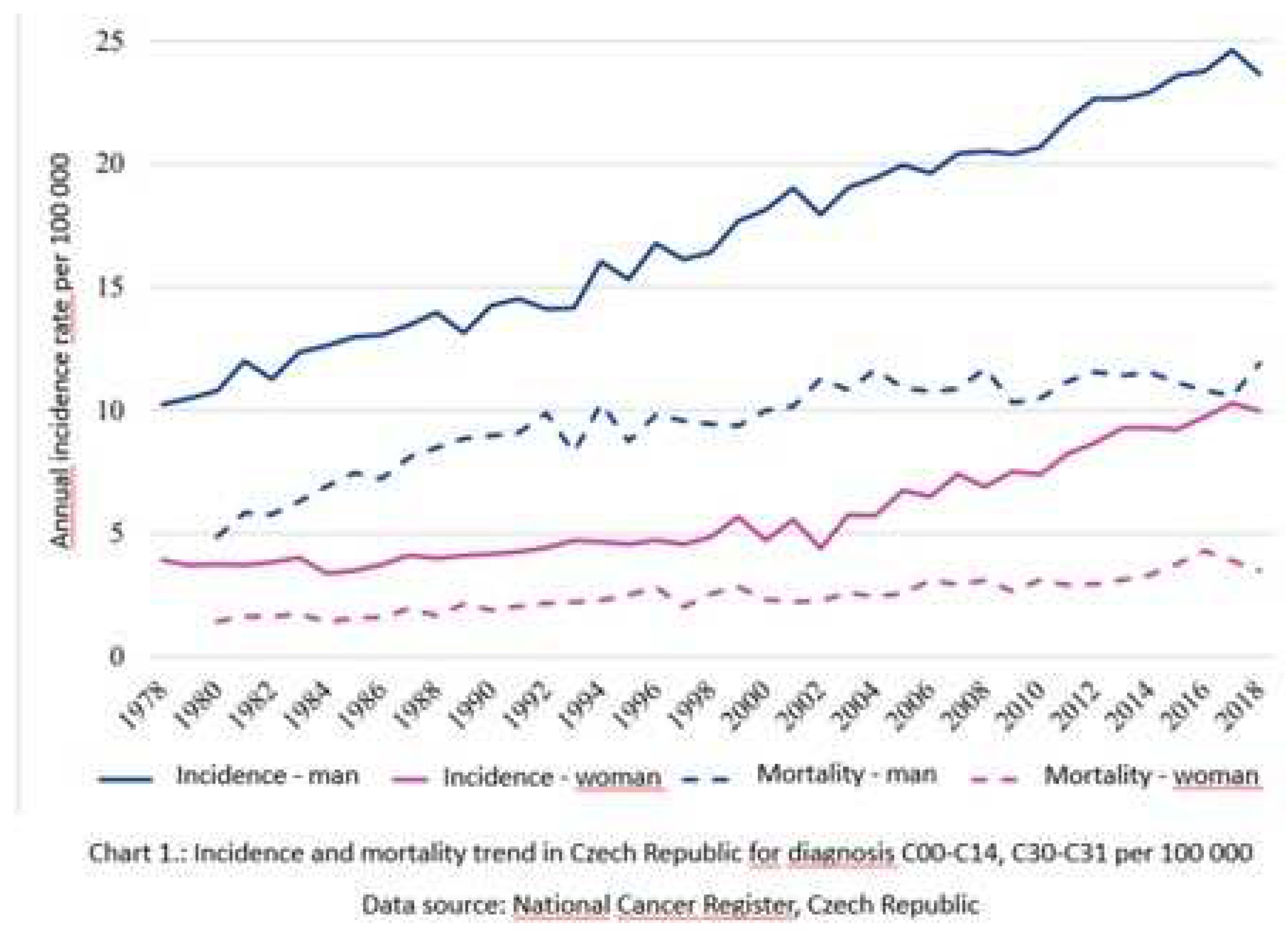
Figure 1.
Optical examination of the oral mucosa affected by the tumor. a) barely visible invasive OSCC of the palato-alveolar area under white light; b) clearly visible loss of fluorescence of the affected area; c) transient marking of the extent of fluorescence loss. d) barely visible invasive OSCC of the right lingual margin under white light; e) clearly visible loss of fluorescence of the affected area; f) transient marking of the extent of fluorescence loss.
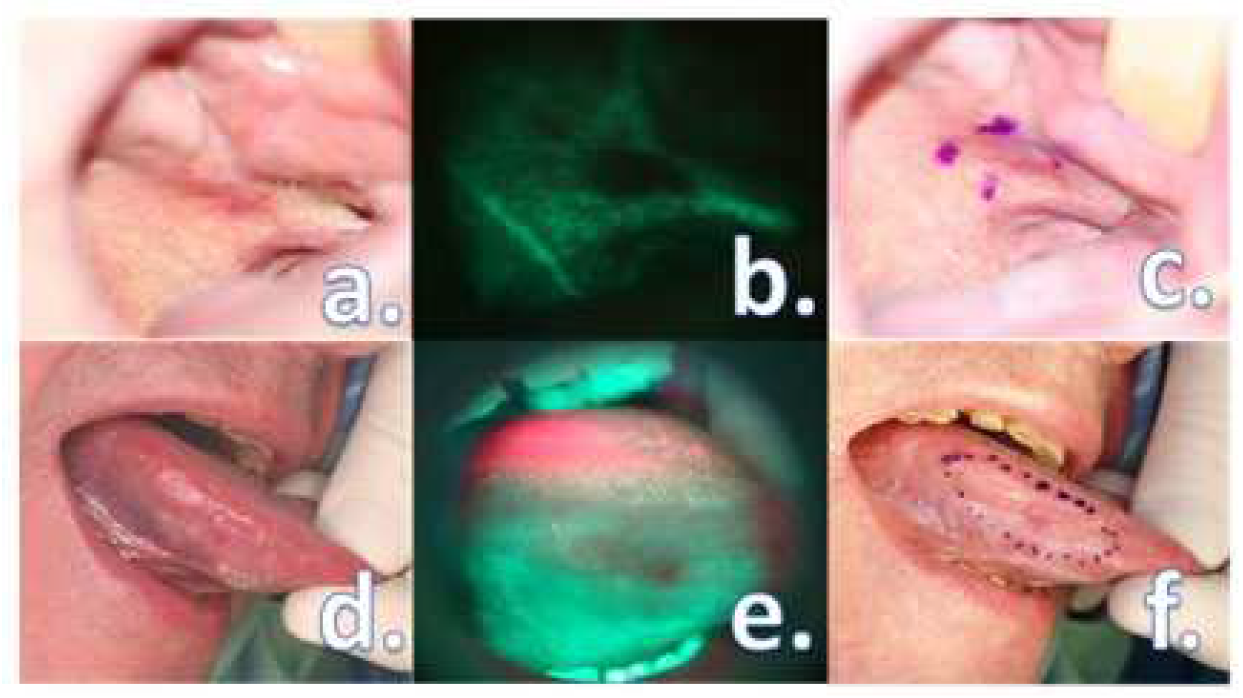
Figure 2.
Permanent marking of loss of fluorescence by tattoo technique. a) various tattoo dyes for immediate specimen orientation; b) a larger syringe filled with a small amount of gentian; insulin syringes filled with a tattoo ink.
Figure 2.
Permanent marking of loss of fluorescence by tattoo technique. a) various tattoo dyes for immediate specimen orientation; b) a larger syringe filled with a small amount of gentian; insulin syringes filled with a tattoo ink.
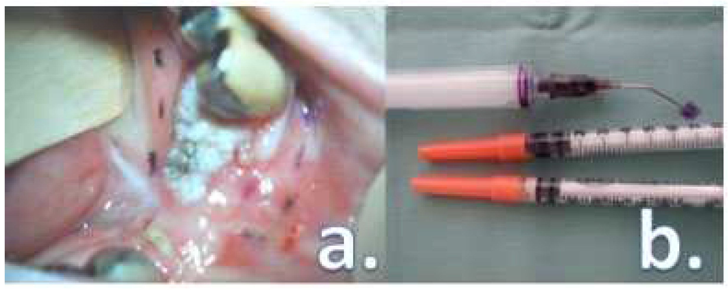
Chart 2.
A box-whisker plot diagram showing the age distribution in both groups.
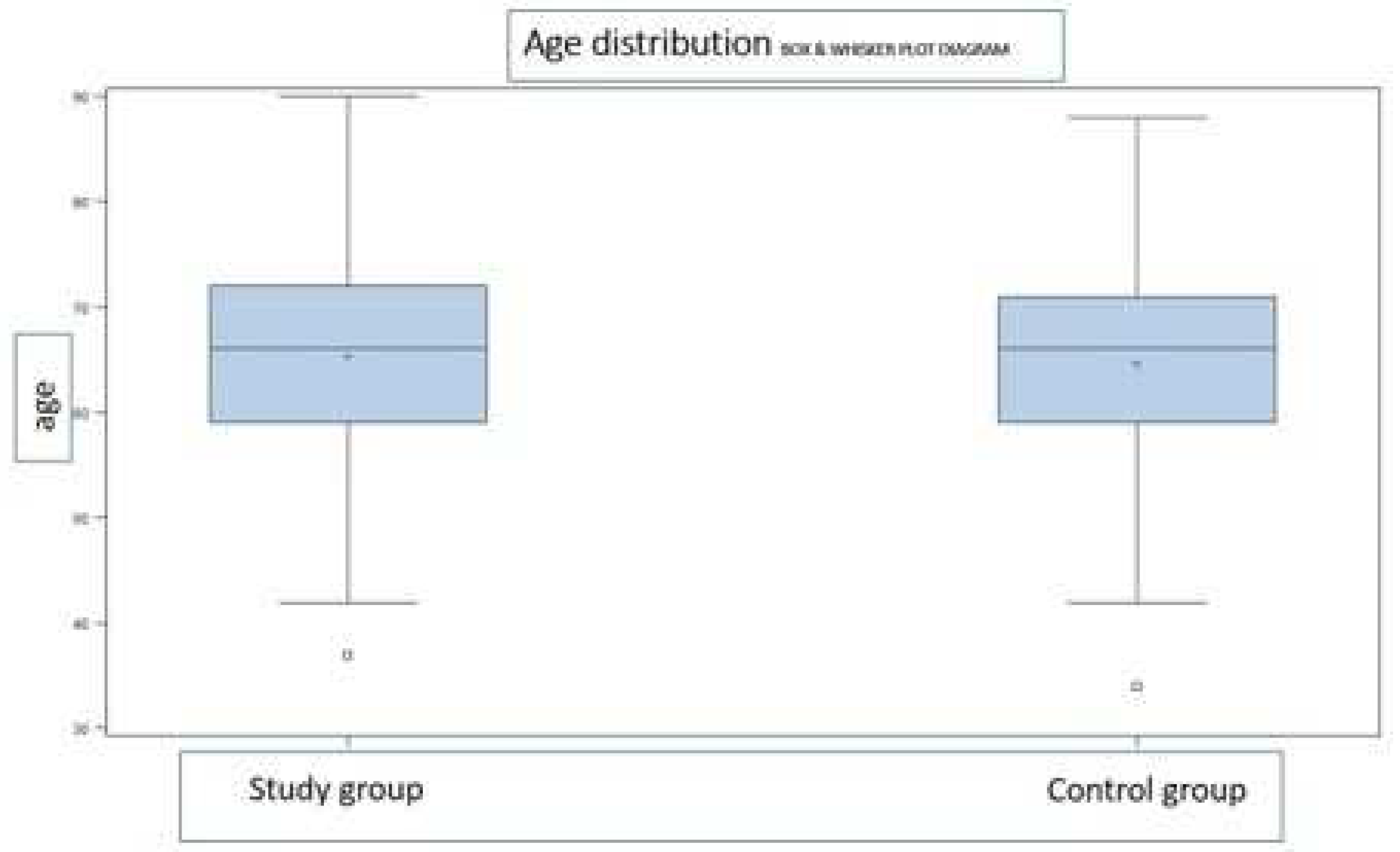
Chart 3.
Mucosal margin status.
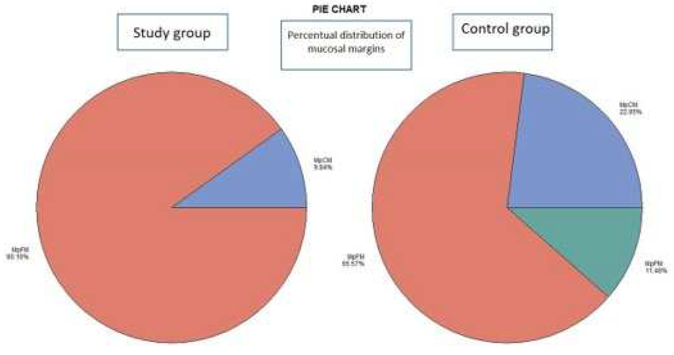
Chart 4.
Deep margin status.
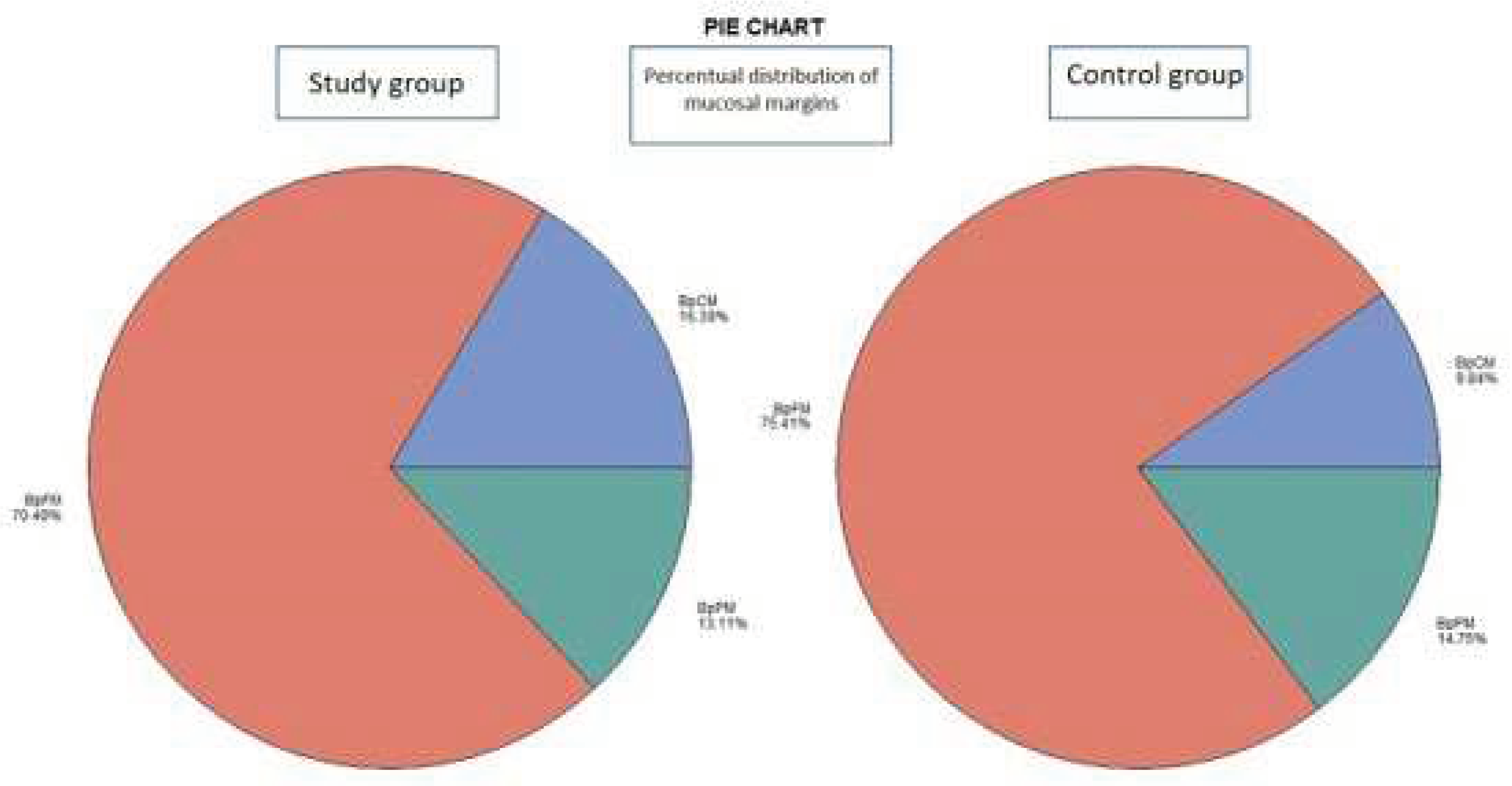
Table 1.
Gender distribution in both groups.
| Gender | Frequency – amount / percentil | Total | |
| Study group | Control group | ||
| Male | 40 / 65.57 | 36 / 59.02 | 76 |
| Female | 21 / 34.43 | 25 / 40.98 | 46 |
| Total | 61 | 61 | 122 |
Table 2.
The distribution of tumor origin in the oral cavity mucosa.
| Diagnosis | Frequency – amount / percentil | Total | |
| Study group | Control group | ||
| C02 | 19 / 31.15 | 21 / 34.43 | 40 |
| C03 | 18 / 29.51 | 14 / 22.95 | 32 |
| C04 | 18 / 29.51 | 18 / 29.51 | 36 |
| C05 | 2 / 3.28 | 1 / 1.64 | 3 |
| C06 | 3 / 4.92 | 7 / 11.48 | 10 |
| C09 | 1 / 1.64 | 0 / 0.00 | 1 |
| Total | 61 | 61 | 122 |
Table 3.
Tumor grade distribution in both groups.
| Grade | Frequency – amount / percentil | Total | |
| Study group | Control group | ||
| G1 | 26 / 42.62 | 28 / 45.90 | 54 |
| G2 | 31 / 50.82 | 25 / 40.98 | 56 |
| G3 | 4 / 6.56 | 8 / 13.11 | 12 |
| Total | 61 | 61 | 122 |
Table 4.
Tumor stage distribution in both groups.
| Stage | Frequency – amount / percentil | Total | |
| Study group | Control group | ||
| I | 16 / 26.23 | 8 / 13.11 | 24 |
| II | 15 / 24.59 | 14 / 22.95 | 29 |
| III | 6 / 9.84 | 13 / 21.31 | 19 |
| Iva | 22 / 36.07 | 23 / 37.70 | 45 |
| IVb | 1 / 1.64 | 3 / 4.92 | 4 |
| IVc | 1 / 1.64 | 0 / 0.00 | 1 |
| Total | 61 | 61 | 122 |
Disclaimer/Publisher’s Note: The statements, opinions and data contained in all publications are solely those of the individual author(s) and contributor(s) and not of MDPI and/or the editor(s). MDPI and/or the editor(s) disclaim responsibility for any injury to people or property resulting from any ideas, methods, instructions or products referred to in the content. |
© 2023 by the authors. Licensee MDPI, Basel, Switzerland. This article is an open access article distributed under the terms and conditions of the Creative Commons Attribution (CC BY) license (http://creativecommons.org/licenses/by/4.0/).
Copyright: This open access article is published under a Creative Commons CC BY 4.0 license, which permit the free download, distribution, and reuse, provided that the author and preprint are cited in any reuse.


