
Submitted:
01 June 2023
Posted:
02 June 2023
You are already at the latest version
Abstract
Chronic wounds are associated with considerable patient morbidity and present a significant economic burden to the healthcare system. Often, chronic wounds are in a state of persistent in-flammation and unable to progress to the next phase of wound healing. Placental-derived bio-materials are recognized for their biocompatibility, biodegradability, angiogenic, an-ti-inflammatory, anti-microbial, anti-fibrotic, immunomodulatory, and immune privileged prop-erties. As such, placental-derived biomaterials have been used in wound management for more than a century. Placental-derived scaffolds are composed of an extracellular matrix (ECM) that can mimic the native tissue, creating a reparative environment to promote ECM remodeling, cell migration, proliferation, and differentiation. Reliable evidence exists throughout the literature to support the safety and effectiveness of placental-derived biomaterials in wound healing. How-ever, differences in source (i.e., anatomical regions of the placenta), preservation techniques, decellularization status, design, and clinical application have not been fully evaluated. This re-view provides an overview of wound healing and placental-derived biomaterials, summarizes the clinical results of placental-derived scaffolds in wound healing, and suggests directions for future work.
Keywords:
biomaterials
; decellularization
; extracellular matrix
; placenta
; placental-derived biomaterials
; wound healing
1. Introduction
Complex, hard-to-heal wounds present a significant clinical challenge and are associated with considerable patient morbidity. Often, chronic wounds are unable to progress past the inflammatory phase of wound healing. Consequently, the wound is burdened by elevated concentrations of pro-inflammatory cytokines and imbalanced proteolytic enzymes and protease inhibitors, resulting in high concentrations of matrix metalloproteinases (MMPs), which destroy the extracellular matrix (ECM) [1, 2].
A promising strategy for the treatment of non-healing wounds is the application of placental-derived biomaterials. Placental-derived biomaterials are known for their biocompatibility, biodegradability, and low immunogenicity [3], making them ideal for use in medical applications. Research has shown that placental-derived biomaterials can be used to promote wound healing by providing an ECM scaffold for tissue repair [4], exerting anti-inflammatory effects [4-6], facilitating cell migration [5, 6], and promoting regeneration [7].
This review provides an overview of wound healing and placental-derived biomaterials, summarizes the clinical results of placental-derived scaffolds in wound healing, and presents directions for future work. The focus of this review is the clinical application of placental-derived scaffolds in wound healing. Although placental-derived cell-based therapy shows promising clinical application, it is beyond the scope of this review.
2. Overview of Wound Healing
Wound healing is the process by which the body repairs and regenerates damaged tissue after an injury. It is a complex and dynamic process that involves a variety of cellular and molecular events, including homeostasis, inflammation, cell migration, proliferation, and tissue remodeling. Wound healing is divided into three distinct phases: the inflammatory phase, the proliferative phase, and the remodeling phase (Figure 1; Table 1) [8].
2.1. Inflammatory Phase
The inflammatory phase of wound healing includes hemostasis and inflammation. The inflammatory phase is initiated immediately following injury to the skin. Wound formation activates a clotting cascade, involving the temporary release of vasoconstrictors to reduce bleeding, and a fibrin clot is formed. The fibrin clot consists of collagen, platelets, thrombin, and fibronectin, which stimulate the release of cytokines and growth factors, such as interleukin-1 (IL-1), tumor necrosis factor alpha (TNF-α), transforming growth factor beta (TGF-β), and platelet factor-4 (PF4) [8]. The fibrin clot serves as a scaffold for infiltrating cells, such as neutrophils, monocytes, fibroblasts, and endothelial cells, and concentrates the growth factors and cytokines [8, 10]. After vasoconstriction, vasodilation occurs, which causes hyperemia and edema [8]. The released chemotactic factors and growth factors complete hemostasis and start inflammation [10].
Neutrophils are first recruited to the wound area and initiate phagocytosis. These cells release reactive oxygen species, proteases, and other enzymes that help debride the wound and remove any debris or bacteria. In addition, these cells release chemokines which serve as chemoattract for other cell types and release pro-inflammatory cytokines. Next, leukocytes, including monocytes, are active in the wound area.
Macrophages are a type of white blood cell that play a key role in the body’s healing response [11]. Approximately 48-96 hours after injury, monocytes differentiate into macrophages [8]. During wound healing, macrophages respond to temporal and spatial cues in their environment by altering their phenotypic polarization [12-14]. The M1 phenotype initiates the inflammatory response [10], and in the later phases of wound healing, the M1 phenotypte transitions to the M2 phenotype, which facilitates tissue remodeling, repair, and the resolution of the healing process [15-17]. In chronic wounds, macrophages are believed to persist in an uncontrolled, pro-inflammatory (M1) activation state [1].
2.2. Proliferative Phase
The proliferative phase of wound healing is the building phase, characterized by epithelialization, angiogenesis, and granulation tissue formation. Fibroblasts and endothelial cells are the primary proliferating cells during this phase. Epithelialization begins as soon as the wound occurs and is stimulated by inflammatory cytokines and growth factors [8, 18]. Keratinocytes are stimulated to migrate into the wound area, proliferate, and differentiate into the epidermis. Angiogenesis occurs simultaneously and is stimulated by local hypoxia, vascular endothelial growth factor (VEGF), platelet-derived growth factor (PDGF), fibroblast growth factor-basic (bFGF) and the serine protease thrombin [10, 18, 19]. Angiogenesis is marked by capillary formation and endothelial cell migration [8]. Lastly, granulation tissue formation occurs. Fibroblasts migrate to the wound site from adjacent tissues and proliferate [8, 10, 18]. In response to PDGF, fibroblasts synthesize a provisional matrix, consisting of type III collagen, glycosaminoglycans, and fibronectin [8].
2.3. Remodeling Phase
During the remodeling phase, granulation tissue formation ends, and wound maturation begins. This phase is characterized by the reorganization of collagen fibers and the contraction of the wound edges. The new tissue is remodeled and organized in an orderly manner to strengthen the repair [18]. Type III collagen is replaced with type I collagen [18], increasing the tensile strength of the wound. Matrix remodeling enzymes, particularly MMPs, play an important role in the remodeling of the local wound environment, as they break down ECM matrix components, such as collagen and elastin, allowing for tissue remodeling and the formation of new blood vessels. Even after a year, however, the wound tissue does not achieve the same strength as that of collagen from uninjured skin due to the formation of scar tissue.
2.4. Wound Formation
When the phases of wound healing are disrupted, abnormal healing occurs, and a chronic wound can develop. A chronic wound is a wound that fails to proceed through a normal, orderly, and timely sequence of repair within an expected timeframe, usually 3 months or longer [20, 21]. Often, these wounds remain in a prolonged state of persistent inflammation and are unable to progress to the next phase of wound healing (Figure 2; List 1) [19]. This state of persistent inflammation is characterized by elevated pro-inflammatory cytokines, dysfunctional macrophages, and high protease concentrations, and an abnormal ECM. In normal wound healing, the ECM regulates macrophage behavior. However, in the chronic wound, a vicious cycle exists between an abnormal ECM and uncontrolled M1 macrophages.
List 1. Characteristics of Persistent Inflammation [1, 2]:
- Elevated pro-inflammatory cytokines
- Dysfunctional macrophages
- Imbalanced proteolytic enzymes and protease inhibitors
- High concentrations of MMPs
- Abnormal ECM.
2.5. Factors That Impair Wound Healing
Impaired healing and wound formation occur when the body's natural healing process is disrupted or compromised [22]. The main risk factors that can lead to chronic wound formation can be grouped into local factors and systemic factors (Table 2) [23]. These factors are often interrelated with systemic factors acting locally to influence wound healing [23]. Local factors include oxygenation, infection, and venous insufficiency [19, 23, 24]. Systemic factors include age, ambulatory status, co-morbidities, medications, oncology interventions, and lifestyle habits [19, 23, 24]. When the healing process is impaired, wounds may take longer to heal or may not heal at all, increasing the risk of complications, such as infection, scarring, and even amputation. Effective treatment may involve addressing the underlying cause of the problem as well as appropriate wound care to support healing and prevent further complications.
Many patients who experience chronic wounds have underlying medical conditions, such as diabetes, venous insufficiency, or obesity. The most common types of chronic wounds include arterial ulcers, diabetic ulcers, pressure ulcers, and venous ulcers [20, 21].
2.6. Economic Impact of Chronic Wounds
It has been estimated that 1-2% of the general population in developed countries will experience a chronic wound [25, 26]. In the United States, chronic non-healing wounds impact 8.2 million Medicare beneficiaries with associated costs ranging from $28.1 to $96.8 billion [22]. The alarming number of patients affected by chronic non-healing wounds is expected to rise, due to the combined effects of an aging population and the rising rates of diabetes and obesity [27-29]. As such, chronic wounds represent a significant economic burden to the healthcare system [30].
2.7. Treatment of Chronic Wounds
The basic tenets of wound care follow the TIME principle: tissue debridement, infection control, moisture balance, and edges of the wound [20, 31]. Once the standard measures have been taken, the ulcer must be diagnosed, and treatment tailored to the specific type of ulcer. Those with arterial ulcers should be directed to a vascular surgeon for immediate attention [20]. Venous ulcers require compression, elevation of the lower limbs, and exercise if possible [20, 32]. Diabetic foot ulcers require offloading, and if necessary, treating any underlying peripheral arterial disease [20, 33, 34]. Pressure ulcers should be managed with a repositioning schedule to reduce pressure on the affected area [20, 35, 36].
While the TIME principle remains a mainstay of treatment, a number of additional therapies have been suggested to improve wound healing (List 2).
List 2. Additional Wound Healing Strategies:
- Negative pressure wound therapy [37, 38];
- Hyperbaric oxygen therapy [39-42];
- Autologous platelet rich plasma [43, 44];
- Growth factors [45, 46];
- Cell therapy [47, 48];
- Scaffolds (e.g., autologous, biologic, synthetic) [49-52].
3. Placental-Derived Biomaterials
3.1. Source of Placental-Derived Biomaterials
The placenta is a vital temporary embryonic and later fetal organ that connects the developing fetus to the maternal uterine wall in humans. Placental-derived biomaterials can be sourced from the amniotic sac, amniotic fluid, placental disc, umbilical cord, or a combination of these sources [53] (Figure 3). This includes not only the tissues themselves, but also the entities within these structures, including fluids, gels, and cells [54].
3.1.1. Amniotic Fluid
The amniotic fluid surrounds the embryo and fetus during development and has a myriad of functions [55]. The amniotic fluid contains a variety of nutrients and growth factors that facilitate fetal growth, provides physical protection by cushioning the fetus and umbilical cord, and has antimicrobial properties, which protect the fetus from infection. The amniotic fluid is primarily composed of water and electrolytes, with signaling molecules, peptides, carbohydrates, lipids, proteins, and hormones making up a small percent [56, 57]. Hyaluronic acid is also suspended within the amniotic fluid, increasing the viscosity [58].
3.1.2. Amniotic Sac
The amniotic sac is a thin semi-transparent membrane that holds the amniotic fluid for the developing fetus [59]. It is composed of an avascular layer, the amnion, and a highly vascularized layer, the chorion (Figure 4) [59, 60].
The amniotic membrane (AM) is the innermost of the two membranes, delimiting the amniotic cavity and bathed in amniotic fluid [61]. The AM measures 0.02 mm – 0.05 mm in thickness and has a multilayered architecture: an epithelium, a basement membrane, and a collagen-rich stromal layer. The epithelium is a monolayer of metabolically active cuboidal cells with microvilli present on the apical surface. The basement membrane is one of the thickest membranes in the human body [3]. It is made up of a rich collagen framework in addition to bioactive molecules, such as fibronectin and laminin [3, 59]. The stromal layer can be further subdivided into three layers: a compact layer, a fibroblast layer, and a spongy layer. The compact layer provides the fibrous structure of the amnion [3]. Interstitial collagens (types I and III) form parallel bundles that provide mechanical integrity, while collagens type V and VI create filamentous connections between the interstitial collagens and the basement membrane [3, 59]. The intermediate spongy layer is composed of a nonfibrillar meshwork of mostly type III collagen as well as an abundance of proteoglycans and glycoproteins [3, 62]. It loosely connects the amnion and chorion membranes, which allows the two membranes to be easily separated by blunt dissection [3] (Figure 5).
The chorion is the outermost membrane of the amniotic sac and is in contact with the amnion on the inner aspect and the maternal decidua on the outer [60]. Like the AM, the chorion membrane also has several layers: a reticular layer, a basement membrane, and a trophoblast layer. The reticular layer is the thickest layer of the chorion and is composed of collagen types I, III, IV, V, and VI and proteoglycans. The basement membrane is a dense layer of connective tissue (type IV collagen, fibronectin, and laminin) that adheres the trophoblasts and the reticular layer [60, 62]. The trophoblast layer interfaces with the maternal decidua on the surface of the placental disc and consists of 2-10 layers of trophoblasts [59, 60].
3.1.3. Placental Disc
The placenta disc provides a link between the developing fetus and the mother, regulating nutrition, waste removal, hormonal balance, and the immune system, while also acting as an immunologically privileged barrier to prevent direct contact between their respective blood supplies [63]. The placental disc is composed of a highly vascularized ECM, containing collagen types I, III, IV and VI as well as a vast distribution of non-collagenous glycoproteins and proteoglycans, such as fibronectin, fibrillin I, laminin, thrombospondin I, tenascin C, decorin, heparan sulfate proteoglycans and elastin [64]. Within the placental disc, a variety of cell types can be found, such as trophoblasts (i.e., syncytiotrophoblasts/cytotrophoblasts), mesenchymal cells and mesenchymal-derived macrophages, fibroblasts, and fetal vascular cells (i.e., vascular smooth muscle cells, perivascular cells, and endothelial cells) [65]. In addition to the various cells types, the placental disc is also rich in nutrients and cytokines [59].
3.1.4. Umbilical Cord
The umbilical cord contains three vessels, the umbilical vein and two umbilical arteries, which are embedded in Wharton’s Jelly and surrounded by a single epithelial layer, derived from the amnion [66, 67]. The umbilical vein transports oxygenated blood from the placenta to the to the fetal heart, and the arteries return deoxygenated blood and waste away from the fetus and to the placenta [67]. Wharton’s Jelly is a mucoid connective tissue, composed of a network of glycoprotein microfibrils and collagen fibrils [68]. Collagen types I, III, V, and VI have been identified in Wharton’s Jelly [69, 70]. Hyaluronic acid, the most abundant glycosaminoglycan in Wharton's Jelly [69], creates a hydrated gel around the fibroblasts and collagen fibrils, which maintains the architecture of the umbilical cord and provides protection from pressure [69, 71, 72]. The cell population of Wharton’s Jelly includes fibroblast-like cells, myofibroblast-like cells, and mesenchymal stem cells [71, 73, 74].
3.2. Properties of Placental-Derived Biomaterials
The human placenta is a temporary vital organ that is usually discarded as medical waste, making it an easily accessible, cost effective, and ethical source of raw material. In addition to its availability, the placenta possesses several desirable biological properties that are innate to healing, including angiogenic, anti-inflammatory, anti-microbial, anti-fibrotic, and immunomodulatory with low immunogenicity (List 3). Complementary to the desirable biological properties, placental tissues have unique ECMs with notable structural and mechanical properties, including elasticity, stiffness, and tensile strength [75]. However, ECM composition varies with source, as described in the previous section.
List 3. Biologic Properties of the Placenta:
3.3. Differences among Placental-Derived Biomaterials
Despite a growing body of evidence demonstrating that placental-derived biomaterials have the capacity to enhance healing, the methods of processing and preparing the tissue are continually evolving. Variations in tissue source, preservation, decellularization, design, and application have the potential to affect the biological and mechanical characteristics of the tissue.
As previously noted, placental-derived biomaterials can be derived from a variety of sources, including the amniotic sac (e.g., amnion, chorion), amniotic fluid, umbilical cord (e.g., umbilical cord blood, umbilical cord tissue, Wharton’s Jelly) or a combination of these sources [53]. While this provides a plethora of biomaterials, it also introduces significant variability. Differences in composition exist between sources. For example, the AM has a collagen-rich ECM and contains several bioactive ECM molecules, such as fibronectin, laminin, elastin, and glycosaminoglycans [87], while Wharton’s Jelly is a mucoid connective tissue, composed of a network of glycoprotein microfibrils and collagen fibrils [68]. In addition, research has shown that the immunomodulatory properties are source dependent [88]. Even when comparing biomaterials from the same source, interdonor and intradonor variability exists [89].
3.4. Preservation Method
Following delivery, the placenta is usually discarded as medical waste. Alternatively, placental-derived biomaterials can be obtained following normal, healthy, full-term pregnancies. Comprehensive screening of the donors is required before the tissue is procured and processed. With appropriate consent, the placenta is collected after delivery and donated. After the tissue is collected, the placenta is transported for processing. Although the specific testing requirements may vary depending on local and regional guidelines, proper screening is required to test for infectious diseases, such as Human Immunodeficiency Virus, Hepatitis B Virus, Hepatis C Virus, West Nile, and Syphilis. The tissue is sterilized to minimize the risk of disease transmission to recipients and is processed to allow prolonged storage. Tissue preservation is usually accomplished by one of several techniques, most commonly cryopreservation, dehydration, or lyophilization [61]. It is important to note that all methods of preservation compromise the tissue's integrity to varying degrees.
Cryopreservation or freezing is the most widely used method of tissue preservation. Cryopreservation is a process in which the structure and function of cells, tissues, or organs are preserved by cooling the samples to very low temperatures [90, 91]. As part of the cryopreservation process, cryoprotectants are mixed with cells and tissues to reduce ice crystal formation. Various cryoprotectants have been used to preserve placental-derived biomaterials, including glycerol, dimethylsulfoxide (DMSO), and ethylene glycol. However, the cryopreservation method is criticized for impairing the viability and proliferative capacity of cells and also requires the tissue to be shipped and stored at -80°C [92].
Dehydration is an alternate method of tissue preservation that removes the water from the tissue sample. This process helps to preserve the structural and biochemical activity of placental tissue and prevent the growth of microorganisms and the breakdown of the tissue. Dehydration is achieved using air or heat to dehydrate the tissue [59]. Unlike cryopreservation, dehydration preserves the tissue without the need for freezers, dry ice, or liquid nitrogen and can be shipped and stored at room temperature with a 5+ year shelf-life. The dehydration of tissue provides an additional advantage of allowing allografts to be terminally sterilized, thus reducing the risk of infectious disease transmission from the donor tissue.
Lyophilization or freeze drying involves cooling the tissue to −80°C and using a sublimation process to remove the water by vacuum desiccation [93]. Like cryopreservation, the freezing step in this process can cause ice crystal formation and damage to the tissues. To reduce tissue damage, sugars can be used as a cryoprotectant to stabilize the proteins [94]. Similar to dehydration, lyophilization allows the tissues to be shipped and stored at room temperature without the need for freezers or liquid nitrogen [93].
3.5. Decellularization
Decellularization is a process by which the cellular components of a tissue or organ (e.g., endogenous cells, cell debris, and genetic materials) are removed, while the structural and regulatory proteins of the ECM are preserved (Figure 6).
This process is used to create acellular tissues and organs for use in regenerative medicine and tissue engineering applications. The elimination of cellular content from natural tissue-derived matrices has the potential to promote healing, integration with host tissues, and limit a foreign body reaction [96]. The decellularization process typically involves the use of chemicals, enzymes, physical forces, and/or a combination of these methods to remove cellular components, while preserving the ECM [97, 98]. Methods of decellularization affect the structure of the ECM, the structure of the tissue, and the biomechanical behavior. Therefore, it is important to find methods that balance the removal of cellular content with the retention of the structures and entities within the ECM. Several methods have been used in the decellularization of placental tissues [9, 98-101]. Table 3 summarizes the methods and agents used to achieve an acellular scaffold.
As indicated, tissue preservation and decellularization methods have specific advantages and disadvantages. The processing of placental tissues aims to remove any hazardous materials, while preserving the structural and biochemical activity of the tissue to optimize healing.
3.6. Clinical Application
The clinical application of placental-derived biomaterials also varies in form, administration, and delivery system. Placental-derived biomaterials are available in many forms, including sheet scaffolds, injectables, extracts, and cells (Figure 7) [54]. Reported administrations include topical application, intradermal/subcutaneous injection, and intravenous or intraperitoneal injection [48]. Delivery systems include hydrogels, synthetic or natural biomaterials as carriers for transplanted cells, and extracts or secretomes [48].
Several cell types exist within the placenta and have different mechanisms of action, which are source-dependent [88]. While the therapeutic benefit of placental-derived mesenchymal stem cells in wound healing has been described throughout the literature [114-116], it is not a subject of this review. Rather, this review focuses on the use of human placental-derived biomaterials as scaffolds for wound healing. Placental-derived biomaterials are available as scaffolds in two principal forms: sheets and injectables [117].
The human AM is one of the most widely used and studied placental-derived biomaterials. Human AM sheets are processed to retain the native ECM structure with its high collagen content and key bioactive molecules, such as fibronectin, laminin, glycosaminoglycans, and elastin [87]. Some scaffolds undergo proprietary processing procedures to retain the native growth factors and cytokines [87], while others are decellularized to remove all residual cells, cell debris, growth factors, and cytokines [118]. Sheet scaffolds are commonly used as a wound covering and can be composed of amnion alone [119, 120], amnion and chorion [121-125], umbilical cord alone [125-130], or umbilical cord and amnion [131]. The sheet ECMs possess the properties associated with their tissue of origin [54]. For example, AM sheet scaffolds are thought to promote healing via epithelialization [132-134], reduction of inflammation [6, 77-79], inhibition of scar tissue formation [83, 84], and the ability to act as an antimicrobial agent [80-82]. In addition, sheet scaffolds are modified to improve the mechanical properties of the tissue and are available in many configurations, including single layer and multilayer [6], full-thickness, and composite grafts. For example, lamination of sheet scaffolds is performed to create biomaterials with improved handleability and tensile strength [6, 135].
Placental-derived biomaterials are also available in an injectable form as suspension allografts [136] and particulates/micronized powders [117, 137, 138]. The micronized powders can be applied directly to the wound or can be rehydrated and injected [137]. Commercially available products are sourced from the amnion, chorion, amniotic fluid, umbilical cord, the placental disc, or a combination of these sources. Similar to the sheet scaffolds, the micronized form provides a connective tissue matrix complete with regulatory proteins, which stimulate cell migration [5], proliferation [5], and epithelialization [139-141]. Unlike the sheet scaffolds, the injectable form offers the added benefits of being minimally invasive with the capacity to fill irregular spaces [117, 138].
3.7. Commercial Products
Placental-derived allografts are processed from human tissue according to the American Association of Tissue Banks (AATB) standards and are regulated as a Human Cell, Tissue, or Cellular or Tissue-based Product (HCT/P) by the US FDA under section 361 of the Public Health Service act as HCT/P (21 CFR, Part 127.10a). According to these guidelines, at a minimum, these products must be minimally manipulated, not combined with drugs or devices, and not reliant on cell metabolic activity as a primary function. In 2017, the company AmnioChor provided a list of placental-derived tissue products sold under section 361 [142]. In total, 116 products were listed. Table 4 and Table 5 provide an updated list of the commercially available placental-derived scaffolds, intended for wound healing applications. This list is not exhaustive and therefore does not represent all of the commercially available HCT/P under section 361. However, it does provide a scope of the available products and variations in source, preservation methods, decellularization status, and unique design characteristics, where applicable.
4. Placental-Derived Biomaterials in Wound Healing: Clinical Results
Given the innate healing properties of the placenta, placental-derived biomaterials have been investigated as advanced wound care therapies for more than 100 years. The first clinical application of placental-derived biomaterials was reported in 1910, when Davis used AM as a substrate for skin transplantation [143]. At that time, only fresh AM was available, which was difficult to procure and carried a significant risk of disease transmission. With advancements in tissue processing and preservation, the use of these biomaterials has expanded considerably and now includes applications in tissue engineering, regenerative medicine, and cell-based therapies [3, 137].
4.1. Outcomes
Randomized Controlled Trials (RCTs) provide the most reliable evidence for determining the effectiveness of a treatment/intervention. A comprehensive literature search was performed to identify RCTs published in the last ten years that evaluated the application of placental-derived scaffolds to treat non-healing wounds. The PubMed database was queried for the terms: “placenta matrix wound healing,” “amnion wound healing,” “chorion wound healing,” “umbilical cord wound healing,” and “amniotic fluid wound healing.” Inclusion criteria included randomized controlled trials reporting on the use of placental-derived scaffolds to treat non-healing wounds, clinical outcomes, and human subjects. Exclusion criteria included animal data, basic science studies, review articles, articles with inadequate sample sizes for statistical analysis, studies evaluating skin grafts, burn wound healing, and punch biopsy wounds, and non-English language literature. The search was limited to the previous 10 years. A summary of the results is provided in Table 6.
Although a large percentage of the randomized controlled trials published within the last 10 years focused on the application of dehydrated human amnion/chorion membranes (dHACMs), the literature also includes studies evaluating the application of dehydrated amnion powder, dried human AM, hypothermically stored AM, and umbilical cord allografts in the treatment of hard-to-heal ulcers. The research consistently demonstrates the effectiveness of placental-derived biomaterials to treat diabetic foot ulcers (DFUs) and venous leg ulcers (VLUs). Several of these high level studies compared the application of placental-derived biomaterials with standard wound care and demonstrated that the application of placental-derived biomaterials significantly improves the proportion of healed ulcers [121, 124, 126, 144-152], time to healing and rates of healing [121, 124, 126, 146, 147, 149-151, 153], and ulcer size [145, 152-155]. In addition, Selena and colleagues [148] reported that 79.5% of patients treated with dehydrated human amnion/chorion membrane (dHACM) and multilayer compression therapy reported reduced VLU pain, compared with 52.4% patients who were treated with multilayer compression therapy alone. This finding suggests that the application of placental-derived biomaterials may also alleviate the pain associated with VLUs.

Table 4.
Placental-derived sheet scaffolds for wound healing.
| # | Company | Product | Source | Preservation Method | Decellularization Status | Unique Design Elements | Reference(s) |
|---|---|---|---|---|---|---|---|
| 1 | AlloSource | AlloWrap® DS | A | Dry; Proprietary Technology | Non-decellularized | Dual Layers, omnidirectional implantation | [156] |
| 2 | Amniox Medical, Inc. | Neox® 1K | UC | Cryopreservation; CryoTek® | Non-decellularized | ||
| 3 | Amniox Medical, Inc. | Neox® 100 | A | Cryopreservation; CryoTek® | Non-decellularized | ||
| 4 | Amniox Medical, Inc. | Neox® Cord RT | UC & A | Dehydration; SteriTek® | Non-decellularized | [129, 157, 158] | |
| 5 | Applied BiologicsTM | Xwrap® | A | Dehydration | Non-decellularized | Chorion free | |
| 6 | Celularity Inc. | Biovance® | A | Dehydration | Decellularized | Decellularized | [4, 31, 87, 119, 120, 159] |
| 7 | Celularity Inc. | Biovance3L® | A | Dehydration | Decellularized | Tri-layer | |
| 8 | Integra Life Sciences | AmnioExcel® | A | Dehydration | Non-decellularized | [7, 160-168] | |
| 9 | Integra LifeSciences | BioDFence® G3 | A & C | Proprietary method | Non-decellularized | Tri-layer (amnion-chorion-amnion) | |
| 10 | Integra LifeSciences | BioDDryFlex® | A | Dehydration | Non-decellularized | ||
| 11 | Integra LifeSciences | BioFix® | A | Dehydration | Decellularized | Omnidirectional placement | |
| 12 | Integra LifeSciences | BioFix® Plus | C | Dehydration; HydraTek® | Decellularized | Omnidirectional placement | |
| 13 | MiMedx Group, Inc. | EPIFIX® | A & C | Dehydration; HydraTek® | Non-decellularized | Retains cytokines & growth factors | [118, 169] |
| 14 | MiMedx Group, Inc. | EPICORD® | UC | Dehydration; PURION® | Non-decellularized | Expandable | [118, 169] |
| 15 | MiMedx Group, Inc. | AMNIOCORD® | UC | Dehydration; PURION® | Non-decellularized | 250+ Regulatory Proteins | [170] |
| 16 | MiMedx Group, Inc. | AMNIOEFFECT® | A, IL & C | Dehydration; PURION® | Non-decellularized | 300+ regulatory proteins | |
| 17 | MiMedx Group, Inc. | AmnioFix® | A & C | Lyophilization; PURION® | Non-decellularized | 300+ regulatory proteins | [118, 169] |
| 18 | MTF Biologics | AmnioBand | PT | Dehydration; PURION® | Non-decellularized | ||
| 19 | Organogenesis | Affinity® | A | Dehydration; Proprietary aseptic method | Non-decellularized | Fresh allograft derived from amnion tissue | [145, 171] |
| 20 | Organogenesis | NuShield® | A & C | Hypothermically stored | Non-decellularized | LayerLoc™ preserves spongy layer | [123, 172-174] |
| 21 | Skye Biologics, Inc., | WoundEx®45 | A | Dehydration; LayerLoc™ | Non-decellularized | Thin | |
| 22 | Skye Biologics, Inc., | WoundEx®200 | C | Dehydration | Non-decellularized | Thick | |
| 23 | Skye Biologics, Inc., | WoundFix™ | A | Dehydration | Non-decellularized | ||
| 24 | Smith & Nephew | GRAFIX® PL | A & C | Dehydration | Non-decellularized | Retains native cells and growth factors | |
| 25 | Smith & Nephew | GRAFIX® | A & C | Lyopreservation | Non-decellularized | Retains native cells and growth factors | [151, 175, 176] |
| 26 | Smith & Nephew | STRAVIX® PL | UC | Cryopreservation | Non-decellularized | ||
| 27 | Smith & Nephew | STRAVIX® | UC | Lyopreservation | Non-decellularized | [177] | |
| 28 | StimLabs, LLC | Revita® | A, IL & C | Cryopreservation | Non-decellularized | ||
| 29 | StimLabs, LLC | Cogenex® | A, IL & C | Dehydration; Clearify™ | Non-decellularized | Fenestrated | |
| 30 | StimLabs, LLC | Enverse® | A, IL & C | Dehydration | Non-decellularized | Translucent | |
| 31 | StimLabs, LLC | Vialize® | A, IL & C | Clearify™ | Non-decellularized | Lyophilized | |
| 32 | Tides Medical | Artacent® Wound | A | Dehydration; Clearify™ | Non-decellularized | Dual layer | |
| 33 | Ventris Medical | CellestaTM | A | Dehydration; Clearify™ | Non-decellularized | Poly mesh backing | |
| 34 | Vivex | CYGNUS® Solo | A | Dehydration | Non-decellularized | Single Layer | |
| 35 | Vivex | CYGNUS® Matrix | A & C | Artacleanse® | Non-decellularized | ||
| 36 | Vivex | CYGNUS® Max | UC | Clearant™ | Non-decellularized | ||
| 37 | Vivex | CYGNUS® Max XL | UC | Dehydrated; INTEGRITY PROCESSING™ | Non-decellularized | Fenestrated |
Abbreviations: A, amnion; C, Chorion; IL, intermediate layer; PT, placental tissue; UC, umbilical cord.
Table 5.
Placental-derived injectable scaffolds for wound healing.
| # | Company | Product | Source | Preservation Method | Decellularization Status | Unique Design Elements | Reference(s) |
|---|---|---|---|---|---|---|---|
| 1 | AediCell | Dermavest®/ Plurivest® | PD, A, C & UC | Dehydration | Decellularized | ||
| 2 | Applied Biologics™ | FLŌGRAFT® | AF | Cryopreservation | Non-decellularized | ||
| 3 | Celularity Inc. | Interfyl® | C | Dehydration | Decellularized | Decellularized | [137] |
| 4 | Integra LifeSciences | AmnioMatrix® | A & AF | Cryopreservation | Non-decellularized | ||
| 5 | Integra LifeSciences | BioDFactor® | A & AF | Cryopreservation | Non-decellularized | ||
| 6 | Integra LifeSciences | BioFix® Flo | PT | Dehydration; HydraTek® | Decellularized | ||
| 7 | MiMedx Group, Inc. | AmnioFill® | A & C | Dehydration; Purion® | Non-decellularized | 300+ regulatory proteins | [118] |
| 8 | MiMedx Group, Inc. | AxioFillTM | PD | Dehydrated; Purion® | Decellularized | ||
| 9 | Skye Biologics, Inc. | BioRenewTM | PT | Cryopreservation | Non-decellularized | Growth factors | [136] |
| 10 | Skye Biologics, Inc. | WoundEx®Flow | PT | Dehydration | Non-decellularized | ||
| 11 | Ventris Medical | CellestaTM Flowable | A | Clearant™ | Non-decellularized |
Abbreviations: A, amnion; AF, amniotic fluid; C, Chorion; IL, PD, placental disc; PT, placental tissue; UC, umbilical cord.
.
To better understand ideal application strategy, two studies evaluated the frequency of placental-derived biomaterial application [148, 152]. In 2014, Serena and colleagues [152] conducted a multicenter study evaluating the use of dHACM and multilayer compression therapy versus multilayer compression therapy alone in the treatment of VLUs. As a secondary aim, the study compared the proportion of VLUs demonstrating ≥40% closure at 4 weeks in patients receiving one application of dHACM versus two applications of dHACM. After 4 weeks, the proportion of wounds demonstrating a ≥40% closure was similar with one or two applications (62% and 63%, respectively). However, the lack of a significant difference in dHACM application frequency may be attributable to the short study period of 4 weeks. In 2014, Zelen and colleagues [148] conducted a similar study over 12 weeks to determine if weekly application of dHACMs reduce the time to healing more effectively than biweekly applications for the management of DFUs. Although the proportion of DFUs that achieved complete healing were similar between the two groups, DFUs receiving weekly application of dHACM healed significantly faster than those receiving biweekly dHACM application. These results suggest that more frequent application reduces the time to healing. However, additional studies are needed to determine the optimal application frequency for placental-derived biomaterials.
Given the significant economic burden associated with treating DFUs and VLUs, investigators have analyzed the economic impact of treating ulcers with placental-derived products [147, 149, 150, 178]. In each of these analyses, the results demonstrate that application of placental-derived biomaterials is a cost-effective treatment for ulcers. For example, in 2015, Zelen and colleagues [149] conducted an interim analysis of 60 patients and compared the application of Apligraf® (Organogenesis, Inc., Canton, MA) and EpiFix® (MiMedx Group Inc.,Marietta, GA) for the treatment of DFUs. The study found that the application of dHACM significantly reduced the median number of grafts (2.15 vs 6.2 grafts) as well as the median graft cost per healed wound ($1,669 vs $9,216). The study was continued, expanding the cohort to 100 patients, and again dHACM was found to significantly reduce the median number of grafts (2.5 vs 6 grafts) and the median graft cost per healed wound ($1,517 vs $8,918) [150]. Using the data from a previously published study by DiDomenico and colleagues [147], Carter [178] conducted an economics health study to estimate the cost-utility of an aseptically processed dehydrated human amnion and chorion allograft (dHACA) (AmnioBand®, Musculoskeletal Transplant Foundation [MTF], Edison, New Jersey) plus standard wound care versus standard wound care alone. This data modeling study demonstrated that the use of a dHACA combined with standard wound care compared with standard wound care alone is a cost-effective treatment for DFUs. When collectively considered, these results confirm superior resource utilization with the application of placental-derived biomaterials.
RCTs have been criticized for excluding a large percentage of the population, due to strict inclusion and exclusion criteria and therefore, not generalizing to the population at large [119]. Although not randomized or controlled, in 2015, Smiell and colleagues [119] conducted a real-world multicenter trial, evaluating wound closure, following treatment of uninfected, full- and partial-thickness wounds with a decellularized dehydrated human amniotic membrane (DDHAM). Chronic wounds included venous ulcers, diabetic ulcers, pressure ulcers, arterial ulcers, and collagen vascular ulcers of varying severity.
Table 6.
Randomized Controlled Trials Evaluating Placental-derived Biomaterials in Wound Healing.
| # | Study Details | Purpose | Results Summary | Conclusion |
|---|---|---|---|---|
| 1 | Study: Mohammadi Tofigh et al. 2022 [154] Study Design: Prospective, Single Center Wound Type: DFU Placental-derived Biomaterial: dAP (AMOR) Patients: 243 (81 dAP, 81 PDGF gel, 81 debridement) |
To compare the therapeutic effects of the three methods of diabetic wound care: surgical debridement and dressing, dressing with dAP, and dressing with PDGF gel | Percent area reduction was significantly different between dehydrated amnion, PDGF gel, and debridement at 4 weeks (49.3 vs 14.8 vs 7.4%), 6 weeks (79 vs 35.8 vs 20.1%), 8 weeks (86.4 vs 56.8 vs 43.7%), weeks 10 and 12 (87.6 vs 61.7 vs 50%) Similar safety profiles between groups |
Shows improved healing with application of dehydrated amnion powder in DFU patients, compared with platelet-derived growth factor dressing and surgical debridement |
| 2 | Study: Serena et al. 2022 [144] Study Design: Prospective, Multicenter Wound Type: VLU Placental-derived Biomaterial: dHACA (AmnioBand®) Patients: 60 (40 dHACA, 20 SOC) |
To evaluate the safety and effectiveness of weekly and biweekly applications of dHACA plus SOC compared to SOC alone on chronic VLUs | Significantly higher proportion of healed VLUs in the two dHACA groups than SOC (75% vs 30%) No significant differences in the proportion of ulcers achieving 40% closure at 4 weeks AE Rate: 63.5%; no graft or procedure related AEs |
Shows dHACA and SOC, either applied weekly or biweekly, healed significantly more VLUs than SOC alone, suggesting that the use of aseptically processed dHACA is a safe and effective treatment option in the healing of chronic VLUs |
| 3 | Study: Game et al. 2021 [155] Study Design: Prospective, Multicenter Wound Type: DFU Placental-derived Biomaterial: dHAM® (Omnigen) Patients: 31 (15 dHAM, 16 SOC) |
To investigate whether 2 weekly additions of the dHAM to standard care versus standard care alone increased the proportion of healed participants' DFUs within 12 weeks | Similar proportion of healed DFUs for dHAM and SOC (27 vs 6.3%) Percent wound area reduction was significantly higher in the dHAM group No difference in AEs |
Shows dHAM preparation is safe treatment for DFUs |
| 4 | Study: Carter, 2020 [178] Study Design: Health Economics Study Wound Type: DFU Placental-derived Biomaterial: dHACA (AmnioBand®) Patients: 80 (40 dHACA, 40 SOC) |
To estimate the cost-utility of an aseptically processed dHACA plus SOC versus SOC alone based on a published randomized controlled trial in which patients who had an eligible Wagner 1 DFU wound were randomized to either of these treatments | ICER at 1 year for group 1 versus group 2 was -$4,373 Group 1 had 69.2% lower cost values with increased positive incremental effectiveness for 94.9% of values A willingness to pay curve showed that about 92% of interventions were cost effective for group 1 when $50,000 was paid |
Demonstrates dHACA added to SOC compared to SOC alone is a cost-effective treatment for DFUs |
| 5 | Study: Serena et al. 2020 [145] Study Design: Prospective, Multicenter Wound Type: DFU Placental-derived Biomaterial: HSAM (Affinity®) Patients: 76 (38 HSAM, 38 SOC) |
To determine the effectiveness of HSAM versus SOC in DFUs | Proportion of wound closure for HSAM was significantly greater at 12 (55 vs 29%) and 16 (58 vs 29%) weeks Incidence of ulcers achieving >60% reductions in area and depth was significantly greater for HSAM (area: 82 vs 58%; depth: 65 vs 39%) |
Demonstrates an increased frequency and probability of wound closure in DFUs with HSAM versus SOC |
| 6 | Study: Tettelbach et al. 2019 [124] Study Design: Prospective, Multicenter Wound Type: DFU Placental-derived Biomaterial: dHACM (EpiFix®) Patients: 110 ITT (54 dHACM, 56 SOC); 98 PP (47 dHACM, 51 SOC) |
To confirm the efficacy of dHACM for the treatment of chronic lower extremity ulcers in persons with diabetes | Significantly higher proportion of complete wound closure in 12 weeks for dHACM (ITT: 70 vs 50%; PP: 81 vs 55%) A Kaplan-Meier analysis showed a significantly improved time to healing with dHACM Higher proportion of wound remained closed at 16 weeks for dHACM (95 vs 86%) 230 AEs; 3 possibly product-related |
Confirms dHACM is an efficacious treatment for lower extremity DFUs |
| 7 | Study: Bianchi et al. 2019 [146] Study Design: Prospective, Multicenter Wound Type: VLU Placental-derived Biomaterial: dHACM (EpiFix®) Patients: 128 (64 dHACM, 64 Control) |
To report ITT results and assess if both ITT and PP data analyses arrive at the same conclusion of the efficacy of dHACM as a treatment for VLU | Kaplan-Meier plot of time to heal showed a superior wound healing trajectory for dHACM in both ITT and PP Proportion of healed ulcers was significantly greater for dHACM (ITT: 50 vs 31%; PP: 60 vs 35%) 65 AEs; none related to dHACM or study procedures |
Provides an additional level of assurance regarding the effectiveness of dHACM |
| 8 | Study: Tettelbach et al. 2019 [126] Study Design: Prospective, Multicenter Wound Type: DFU Placental-derived Biomaterial: dHUC (EpiCord®) Patients: 155 (101 dHUC, 54 Alginate); 134 PP (86 dHUC; 48 Alginate); 107 AD (67 dHUC; 40 Alginate) |
To determine the safety and effectiveness of dHUC allograft compared with alginate wound dressings for the treatment of chronic, non-healing DFUs | Proportion of patients with complete wound closure at 12 weeks was significantly greater for dHUC (70 vs 48%) Proportion of AD patients with complete wound closure at 12 weeks was significantly greater for dHUC (96 vs 65%) Rate of healing at 12 weeks in PP patients was significantly greater for dHUC (81 vs 54%) 160 AEs; none related to dHUC or alginate dressing |
Demonstrates the safety and efficacy of dHUC as a treatment for non-healing DFUs |
| 9 | Study: DiDomenico et al. 2018 [147] Study Design: Prospective, Multicenter Wound Type: DFU Placental-derived Biomaterial: dHACA (AmnioBand®) Patients: 80 (40 dHACA, 40 SOC) |
To compare dHACA with SOC in achieving wound closure in non-healing DFUs | Higher proportion of healed DFUs at 12 weeks for dHACA (85 vs 33%) Significantly faster mean time to heal for dHACA (37 vs 67 days) Mean number of grafts used per healed DFU was 4.0 Mean graft cost per healed DFU was $1,771 11 AEs; None dHACA related |
Shows aseptically processed dHACA heals DFUs significantly faster than SOC at 12 weeks |
| 10 | Study: Bianchi et al. 2018 [121] Study Design: Prospective, Multicenter Wound Type: VLU Placental-derived Biomaterial: dHACM (EpiFix®) Patients: 109 (52 dHACM, 57 Control) |
To evaluate the efficacy of dHACM as an adjunct to multilayer compression therapy for the treatment of non-healing full-thickness VLUs | Kaplan-Meier plot of time to heal showed a superior wound healing trajectory for dHACM Proportion of healed ulcers was significantly greater for dHACM at 12 weeks (60 vs 35%) and 16 weeks (71 vs 44%) 65 AEs; none related to dHACM or study procedures |
Confirms dHACM as an adjunct to multilayer compression therapy for the treatment of non-healing, full-thickness VLUs |
| 11 | Study: Zelen et al. 2016 [150] Study Design: Prospective, Multicenter Wound Type: DFU Placental-derived Biomaterial: dHACM (EpiFix®) Patients: 100 (33 Apligraf, 32 dHACM, 35 SOC) |
To compare clinical outcomes at 12 weeks in 100 patients with chronic lower extremity DFUs treated with weekly applications of Apligraf®, dHACM, or SOC with collagen-alginate dressing as controls | Significantly higher proportion of healed ulcers for dHACM versus Apligraft versus SOC (97 vs 73 vs 51%) Significantly faster time to healing for dHACM versus Apligraft versus SOC (23.6 vs 47.9 vs 57.4 days) Median number of grafts per healed wound was significantly lower for dHACM (2.5 vs 6) Median graft cost per healed wound was significantly lower for dHACM ($1,517 vs $8,918) 10 AEs; none dHACM related |
Provides further evidence of the clinical and resource utilization superiority of dHACM for the treatment of lower extremity DFUs |
| 12 | Study: Zelen et al. 2015 [149] Study Design: Prospective, Multicenter Wound Type: DFU Placental-derived Biomaterial: dHACM (EpiFix®) Patients: 60 (20 Apligraf, 20 dHACM, 20 SOC) |
To compare the healing effectiveness of treatment of chronic lower extremity diabetic ulcers with either weekly applications of Apligraf®, dHACM, or SOC with collagen-alginate dressing | Significantly higher proportion of complete wound closure for dHACM versus Apligraf and SOC at 4 weeks (85 vs 35 and 30%) and 6 weeks (95 vs 45 and 35%) At each week 1-6, mean percent wound size reduction was greatest for dHACM Significantly faster median time to healing for dHACM versus Apligraf and SOC (13 vs 49 and 49 days) Significantly fewer grafts for dHACM (2.15 vs 6.2) Significantly lower graft cost per patient for dHACM ($1,669 vs $9,216) 5 AEs; none related to treatment |
Demonstrates superior clinical and resource utilisation for dHACM compared with Apligraf and SOC for the treatment of DFUs |
| 13 | Study: Serena et al. 2015 [179] Study Design: Retrospective, Multicenter Wound Type: VLU Placental-derived Biomaterial: dHACM (EpiFix®) Patients: 44 (20 ≥40%, 24 <40%) |
To evaluate correct correlation between an intermediate rate of wound reduction (40% wound area reduction after 4-weeks of treatment) and complete healing at 24 weeks in patients with a VLU | Complete healing occurred in 16/20 of the ≥40% group at a mean of 46 days 8/24 of the <40% group at a mean of 103.6 days Correct correlation of status at 4 weeks and ultimate healing status of VLU occurred in 32/44 patients (73%) |
Confirms the intermediate outcome is a viable predictor of VLU healing |
| 14 | Study: Serena et al. 2014 [152] Study Design: Prospective, Multicenter Wound Type: VLU Placental-derived Biomaterial: dHACM (EpiFix®) Patients: 84 (53 dHACM, 31 Control) |
To evaluate the safety and efficacy of one or two applications of dHACM and multilayer compression therapy versus multilayer compression therapy alone in the treatment of VLUs | Proportion of patients achieving >40% wound closure at 4 weeks was significantly greater for dHACM (62 vs 32%). Significant reduction in mean ulcer size for dHACM (48 vs 19%) 14 AEs (9 dHACM, 5 Control) |
Shows dHACM significantly improved VLU healing at 4 weeks |
| 15 | Study: Zelen et al. 2014 [148] Study Design: Prospective, Single-Center Wound Type: DFU Placental-derived Biomaterial: dHACM (EpiFix®) Patients: 40 (20 weekly, 20 bi-weekly) |
To determine if weekly application of dHACM allograft reduce time to heal more effectively than biweekly application for treatment of DFUs | Significantly shorter mean time to complete healing in the weekly application group (2.4 ± 1.8 vs 4.1 ± 2.9 weeks) Proportion of completely healed wounds at 4 weeks was significantly greater in the weekly application group (90% vs 50%) Similar number of grafts applied to healed wounds (weekly: 2.3 ± 1.8; biweekly: 2.4 ± 1.5) 8 AEs; none attributed to dHACM |
Shows dHACM is an effective treatment for DFUs, and DFUs heal more rapidly with weekly application |
| 16 | Study: Lavery et al. 2014 [151] Study Design: Prospective, Multicenter Wound Type: DFU Placental-derived Biomaterial: hVWM (Grafix®) Patients: 97 (50 hVWM, 47 Control) |
To compare the efficacy of a hVWM with standard wound care to heal DFUs | Proportion of patients with complete wound closure at 12 weeks was significantly greater for hVWM (62 vs 21%) Significantly shorter median time to healing for hVWM (42 vs 69.5 days) Significantly fewer AEs for hVWM (44 vs 66%) Significantly fewer wound-related infections for hVWM (18 vs 36%) Similar proportion of wounds remained closed in crossover phase (hVWM: 82%; Control: 70%) |
Shows that hVWM is a safe and effective therapy for treating DFUs |
| 17 | Study: Zelen et al. 2013 [153] Study Design: Prospective, Single-Center Wound Type: DFU Placental-derived Biomaterial: dHACM (EpiFix®) Patients: 25 (13 dHACM, 12 SOC) |
To compare healing characteristics of DFUs treated with dHACM versus standard of care | Reductions in wound size were significantly greater for dHACM at 4 weeks (97 vs 32%) and 6 weeks (98 vs -2%) Healing rates were significantly higher for dHACM at 4 weeks (77 vs 0%) and 6 weeks (92 vs 8%) 5 AEs; none dHACM related |
Shows dHACM in addition to the SOC is efficacious for wound healing |
Abbreviations: AD, adequate debridement; dAP, dehydrated amnion powder; DFU, diabetic foot ulcer; dHACA, dehydrated human amnion and chorion allograft; dHACM, dehydrated human amnion/chorion membrane; dHAM, dried human amnion membrane; dHUC, dehydrated human umbilical cord; HAA, human amniotic allograft; HSAM, hypothermically stored amniotic membrane; hVWM, human viable wound matrix; ICER, incremental cost-effectiveness ratio; ITT, intent to treat; PDGF, platelet derived growth factor; PP, per protocol; SOC, standard of care; VLU, venous leg ulcer.and duration. In total, 179 wounds in 165 patients were included. The two most common ulcer types were venous ulcers (50%) and diabetic ulcers (26%). The median time to closure in the Good Wound Care group, a subset of compliant patients, was 6.3 weeks. Most notably, 50% of patients who had failed treatment with one or more advanced biologic therapies, achieved complete closure after treatment with DDHAM. No serious or unexpected adverse events were considered related to DDHAM application. The results from this real-world population are compelling and, in many instances, may be considered more useful than “controlled” studies.
4.2. Safety
Several of the recently published RCTs evaluated the safety profile of placental-derived biomaterials and concluded that placental-derived biomaterials are safe in the management of hard-to-heal ulcers [121, 126, 144, 146-149, 151, 154, 155]. Although most investigators did not attribute any adverse events (AEs) to the application of placental-derived biomaterials [121, 126, 144, 146-150], a few were unable to rule out the possibility that an AE was product-related [124, 152]. In the 2019 study by Tettelbach and colleagues [124], there were 230 AEs reported during the study period. Of these 230, three were deemed possibly product related. There was one case of wound maceration and two positive wound cultures. Similarly, in the 2014 study by Serena and colleagues [152], there were 14 AEs reported. Nine AEs occurred in patients treated with dHACM. Five of the nine AEs were unrelated to treatment, but the remaining four were considered potentially product related. There were two cases of cellulitis on the affected extremity, one wound infection, and one wound with increased drainage and abscess. Also in 2014, Lavery and colleagues conducted a multicenter study, comparing the efficacy of a human viable wound matrix (HVWM, Grafix®, Osiris Therapeutics, Inc., Columbia, MD) with standard wound care in the treatment of DFUs. Outcome variables included the proportion of AEs and wound related infections. The study found that patients treated with the HVWM had significantly fewer AEs (44% versus 66%) and wound related infections (18% versus 36%), allowing the authors to conclude that HVWM is a safe treatment for DFUs. The results from RCTs in the past 10 years provide reliable evidence that the placental-membrane biomaterials are safe for the management of DFUs and VLUs.
In summary, the available evidence demonstrates that placental-derived biomaterials are a safe and effective means to increase the rate of wound closure compared with conventional wound care alone [180, 181]. Increased frequency of application appears to accelerate wound closure [148], however, additional work is needed to determine optimal application frequency. Additionally, it is important to note that these study results are generalized. Although the products are correctly classified as placental-derived biomaterials, there are many notable differences, namely, in source, form, preservation, decellularization status, and design. These specific differences have the potential to influence the morphological, physical-chemical, and biological properties of the ECM [6, 182] and perhaps, the clinical effectiveness of the product. Additional RCTs are needed to directly compare the efficacy of different commercially available products in the treatment of complex, hard-to-heal ulcers, taking into account differences in patient (e.g., age, comorbidities, activity level, and ability to comply with protocol) and wound characteristics (e.g., wound etiology, duration, depth, surface area, exudate, bacterial burden, and location) [116].
5. Discussion
Placental-derived biomaterials are a promising new class of materials for wound healing applications. Placental tissues are a rich source of ECM components, which can be used to create a variety of scaffolds for wound healing. This review article focused on the use of placental-derived biomaterials as scaffolds in the management of complex, hard-to-heal wounds, such as DFUs and VLUs. Within the last 10 years, reliable evidence has been published demonstrating the safety and effectiveness of placental-derived biomaterials to improve the time to healing. However, there are significant gaps in the literature that require further investigation. Despite increased market availability of placental-derived scaffolds, it remains unclear how differences in source, preservation techniques, decellularization status, design, and clinical application influence clinical outcomes.
In vitro and clinical evidence supports the application of decellularized placental-derived biomaterials scaffolds in healing [4-6, 31, 137, 159]. However, no RCTs have been published comparing decellularized and non-decellularized products in the management of complex wounds. Decellularization of placental-derived biomaterials is performed to limit the immune reaction and inflammatory response induced by the cells, cell debris, and genetic material in the implanted tissue [183]. Decellularized placental tissues have been shown to retain the ECM with a high collagen content (Types I, III, and IV) along with key bioactive ECM molecules, such as fibronectin, laminin, glycosaminoglycans, and elastin [87], mimicking the native ECM and creating a reparative environment to promote ECM remodeling, cell migration, proliferation, and differentiation.
Using an in vitro wound healing model, a decellularized, dehydrated human AM (DDHAM, BIOVANCE®, Celularity, Florham Park, NJ) was shown to actively direct macrophage polarization into the M2 phenotype [4], mediating a regenerative response and the resolution of wound healing. Biomaterials, capable of directing macrophage polarization, have the potential to restore a timely transition through the phases of wound healing [185-187]. Other in vitro models have also demonstrated that decellularized placental-derived biomaterials attenuate the inflammatory response to a greater extent than other non-decellularized placental-derived scaffolds [5, 6]. The more pronounced inflammatory response in non-decellularized products may be attributable to the presence of non-viable cells, growth factors, and cytokines [5]. Moreover, the decellularized products appear to promote the activity of various cell types to a greater extent than their non-decellularized counterparts [5, 6]. These in vitro reports suggest that decellularized placental-derived biomaterials may more effectively improve healing in clinical applications.
As reviewed in the Outcomes section, Smiell and colleagues [119] conducted a real-world, multicenter trial to observe the outcomes associated with the use of DDHAM for the treatment of uninfected, partial- and full-thickness wounds. Wound management with DDHAM resulted in wound closure after 6.3 weeks, and no DDHAM-related adverse events were reported. DDHAM also achieved wound closure in patients that previously failed one more advanced biologic therapy. The results from this real-world population demonstrate the effectiveness of DDHAM to treat several wound types.
In 2009, Letendre and colleagues [120] conducted an open-label study to determine the healing rates for partial- and full-thickness DFUs treated with DDHAM. The secondary objective was to determine a safety profile. Of the 14 patients enrolled in the study, 9 patients completed the 12-week study without deviation. All but one wound responded to treatment with DDHAM. Fifty-six percent of patients achieved complete wound closure (Group 1), 33% achieved 50-80% wound closure (Group 2), and 11% achieved less than 50% wound closure (Group 3). No adverse events were associated with DDHAM application. These findings demonstrate that DDHAM promotes wound healing and is a safe treatment for partial- and full-thickness DFUs. While the preliminary results evaluating decellularized placental-derived biomaterials show promise, comparative investigations are needed to assess the effect of decellularization status on clinical outcomes.
Moreover, there is a paucity of evidence evaluating placental-derived injectable scaffolds. None of the identified RCTs evaluated placental-derived injectable scaffolds for wound healing. Like sheet scaffolds, placental-derived injectable scaffolds provide structural and biochemical ECM components, but they also have the added benefits of being minimally invasive with the capacity to fill irregular spaces [137, 138]. This is highly desirable in the treatment of deep, tunneling wounds [117]. To date, clinical research is limited. Although case studies have reported complete epithelialization of complex wounds following treatment with a decellularized flowable placental-derived biomaterial [117, 184], RCTs are warranted.
6. Next Steps
The use of placental-derived biomaterials in wound healing is a rapidly growing field of research. The existing research establishes placental-derived biomaterials as a safe and effective treatment for the management of complex ulcers. However, additional research is needed to fully understand how the differences in placental-derived biomaterial source, preservation techniques, decellularization methods, design, forms, frequency of application, and methods of administration influence clinical outcomes. Future studies are needed to compare the clinical outcomes associated with the application of decellularized and non-decellularized placental-derived biomaterials in wound management.
Author Contributions
Conceptualization, N.M.P., Y.M., R.S., and A.G.; resources, D.L., R.S., A.G., S.A.B., and R.J.H.; writing—original draft preparation, N.M.P.; writing—review and editing, N.M.P., Y.M., R.S., and A.G.; visualization, N.M.P., Y.M., R.S., and A.G.; project administration, A.G. All authors have read and agreed to the published version of the manuscript.
Funding
This research received no external funding.
Data Availability Statement
No new data were created or analyzed in this study. Data sharing is not applicable to this article.
Conflicts of Interest
Desiree Long, Raja Sivalenka, Anna Gosiewska, Stephen A. Brigido, and Robert J. Hariri are salaried employees at Celularity Inc. Nicole M. Protzman serves as an independent contractor for Celularity Inc. and reports personal fees from Celularity Inc. during the study. Yong Mao has nothing to disclose.
References
- Sindrilaru A., Peters T., Wieschalka S., Baican C., Baican A., Peter H., Hainzl A., Schatz S., Qi Y., Schlecht A., Weiss J.M., Wlaschek M., Sunderkötter C., Scharffetter-Kochanek K. An unrestrained proinflammatory m1 macrophage population induced by iron impairs wound healing in humans and mice. J Clin Invest. 2011;121:985-997. https://doi.org/10.1172/jci44490. [CrossRef]
- Choi J.S., Kim J.D., Yoon H.S., Cho Y.W. Full-thickness skin wound healing using human placenta-derived extracellular matrix containing bioactive molecules. Tissue Eng Part A. 2013;19:329-339. https://doi.org/10.1089/ten.TEA.2011.0738. [CrossRef]
- Niknejad H., Peirovi H., Jorjani M., Ahmadiani A., Ghanavi J., Seifalian A.M. Properties of the amniotic membrane for potential use in tissue engineering. Eur Cell Mater. 2008;15:88-99. https://doi.org/10.22203/ecm.v015a07. [CrossRef]
- Gleason J., Guo X., Protzman N.M., Mao Y., Kuehn A., Sivalenka R., Gosiewska A., Hariri R., Brigido S.A. Decellularized and dehydrated human amniotic membrane in wound management: Modulation of macrophage differentiation and activation. J Biotechnol Biomater. 2022;12:1000288. https://doi.org/10.4172/2155-952X.1000289. [CrossRef]
- Mao Y., John N., Protzman N.M., Kuehn A., Long D., Sivalenka R., Junka R.A., Gosiewska A., Hariri R.J., Brigido S.A. A decellularized flowable placental connective tissue matrix supports cellular functions of human tenocytes in vitro. J Exp Orthop. 2022;9:69. https://doi.org/10.1186/s40634-022-00509-4. [CrossRef]
- Mao Y., Protzman N.M., John N., Kuehn A., Long D., Sivalenka R., Junka R.A., Shah A.U., Gosiewska A., Hariri R.J., Brigido S.A. An in vitro comparison of human corneal epithelial cell activity and inflammatory response on differently designed ocular amniotic membranes and a clinical case study. J Biomed Mater Res B Appl Biomater. 2022. https://doi.org/10.1002/jbm.b.35186. [CrossRef]
- Thompson P., Hanson D.S., Langemo D., Anderson J. Comparing human amniotic allograft and standard wound care when using total contact casting in the treatment of patients with diabetic foot ulcers. Adv Skin Wound Care. 2019;32:272-277. https://doi.org/10.1097/01.Asw.0000557831.78645.85. [CrossRef]
- Broughton G., 2nd, Janis J.E., Attinger C.E. Wound healing: An overview. Plast Reconstr Surg. 2006;117:1e-S-32e-S. https://doi.org/10.1097/01.prs.0000222562.60260.f9. [CrossRef]
- Solarte David V.A., Guiza-Arguello V.R., Arango-Rodriguez M.L., Sossa C.L., Becerra-Bayona S.M. Decellularized tissues for wound healing: Towards closing the gap between scaffold design and effective extracellular matrix remodeling. Front Bioeng Biotechnol. 2022;10:821852. https://doi.org/10.3389/fbioe.2022.821852. [CrossRef]
- Ozgok Kangal M.K., Regan J.P. Wound healing. In: Statpearls. Treasure Island, FL: StatPearls Publishing LLC.; 2022.
- Wynn T.A., Chawla A., Pollard J.W. Macrophage biology in development, homeostasis and disease. Nature. 2013;496:445-455. https://doi.org/10.1038/nature12034. [CrossRef]
- Porcheray F., Viaud S., Rimaniol A.C., Leone C., Samah B., Dereuddre-Bosquet N., Dormont D., Gras G. Macrophage activation switching: An asset for the resolution of inflammation. Clin Exp Immunol. 2005;142:481-489. https://doi.org/10.1111/j.1365-2249.2005.02934.x. [CrossRef]
- Stout R.D., Suttles J. Immunosenescence and macrophage functional plasticity: Dysregulation of macrophage function by age-associated microenvironmental changes. Immunol Rev. 2005;205:60-71. https://doi.org/10.1111/j.0105-2896.2005.00260.x. [CrossRef]
- Stout R.D., Jiang C., Matta B., Tietzel I., Watkins S.K., Suttles J. Macrophages sequentially change their functional phenotype in response to changes in microenvironmental influences. J Immunol. 2005;175:342-349. https://doi.org/10.4049/jimmunol.175.1.342. [CrossRef]
- Arnold L., Henry A., Poron F., Baba-Amer Y., van Rooijen N., Plonquet A., Gherardi R.K., Chazaud B. Inflammatory monocytes recruited after skeletal muscle injury switch into antiinflammatory macrophages to support myogenesis. J Exp Med. 2007;204:1057-1069. https://doi.org/10.1084/jem.20070075. [CrossRef]
- Spiller K.L., Koh T.J. Macrophage-based therapeutic strategies in regenerative medicine. Adv Drug Deliv Rev. 2017;122:74-83. https://doi.org/10.1016/j.addr.2017.05.010. [CrossRef]
- O'Brien E.M., Risser G.E., Spiller K.L. Sequential drug delivery to modulate macrophage behavior and enhance implant integration. Adv Drug Deliv Rev. 2019;149-150:85-94. https://doi.org/10.1016/j.addr.2019.05.005. [CrossRef]
- Reinke J.M., Sorg H. Wound repair and regeneration. Eur Surg Res. 2012;49:35-43. https://doi.org/10.1159/000339613. [CrossRef]
- Demidova-Rice T.N., Hamblin M.R., Herman I.M. Acute and impaired wound healing: Pathophysiology and current methods for drug delivery, part 1: Normal and chronic wounds: Biology, causes, and approaches to care. Adv Skin Wound Care. 2012;25:304-314. https://doi.org/10.1097/01.ASW.0000416006.55218.d0. [CrossRef]
- Bowers S., Franco E. Chronic wounds: Evaluation and management. Am Fam Physician. 2020;101:159-166.
- Mustoe T.A., O'Shaughnessy K., Kloeters O. Chronic wound pathogenesis and current treatment strategies: A unifying hypothesis. Plast Reconstr Surg. 2006;117:35s-41s. https://doi.org/10.1097/01.prs.0000225431.63010.1b. [CrossRef]
- Nussbaum S.R., Carter M.J., Fife C.E., DaVanzo J., Haught R., Nusgart M., Cartwright D. An economic evaluation of the impact, cost, and medicare policy implications of chronic nonhealing wounds. Value Health. 2018;21:27-32. https://doi.org/10.1016/j.jval.2017.07.007. [CrossRef]
- Guo S., Dipietro L.A. Factors affecting wound healing. J Dent Res. 2010;89:219-229. https://doi.org/10.1177/0022034509359125. [CrossRef]
- Anderson K., Hamm R.L. Factors that impair wound healing. J Am Coll Clin Wound Spec. 2012;4:84-91. https://doi.org/10.1016/j.jccw.2014.03.001. [CrossRef]
- Heyer K., Herberger K., Protz K., Glaeske G., Augustin M. Epidemiology of chronic wounds in germany: Analysis of statutory health insurance data. Wound Repair Regen. 2016;24:434-442. https://doi.org/10.1111/wrr.12387. [CrossRef]
- Guest J.F., Ayoub N., McIlwraith T., Uchegbu I., Gerrish A., Weidlich D., Vowden K., Vowden P. Health economic burden that wounds impose on the national health service in the uk. BMJ Open. 2015;5:e009283. https://doi.org/10.1136/bmjopen-2015-009283. [CrossRef]
- Sun H., Saeedi P., Karuranga S., Pinkepank M., Ogurtsova K., Duncan B.B., Stein C., Basit A., Chan J.C.N., Mbanya J.C., Pavkov M.E., Ramachandaran A., Wild S.H., James S., Herman W.H., Zhang P., Bommer C., Kuo S., Boyko E.J., Magliano D.J. Idf diabetes atlas: Global, regional and country-level diabetes prevalence estimates for 2021 and projections for 2045. Diabetes Res Clin Pract. 2022;183:109119. https://doi.org/10.1016/j.diabres.2021.109119. [CrossRef]
- Ogurtsova K., da Rocha Fernandes J.D., Huang Y., Linnenkamp U., Guariguata L., Cho N.H., Cavan D., Shaw J.E., Makaroff L.E. Idf diabetes atlas: Global estimates for the prevalence of diabetes for 2015 and 2040. Diabetes Res Clin Pract. 2017;128:40-50. https://doi.org/10.1016/j.diabres.2017.03.024. [CrossRef]
- Hossain P., Kawar B., El Nahas M. Obesity and diabetes in the developing world--a growing challenge. N Engl J Med. 2007;356:213-215. https://doi.org/10.1056/NEJMp068177. [CrossRef]
- Järbrink K., Ni G., Sönnergren H., Schmidtchen A., Pang C., Bajpai R., Car J. The humanistic and economic burden of chronic wounds: A protocol for a systematic review. Syst Rev. 2017;6:15. https://doi.org/10.1186/s13643-016-0400-8. [CrossRef]
- Guo X., Kaplunovsky A., Zaka R., Wang C., Rana H., Turner J., Ye Q., Djuretic I., Gleason J., Jankovic V., Smiell J.M., Bhatia M., Hofgartner W., Hariri R. Modulation of cell attachment, proliferation, and angiogenesis by decellularized, dehydrated human amniotic membrane in in vitro models. Wounds. 2017;29:28-38.
- O'Meara S., Cullum N.A., Nelson E.A. Compression for venous leg ulcers. Cochrane Database Syst Rev. 2009:Cd000265. https://doi.org/10.1002/14651858.CD000265.pub2. [CrossRef]
- Lavery L.A., Higgins K.R., La Fontaine J., Zamorano R.G., Constantinides G.P., Kim P.J. Randomised clinical trial to compare total contact casts, healing sandals and a shear-reducing removable boot to heal diabetic foot ulcers. Int Wound J. 2015;12:710-715. https://doi.org/10.1111/iwj.12213. [CrossRef]
- Lewis J., Lipp A. Pressure-relieving interventions for treating diabetic foot ulcers. Cochrane Database Syst Rev. 2013:Cd002302. https://doi.org/10.1002/14651858.CD002302.pub2. [CrossRef]
- Kottner J., Haesler E. The dissemination of the prevention and treatment of pressure ulcers clinical practice guideline 2014 in the academic literature. Wound Repair Regen. 2020;28:580-583. https://doi.org/10.1111/wrr.12823. [CrossRef]
- Gould L., Stuntz M., Giovannelli M., Ahmad A., Aslam R., Mullen-Fortino M., Whitney J.D., Calhoun J., Kirsner R.S., Gordillo G.M. Wound healing society 2015 update on guidelines for pressure ulcers. Wound Repair Regen. 2016;24:145-162. https://doi.org/10.1111/wrr.12396. [CrossRef]
- Liu X., Zhang H., Cen S., Huang F. Negative pressure wound therapy versus conventional wound dressings in treatment of open fractures: A systematic review and meta-analysis. Int J Surg. 2018;53:72-79. https://doi.org/10.1016/j.ijsu.2018.02.064. [CrossRef]
- Chen L., Zhang S., Da J., Wu W., Ma F., Tang C., Li G., Zhong D., Liao B. A systematic review and meta-analysis of efficacy and safety of negative pressure wound therapy in the treatment of diabetic foot ulcer. Ann Palliat Med. 2021;10:10830-10839. https://doi.org/10.21037/apm-21-2476. [CrossRef]
- Andrade S.M., Santos I.C. Hyperbaric oxygen therapy for wound care. Rev Gaucha Enferm. 2016;37:e59257. https://doi.org/10.1590/1983-1447.2016.02.59257. [CrossRef]
- Nik Hisamuddin N.A.R., Wan Mohd Zahiruddin W.N., Mohd Yazid B., Rahmah S. Use of hyperbaric oxygen therapy (hbot) in chronic diabetic wound - a randomised trial. Med J Malaysia. 2019;74:418-424.
- Thistlethwaite K.R., Finlayson K.J., Cooper P.D., Brown B., Bennett M.H., Kay G., O'Reilly M.T., Edwards H.E. The effectiveness of hyperbaric oxygen therapy for healing chronic venous leg ulcers: A randomized, double-blind, placebo-controlled trial. Wound Repair Regen. 2018;26:324-331. https://doi.org/10.1111/wrr.12657. [CrossRef]
- Salama S.E., Eldeeb A.E., Elbarbary A.H., Abdelghany S.E. Adjuvant hyperbaric oxygen therapy enhances healing of nonischemic diabetic foot ulcers compared with standard wound care alone. Int J Low Extrem Wounds. 2019;18:75-80. https://doi.org/10.1177/1534734619829939. [CrossRef]
- Qu W., Wang Z., Hunt C., Morrow A.S., Urtecho M., Amin M., Shah S., Hasan B., Abd-Rabu R., Ashmore Z., Kubrova E., Prokop L.J., Murad M.H. The effectiveness and safety of platelet-rich plasma for chronic wounds: A systematic review and meta-analysis. Mayo Clin Proc. 2021;96:2407-2417. https://doi.org/10.1016/j.mayocp.2021.01.030. [CrossRef]
- Qu S., Hu Z., Zhang Y., Wang P., Li S., Huang S., Dong Y., Xu H., Rong Y., Zhu W., Tang B., Zhu J. Clinical studies on platelet-rich plasma therapy for chronic cutaneous ulcers: A systematic review and meta-analysis of randomized controlled trials. Adv Wound Care (New Rochelle). 2022;11:56-69. https://doi.org/10.1089/wound.2020.1186. [CrossRef]
- Lee Y., Lee M.H., Phillips S.A., Stacey M.C. Growth factors for treating chronic venous leg ulcers: A systematic review and meta-analysis. Wound Repair Regen. 2022;30:117-125. https://doi.org/10.1111/wrr.12982. [CrossRef]
- Brown G.L., Nanney L.B., Griffen J., Cramer A.B., Yancey J.M., Curtsinger L.J., 3rd, Holtzin L., Schultz G.S., Jurkiewicz M.J., Lynch J.B. Enhancement of wound healing by topical treatment with epidermal growth factor. N Engl J Med. 1989;321:76-79. https://doi.org/10.1056/nejm198907133210203. [CrossRef]
- Dong Y., Yang Q., Sun X. Comprehensive analysis of cell therapy on chronic skin wound healing: A meta-analysis. Hum Gene Ther. 2021;32:787-795. https://doi.org/10.1089/hum.2020.275. [CrossRef]
- Pichlsberger M., Jerman U.D., Obradović H., Tratnjek L., Macedo A.S., Mendes F., Fonte P., Hoegler A., Sundl M., Fuchs J., Schoeberlein A., Kreft M.E., Mojsilović S., Lang-Olip I. Systematic review of the application of perinatal derivatives in animal models on cutaneous wound healing. Front Bioeng Biotechnol. 2021;9:742858. https://doi.org/10.3389/fbioe.2021.742858. [CrossRef]
- Jones J.E., Nelson E.A., Al-Hity A. Skin grafting for venous leg ulcers. Cochrane Database Syst Rev. 2013;2013:Cd001737. https://doi.org/10.1002/14651858.CD001737.pub4. [CrossRef]
- Turner N.J., Badylak S.F. The use of biologic scaffolds in the treatment of chronic nonhealing wounds. Adv Wound Care (New Rochelle). 2015;4:490-500. https://doi.org/10.1089/wound.2014.0604. [CrossRef]
- Dai C., Shih S., Khachemoune A. Skin substitutes for acute and chronic wound healing: An updated review. J Dermatolog Treat. 2020;31:639-648. https://doi.org/10.1080/09546634.2018.1530443. [CrossRef]
- Towler M.A., Rush E.W., Richardson M.K., Williams C.L. Randomized, prospective, blinded-enrollment, head-to-head venous leg ulcer healing trial comparing living, bioengineered skin graft substitute (apligraf) with living, cryopreserved, human skin allograft (theraskin). Clin Podiatr Med Surg. 2018;35:357-365. https://doi.org/10.1016/j.cpm.2018.02.006. [CrossRef]
- Roy A., Mantay M., Brannan C., Griffiths S. Placental tissues as biomaterials in regenerative medicine. Biomed Res Int. 2022;2022:6751456. https://doi.org/10.1155/2022/6751456. [CrossRef]
- Pogozhykh O., Prokopyuk V., Figueiredo C., Pogozhykh D. Placenta and placental derivatives in regenerative therapies: Experimental studies, history, and prospects. Stem Cells Int. 2018;2018:4837930. https://doi.org/10.1155/2018/4837930. [CrossRef]
- Underwood M.A., Gilbert W.M., Sherman M.P. Amniotic fluid: Not just fetal urine anymore. J Perinatol. 2005;25:341-348. https://doi.org/10.1038/sj.jp.7211290. [CrossRef]
- Tong X.L., Wang L., Gao T.B., Qin Y.G., Qi Y.Q., Xu Y.P. Potential function of amniotic fluid in fetal development---novel insights by comparing the composition of human amniotic fluid with umbilical cord and maternal serum at mid and late gestation. J Chin Med Assoc. 2009;72:368-373. https://doi.org/10.1016/s1726-4901(09)70389-2. [CrossRef]
- Suliburska J., Kocyłowski R., Komorowicz I., Grzesiak M., Bogdański P., Barałkiewicz D. Concentrations of mineral in amniotic fluid and their relations to selected maternal and fetal parameters. Biol Trace Elem Res. 2016;172:37-45. https://doi.org/10.1007/s12011-015-0557-3. [CrossRef]
- Nyman E., Huss F., Nyman T., Junker J., Kratz G. Hyaluronic acid, an important factor in the wound healing properties of amniotic fluid: In vitro studies of re-epithelialisation in human skin wounds. J Plast Surg Hand Surg. 2013;47:89-92. https://doi.org/10.3109/2000656x.2012.733169. [CrossRef]
- Lim J.J., Koob T.J. Placental cells and tissues: The transformative rise in advanced wound care. In: Worldwide wound healing - innovation in natural and conventional methods. 2016.
- Bourne G. The foetal membranes. A review of the anatomy of normal amnion and chorion and some aspects of their function. Postgrad Med J. 1962;38:193-201. https://doi.org/10.1136/pgmj.38.438.193. [CrossRef]
- Mamede A.C., Carvalho M.J., Abrantes A.M., Laranjo M., Maia C.J., Botelho M.F. Amniotic membrane: From structure and functions to clinical applications. Cell Tissue Res. 2012;349:447-458. https://doi.org/10.1007/s00441-012-1424-6. [CrossRef]
- Parry S., Strauss J.F., 3rd. Premature rupture of the fetal membranes. N Engl J Med. 1998;338:663-670. https://doi.org/10.1056/nejm199803053381006. [CrossRef]
- Gude N.M., Roberts C.T., Kalionis B., King R.G. Growth and function of the normal human placenta. Thromb Res. 2004;114:397-407. https://doi.org/10.1016/j.thromres.2004.06.038. [CrossRef]
- Chen C.P., Aplin J.D. Placental extracellular matrix: Gene expression, deposition by placental fibroblasts and the effect of oxygen. Placenta. 2003;24:316-325. https://doi.org/10.1053/plac.2002.0904. [CrossRef]
- Wang Y., Zhao S. Integrated systems physiology: From molecules to function to disease. In: Vascular biology of the placenta. San Rafael (CA): Morgan & Claypool Life Sciences Copyright © 2010 by Morgan & Claypool Life Sciences.; 2010.
- Ferguson V.L., Dodson R.B. Bioengineering aspects of the umbilical cord. Eur J Obstet Gynecol Reprod Biol. 2009;144 Suppl 1:S108-113. https://doi.org/10.1016/j.ejogrb.2009.02.024. [CrossRef]
- Spurway J., Logan P., Pak S. The development, structure and blood flow within the umbilical cord with particular reference to the venous system. Australas J Ultrasound Med. 2012;15:97-102. https://doi.org/10.1002/j.2205-0140.2012.tb00013.x. [CrossRef]
- Meyer F.A., Laver-Rudich Z., Tanenbaum R. Evidence for a mechanical coupling of glycoprotein microfibrils with collagen fibrils in wharton's jelly. Biochim Biophys Acta. 1983;755:376-387. https://doi.org/10.1016/0304-4165(83)90241-6. [CrossRef]
- Sobolewski K., Bańkowski E., Chyczewski L., Jaworski S. Collagen and glycosaminoglycans of wharton's jelly. Biol Neonate. 1997;71:11-21. https://doi.org/10.1159/000244392. [CrossRef]
- Franc S., Rousseau J.C., Garrone R., van der Rest M., Moradi-Améli M. Microfibrillar composition of umbilical cord matrix: Characterization of fibrillin, collagen vi and intact collagen v. Placenta. 1998;19:95-104. https://doi.org/10.1016/s0143-4004(98)90104-7. [CrossRef]
- Wang H.S., Hung S.C., Peng S.T., Huang C.C., Wei H.M., Guo Y.J., Fu Y.S., Lai M.C., Chen C.C. Mesenchymal stem cells in the wharton's jelly of the human umbilical cord. Stem Cells. 2004;22:1330-1337. https://doi.org/10.1634/stemcells.2004-0013. [CrossRef]
- Sobolewski K., Małkowski A., Bańkowski E., Jaworski S. Wharton's jelly as a reservoir of peptide growth factors. Placenta. 2005;26:747-752. https://doi.org/10.1016/j.placenta.2004.10.008. [CrossRef]
- McElreavey K.D., Irvine A.I., Ennis K.T., McLean W.H. Isolation, culture and characterisation of fibroblast-like cells derived from the wharton's jelly portion of human umbilical cord. Biochem Soc Trans. 1991;19:29s. https://doi.org/10.1042/bst019029s. [CrossRef]
- Kobayashi K., Kubota T., Aso T. Study on myofibroblast differentiation in the stromal cells of wharton's jelly: Expression and localization of alpha-smooth muscle actin. Early Hum Dev. 1998;51:223-233. https://doi.org/10.1016/s0378-3782(97)00123-0. [CrossRef]
- Fénelon M., Catros S., Meyer C., Fricain J.C., Obert L., Auber F., Louvrier A., Gindraux F. Applications of human amniotic membrane for tissue engineering. Membranes (Basel). 2021;11. https://doi.org/10.3390/membranes11060387. [CrossRef]
- Koob T.J., Lim J.J., Massee M., Zabek N., Rennert R., Gurtner G., Li W.W. Angiogenic properties of dehydrated human amnion/chorion allografts: Therapeutic potential for soft tissue repair and regeneration. Vasc Cell. 2014;6:10. https://doi.org/10.1186/2045-824x-6-10. [CrossRef]
- Shimmura S., Shimazaki J., Ohashi Y., Tsubota K. Antiinflammatory effects of amniotic membrane transplantation in ocular surface disorders. Cornea. 2001;20:408-413. https://doi.org/10.1097/00003226-200105000-00015. [CrossRef]
- Moreno S.E., Massee M., Koob T.J. Dehydrated human amniotic membrane regulates tenocyte expression and angiogenesis in vitro: Implications for a therapeutic treatment of tendinopathy. J Biomed Mater Res B Appl Biomater. 2022;110:731-742. https://doi.org/10.1002/jbm.b.34951. [CrossRef]
- Hao Y., Ma D.H., Hwang D.G., Kim W.S., Zhang F. Identification of antiangiogenic and antiinflammatory proteins in human amniotic membrane. Cornea. 2000;19:348-352. https://doi.org/10.1097/00003226-200005000-00018. [CrossRef]
- King A.E., Paltoo A., Kelly R.W., Sallenave J.M., Bocking A.D., Challis J.R. Expression of natural antimicrobials by human placenta and fetal membranes. Placenta. 2007;28:161-169. https://doi.org/10.1016/j.placenta.2006.01.006. [CrossRef]
- Mencucci R., Paladini I., Menchini U., Gicquel J.J., Dei R. Inhibition of viral replication in vitro by antiviral-treated amniotic membrane. Possible use of amniotic membrane as drug-delivering tool. Br J Ophthalmol. 2011;95:28-31. https://doi.org/10.1136/bjo.2010.179556. [CrossRef]
- Ramuta T., Šket T., Starčič Erjavec M., Kreft M.E. Antimicrobial activity of human fetal membranes: From biological function to clinical use. Front Bioeng Biotechnol. 2021;9:691522. https://doi.org/10.3389/fbioe.2021.691522. [CrossRef]
- Mamede K.M., Sant'anna L.B. Antifibrotic effects of total or partial application of amniotic membrane in hepatic fibrosis. An Acad Bras Cienc. 2019;91:e20190220. https://doi.org/10.1590/0001-3765201920190220. [CrossRef]
- Niknejad H., Deihim T., Solati-Hashjin M., Peirovi H. The effects of preservation procedures on amniotic membrane's ability to serve as a substrate for cultivation of endothelial cells. Cryobiology. 2011;63:145-151. https://doi.org/10.1016/j.cryobiol.2011.08.003. [CrossRef]
- Wassmer C.H., Berishvili E. Immunomodulatory properties of amniotic membrane derivatives and their potential in regenerative medicine. Curr Diab Rep. 2020;20:31. https://doi.org/10.1007/s11892-020-01316-w. [CrossRef]
- Warning J.C., McCracken S.A., Morris J.M. A balancing act: Mechanisms by which the fetus avoids rejection by the maternal immune system. Reproduction. 2011;141:715-724. https://doi.org/10.1530/rep-10-0360. [CrossRef]
- Bhatia M., Pereira M., Rana H., Stout B., Lewis C., Abramson S., Labazzo K., Ray C., Liu Q., Hofgartner W., Hariri R. The mechanism of cell interaction and response on decellularized human amniotic membrane: Implications in wound healing. Wounds. 2007;19:207-217.
- Cheng H.Y. The impact of mesenchymal stem cell source on proliferation, differentiation, immunomodulation and therapeutic efficacy. Journal of Stem Cell Research & Therapy. 2014;04. https://doi.org/10.4172/2157-7633.1000237. [CrossRef]
- Hopkinson A., McIntosh R.S., Tighe P.J., James D.K., Dua H.S. Amniotic membrane for ocular surface reconstruction: Donor variations and the effect of handling on tgf-beta content. Invest Ophthalmol Vis Sci. 2006;47:4316-4322. https://doi.org/10.1167/iovs.05-1415. [CrossRef]
- Jang T.H., Park S.C., Yang J.H., Kim J.Y., Seok J.H., Park U.S., Choi C.W., Lee S.R., Han J. Cryopreservation and its clinical applications. Integr Med Res. 2017;6:12-18. https://doi.org/10.1016/j.imr.2016.12.001. [CrossRef]
- Hunt C.J. Cryopreservation: Vitrification and controlled rate cooling. Methods Mol Biol. 2017;1590:41-77. https://doi.org/10.1007/978-1-4939-6921-0_5. [CrossRef]
- Kruse F.E., Joussen A.M., Rohrschneider K., You L., Sinn B., Baumann J., Völcker H.E. Cryopreserved human amniotic membrane for ocular surface reconstruction. Graefes Arch Clin Exp Ophthalmol. 2000;238:68-75. https://doi.org/10.1007/s004170050012. [CrossRef]
- Rodríguez-Ares M.T., López-Valladares M.J., Touriño R., Vieites B., Gude F., Silva M.T., Couceiro J. Effects of lyophilization on human amniotic membrane. Acta Ophthalmol. 2009;87:396-403. https://doi.org/10.1111/j.1755-3768.2008.01261.x. [CrossRef]
- Mensink M.A., Frijlink H.W., van der Voort Maarschalk K., Hinrichs W.L. How sugars protect proteins in the solid state and during drying (review): Mechanisms of stabilization in relation to stress conditions. Eur J Pharm Biopharm. 2017;114:288-295. https://doi.org/10.1016/j.ejpb.2017.01.024. [CrossRef]
- Mao Y., Protzman N.M., John N., Kuehn A., Long D., Sivalenka R., Junka R.A., Shah A.U., Gosiewska A., Hariri R.J., Brigido S.A. An in vitro comparison of human corneal epithelial cell activity and inflammatory response on differently designed ocular amniotic membranes and a clinical case study. J Biomed Mater Res B Appl Biomater. 2023;111:684-700. https://doi.org/10.1002/jbm.b.35186. [CrossRef]
- Aamodt J.M., Grainger D.W. Extracellular matrix-based biomaterial scaffolds and the host response. Biomaterials. 2016;86:68-82. https://doi.org/10.1016/j.biomaterials.2016.02.003. [CrossRef]
- Cesur N.P., Yalman V., LaÇİN TÜRkoĞLu N. Decellularization of tissues and organs. Cumhuriyet Medical Journal. 2020. https://doi.org/10.7197/cmj.vi.609592. [CrossRef]
- Gilpin A., Yang Y. Decellularization strategies for regenerative medicine: From processing techniques to applications. Biomed Res Int. 2017;2017:9831534. https://doi.org/10.1155/2017/9831534. [CrossRef]
- Keane T.J., Swinehart I.T., Badylak S.F. Methods of tissue decellularization used for preparation of biologic scaffolds and in vivo relevance. Methods. 2015;84:25-34. https://doi.org/10.1016/j.ymeth.2015.03.005. [CrossRef]
- Crapo P.M., Gilbert T.W., Badylak S.F. An overview of tissue and whole organ decellularization processes. Biomaterials. 2011;32:3233-3243. https://doi.org/10.1016/j.biomaterials.2011.01.057. [CrossRef]
- Neishabouri A., Soltani Khaboushan A., Daghigh F., Kajbafzadeh A.M., Majidi Zolbin M. Decellularization in tissue engineering and regenerative medicine: Evaluation, modification, and application methods. Front Bioeng Biotechnol. 2022;10:805299. https://doi.org/10.3389/fbioe.2022.805299. [CrossRef]
- Leonel L., Miranda C., Coelho T.M., Ferreira G.A.S., Caãada R.R., Miglino M.A., Lobo S.E. Decellularization of placentas: Establishing a protocol. Braz J Med Biol Res. 2017;51:e6382. https://doi.org/10.1590/1414-431x20176382. [CrossRef]
- Schneider K.H., Enayati M., Grasl C., Walter I., Budinsky L., Zebic G., Kaun C., Wagner A., Kratochwill K., Redl H., Teuschl A.H., Podesser B.K., Bergmeister H. Acellular vascular matrix grafts from human placenta chorion: Impact of ecm preservation on graft characteristics, protein composition and in vivo performance. Biomaterials. 2018;177:14-26. https://doi.org/10.1016/j.biomaterials.2018.05.045. [CrossRef]
- Asgari F., Asgari H.R., Najafi M., Eftekhari B.S., Vardiani M., Gholipourmalekabadi M., Koruji M. Optimization of decellularized human placental macroporous scaffolds for spermatogonial stem cells homing. J Mater Sci Mater Med. 2021;32:47. https://doi.org/10.1007/s10856-021-06517-7. [CrossRef]
- Henry J.J.D., Delrosario L., Fang J., Wong S.Y., Fang Q., Sievers R., Kotha S., Wang A., Farmer D., Janaswamy P., Lee R.J., Li S. Development of injectable amniotic membrane matrix for postmyocardial infarction tissue repair. Adv Healthc Mater. 2020;9:e1900544. https://doi.org/10.1002/adhm.201900544. [CrossRef]
- Wilshaw S.P., Kearney J., Fisher J., Ingham E. Biocompatibility and potential of acellular human amniotic membrane to support the attachment and proliferation of allogeneic cells. Tissue Eng Part A. 2008;14:463-472. https://doi.org/10.1089/tea.2007.0145. [CrossRef]
- Schneider K.H., Aigner P., Holnthoner W., Monforte X., Nürnberger S., Rünzler D., Redl H., Teuschl A.H. Decellularized human placenta chorion matrix as a favorable source of small-diameter vascular grafts. Acta Biomater. 2016;29:125-134. https://doi.org/10.1016/j.actbio.2015.09.038. [CrossRef]
- Flynn L., Semple J.L., Woodhouse K.A. Decellularized placental matrices for adipose tissue engineering. J Biomed Mater Res A. 2006;79:359-369. https://doi.org/10.1002/jbm.a.30762. [CrossRef]
- Gholipourmalekabadi M., Bandehpour M., Mozafari M., Hashemi A., Ghanbarian H., Sameni M., Salimi M., Gholami M., Samadikuchaksaraei A. Decellularized human amniotic membrane: More is needed for an efficient dressing for protection of burns against antibiotic-resistant bacteria isolated from burn patients. Burns. 2015;41:1488-1497. https://doi.org/10.1016/j.burns.2015.04.015. [CrossRef]
- Funamoto S., Nam K., Kimura T., Murakoshi A., Hashimoto Y., Niwaya K., Kitamura S., Fujisato T., Kishida A. The use of high-hydrostatic pressure treatment to decellularize blood vessels. Biomaterials. 2010;31:3590-3595. https://doi.org/10.1016/j.biomaterials.2010.01.073. [CrossRef]
- Sevastianov V.I., Basok Y.B., Grigoriev A.M., Nemets E.A., Kirillova A.D., Kirsanova L.A., Lazhko A.E., Subbot A., Kravchik M.V., Khesuani Y.D., Koudan E.V., Gautier S.V. Decellularization of cartilage microparticles: Effects of temperature, supercritical carbon dioxide and ultrasound on biochemical, mechanical, and biological properties. J Biomed Mater Res A. 2023;111:543-555. https://doi.org/10.1002/jbm.a.37474. [CrossRef]
- Nonaka P.N., Campillo N., Uriarte J.J., Garreta E., Melo E., de Oliveira L.V., Navajas D., Farré R. Effects of freezing/thawing on the mechanical properties of decellularized lungs. J Biomed Mater Res A. 2014;102:413-419. https://doi.org/10.1002/jbm.a.34708. [CrossRef]
- Pulver, Shevtsov A., Leybovich B., Artyuhov I., Maleev Y., Peregudov A. Production of organ extracellular matrix using a freeze-thaw cycle employing extracellular cryoprotectants. Cryo Letters. 2014;35:400-406.
- Luo G., Cheng W., He W., Wang X., Tan J., Fitzgerald M., Li X., Wu J. Promotion of cutaneous wound healing by local application of mesenchymal stem cells derived from human umbilical cord blood. Wound Repair Regen. 2010;18:506-513. https://doi.org/10.1111/j.1524-475X.2010.00616.x. [CrossRef]
- Abd-Allah S.H., El-Shal A.S., Shalaby S.M., Abd-Elbary E., Mazen N.F., Abdel Kader R.R. The role of placenta-derived mesenchymal stem cells in healing of induced full-thickness skin wound in a mouse model. IUBMB Life. 2015;67:701-709. https://doi.org/10.1002/iub.1427. [CrossRef]
- Meamar R., Ghasemi-Mobarakeh L., Norouzi M.R., Siavash M., Hamblin M.R., Fesharaki M. Improved wound healing of diabetic foot ulcers using human placenta-derived mesenchymal stem cells in gelatin electrospun nanofibrous scaffolds plus a platelet-rich plasma gel: A randomized clinical trial. Int Immunopharmacol. 2021;101:108282. https://doi.org/10.1016/j.intimp.2021.108282. [CrossRef]
- Protzman N.M., Brigido S.A. Recent advances in acellular regenerative tissue scaffolds. Clin Podiatr Med Surg. 2015;32:147-159. https://doi.org/10.1016/j.cpm.2014.09.008. [CrossRef]
- Koob T.J., Lim J.J., Massee M., Zabek N., Denoziere G. Properties of dehydrated human amnion/chorion composite grafts: Implications for wound repair and soft tissue regeneration. J Biomed Mater Res B Appl Biomater. 2014;102:1353-1362. https://doi.org/10.1002/jbm.b.33141. [CrossRef]
- Smiell J.M., Treadwell T., Hahn H.D., Hermans M.H. Real-world experience with a decellularized dehydrated human amniotic membrane allograft. Wounds. 2015;27:158-169.
- Letendre S., LaPorta G., O'Donnell E., Dempsey J., Leonard K. Pilot trial of biovance collagen-based wound covering for diabetic ulcers. Adv Skin Wound Care. 2009;22:161-166. https://doi.org/10.1097/01.Asw.0000305463.32800.32. [CrossRef]
- Bianchi C., Cazzell S., Vayser D., Reyzelman A.M., Dosluoglu H., Tovmassian G. A multicentre randomised controlled trial evaluating the efficacy of dehydrated human amnion/chorion membrane (epifix(®) ) allograft for the treatment of venous leg ulcers. Int Wound J. 2018;15:114-122. https://doi.org/10.1111/iwj.12843. [CrossRef]
- Mrugala A., Sui A., Plummer M., Altman I., Papineau E., Frandsen D., Hill D., Ennis W.J. Amniotic membrane is a potential regenerative option for chronic non-healing wounds: A report of five cases receiving dehydrated human amnion/chorion membrane allograft. Int Wound J. 2016;13:485-492. https://doi.org/10.1111/iwj.12458. [CrossRef]
- Caporusso J., Abdo R., Karr J., Smith M., Anaim A. Clinical experience using a dehydrated amnion/chorion membrane construct for the management of wounds. Wounds. 2019;31:S19-s27.
- Tettelbach W., Cazzell S., Reyzelman A.M., Sigal F., Caporusso J.M., Agnew P.S. A confirmatory study on the efficacy of dehydrated human amnion/chorion membrane dhacm allograft in the management of diabetic foot ulcers: A prospective, multicentre, randomised, controlled study of 110 patients from 14 wound clinics. Int Wound J. 2019;16:19-29. https://doi.org/10.1111/iwj.12976. [CrossRef]
- Tettelbach W.H., Cazzell S.M., Hubbs B., Jong J.L., Forsyth R.A., Reyzelman A.M. The influence of adequate debridement and placental-derived allografts on diabetic foot ulcers. J Wound Care. 2022;31:S16-s26. https://doi.org/10.12968/jowc.2022.31.Sup9.S16. [CrossRef]
- Tettelbach W., Cazzell S., Sigal F., Caporusso J.M., Agnew P.S., Hanft J., Dove C. A multicentre prospective randomised controlled comparative parallel study of dehydrated human umbilical cord (epicord) allograft for the treatment of diabetic foot ulcers. Int Wound J. 2019;16:122-130. https://doi.org/10.1111/iwj.13001. [CrossRef]
- Garoufalis M., Tettelbach W. Expandable dehydrated human umbilical cord allograft for the management of nonhealing lower extremity wounds in patients with diabetes: A case series. Wounds. 2022;34:E91-e95. https://doi.org/10.25270/wnds/21126. [CrossRef]
- Couture M. A single-center, retrospective study of cryopreserved umbilical cord for wound healing in patients suffering from chronic wounds of the foot and ankle. Wounds. 2016;28:217-225.
- Raphael A. A single-centre, retrospective study of cryopreserved umbilical cord/amniotic membrane tissue for the treatment of diabetic foot ulcers. J Wound Care. 2016;25:S10-s17. https://doi.org/10.12968/jowc.2016.25.Sup7.S10. [CrossRef]
- Raphael A., Grimes L. Implantation of cryopreserved umbilical cord allograft in hard-to-heal foot wounds: A retrospective study. J Wound Care. 2020;29:S12-s17. https://doi.org/10.12968/jowc.2020.29.Sup8.S12. [CrossRef]
- Bemenderfer T.B., Anderson R.B., Odum S.M., Davis W.H. Effects of cryopreserved amniotic membrane-umbilical cord allograft on total ankle arthroplasty wound healing. J Foot Ankle Surg. 2019;58:97-102. https://doi.org/10.1053/j.jfas.2018.08.014. [CrossRef]
- Koizumi N.J., Inatomi T.J., Sotozono C.J., Fullwood N.J., Quantock A.J., Kinoshita S. Growth factor mrna and protein in preserved human amniotic membrane. Curr Eye Res. 2000;20:173-177.
- Mermet I., Pottier N., Sainthillier J.M., Malugani C., Cairey-Remonnay S., Maddens S., Riethmuller D., Tiberghien P., Humbert P., Aubin F. Use of amniotic membrane transplantation in the treatment of venous leg ulcers. Wound Repair Regen. 2007;15:459-464. https://doi.org/10.1111/j.1524-475X.2007.00252.x. [CrossRef]
- Mohajeri-Tehrani M.R., Variji Z., Mohseni S., Firuz A., Annabestani Z., Zartab H., Rad M.A., Tootee A., Dowlati Y., Larijani B. Comparison of a bioimplant dressing with a wet dressing for the treatment of diabetic foot ulcers: A randomized, controlled clinical trial. Wounds. 2016;28:248-254.
- Moore M.C., Van De Walle A., Chang J., Juran C., McFetridge P.S. Human perinatal-derived biomaterials. Adv Healthc Mater. 2017;6. https://doi.org/10.1002/adhm.201700345. [CrossRef]
- Irvin J., Danchik C., Rall J., Babcock A., Pine M., Barnaby D., Pathakamuri J., Kuebler D. Bioactivity and composition of a preserved connective tissue matrix derived from human placental tissue. J Biomed Mater Res B Appl Biomater. 2018;106:2731-2740. https://doi.org/10.1002/jbm.b.34054. [CrossRef]
- Brigido S.A., Carrington S.C., Protzman N.M. The use of decellularized human placenta in full-thickness wound repair and periarticular soft tissue reconstruction: An update on regenerative healing. Clin Podiatr Med Surg. 2018;35:95-104. https://doi.org/10.1016/j.cpm.2017.08.010. [CrossRef]
- Protzman N.M., Mao Y., Sivalenka R., Long D., Gosiewska A., Hariri R., Brigido S.A. Flowable placental connective tissue matrices for tendon repair: A review. J Biol Med. 2022;6:010-020. https://doi.org/10.17352/jbm.000030. [CrossRef]
- Zheng Y., Ji S., Wu H., Tian S., Zhang Y., Wang L., Fang H., Luo P., Wang X., Hu X., Xiao S., Xia Z. Topical administration of cryopreserved living micronized amnion accelerates wound healing in diabetic mice by modulating local microenvironment. Biomaterials. 2017;113:56-67. https://doi.org/10.1016/j.biomaterials.2016.10.031. [CrossRef]
- Garoufalis M., Nagesh D., Sanchez P.J., Lenz R., Park S.J., Ruff J.G., Tien A., Goldsmith J., Seat A. Use of dehydrated human amnion/chorion membrane allografts in more than 100 patients with six major types of refractory nonhealing wounds. J Am Podiatr Med Assoc. 2018;108:84-89. https://doi.org/10.7547/17-039. [CrossRef]
- Hawkins B. The use of micronized dehydrated human amnion/chorion membrane allograft for the treatment of diabetic foot ulcers: A case series. Wounds. 2016;28:152-157.
- Amniochor. Available online: https://celltrials.org/sites/default/files/report_files/Amniotic-Membrane-Tissue-Allograft-Products_AmnioChor20170529.pdf. Accessed on: 21 05 2023.
- Davis J. Skin transplantation with a review of 550 cases at the johns hopkins hospital. Johns Hopkins Med. 1910;15:307.
- Serena T.E., Orgill D.P., Armstrong D.G., Galiano R.D., Glat P.M., Carter M.J., Kaufman J.P., Li W.W., Zelen C.M. A multicenter, randomized, controlled, clinical trial evaluating dehydrated human amniotic membrane in the treatment of venous leg ulcers. Plast Reconstr Surg. 2022;150:1128-1136. https://doi.org/10.1097/prs.0000000000009650. [CrossRef]
- Serena T.E., Yaakov R., Moore S., Cole W., Coe S., Snyder R., Patel K., Doner B., Kasper M.A., Hamil R., Wendling S., Sabolinski M.L. A randomized controlled clinical trial of a hypothermically stored amniotic membrane for use in diabetic foot ulcers. J Comp Eff Res. 2020;9:23-34. https://doi.org/10.2217/cer-2019-0142. [CrossRef]
- Bianchi C., Tettelbach W., Istwan N., Hubbs B., Kot K., Harris S., Fetterolf D. Variations in study outcomes relative to intention-to-treat and per-protocol data analysis techniques in the evaluation of efficacy for treatment of venous leg ulcers with dehydrated human amnion/chorion membrane allograft. Int Wound J. 2019;16:761-767. https://doi.org/10.1111/iwj.13094. [CrossRef]
- DiDomenico L.A., Orgill D.P., Galiano R.D., Serena T.E., Carter M.J., Kaufman J.P., Young N.J., Jacobs A.M., Zelen C.M. Use of an aseptically processed, dehydrated human amnion and chorion membrane improves likelihood and rate of healing in chronic diabetic foot ulcers: A prospective, randomised, multi-centre clinical trial in 80 patients. Int Wound J. 2018;15:950-957. https://doi.org/10.1111/iwj.12954. [CrossRef]
- Zelen C.M., Serena T.E., Snyder R.J. A prospective, randomised comparative study of weekly versus biweekly application of dehydrated human amnion/chorion membrane allograft in the management of diabetic foot ulcers. Int Wound J. 2014;11:122-128. https://doi.org/10.1111/iwj.12242. [CrossRef]
- Zelen C.M., Gould L., Serena T.E., Carter M.J., Keller J., Li W.W. A prospective, randomised, controlled, multi-centre comparative effectiveness study of healing using dehydrated human amnion/chorion membrane allograft, bioengineered skin substitute or standard of care for treatment of chronic lower extremity diabetic ulcers. Int Wound J. 2015;12:724-732. https://doi.org/10.1111/iwj.12395. [CrossRef]
- Zelen C.M., Serena T.E., Gould L., Le L., Carter M.J., Keller J., Li W.W. Treatment of chronic diabetic lower extremity ulcers with advanced therapies: A prospective, randomised, controlled, multi-centre comparative study examining clinical efficacy and cost. Int Wound J. 2016;13:272-282. https://doi.org/10.1111/iwj.12566. [CrossRef]
- Lavery L.A., Fulmer J., Shebetka K.A., Regulski M., Vayser D., Fried D., Kashefsky H., Owings T.M., Nadarajah J. The efficacy and safety of grafix® for the treatment of chronic diabetic foot ulcers: Results of a multi-centre, controlled, randomised, blinded, clinical trial. Int Wound J. 2014;11:554-560. https://doi.org/10.1111/iwj.12329. [CrossRef]
- Serena T.E., Carter M.J., Le L.T., Sabo M.J., DiMarco D.T. A multicenter, randomized, controlled clinical trial evaluating the use of dehydrated human amnion/chorion membrane allografts and multilayer compression therapy vs. Multilayer compression therapy alone in the treatment of venous leg ulcers. Wound Repair Regen. 2014;22:688-693. https://doi.org/10.1111/wrr.12227. [CrossRef]
- Zelen C.M., Poka A., Andrews J. Prospective, randomized, blinded, comparative study of injectable micronized dehydrated amniotic/chorionic membrane allograft for plantar fasciitis--a feasibility study. Foot Ankle Int. 2013;34:1332-1339. https://doi.org/10.1177/1071100713502179. [CrossRef]
- Mohammadi Tofigh A., Tajik M. Comparing the standard surgical dressing with dehydrated amnion and platelet-derived growth factor dressings in the healing rate of diabetic foot ulcer: A randomized clinical trial. Diabetes Res Clin Pract. 2022;185:109775. https://doi.org/10.1016/j.diabres.2022.109775. [CrossRef]
- Game F., Gray K., Davis D., Sherman R., Chokkalingam K., Connan Z., Fakis A., Jones M. The effectiveness of a new dried human amnion derived membrane in addition to standard care in treating diabetic foot ulcers: A patient and assessor blind, randomised controlled pilot study. Int Wound J. 2021;18:692-700. https://doi.org/10.1111/iwj.13571. [CrossRef]
- Duarte I.G., Duval-Araujo I. Amniotic membrane as a biological dressing in infected wound healing in rabbits. Acta Cir Bras. 2014;29:334-339. https://doi.org/10.1590/s0102-86502014000500008. [CrossRef]
- Caputo W.J., Vaquero C., Monterosa A., Monterosa P., Johnson E., Beggs D., Fahoury G.J. A retrospective study of cryopreserved umbilical cord as an adjunctive therapy to promote the healing of chronic, complex foot ulcers with underlying osteomyelitis. Wound Repair Regen. 2016;24:885-893. https://doi.org/10.1111/wrr.12456. [CrossRef]
- Cooke M., Tan E.K., Mandrycky C., He H., O'Connell J., Tseng S.C. Comparison of cryopreserved amniotic membrane and umbilical cord tissue with dehydrated amniotic membrane/chorion tissue. J Wound Care. 2014;23:465-474, 476. https://doi.org/10.12968/jowc.2014.23.10.465. [CrossRef]
- Brigido S.A., Riniker M.L., Protzman N.M., Constant D.D. Application of acellular amniotic scaffold following total ankle replacement: A retrospective comparison. Clinical Research on Foot & Ankle. 2018;06. https://doi.org/10.4172/2329-910x.1000275. [CrossRef]
- Snyder R.J., Shimozaki K., Tallis A., Kerzner M., Reyzelman A., Lintzeris D., Bell D., Rutan R.L., Rosenblum B. A prospective, randomized, multicenter, controlled evaluation of the use of dehydrated amniotic membrane allograft compared to standard of care for the closure of chronic diabetic foot ulcer. Wounds. 2016;28:70-77.
- Lintzeris D., Yarrow K., Johnson L., White A., Hampton A., Strickland A., Albert K., Cook A. Use of a dehydrated amniotic membrane allograft on lower extremity ulcers in patients with challenging wounds: A retrospective case series. Ostomy Wound Manage. 2015;61:30-36.
- Barr S.M. Dehydrated amniotic membrane allograft for treatment of chronic leg ulcers in patients with multiple comorbidities: A case series. J Am Coll Clin Wound Spec. 2014;6:38-45. https://doi.org/10.1016/j.jccw.2016.01.002. [CrossRef]
- Abdo R.J. Treatment of diabetic foot ulcers with dehydrated amniotic membrane allograft: A prospective case series. J Wound Care. 2016;25:S4-s9. https://doi.org/10.12968/jowc.2016.25.Sup7.S4. [CrossRef]
- Rosenblum B.I. A retrospective case series of a dehydrated amniotic membrane allograft for treatment of unresolved diabetic foot ulcers. J Am Podiatr Med Assoc. 2016;106:328-337. https://doi.org/10.7547/15-139. [CrossRef]
- Doucette M., Payne K.M., Lough W., Beck A., Wayment K., Huffman J., Bond L., Thomas-Vogel A., Langley S. Early advanced therapy for diabetic foot ulcers in high amputation risk veterans: A cohort study. Int J Low Extrem Wounds. 2022;21:111-119. https://doi.org/10.1177/1534734620928151. [CrossRef]
- Tsai G.L., Zilberbrand D., Liao W.J., Horl L.P. Healing hard-to-heal diabetic foot ulcers: The role of dehydrated amniotic allograft with cross-linked bovine-tendon collagen and glycosaminoglycan matrix. J Wound Care. 2021;30:S47-s53. https://doi.org/10.12968/jowc.2021.30.Sup7.S47. [CrossRef]
- Moore M.C., Bonvallet P.P., Damaraju S.M., Modi H.N., Gandhi A., McFetridge P.S. Biological characterization of dehydrated amniotic membrane allograft: Mechanisms of action and implications for wound care. J Biomed Mater Res B Appl Biomater. 2020;108:3076-3083. https://doi.org/10.1002/jbm.b.34635. [CrossRef]
- Boyar V., Galiczewski C. Efficacy of dehydrated human amniotic membrane allograft for the treatment of severe extravasation injuries in preterm neonates. Wounds. 2018;30:224-228.
- Lei J., Priddy L.B., Lim J.J., Massee M., Koob T.J. Identification of extracellular matrix components and biological factors in micronized dehydrated human amnion/chorion membrane. Adv Wound Care (New Rochelle). 2017;6:43-53. https://doi.org/10.1089/wound.2016.0699. [CrossRef]
- Bullard J.D., Lei J., Lim J.J., Massee M., Fallon A.M., Koob T.J. Evaluation of dehydrated human umbilical cord biological properties for wound care and soft tissue healing. J Biomed Mater Res B Appl Biomater. 2019;107:1035-1046. https://doi.org/10.1002/jbm.b.34196. [CrossRef]
- McQuilling J.P., Vines J.B., Mowry K.C. In vitro assessment of a novel, hypothermically stored amniotic membrane for use in a chronic wound environment. Int Wound J. 2017;14:993-1005. https://doi.org/10.1111/iwj.12748. [CrossRef]
- McQuilling J.P., Vines J.B., Kimmerling K.A., Mowry K.C. Proteomic comparison of amnion and chorion and evaluation of the effects of processing on placental membranes. Wounds. 2017;29:E36-e40.
- McQuilling J.P., Sanders M., Poland L., Sanders M., Basadonna G., Waldrop N.E., Mowry K.C. Dehydrated amnion/chorion improves achilles tendon repair in a diabetic animal model. Wounds. 2019;31:19-25.
- McQuilling J.P., Burnette M., Kimmerling K.A., Kammer M., Mowry K.C. A mechanistic evaluation of the angiogenic properties of a dehydrated amnion chorion membrane in vitro and in vivo. Wound Repair Regen. 2019;27:609-621. https://doi.org/10.1111/wrr.12757. [CrossRef]
- Farivar B.S., Toursavadkohi S., Monahan T.S., Sharma J., Ucuzian A.A., Kundi R., Sarkar R., Lal B.K. Prospective study of cryopreserved placental tissue wound matrix in the management of chronic venous leg ulcers. J Vasc Surg Venous Lymphat Disord. 2019;7:228-233. https://doi.org/10.1016/j.jvsv.2018.09.016. [CrossRef]
- Gibbons G.W. Grafix(®), a cryopreserved placental membrane, for the treatment of chronic/stalled wounds. Adv Wound Care (New Rochelle). 2015;4:534-544. https://doi.org/10.1089/wound.2015.0647. [CrossRef]
- Cole A., Lee M., Koloff Z., Ghani K.R. Complex re-do robotic pyeloplasty using cryopreserved placental tissue: An adjunct for success. Int Braz J Urol. 2021;47:214-215. https://doi.org/10.1590/s1677-5538.Ibju.2020.0130. [CrossRef]
- Carter M.J. Dehydrated human amnion and chorion allograft versus standard of care alone in treatment of wagner 1 diabetic foot ulcers: A trial-based health economics study. J Med Econ. 2020;23:1273-1283. https://doi.org/10.1080/13696998.2020.1803888. [CrossRef]
- Serena T.E., Yaakov R., DiMarco D., Le L., Taffe E., Donaldson M., Miller M. Dehydrated human amnion/chorion membrane treatment of venous leg ulcers: Correlation between 4-week and 24-week outcomes. J Wound Care. 2015;24:530-534. https://doi.org/10.12968/jowc.2015.24.11.530. [CrossRef]
- Lakmal K., Basnayake O., Hettiarachchi D. Systematic review on the rational use of amniotic membrane allografts in diabetic foot ulcer treatment. BMC Surg. 2021;21:87. https://doi.org/10.1186/s12893-021-01084-8. [CrossRef]
- Paggiaro A.O., Menezes A.G., Ferrassi A.D., De Carvalho V.F., Gemperli R. Biological effects of amniotic membrane on diabetic foot wounds: A systematic review. J Wound Care. 2018;27:S19-s25. https://doi.org/10.12968/jowc.2018.27.Sup2.S19. [CrossRef]
- Gholipourmalekabadi M., Farhadihosseinabadi B., Faraji M., Nourani M.R. How preparation and preservation procedures affect the properties of amniotic membrane? How safe are the procedures? Burns. 2020;46:1254-1271. https://doi.org/10.1016/j.burns.2019.07.005. [CrossRef]
- Mendibil U., Ruiz-Hernandez R., Retegi-Carrion S., Garcia-Urquia N., Olalde-Graells B., Abarrategi A. Tissue-specific decellularization methods: Rationale and strategies to achieve regenerative compounds. Int J Mol Sci. 2020;21. https://doi.org/10.3390/ijms21155447. [CrossRef]
- Mrdjenovich D.E. Human placental connective tissue matrix (hctm) a pathway toward improved outcomes. In Proceedings of' Symposium on Advanced Wound Care & Wound Healing Society, San Diego, California, United States, 05 04 2017.
Figure 1.
Phases of wound healing. Wound healing is divided into three distinct phases: the inflammatory phase, the proliferative phase, and the remodeling phase. The figure was originally published by Frontiers under the Creative Commons Attribution License (CC-BY), permitting unrestricted use [9]. The figure has been adapted.
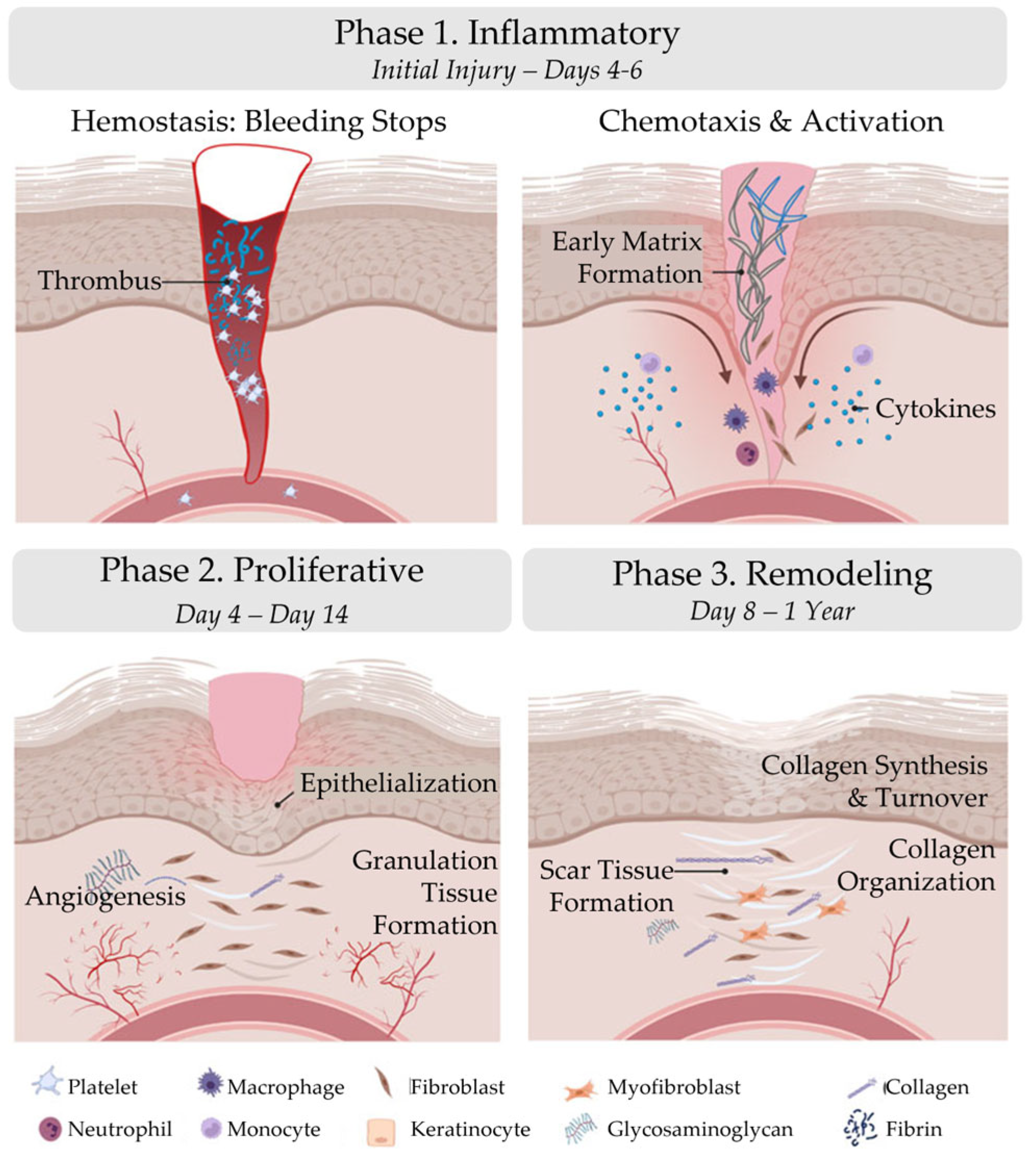
Figure 2.
Chronic wound formation. When the phases of wound healing are disrupted, abnormal healing occurs, and the wound fails to proceed through a normal, orderly, and timely sequence of repair. Often, chronic wounds remain in a prolonged state of persistent inflammation and are characterized by elevated concentrations of pro-inflammatory cytokines, pro-inflammatory (M1) macrophages, and proteases, which destroy the extracellular matrix. In normal wound healing, the extracellular matrix regulates macrophage behavior. In the chronic wound, however, a vicious cycle exists between a dysfunctional extracellular matrix and uncontrolled M1 macrophages. Abbreviations: ECM, extracellular matrix; IFN-γ, interferon gamma; IL-1, interleukin-1; IL-6, interleukin-6; IL-17, interleukin-17; MMPs, matrix metalloproteinases; ROS, reactive oxygen species; TIMPs, tissue inhibitors of metalloproteinases; TNF-α, tumor necrosis factor-alpha. The figure was originally published by Frontiers under the Creative Commons Attribution License (CC-BY), permitting unrestricted use [9].
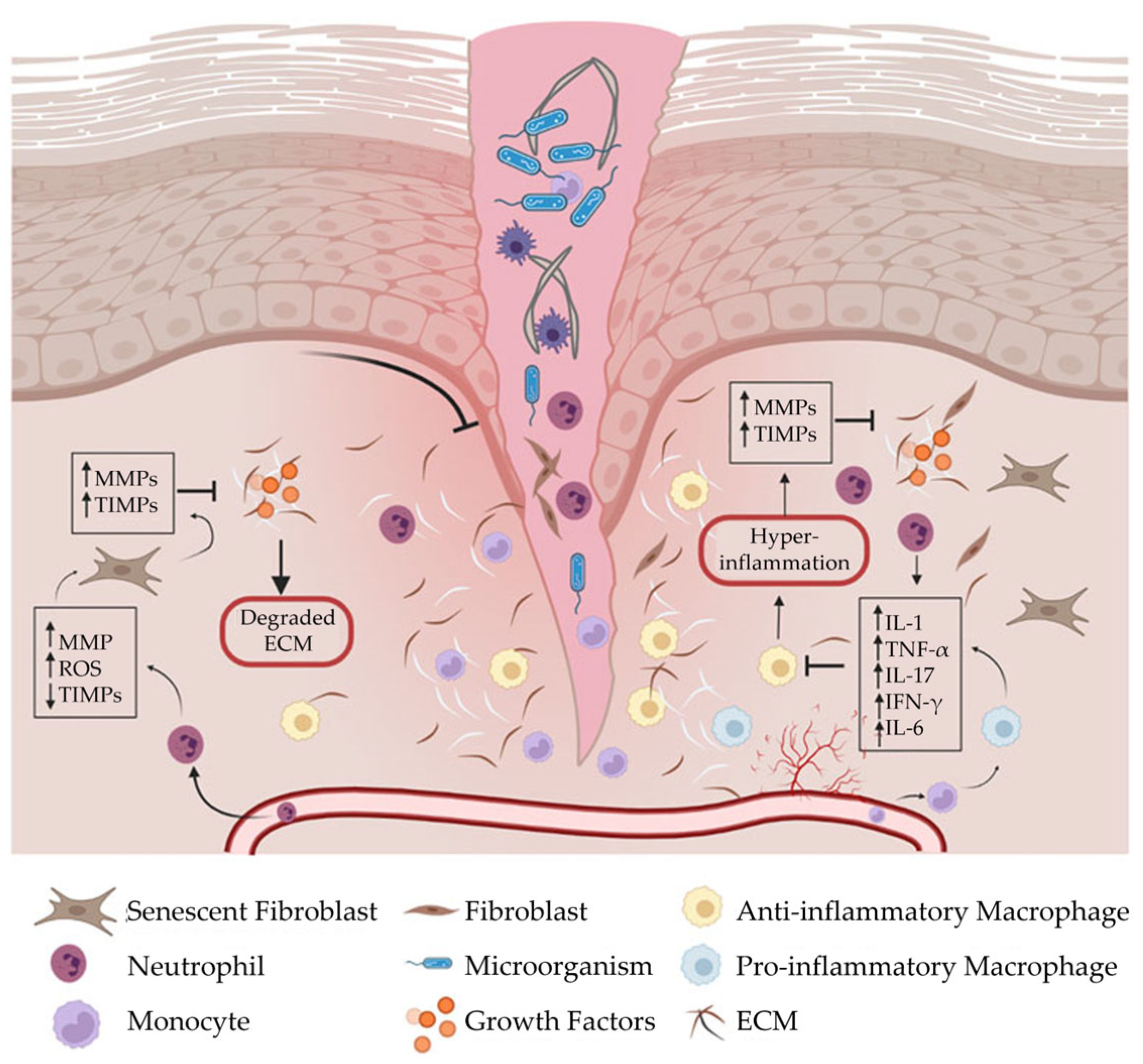
Figure 3.
Sources of placental-derived biomaterials. Placental-derived biomaterials can be sourced from the amniotic sac, amniotic fluid, placental disc, umbilical cord, or a combination of these sources. The amniotic sac is composed of the amnion and chorion layers.
Figure 3.
Sources of placental-derived biomaterials. Placental-derived biomaterials can be sourced from the amniotic sac, amniotic fluid, placental disc, umbilical cord, or a combination of these sources. The amniotic sac is composed of the amnion and chorion layers.
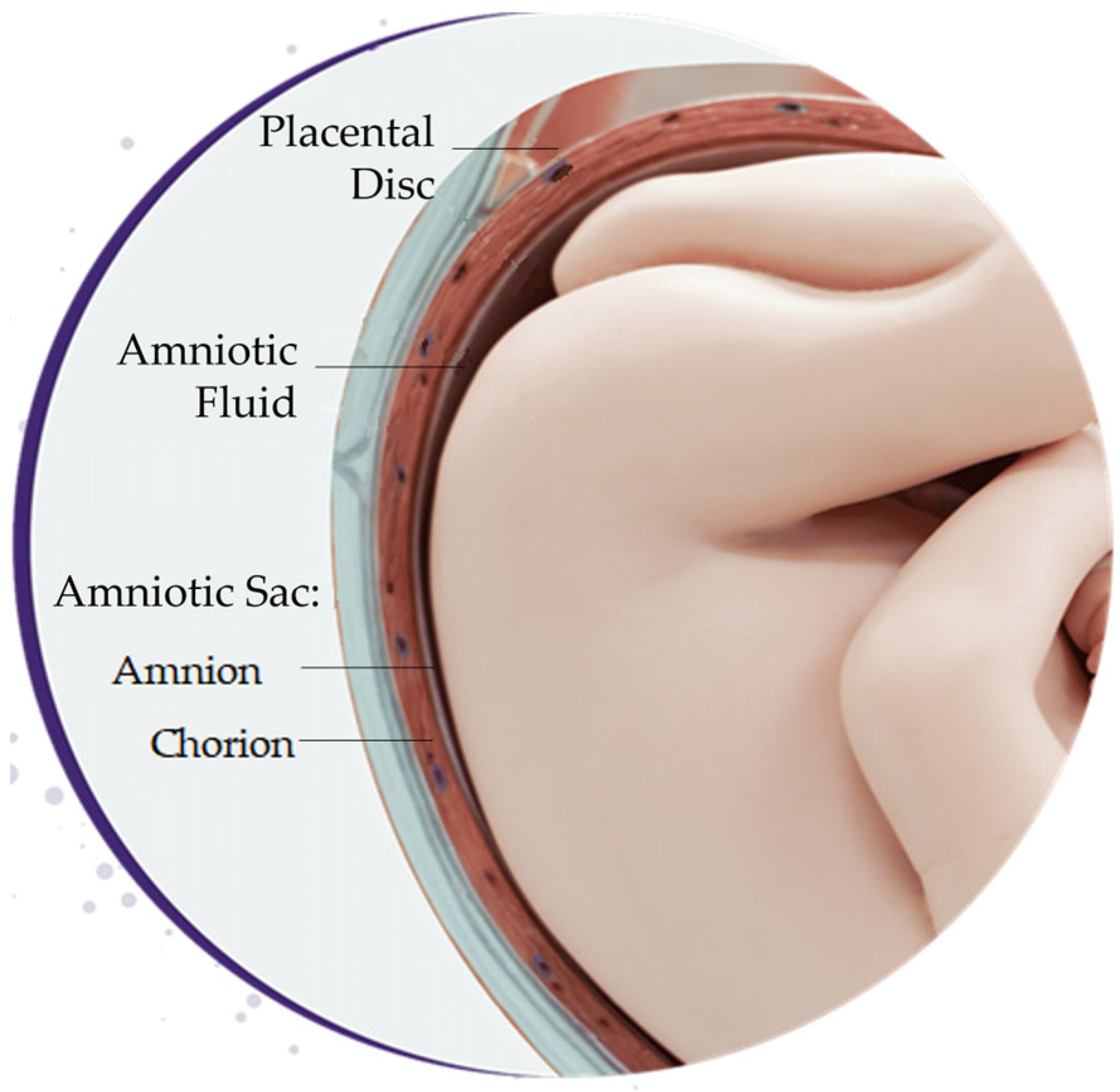
Figure 4.
Subdivision of the amnion and chorion layers. The red structures, visible in the chorion, are blood vessels.
Figure 4.
Subdivision of the amnion and chorion layers. The red structures, visible in the chorion, are blood vessels.
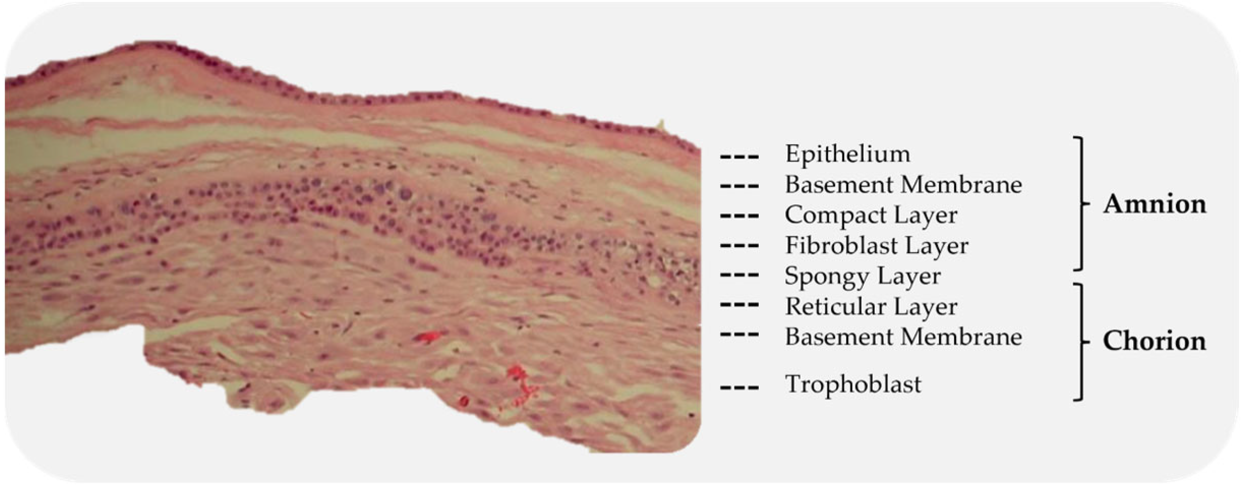
Figure 5.
Cross-section of a placental membrane with the chorion layer removed.
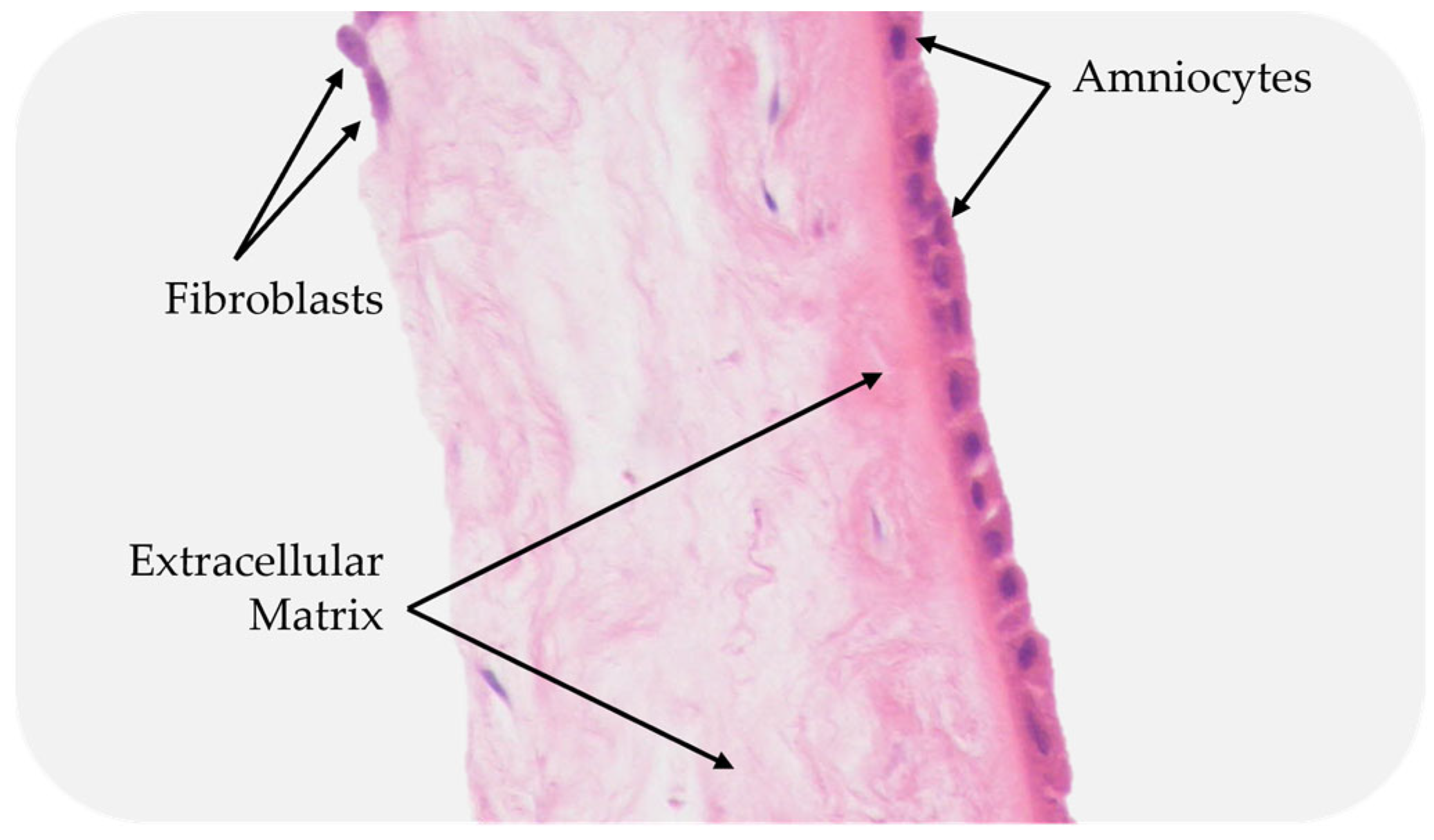
Figure 6.
Comparison of decellularized and non-decellularized amniotic membranes. (a) A proprietarily processed decellularized dehydrated amniotic membrane (BIOVANCE®, Celularity Inc., Florham Park, NJ) is shown. The decellularization process completely removes the residual cellular components, cells, cells debris, and DNA as well as growth factors and cytokines. The collagen framework remains intact, in its native 3-dimensional form with essential extracellular matrix molecules; (b) non-decellularized dehydrated amniotic membrane and (c) non-decellularized cryopreserved amniotic membranes are shown. These membranes retain residual cellular components, cells, cell debris, and DNA as well as growth factors and cytokines. The images are used with permission from the original publisher [95]. Images were originally published by and used with permission from John Wiley and Sons.
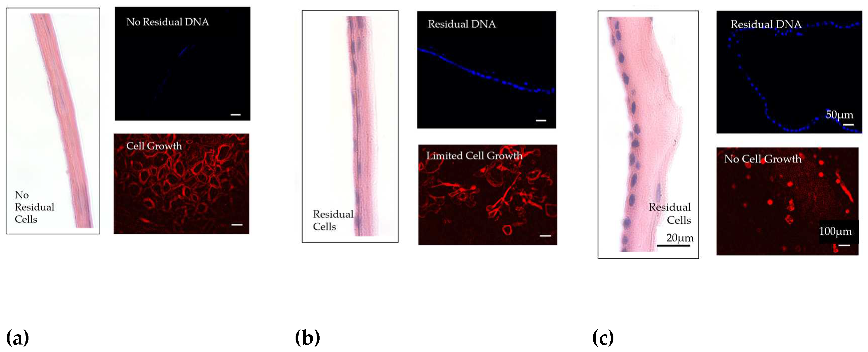
Figure 7.
Forms of placental-derived biomaterials. Various forms of biomaterials are extracted from the placenta, including: 1) sheet scaffolds; 2) injectables; 3) extracts/serums; and 4) cells.
Figure 7.
Forms of placental-derived biomaterials. Various forms of biomaterials are extracted from the placenta, including: 1) sheet scaffolds; 2) injectables; 3) extracts/serums; and 4) cells.
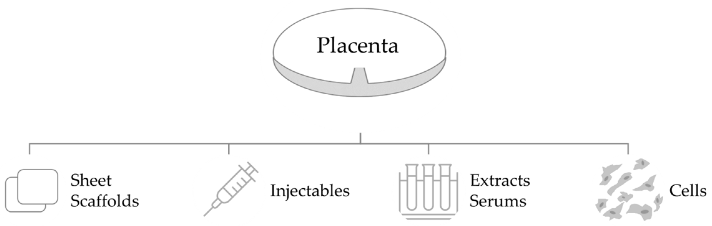
Table 1.
Phases of wound healing. Wound healing is divided into three distinct phases: the inflammatory phase, the proliferative phase, and the remodeling phase.
Table 1.
Phases of wound healing. Wound healing is divided into three distinct phases: the inflammatory phase, the proliferative phase, and the remodeling phase.
| Phase 1. Inflammatory | Phase 2. Proliferative | Phase 3. Remodeling | |
|---|---|---|---|
| Initial Injury – Days 4-6 | Day 4 – Day 14 | Day 8 – 1 Year | |
| Hemostasis: Bleeding stops | Epithelialization | Collagen Synthesis | |
| Chemotaxis | Angiogenesis | Collagen Turnover | |
| Activation | Granulation Tissue Formation | Collagen Organization |
Table 2.
Factors that impair wound healing.
| Local Factors | Systemic Factors |
|---|---|
| Oxygenation | Age |
| Infection | Ambulatory status |
| Venous insufficiency | Co-morbidities (e.g., diabetes, obesity, malnutrition, ischemia) |
| Medications (e.g., steroids, non-steroidal anti-inflammatory drugs) | |
| Oncology interventions (e.g., radiation, chemotherapy) | |
| Lifestyle habits (e.g., smoking, alcohol abuse) |
Table 3.
Methods used to decellularize placental tissues.
| Method | Agent | Example(s) | Reference(s) | |
|---|---|---|---|---|
| Chemical | Ionic Detergents | SDS SDC Triton-X-200 |
[2, 102-104] | |
| Non-ionic detergents | Triton-X-100 | [102-104] | ||
| Zwitterionic | CHAPS SB-10 SB-16 |
[105] | ||
| Acids | Peracetic Acid Hydrochloric Acid |
[106] | ||
| Hypertonic Solutions | Sodium chloride | [105, 107] | ||
| Hypotonic Solutions | Tris-HCl | [105, 107] | ||
| Chelating Agents | EDTA EGTA |
[102, 105, 108, 109] | ||
| Organic Solvents | Ethanol Methanol Acetones |
[102] | ||
| Enzymatic | Proteases | Trypsin | [102] | |
| Nucleases | DNase RNase |
[2, 108] | ||
| Physical | Pressure | High Hydrostatic Pressure Supercritical Fluids |
[5, 110, 111] | |
| Temperature | Freeze-thaw | [107, 111-113] | ||
| Force | Immersion & Agitation Shaking Scraping |
[2, 107, 109] |
Abbreviations: CHAM, 3-(3-cholamidopropyl) dimethylammonio]-1-proppanesulfonate; DNase, Deoxyribonuclease; EDTA, Ethylenediaminetetraacetic acid; EGTA, Ethylene glycol tetracetic acid; SB-10, RNase, Ribonuclease; Sulfobetaine-10; SB-16, Sulfobetaine-16; SDC, Sodium deoxycholate; SDS, Sodium dodecyl sulfate, Tris-HCl, Tris-Hydrochloride.
Disclaimer/Publisher’s Note: The statements, opinions and data contained in all publications are solely those of the individual author(s) and contributor(s) and not of MDPI and/or the editor(s). MDPI and/or the editor(s) disclaim responsibility for any injury to people or property resulting from any ideas, methods, instructions or products referred to in the content. |
© 2023 by the authors. Licensee MDPI, Basel, Switzerland. This article is an open access article distributed under the terms and conditions of the Creative Commons Attribution (CC BY) license (http://creativecommons.org/licenses/by/4.0/).
Copyright: This open access article is published under a Creative Commons CC BY 4.0 license, which permit the free download, distribution, and reuse, provided that the author and preprint are cited in any reuse.


