
Submitted:
23 May 2023
Posted:
26 May 2023
You are already at the latest version
Abstract
CD44 is known as a cancer stem cell marker of head and neck squamous cell carcinoma (HNSCC) and plays a critical role in cancer malignant progression. Splicing variant isoforms of CD44 (CD44v) are overexpressed in cancers and considered a promising target for cancer therapy. Several monoclonal antibodies (mAbs) against CD44 have been developed by immunizing mice with CD44v3–10-overexpressed cancer cells. In this study, we characterized a novel clone, C44Mab-18 (IgM, kappa). C44Mab-18 reacted with CHO/CD44v3–10, but not with CHO/CD44s by flow cytometry. Enzyme-linked immunosorbent assay revealed that the epitope of C44Mab-18 is determined to be the border sequence between variant 10 and the constant exon 16-encoded sequence. Flow cytometry showed that C44Mab-18 recognizes HSC-3, an oral squamous cell carcinoma(OSCC) cell line. The apparent dissociation constant (KD) of C44Mab-18 for CHO/CD44v3–10 and HSC-3 was determined to be 1.6 × 10⁻⁷ M and 1.7 × 10⁻⁷ M, respectively. C44Mab-18 detected CD44v3–10, but not CHO/CD44s in western blotting. Furthermore, C44Mab-18 detected endogenous CD44v10 in immunohistochemistry using OSCC tissues. Taken together, C44Mab-18 is useful for detecting CD44v10 in flow cytometry and immunohistochemistry.
Keywords:
CD44
; CD44v10
; monoclonal antibody
; oral squamous cell carcinoma
; immunohistochemistry
1. Introduction
Head and neck cancer is the seventh most common cancer type throughout the world, and makes the quality of life of patients worse after surgical ablation and therapies [1]. Head and neck squamous cell carcinoma (HNSCC) is the most common type of head and neck cancer. Surgery, chemotherapy, radiation therapy, immunotherapy, molecular targeted therapy, or a combination of those modalities are the main treatment of HNSCC [2]. Survival of patients can be improved by the development of those treatments; however, drug resistance and cancer metastasis remain the main causes of death [3]. The rate of 5-year survival remains stagnant at approximately 50% [4].
CD44 is a type I transmembrane glycoprotein, which possesses many functions, and mediates metastasis and drug resistance in cancer cells. HNSCC is revealed as the second-highest CD44-expressing cancer in the Pan-Cancer Atlas [5]. The alternative splicing of CD44 mRNA produces the various isoforms [6]. The constant exons, the first five (1 to 5) and the last five (16 to 20), exist in all variants and make up the standard isoform (CD44s). In various combinations with the constant exons of CD44s, the CD44 variant (CD44v) isoforms are generated by the alternative splicing of variant exons (v1 to v10) [7]. Both CD44s and CD44v (pan-CD44) attach to the hyaluronic acid (HA) and facilitate the activation of metastasis-associated intracellular signaling pathways [8].
Cancer metastasis is a multistep process, which includes (1) dissemination from primary sites, (2) the acquisition of migration/invasion phenotype, (3) intra/extravasation, (4) survival in circulation, and (5) adaptation and colonization in a distant organ [9]. Moreover, (6) cancer associate fibroblasts and tumor-infiltrating lymphocytes in the tumor microenvironment are involved in the promotion of tumor metastasis [10]. CD44 mediates the multiple steps of the invasion-metastasis cascade through interaction with HA [11] and CD44v-specific functions [12].
CD44 has been investigated as a cancer stem cell (CSC) or tumor-initiating cell (TIC) marker in various tumors [13]. Monoclonal antibodies (mAbs) against CD44s or CD44v are utilized to isolate the CD44-high CSCs [13]. The CD44-high population exhibited the increased self-renewing property, metastatic colonization in vivo, and drug resistance [13]. CD44 is the first established CSC marker to select HNSCC-derived CSCs [14]. Notably, CD44-high CSCs from HNSCC tumors showed the properties of epithelial to mesenchymal transition (EMT). The activation of the EMT gives tumor cells the ability to migrate, invade, and extravasate [15]. CD44-high cells could make colonization in the lungs of immunodeficient mice, compared to CD44-low ones, which failed to form the metastatic colonization [16].
CD44v8–10 mediates the resistance to treatment [17]. The v8–10-encoded region binds to and stabilizes a cystine–glutamate transporter (xCT), which enhances cystine uptake and glutathione synthesis [17]. The elevation of reduced glutathione (GSH) mediates the defense to reactive oxygen species (ROS) [17], radiation [18], and chemotherapeutic drugs [19]. CD44v8–10 expression is associated with the xCT function or redox status, and links to the poor prognosis of patients [18]. The establishment of each CD44v-specific mAb is important to reveal the function and develop CD44-targeting cancer therapy. Because anti-CD44v10 mAbs have not been developed, the function or tissue distribution of the variant 10-encoded region has not been fully understood.
Until now, we have developed an anti-pan-CD44 mAb, C44Mab-5 (IgG1, kappa) using the Cell-Based Immunization and Screening (CBIS) method [20]. Another anti-pan-CD44 mAb, C44Mab-46 [21] was established by immunizing mice with CD44v3–10 ectodomain. Both C44Mab-5 and C44Mab-46 have the epitopes within the constant exon 2 and 5-encoding sequences [22-24] and apply to immunohistochemistry in oral squamous cell carcinomas (OSCC) [20] and esophageal SCC [21], respectively. Furthermore, we established a class-switched and defucosylated type of recombinant C44Mab-5 (5-mG2a-f) using fucosyltransferase 8 (Fut8)-deficient ExpiCHO-S cells and investigated the antitumor activity in OSCC xenograft transplanted mice [25]. We have developed various anti-CD44v mAbs, including C44Mab-6 (anti-CD44v3 mAb) [26], C44Mab-108 (anti-CD44v4 mAb) [27], C44Mab-3 (anti-CD44v5 mAb) [28], C44Mab-9 (anti-CD44v6 mAb) [29], C44Mab-34 (anti-CD44v7/8 mAb) [30], and C44Mab-1 (anti-CD44v9 mAb) [31].
In this study, we developed a novel anti-CD44v10 mAb, C44Mab-18 (IgM, kappa) using CBIS method, and evaluated its applications, including flow cytometry, western blotting, and immunohistochemical analyses of OSCC tissues.
2. Materials and Methods
2.1. Cell Lines
Chinese hamster ovary (CHO)-K1 and P3X63Ag8U.1 (P3U1; a mouse multiple myeloma) were obtained from the American Type Culture Collection (ATCC, Manassas, VA, USA). PANC-1 (a human pancreatic cancer cell line) was obtained from the Cell Resource Center for Biomedical Research Institute of Development, Aging, and Cancer at Tohoku University (Sendai, Japan). We used RPMI-1640 medium [Nacalai Tesque, Inc. (Nacalai), Kyoto, Japan], which is supplemented with 10% heat-inactivated fetal bovine serum [FBS; Thermo Fisher Scientific, Inc. (Thermo), Waltham, MA, USA] to culture these cell lines. We added the antibiotics, including 100 μg/mL streptomycin, 100 U/mL penicillin, and 0.25 μg/mL amphotericin B (Nacalai) to the media. HSC-3 (a human OSCC cell line) was obtained from the Japanese Collection of Research Bioresources (Osaka, Japan), and cultured in DMEM medium (Nacalai), supplemented as indicated above. All cell lines were grown in a humidified incubator at 37°C with 5% CO2.
CD44s and CD44v3–10 cDNAs were obtained as described previously [20]. The cDNAs were cloned into pCAG-zeo-ssPA16 and pCAG-neo-ssPA16 vectors possessing a signal sequence and N-terminal PA16 tag (GLEGGVAMPGAEDDVV) [20,32-35]. We previously developed NZ-1 as an anti-human podoplanin (PDPN) mAb [36-48], which also detects a PA16 tag. We produced stable transfectants, such as PANC-1/CD44v3–10, CHO/CD44v3–10, and CHO/CD44s, by introducing corresponding vectors into the cells using a Neon transfection system (Thermo).
2.2. Production of hybridoma cells
PANC-1/CD44v3–10 (1 × 10⁸ cells) was administrated intraperitoneally into the 6-week-old female BALB/c mice (CLEA Japan, Tokyo, Japan) with Imject Alum (Thermo). Additional three times immunizations of PANC-1/CD44v3–10 (1 × 10⁸ cells) and a booster injection of PANC-1/CD44v3–10 (1 × 10⁸ cells) two days before the sacrifice was performed. Hybridomas were developed as described previously [28]. The supernatants were selected by flow cytometer (SA3800 Cell Analyzer) and SA3800 software (ver. 2.05, Sony Corp. Tokyo, Japan).
2.3. Enzyme−linked immunosorbent assay (ELISA)
Thirty-four peptides, which cover the variant region of CD44v3–10 [22], were synthesized by Sigma-Aldrich Corp. (Sigma; St. Louis, MO, USA). Peptides were immobilized on Nunc Maxisorp 96-well plates (Thermo) at 20 µg/mL. After the blocking with 1% (w/v) bovine serum albumin (BSA) in phosphate-buffered saline (PBS) containing 0.05% (v/v) Tween 20 (PBST; Nacalai), C44Mab-18 (1 µg/mL) was added to each well. Then, the wells were further treated with anti-mouse immunoglobulins peroxidase-conjugate [1:2000 diluted; Agilent Technologies Inc. (Agilent), Santa Clara, CA, USA]. We used the ELISA POD Substrate TMB Kit (Nacalai) for enzymatic reactions. The optical density (655 nm) was measured using an iMark microplate reader (Bio-Rad Laboratories, Inc., Berkeley, CA, USA).
2.4. Flow Cytometric Analysis
HSC-3, CHO/CD44v3–10, and CHO-K1 cells (1 × 10⁵ cells/sample) were incubated with C44Mab-18, C44Mab-46, or blocking buffer (0.1% BSA in PBS; control) for 30 min at 4°C. Then, the cells were treated with anti-mouse IgG conjugated with Alexa Fluor 488 (1:2000; Cell Signaling Technology, Inc., Danvers, MA, USA) for 30 min at 4°C. Fluorescence data were collected and analyzed as indicated above.
2.5. Determination of Apparent Dissociation Constant (KD) by Flow Cytometry
The serially diluted C44Mab-18 at the indicated concentrations was suspended with 2 × 10⁵ of HSC-3 and CHO/CD44v3–10 cells. Then, the cells were treated with anti-mouse IgG conjugated with Alexa Fluor 488 (1:200). Fluorescence data were analyzed, and the apparent dissociation constant (KD) was determined by the fitting binding isotherms to built-in one-site binding models of GraphPad Prism 8 (GraphPad Software, Inc., La Jolla, CA, USA).
2.6. Western Blot Analysis
The 10 μg of cell lysates were subjected to SDS-polyacrylamide gel for electrophoresis using polyacrylamide gels [5–20%; FUJIFILM Wako Pure Chemical Corporation (Wako), Osaka, Japan] and electrotransferred onto polyvinylidene difluoride membranes (Merck KGaA, Darmstadt, Germany). After the blocking using 4% skim milk (Nacalai) in PBST, the membranes were incubated with 10 μg/mL of C44Mab-18, 10 μg/mL of C44Mab-46, or 0.5 μg/mL of an anti-β-actin mAb (clone AC-15; Sigma). The membranes were further treated with peroxidase-conjugated anti-mouse immunoglobulins (diluted 1:1000; Agilent). Finally, the chemiluminescence signal was obtained using ImmunoStar LD (Wako) and was detected by a Sayaca-Imager (DRC Co. Ltd., Tokyo, Japan).
2.7. Immunohistochemical Analysis
Formalin-fixed paraffin-embedded (FFPE) sections of OSCC tissue array (OR601c) were purchased from US Biomax Inc. (Rockville, MD, USA) Tissue sections were autoclaved using EnVision FLEX Target Retrieval Solution High pH (Agilent). After blocking with SuperBlock T20 (Thermo), the tissue sections were incubated with C44Mab-18 (1 μg/mL) and C44Mab-46 (1 μg/mL) for 1 h at room temperature. The sections were further treated with the EnVision+ Kit for a mouse (Agilent) for 30 min at room temperature. The chromogenic reaction and counterstaining were performed using 3,3′-diaminobenzidine tetrahydrochloride (DAB; Agilent) and hematoxylin (Wako), respectively.
3. Results
3.1. Establishment of an Anti-CD44 mAbs by immunization of PANC-1/CD44v3–10 cells
We have developed anti-CD44 mAbs, including C44Mab-5 (pan-CD44) [20], C44Mab-6 (v3) [26], C44Mab-3 (v5) [28], C44Mab-9 (v6) [29], and C44Mab-1 (v9) [31] using CHO/CD44v3–10 cells as an immunogen. In this study, we established another stable transfectant (PANC-1/CD44v3–10 cells) (Figure 1A). Mice were immunized with PANC-1/CD44v3–10 cells (Figure 1B), and hybridomas were produced by fusion between the splenocyte and P3U1 cells (Figure 1C). Then, the supernatants, which were reactive to CHO/CD44v3–10 cells, but not to CHO-K1, were selected by flow cytometry-based high throughput screening (Figure 1D). After cloning, anti-CD44 mAb-producing clones were finally established (Figure 1E).
3.2. Flow Cytometric Analysis of C44Mab-18 to CD44-Expressing Cells
In this study, established clones, the epitope of which includes CD44v10, were mainly determined to be IgM although all mAbs against other CD44 variants are IgG [26-31]. Among those clones, we examined the reactivity of C44Mab-18 (IgM, kappa) against CHO/CD44v3–10 and CHO/CD44s cells by flow cytometry. C44Mab-18 dose-dependently recognized CHO/CD44v3–10 cells (Figure 2A). In contrast, C44Mab-18 neither recognized CHO/CD44s (Figure 2B) nor CHO-K1 (Figure 2C) cells. We confirmed that an anti-pan-CD44 mAb, C44Mab-46 [21], recognized CHO/CD44s cells, but not CHO-K1 cells (Supplementary Figure S1). Furthermore, C44Mab-18 could recognize HSC-3 cells (Figure 2D) in a dose-dependent manner. These results indicated that C44Mab-18 recognizes the variant exon-encoded region between v3 and v10 (Figure 1A).
3.3. Epitope Mapping of C44Mab-18 by ELISA
To determine the epitope of C44Mab-18, we performed the ELISA using synthetic peptides, which cover the variant exon-encoded region between v3 and v10 [22]. As shown in Figure 3, C44Mab-18 recognized the CD44 p551–570 peptide (SNSNVNRSLSGDQDTFHPSG), which is corresponding to variant 10 and constant exon 16-encoded sequence (Supplementary Table S1). In contrast, C44Mab-18 never recognized other v3 and v10-encoded peptides. This and Figure 2 results indicated that C44Mab-18 specifically recognizes the variant 10-containing CD44.
3.3. Determination of the apparent Binding Affinity of C44Mab-18 by Flow Cytometry
We next measured the apparent binding affinity of C44Mab-18 to CHO/CD44v3–10 and HSC-3 cells using flow cytometry. The dissociation constant (KD) of C44Mab-18 for CHO/CD44v3–10 (Figure 4A) and HSC-3 (Figure 4B) were 1.6 × 10⁻⁷ M and 1.7 × 10⁻⁷ M, respectively. These results indicated that C44Mab-18 possesses a moderate binding affinity for CD44v3–10 or endogenous CD44v10-expressing cells.
3.4. Western Blot Analysis
To assess the sensitivity of C44Mab-18 in western blot analysis, we prepared the cell lysates of CHO-K1, CHO/CD44s, and CHO/CD44v3–10. C44Mab-18 mainly detected CD44v3–10 as more than 180-kDa and ~70-kDa bands. However, C44Mab-18 did not detect any bands from lysates of CHO/CD44s and CHO-K1 cells (Figure 5A). An anti-pan-CD44 mAb, C44Mab-46, recognized both CD44s (~75 kDa) and CD44v3–10 (>180 kDa) bands in the lysates of CHO/CD44s and CHO/CD44v3–10, respectively (Figure 5B). We used β-actin as a loading control (Figure 5C). These results indicated that C44Mab-18 can detect exogenous CD44v3–10.
3.5. Immunohistochemical Analysis using C44Mab-18 against Tumor Tissues
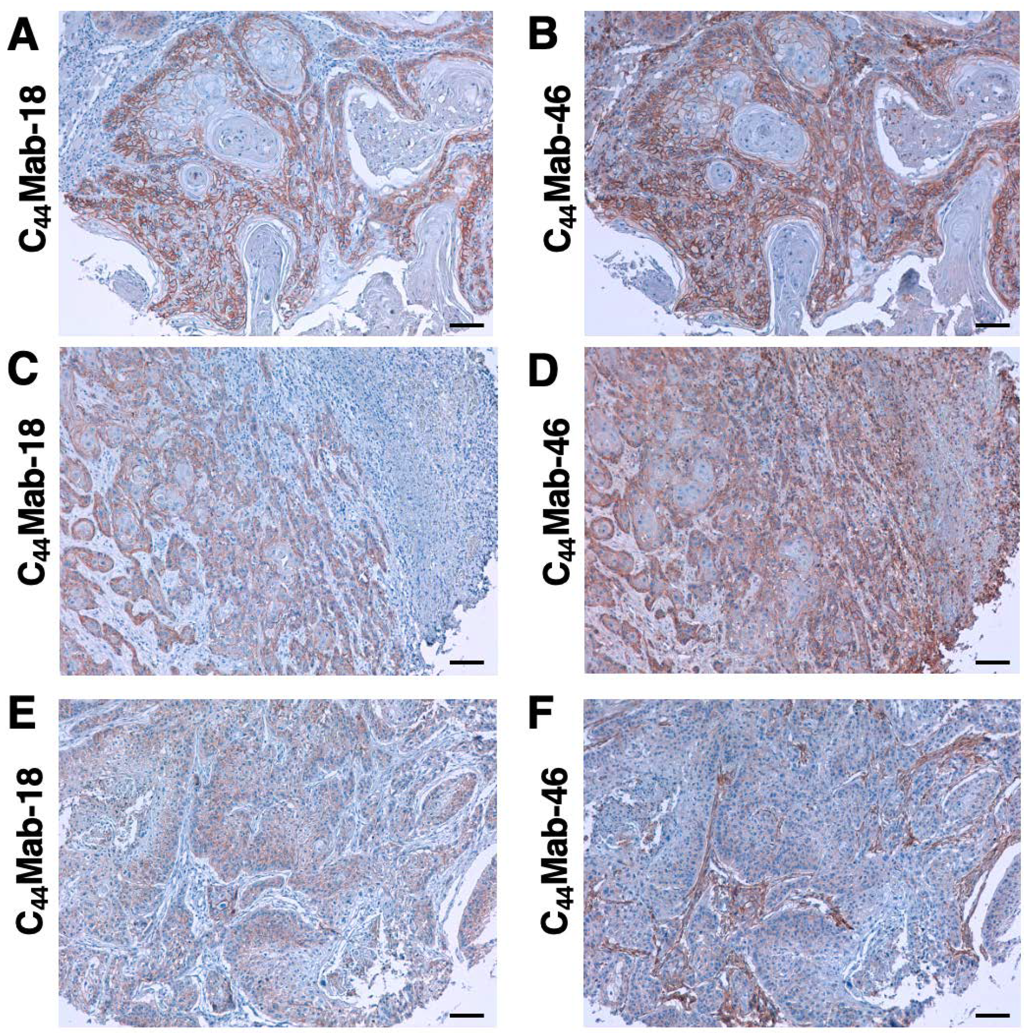
Since HNSCC is revealed as the second highest CD44-expressing cancer type in the Pan-Cancer Atlas [5], we examined the reactivity of C44Mab-18 and C44Mab-46 in immunohistochemical analyses using FFPE sections of OSCC tissue array. As shown in Figure 6, C44Mab-18 exhibited membranous staining and was able to distinguish tumor cells from stromal tissues. In contrast, C44Mab-46 stained both. We summarized the data of immunohistochemical analyses in Supplementary Table S2; C44Mab-18 stained 41 out of 50 cases (82%) in OSCC. These results indicated that C44Mab-18 applies to immunohistochemical analysis of FFPE tumor sections.
4. Discussion
We have established several anti-CD44 mAbs using CHO/CD44v3–10 [20,26,28,29,31], purified CD44v3–10 ectodomain [21,30], or PANC-1/CD44v3–10 (this study) as antigens. We further developed a novel anti-CD44v antibody (C44Mab-18), which recognizes the border sequence between variant 10 and constant exon 16 (Figure 3) in this study. C44Mab-18 could recognize CHO/CD44v3–10, but not CHO/CD44s in flow cytometry (Figure 2) and western blot analyses (Figure 5). Endogenous CD44v10 was not detected by C44Mab-18 in western blot analyses. In contrast, C44Mab-18 could stain tumor cells, but not stromal tissues, which could be stained by C44Mab-46, an anti-pan-CD44 mAb (Figure 6). These results indicate that C44Mab-18 is a specific mAb against CD44v10.
Heider et al. previously established the VFF series against CD44v by the immunization of glutathione S-transferase fused CD44v3–10 protein, which was produced by bacteria [49,50]. VFF-8 (anti-CD44v5), VFF-18 (anti-CD44v6), VFF-9 (anti-CD44v7), VFF-17 (anti-CD44v7/8), and VFF-14 (anti-CD44v10) have been used for various applications [51]. Although VFF-14 was shown to apply to immunohistochemistry [52], the detailed binding epitope of VFF-14 has not been reported. In this study, we determined the epitope of C44Mab-18 as the CD44 p551–570 peptide (SNSNVNRSLSGDQDTFHPSG), which is corresponding to variant 10 (underlined) and constant exon 16-encoded region. In contrast, C44Mab-18 never recognizes the p541–560 peptide (FGVTAVTVGDSNSNVNRSLS) in the variant 10 region. Therefore, C44Mab-18 could have the epitope in the border region, but the inclusion of variant 10 is essential for the recognition.
By posttranslational modification such as N-glycans and O-glycans to CD44, the molecular weight of CD44v isoforms surpasses 200 kDa [53]. C44Mab-18 detected both more than 180-kDa and ~70-kDa bands (Figure 5A) in the lysate from CHO/CD44v3–10. The 70 kDa is approximately identical to the predicted molecular weight of CD44v3–10 from the amino acid sequence. Therefore, C44Mab-18 could recognize CD44v3–10 regardless of the glycosylation. The detailed epitope mapping and the influence of glycosylation on C44Mab-18 recognition should be investigated in future studies.
CD44v8–10 was shown to interact with a glutamate-cystine transporter xCT, and regulate the level of reduced glutathione in cancer cells. The interaction is important for the stabilization of xCT on the cell surface, which promotes the defense against reactive oxygen species [17]. Furthermore, the interaction failed in CD44v8–10 (S301A), an N-linked glycosylation consensus motif (Asn-X-Ser/Thr) mutant in the variant 10-encoded region [17]. Therefore, it is worthwhile to investigate whether C44Mab-18 interferes with the interaction between CD44v8–10 and xCT in future studies. Furthermore, several studies revealed that CD44v9 is used as a predictive marker for recurrence [54] and a biomarker for patient selection and efficacy of xCT inhibitors, sulfasalazine in gastric cancer [55]. Further investigations are also required to clarify the clinical significance of CD44v10 expression using C44Mab-94.
The mAbs against CD44 have been considered a therapeutic option for solid tumors and leukemia [12]. However, anti-pan-CD44 mAbs can affect normal tissues such as epithelium and hematopoiesis. In a preclinical study using a murine thymoma model, a comparative study between an anti-pan-CD44 mAb (IM-7) and an anti-murine CD44v10 mAb (K926) was performed in CD44v10-transfected EL4 thymoma (EL4-v10) [56]. The results showed that a blockade of CD44v10 by K926 was superior to that of IM-7 in intra-marrow EL4-v10 growth retardation. Furthermore, K926 hardly disturbed the hematopoietic stem cell (HSC) interaction with the bone marrow stroma. In contrast, IM-7 strongly affected the embedding of HSC in the bone marrow stroma [56], indicating that the therapeutic use of anti-pan-CD44 mAbs should be avoided in favor of CD44v-specific mAbs as far as leukemic cells express CD44v isoforms.
CD44v8–10 was elevated during chronic myeloid leukemia progression from chronic phase to blast crisis in a humanized mouse model [57]. Furthermore, increased transcription of CD44 mRNA was detected in human acute myeloid leukemia (AML) patients with FLT3 or DNMT3A mutations through suppression of CpG islands methylation in the promoter [58]. BIWA-8 derived from VFF-18 against CD44v6 was engineered to develop chimeric antigen receptors (CARs) for AML with FLT3 or DNMT3A mutations [59]. The CD44v6 CAR-T cells showed potent anti-leukemic effects [58]. We have developed class-switched and defucosylated IgG2a recombinant mAbs and investigated the antitumor activity using xenograft models [25,60-66]. Therefore, the development of class-switched and defucosylated C44Mab-18 is one of the important strategies to evaluate the antitumor effect in preclinical models.
Anti-pan-CD44 and anti-CD44v mAbs still have the possibility of adverse events by affecting normal tissues; therefore, the clinical applications are limited. We used tumor cell-expressed CD44v3–10 as an immunogen in this study. The strategy is critical for the development of cancer-specific mAbs (CasMabs). We developed podocalyxin-targeting CasMabs [67] and PDPN-targeting CasMabs [68-70], which react with the aberrantly glycosylated targets selectively expressed in cancers [71]. Anti-PDPN-CasMabs have been applied to CAR-T therapy in preclinical studies [72-74]. For CasMab development, we should perform a further selection of our established anti-CD44 mAbs by comparing the reactivity against normal cells and tissues. Anti-CD44 CasMabs could be applicable for designing the modalities, including antibody-drug conjugates and CAR-T.
Supplementary Materials
Supplementary Figure S1, Recognition of CHO/CD44s and CHO/CD44v3–10 by C44Mab-46 by flow cytometry. Supplementary Table S1, The determination of the binding epitope of C44Mab-18 by ELISA. Supplementary Table S2, Immunohistochemical analysis using C44Mab-18 against OSCC tissue array.
References
- Mody, M.D.; Rocco, J.W.; Yom, S.S.; Haddad, R.I.; Saba, N.F. Head and neck cancer. Lancet 2021, 398, 2289–2299. [Google Scholar] [CrossRef] [PubMed]
- Xing, D.T.; Khor, R.; Gan, H.; Wada, M.; Ermongkonchai, T.; Ng, S.P. Recent Research on Combination of Radiotherapy with Targeted Therapy or Immunotherapy in Head and Neck Squamous Cell Carcinoma: A Review for Radiation Oncologists. Cancers (Basel) 2021, 13. [Google Scholar] [CrossRef]
- Muzaffar, J.; Bari, S.; Kirtane, K.; Chung, C.H. Recent Advances and Future Directions in Clinical Management of Head and Neck Squamous Cell Carcinoma. Cancers (Basel) 2021, 13. [Google Scholar] [CrossRef]
- Johnson, D.E.; Burtness, B.; Leemans, C.R.; Lui, V.W.Y.; Bauman, J.E.; Grandis, J.R. Head and neck squamous cell carcinoma. Nat Rev Dis Primers 2020, 6, 92. [Google Scholar] [CrossRef]
- Ludwig, N.; Szczepanski, M.J.; Gluszko, A.; Szafarowski, T.; Azambuja, J.H.; Dolg, L.; Gellrich, N.C.; Kampmann, A.; Whiteside, T.L.; Zimmerer, R.M. CD44(+) tumor cells promote early angiogenesis in head and neck squamous cell carcinoma. Cancer Lett 2019, 467, 85–95. [Google Scholar] [CrossRef] [PubMed]
- Ponta, H.; Sherman, L.; Herrlich, P.A. CD44: from adhesion molecules to signalling regulators. Nat Rev Mol Cell Biol 2003, 4, 33–45. [Google Scholar] [CrossRef] [PubMed]
- Chen, C.; Zhao, S.; Karnad, A.; Freeman, J.W. The biology and role of CD44 in cancer progression: therapeutic implications. J Hematol Oncol 2018, 11, 64. [Google Scholar] [CrossRef] [PubMed]
- Slevin, M.; Krupinski, J.; Gaffney, J.; Matou, S.; West, D.; Delisser, H.; Savani, R.C.; Kumar, S. Hyaluronan-mediated angiogenesis in vascular disease: uncovering RHAMM and CD44 receptor signaling pathways. Matrix Biol 2007, 26, 58–68. [Google Scholar] [CrossRef]
- Valastyan, S.; Weinberg, R.A. Tumor metastasis: molecular insights and evolving paradigms. Cell 2011, 147, 275–292. [Google Scholar] [CrossRef]
- de Visser, K.E.; Joyce, J.A. The evolving tumor microenvironment: From cancer initiation to metastatic outgrowth. Cancer Cell 2023, 41, 374–403. [Google Scholar] [CrossRef]
- Zöller, M. CD44, Hyaluronan, the Hematopoietic Stem Cell, and Leukemia-Initiating Cells. Front Immunol 2015, 6, 235. [Google Scholar] [CrossRef] [PubMed]
- Hassn Mesrati, M.; Syafruddin, S.E.; Mohtar, M.A.; Syahir, A. CD44: A Multifunctional Mediator of Cancer Progression. Biomolecules 2021, 11. [Google Scholar] [CrossRef] [PubMed]
- Zöller, M. CD44: can a cancer-initiating cell profit from an abundantly expressed molecule? Nat Rev Cancer 2011, 11, 254–267. [Google Scholar] [CrossRef] [PubMed]
- Prince, M.E.; Sivanandan, R.; Kaczorowski, A.; Wolf, G.T.; Kaplan, M.J.; Dalerba, P.; Weissman, I.L.; Clarke, M.F.; Ailles, L.E. Identification of a subpopulation of cells with cancer stem cell properties in head and neck squamous cell carcinoma. Proc Natl Acad Sci U S A 2007, 104, 973–978. [Google Scholar] [CrossRef]
- Yang, J.; Antin, P.; Berx, G.; Blanpain, C.; Brabletz, T.; Bronner, M.; Campbell, K.; Cano, A.; Casanova, J.; Christofori, G.; et al. Guidelines and definitions for research on epithelial-mesenchymal transition. Nat Rev Mol Cell Biol 2020, 21, 341–352. [Google Scholar] [CrossRef] [PubMed]
- Davis, S.J.; Divi, V.; Owen, J.H.; Bradford, C.R.; Carey, T.E.; Papagerakis, S.; Prince, M.E. Metastatic potential of cancer stem cells in head and neck squamous cell carcinoma. Arch Otolaryngol Head Neck Surg 2010, 136, 1260–1266. [Google Scholar] [CrossRef]
- Ishimoto, T.; Nagano, O.; Yae, T.; Tamada, M.; Motohara, T.; Oshima, H.; Oshima, M.; Ikeda, T.; Asaba, R.; Yagi, H.; et al. CD44 variant regulates redox status in cancer cells by stabilizing the xCT subunit of system xc(-) and thereby promotes tumor growth. Cancer Cell 2011, 19, 387–400. [Google Scholar] [CrossRef]
- Kagami, T.; Yamade, M.; Suzuki, T.; Uotani, T.; Tani, S.; Hamaya, Y.; Iwaizumi, M.; Osawa, S.; Sugimoto, K.; Baba, S.; et al. High expression level of CD44v8-10 in cancer stem-like cells is associated with poor prognosis in esophageal squamous cell carcinoma patients treated with chemoradiotherapy. Oncotarget 2018, 9, 34876–34888. [Google Scholar] [CrossRef]
- Hagiwara, M.; Kikuchi, E.; Tanaka, N.; Kosaka, T.; Mikami, S.; Saya, H.; Oya, M. Variant isoforms of CD44 involves acquisition of chemoresistance to cisplatin and has potential as a novel indicator for identifying a cisplatin-resistant population in urothelial cancer. BMC Cancer 2018, 18, 113. [Google Scholar] [CrossRef]
- Yamada, S.; Itai, S.; Nakamura, T.; Yanaka, M.; Kaneko, M.K.; Kato, Y. Detection of high CD44 expression in oral cancers using the novel monoclonal antibody, C(44)Mab-5. Biochem Biophys Rep 2018, 14, 64–68. [Google Scholar] [CrossRef]
- Goto, N.; Suzuki, H.; Tanaka, T.; Asano, T.; Kaneko, M.K.; Kato, Y. Development of a Novel Anti-CD44 Monoclonal Antibody for Multiple Applications against Esophageal Squamous Cell Carcinomas. Int J Mol Sci 2022, 23. [Google Scholar] [CrossRef]
- Takei, J.; Asano, T.; Suzuki, H.; Kaneko, M.K.; Kato, Y. Epitope Mapping of the Anti-CD44 Monoclonal Antibody (C44Mab-46) Using Alanine-Scanning Mutagenesis and Surface Plasmon Resonance. Monoclon Antib Immunodiagn Immunother 2021, 40, 219–226. [Google Scholar] [CrossRef] [PubMed]
- Asano, T.; Kaneko, M.K.; Takei, J.; Tateyama, N.; Kato, Y. Epitope Mapping of the Anti-CD44 Monoclonal Antibody (C44Mab-46) Using the REMAP Method. Monoclon Antib Immunodiagn Immunother 2021, 40, 156–161. [Google Scholar] [CrossRef]
- Asano, T.; Kaneko, M.K.; Kato, Y. Development of a Novel Epitope Mapping System: RIEDL Insertion for Epitope Mapping Method. Monoclon Antib Immunodiagn Immunother 2021, 40, 162–167. [Google Scholar] [CrossRef]
- Takei, J.; Kaneko, M.K.; Ohishi, T.; Hosono, H.; Nakamura, T.; Yanaka, M.; Sano, M.; Asano, T.; Sayama, Y.; Kawada, M.; et al. A defucosylated antiCD44 monoclonal antibody 5mG2af exerts antitumor effects in mouse xenograft models of oral squamous cell carcinoma. Oncol Rep 2020, 44, 1949–1960. [Google Scholar] [CrossRef] [PubMed]
- Suzuki, H.; Kitamura, K.; Goto, N.; Ishikawa, K.; Ouchida, T.; Tanaka, T.; Kaneko, M.K.; Kato, Y. A Novel Anti-CD44 Variant 3 Monoclonal Antibody C(44)Mab-6 Was Established for Multiple Applications. Int J Mol Sci 2023, 24. [Google Scholar] [CrossRef] [PubMed]
- Suzuki, H.; Tanaka, T.; Goto, N.; Kaneko, M.K.; Kato, Y. Development of a Novel Anti-CD44 Variant 4 Monoclonal Antibody C44Mab-108 for Immunohistochemistry. Curr Issues Mol Biol 2023, 45, 1875–1888. [Google Scholar] [CrossRef]
- Kudo, Y.; Suzuki, H.; Tanaka, T.; Kaneko, M.K.; Kato, Y. Development of a Novel Anti-CD44 variant 5 Monoclonal Antibody C44Mab-3 for Multiple Applications against Pancreatic Carcinomas. Antibodies 2023, 12, 31. [Google Scholar] [CrossRef]
- Ejima, R.; Suzuki, H.; Tanaka, T.; Asano, T.; Kaneko, M.K.; Kato, Y. Development of a Novel Anti-CD44 Variant 6 Monoclonal Antibody C(44)Mab-9 for Multiple Applications against Colorectal Carcinomas. Int J Mol Sci 2023, 24. [Google Scholar] [CrossRef]
- Suzuki, H.; Ozawa, K.; Tanaka, T.; Kaneko, M.K.; Kato, Y. Development of a Novel Anti-CD44 Variant 7/8 Monoclonal Antibody, C44Mab-34, for Multiple Applications against Oral Carcinomas. Biomedicines 2023, 11, 1099. [Google Scholar] [CrossRef]
- Tawara, M.; Suzuki, H.; Goto, N.; Tanaka, T.; Kaneko, M.K.; Kato, Y. A Novel Anti-CD44 Variant 9 Monoclonal Antibody C44Mab-1 was Developed for Immunohistochemical Analyses Against Colorectal Cancers Curr. Issues Mol. Biol. 2023, 45, 3658–3673. [Google Scholar] [CrossRef] [PubMed]
- Kato, Y.; Yamada, S.; Furusawa, Y.; Itai, S.; Nakamura, T.; Yanaka, M.; Sano, M.; Harada, H.; Fukui, M.; Kaneko, M.K. PMab-213: A Monoclonal Antibody for Immunohistochemical Analysis Against Pig Podoplanin. Monoclon Antib Immunodiagn Immunother 2019, 38, 18–24. [Google Scholar] [CrossRef]
- Furusawa, Y.; Yamada, S.; Itai, S.; Sano, M.; Nakamura, T.; Yanaka, M.; Fukui, M.; Harada, H.; Mizuno, T.; Sakai, Y.; et al. PMab-210: A Monoclonal Antibody Against Pig Podoplanin. Monoclon Antib Immunodiagn Immunother 2019, 38, 30–36. [Google Scholar] [CrossRef]
- Furusawa, Y.; Yamada, S.; Itai, S.; Nakamura, T.; Yanaka, M.; Sano, M.; Harada, H.; Fukui, M.; Kaneko, M.K.; Kato, Y. PMab-219: A monoclonal antibody for the immunohistochemical analysis of horse podoplanin. Biochem Biophys Rep 2019, 18, 100616. [Google Scholar] [CrossRef] [PubMed]
- Furusawa, Y.; Yamada, S.; Itai, S.; Nakamura, T.; Takei, J.; Sano, M.; Harada, H.; Fukui, M.; Kaneko, M.K.; Kato, Y. Establishment of a monoclonal antibody PMab-233 for immunohistochemical analysis against Tasmanian devil podoplanin. Biochem Biophys Rep 2019, 18, 100631. [Google Scholar] [CrossRef]
- Kato, Y.; Kaneko, M.K.; Kuno, A.; Uchiyama, N.; Amano, K.; Chiba, Y.; Hasegawa, Y.; Hirabayashi, J.; Narimatsu, H.; Mishima, K.; et al. Inhibition of tumor cell-induced platelet aggregation using a novel anti-podoplanin antibody reacting with its platelet-aggregation-stimulating domain. Biochem Biophys Res Commun 2006, 349, 1301–1307. [Google Scholar] [CrossRef]
- Tamura-Sakaguchi, R.; Aruga, R.; Hirose, M.; Ekimoto, T.; Miyake, T.; Hizukuri, Y.; Oi, R.; Kaneko, M.K.; Kato, Y.; Akiyama, Y.; et al. Moving toward generalizable NZ-1 labeling for 3D structure determination with optimized epitope-tag insertion. Acta Crystallogr D Struct Biol 2021, 77, 645–662. [Google Scholar] [CrossRef] [PubMed]
- Kaneko, M.K.; Ohishi, T.; Nakamura, T.; Inoue, H.; Takei, J.; Sano, M.; Asano, T.; Sayama, Y.; Hosono, H.; Suzuki, H.; et al. Development of Core-Fucose-Deficient Humanized and Chimeric Anti-Human Podoplanin Antibodies. Monoclon Antib Immunodiagn Immunother 2020, 39, 167–174. [Google Scholar] [CrossRef] [PubMed]
- Fujii, Y.; Matsunaga, Y.; Arimori, T.; Kitago, Y.; Ogasawara, S.; Kaneko, M.K.; Kato, Y.; Takagi, J. Tailored placement of a turn-forming PA tag into the structured domain of a protein to probe its conformational state. J Cell Sci 2016, 129, 1512–1522. [Google Scholar] [CrossRef]
- Abe, S.; Kaneko, M.K.; Tsuchihashi, Y.; Izumi, T.; Ogasawara, S.; Okada, N.; Sato, C.; Tobiume, M.; Otsuka, K.; Miyamoto, L.; et al. Antitumor effect of novel anti-podoplanin antibody NZ-12 against malignant pleural mesothelioma in an orthotopic xenograft model. Cancer Sci 2016, 107, 1198–1205. [Google Scholar] [CrossRef]
- Kaneko, M.K.; Abe, S.; Ogasawara, S.; Fujii, Y.; Yamada, S.; Murata, T.; Uchida, H.; Tahara, H.; Nishioka, Y.; Kato, Y. Chimeric Anti-Human Podoplanin Antibody NZ-12 of Lambda Light Chain Exerts Higher Antibody-Dependent Cellular Cytotoxicity and Complement-Dependent Cytotoxicity Compared with NZ-8 of Kappa Light Chain. Monoclon Antib Immunodiagn Immunother 2017, 36, 25–29. [Google Scholar] [CrossRef] [PubMed]
- Ito, A.; Ohta, M.; Kato, Y.; Inada, S.; Kato, T.; Nakata, S.; Yatabe, Y.; Goto, M.; Kaneda, N.; Kurita, K.; et al. A Real-Time Near-Infrared Fluorescence Imaging Method for the Detection of Oral Cancers in Mice Using an Indocyanine Green-Labeled Podoplanin Antibody. Technol Cancer Res Treat 2018, 17, 1533033818767936. [Google Scholar] [CrossRef]
- Tamura, R.; Oi, R.; Akashi, S.; Kaneko, M.K.; Kato, Y.; Nogi, T. Application of the NZ-1 Fab as a crystallization chaperone for PA tag-inserted target proteins. Protein Sci 2019, 28, 823–836. [Google Scholar] [CrossRef]
- Kuwata, T.; Yoneda, K.; Mori, M.; Kanayama, M.; Kuroda, K.; Kaneko, M.K.; Kato, Y.; Tanaka, F. Detection of Circulating Tumor Cells (CTCs) in Malignant Pleural Mesothelioma (MPM) with the "Universal" CTC-Chip and An Anti-Podoplanin Antibody NZ-1.2. Cells 2020, 9. [Google Scholar] [CrossRef] [PubMed]
- Nishinaga, Y.; Sato, K.; Yasui, H.; Taki, S.; Takahashi, K.; Shimizu, M.; Endo, R.; Koike, C.; Kuramoto, N.; Nakamura, S.; et al. Targeted Phototherapy for Malignant Pleural Mesothelioma: Near-Infrared Photoimmunotherapy Targeting Podoplanin. Cells 2020, 9. [Google Scholar] [CrossRef]
- Fujii, Y.; Kaneko, M.; Neyazaki, M.; Nogi, T.; Kato, Y.; Takagi, J. PA tag: a versatile protein tagging system using a super high affinity antibody against a dodecapeptide derived from human podoplanin. Protein Expr Purif 2014, 95, 240–247. [Google Scholar] [CrossRef] [PubMed]
- Kato, Y.; Kaneko, M.K.; Kunita, A.; Ito, H.; Kameyama, A.; Ogasawara, S.; Matsuura, N.; Hasegawa, Y.; Suzuki-Inoue, K.; Inoue, O.; et al. Molecular analysis of the pathophysiological binding of the platelet aggregation-inducing factor podoplanin to the C-type lectin-like receptor CLEC-2. Cancer Sci 2008, 99, 54–61. [Google Scholar] [CrossRef]
- Kato, Y.; Vaidyanathan, G.; Kaneko, M.K.; Mishima, K.; Srivastava, N.; Chandramohan, V.; Pegram, C.; Keir, S.T.; Kuan, C.T.; Bigner, D.D.; et al. Evaluation of anti-podoplanin rat monoclonal antibody NZ-1 for targeting malignant gliomas. Nucl Med Biol 2010, 37, 785–794. [Google Scholar] [CrossRef]
- Heider, K.H.; Sproll, M.; Susani, S.; Patzelt, E.; Beaumier, P.; Ostermann, E.; Ahorn, H.; Adolf, G.R. Characterization of a high-affinity monoclonal antibody specific for CD44v6 as candidate for immunotherapy of squamous cell carcinomas. Cancer Immunol Immunother 1996, 43, 245–253. [Google Scholar] [CrossRef]
- Heider, K.H.; Mulder, J.W.; Ostermann, E.; Susani, S.; Patzelt, E.; Pals, S.T.; Adolf, G.R. Splice variants of the cell surface glycoprotein CD44 associated with metastatic tumour cells are expressed in normal tissues of humans and cynomolgus monkeys. Eur J Cancer 1995, 31a, 2385–2391. [Google Scholar] [CrossRef] [PubMed]
- Gansauge, F.; Gansauge, S.; Zobywalski, A.; Scharnweber, C.; Link, K.H.; Nussler, A.K.; Beger, H.G. Differential expression of CD44 splice variants in human pancreatic adenocarcinoma and in normal pancreas. Cancer Res 1995, 55, 5499–5503. [Google Scholar] [PubMed]
- Beham-Schmid, C.; Heider, K.H.; Hoefler, G.; Zatloukal, K. Expression of CD44 splice variant v10 in Hodgkin's disease is associated with aggressive behaviour and high risk of relapse. J Pathol 1998, 186, 383–389. [Google Scholar] [CrossRef]
- Mishra, M.N.; Chandavarkar, V.; Sharma, R.; Bhargava, D. Structure, function and role of CD44 in neoplasia. J Oral Maxillofac Pathol 2019, 23, 267–272. [Google Scholar] [CrossRef]
- Hirata, K.; Suzuki, H.; Imaeda, H.; Matsuzaki, J.; Tsugawa, H.; Nagano, O.; Asakura, K.; Saya, H.; Hibi, T. CD44 variant 9 expression in primary early gastric cancer as a predictive marker for recurrence. Br J Cancer 2013, 109, 379–386. [Google Scholar] [CrossRef]
- Shitara, K.; Doi, T.; Nagano, O.; Imamura, C.K.; Ozeki, T.; Ishii, Y.; Tsuchihashi, K.; Takahashi, S.; Nakajima, T.E.; Hironaka, S.; et al. Dose-escalation study for the targeting of CD44v(+) cancer stem cells by sulfasalazine in patients with advanced gastric cancer (EPOC1205). Gastric Cancer 2017, 20, 341–349. [Google Scholar] [CrossRef] [PubMed]
- Erb, U.; Megaptche, A.P.; Gu, X.; Büchler, M.W.; Zöller, M. CD44 standard and CD44v10 isoform expression on leukemia cells distinctly influences niche embedding of hematopoietic stem cells. J Hematol Oncol 2014, 7, 29. [Google Scholar] [CrossRef] [PubMed]
- Holm, F.; Hellqvist, E.; Mason, C.N.; Ali, S.A.; Delos-Santos, N.; Barrett, C.L.; Chun, H.J.; Minden, M.D.; Moore, R.A.; Marra, M.A.; et al. Reversion to an embryonic alternative splicing program enhances leukemia stem cell self-renewal. Proc Natl Acad Sci U S A 2015, 112, 15444–15449. [Google Scholar] [CrossRef]
- Tang, L.; Huang, H.; Tang, Y.; Li, Q.; Wang, J.; Li, D.; Zhong, Z.; Zou, P.; You, Y.; Cao, Y.; et al. CD44v6 chimeric antigen receptor T cell specificity towards AML with FLT3 or DNMT3A mutations. Clin Transl Med 2022, 12, e1043. [Google Scholar] [CrossRef]
- Verel, I.; Heider, K.H.; Siegmund, M.; Ostermann, E.; Patzelt, E.; Sproll, M.; Snow, G.B.; Adolf, G.R.; van Dongen, G.A. Tumor targeting properties of monoclonal antibodies with different affinity for target antigen CD44V6 in nude mice bearing head-and-neck cancer xenografts. Int J Cancer 2002, 99, 396–402. [Google Scholar] [CrossRef]
- Li, G.; Suzuki, H.; Ohishi, T.; Asano, T.; Tanaka, T.; Yanaka, M.; Nakamura, T.; Yoshikawa, T.; Kawada, M.; Kaneko, M.K.; et al. Antitumor activities of a defucosylated anti-EpCAM monoclonal antibody in colorectal carcinoma xenograft models. Int J Mol Med 2023, 51. [Google Scholar] [CrossRef]
- Nanamiya, R.; Takei, J.; Ohishi, T.; Asano, T.; Tanaka, T.; Sano, M.; Nakamura, T.; Yanaka, M.; Handa, S.; Tateyama, N.; et al. Defucosylated Anti-Epidermal Growth Factor Receptor Monoclonal Antibody (134-mG(2a)-f) Exerts Antitumor Activities in Mouse Xenograft Models of Canine Osteosarcoma. Monoclon Antib Immunodiagn Immunother 2022, 41, 1–7. [Google Scholar] [CrossRef] [PubMed]
- Kawabata, H.; Suzuki, H.; Ohishi, T.; Kawada, M.; Kaneko, M.K.; Kato, Y. A Defucosylated Mouse Anti-CD10 Monoclonal Antibody (31-mG(2a)-f) Exerts Antitumor Activity in a Mouse Xenograft Model of CD10-Overexpressed Tumors. Monoclon Antib Immunodiagn Immunother 2022, 41, 59–66. [Google Scholar] [CrossRef] [PubMed]
- Kawabata, H.; Ohishi, T.; Suzuki, H.; Asano, T.; Kawada, M.; Suzuki, H.; Kaneko, M.K.; Kato, Y. A Defucosylated Mouse Anti-CD10 Monoclonal Antibody (31-mG(2a)-f) Exerts Antitumor Activity in a Mouse Xenograft Model of Renal Cell Cancers. Monoclon Antib Immunodiagn Immunother 2022, 41, 320–327. [Google Scholar] [CrossRef]
- Asano, T.; Tanaka, T.; Suzuki, H.; Li, G.; Ohishi, T.; Kawada, M.; Yoshikawa, T.; Kaneko, M.K.; Kato, Y. A Defucosylated Anti-EpCAM Monoclonal Antibody (EpMab-37-mG(2a)-f) Exerts Antitumor Activity in Xenograft Model. Antibodies (Basel) 2022, 11. [Google Scholar] [CrossRef]
- Tateyama, N.; Nanamiya, R.; Ohishi, T.; Takei, J.; Nakamura, T.; Yanaka, M.; Hosono, H.; Saito, M.; Asano, T.; Tanaka, T.; et al. Defucosylated Anti-Epidermal Growth Factor Receptor Monoclonal Antibody 134-mG(2a)-f Exerts Antitumor Activities in Mouse Xenograft Models of Dog Epidermal Growth Factor Receptor-Overexpressed Cells. Monoclon Antib Immunodiagn Immunother 2021, 40, 177–183. [Google Scholar] [CrossRef] [PubMed]
- Takei, J.; Ohishi, T.; Kaneko, M.K.; Harada, H.; Kawada, M.; Kato, Y. A defucosylated anti-PD-L1 monoclonal antibody 13-mG(2a)-f exerts antitumor effects in mouse xenograft models of oral squamous cell carcinoma. Biochem Biophys Rep 2020, 24, 100801. [Google Scholar] [CrossRef]
- Kaneko, M.K.; Ohishi, T.; Kawada, M.; Kato, Y. A cancer-specific anti-podocalyxin monoclonal antibody (60-mG(2a)-f) exerts antitumor effects in mouse xenograft models of pancreatic carcinoma. Biochem Biophys Rep 2020, 24, 100826. [Google Scholar] [CrossRef]
- Kato, Y.; Kaneko, M.K. A cancer-specific monoclonal antibody recognizes the aberrantly glycosylated podoplanin. Sci Rep 2014, 4, 5924. [Google Scholar] [CrossRef]
- Kaneko, M.K.; Nakamura, T.; Kunita, A.; Fukayama, M.; Abe, S.; Nishioka, Y.; Yamada, S.; Yanaka, M.; Saidoh, N.; Yoshida, K.; et al. ChLpMab-23: Cancer-Specific Human-Mouse Chimeric Anti-Podoplanin Antibody Exhibits Antitumor Activity via Antibody-Dependent Cellular Cytotoxicity. Monoclon Antib Immunodiagn Immunother 2017, 36, 104–112. [Google Scholar] [CrossRef]
- Kaneko, M.K.; Yamada, S.; Nakamura, T.; Abe, S.; Nishioka, Y.; Kunita, A.; Fukayama, M.; Fujii, Y.; Ogasawara, S.; Kato, Y. Antitumor activity of chLpMab-2, a human-mouse chimeric cancer-specific antihuman podoplanin antibody, via antibody-dependent cellular cytotoxicity. Cancer Med 2017, 6, 768–777. [Google Scholar] [CrossRef]
- Suzuki, H.; Kaneko, M.K.; Kato, Y. Roles of Podoplanin in Malignant Progression of Tumor. Cells 2022, 11. [Google Scholar] [CrossRef] [PubMed]
- Ishikawa, A.; Waseda, M.; Ishii, T.; Kaneko, M.K.; Kato, Y.; Kaneko, S. Improved anti-solid tumor response by humanized anti-podoplanin chimeric antigen receptor transduced human cytotoxic T cells in an animal model. Genes Cells 2022, 27, 549–558. [Google Scholar] [CrossRef] [PubMed]
- Chalise, L.; Kato, A.; Ohno, M.; Maeda, S.; Yamamichi, A.; Kuramitsu, S.; Shiina, S.; Takahashi, H.; Ozone, S.; Yamaguchi, J.; et al. Efficacy of cancer-specific anti-podoplanin CAR-T cells and oncolytic herpes virus G47Δ combination therapy against glioblastoma. Molecular Therapy - Oncolytics 2022, 26, 265–274. [Google Scholar] [CrossRef] [PubMed]
- Shiina, S.; Ohno, M.; Ohka, F.; Kuramitsu, S.; Yamamichi, A.; Kato, A.; Motomura, K.; Tanahashi, K.; Yamamoto, T.; Watanabe, R.; et al. CAR T Cells Targeting Podoplanin Reduce Orthotopic Glioblastomas in Mouse Brains. Cancer Immunol Res 2016, 4, 259–268. [Google Scholar] [CrossRef]
Figure 1.
A schematic illustration of the CBIS method to establish ant-human CD44 mAbs. (A) Structure of CD44. The CD44s mRNA contains the constant exons (1 to 5) and (16 to 20). The CD44v mRNAs are produced by the alternative splicing of variant exons such as CD44v3–10, CD44v4–10, CD44v6–10, and CD44v8–10. (B) PANC-1/CD44v3–10 cells were intraperitoneally injected into BALB/c mice. (C) Hybridomas were produced by the fusion of the splenocytes and P3U1 cells (D) The screening was performed by flow cytometry using CHO/CD44v3–10 and parental CHO-K1 cells. (E) After cloning and additional screening, a clone C44Mab-18 (IgM, kappa) was established.
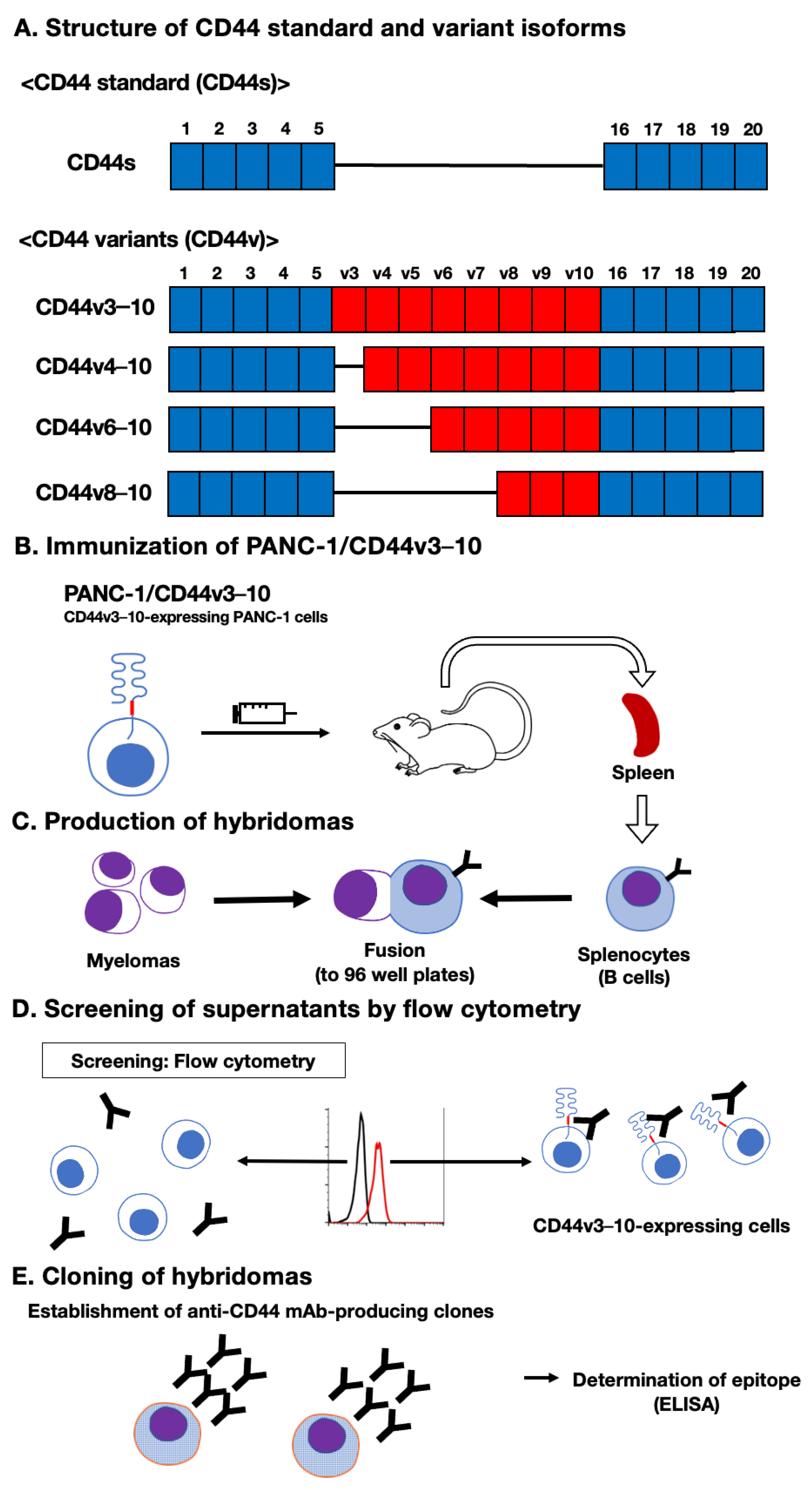
Figure 2.
Flow cytometry using C44Mab-18. CHO/CD44v3–10 (A), CHO/CD44s (B), CHO-K1 (C), and HSC-3 (D) cells were treated with 0.01–10 µg/mL of C44Mab-18. Then, cells were treated with Alexa Fluor 488-conjugated anti-mouse IgG (Red line). The black line represents the negative control (blocking buffer).
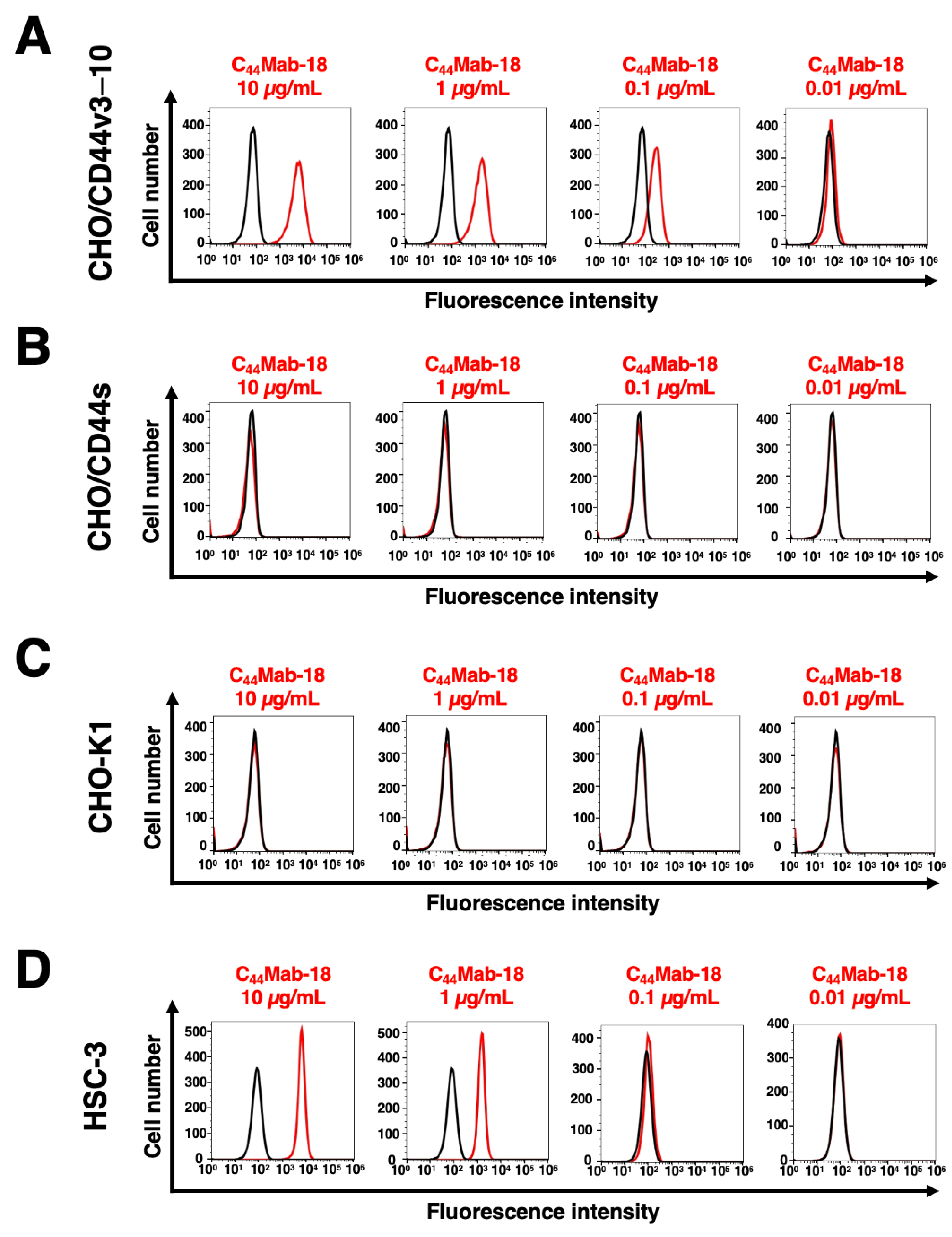
Figure 3.
Determination of C44Mab-18 epitope by ELISA. The synthesized peptides, which cover the variant exon-encoded region between v3 and v10, were immobilized on 96-well plates. The plates were incubated with C44Mab-18, followed by incubation with peroxidase-conjugated anti-mouse immunoglobulins. Optical density was measured at 655 nm. The CD44 p551–570 sequence (SNSNVNRSLSGDQDTFHPSG) is corresponding to variant 10 and the constant exon 16-encoded sequence. Error bars represent means ± SDs. NC, negative control (0.1% DMSO [solvent] in PBS).
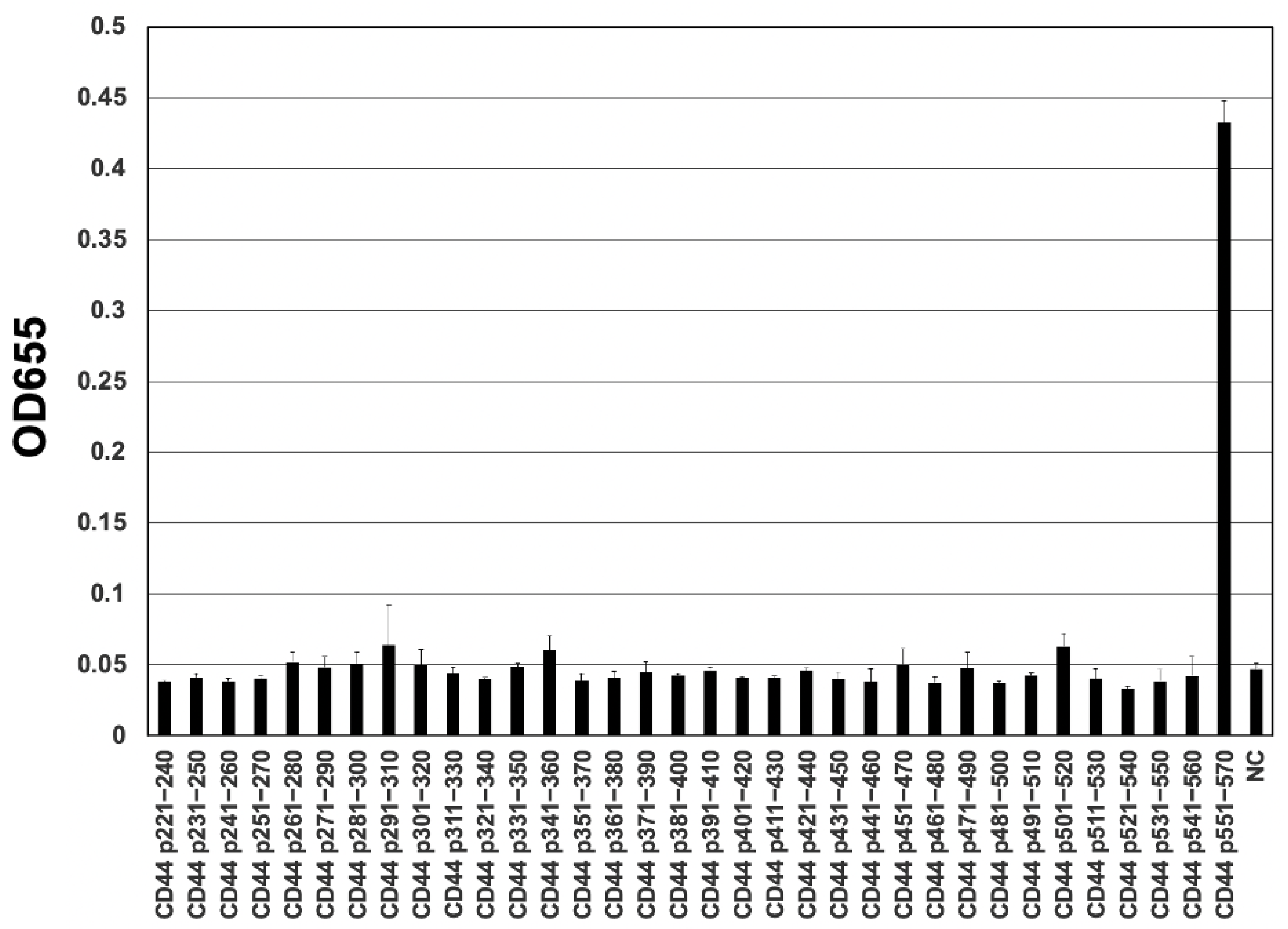
Figure 4.
The determination of the binding affinity of C44Mab-18. C44Mab-18 at indicated concentrations was treated with CHO/CD44v3–10 (A) and HSC-3 (B). Then, cells were treated with anti-mouse IgG conjugated with Alexa Fluor 488. Fluorescence data were collected, followed by the calculation of the apparent dissociation constant (KD) by GraphPad PRISM 8.
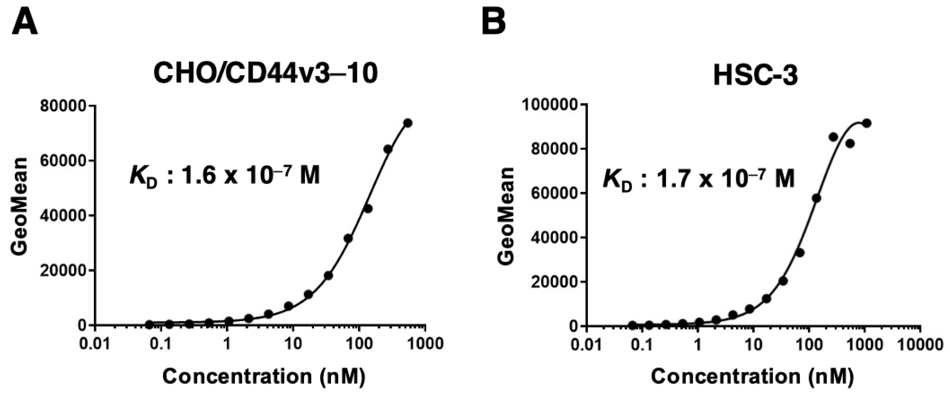
Figure 5.
Western blot analysis by C44Mab-18. The total cell lysates (10 µg of protein) were separated and transferred onto polyvinylidene difluoride (PVDF) membranes. The membranes were incubated with 10 µg/mL of C44Mab-18 (A), 10 µg/mL of C44Mab-46 (B), or 0.5 µg/mL of an anti-β-actin mAb (C), followed by incubation with peroxidase-conjugated anti-mouse immunoglobulins. The red arrows indicate the CD44v3–10 (>180 kDa). The black arrow indicates the CD44s (~75 kDa). The white arrow indicates a lower molecular weight band recognized by C44Mab-18 in CHO/CD44v3–10 lysate (~70 kDa).
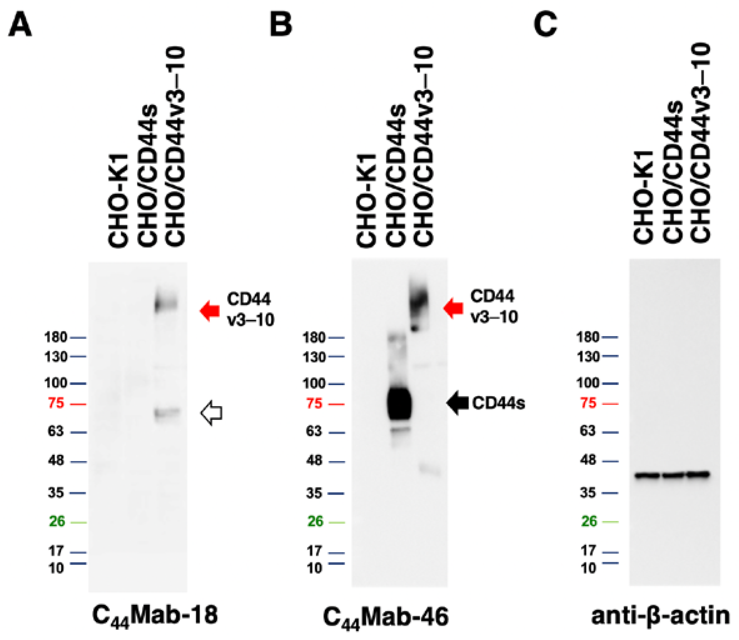
Figure 6.
Immunohistochemical analysis using C44Mab-18 and C44Mab-46 against OSCC tissues. (A–F) Serial sections of the OSCC tissue array (OR601c) were incubated with 1 µg/mL of C44Mab-18 or C44Mab-46 followed by treatment with the Envision+ kit. The color was developed using 3,3’-diaminobenzidine tetrahydrochloride (DAB), and the sections were counterstained with hematoxylin. Scale bar = 100 µm.
Disclaimer/Publisher’s Note: The statements, opinions and data contained in all publications are solely those of the individual author(s) and contributor(s) and not of MDPI and/or the editor(s). MDPI and/or the editor(s) disclaim responsibility for any injury to people or property resulting from any ideas, methods, instructions or products referred to in the content. |
© 2023 by the authors. Licensee MDPI, Basel, Switzerland. This article is an open access article distributed under the terms and conditions of the Creative Commons Attribution (CC BY) license (http://creativecommons.org/licenses/by/4.0/).
Copyright: This open access article is published under a Creative Commons CC BY 4.0 license, which permit the free download, distribution, and reuse, provided that the author and preprint are cited in any reuse.


