
Submitted:
18 May 2023
Posted:
19 May 2023
You are already at the latest version
Abstract
Children from rural areas face numerous possibilities for neurodevelopmental conditions that may compromise their well-being and optimal development. Neuropsychology and electroencephalography (EEG) have shown strong agreement in detecting correlations between these two variables and suggest an association with specific environmental and social risk factors. The present meta-analysis aims to integrate the qualitative and quantitative EEG findings, their relationship with cognitive impairment in children living in vulnerable or rural environments, and the main risk factors influencing EEG abnormalities. The method for this purpose was based on a systematic string-based review from Ebscohost and Web of Science, following the Preferred Reporting Items for Systematic Reviews and Meta-Analysis (PRISMA). Qualitative and quantitative analyses were conducted from the outcomes that complied with the selected criteria. A total of 92 records were identified; however, only 20 were eligible, considering 11 for qualitative and 9 for quantitative analysis. The findings highlight a significant amount of literature on EEG and its relation with cognitive impairment from studies in children with epilepsy and malnutrition. In general, there is evidence about the advantages of implementing EEG diagnosis and research techniques in children living under risk conditions. Further research is needed to better describe and integrate the state of the art regarding EEG features extraction.
Keywords:
Rural
; well-being
; infant welfare
; risk factors
1. Introduction
Impaired cognitive development under risk conditions depends on different factors and how much they affect one another. Common causes compromising the cognitive and central nervous system (CNS) are often observed in disadvantaged social groups such as rural entities living in poverty [1]. Compared to other neuroimaging techniques, electroencephalography (EEG) is a viable clinical diagnostic and research technique; it is low-cost, non-invasive, functionally sensitive, temporarily precise, and continuously improved for further complex features extraction in different clinical conditions [2,3]. Classification methods for the sensitive detection of differences between normal and cognitively impaired subjects through the analysis of the electrical activity in the brain have been explored and have shown good reliability, even using a small number of electrodes. The EEG functionally probes the CNS to obtain a real-time record of electrical activity [4]. The origin of these electrical signals is in the pyramidal cells of the cerebral cortex; each neuron contains an electrical dipole that can be inhibitory or excitatory depending on the cell [5]. In this way, it collects and records information about the electrical activity in each brain region through electrodes placed on the surface of the skull to capture the potential difference between them [6]. The standard EEG is a non-invasive, painless, and inexpensive technique in which surface electrodes are attached to the scalp by a conductive gel, positioned according to the international 10-20 system; the voltage is measured between two electrodes and usually 16 to 24 electrodes are used for clinical purposes [7,8], however for research purposes the number of electrodes can go up to 128 electrode channels. Today, digital amplifiers are used, which facilitate signal analysis and storage, as well as the ability to change parameters such as filters, sensitivity, recording time, and settings. To obtain an EEG recording, the patient must be relaxed, in a semi-dark environment, and with eyes closed. A standard EEG, including activation techniques, primarily intermittent photostimulation and hyperventilation, should last at least 30 minutes [9]. These techniques are designed to provoke or enhance the appearance of abnormalities in brain activity. Neuron-related potentials recorded in the EEG are derived from the electrical activity of excitable tissues and gathered by measuring the potential difference between a scanner electrode and a reference electrode to investigate the biophysical basis of potentials of neuronal origin.
On the other hand, the conditions of rural development are part of society’s history. They are somehow not a new public health concern. However, most diagnoses related to brain maturation, specifically using EEG techniques, prevention, or treatment methods, are often developed and implemented in urban facilities, leading to challenges for rural living children [10,11]. Rurality in definition has wide variances; the Office Management and Budget (OMB) and other census institutions are often good resources for defining conditions for rural development. Accordingly, a rural is defined as a nonmetropolitan area in which there is neither a city nor an urbanized area with 50,000 or more inhabitants, and the culture of the population who live in this condition is shaped by density, geography, agricultural heritage, economic conditions [12], social styles [13], religion, behavioral norms, health care, and distance to health care services different from urban communities [14]; particularly neuropsychology, and psychophysiology practice in rural settings [15]. Rural represent disparities in health risk factors compared to urban residential assets; rural residents usually demonstrate higher rates of smoking and crude alcohol consumption [16]. Status of poverty or malnutrition has been found to be associated with behavioral conditions, such as depression in children [17].
These issues considerably influence and mold behavioral, mental health, and cognitive features. For example, the impact of mental health disorders and cognitive impairment in rural areas is higher compared to urban areas [18], children lack basic assets for a good quality of life, which is directly related to risk-taking behaviors, and non-optimal cognitive development [19]. The literature suggests that neonatal mortality and morbidity are higher in rural and marginalized regions due to economic constraints, the absence of specialized obstetric and neonatal services, and the lack of awareness of the dangers to maternal and fetal health during pregnancy[20], which are linked to a higher prevalence of risk factors for early brain damage such as very preterm birth, low birth weight, hypoxic-ischemic encephalopathy, exposure to substances and maternal diseases, as well as septic processes [21]. In the case of the presence of early signs of neonatal seizures, EEG monitoring provides predictors related to unfavorable neurological, particularly in infants who experienced perinatal risks [22]. All these variables have been associated with an increased risk of neurocognitive developmental disorders. However, according to a systematic review [23] current capacity to predict at-risk infants are sub-optimal; even indirect clinical familial of particular mood conditions, such as depression in mothers, is also considered a factor capable of modifying frontal alpha EEG activity in their children [24].
Several research reports the findings from the association of EEG abnormalities, patterns, or features. Scholarly literature address the relationship between congenital risk factors in children’s development and EEG patterns. However, only a few integrate this information with neuropsychological, clinical, and behavioral conditions when children were exposed to neurodevelopmental risk factors. The present meta-analysis aims to integrate the qualitative and quantitative EEG findings, their relationship with cognitive impairment in children living in vulnerable or rural environments, and the main risk factors influencing EEG abnormalities.
2. Methodology
2.1. Search strategy
The literature review was conducted following the Preferred Reporting Items for Systematic Reviews and Meta-Analysis (PRISMA) guidelines, identification, screening, eligibility, and inclusion for analysis [25] (Figure 1). The electronic databases for the search were: EBSCOHost and Web of Science (WoS), considering their disciplinary focus on the topic of interest, the selection of these databases is optimal for research in health sciences. We used keyword combinations as follows: ("EEG" OR "Electroencephalography", "EEG" OR "Cognitive Impairment") AND ("Neurodevelopmental" OR "Cognitive Impairment"). For this review, we considered articles published through January 2023.
2.2. Selection process and data analysis
Outcomes from the search were divided into two groups. The first group of literature for qualitative analysis included research that was not specifically reporting the same measures of cognitive impairment and EEG signals. The description was subdivided from the relevance of the information reported, considering first, EEG in children at neurodevelopmental risk and second, EEG, environmental, and social risk factors. A second group, which particularly reported the same measures and EEG signals, including a sample of children from rural or vulnerable environments, also reported an EEG feature and its relation with cognitive impairment using p-value. Fisher’s Z effect sizes, correlations, and confidence intervals estimated from sample sizes were calculated by extracting and processing information from each article p-value. In both groups, the articles were included if they were published in peer-reviewed journals, were original, and English or Spanish wrote, in which the participants were children from rural environments or were raised under vulnerable conditions, and the use of any EEG recording of brain activity, including ERPs, power spectrum, and frequency domains.
3. Results
3.1. Qualtitative synthesis
3.1.1. EEG in children at neurodevelopmental risk
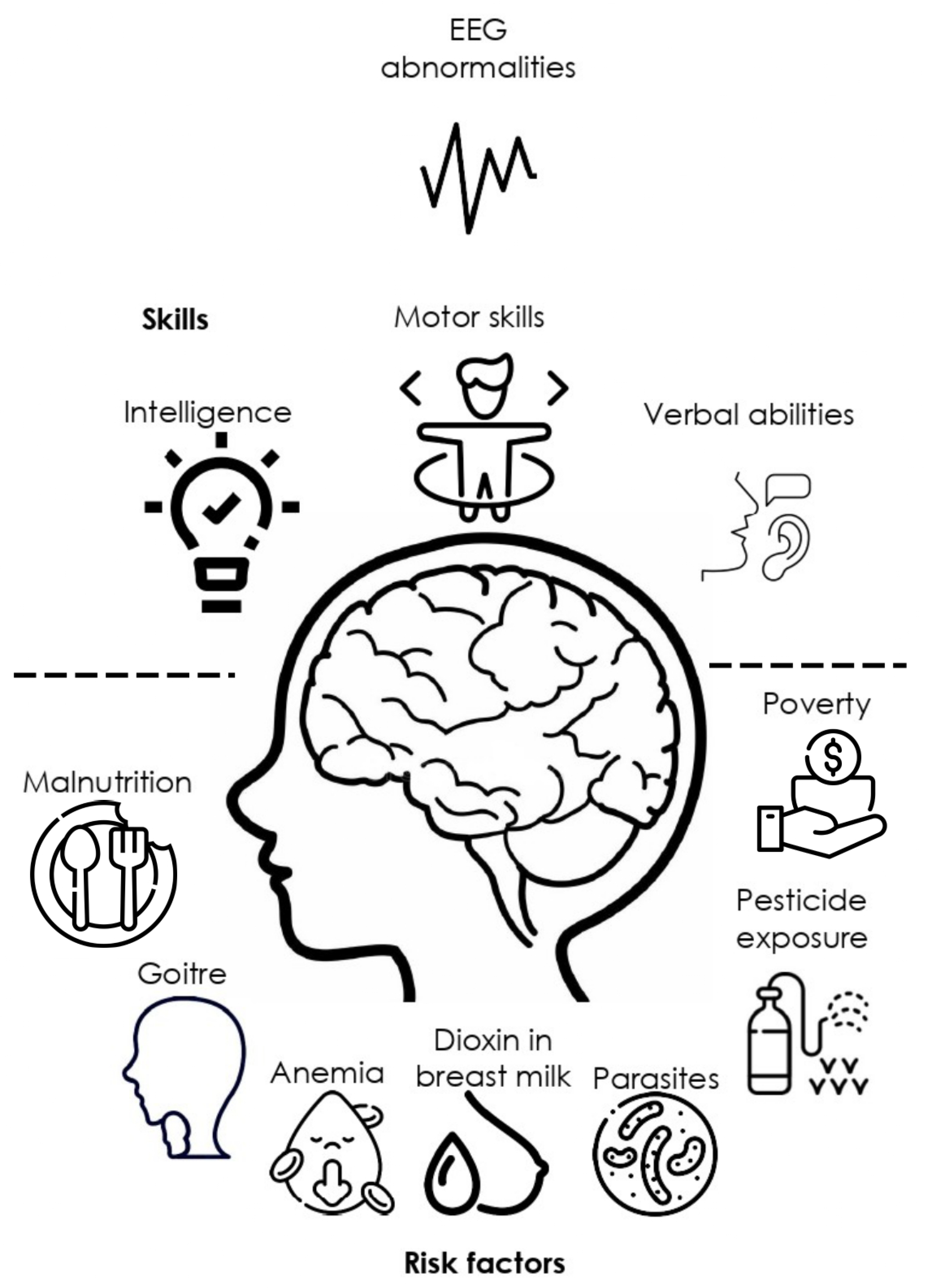
Living in rural areas is not a cognitive or central nervous system (CNS) development risk factor per-se. However, this condition is frequently associated with exposure to pesticides affecting health [26], low-income [27], malnutrition in children and their parents [28]; conditions which may directly or indirectly lead to cognitive impairment and electrophysiological abnormalities (Figure 2). Previous research seeking to describe an association between rurality and risk factors affecting mental health has even shown that low schooling is the main related factor [29]. This problem is increased with the characteristics of rural communities that challenge the neuropsychological service delivery due to resource limitations, distance and costs, professional isolation, and beliefs about psychological services that reduce reliability all over these services [15].
Ueda and collaborators demonstrated a difference in frontal and occipital power variance function in the theta and alpha bands to be smaller in participants presenting mild cognitive impairment compared with healthy subjects [30]. Few studies have reported EEG features of suboptimal or impaired cognitive development in recent years. Particularly under rural or vulnerable environment development conditions, characterized by low to middle income, farm-working as sustainable productive activity, lack or deficient drain systems, treated water services at homes, nutrition deficiencies, and toxins exposure. All of these factors have been linked to different particular neuropsychological and neurophysiological EEG features [31], which could be a predictive factor for anticipating risk or protective factors. However, these studies offer a clear direction about the relevance of EEG features for analyzing cognitive development in children from at-risk rural environments. Particularly, for analyzing the effects of development risk factors and EEG techniques for its description, we selected a group of reports systematic review on neuropsychological and neurophysiological studies in subjects with developmental risks with different neurophysiological characteristics associated with cognitive impairment. We summarize the studies in Table 1, which present data of children aged 0 to 13 years who were exposed to risk factors from rural environments. Within the cognitive domain, research has highlighted language and IQ deficits, as well as differences in motor and executive function skills between the study groups. These findings indicate a disadvantage in cognitive development for the at-risk study groups. Deviant EEG activity or cognitive skills can be detected through different sources of clinical analysis, which could be within a clinician’s scope. It is strongly suggested that diagnostic and intervention practices performed by the neurophysiologist, neuropsychologist, or psychologist should pay special attention to possible predictive or alerting signals. For example, severe malnutrition is a common pattern in children from rural areas and has been shown to affect motor task performance, which is also evident in EEG patterns. In a series of case studies, [31], visually analyzed EEGs of children from the rural undernourished condition and described that these children had motor soft neurological signs related to specific abnormalities in frontal, sharp, and slow waves becoming generalized, parietal sharp and slow waves, and temporal slow waves. Rural children as compared with urban, show lower accuracy in visuospatial tasks measured by global and local visual features recognition and speed processing; they also present difficulties in internal image objects and visual memory [32].
Later IQ scores can be predicted employing EEG patterns measure strategies even during early neonatal development periods, as suggested by previous studies. For example, in their study Beckwith and Parmelee [33] proposed three visual methods to analyze EEG in preterm infants, one to describe unusual EEG patterns, second, to code persistent remarkable delay on EEG maturation, and third, to describe the distribution of patterns within and across states of consciousness. Their results suggest that the integrity of the electrical neurophysiological organization of the brain has implications for later development, as measured in five years follow-ups. Other severe EEG features abnormalities such as epileptiform signs [34] and stress exposure have also been underestimated in at-risk populations. Several neuropsychological and EEG features have proven to have a high sensitivity to indicate a possible neurophysiological decline in conditions such as Parkinson’s disease [35], autism [36], epilepsy [37], parental history of alcohol consumption [38], as well as cognitive impairment in areas such as memory, attention, language, and school success [39].
Thus, neuropsychological tests and EEG analysis are useful tools for studying cognitive impairment in at-risk populations. For example, qEEG analysis in Pakistani children suggested gamma power as a neural marker of cognitive function, specifically associated with executive function and verbal IQ [40]. Cognitive impairment in vulnerable rural populations is often severely underestimated or mistaken for other common developmental disorders. This is partly because most children with, for example, attention problems are impulsive and have low academic achievement. However, the cognitive and behavioral deficits associated with attention disorders are related to different brain impairments, such as less effective executive function, increased frontal theta waves, and a deficit in selective modulation of cortical activity [41]. Moreover, this underestimation is also due to the difficulty in distinguishing between neurotypical development and mild cognitive impairment at the cognitive and electrophysiological levels. The diversity of cognitive and electrophysiological symptoms, according to the literature presented in Table 1, leads to the conclusion that there is not a general developmental disorder but a complex of non-optimal or mild multifactorial information processing conditions in children with attention, learning, or deviant behavior, which can be described by characterization of EEG features.
In the same direction, literature had widely reported that severe health risks are not the only condition causing abnormalities in children’s bioelectrical functional state with a significant association with other cognitive processes. In a study with 172 children, 10 to 12 years old, children with slight deficits in executive functions showed signs of suboptimal functional states of the limbic structures. Most evident behavioral deficits were related to motor and tactile perseverations and deviations in emotional-motivational regulation, such as poor motivation in task performance and poor communication skills, according to the authors [42]. Systematically, in previous studies on children with learning difficulties, bilateral synchronous slow waves over the frontal and/or frontal and central cortices-frontal theta-waves seem to be the vector for the cortico-cortical functional connectivity for frontal regions and surrounding structures on the EEG, suggesting that frontal theta waves are most probably caused by the common for the frontal and central cortices neuronal theta activity synchronized via cortico-subcortical links [43]. The findings consistently suggest that children with learning difficulties present functional connectivity abnormal patterns in theta, alpha, and beta frequency bands observed during resting state in the coherence of the functional coupling between the frontal and anterior temporal cortices found predominantly in the left hemisphere of frontal bilateral synchronous theta waves [44].

Table 1.
Qualitative analysis of neuropsychological and electrophysiological features, and its relation with risk factors and covariates commonly found under rural development conditions.
Table 1.
Qualitative analysis of neuropsychological and electrophysiological features, and its relation with risk factors and covariates commonly found under rural development conditions.
| EEG finding | Brain Region | Neuropsychological finding | n | Covariates | Age | Country | Associated with factor | Reference |
|---|---|---|---|---|---|---|---|---|
| Sharp slow waves, Slow waves, Generalized sharp and slow waves, Sharp and slow waves | Right parietal, Bilateral centroparietal, Right frontal, Bifrontal | Soft neurological signs, poor performance in motor tasks, successive finger tapping, heel-toe tapping, alternating hand pronation supination | 208 | Movement coordination disorders | 8-10 yr | India | Malnourish | [31] |
| Lower gamma power | Frontal, and parietal | Better Executive function performance, verbal intelligence | 105 | Anemia | From birth, 24, and 48 months | Pakistan | Poverty | [40] |
| Decrease in relative Delta and increase in alpha and beta powers | Right frontal, and parietal | Positive correlation with language, and motor development | 55 | Gestational age, body length and head circumference | Prenatal-2-year follow-up | Vietnam (US) | Dioxin in breast milk | [45] |
| Lower relative alpha, and higher relative theta power | Bilateral central, temporal, and parietal | Delay gratification and non-verbal cognitive ability. Lower scores in risk exposure group for visual reception | 143 | Friendliness | 18 months | US (International adoption) | Adoption, deprivation, parental exposure to drugs, parental malnourishment and premature birth | [46] |
| Decrease in Alpha, high theta | Lingual gyrus, and inferior frontal gyrus orbital right Middle temporal gyrus | WISC Full-Scale IQ | 108 | Classification techniques | 5-11 yr | Caribean islands | Protein undernutrition | [47] |
| Centro-parietal slow-wave, paroxysmal, and focal abnormalities. Increment of slow (<5 Hz). Decrease of alpha power (8.9 Hz) | Fronto-central. Centro-parietal, frontal | Non | 108 | Non | 5-11 yr | Barbados | Protein undernutrition | [48] |
| Abnormal slow wave background EEG tracings, Paroxysmal activity | Non | 194 | Parasitism, and goitre, iodine level | 9-13 yr | Ecuador | Malnutrition | [49] | |
| Bilateral slow waves, Slow abnormal waves, Sharp abnormal waves | Anterior brain areas, subcortical origin, Posterior regions | Reduced verbal abilities, problem solving/concentration, and focusing, and inhibition-control/flexibility in at-risk groups | 194 | Infection protozoan parasite, Parent’s education | 9-13 yr | Ecuador | Malnutrition | [50] |
| Alpha 1 band, and alpha-beta power ratio under driving 8 Hz | Temporo-occipital | Non | 20 | Lethargic movement, depressed oxygen consumption, and sodium pump activity | 5-23 months | Jamaica | Malnutrition, Marasmus and Kwashiorkor | [51] |
| Synchronous theta waves | Frontal and limbic | Motor and tactile perseverations, emotional-motivational regulation, poor communication skills | 172 | Learning difficulties | 10-12 yr | Russia | Non | [42,43] |
3.1.2. EEG, environmental and social risk factors
Environmental exposure to toxins is present in both rural and urban facilities, which leads to the presence of toxic concentrations of these compounds in different regions of the CNS. Furthermore, they may have a neurobiological effect even during prenatal in utero stages [52], leading to future developmental consequences. For example, exposure to lead and cadmium in children with learning disabilities in a rural population is related to differences in IQ performance, demonstrating that higher lead concentrations measured from hair are inversely correlated with intelligence and brain functioning [53]. In their study, Thatcher and Lester demonstrated, using a series of mixed methods of qEEG, and EEG visual analysis, that higher concentrations of lead or cadmium are associated with an increase in slow wave activity and a decrease in the amplitude observed in the EEG. The authors describe the different effects of exposure to heavy toxins on brain electrical activity. In particular, there is a strong correlation between the evoked potential measures obtained from the central leads and those obtained from the posterior occipital and parietal leads.
The low income in rural areas is, at the same time, a multiphasic factor that is allocated to different situations that give place to adverse developmental settings. Children growing up under vulnerable conditions are frequently exposed to higher levels of stress due to stressful home environments explained by low-income [54], anxiety [55] as well as prenatal cortisol exposure from the stressed mother [56]. Stress-related neurochemicals in early childhood, measured utilizing cortisol concentrations, are associated with impaired development of executive functions. In their study, Blair and Berry [57] describe findings that indicate children from 7 to 48 months with lower cortisol levels under resting conditions perform better on a battery of executive functions tasks. On the other hand, there is currently an increased advance in the understanding of electrical neural activity assessed by the use of EEG. EEG Power can be quantified among the different frequency bands and has been proven to be associated with different cognitive processes so studies suggest the analysis of power spectra distributions to better understand how neurons grow and become myelinated [58], which is closely related to cognitive development. External factors in children in vulnerable conditions represent a strong influence on brain development. For example, sleep deprivation associated with external stressors affects white matter myelin microstructure growth [59], and economic problems increase cerebrovascular diseases [60]. Neurological activation is dependent on cortisol concentrations during early infancy. In a study by St John [56], breastfeeding did not explain links between maternal cortisol and infant physiology. However, maternal cortisol at six months of age has proven to be a predictor of infant cortisol slope and EEG power even at 12 months, indexed by greater neural activation and reduced 6-9 Hz power, under a social interaction task. And even in an advantaged, low-risk sample, infant neural activation, as indexed by 6-9 Hz power, is sensitive to subtle variations, especially maternal physiological regulation.
3.2. Quantitative analysis
Quantitative analysis was taken from 11 research reporting EEG abnormalities, mainly related to epileptic EEG features and seizure history. According to the data from children from rural areas, the reports have in common a first recognition based on epileptic history and secondly a particular characterization of EEG features. Table 2 shows confidence intervals and standardized Fisher’s correlation from sample size and p-values from the studies reporting an association of abnormal EEG and cognitive impairment. Six of the studies report a significant association between EEG abnormalities and cognitive impairment; besides, the standard calculation from three of the studies showed a significant correlation between the two variables.
Table 2.
Quantitative analysis of EEG reports and cognitive impairment correlation.
| Reference | EEG technique | n | Age | p-value | r | 95% CI Upper limit |
95% CI Lower limit |
Fisher’s Zr |
|---|---|---|---|---|---|---|---|---|
| [61] | EEG Seizures report | 1014 | 0-17 yr | <0.01 | 0.0809 | 0.0194 | 0.1417 | 0.081 |
| [62] | ERPs | 50 | 6-7 yr | .450 | - | - | - | - |
| [63] | ERPs | 178 | 4-12 yr | .651 | - | - | - | - |
| [64] | EEG Seizures report | 494 | * | <0.001 | 0.1476 | 0.0602 | 0.2328 | 0.1487 |
| [65] | EEG Seizures report | 16 | * | <0.001 | 0.7419 | 0.3895 | 0.3895 | 0.9548** |
| [66] | EEG Seizures report | 72 | 6-14 yr | * | 0.3798 | 0.1624 | 0.562 | 0.3998** |
| [67] | EEG Seizures report | 679 | * | * | 0.0833 | 0.0081 | 0.1575 | 0.0835 |
| [68] | EEG Seizures report | 112 | 6-14 yr | <0.001 | 0.126 | 0.0513 | 0.1994 | 0.1267 |
| [69] | ERPs | 148 | 1-5 month | 0.356 | 0.356 | 0.2065 | 0.4892 | 0.3723** |
| [40] | EEG Gamma power | 105 | 0-24 months | 0.036 | 0.2049 | 0.0138 | 0.3816 | 0.2079 |
| [70] | EEG alpha and gamma power | 41 | 12-16 yr | <0.01 | 0.0016 | -0.3062 | 0.3091 | 0.0016 |
* Data not directly reported from variables of interest. **Significance < 0.001.
3.2.1. EEG abnormalities and frequent co-morbidity in rural areas
A variety of poverty-related risk factors are commonly presented to infants and young children in low- and middle-income countries, increasing the possibility that these individuals may experience poor neurodevelopmental outcomes [69]. Access inequities to treatments in children who present neurodevelopmental risks are commonly reported; for example, sociodemographic research [61] showed that children with epilepsy aged 1 to 5 years old as opposed to other children and adolescents and children with epilepsy as opposed to children living in smaller cities and rural areas were more likely to receive specialized attention by neuro pediatricians. Other countries, for example, reported that ERPs are effective for assessing children in rural areas [62,63], and are a useful tool to describe the neurophysiological maturity of the brain for the processing of visual novel information, face, and auditory processing. Non-optimal development of sensorial brain systems such as brain somatomotor [71], or frontostriatal and cerebellar circuitry [72,73] can lead to clinical co-morbidity with ADHD. In literature [65], this factor has suggested a complex clinical symptomatology that goes beyond that explained by the EEG features of status epilepticus. For example, in the research by Chidi and collaborators in Nigeria, the cross-sectional study in children with epilepsy, 14.2% of the sample had ADHD co-morbidity, being the inattentive subtype the most common and significantly associated with poor academic achievement, and living in rural areas.
According to a case-control study [66], co-morbidity in children with a history of epilepsy is very common among cases and consistently associated with cognitive impairment (64%), behavior disorders (61%), and motor difficulties (26%).
4. Integrated analysis: current limitations and future directions
EEG is perhaps the most advantaged technique for research and diagnosis to implement in at-risk rural conditions. Neurophysiological factors underlie different expressions of cognitive impairment, which often are underestimated in rural environments. Many cognitive symptoms are often generalized and erroneously used in common clinical practice. Basic neuropsychological assessments offer sensitive information about the cognitive performance of children and adults under rural risk living conditions.
Research reveals that perhaps one of the most widely used techniques in rural areas is EEG features description for seizure detection, being at a primary level accessible to a broad population. Despite this being a clinically sensitive technique. According to the revised literature for this study, there is a significant relation suggested in at least 16 reports between EEG features and cognitive impairment in children with neurodevelopmental risk factors. These findings underscore a need for cultural indicators to inform neonatal, child, and adolescent assessment from birth onward, enabling early detection and, ultimately, intervention at critical neurodevelopmental stages. As protective factors, nutritional considerations such as blood iron biomarkers, behavioral measures of cognition, and EEG measurements of brain function have all been found to improve with the use of a biofortified staple grain in dietary interventions [70]. The numerous processes by which blood iron content impacts brain function, and cognition are strongly suggested by modeling the interactions among these variables.
This study presents evidence from at least two decades of EEG and cognitive impairment research, including the effects of neurodevelopmental risk factors among these variables. The results are clinically relevant and consistent across countries and large populations of children. In addition, we can observe an advance in EEG measurement techniques and feature extraction with a reduction in cost and an increase in clinical sensitivity and classification of abnormalities. The descriptions in this report can serve as a guide for clinical and research decisions in countries or cultures facing the above challenges in children’s mental health. Epidemiologic data on epilepsy demonstrate a frequent interaction of this neurological disorder with cognitive impairment. Finally, we emphasize the relevance of using electrophysiological and neuropsychological assessment procedures together for research and clinical purposes.
This meta-analysis has several limitations. The main effect of child abuse on neurodevelopment was not studied, being another widely studied variable reported in the literature. Drug abuse and parental neglect are, for instance, significant risk factors commonly associated with neurodevelopmental clinical conditions and also frequently present in families living in rural areas in low-income socio-economical communities. Another limitation of the study relates to the lack of information concerning protective factors; case-control studies, including the variables of interest in this analysis, are scarce, and there is a need to increase efforts to provide access to diagnostic services in children living in vulnerable or rural environments. It is also necessary to incorporate analysis related to EEG feature extraction, given the emerging developed techniques in this issue, which suggest being promising to facilitate the identification of EEG abnormalities.
Author Contributions
Conceptualization, G.G.A., and C.T.G.; methodology, G.G.A.; formal analysis, G.G.A.; writing original draft preparation, G.G.A.; writing review and editing, G.G.A.; visualization, C.T.G.; Y. All authors have read and agreed to the published version of the manuscript.
Funding
This research received no external funding.
Institutional Review Board Statement
Not applicable.
Informed Consent Statement
No informed consent was needed for this study.
Data Availability Statement
The data described in this study are available on request from the corresponding author.
Conflicts of Interest
The authors declare no conflict of interest.
Abbreviations
The following abbreviations are used in this manuscript:
| EEG | Electroencephalography |
| qEEG | Quantitative Electroencephalography |
| CNS | Central Nervous System |
| IQ | Intellectual Quotient |
References
- Buda, A.; Dean, O.; Adams, H.R.; Mwanza-Kabaghe, S.; Potchen, M.J.; Mbewe, E.G.; Kabundula, P.P.; Mweemba, M.; Matoka, B.; Mathews, M.; Menon, J.A.; Wang, B.; Birbeck, G.L.; Bearden, D.R. Neighborhood-Based Socioeconomic Determinants of Cognitive Impairment in Zambian Children With HIV: A Quantitative Geographic Information Systems Approach. Journal of the Pediatric Infectious Diseases Society 2021, 10, 1071–1079. [Google Scholar] [CrossRef]
- Esqueda-Elizondo, J.J.; Juárez-Ramírez, R.; López-Bonilla, O.R.; García-Guerrero, E.E.; Galindo-Aldana, G.M.; Jiménez-Beristáin, L.; Serrano-Trujillo, A.; Tlelo-Cuautle, E.; Inzunza-González, E. Attention Measurement of an Autism Spectrum Disorder User Using EEG Signals: A Case Study. Mathematical and Computational Applications 2022, 27. [Google Scholar] [CrossRef]
- Ramírez-Arias, F.J.; García-Guerrero, E.E.; Tlelo-Cuautle, E.; Colores-Vargas, J.M.; García-Canseco, E.; López-Bonilla, O.R.; Galindo-Aldana, G.M.; Inzunza-González, E. Evaluation of Machine Learning Algorithms for Classification of EEG Signals. Technologies 2022, 10. [Google Scholar] [CrossRef]
- Pfurtscheller, G.; Lopes da Silva, F.H. Event-related EEG/MEG synchronization and desynchronization: basic principles. Clinical neurophysiology : official journal of the International Federation of Clinical Neurophysiology 1999, 110, 1842–1857. [Google Scholar] [CrossRef] [PubMed]
- Selim R. Benbadis, M.; Aatif M. Husain, M.; Peter W. Kaplan, M.; William O. Tatum, IV, D. Handbook of EEG Interpretation.; Demos Medical, 2008.
- Fried, S.; Moshé, S.L. Basic physiology of the EEG. Neurology Asia 2011, 16, 23–25. [Google Scholar]
- Schomer, D.L.; Lopes da Silva, F.H. Niedermeyer’s Electroencephalography: Basic Principles, Clinical Applications, and Related Fields; Oxford University Press, 2017. [CrossRef]
- Jellinger, K.A. Niedermeyer’s Electroencephalography: Basic Principles, Clinical Applications, and Related Fields, 6th edn. European Journal of Neurology 2011, 18, e126. [Google Scholar] [CrossRef]
- Miley, C.E.; Forster, F.M. Activation of partial complex seizures by hyperventilation. Archives of neurology 1977, 34, 371–373. [Google Scholar] [CrossRef]
- Blackstock, J.S.; Chae, K.B.; Mauk, G.W.; McDonald, A. Achieving Access to Mental Health Care for School-Aged Children in Rural Communities: A Literature Review. The rural educator 2018, 39, 12–25. [Google Scholar] [CrossRef]
- Smith, S.L.; Franke, M.F.; Rusangwa, C.; Mukasakindi, H.; Nyirandagijimana, B.; Bienvenu, R.; Uwimana, E.; Uwamaliya, C.; Ndikubwimana, J.S.; Dorcas, S.; et al. Outcomes of a primary care mental health implementation program in rural Rwanda: A quasi-experimental implementation-effectiveness study. PloS one 2020, 15, e0228854. [Google Scholar] [CrossRef]
- Crichlow, M.A.; Northover, P.; others. Race and Rurality in the Global Economy; SUNY Press, 2018.
- BOOK REVIEWS. The Professional Geographer 1986, 38, 430–457. [CrossRef]
- K. Bryant Smalley, PhD, P.; Jacob C. Warren, P. Rural Public Health : Best Practices and Preventive Models.; Springer Publishing Company, 2014.
- Allott, K.; Lloyd, S. The provision of neuropsychological services in rural/regional settings: professional and ethical issues. Applied neuropsychology 2009, 16, 193–206. [Google Scholar] [CrossRef] [PubMed]
- Luo, D.; Du, J.; Wang, P.; Yang, W. Urban-rural comparisons in health risk factor, health status and outcomes in Tianjin, China: A cross-sectional survey (2009-2013). Australian Journal of Rural Health 2019, 27, 535–541. [Google Scholar] [CrossRef] [PubMed]
- Jiménez-Ceballos, B.; Martínez-Herrera, E.; Ocharan-Hernández, M.E.; Guerra-Araiza, C.; Farfán García, E.D.; Muñoz-Ramírez, U.E.; Fuentes-Venado, C.E.; Pinto-Almazán, R. Nutritional Status and Poverty Condition Are Associated with Depression in Preschoolers. Children 2023, 10. [Google Scholar] [CrossRef]
- Smalley, K.B.; Warren, J.C.; Rainer, J. Rural mental health: Issues, policies, and best practices; Springer Publishing Company, 201.
- Bell, E.; Merrick, J. Rural Child Health: International Aspects.; Health and Human Development, Nova Science Publishers, Inc, 2010.
- Beck, S.; Wojdyla, D.; Say, L.; Betran, A.P.; Merialdi, M.; Requejo, J.H.; Rubens, C.; Menon, R.; Van Look, P.F.A. The worldwide incidence of preterm birth: a systematic review of maternal mortality and morbidity. Bulletin of the World Health Organization 2010, 88, 31–38. [Google Scholar] [CrossRef]
- Chattopadhyay, N.; Mitra, K. Neurodevelopmental outcome of high risk newborns discharged from special care baby units in a rural district in India. Journal of Public Health Research 2015, 4, 7–12. [Google Scholar] [CrossRef]
- Doandes, F.M.; Manea, A.M.; Lungu, N.; Brandibur, T.; Cioboata, D.; Costescu, O.C.; Zaharie, M.; Boia, M. The Role of Amplitude-Integrated Electroencephalography (aEEG) in Monitoring Infants with Neonatal Seizures and Predicting Their Neurodevelopmental Outcome. Children 2023, 10. [Google Scholar] [CrossRef] [PubMed]
- Kong, A.H.; Lai, M.M.; Finnigan, S.; Ware, R.S.; Boyd, R.N.; Colditz, P.B. Background EEG features and prediction of cognitive outcomes in very preterm infants: A systematic review. Early Human Development 2018, 127, 74–84. [Google Scholar] [CrossRef]
- Lopez-Duran, N.L.; Nusslock, R.; George, C.; Kovacs, M. Frontal EEG asymmetry moderates the effects of stressful life events on internalizing symptoms in children at familial risk for depression. Psychophysiology 2012, 49, 510–521. [Google Scholar] [CrossRef]
- Page, M.J.; McKenzie, J.E.; Bossuyt, P.M.; Boutron, I.; Hoffmann, T.C.; Mulrow, C.D.; Shamseer, L.; Tetzlaff, J.M.; Akl, E.A.; Brennan, S.E.; Chou, R.; Glanville, J.; Grimshaw, J.M.; Hróbjartsson, A.; Lalu, M.M.; Li, T.; Loder, E.W.; Mayo-Wilson, E.; McDonald, S.; McGuinness, L.A.; Stewart, L.A.; Thomas, J.; Tricco, A.C.; Welch, V.A.; Whiting, P.; Moher, D. The PRISMA 2020 statement: An updated guideline for reporting systematic reviews. Journal of clinical epidemiology 2021, 134, 178–189. [Google Scholar] [CrossRef]
- Corcino, C.O.; Teles, R.B.d.A.; Almeida, J.R.G.d.S.; Lirani, L.d.S.; Araújo, C.R.M.; Gonsalves, A.d.A.; Maia, G.L.d.A. [Evaluation of the effect of pesticide use on the health of rural workers in irrigated fruit farming]. Ciencia & saude coletiva 2019, 24, 3117–3128. [Google Scholar]
- Correction: Perceptions and practices related to birthweight in rural Bangladesh: Implications for neonatal health programs in low- and middle-income settings. PLoS ONE 2020, 15, 1.
- Chanchani, D. Maternal and child nutrition in rural Chhattisgarh: the role of health beliefs and practices. Anthropology & Medicine 2019, 26, 142–158. [Google Scholar]
- Rohrer, J.E.; Borders, T.F.; Blanton, J. Rural residence is not a risk factor for frequent mental distress: a behavioral risk factor surveillance survey. BMC public health 2005, 5, 46. [Google Scholar] [CrossRef]
- UEDA, T.; MUSHA, T.; ASADA, T.; YAGI, T. Classification Method for Mild Cognitive Impairment Based on Power Variability of EEG Using Only a Few Electrodes. Electronics & Communications in Japan 2016, 99, 107–114. [Google Scholar]
- Agarwal, K.N.; Das, D.; Agarwal, D.K.; Upadhyay, S.K.; Mishra, S. Soft neurological signs and EEG pattern in rural malnourished children. Acta paediatrica Scandinavica 1989, 78, 873–878. [Google Scholar] [CrossRef]
- Galindo, G.; Solovieva, Y.; Machinskaya, R.; Quintanar, L. Atención selectiva visual en el procesamiento de letras: un estudio comparative. OCNOS: Revista de Estudios sobre Lectura 2016, 15, 69–80. [Google Scholar] [CrossRef]
- Beckwith, L.; Parmelee, A H, J. EEG patterns of preterm infants, home environment, and later IQ. Child development 1986, 57, 777–789. [CrossRef]
- Placencia, M.; Sander, J.W.; Roman, M.; Madera, A.; Crespo, F.; Cascante, S.; Shorvon, S.D. The characteristics of epilepsy in a largely untreated population in rural Ecuador. Journal of neurology, neurosurgery, and psychiatry 1994, 57, 320–325. [Google Scholar] [CrossRef]
- Cozac, V.V.; Chaturvedi, M.; Hatz, F.; Meyer, A.; Fuhr, P.; Gschwandtner, U. Increase of EEG Spectral Theta Power Indicates Higher Risk of the Development of Severe Cognitive Decline in Parkinson’s Disease after 3 Years. Frontiers in aging neuroscience 2016, 8, 284. [Google Scholar] [CrossRef]
- Levin, A.R.; Varcin, K.J.; O’Leary, H.M.; Tager-Flusberg, H.; Nelson, C.A. EEG power at 3 months in infants at high familial risk for autism. Journal of neurodevelopmental disorders 2017, 9, 34. [Google Scholar] [CrossRef]
- Kanemura, H.; Sano, F.; Ohyama, T.; Mizorogi, S.; Sugita, K.; Aihara, M. EEG characteristics predict subsequent epilepsy in children with their first unprovoked seizure. Epilepsy research 2015, 115, 58–62. [Google Scholar] [CrossRef] [PubMed]
- Ehlers, C.L.; Wall, T.L.; Garcia-Andrade, C.; Phillips, E. Effects of age and parental history of alcoholism on EEG findings in mission Indian children and adolescents. Alcoholism, clinical and experimental research 2001, 25, 672–679. [Google Scholar] [CrossRef] [PubMed]
- Amores-Villalba, A.; Mateos-Mateos, R. Revisión de la neuropsicología del maltrato infantil: la neurobiología y el perfil neuropsicológico de las víctimas de abusos en la infancia. Psicologia Educativa 2017, 23, 81–88. [Google Scholar] [CrossRef]
- Tarullo, A.R.; Obradović, J.; Keehn, B.; Rasheed, M.A.; Siyal, S.; Nelson, C.A.; Yousafzai, A.K. Gamma power in rural Pakistani children: Links to executive function and verbal ability. Developmental cognitive neuroscience 2017, 26, 1–8. [Google Scholar] [CrossRef]
- Machinskaya, R.I.; Semenova, O.A.; Absatova, K.A.; Sugrobova, G.A. Neurophysiological factors associated with cognitive deficits in children with ADHD symptoms: EEG and neuropsychological analysis. Psychology & Neuroscience 2014, 7, 461–473. [Google Scholar] [CrossRef]
- Semenova, O.; Machinskaya, R. The influence of the functional state of brain regulatory systems on the efficiency of voluntary regulation of cognitive activity in children: II. neuropsychological and EEG analysis of brain regulatory functions in 10-12-year-old children with learning difficulties. Human Physiology 2015, 41, 478–486. [Google Scholar]
- Kurgansky, A.; Machinskaya, R. Bilateral frontal theta-waves in EEG of 7-8-year-old children with learning difficulties: Qualitative and quantitative analysis. Human Physiology 2012, 38, 255–263. [Google Scholar] [CrossRef]
- Machinskaya, R.; Kurgansky, A. Frontal bilateral synchronous theta waves and the resting EEG coherence in children aged 7-8 and 9-10 with learning difficulties. Human Physiology 2013, 39, 58–67. [Google Scholar] [CrossRef]
- Nghiem, G.T.; Nishijo, M.; Pham, T.N.; Ito, M.; Pham, T.T.; Tran, A.H.; Nishimaru, H.; Nishino, Y.; Nishijo, H. Adverse effects of maternal dioxin exposure on fetal brain development before birth assessed by neonatal electroencephalography (EEG) leading to poor neurodevelopment; a 2-year follow-up study. Science of the Total Environment 2019, 667, 718–729. [Google Scholar] [CrossRef]
- Tarullo, A.R.; Gunnar, M.R.; Garvin, M.C. Atypical EEG Power Correlates With Indiscriminately Friendly Behavior in Internationally Adopted Children. Developmental Psychology 2011, 47, 417–431. [Google Scholar] [CrossRef]
- Bringas Vega, M.L.; Guo, Y.; Tang, Q.; Razzaq, F.A.; Calzada Reyes, A.; Ren, P.; Paz Linares, D.; Galan Garcia, L.; Rabinowitz, A.G.; Galler, J.R.; Bosch-Bayard, J.; Valdes Sosa, P.A. An Age-Adjusted EEG Source Classifier Accurately Detects School-Aged Barbadian Children That Had Protein Energy Malnutrition in the First Year of Life. Frontiers in neuroscience 2019, 13, 1222. [Google Scholar] [CrossRef] [PubMed]
- Bringas-Vega, M.L.; Taboada-Crispi, A.; Bosch-Bayard, J.; Galán-García, L.; Bryce, C.; Rabinowitz, A.G.; Prichep, L.S.; Isenhart, R.; CALZADA-REYES, A.A.; Virues, T.; Galler, J.R.; Sosa, P.V. F168. An EEG fingerprint of early protein-energy malnutrition. Clinical Neurophysiology 2018, 129, e131. [Google Scholar] [CrossRef]
- Levav, M.; Cruz, M.E.; Mirsky, A.F. EEG abnormalities, malnutrition, parasitism and goitre: a study of schoolchildren in Ecuador. Acta paediatrica (Oslo, Norway : 1992) 1995, 84, 197–202. [Google Scholar] [CrossRef]
- Levav, M.; Mirsky, A.F.; Schantz, P.M.; Castro, S.; Cruz, M.E. Parasitic infection in malnourished school children: effects on behaviour and EEG. Parasitology 1995, 110 Pt 1, 103–111. [Google Scholar] [CrossRef] [PubMed]
- Robinson, S.; Young, R.E.; Golden, M.H. Electrophysiological assessment of brain function in severe malnutrition. Acta paediatrica (Oslo, Norway : 1992) 1995, 84, 1245–1251. [Google Scholar] [CrossRef] [PubMed]
- Cottrell, J.N.; Thomas, D.S.; Mitchell, B.L.; Childress, J.E.; Dawley, D.M.; Harbrecht, L.E.; Jude, D.A.; Valentovic, M.A. Rural and urban differences in prenatal exposure to essential and toxic elements. Journal of toxicology and environmental health. Part A 2018, 81, 1214–1223. [Google Scholar] [CrossRef] [PubMed]
- Thatcher, R.W.; Lester, M.L. Nutrition, environmental toxins and computerized EEG: a mini-max approach to learning disabilities. Journal of learning disabilities 1985, 18, 287–297. [Google Scholar] [CrossRef]
- Urizar, Guido G, J.; Caliboso, M.; Gearhart, C.; Yim, I.S.; Dunkel Schetter, C. Process Evaluation of a Stress Management Program for Low-Income Pregnant Women: The SMART Moms/Mamás LÍSTAS Project. Health education & behavior : the official publication of the Society for Public Health Education 2019, 46, 930–941.
- Atif, N.; Nazir, H.; Zafar, S.; Chaudhri, R.; Atiq, M.; Mullany, L.C.; Rowther, A.A.; Malik, A.; Surkan, P.J.; Rahman, A. Development of a Psychological Intervention to Address Anxiety During Pregnancy in a Low-Income Country. Frontiers in psychiatry 2020, 10, 927. [Google Scholar] [CrossRef]
- St John, A.M.; Kao, K.; Liederman, J.; Grieve, P.G.; Tarullo, A.R. Maternal cortisol slope at 6 months predicts infant cortisol slope and EEG power at 12 months. Developmental psychobiology 2017, 59, 787–801. [Google Scholar] [CrossRef]
- Blair, C.; Berry, D.J. Moderate within-person variability in cortisol is related to executive function in early childhood. Psychoneuroendocrinology 2017, 81, 88–95. [Google Scholar] [CrossRef]
- John, E.R.; Ahn, H.; Prichep, L.; Trepetin, M.; Brown, D.; Kaye, H. Developmental equations for the electroencephalogram. Science (New York, N.Y.) 1980, 210, 1255–1258. [Google Scholar] [CrossRef] [PubMed]
- LeBourgeois, M.K.; Dean, D.C.; Deoni, S.C.L.; Kohler, M.; Kurth, S. A simple sleep EEG marker in childhood predicts brain myelin 3.5 years later. NeuroImage 2019, 199, 342–350. [Google Scholar] [CrossRef] [PubMed]
- Ghaffari-Rafi, A.; Mehdizadeh, R.; Ghaffari-Rafi, S.; Leon-Rojas, J. Demographic and socioeconomic disparities of benign and malignant spinal meningiomas in the United States. Neuro-Chirurgie 2021, 67, 112–118. [Google Scholar] [CrossRef]
- Mattsson, P.; Tomson, T.; Edebol Eeg-Olofsson, K.; Brännström, L.; Ringbäck Weitoft, G. Association between sociodemographic status and antiepileptic drug prescriptions in children with epilepsy. Epilepsia 2012, 53, 2149–2155. [Google Scholar] [CrossRef] [PubMed]
- Kihara, M.; de Haan, M.; Garrashi, H.H.; Neville, B.G.R.; Newton, C.R.J.C. Atypical brain response to novelty in rural African children with a history of severe falciparum malaria. Journal of the neurological sciences 2010, 296, 88–95. [Google Scholar] [CrossRef]
- Kihara, M.; Hogan, A.M.; Newton, C.R.; Garrashi, H.H.; Neville, B.R.; de Haan, M. Auditory and visual novelty processing in normally-developing Kenyan children. Clinical Neurophysiology 2010, 121, 564–576. [Google Scholar] [CrossRef]
- Kariuki, S.M.; Matuja, W.; Akpalu, A.; Kakooza-Mwesige, A.; Chabi, M.; Wagner, R.G.; Connor, M.; Chengo, E.; Ngugi, A.K.; Odhiambo, R.; Bottomley, C.; White, S.; Sander, J.W.; Neville, B.G.R.; Newton, C.R.J.C.; Twine, R.; Gómez Olivé, F.X.; Collinson, M.; Kahn, K.; Tollman, S.; Masanja, H.; Mathew, A.; Pariyo, G.; Peterson, S.; Ndyomughenyi, D.; Bauni, E.; Kamuyu, G.; Odera, V.M.; Mageto, J.O.; Ae-Ngibise, K.; Akpalu, B.; Agbokey, F.; Adjei, P.; Owusu-Agyei, S.; Kleinschmidt, I.; Doku, V.C.K.; Odermatt, P.; Nutman, T.; Wilkins, P.; Noh, J. Clinical features, proximate causes, and consequences of active convulsive epilepsy in Africa. Epilepsia 2014, 55, 76–85. [Google Scholar] [CrossRef]
- Chidi, I.R.; Chidi, N.A.; Ebele, A.A.; Chinyelu, O.N. Co-Morbidity of attention deficit Hyperactivity Disorder (ADHD) and epilepsy In children seen In University of Nigeria Teaching Hospital Enugu: Prevalence, Clinical and social correlates. The Nigerian postgraduate medical journal 2014, 21, 273–278. [Google Scholar] [CrossRef]
- Burton, K.; Rogathe, J.; Whittaker, R.G.; Mankad, K.; Hunter, E.; Burton, M.J.; Todd, J.; Neville, B.G.R.; Walker, R.; Newton, C.R.J.C. Co-morbidity of epilepsy in Tanzanian children: a community-based case-control study. Seizure 2012, 21, 169–174. [Google Scholar] [CrossRef]
- Kariuki, S.M.; White, S.; Chengo, E.; Wagner, R.G.; Ae-Ngibise, K.A.; Kakooza-Mwesige, A.; Masanja, H.; Ngugi, A.K.; Sander, J.W.; Neville, B.G.; Newton, C.R. Electroencephalographic features of convulsive epilepsy in Africa: A multicentre study of prevalence, pattern and associated factors. Clinical neurophysiology : official journal of the International Federation of Clinical Neurophysiology 2016, 127, 1099–1107. [Google Scholar] [CrossRef]
- Burton, K.J.; Rogathe, J.; Whittaker, R.; Mankad, K.; Hunter, E.; Burton, M.J.; Todd, J.; Neville, B.G.R.; Walker, R.; Newton, C.R.J.C. Epilepsy in Tanzanian children: association with perinatal events and other risk factors. Epilepsia 2012, 53, 752–760. [Google Scholar] [CrossRef] [PubMed]
- Katus, L.; Mason, L.; Milosavljevic, B.; McCann, S.; Rozhko, M.; Moore, S.E.; Elwell, C.E.; Lloyd-Fox, S.; de Haan, M. ERP markers are associated with neurodevelopmental outcomes in 1-5 month old infants in rural Africa and the UK. NeuroImage 2020, 210, 116591. [Google Scholar] [CrossRef] [PubMed]
- Wenger, M.J.; Murray Kolb, L.E.; Scott, S.P.; Boy, E.; Haas, J.D. Modeling relationships between iron status, behavior, and brain electrophysiology: evidence from a randomized study involving a biofortified grain in Indian adolescents. BMC Public Health 2022, 22, 1–14. [Google Scholar]
- Marcos-Vidal, L.; Martínez-García, M.; Pretus, C.; Garcia-Garcia, D.; Martínez, K.; Janssen, J.; Vilarroya, O.; Castellanos, F.X.; Desco, M.; Sepulcre, J.; Carmona, S. Local functional connectivity suggests functional immaturity in children with attention-deficit/hyperactivity disorder. Human Brain Mapping 2018, 39, 2442–2454. [Google Scholar] [CrossRef]
- Cao, Q.; Zang, Y.; Sun, L.; Sui, M.; Long, X.; Zou, Q.; Wang, Y. Abnormal neural activity in children with attention deficit hyperactivity disorder: a resting-state functional magnetic resonance imaging study. Neuroreport 2006, 17, 1033–1036. [Google Scholar] [CrossRef]
- Casey, B.J.; Castellanos, F.X.; Giedd, J.N.; Marsh, W.L.; Hamburger, S.D.; Schubert, A.B.; Vauss, Y.C.; Vaituzis, A.C.; Dickstein, D.P.; Sarfatti, S.E.; Rapoport, J.L. Implication of right frontostriatal circuitry in response inhibition and attention-deficit/hyperactivity disorder. Journal of the American Academy of Child and Adolescent Psychiatry 1997, 36, 374–383. [Google Scholar] [CrossRef]
Figure 1.
Search procedure flow chart.
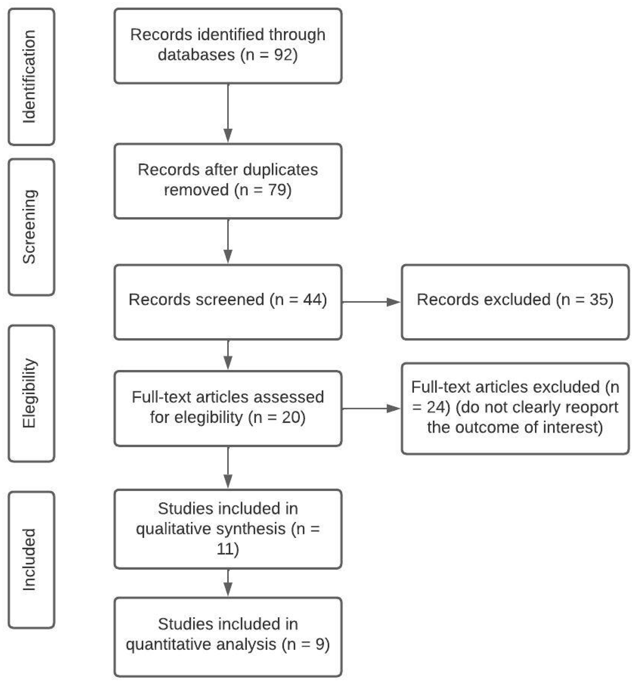
Figure 2.
Main social, ecological, and health factors associated with EEG abnormalities and cognitive impairment, according to literature analyzed for this study.
Figure 2.
Main social, ecological, and health factors associated with EEG abnormalities and cognitive impairment, according to literature analyzed for this study.
Disclaimer/Publisher’s Note: The statements, opinions and data contained in all publications are solely those of the individual author(s) and contributor(s) and not of MDPI and/or the editor(s). MDPI and/or the editor(s) disclaim responsibility for any injury to people or property resulting from any ideas, methods, instructions or products referred to in the content. |
© 2023 by the authors. Licensee MDPI, Basel, Switzerland. This article is an open access article distributed under the terms and conditions of the Creative Commons Attribution (CC BY) license (http://creativecommons.org/licenses/by/4.0/).
Copyright: This open access article is published under a Creative Commons CC BY 4.0 license, which permit the free download, distribution, and reuse, provided that the author and preprint are cited in any reuse.


