Submitted:
10 May 2023
Posted:
11 May 2023
You are already at the latest version
Abstract
Flavonoids are polyphenolic phytochemical compounds found in many plants, fruits, vegetables, and leaves. They have a multitude of medicinal applications due to their anti-inflammatory, anti-oxidative, anti-viral, and anti-carcinogenic properties. Furthermore, they also have neuroprotective and cardioprotective effects. Their biological properties depend on the chemical structure of flavonoids, their mechanism of action, and their bioavailability. The flavonoid’s beneficial effects have been proven for a variety of diseases. In the last few years, it is demonstrated that flavonoids’ effects are mediated by inhibiting the NF-κB (Nuclear Factor-κB) pathway. In this review, we have summarized the effects of some flavonoids on the most common diseases such as cancer, cardiovascular, and neurodegenerative human diseases. In particular, here we collected all recent studies describing plant-derived flavonoids’ protective and prevention role, by specifically focusing their action on the NF-κB signaling pathway.
Keywords:
Flavonoids
; NF-κB
; cancer
; neurodegenerative diseases
; cardiac diseases
1. Introduction
Flavonoids are a class of natural compounds characterized by a polyphenolic group and contained in different plants, fruits, grains, and flowers as well as in beverages such as tea or wine [1]. A healthy diet is recognized all over the world as fundamental to preventing or ameliorating several diseases such as cardiovascular and neurodegenerative diseases, or cancer and its progression [2,3,4]. Hence, they are recognized as dietary flavonoids.
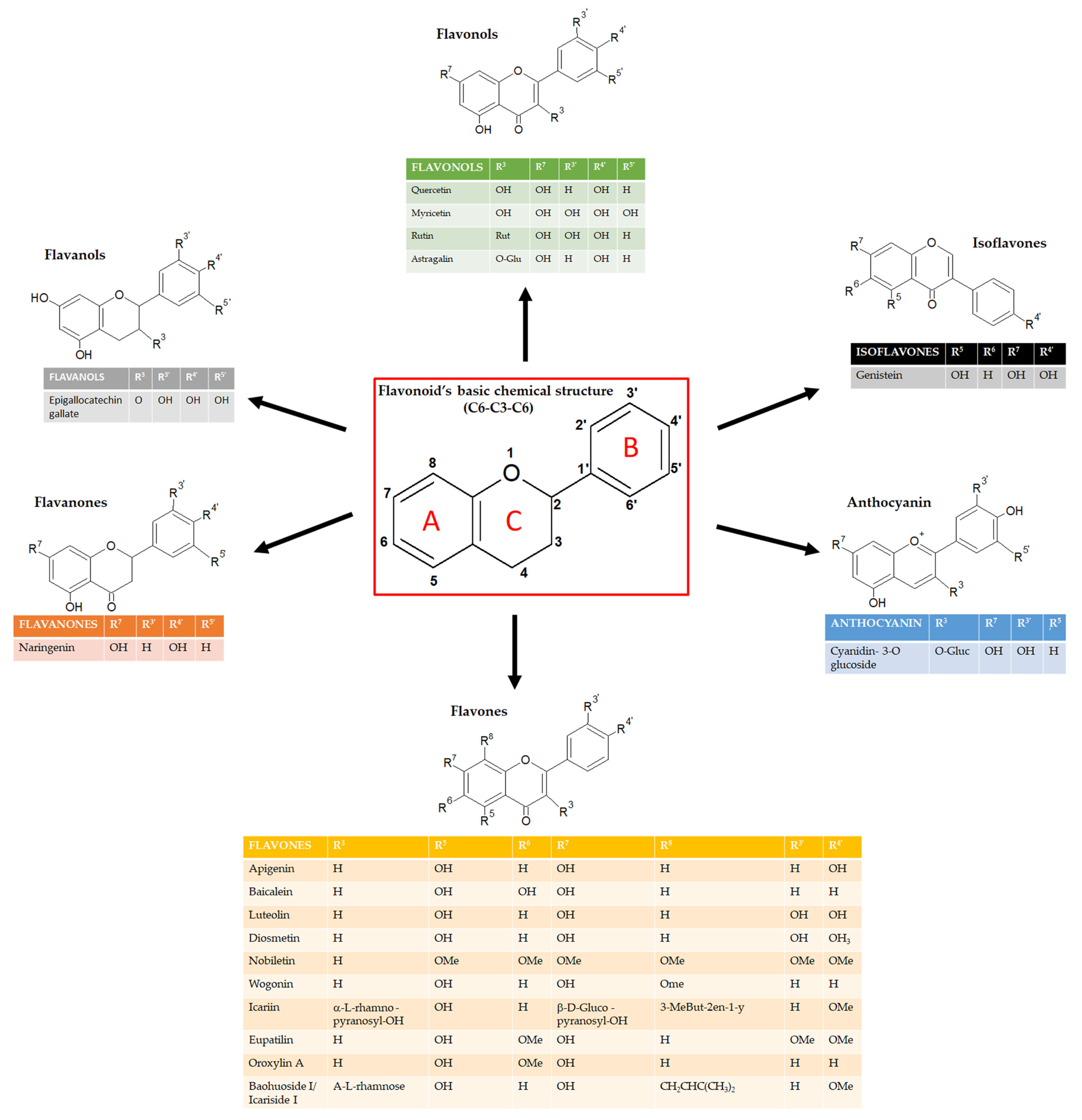
Flavonoids can be classified into several subgroups depending on the carbon of the C ring on which the B ring is attached and the degree of unsaturation and oxidation of the C ring [5]. According to these differences, we can distinguish isoflavones, neoflavonoids, flavones, flavonols, flavanones, flavanonols, flavanols or catechins, anthocyanins, and chalcones (figure 1). Some natural compounds contain specific subgroups, e.g., onions and tea are major dietary sources of flavonols and flavones; isoflavones are mainly found in leguminous species; proanthocyanidins or condensed tannins are found in flowers, seeds, and fruits of diverse plant species such as grapes, apples, blueberry, and some cereals; flavanols (epicatechin), flavonols (e.g., myricetin and quercetin) and anthocyanins (e.g., malvidin-3-glucoside) are enriched in red wine together with the resveratrol, well known for its antioxidant properties [6,7].For the specific concentration of each flavonoid in the most common foods, refer to USDA (United States Department Of Agriculture) database for Flavonoid Content of Selected Foods (https://www.ars.usda.gov/ARSUserFiles/80400535/Data/Flav/Flav3.3.pdf).
Figure 1.
Schematic representation of Flavonoids’, and its subgroups, chemical structure.
Flavonoids absorb UV wavelength, and their biosynthesis is activated after UV exposure. For this reason, they act as unique UV filters thus protecting several plants [8]. Garcia Forero and collaborators demonstrated the photoprotective and antigenotoxic effects in bacteria and human cells of three flavonoids: apigenin, mainly contained in chamomile tea; pinocembrin, mainly present in honey, propolis, piper leaves, and oregano; naringenin, distributed in tomatoes and several citrus fruits such as bergamot [9]
Nowadays it is common to observe bacteria that have acquired resistance to different antibiotics, and it is a priority to study plant-derived compounds that represent a source of antimicrobial agents. Eucalyptus camaldulensis has these antimicrobial effects, due to its active components such as flavonoids and essential oils that can eradicate several bacterial and viral infections [10]. It is published that 5 flavanones isolated from the fruit of Macaranga tanarius, a plant common in Asian areas and eastern Australia, have antimicrobial activity against 14 microorganisms [11]. For a complete review of plants with antibacterial activities, refer to Chassagne and colleagues 2020 [12].
In the last decade, the scientific community’s interest in these compounds has increased and their beneficial or protective effects against different human diseases have been widely recognized.
Nutraceutical compounds have multiple biological activities such as anti-oxidant, anti-inflammatory, or anti-platelet properties. The plant-derived flavonoids have a wide range of activities that could make them particularly effective against cardiovascular and neurodegenerative diseases [2,3]. Moreover, many scientists focused on the anti-cancer activities of several flavonoids demonstrating their beneficial effects [13,14,15].
For these properties and together with absent or very low toxicity they can be a valuable resource for the prevention of several diseases or to reduce tumor progression.
Nowadays, flavonoids are essential components of nutraceutical, pharmaceutical, and cosmetic applications because they have a huge spectrum of biological activity. However, the main mechanism through which they exert these effects is not well known. Recent manuscripts describe the role of flavonoids as anti-inflammatory agents targeting the NF-kB pathway in a specific disease such as cardiovascular diseases or cancer [16,17,18]. This review intends to summarize and discuss the newest results regarding the role of flavonoids on the diseases that affect most people around the world nowadays: cancer, cardiovascular, and neurodegenerative diseases. Moreover, we decided to focus only on the flavonoids that exert their protective or preventive role by affecting the NF-kB signaling pathway.
2. The NF-κB pathway
The NF-κB proteins can regulate the expression of hundreds of genes, which affect important physiological processes such as inflammation, immunity, proliferation, and cell death [19]. Since NF-κB activity is spontaneously regulated by several different stimuli, NF-κB proteins can be considered important regulators of cellular homeostasis [20,21].
NF-κB is a key mediator in the inflammatory response; its activation promotes the transcription of several target genes, many of which are pro-inflammatory [22]. Two pathways lead to the activation of the NF-κB signaling: the canonical and the non-canonical pathways [22,23]. In both pathways, the activation of the enzyme IκB Kinase (IKK) represents the common regulatory step. Notably, the canonical pathway is the most studied and plays a critical role in inflammatory responses. In this pathway, the NF-κB (p65/p50) dimer, in its inactive state, is sequestered in the cytoplasm by IκB (inhibitor of NF-κB) that covers the nuclear localization signal (NLS) of NF- κB protein.
Upon exposure to pro-inflammatory stimuli such as cytokines, pathogens, and danger-associated molecular patterns, the p65/p50 dimer is released from IκB due to the phosphorylation cascade resulting in the proteasomal degradation of IκB. Subsequently, p65/p50 translocate into the nucleus, where it promotes the activation and expression of NF-κB target genes [22]. On the other hand, the non-canonical pathway is activated through a subset of Tumor Necrosis Factor Receptor (TNFR) superfamily members, leading to the activation of NF-κB-inducing kinase (NIK). NIK phosphorylates IKKα, which phosphorylates the C-terminal of NF-κB, allowing it to translocate into the nucleus. Once into the nucleus, NF-κB induces the expression of its target genes that play a role in cell proliferation and survival, as well as in immune cells’ development and inflammation finally inducing the progression of several diseases, as discussed below [22].
3. Flavonoids and cancer: role of NF-κB signaling pathway
Cancer is one of the major health problems in the twenty-first century, due to its high incidence rate, and the second leading cause of mortality globally. Approximately, 15 million people die every year due to the persistence of malignant cells, and the number of cases significantly increases day by day. For these reasons, cancer diagnosis and treatment require great effort [24,25].
Surgery, chemotherapy, radiation, hormone- and targeted therapies are examples of traditional cancer treatment approaches. However, multiple short- and long-term adverse effects may significantly affect patient prognosis depending on treatment-associated clinical factors. Several natural compounds have been studied to find new therapeutic agents, among which the bioactive elements derived from plants have gained the greatest interest in the scientific community.
The scientific community nowadays consider the chemoprevention one of the main topics in order to counteract the continuous increase of neoplasms. Natural compounds are largely diffuse due to their anti-cancerous ability and their potential to overcome resistance. Many scientists focused on the anti-cancer properties of several flavonoids. Several in vitro studies showed the flavonoids’ activities on tumor cells such as inhibition of cell growth, and alteration of tumor invasive behavior [26]. Javier Fernández et al. studied the beneficial effects of five flavonoids, together with conventional chemotherapy, on three colorectal cancer cell lines (HCT116, HT-29, and T84) [27]. Minami Hatono’s group demonstrated the effect of isoflavones on breast cancer cell development and their impact on breast cancer treatments [28]. Joanna Xuan Hui Goh and collaborators demonstrated the chemopreventive role of nobiletin, extracted from citrus fruit, in colon cancer cells [29].
Xhantohumol and nobiletin, are the most abundant flavonoids extracted respectively from the hop plant and red-orange. Recently, authors showed the effects of these two compounds on colon cancer cells, demonstrating that they sensitize colorectal cancer stem cells to 5-fluorouracil (5-Fu) and oxaliplatin (FOX)–based chemotherapy and affect angiogenesis [30,31].
It has been demonstrated that inflammation favors the development of cancer and metastasis. Inflammation represents the first immune response to exogenous or endogenous damage and includes the release of inflammatory cytokines and mediators such as the interleukins IL-1, IL-6, IL-10, TNF-α (Tumor Necrosis Factor α), NF-κB, NO (Nitric Oxide), iNOS (inducible Nitric Oxide Synthase), and COX (CycloOXygenase) [32]. Constitutive induction of the NF-κB pathway has been found in lung, breast, and leukemia cells and its up-regulation correlates with tumor progression and poor prognosis [33,34,35,36,37].
For this reason, in the last few years, researchers’ attention has been focused to identify natural compounds, including flavonoids, that selectively affect or inhibit the NF-κB pathway [38,39,40].
Quercetin is a pentahydroxyflavone, a flavonoid isolated from numerous plant species like horse chestnut, calendula, chamomile, and Ginkgo biloba. Moreover, quercetin is contained in several fruits, vegetables, green tea, and in red wine [41]. It has beneficial effects on human health by mediating anti-oxidant activities and having a role in the modulation of metabolic pathways [42,43]. Several in vitro studies have verified the antiproliferative effects of quercetin and its effect on the expression of apoptotic genes and cell cycle-related genes. Specifically, it has been well-documented that quercetin, by modulating PI3K/AKT/NF-κB (Phosphoinositide 3-kinases/Protein kinase B/NF-κB) or STAT3 (Signal transducer and activator of transcription 3), could exert anti-proliferative, anti-inflammatory, and anti-cancer effects and can modulate the expression of many genes [44,45].
Quercetin produces a beneficial effect through the activation of Nrf2/ARE (Nuclear factor erythroid 2-related factor 2/ARE) that promotes the expression of anti-oxidant enzymes like superoxide dismutase. Moreover, quercetin inhibits the expression of pro-apoptotic and pro-inflammatory genes regulated by NF-κB [46,47]. Constitutive activation of the NF-κB transcription factor was detected in acute myeloid leukemia blasts and other hematopoietic cancers as well as in various solid tumors [48]. Quercetin shows inhibitory effects on the growth of malignant cells such as ovary, liver, bladder, or colorectal cancer cells [49,50]. More recently, Rubio and colleagues showed that in vitro leukemia cells’ treatment with 25 μM of quercetin for 24h affects leukemia cell proliferation by modulating the NF-κB /Nrf2 pathway [51]. Furthermore, quercetin, by interfering with the NF-κB signaling pathway, induces apoptosis in colon cancer cells and decreases prostate cancer cells’ survival [52,53]. More recently, Sahyon and coworkers proved in vitro that combination therapy comprising quercetin (27.98 ± 1.74 μg/ml) and sulfamethoxazole exerts anti-cancer effects by inducing apoptosis via caspase-3 and NF-κB gene regulation, and exhibits selective toxicity against cancer cells [54]. Moreover, the authors obtained same results in vivo, by using EAC (Ehrlich ascites carcinoma)-inoculated mice as animal model. EAC is a carcinoma that can spread in the abdominal region affecting both liver and kidney. For their experiment the authors treated EAC-inoculated mice with the combination of sulfamethoxazole and quercetin (200 mg/kg/day) for 14 days.
It was reported that a common dietary flavonoid apigenin, a trihydroxyflavone extracted by plants such as parsley (Petroselinum crispum), celery (Apium graveolens), and chamomile (Matricaria chamomilla), may prevent cancer therapy. A recent study conducted by Shukla and coworkers revealed that in TRAMP (TRansgenic Adenocarcinoma Mouse Prostate) mice model, treatment with apigenin (20 and 50 μg/mouse/day for 20 days) inhibits phosphorylation and degradation of IκB, leading to suppression of NF-κB activation and finally affect prostate tumorigenesis. Moreover, they observed, after apigenin treatment, a downregulation of different NF-κB-regulated genes involved in proliferation (COX-2 and cyclin D1), angiogenesis (vascular endothelial growth factor), and apoptosis (B-cell lymphoma 2, Bcl-2, and B-cell lymphoma-extra-large, Bcl-xL) [55]. Another study conducted by the same group, using human prostate cancer cell lines, demonstrated the role of apigenin cells’ treatment (2.5 to 20 μM up to 16 h) in the suppression of NF-kB signaling through direct binding and consequent inhibition of IKKα [56]. It was also found that NF-κB expression and activity decreased following apigenin treatment in colon carcinoma cells and in non-small lung cancer cells. Tong et al., showed that, in colon carcinoma cell lines, 20 μM apigenin also inhibited epithelial-mesenchymal transition by blocking NF-κB translocation to the nucleus [57]. Moreover, their in vivo experiments showed the efficacy of apigenin (300 mg/kg for two weeks) in the inhibition of the metastasis of HCT-116 cells (colorectal cancer cell line) in a xenograft model of nude mice, mediated by NF-kB signaling pathway. Recently, Chen and colleagues proved that 20 μM apigenin enhanced TRAIL-induced apoptosis by suppressing the NF-κB nuclear translocation in non-small lung cancer cells [58]. In addition, in malignant mesothelioma, apigenin treatment (50 μM for 24h) showed anticancer effects in vitro and in vivo by inhibiting NF-κB nuclear translocation [59].
Another interesting compound is represented by Diosmetin, a monomethoxyflavone isolated from citrus fruits, that is known for its anti-inflammatory and anti-oxidant properties [60]. Several studies proved that diosmetin has anti-proliferative, anti-metastasis, and pro-apoptotic effects on breast cancer and hepatocellular carcinoma cells [60,61,62]. Moreover, the cytotoxic effect of diosmetin against Colo205, HT-29, and Caco-2 cells demonstrates that it has anti-tumorigenesis properties against human colorectal cancer [60,63]. Koosha et al. in their study demonstrated that diosmetin inhibits cell proliferation and activates apoptosis signaling pathways in human colorectal cancer through the inhibition of the BMP (Bone morphogenetic proteins) and NF-κB pathways [64]. They showed that in diosmetin-treated cells (3.58 µg/ml for 48 h), TNF-α receptors expression was increased, which in turn activates the extrinsic apoptosis pathway. It is well known that TNF-α is an NF-κB inducer [65,66]. In their study, despite the upregulation of TNF-α receptors, translocation of NF-κB was inhibited via overexpression of IκB-α. Moreover, the reduction in protein expression of survivin confirms NF-κB inhibition [64]. Hence, diosmetin activates apoptosis both through the engagement of apoptotic factors and inhibition of NF-κB.
Sun et al. in their study evaluated the possible anticancer effects of Baohuoside-I or Icariside II, a glycosyloxyflavone isolated from Epimedium koreanum Nakai I, in contrasting hepatocellular carcinoma progression [67]. They proved that the treatment with 5-10 μM for 24h of baohuoside-I significantly inhibited the proliferation of hepatocellular carcinoma cells by inducing apoptosis and downregulating the NF-κB signaling pathway. Thus, Baohuoside-I could be a potentially novel drug, opening new possibilities in the treatment of hepatocellular carcinoma [67].
El-Shitany’s group studied the role of an interesting compound, Icariin [68]. It is a prenylated flavonol glucoside, extracted from Epimedium koreanum Nakai, commonly known as horny goat weed, which offers different pharmacological properties, including reduction of inflammation and anti-oxidant effect. This group evaluated the anti-inflammatory mechanisms of icariin focusing on both HO-1 (heme oxygenase-1)/Nrf2 and NF-κB pathways. They evidenced that in rats with induced acute inflammation the pre-treatment with icariin (50 mg/kg) reduced levels of several inflammatory cytokines, COX-2 gene expression, and NF-κB activation [68].
Baicalein is a trihydroxyflavone extracted from the roots of Scutellaria baicalensis and showed antitumor activity in many cancer types. The anticancer potential of Baicalein has been investigated in a great number of studies [69,70,71,72]. It inhibits the proliferation of human cervical cancer cells by inducing apoptosis [73]. Similarly, it was found also active in inhibiting human prostate and ovarian cancer cells [74,75]. In the manuscript of Sheatta and colleagues, for their in vivo experiments, EAC-inoculated mice were treated with baicalin (50 mg/kg/day for three weeks) and with baicalin and/or 5-FU. Their results proved that baicalein significantly reduced inflammation and angiogenesis via suppression of NF-κB/IL-1β and VEGF (Vascular Endothelial Growth factor) as well as apoptotic pathways activation through up-regulation of pro-apoptotic and downregulation of anti-apoptotic genes [76].
Luteolin is a tetrahydroxyflavone found in plant leaves, fruits, cloves, and foods such as chamomile tea and green pepper [77]. Several studies showed that it is involved in sensitizing cancer cells to therapeutic-induced cytotoxicity by suppressing cell survival regulated by the NF-κB pathway [15]. The study conducted by Huang and coworkers showed that treatment with 10 and 30 µM of luteolin for 24 h blocked invasion and cell cycle progression of breast cancer cell lines inhibiting the NF-κB pathway and as a consequence reducing c-Myc proto-oncogene and hTERT (human telomerase reverse transcriptase) expression levels [78].
Nobiletin is a natural polymethoxy flavone extracted from the fruit peel of citrus. Nobiletin is well known to show anti-inflammatory properties and in addition, it has been revealed to suppress the proliferation of human skin, prostate, colon, and breast carcinoma cell lines [79,80]. Jiang’s group found that nobiletin was actively involved in inducing autophagy, G0/G1 cell cycle arrest, and NF-κB signaling pathway inhibition along with reducing cell migration and invasion in human pancreatic carcinoma cells [81]. Specifically, in their experiments the authors treated the cells with different doses of nobiletin (6.12, 12.5 and 25 μM) for 24h, and they found a dose-dependent reduction of the invasion capability of the cells and the inhibition of the NF-κB protein expression [81]. Based on these results, nobiletin could be evaluated as a potential lead molecule for the systemic treatment of cancer and merits more investigations.
Several experimental evidence suggested that the extracts of Litchi seeds have anticancer effects and can inhibit metastatic tumor growth [82,83]. It was proved that Litchi seed extracts, particularly enriched in flavonoids, show anti-prostate cancer properties both in vitro and in vivo [84]. Recently, Chang and coworkers, evaluating the effects of the treatment with 100 μg/ml of total flavonoids of litchi seeds (TFLS) for 48h on prostate cancer cell lines, found that this compound inhibited the NF-κB signaling pathway by decreasing the phosphorylation of IκBα and NF-κB nuclear expression. Moreover, their results illustrated that NF-κB influenced EMT (epithelial-mesenchymal transition). In addition, they showed that TFLS increased the level of cleaved-PARP (Poli ADP-ribose polymerase) and Bax (Bcl-2 associated X protein) and reduced the level of Bcl-2 (B-cell lymphoma 2). Hence, their results demonstrated that TFLS exerted pro-apoptotic and anti-metastatic effects in prostate cancer cells by inhibiting the activation of the AKT/NF-κB signaling pathway [85].
Recently, it was evaluated the possible antitumoral role of Genistein, a 7-hydroxyisoflavone contained in soybean with different biological activities [86]. It was proved that genistein inhibits carcinogenesis in animal models [87]. Research results, obtained by Xie and colleagues, revealed that 20 µM genistein induces multiple myeloma cell proliferation inhibition and apoptosis by suppressing the NF-κB signaling pathway [88]. More recently, the Ozturk group revealed a role of genistein in thyroid cancer cell apoptosis through the downregulation of the NF-κB pathway [89].
In the last years, it was proved the anticancer role of Wogonin, a dihydroxy- and monomethoxy-flavone extracted from Scutellaria baicalensis Georgi, by inhibiting NF-κB activity. In particular, results obtained by Liu and colleagues demonstrated that treatment with increasing doses of wogonin (0, 25, 50 and 100 μM, for 24 h) can act as a natural sensitizer of human chemoresistant myelogenous leukemia cells through inhibition of Nrf2 via NF-κB signaling [90].
The major polyphenolic component of dried green tea extracts is epigallocatechin-gallate (EGCG), the ester of gallic acid and epigallocatechin, belong to the family of flavanol [91]. Several in vitro, in vivo, and clinical studies have shown multiple EGCG anticancer actions. Among them, there are anti-proliferative, pro-apoptotic, anti-angiogenic, and anti-invasive functions [92]. Furthermore, EGCG has been observed to impair other processes that are involved in carcinogenesis as inflammation, oxidative stress, and hypoxia, and to target tumor microenvironment components [93]. EGCG negatively modulates the expression of various transcription factors such as Sp1 (Specific protein 1), AP-1 (activator protein 1), and NF-κB, finally preventing cancer formation [94,95]. More recently, it was revealed by Tian and colleagues that 20 μM EGCG may significantly sensitize ovarian cancer cells to cisplatin through modulation of the c-Myb-induced NF-kB -STAT3 signaling pathway [96].
Anticancer properties were demonstrated also for Eupatilin and Oroxylin natural flavonoids. Wang and colleagues showed that Eupatilin, a trimethoxyflavone mainly found in Artemisa asiatica, could be a good anti-cancer compound. They proved that Eupatilin plays a role in the prevention of gastric cancer. In particular, they demonstrated that eupatilin treatment (12.5, 25 and 50 µM of for 24 h) of gastric cancer cells can reduce NF-κB activity, as well as down-regulating pro-inflammatory cytokines and metalloproteinases (MMP-2, MMP-9) [97]. Sun’s group suggests a possible role of Oroxylin A, a dihydroxy- and monomethoxy-flavone extracted from the Oroxylum Indicum tree and the Scutellariae radix, as a therapeutic candidate for the treatment of breast cancer. Their data reported that 24h-treatment with 20 μM of Oroxylin A can inhibit the invasion, cell proliferation, migration, and EMT through inactivating the NF-κB signaling pathway in human breast cancer cells [98].
Recently another natural flavonoid, the astragalin, a trihydroxyflavone extracted from different medicinal plants such as Eucommia ulmoides, Moringa oleifera, Morus alba, and Radix astragali, has been reported to induce cell death in a caspase-dependent way enhancing the sensibility of lung cancer cells to TNF-α and inhibiting NF-κB activation [99]. Specifically, Chen and coworkers showed, by treating lung cancer cells for 24h with different astragalin doses (2.5, 5, 10, 20 μg/mL), that this compound inhibits NF-κB/p65 nuclear translocation and IκBα degradation in a dose dependent manner.
The literature data cited in this paragraph and summarized in table 1, let us suppose that natural compounds widely present in the daily human diet, such as flavons, flavonols and flavanols, through modulation of the NF-κB signaling pathway, could play a critical role in inhibiting tumor growth and could be considered as natural chemopreventive compounds.

Table 1.
Summary of the effects of flavonoids on cancer mediated by the NF-kB signaling pathway.
| Flavonoids | Subgroups | Chemical Structure | Activity as NF-kB signaling inhibitor with proven anticancer effects | REFS. |
|---|---|---|---|---|
| Quercetin | Flavonol | C15H10O7 | Leukemia cells. Cervical, non-small lung, colon, liver, and prostate cancer cells | [49,50,51,52,53,54] |
| Apigenin | Flavone | C15H10O5 | Prostate adenocarcinoma, colon, prostate, and lung cancer cells. Mesothelioma. | [56,57,58,59] |
| Diosmetin | Flavone | C16H12O6 | Colorectal cancer cells | [60,61,62,64] |
| Baohuoside-I | Flavonol | C27H30O10 | Hepatocellular carcinoma, | [67] |
| Baicalein | Flavone | C15H10O5 | Breast cancer | [76] |
| Luteolin | Flavone | C15H10O6 | Glioblastoma cells, breast and pancreatic cancer cells, hepatocarcinoma cells | [78] |
| Nobiletin | Flavone | C21H22O8 | Pancreatic carcinoma, and colorectal cancer cells | [81] |
| TFLS | Flavone | C15H10O5 | Prostate cancer cells | [85] |
| Genistein | Flavone | C15H10O5 | Breast, thyroid, and colon cancer cells and multiple myeloma cells | [88,89] |
| Wogonin | Flavone | C16H12O5 | Hepatocellular carcinoma, colon cancer, and myelogenous leukemia | [90] |
| EGCG | Flavan-3-ol | C22H18O11 | Ovarian cancer cells | [96] |
| Eupatilin | Flavone | C18H16O7 | Gastric cancer cells | [97] |
| Oroxylin A | Flavone | C16H12O5 | Breast cancer cells | [98] |
| Astragalin | Flavone | C21H20O11 | Colon, and lung cancer cells | [99] |
4. Flavonoids and cardiovascular diseases: role of NF-κB signaling pathway
Nowadays, cardiovascular disease (CVD) is the leading cause of death, especially in Europe, where it remains the primary cause of 42% of mortalities in men and 52% in women [100]. In the United States, it is estimated that 31% of all global deaths in 2016 are from CVDs, mainly heart attacks, and stroke [101].
Inflammation is a key risk factor for CVD. Several evidence demonstrate that chronic inflammation can affect atherosclerosis, myocardial infarction, and heart failure [102]. The upregulation of cytokines in the heart activates intracellular signaling pathways such as NF-κB thus finally altering cardiac cell biology. It is well known that NF-κB activity is strictly correlated with the development and progression of inflammation and cardiovascular damage, as discussed by Fiordelisi and collaborators in their recent review [103].
Atherosclerosis (AS) is the primary cause of CVD that can determine myocardial infarction. AS can affect immune inflammatory activity through NF-κB activity thus finally contributing to platelet migration and aggregation [104].
Platelet activation can induce aggregation and thrombus formation, ultimately contributing to ischemia and myocardial infarction. Several drugs are available to treat and prevent platelet hyperactivation, but they have many collateral negative effects. Diet and lifestyle have an impressive effect on LDL (Low-density lipoprotein)-cholesterol levels and CVD risk. For these reasons, recent studies reported the cardioprotective effects of medicinal plants and the Mediterranean diet in primary and secondary CVD prevention [105]. Hypertension is one of the most common diseases and a major risk factor for stroke and myocardial infarction; many patients affected by cardiovascular diseases are characterized by hypertension and/or cardiac dysfunction. In particular, hypertension can lead to a sustained increase in heart pressure overload, resulting in excessive proliferation and accumulation of collagen fibers in the interstitium and perivascular vessels, which can lead to myocardial fibrosis. For this reason, it is very important to decrease myocardial fibrosis thus reducing the risk of cardiovascular disease.
Several epidemiological studies demonstrated the benefits of a diet enriched with flavonoids, especially for heart diseases.
The effect of Quercetin on CVD is well known; in fact, quercetin can reduce the expression of adhesion molecules and inflammatory markers [106]. It has been demonstrated that 25 μM of quercetin modulates TLR-NF-κB (Toll-Like Receptor-Nuclear Factor kappa B) signaling pathway on endothelial cells in vitro, thus affecting the expression of adhesion molecules involved in the leukocyte-endothelial interaction. Moreover, authors demonstrated the anti-inflammatory activity of quercetin in rats treated for 60 days with 25 mg quercetin/kg body weight, in a condition of hypercholesterolemic diet [107]. Furthermore, by reducing MMP2 (Matrix Metalloproteinase 2) activity and oxidative stress, chronic treatment with quercetin (10 mg/kg/day) ameliorates hypertension-induced coronary hypertrophic remodeling in renovascular hypertensive rats, even if it does not reduce cardiac dysfunction [108]. An interesting study on patients with stable coronary artery disease demonstrated that quercetin therapy reduces the levels of IL-1β in the blood but also the level of IκBα mRNA, thus affecting the transcriptional activity of NF-κB signaling, and finally ameliorating the indicators of chronic systemic inflammation after two-months of quercetin treatment at the daily dose of 120 mg [109].
Some studies suggested the involvement of the TGF-β (tumor growth factor β)/Smad and the NF-κB signaling pathways in the progression of myocardial fibrosis as well as the role of pro-inflammatory cytokines in the development and progression of myocardial fibrosis [102,103]. Therefore, as discussed before, an alternative strategy for the treatment of CVD, such as myocardial fibrosis, is the inhibition of inflammation.
Icariside II, the main metabolite of Icariin, is native to Korea and China, where is well known as a nutraceutical compound in functional foods or as a phytopharmaceutical agent with anti-inflammatory and anti-oxidant activities that protects against cardiovascular diseases [110,111]. It is demonstrated that Icariin inhibits IL1β-induced activation of NF-κB-related signaling pathways thus playing a significant anti-inflammatory role [112]. Fe Shu and collaborators demonstrated that Icariside II chronic treatment (4, 8, and 16 mg/kg) reduces myocardial fibrosis in spontaneously hypertensive rats through suppression of NF-κB signaling and the TGF-β1/Smad2 signaling pathways [113].
One of the major negative effects of Doxorubicin (DOXO) treatment in anticancer therapy is cardiotoxicity which can occur after 3 days (acute cardiotoxicity) or after 30 days of treatment (chronic cardiotoxicity) [114]. Doxorubicin is firstly responsible for inflammatory effects in the myocardium, characterized by the increased expression of NF-κB; moreover, cardiac inflammation contributes to cardiotoxicity. Proanthocyanidins are a class of oligomeric flavonoids derived from catechins and epicatechins, found in many plants, such as cranberry, blueberry, and grape seeds. Proanthocyanidins derived from grape seed extracts exert anti-oxidant effects, may reduce neutrophil infiltration and cyclooxygenase expression and, finally, reduce the adverse effects of doxorubicin treatment in anticancer therapy [115]. It is recently demonstrated that the NF-κB pathway is responsible for the prophylactic effects of proanthocyanidin extract against DOXO-induced heart injury and fibrosis [116]. Rats intraperitoneally injected with DOXO (10 mg/kg on Days 3, 9, 15, and 21 of the experiment) and proanthocyanidin extract (50 mg/kg /once daily) for 4 weeks showed a significant reduction of DOXO induced cardiomyocyte inflammation, thus suggesting a possible role of proanthocyanidin extract against DOXO-induced cardiac toxicity [116].
Morin, a pentahydroxyflavone isolated from Chinese herbs of the Moraceae family and present also in many fruits and wines, can exert similar effects. Kuzu and collaborators demonstrated that Morin significantly decreases the expression of NF-κB, TNF-α, and IL-1β in DOXO-treated rats, inhibiting DOXO-induced oxidative stress and heart damage [117]. For the experiments reported in the manuscript, rats were treated with 50-100 mg/kg of morin (orally administered) for 10 days and with DOXO 40 mg/kg body weight by single dose intraperitoneal injection on the 8th day of the study. After analysis of different inflammation and cardiac markers authors demonstrated the protective role of morin against DOXO-induced heart damage [117].
Curcumin, extracted from the rhizomes of Curcuma longa which belongs to the ginger family, is well known as the main component of the dietary spice turmeric. Several studies demonstrated its anti-oxidant, anti-inflammatory and anti-cancer effects through inhibition of the NF-κB pathway [118,119,120]. Benzer et al. obtained similar results demonstrating the beneficial effect of curcumin against DOXO-induced cardiotoxicity in rats [121]. For their experiments, rats were treated orally for 7 days with 100-200mg/kg body weight of curcumin and authors induced cardiotoxicity by injection of the single dose 40mg/kg of DOXO on the 5th day. Rats were sacrificed at 8th day and several analysis demonstrated the cardioprotective effects of curcumin after DOXO treatment [121].
Finally, we can conclude that flavonoids, as well as many other natural compounds, are able to decrease the risk of several CVD by reducing the activation of inflammatory pathways and especially of the NF-κB signaling pathway (Table 2).
5. Flavonoids and neurodegenerative diseases: role of NF-κB signaling pathway
Neuroinflammation is one of the main causes of different neurodegenerative diseases such as dementia, atherosclerosis, and Alzheimer’s disease (AD). Several signals can interfere with the physiological communication between neurons and glial cells. NF-κB activation is mediated by Toll-like receptors (TLRs), that mediate the intracellular inflammatory response. Focusing on the brain, TLRs are mainly expressed in microglia and astrocytes. Once activated, the microglia can induce the expression of the NF-κB pathway and the subsequent synthesis of pro-inflammatory mediators. These pro-inflammatory cytokines finally activate kinases responsible for the hyperphosphorylation of proteins, such as tau protein, involved in the stability of cytotypes of the central nervous system. The tau proteins, in fact, became insoluble, aggregate, and form the neurofibrillary tangles that destroy the functionality of the cells. Dementia, AD, or Parkinson’s disease are specifically correlated with tau hyperphosphorylation.
Several studies described the role of NF-κB signaling in neurodegenerative diseases [122]. For example, in the brain section of Parkinson’s patients, it is demonstrated a huge increase in NF-κB activation and a strong nuclear immunoreactivity of p65 [123]. Recently, Debashis Dutta et al. demonstrated that, by specifically targeting the TLR2/NF-κB pathway, it is possible to reduce the α-synuclein spreading in vitro and in vivo, finally contributing to the treatment of neurodegenerative diseases such as Parkinson’s disease (PD) [124]. Clinical studies highlight the preventive effects of correct and healthy nutrition in the field of neurodegenerative diseases and especially AD or atherosclerosis [125,126]. Many nutraceutical bioactive principles have anti-platelet activity as well as anti-inflammatory activities [125].
It has recently been demonstrated that several flavonoids and tea catechins have a protective role toward neurons and tau protein hyperphosphorylation mainly inhibiting the activity of MAPK (Mitogen-Activated Protein Kinase) and GSK3 pathways (Glycogen Synthase Kinase-3)[127,128,129,130]. Ching-Hsia Hung and collaborators demonstrated the antioxidant effects of quercetin. Specifically, quercetin treatment for 24 hours (2.5-10 uM) and subsequent exposure to oxidixed LDL protects endothelial cells from damage induced by oxidized LDL. Interestingly, quercetin exerts this effects through the activation of Sirtuin1 (SIRT1), involved in the regulation of several chronic conditions such as neurodegenerative diseases [131].
Naringenin is a flavanone found in different herbs and fruits, such as grapefruit, bergamot, tart cherries, and tomatoes. It is well known that naringenin has antibacterial activity but can also reduce hepatitis C virus production in liver cells in vitro [132,133]. Importantly, Naringenin represents a promising compound in the treatment of AD because it activates the expression of Amiloid-β degradation enzymes and induces M2 polarization of microglia [134]. Sarubbo and collaborators demonstrated that naringenin, as well as quercetin, negatively regulates inflammatory pathways thus decreasing TNF-α, IL-6, and IL-1 expression through the regulation of the nuclear translocation of p65 and the increased expression of SIRT1 in the hippocampus of rat [135]. As discussed previously, SIRT1 has a neuroprotective function and regulates NF-kB expression, while aging reduces SIRT1 hippocampal expression. Old rats were subjected to chronic treatment with naringenin or quercetin (20 mg/kg/day) for 28 days. This chronic treatment, due to the antioxidant properties of polyphenolic compounds, stabilize hippocampal SIRT1 expression, thus affecting NF-kB signaling and the inflamm-aging pathway [135].
Epigallocatechin is the most abundant component of tea catechins, found in solid green tea extract. For this reason, it is responsible for green tea’s anti-oxidant, anti-inflammatory, and anti-apoptotic activities [136]. Epigallocatechin 3-gallate (EGCG) overpasses the blood-brain barrier and reduces the β-amyloid (Aβ) accumulation through the suppression of the ERK/NF-κB pathway finally exerting a neuroprotective effect [137,138] .
Chang and Rong demonstrated that EGCG inhibited neurotoxic Aβ accumulation in a model of a senescent mouse (SAMP8, Senescence-accelerated mouse prone 8) subjected to EGCG chronic treatment of 5 and 15 mg/kg for 60 days. Moreover, authors demonstrated that EGCG induced the expression of neprilysin, a protease involved in brain Aβ metabolism in vivo, thus reducing the Aβ accumulation [139].
Myricetin is an hexahydroxyflavone present in several vegetables and fruits [140]. The compound exerts various beneficial effects such as anti-hypertensive, anti-inflammatory, and immunomodulatory [141,142]. As discussed, neuroinflammation is caused by microglia activation. Long Sun et al. utilize a rat model of permanent middle cerebral artery occlusion (pMCAO) and administered them three different concentrations of myricetin (1-5-25mg/kg), once per day for 1 week. They demonstrated that myricetin reduces ischemic cerebral injury in the rat middle cerebral artery occlusion model by decreasing the expression of several pro-inflammatory cytokines, such as IL-1β, IL-6, TNF-α, the phosphorylation of p38 MAPK, and the level of NF-κB/p65 [143]. Jang and collaborators have recently demonstrated the inhibitory effects of Myricetin on microglia neuroinflammation through the production of proinflammatory molecules and cytokines [144]. Authors demonstrated their results in vitro on microglial BV-2 cells treated with myricetin 10-25 μM for 1 hour, and then stimulated with LPS. Moreover, myricetin (50-100mg/kg), once a day for 7 days, inhibited LPS-induced macrophages and microglial activation in the hippocampus and cortex of mice, in vivo [144]. Kaewmool et al. obtained similar results studying the effects of Cyanidin 3-O-glucoside, anthocyanins responsible for the red and blue pigments of different fruits, and well known for its neuroprotective effects [145]. Authors demonstrated that cyanidin treatment (2.5-5-10 μM) for 4 hours inhibits the NF-κB and p38 pathways thus affecting the expression of pro-inflammatory cytokines in microglial BV2 cells [145,146].
Recently, Chiocchio et al. demonstrated the neuroprotective role of a flavonoid blend contained in leaves of Castanea sativa and mainly enriched in astragalin, isorhamnetin glucoside, and myricetin. Authors demonstrated that chestnut extracts significantly decrease the mRNA expression of pro-inflammatory cytokines and the activation of the NF-κB pathway on BV2 microglial cells treated for 3 hours with 0.5 mg/ml of the extract [147].
It is well known that PD patients have a high level of pro-inflammatory cytokines in their brains and that bacterial LPS (lipopolysaccharides) can induce microglia activation, inflammatory cytokines expression, and PD-like symptoms in animal models [148]. Hartmann and colleagues demonstrated that activation and nuclear translocation of NF-κB are specifically involved in this process [149]. Patel and Singh recently demonstrated that apigenin treatment (25-50 mg/kg) for 2 weeks attenuates LPS-induced parkinsonism in experimental rats targeting TLR/NF-κB and Nrf2/HO-1 signaling pathways. Apigenin reduces neuroinflammation in rat striatum as demonstrated by a significant decrease in TNF-α, IL-1β and IL-6 expression. Moreover, it reduces the nuclear translocation of phospho-NF-κB in the rat brain, finally exerting a neuroprotective effect [150].
Rutin, a natural flavonoid glycoside derived from quercetin, exerts similar neuroprotective effects on different neurodiseases including AD, and PD [151]. Sun and collaborators demonstrated that rutin treatment (100 mg/kg) for 30 days prevents neuroinflammation in mouse models of AD by decreasing IL-1β and TNF-α production. Interestingly, authors evaluated the level of IKK-β, p65, and phospho-p65 in the brain protein lysates of mice to demonstrate that the rutin’s neuroprotective effects are mediated by the downregulation of NF-κB activation [152].
Already in 2014 Wang and collaborators showed that Genistein at the dose of 10mg/kg once daily for 2 weeks, ameliorated brain damage [153]. Recently it has been demonstrated that genistein reduces the inflammation of the Hippocampus in cecal ligation and puncture (CLP)-induced septic rats treated with the dose of 15 mg/kg daily for 10 days. In this study authors focused on the hippocampus NF-κB activation showing that genistein reduces the degradation of IκBα and the nuclear translocation of NF-κB [154].
It is known that nobiletin showed anti-dementia properties attenuating cognitive impairment in the AD mice model [155,156]. Qi et al. recently demonstrated that nobiletin treatment (25-50-100 μM) for 4 hours counteracts the microglia activation and the secretion of proinflammatory mediators in BV2 microglia cells. Moreover, authors demonstrated the protective effects of nobiletin on mice treated with the dose of 100 mg/kg once daily for 7 weeks. These effects are mediated by the modulation of MAPKs, PI3K/AKT, and NF-κB signaling pathways [157]. Reihaneh Ghasemi-Tarie showed similar results in the AD rat model where nobiletin treatment (mg/kg/day), for 7 days prevents amyloid-induced cognitive defects by blocking p65 nuclear translocation and IκBα phosphorylation [158].
Here, we summarize the role of some natural compounds that through affecting inflammatory pathways, and specifically the NF-κB pathway, can reduce neuroinflammation and the subsequent neuronal degeneration (Table 3).
5. Methods
The literature review was conducted using recommended five-step scoping review guidelines [160]: (1) identify a research question (Roles of NF-κB signaling pathways in mediating the effects of flavonoids on human diseases); (2) identify relevant studies; (3) select relevant studies; (4) chart data from these studies; and (5) collate, summarize, and report the results.
We have identified studies from the PubMed/Medline database about flavonoids and NF-kB signaling pathways in human diseases. The aim was to access original research on subjects related to human pathology. Keywords used to search the database were Flavonoids, NF-κB, cancer, neurological diseases, and cardiovascular diseases. We have excluded non-English reviews and we included papers published within recent 5 years (except for the cardinal papers on the topic).
6. Conclusions, limitations and future perspectives
To date, NF-κB represents a key signaling molecule that regulates the expression of a great number of genes, thus modulating several cell signaling pathways such as cell survival and proliferation pathways, or immune regulatory and inflammation pathways. For this reason, in the last decades, researchers’ attention was focused on the possibility that NF-κB could be a good target for several diseases. Therefore, one of the researcher’s challenges is the development of NF-κB inhibitors to treat different diseases and avoid the risk of side effects related to conventional therapy.
A healthy diet is recognized all over the world to prevent or ameliorate several diseases such as cardiovascular and neurodegenerative diseases, or cancer and its progression. Several natural compounds are now being explored for their capability to affect and reduce the progression of many diseases with low collateral effects or toxicity. Moreover, recent manuscripts describe the role of flavonoids as negative regulators of the NF-κB signaling pathway. For these reasons, the scientific community is now focusing on flavonoids, contained in many plants, fruits, and beverages as a possible alternative strategy to overcome or prevent different pathologies. The literature data reported in this review suggest the potential capability of flavonoids to modulate pathological diseases such as cancer, cardiovascular and neurodegenerative diseases, by affecting directly or indirectly the NF-κB pathway (Figure 2). Furthermore, the researcher’s results might provide substantial support for further investigation to assess flavonoid compounds’ efficacy and safety as part of a healthy diet or as co-treatment of human cancers, cardiovascular and neurodegenerative diseases.
It is well known that flavonoids are lipophilic compounds but their interaction with biological membranes is not clear and needs to be better clarified. Many flavonoids interact with receptors on the membrane, protein component of the membrane, and often with lipid rafts. The interaction with the membrane, and/or the speed of the diffusion depend on the different composition of the membrane in each tissue and, on the other hand, on the specific chemical structure of each flavonoid [161]. The presence of definite groups facilitates the adsorption of the compounds, e.g., gallate groups in catechins make EGCG better adsorbed by the membrane [162,163,164]. Flavonols, because of their planar structure, interact with the membrane with higher affinity than flavanones, even if flavanones are more hydrophobic [165].
Another limitation, that we have to take into account if we want to consider flavonoids as food supplements, is the toxicity of these compounds. Even if the beneficial properties of all these flavonoids are clear, it is important to consider that they have to pass clinical trials for efficacy and toxicity in humans. Many flavonoids have been demonstrated to be mutagenic in mammalian cells or in mice [166,167,168]. Recently Zhang and colleagues analyzed the toxicities of 4 flavonoids (luteolin, apigenin, quercetin, and genistein) demonstrating, through in silico, in vitro, and in vivo analysis, the developmental and mutagenic toxicity together with estrogen activity that makes them dangerous especially in young people [168].
Further investigations to optimize the bioavailability, biodistribution, and safety of the compounds for future human studies are needed. Specifically, it is necessary: i) to clarify how each flavonoid, according to its specific chemical structure, can cross the membrane; ii) that the scientific community deepens knowledge and clarifies the real toxicity of each compound.
Author Contributions
Conceptualization, M.M.B., and C.C.; writing—original draft preparation, M.M.B., and C.C.; writing—review and editing, M.M.B., R.A., and C.C.; supervision, C.C., and R.A. All authors have read and agreed to the published version of the manuscript.
Funding
This study was funded by the European Union 2014–2020 PON Ricerca e Innovazione grant from the Italian Ministry of Education, University and Research, entitled “PROGEMA-Processi Green per l’Estrazione di Principi Attivi e la Depurazione di Matrici di Scarto e Non” (ARS01_00432) to Riccardo Alessandro.
Institutional Review Board Statement
Not applicable.
Informed Consent Statement
Not applicable since this study is a narrative review of the literature. All included studies were ethically conducted.
Data Availability Statement
No new data were created or analyzed in this study. Data sharing does not apply to this review article.
Conflicts of Interest
The authors declare no conflict of interest “The funders had no role in the design of the study; in the collection, analyses, or interpretation of data; in the writing of the manuscript; or in the decision to publish the results”.
Abbreviations
| NF-κB | Nuclear Factor-kB |
| USDA | United States Department Of Agriculture |
| UV | ultraviolet |
| IKK | IκB Kinase |
| IκB | inhibitor of NF-κB |
| NLS | nuclear localization signal |
| TNFR | Tumor Necrosis Factor Receptor |
| NIK | NF-κB-inducing kinase |
| 5-FU | 5-Fluorouracil |
| TNF-α | Tumor Necrosis Factor α |
| IL-1 | Interleukin-1 |
| IL-6 | Interleukin-6 |
| IL-10 | Interleukin-10 |
| NO | Nitric Oxide |
| iNOS | inducible Nitric Oxide Synthase |
| COX | CycloOXygenase |
| PI3K | Phosphoinositide 3-kinases |
| AKT | Protein kinase B |
| STAT3 | Signal transducer and activator of transcription 3 |
| Nrf2 | Nuclear factor erythroid 2-related factor 2 |
| ARE | antioxidant response element |
| EAC | Ehrlich ascites carcinoma |
| TRAMP | TRansgenic Adenocarcinoma Mouse Prostate |
| Bcl-2 | B-cell lymphoma 2 |
| Bcl-xL | B-cell lymphoma-extra-large |
| TRAIL | tumor-necrosis factor related apoptosis-inducing ligand, CD253 |
| BMP | Bone morphogenetic proteins |
| HO-1 | heme oxygenase-1 |
| VEGF | Vascular Endothelial Growth factor |
| hTERT | human telomerase reverse transcriptase |
| TFLS | total flavonoids of litchi seeds |
| EMT | epithelial-mesenchymal transition |
| PARP | Poli ADP-ribose polymerase |
| Bax | Bcl-2 associated X protein |
| EGCG | epigallocatechin-gallate |
| Sp1 | Specific protein 1 |
| AP-1 | activator protein 1 |
| MMP-2 | metalloproteinase-2 |
| MMP-9 | metalloproteinase-9 |
| CVD | cardiovascular disease |
| AS | Atherosclerosis |
| LDL | Low-density lipoprotein |
| TLR | Toll-Like Receptor |
| TGF- | tumor growth factor- |
| DOXO | Doxorubicin |
| AD | Alzheimer’s disease |
| PD | Parkinson’s disease |
| MAPK | Mitogen-Activated Protein Kinase |
| GSK3 | Glycogen Synthase Kinase-3 |
| SIRT1 | Sirtuin1 |
| Aβ | β-amyloid |
| Erk | extracellular signal-regulated kinases |
| SAMP8 | Senescence-accelerated mouse prone 8 |
| pMCAO | permanent middle cerebral artery occlusion |
| LPS | Lipopolysaccharide |
| CLP | cecal ligation and puncture |
References
- Sun, W.; Shahrajabian, M.H. Therapeutic Potential of Phenolic Compounds in Medicinal Plants-Natural Health Products for Human Health. Molecules 2023, 28. [CrossRef]
- Calis, Z.; Mogulkoc, R.; Baltaci, A.K. The Roles of Flavonols/Flavonoids in Neurodegeneration and Neuroinflammation. Mini Rev Med Chem 2020, 20, 1475-1488. [CrossRef]
- Micek, A.; Godos, J.; Del Rio, D.; Galvano, F.; Grosso, G. Dietary Flavonoids and Cardiovascular Disease: A Comprehensive Dose-Response Meta-Analysis. Mol Nutr Food Res 2021, 65, e2001019. [CrossRef]
- Aiello, P.; Consalvi, S.; Poce, G.; Raguzzini, A.; Toti, E.; Palmery, M.; Biava, M.; Bernardi, M.; Kamal, M.A.; Perry, G.; et al. Dietary flavonoids: Nano delivery and nanoparticles for cancer therapy. Semin Cancer Biol 2021, 69, 150-165, doi:S1044-579X(19)30217-2. [CrossRef]
- Shen, N.; Wang, T.; Gan, Q.; Liu, S.; Wang, L.; Jin, B. Plant flavonoids: Classification, distribution, biosynthesis, and antioxidant activity. Food Chem 2022, 383, 132531, doi:S0308-8146(22)00493-9. [CrossRef]
- Fernandes, I.; Perez-Gregorio, R.; Soares, S.; Mateus, N.; de Freitas, V. Wine Flavonoids in Health and Disease Prevention. Molecules 2017, 22. [CrossRef]
- Yang, L.; Xian, D.; Xiong, X.; Lai, R.; Song, J.; Zhong, J. Proanthocyanidins against Oxidative Stress: From Molecular Mechanisms to Clinical Applications. Biomed Res Int 2018, 2018, 8584136. [CrossRef]
- Ferreyra, M.L.F.; Serra, P.; Casati, P. Recent advances on the roles of flavonoids as plant protective molecules after UV and high light exposure. Physiol Plant 2021, 173, 736-749. [CrossRef]
- Garcia Forero, A.; Villamizar Mantilla, D.A.; Nunez, L.A.; Ocazionez, R.E.; Stashenko, E.E.; Fuentes, J.L. Photoprotective and Antigenotoxic Effects of the Flavonoids Apigenin, Naringenin and Pinocembrin. Photochem Photobiol 2019, 95, 1010-1018. [CrossRef]
- Ghasemian, A.; Eslami, M.; Hasanvand, F.; Bozorgi, H.; Al-Abodi, H.R. Eucalyptus camaldulensis properties for use in the eradication of infections. Comp Immunol Microbiol Infect Dis 2019, 65, 234-237, doi:S0147-9571(19)30076-1. [CrossRef]
- Lee, J.H.; Kim, Y.G.; Khadke, S.K.; Yamano, A.; Woo, J.T.; Lee, J. Antimicrobial and antibiofilm activities of prenylated flavanones from Macaranga tanarius. Phytomedicine 2019, 63, 153033, doi:S0944-7113(19)30199-0. [CrossRef]
- Chassagne, F.; Samarakoon, T.; Porras, G.; Lyles, J.T.; Dettweiler, M.; Marquez, L.; Salam, A.M.; Shabih, S.; Farrokhi, D.R.; Quave, C.L. A Systematic Review of Plants With Antibacterial Activities: A Taxonomic and Phylogenetic Perspective. Front Pharmacol 2020, 11, 586548. [CrossRef]
- Selvakumar, P.; Badgeley, A.; Murphy, P.; Anwar, H.; Sharma, U.; Lawrence, K.; Lakshmikuttyamma, A. Flavonoids and Other Polyphenols Act as Epigenetic Modifiers in Breast Cancer. Nutrients 2020, 12. [CrossRef]
- Zhai, K.; Mazurakova, A.; Koklesova, L.; Kubatka, P.; Busselberg, D. Flavonoids Synergistically Enhance the Anti-Glioblastoma Effects of Chemotherapeutic Drugs. Biomolecules 2021, 11. [CrossRef]
- Imran, M.; Rauf, A.; Abu-Izneid, T.; Nadeem, M.; Shariati, M.A.; Khan, I.A.; Imran, A.; Orhan, I.E.; Rizwan, M.; Atif, M.; et al. Luteolin, a flavonoid, as an anticancer agent: A review. Biomed Pharmacother 2019, 112, 108612, doi:S0753-3322(18)36718-0.
- Khan, H.; Ullah, H.; Castilho, P.; Gomila, A.S.; D'Onofrio, G.; Filosa, R.; Wang, F.; Nabavi, S.M.; Daglia, M.; Silva, A.S.; et al. Targeting NF-kappaB signaling pathway in cancer by dietary polyphenols. Crit Rev Food Sci Nutr 2020, 60, 2790-2800. [CrossRef]
- Choy, K.W.; Murugan, D.; Leong, X.F.; Abas, R.; Alias, A.; Mustafa, M.R. Flavonoids as Natural Anti-Inflammatory Agents Targeting Nuclear Factor-Kappa B (NFkappaB) Signaling in Cardiovascular Diseases: A Mini Review. Front Pharmacol 2019, 10, 1295. [CrossRef]
- Kariagina, A.; Doseff, A.I. Anti-Inflammatory Mechanisms of Dietary Flavones: Tapping into Nature to Control Chronic Inflammation in Obesity and Cancer. Int J Mol Sci 2022, 23. [CrossRef]
- Giuliani, C.; Bucci, I.; Napolitano, G. The Role of the Transcription Factor Nuclear Factor-kappa B in Thyroid Autoimmunity and Cancer. Front Endocrinol (Lausanne) 2018, 9, 471. [CrossRef]
- Barnabei, L.; Laplantine, E.; Mbongo, W.; Rieux-Laucat, F.; Weil, R. NF-kappaB: At the Borders of Autoimmunity and Inflammation. Front Immunol 2021, 12, 716469. [CrossRef]
- Nathan, C. Points of control in inflammation. Nature 2002, 420, 846-852, doi:nature01320.
- Zinatizadeh, M.R.; Schock, B.; Chalbatani, G.M.; Zarandi, P.K.; Jalali, S.A.; Miri, S.R. The Nuclear Factor Kappa B (NF-kB) signaling in cancer development and immune diseases. Genes Dis 2021, 8, 287-297. [CrossRef]
- Sun, S.C. Non-canonical NF-kappaB signaling pathway. Cell Res 2011, 21, 71-85. [CrossRef]
- Block, K.I.; Gyllenhaal, C.; Lowe, L.; Amedei, A.; Amin, A.; Amin, A.; Aquilano, K.; Arbiser, J.; Arreola, A.; Arzumanyan, A.; et al. Designing a broad-spectrum integrative approach for cancer prevention and treatment. Semin Cancer Biol 2015, 35 Suppl, S276-S304, doi:S1044-579X(15)00088-7. [CrossRef]
- Bray, F.; Ren, J.S.; Masuyer, E.; Ferlay, J. Global estimates of cancer prevalence for 27 sites in the adult population in 2008. Int J Cancer 2013, 132, 1133-1145. [CrossRef]
- Cragg, G.M.; Pezzuto, J.M. Natural Products as a Vital Source for the Discovery of Cancer Chemotherapeutic and Chemopreventive Agents. Med Princ Pract 2016, 25 Suppl 2, 41-59. [CrossRef]
- Fernandez, J.; Silvan, B.; Entrialgo-Cadierno, R.; Villar, C.J.; Capasso, R.; Uranga, J.A.; Lombo, F.; Abalo, R. Antiproliferative and palliative activity of flavonoids in colorectal cancer. Biomed Pharmacother 2021, 143, 112241, doi:S0753-3322(21)01025-8. [CrossRef]
- Hatono, M.; Ikeda, H.; Suzuki, Y.; Kajiwara, Y.; Kawada, K.; Tsukioki, T.; Kochi, M.; Suzawa, K.; Iwamoto, T.; Yamamoto, H.; et al. Effect of isoflavones on breast cancer cell development and their impact on breast cancer treatments. Breast Cancer Res Treat 2021, 185, 307-316. [CrossRef]
- Goh, J.X.H.; Tan, L.T.; Goh, J.K.; Chan, K.G.; Pusparajah, P.; Lee, L.H.; Goh, B.H. Nobiletin and Derivatives: Functional Compounds from Citrus Fruit Peel for Colon Cancer Chemoprevention. Cancers (Basel) 2019, 11. [CrossRef]
- Turdo, A.; Glaviano, A.; Pepe, G.; Calapa, F.; Raimondo, S.; Fiori, M.E.; Carbone, D.; Basilicata, M.G.; Di Sarno, V.; Ostacolo, C.; et al. Nobiletin and Xanthohumol Sensitize Colorectal Cancer Stem Cells to Standard Chemotherapy. Cancers (Basel) 2021, 13. [CrossRef]
- Corrado, C.; Barreca, M.M.; Raimondo, S.; Diana, P.; Pepe, G.; Basilicata, M.G.; Conigliaro, A.; Alessandro, R. Nobiletin and xanthohumol counteract the TNFalpha-mediated activation of endothelial cells through the inhibition of the NF-kappaB signaling pathway. Cell Biol Int 2023, 47, 634-647. [CrossRef]
- Pereira, M.F.; Martino, T.; Dalmau, S.R.; Paes, M.C.; Barja-Fidalgo, C.; Albano, R.M.; Coelho, M.G.; Sabino, K.C. Terpenic fraction of Pterodon pubescens inhibits nuclear factor kappa B and extracellular signal-regulated protein kinase 1/2 activation and deregulates gene expression in leukemia cells. BMC Complement Altern Med 2012, 12, 231. [CrossRef]
- Tew, G.W.; Lorimer, E.L.; Berg, T.J.; Zhi, H.; Li, R.; Williams, C.L. SmgGDS regulates cell proliferation, migration, and NF-kappaB transcriptional activity in non-small cell lung carcinoma. J Biol Chem 2008, 283, 963-976, doi:S0021-9258(20)69006-8. [CrossRef]
- Chua, H.L.; Bhat-Nakshatri, P.; Clare, S.E.; Morimiya, A.; Badve, S.; Nakshatri, H. NF-kappaB represses E-cadherin expression and enhances epithelial to mesenchymal transition of mammary epithelial cells: Potential involvement of ZEB-1 and ZEB-2. Oncogene 2007, 26, 711-724, doi:1209808. [CrossRef]
- Vilimas, T.; Mascarenhas, J.; Palomero, T.; Mandal, M.; Buonamici, S.; Meng, F.; Thompson, B.; Spaulding, C.; Macaroun, S.; Alegre, M.L.; et al. Targeting the NF-kappaB signaling pathway in Notch1-induced T-cell leukemia. Nat Med 2007, 13, 70-77, doi:nm1524. [CrossRef]
- Annunziata, C.M.; Stavnes, H.T.; Kleinberg, L.; Berner, A.; Hernandez, L.F.; Birrer, M.J.; Steinberg, S.M.; Davidson, B.; Kohn, E.C. Nuclear factor kappaB transcription factors are coexpressed and convey a poor outcome in ovarian cancer. Cancer 2010, 116, 3276-3284. [CrossRef]
- Brown, R.E.; Law, A. Morphoproteomic demonstration of constitutive nuclear factor-kappaB activation in glioblastoma multiforme with genomic correlates and therapeutic implications. Ann Clin Lab Sci 2006, 36, 421-426, doi:36/4/421.
- Li, Y.; Ahmed, F.; Ali, S.; Philip, P.A.; Kucuk, O.; Sarkar, F.H. Inactivation of nuclear factor kappaB by soy isoflavone genistein contributes to increased apoptosis induced by chemotherapeutic agents in human cancer cells. Cancer Res 2005, 65, 6934-6942, doi:65/15/6934. [CrossRef]
- Avci, N.G.; Ebrahimzadeh-Pustchi, S.; Akay, Y.M.; Esquenazi, Y.; Tandon, N.; Zhu, J.J.; Akay, M. NF-kappaB inhibitor with Temozolomide results in significant apoptosis in glioblastoma via the NF-kappaB(p65) and actin cytoskeleton regulatory pathways. Sci Rep 2020, 10, 13352. [CrossRef]
- Rasmi, R.R.; Sakthivel, K.M.; Guruvayoorappan, C. NF-kappaB inhibitors in treatment and prevention of lung cancer. Biomed Pharmacother 2020, 130, 110569, doi:S0753-3322(20)30762-9. [CrossRef]
- Vargas, A.J.; Burd, R. Hormesis and synergy: Pathways and mechanisms of quercetin in cancer prevention and management. Nutr Rev 2010, 68, 418-428. [CrossRef]
- Ghafouri-Fard, S.; Shoorei, H.; Khanbabapour Sasi, A.; Taheri, M.; Ayatollahi, S.A. The impact of the phytotherapeutic agent quercetin on expression of genes and activity of signaling pathways. Biomed Pharmacother 2021, 141, 111847, doi:S0753-3322(21)00629-6. [CrossRef]
- Kelly, G.S. Quercetin. Monograph. Altern Med Rev 2011, 16, 172-194.
- Lai, W.W.; Hsu, S.C.; Chueh, F.S.; Chen, Y.Y.; Yang, J.S.; Lin, J.P.; Lien, J.C.; Tsai, C.H.; Chung, J.G. Quercetin inhibits migration and invasion of SAS human oral cancer cells through inhibition of NF-kappaB and matrix metalloproteinase-2/-9 signaling pathways. Anticancer Res 2013, 33, 1941-1950, doi:33/5/1941.
- Anand David, A.V.; Arulmoli, R.; Parasuraman, S. Overviews of Biological Importance of Quercetin: A Bioactive Flavonoid. Pharmacogn Rev 2016, 10, 84-89. [CrossRef]
- Ramyaa, P.; Krishnaswamy, R.; Padma, V.V. Quercetin modulates OTA-induced oxidative stress and redox signalling in HepG2 cells - up regulation of Nrf2 expression and down regulation of NF-kappaB and COX-2. Biochim Biophys Acta 2014, 1840, 681-692, doi:S0304-4165(13)00464-9. [CrossRef]
- Acharya, A.; Das, I.; Chandhok, D.; Saha, T. Redox regulation in cancer: A double-edged sword with therapeutic potential. Oxid Med Cell Longev 2010, 3, 23-34. [CrossRef]
- Rushworth, S.A.; Zaitseva, L.; Murray, M.Y.; Shah, N.M.; Bowles, K.M.; MacEwan, D.J. The high Nrf2 expression in human acute myeloid leukemia is driven by NF-kappaB and underlies its chemo-resistance. Blood 2012, 120, 5188-5198. [CrossRef]
- Tao, S.F.; He, H.F.; Chen, Q. Quercetin inhibits proliferation and invasion acts by up-regulating miR-146a in human breast cancer cells. Mol Cell Biochem 2015, 402, 93-100. [CrossRef]
- Yuan, Z.; Long, C.; Junming, T.; Qihuan, L.; Youshun, Z.; Chan, Z. Quercetin-induced apoptosis of HL-60 cells by reducing PI3K/Akt. Mol Biol Rep 2012, 39, 7785-7793. [CrossRef]
- Rubio, V.; Garcia-Perez, A.I.; Herraez, A.; Diez, J.C. Different roles of Nrf2 and NFKB in the antioxidant imbalance produced by esculetin or quercetin on NB4 leukemia cells. Chem Biol Interact 2018, 294, 158-166, doi:S0009-2797(18)30610-0. [CrossRef]
- Zhang, X.A.; Zhang, S.; Yin, Q.; Zhang, J. Quercetin induces human colon cancer cells apoptosis by inhibiting the nuclear factor-kappa B Pathway. Pharmacogn Mag 2015, 11, 404-409. [CrossRef]
- Ward, A.B.; Mir, H.; Kapur, N.; Gales, D.N.; Carriere, P.P.; Singh, S. Quercetin inhibits prostate cancer by attenuating cell survival and inhibiting anti-apoptotic pathways. World J Surg Oncol 2018, 16, 108. [CrossRef]
- Sahyon, H.A.E.; Ramadan, E.N.M.; Althobaiti, F.; Mashaly, M.M.A. Anti-proliferative effects of the combination of Sulfamethoxazole and Quercetin via caspase3 and NFkB gene regulation: An in vitro and in vivo study. Naunyn Schmiedebergs Arch Pharmacol 2022, 395, 227-246. [CrossRef]
- Shukla, S.; Shankar, E.; Fu, P.; MacLennan, G.T.; Gupta, S. Suppression of NF-kappaB and NF-kappaB-Regulated Gene Expression by Apigenin through IkappaBalpha and IKK Pathway in TRAMP Mice. PLoS ONE 2015, 10, e0138710. [CrossRef]
- Shukla, S.; Kanwal, R.; Shankar, E.; Datt, M.; Chance, M.R.; Fu, P.; MacLennan, G.T.; Gupta, S. Apigenin blocks IKKalpha activation and suppresses prostate cancer progression. Oncotarget 2015, 6, 31216-31232. [CrossRef]
- Tong, J.; Shen, Y.; Zhang, Z.; Hu, Y.; Zhang, X.; Han, L. Apigenin inhibits epithelial-mesenchymal transition of human colon cancer cells through NF-kappaB/Snail signaling pathway. Biosci Rep 2019, 39, doi:BSR20190452.
- Chen, M.; Wang, X.; Zha, D.; Cai, F.; Zhang, W.; He, Y.; Huang, Q.; Zhuang, H.; Hua, Z.C. Apigenin potentiates TRAIL therapy of non-small cell lung cancer via upregulating DR4/DR5 expression in a p53-dependent manner. Sci Rep 2016, 6, 35468. [CrossRef]
- Masuelli, L.; Benvenuto, M.; Mattera, R.; Di Stefano, E.; Zago, E.; Taffera, G.; Tresoldi, I.; Giganti, M.G.; Frajese, G.V.; Berardi, G.; et al. In Vitro and In Vivo Anti-tumoral Effects of the Flavonoid Apigenin in Malignant Mesothelioma. Front Pharmacol 2017, 8, 373. [CrossRef]
- Androutsopoulos, V.P.; Mahale, S.; Arroo, R.R.; Potter, G. Anticancer effects of the flavonoid diosmetin on cell cycle progression and proliferation of MDA-MB 468 breast cancer cells due to CYP1 activation. Oncol Rep 2009, 21, 1525-1528. [CrossRef]
- Liu, B.; Shi, Y.; Peng, W.; Zhang, Q.; Liu, J.; Chen, N.; Zhu, R. Diosmetin induces apoptosis by upregulating p53 via the TGF-beta signal pathway in HepG2 hepatoma cells. Mol Med Rep 2016, 14, 159-164. [CrossRef]
- Liu, J.; Wen, X.; Liu, B.; Zhang, Q.; Zhang, J.; Miao, H.; Zhu, R. Diosmetin inhibits the metastasis of hepatocellular carcinoma cells by downregulating the expression levels of MMP-2 and MMP-9. Mol Med Rep 2016, 13, 2401-2408. [CrossRef]
- Xie, Y.Y.; Yuan, D.; Yang, J.Y.; Wang, L.H.; Wu, C.F. Cytotoxic activity of flavonoids from the flowers of Chrysanthemum morifolium on human colon cancer Colon205 cells. J Asian Nat Prod Res 2009, 11, 771-778. [CrossRef]
- Koosha, S.; Mohamed, Z.; Sinniah, A.; Alshawsh, M.A. Investigation into the Molecular Mechanisms underlying the Anti-proliferative and Anti-tumorigenesis activities of Diosmetin against HCT-116 Human Colorectal Cancer. Sci Rep 2019, 9, 5148. [CrossRef]
- Schnare, M.; Barton, G.M.; Holt, A.C.; Takeda, K.; Akira, S.; Medzhitov, R. Toll-like receptors control activation of adaptive immune responses. Nat Immunol 2001, 2, 947-950, doi:ni712. [CrossRef]
- Baldwin, A.S., Jr. Series introduction: The transcription factor NF-kappaB and human disease. J Clin Invest 2001, 107, 3-6, doi:11891. [CrossRef]
- Sun, Y.; Pang, B.; Wang, Y.; Xiao, J.; Jiang, D. Baohuoside I Inhibits the Proliferation of Hepatocellular Carcinoma Cells via Apoptosis Signaling and NF-kB Pathway. Chem Biodivers 2021, 18, e2100063. [CrossRef]
- El-Shitany, N.A.; Eid, B.G. Icariin modulates carrageenan-induced acute inflammation through HO-1/Nrf2 and NF-kB signaling pathways. Biomed Pharmacother 2019, 120, 109567, doi:S0753-3322(19)35190-X. [CrossRef]
- Ikemoto, S.; Sugimura, K.; Yoshida, N.; Yasumoto, R.; Wada, S.; Yamamoto, K.; Kishimoto, T. Antitumor effects of Scutellariae radix and its components baicalein, baicalin, and wogonin on bladder cancer cell lines. Urology 2000, 55, 951-955, doi:S0090429500004672. [CrossRef]
- Li-Weber, M. New therapeutic aspects of flavones: The anticancer properties of Scutellaria and its main active constituents Wogonin, Baicalein and Baicalin. Cancer Treat Rev 2009, 35, 57-68. [CrossRef]
- Parajuli, P.; Joshee, N.; Rimando, A.M.; Mittal, S.; Yadav, A.K. In vitro antitumor mechanisms of various Scutellaria extracts and constituent flavonoids. Planta Med 2009, 75, 41-48. [CrossRef]
- Chiu, Y.W.; Lin, T.H.; Huang, W.S.; Teng, C.Y.; Liou, Y.S.; Kuo, W.H.; Lin, W.L.; Huang, H.I.; Tung, J.N.; Huang, C.Y.; et al. Baicalein inhibits the migration and invasive properties of human hepatoma cells. Toxicol Appl Pharmacol 2011, 255, 316-326. [CrossRef]
- Peng, Y.; Guo, C.; Yang, Y.; Li, F.; Zhang, Y.; Jiang, B.; Li, Q. Baicalein induces apoptosis of human cervical cancer HeLa cells in vitro. Mol Med Rep 2015, 11, 2129-2134. [CrossRef]
- Miocinovic, R.; McCabe, N.P.; Keck, R.W.; Jankun, J.; Hampton, J.A.; Selman, S.H. In vivo and in vitro effect of baicalein on human prostate cancer cells. Int J Oncol 2005, 26, 241-246. [CrossRef]
- Chen, J.; Li, Z.; Chen, A.Y.; Ye, X.; Luo, H.; Rankin, G.O.; Chen, Y.C. Inhibitory effect of baicalin and baicalein on ovarian cancer cells. Int J Mol Sci 2013, 14, 6012-6025. [CrossRef]
- Shehatta, N.H.; Okda, T.M.; Omran, G.A.; Abd-Alhaseeb, M.M. Baicalin; a promising chemopreventive agent, enhances the antitumor effect of 5-FU against breast cancer and inhibits tumor growth and angiogenesis in Ehrlich solid tumor. Biomed Pharmacother 2022, 146, 112599, doi:S0753-3322(21)01386-X. [CrossRef]
- Lin, Y.; Shi, R.; Wang, X.; Shen, H.M. Luteolin, a flavonoid with potential for cancer prevention and therapy. Curr Cancer Drug Targets 2008, 8, 634-646. [CrossRef]
- Huang, L.; Jin, K.; Lan, H. Luteolin inhibits cell cycle progression and induces apoptosis of breast cancer cells through downregulation of human telomerase reverse transcriptase. Oncol Lett 2019, 17, 3842-3850. [CrossRef]
- Murakami, A.; Nakamura, Y.; Torikai, K.; Tanaka, T.; Koshiba, T.; Koshimizu, K.; Kuwahara, S.; Takahashi, Y.; Ogawa, K.; Yano, M.; et al. Inhibitory effect of citrus nobiletin on phorbol ester-induced skin inflammation, oxidative stress, and tumor promotion in mice. Cancer Res 2000, 60, 5059-5066.
- Kandaswami, C.; Perkins, E.; Soloniuk, D.S.; Drzewiecki, G.; Middleton, E., Jr. Antiproliferative effects of citrus flavonoids on a human squamous cell carcinoma in vitro. Cancer Lett 1991, 56, 147-152, doi:0304-3835(91)90089-Z. [CrossRef]
- Jiang, H.; Chen, H.; Jin, C.; Mo, J.; Wang, H. Nobiletin flavone inhibits the growth and metastasis of human pancreatic cancer cells via induction of autophagy, G0/G1 cell cycle arrest and inhibition of NF-kB signalling pathway. J BUON 2020, 25, 1070-1075.
- Hsu, C.P.; Lin, C.C.; Huang, C.C.; Lin, Y.H.; Chou, J.C.; Tsia, Y.T.; Su, J.R.; Chung, Y.C. Induction of apoptosis and cell cycle arrest in human colorectal carcinoma by Litchi seed extract. J Biomed Biotechnol 2012, 2012, 341479. [CrossRef]
- Emanuele, S.; Notaro, A.; Palumbo Piccionello, A.; Maggio, A.; Lauricella, M.; D'Anneo, A.; Cernigliaro, C.; Calvaruso, G.; Giuliano, M. Sicilian Litchi Fruit Extracts Induce Autophagy versus Apoptosis Switch in Human Colon Cancer Cells. Nutrients 2018, 10. [CrossRef]
- Guo, H.; Luo, H.; Yuan, H.; Xia, Y.; Shu, P.; Huang, X.; Lu, Y.; Liu, X.; Keller, E.T.; Sun, D.; et al. Author Correction: Litchi seed extracts diminish prostate cancer progression via induction of apoptosis and attenuation of EMT through Akt/GSK-3beta signaling. Sci Rep 2021, 11, 15201. [CrossRef]
- Chang, M.; Zhu, D.; Chen, Y.; Zhang, W.; Liu, X.; Li, X.L.; Cheng, Z.; Su, Z.; Zhang, J.; Lu, Y.; et al. Total Flavonoids of Litchi Seed Attenuate Prostate Cancer Progression Via Inhibiting AKT/mTOR and NF-kB Signaling Pathways. Front Pharmacol 2021, 12, 758219. [CrossRef]
- Zamani-Garmsiri, F.; Emamgholipour, S.; Rahmani Fard, S.; Ghasempour, G.; Jahangard Ahvazi, R.; Meshkani, R. Polyphenols: Potential anti-inflammatory agents for treatment of metabolic disorders. Phytother Res 2022, 36, 415-432. [CrossRef]
- Li, H.Q.; Luo, Y.; Qiao, C.H. The mechanisms of anticancer agents by genistein and synthetic derivatives of isoflavone. Mini Rev Med Chem 2012, 12, 350-362, doi:MRMC-EPUB-20120201-011. [CrossRef]
- Xie, J.; Wang, J.; Zhu, B. Genistein inhibits the proliferation of human multiple myeloma cells through suppression of nuclear factor-kappaB and upregulation of microRNA-29b. Mol Med Rep 2016, 13, 1627-1632. [CrossRef]
- Ozturk, S.A.; Alp, E.; Yar Saglam, A.S.; Konac, E.; Menevse, E.S. The effects of thymoquinone and genistein treatment on telomerase activity, apoptosis, angiogenesis, and survival in thyroid cancer cell lines. J Cancer Res Ther 2018, 14, 328-334. [CrossRef]
- Liu, X.; Tian, S.; Liu, M.; Jian, L.; Zhao, L. Wogonin inhibits the proliferation and invasion, and induces the apoptosis of HepG2 and Bel7402 HCC cells through NF-kappaB/Bcl-2, EGFR and EGFR downstream ERK/AKT signaling. Int J Mol Med 2016, 38, 1250-1256. [CrossRef]
- Khan, N.; Afaq, F.; Saleem, M.; Ahmad, N.; Mukhtar, H. Targeting multiple signaling pathways by green tea polyphenol (-)-epigallocatechin-3-gallate. Cancer Res 2006, 66, 2500-2505, doi:66/5/2500. [CrossRef]
- Gan, R.Y.; Li, H.B.; Sui, Z.Q.; Corke, H. Absorption, metabolism, anti-cancer effect and molecular targets of epigallocatechin gallate (EGCG): An updated review. Crit Rev Food Sci Nutr 2018, 58, 924-941. [CrossRef]
- Zubair, H.; Azim, S.; Ahmad, A.; Khan, M.A.; Patel, G.K.; Singh, S.; Singh, A.P. Cancer Chemoprevention by Phytochemicals: Nature's Healing Touch. Molecules 2017, 22. [CrossRef]
- Shimizu, M.; Shirakami, Y.; Moriwaki, H. Targeting receptor tyrosine kinases for chemoprevention by green tea catechin, EGCG. Int J Mol Sci 2008, 9, 1034-1049. [CrossRef]
- Shimizu, M.; Weinstein, I.B. Modulation of signal transduction by tea catechins and related phytochemicals. Mutat Res 2005, 591, 147-160, doi:S0027-5107(05)00156-9. [CrossRef]
- Tian, M.; Tian, D.; Qiao, X.; Li, J.; Zhang, L. Modulation of Myb-induced NF-kB -STAT3 signaling and resulting cisplatin resistance in ovarian cancer by dietary factors. J Cell Physiol 2019, 234, 21126-21134. [CrossRef]
- Wang, Y.; Hou, H.; Li, M.; Yang, Y.; Sun, L. Anticancer effect of eupatilin on glioma cells through inhibition of the Notch-1 signaling pathway. Mol Med Rep 2016, 13, 1141-1146. [CrossRef]
- Sun, X.; Chang, X.; Wang, Y.; Xu, B.; Cao, X. Oroxylin A Suppresses the Cell Proliferation, Migration, and EMT via NF-kappaB Signaling Pathway in Human Breast Cancer Cells. Biomed Res Int 2019, 2019, 9241769. [CrossRef]
- Chen, M.; Cai, F.; Zha, D.; Wang, X.; Zhang, W.; He, Y.; Huang, Q.; Zhuang, H.; Hua, Z.C. Astragalin-induced cell death is caspase-dependent and enhances the susceptibility of lung cancer cells to tumor necrosis factor by inhibiting the NF-small ka, CyrillicB pathway. Oncotarget 2017, 8, 26941-26958. [CrossRef]
- Townsend, N.; Nichols, M.; Scarborough, P.; Rayner, M. Cardiovascular disease in Europe--epidemiological update 2015. Eur Heart J 2015, 36, 2696-2705. [CrossRef]
- Micha, R.; Penalvo, J.L.; Cudhea, F.; Imamura, F.; Rehm, C.D.; Mozaffarian, D. Association Between Dietary Factors and Mortality From Heart Disease, Stroke, and Type 2 Diabetes in the United States. JAMA 2017, 317, 912-924. [CrossRef]
- Zhang, Z.; Zhao, L.; Zhou, X.; Meng, X. Role of inflammation, immunity, and oxidative stress in hypertension: New insights and potential therapeutic targets. Front Immunol 2022, 13, 1098725. [CrossRef]
- Fiordelisi, A.; Iaccarino, G.; Morisco, C.; Coscioni, E.; Sorriento, D. NFkappaB is a Key Player in the Crosstalk between Inflammation and Cardiovascular Diseases. Int J Mol Sci 2019, 20. [CrossRef]
- Wolf, D.; Ley, K. Immunity and Inflammation in Atherosclerosis. Circ Res 2019, 124, 315-327. [CrossRef]
- Irfan, M.; Kwon, T.H.; Yun, B.S.; Park, N.H.; Rhee, M.H. Eisenia bicyclis (brown alga) modulates platelet function and inhibits thrombus formation via impaired P(2)Y(12) receptor signaling pathway. Phytomedicine 2018, 40, 79-87, doi:S0944-7113(18)30003-5. [CrossRef]
- Patel, R.V.; Mistry, B.M.; Shinde, S.K.; Syed, R.; Singh, V.; Shin, H.S. Therapeutic potential of quercetin as a cardiovascular agent. Eur J Med Chem 2018, 155, 889-904, doi:S0223-5234(18)30544-0. [CrossRef]
- Bhaskar, S.; Sudhakaran, P.R.; Helen, A. Quercetin attenuates atherosclerotic inflammation and adhesion molecule expression by modulating TLR-NF-kappaB signaling pathway. Cell Immunol 2016, 310, 131-140, doi:S0008-8749(16)30084-3. [CrossRef]
- da Rocha, E.V.; Falchetti, F.; Pernomian, L.; de Mello, M.M.B.; Parente, J.M.; Nogueira, R.C.; Gomes, B.Q.; Bertozi, G.; Sanches-Lopes, J.M.; Tanus-Santos, J.E.; et al. Quercetin decreases cardiac hypertrophic mediators and maladaptive coronary arterial remodeling in renovascular hypertensive rats without improving cardiac function. Naunyn Schmiedebergs Arch Pharmacol 2022. [CrossRef]
- Chekalina, N.; Burmak, Y.; Petrov, Y.; Borisova, Z.; Manusha, Y.; Kazakov, Y.; Kaidashev, I. Quercetin reduces the transcriptional activity of NF-kB in stable coronary artery disease. Indian Heart J 2018, 70, 593-597, doi:S0019-4832(17)30210-9. [CrossRef]
- Fu, S.; Li, Y.; Wu, Y.; Yue, Y.; Yang, D. Icariside II improves myocardial fibrosis in spontaneously hypertensive rats by inhibiting collagen synthesis. J Pharm Pharmacol 2020, 72, 227-235. [CrossRef]
- Irfan, M.; Kwon, T.H.; Lee, D.H.; Hong, S.B.; Oh, J.W.; Kim, S.D.; Rhee, M.H. Antiplatelet and Antithrombotic Effects of Epimedium koreanum Nakai. Evid Based Complement Alternat Med 2021, 2021, 7071987. [CrossRef]
- Hua, W.; Zhang, Y.; Wu, X.; Kang, L.; Tu, J.; Zhao, K.; Li, S.; Wang, K.; Song, Y.; Luo, R.; et al. Icariin Attenuates Interleukin-1beta-Induced Inflammatory Response in Human Nucleus Pulposus Cells. Curr Pharm Des 2018, 23, 6071-6078. [CrossRef]
- Fu, S.; Li, Y.L.; Wu, Y.T.; Yue, Y.; Qian, Z.Q.; Yang, D.L. Icariside II attenuates myocardial fibrosis by inhibiting nuclear factor-kappaB and the TGF-beta1/Smad2 signalling pathway in spontaneously hypertensive rats. Biomed Pharmacother 2018, 100, 64-71, doi:S0753-3322(17)36155-3. [CrossRef]
- Hao, G.; Yu, Y.; Gu, B.; Xing, Y.; Xue, M. Protective effects of berberine against doxorubicin-induced cardiotoxicity in rats by inhibiting metabolism of doxorubicin. Xenobiotica 2015, 45, 1024-1029. [CrossRef]
- Bagchi, D.; Swaroop, A.; Preuss, H.G.; Bagchi, M. Free radical scavenging, antioxidant and cancer chemoprevention by grape seed proanthocyanidin: An overview. Mutat Res 2014, 768, 69-73, doi:S0027-5107(14)00070-0. [CrossRef]
- Sadek, K.M.; Mahmoud, S.F.E.; Zeweil, M.F.; Abouzed, T.K. Proanthocyanidin alleviates doxorubicin-induced cardiac injury by inhibiting NF-kB pathway and modulating oxidative stress, cell cycle, and fibrogenesis. J Biochem Mol Toxicol 2021, 35, e22716. [CrossRef]
- Kuzu, M.; Kandemir, F.M.; Yildirim, S.; Kucukler, S.; Caglayan, C.; Turk, E. Morin attenuates doxorubicin-induced heart and brain damage by reducing oxidative stress, inflammation and apoptosis. Biomed Pharmacother 2018, 106, 443-453, doi:S0753-3322(18)32527-7. [CrossRef]
- Li, W.; Jiang, L.; Lu, X.; Liu, X.; Ling, M. Curcumin protects radiation-induced liver damage in rats through the NF-kappaB signaling pathway. BMC Complement Med Ther 2021, 21, 10. [CrossRef]
- Chung, S.S.; Dutta, P.; Chard, N.; Wu, Y.; Chen, Q.H.; Chen, G.; Vadgama, J. A novel curcumin analog inhibits canonical and non-canonical functions of telomerase through STAT3 and NF-kappaB inactivation in colorectal cancer cells. Oncotarget 2019, 10, 4516-4531. [CrossRef]
- Hassan, F.U.; Rehman, M.S.; Khan, M.S.; Ali, M.A.; Javed, A.; Nawaz, A.; Yang, C. Curcumin as an Alternative Epigenetic Modulator: Mechanism of Action and Potential Effects. Front Genet 2019, 10, 514. [CrossRef]
- Benzer, F.; Kandemir, F.M.; Ozkaraca, M.; Kucukler, S.; Caglayan, C. Curcumin ameliorates doxorubicin-induced cardiotoxicity by abrogation of inflammation, apoptosis, oxidative DNA damage, and protein oxidation in rats. J Biochem Mol Toxicol 2018, 32. [CrossRef]
- Singh, S.; Singh, T.G. Role of Nuclear Factor Kappa B (NF-kappaB) Signalling in Neurodegenerative Diseases: An Mechanistic Approach. Curr Neuropharmacol 2020, 18, 918-935. [CrossRef]
- Camandola, S.; Mattson, M.P. NF-kappa B as a therapeutic target in neurodegenerative diseases. Expert Opin Ther Targets 2007, 11, 123-132. [CrossRef]
- Dutta, D.; Jana, M.; Majumder, M.; Mondal, S.; Roy, A.; Pahan, K. Selective targeting of the TLR2/MyD88/NF-kappaB pathway reduces alpha-synuclein spreading in vitro and in vivo. Nat Commun 2021, 12, 5382. [CrossRef]
- Calfio, C.; Gonzalez, A.; Singh, S.K.; Rojo, L.E.; Maccioni, R.B. The Emerging Role of Nutraceuticals and Phytochemicals in the Prevention and Treatment of Alzheimer's Disease. J Alzheimers Dis 2020, 77, 33-51. [CrossRef]
- Maccioni, R.B.; Calfio, C.; Gonzalez, A.; Luttges, V. Novel Nutraceutical Compounds in Alzheimer Prevention. Biomolecules 2022, 12. [CrossRef]
- Zaplatic, E.; Bule, M.; Shah, S.Z.A.; Uddin, M.S.; Niaz, K. Molecular mechanisms underlying protective role of quercetin in attenuating Alzheimer's disease. Life Sci 2019, 224, 109-119, doi:S0024-3205(19)30219-X. [CrossRef]
- Atrahimovich, D.; Avni, D.; Khatib, S. Flavonoids-Macromolecules Interactions in Human Diseases with Focus on Alzheimer, Atherosclerosis and Cancer. Antioxidants (Basel) 2021, 10. [CrossRef]
- Maher, P. The Potential of Flavonoids for the Treatment of Neurodegenerative Diseases. Int J Mol Sci 2019, 20. [CrossRef]
- Zu, G.; Sun, K.; Li, L.; Zu, X.; Han, T.; Huang, H. Mechanism of quercetin therapeutic targets for Alzheimer disease and type 2 diabetes mellitus. Sci Rep 2021, 11, 22959. [CrossRef]
- Hung, C.H.; Chan, S.H.; Chu, P.M.; Tsai, K.L. Quercetin is a potent anti-atherosclerotic compound by activation of SIRT1 signaling under oxLDL stimulation. Mol Nutr Food Res 2015, 59, 1905-1917. [CrossRef]
- Bouchelaghem, S. Propolis characterization and antimicrobial activities against Staphylococcus aureus and Candida albicans: A review. Saudi J Biol Sci 2022, 29, 1936-1946. [CrossRef]
- Nahmias, Y.; Goldwasser, J.; Casali, M.; van Poll, D.; Wakita, T.; Chung, R.T.; Yarmush, M.L. Apolipoprotein B-dependent hepatitis C virus secretion is inhibited by the grapefruit flavonoid naringenin. Hepatology 2008, 47, 1437-1445. [CrossRef]
- Yang, Z.; Kuboyama, T.; Tohda, C. Naringenin promotes microglial M2 polarization and Abeta degradation enzyme expression. Phytother Res 2019, 33, 1114-1121. [CrossRef]
- Sarubbo, F.; Ramis, M.R.; Kienzer, C.; Aparicio, S.; Esteban, S.; Miralles, A.; Moranta, D. Chronic Silymarin, Quercetin and Naringenin Treatments Increase Monoamines Synthesis and Hippocampal Sirt1 Levels Improving Cognition in Aged Rats. J Neuroimmune Pharmacol 2018, 13, 24-38. [CrossRef]
- Khalatbary, A.R.; Khademi, E. The green tea polyphenolic catechin epigallocatechin gallate and neuroprotection. Nutr Neurosci 2020, 23, 281-294. [CrossRef]
- Calderaro, A.; Patane, G.T.; Tellone, E.; Barreca, D.; Ficarra, S.; Misiti, F.; Lagana, G. The Neuroprotective Potentiality of Flavonoids on Alzheimer's Disease. Int J Mol Sci 2022, 23. [CrossRef]
- Arafa, M.H.; Atteia, H.H. Protective Role of Epigallocatechin Gallate in a Rat Model of Cisplatin-Induced Cerebral Inflammation and Oxidative Damage: Impact of Modulating NF-kappaB and Nrf2. Neurotox Res 2020, 37, 380-396. [CrossRef]
- Chang, X.; Rong, C.; Chen, Y.; Yang, C.; Hu, Q.; Mo, Y.; Zhang, C.; Gu, X.; Zhang, L.; He, W.; et al. (-)-Epigallocatechin-3-gallate attenuates cognitive deterioration in Alzheimer's disease model mice by upregulating neprilysin expression. Exp Cell Res 2015, 334, 136-145, doi:S0014-4827(15)00136-6. [CrossRef]
- Nardini, M.; Garaguso, I. Characterization of bioactive compounds and antioxidant activity of fruit beers. Food Chem 2020, 305, 125437, doi:S0308-8146(19)31552-3. [CrossRef]
- Taheri, Y.; Suleria, H.A.R.; Martins, N.; Sytar, O.; Beyatli, A.; Yeskaliyeva, B.; Seitimova, G.; Salehi, B.; Semwal, P.; Painuli, S.; et al. Myricetin bioactive effects: Moving from preclinical evidence to potential clinical applications. BMC Complement Med Ther 2020, 20, 241. [CrossRef]
- Imran, M.; Saeed, F.; Hussain, G.; Imran, A.; Mehmood, Z.; Gondal, T.A.; El-Ghorab, A.; Ahmad, I.; Pezzani, R.; Arshad, M.U.; et al. Myricetin: A comprehensive review on its biological potentials. Food Sci Nutr 2021, 9, 5854-5868. [CrossRef]
- Sun, L.; Xu, P.; Fu, T.; Huang, X.; Song, J.; Chen, M.; Tian, X.; Yin, H.; Han, J. Myricetin against ischemic cerebral injury in rat middle cerebral artery occlusion model. Mol Med Rep 2018, 17, 3274-3280. [CrossRef]
- Jang, J.H.; Lee, S.H.; Jung, K.; Yoo, H.; Park, G. Inhibitory Effects of Myricetin on Lipopolysaccharide-Induced Neuroinflammation. Brain Sci 2020, 10. [CrossRef]
- Kaewmool, C.; Udomruk, S.; Phitak, T.; Pothacharoen, P.; Kongtawelert, P. Cyanidin-3-O-Glucoside Protects PC12 Cells Against Neuronal Apoptosis Mediated by LPS-Stimulated BV2 Microglial Activation. Neurotox Res 2020, 37, 111-125. [CrossRef]
- Wang, W.; Zhu, G.; Wang, Y.; Li, W.; Yi, S.; Wang, K.; Fan, L.; Tang, J.; Chen, R. Multi-Omics Integration in Mice With Parkinson's Disease and the Intervention Effect of Cyanidin-3-O-Glucoside. Front Aging Neurosci 2022, 14, 877078. [CrossRef]
- Chiocchio, I.; Prata, C.; Mandrone, M.; Ricciardiello, F.; Marrazzo, P.; Tomasi, P.; Angeloni, C.; Fiorentini, D.; Malaguti, M.; Poli, F.; et al. Leaves and Spiny Burs of Castanea Sativa from an Experimental Chestnut Grove: Metabolomic Analysis and Anti-Neuroinflammatory Activity. Metabolites 2020, 10. [CrossRef]
- Badanjak, K.; Fixemer, S.; Smajic, S.; Skupin, A.; Grunewald, A. The Contribution of Microglia to Neuroinflammation in Parkinson's Disease. Int J Mol Sci 2021, 22. [CrossRef]
- Hartmann, A. Postmortem studies in Parkinson's disease. Dialogues Clin Neurosci 2004, 6, 281-293. [CrossRef]
- Patel, M.; Singh, S. Apigenin Attenuates Functional and Structural Alterations via Targeting NF-kB/Nrf2 Signaling Pathway in LPS-Induced Parkinsonism in Experimental Rats : Apigenin Attenuates LPS-Induced Parkinsonism in Experimental Rats. Neurotox Res 2022, 40, 941-960. [CrossRef]
- Budzynska, B.; Faggio, C.; Kruk-Slomka, M.; Samec, D.; Nabavi, S.F.; Sureda, A.; Devi, K.P.; Nabavi, S.M. Rutin as Neuroprotective Agent: From Bench to Bedside. Curr Med Chem 2019, 26, 5152-5164. [CrossRef]
- Sun, X.Y.; Li, L.J.; Dong, Q.X.; Zhu, J.; Huang, Y.R.; Hou, S.J.; Yu, X.L.; Liu, R.T. Rutin prevents tau pathology and neuroinflammation in a mouse model of Alzheimer's disease. J Neuroinflammation 2021, 18, 131. [CrossRef]
- Wang, S.; Wei, H.; Cai, M.; Lu, Y.; Hou, W.; Yang, Q.; Dong, H.; Xiong, L. Genistein attenuates brain damage induced by transient cerebral ischemia through up-regulation of ERK activity in ovariectomized mice. Int J Biol Sci 2014, 10, 457-465. [CrossRef]
- Shi, B.; Chen, M.; Xia, Z.; Tang, W.; Li, Y.; Qin, C.; Ahmadi, A.; Huang, C.; Xu, H. Genistein attenuates neuroinflammation and oxidative stress and improves cognitive impairment in a rat model of sepsis-associated encephalopathy: Potential role of the Nrf2 signaling pathway. Metab Brain Dis 2023, 38, 339-347. [CrossRef]
- Nakajima, A.; Ohizumi, Y. Potential Benefits of Nobiletin, A Citrus Flavonoid, against Alzheimer's Disease and Parkinson's Disease. Int J Mol Sci 2019, 20. [CrossRef]
- Kimura, J.; Shimizu, K.; Kajima, K.; Yokosuka, A.; Mimaki, Y.; Oku, N.; Ohizumi, Y. Nobiletin Reduces Intracellular and Extracellular beta-Amyloid in iPS Cell-Derived Alzheimer's Disease Model Neurons. Biol Pharm Bull 2018, 41, 451-457. [CrossRef]
- Qi, G.; Mi, Y.; Fan, R.; Li, R.; Liu, Z.; Liu, X. Nobiletin Protects against Systemic Inflammation-Stimulated Memory Impairment via MAPK and NF-kappaB Signaling Pathways. J Agric Food Chem 2019, 67, 5122-5134. [CrossRef]
- Ghasemi-Tarie, R.; Kiasalari, Z.; Fakour, M.; Khorasani, M.; Keshtkar, S.; Baluchnejadmojarad, T.; Roghani, M. Nobiletin prevents amyloid beta(1-40)-induced cognitive impairment via inhibition of neuroinflammation and oxidative/nitrosative stress. Metab Brain Dis 2022, 37, 1337-1349. [CrossRef]
- Hua, F.Z.; Ying, J.; Zhang, J.; Wang, X.F.; Hu, Y.H.; Liang, Y.P.; Liu, Q.; Xu, G.H. Naringenin pre-treatment inhibits neuroapoptosis and ameliorates cognitive impairment in rats exposed to isoflurane anesthesia by regulating the PI3/Akt/PTEN signalling pathway and suppressing NF-kappaB-mediated inflammation. Int J Mol Med 2016, 38, 1271-1280. [CrossRef]
- Levac, D.; Colquhoun, H.; O'Brien, K.K. Scoping studies: Advancing the methodology. Implement Sci 2010, 5, 69. [CrossRef]
- Zhang, H.; Hassan, Y.I.; Liu, R.; Mats, L.; Yang, C.; Liu, C.; Tsao, R. Molecular Mechanisms Underlying the Absorption of Aglycone and Glycosidic Flavonoids in a Caco-2 BBe1 Cell Model. ACS Omega 2020, 5, 10782-10793. [CrossRef]
- Kosinova, P.; Berka, K.; Wykes, M.; Otyepka, M.; Trouillas, P. Positioning of antioxidant quercetin and its metabolites in lipid bilayer membranes: Implication for their lipid-peroxidation inhibition. J Phys Chem B 2012, 116, 1309-1318. [CrossRef]
- Raghunathan, M.; Zubovski, Y.; Venable, R.M.; Pastor, R.W.; Nagle, J.F.; Tristram-Nagle, S. Structure and elasticity of lipid membranes with genistein and daidzein bioflavinoids using X-ray scattering and MD simulations. J Phys Chem B 2012, 116, 3918-3927. [CrossRef]
- Tarahovsky, Y.S.; Kim, Y.A.; Yagolnik, E.A.; Muzafarov, E.N. Flavonoid-membrane interactions: Involvement of flavonoid-metal complexes in raft signaling. Biochim Biophys Acta 2014, 1838, 1235-1246, doi:S0005-2736(14)00029-7. [CrossRef]
- Hendrich, A.B. Flavonoid-membrane interactions: Possible consequences for biological effects of some polyphenolic compounds. Acta Pharmacol Sin 2006, 27, 27-40. [CrossRef]
- Middleton, E., Jr.; Kandaswami, C.; Theoharides, T.C. The effects of plant flavonoids on mammalian cells: Implications for inflammation, heart disease, and cancer. Pharmacol Rev 2000, 52, 673-751.
- Singh, P.; Mishra, S.K.; Noel, S.; Sharma, S.; Rath, S.K. Acute exposure of apigenin induces hepatotoxicity in Swiss mice. PLoS ONE 2012, 7, e31964. [CrossRef]
- Zhang, Z.; Chen, S.; Mei, H.; Xuan, J.; Guo, X.; Couch, L.; Dobrovolsky, V.N.; Guo, L.; Mei, N. Ginkgo biloba leaf extract induces DNA damage by inhibiting topoisomerase II activity in human hepatic cells. Sci Rep 2015, 5, 14633. [CrossRef]
Figure 2.
A schematic illustration of the effects of flavonoids in different human diseases mediated by NF-κB signaling pathway.
Figure 2.
A schematic illustration of the effects of flavonoids in different human diseases mediated by NF-κB signaling pathway.
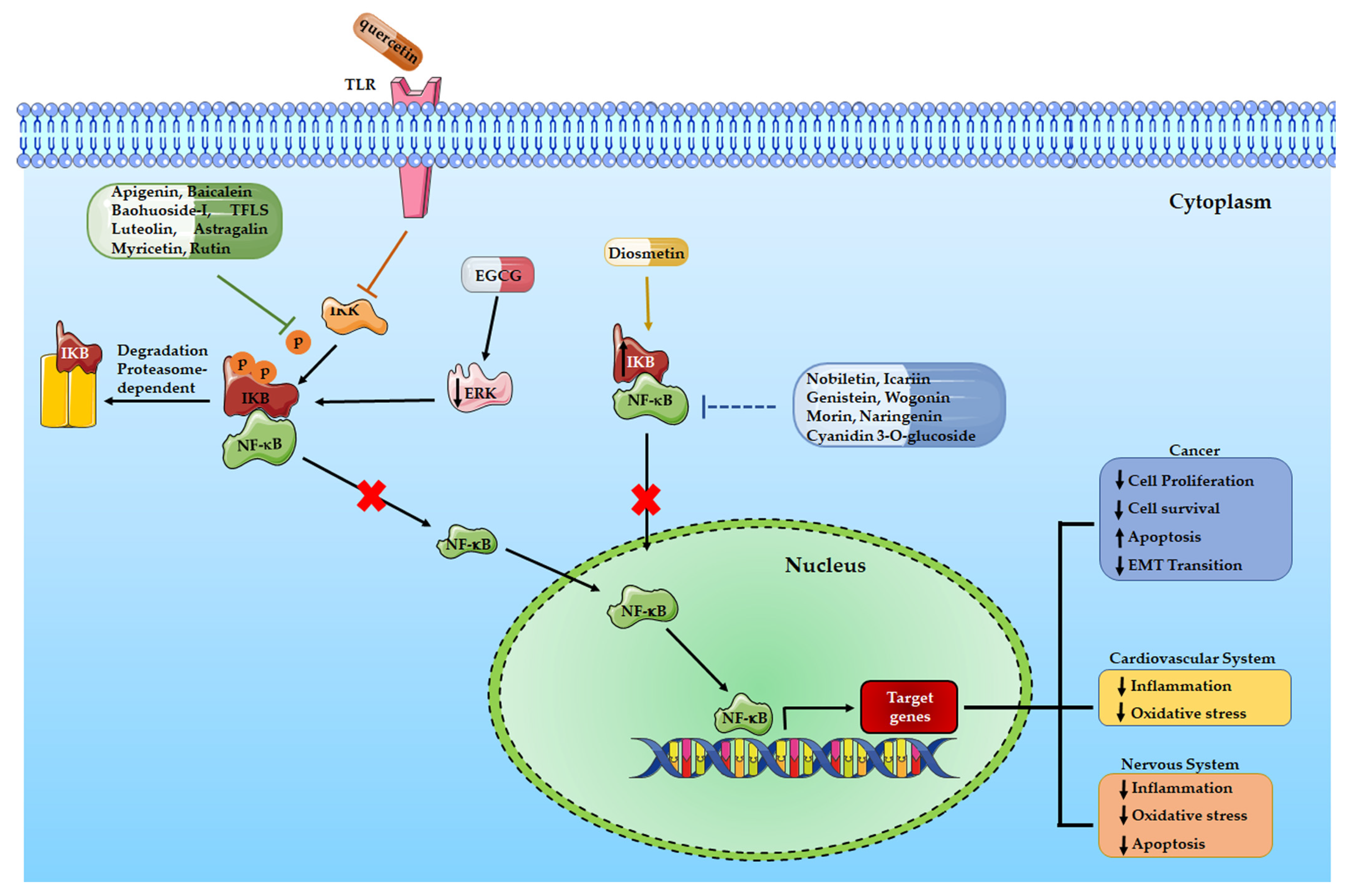
Table 2.
Summary of the effects of flavonoids on cardiovascular diseases mediated by the NF-kB signaling pathway.
Table 2.
Summary of the effects of flavonoids on cardiovascular diseases mediated by the NF-kB signaling pathway.
| Flavonoids | Subgroups | Chemical Structure | Effects on NF-kB signaling with proven positive effect on cardiovascular diseases | REFS. |
|---|---|---|---|---|
| Quercetin | Flavonols | C15H10O7 | Hypertension, coronary artery diseases | [108,109] |
| Icariside II | Flavonol | C27H30O10 | Myocardial fibrosis | [113] |
| Pronthocyanidin | polyphenols | Heart injury and fibrosis | [116] | |
| Morin | Flavone | C15H10O7 | Heart damage | [117] |
| Curcumin | Diarylheptanoid | Cardiotoxicity | [118] |
Table 3.
Summary of the effects of flavonoids on neurodegenerative diseases mediated by the NF-kB signaling pathway.
Table 3.
Summary of the effects of flavonoids on neurodegenerative diseases mediated by the NF-kB signaling pathway.
| Flavonoids | Subgroups | Chemical Structure | Effects on NF-kB signaling with proven positive effect on neurodegenerative diseases | REFS. |
|---|---|---|---|---|
| Quercetin | Flavonols | C15H10O7 | Atherosclerosis | [132] |
| Apigenin | Flavone | C15H10O5 | Parkinson disease | [150] |
| Naringenin | Flavanones | C15H12O5 | Alzheimer disease cognitive impairment |
[134,159] |
| EGCG | Flavan-3-ols | C22H18O11 | Alzheimer disease | [138] |
| Myricetin | Flavonols | C15H10O8 | Ischemic cerebral injury, microglia inflammation | [143,144] |
| Cyanidin 3-O-glucoside | Anthocyanin | C21H21ClO11 | microglia inflammation | [145,146] |
| Rutin | Flavonols | C27H30O16 | Alzheimer disease | [152] |
| Genistein | Flavone | C15H10O5 | Hippocampus inflammation | [154]. |
| Nobiletin | Flavone | C21H22O8 | Microglia inflammation Alzheimer disease |
[157,158] |
Disclaimer/Publisher’s Note: The statements, opinions and data contained in all publications are solely those of the individual author(s) and contributor(s) and not of MDPI and/or the editor(s). MDPI and/or the editor(s) disclaim responsibility for any injury to people or property resulting from any ideas, methods, instructions or products referred to in the content. |
© 2023 by the authors. Licensee MDPI, Basel, Switzerland. This article is an open access article distributed under the terms and conditions of the Creative Commons Attribution (CC BY) license (http://creativecommons.org/licenses/by/4.0/).
Copyright: This open access article is published under a Creative Commons CC BY 4.0 license, which permit the free download, distribution, and reuse, provided that the author and preprint are cited in any reuse.


