
Submitted:
06 May 2023
Posted:
08 May 2023
You are already at the latest version
Abstract
: Given that cancer is a disease that is rampant in the world and especially in Africa where the population has enormous difficulty in treating it, plants are a safer and less expensive alterna-tive. Cassava is one of the plant species valued in Benin because of its numerous medicinal and nutritional virtues. This study evaluated the biological activities of amygdalin from the organs of three cassava varieties most produced in Benin (BEN, RB, and MJ). HPLC analysis was used to quantify amygdalin in cassava organs and derivatives. Phytochemical screening was performed to determine secondary metabolite groups. DPPH and FRAP methods were used to assess anti-oxidant activity. Cytotoxicity of the extracts was tested on Artemia salina larvae. The an-ti-inflammatory activity was evaluated in vivo on albino mouse paw edema model induced by 5% formalin. The anticancer activity was evaluated in vivo on Wistar rats rendered cancerous by 1,2-dimethylhydrazine (DMH) using 5-fluorouracil as reference molecule. The results showed that the organs of all three-cassava varieties contained glycosides, flavonoids, saponosides, ster-oids, tannins, coumarins, and cyanogenic derivatives. Young stems and fresh leaves of cassava had the highest amygdalin concentration with 11142.99 µg 10 g-1 and 9251.14 µg 10 g-1 respec-tively. The Agbeli derivative was more concentrated in amygdalin with a content of 401.56 µg 10 g-1 than the others derivatives. The antioxidant activity results showed that the amygdalin ex-tracts were found to be DPPH radical scavengers with IC50 values ranging from 0.18 mg mL-1 to 2.35 mg mL-1. The cytotoxicity test showed no toxicity of the extracts toward shrimp larvae. Ad-ministration of amygdalin extracts from the leaves of BEN and MJ varieties prevents inflamma-tory edema. The percentages of edema inhibition varied between 21.77% and 27.89%. These val-ues are similar (p> 0.05) to that of acetylsalicylic acid (25.20%). Amygdalin extract of BEN variety significantly (p<0.0001) reduces edema. Cancer induction with DMH was inhibited by both BEN extract. In both preventive and curative treatments, rats fed with amygdalin extracts showed low anti-cancer activity under the effect of DMH and the significant difference in biochemical results. Thus, the organs of all three cassava varieties studied have secondary metabolites and good an-tioxydant activity. The leaves contain high levels of amygdalin and can be used as an-ti-inflammatory and anticancer agents.
Keywords:
Amygdalin
; cassava
; phytochemistry
; biological activities
; Benin
1. Introduction
Cancer is now a major public health problem in the word, with over 18 million new cases and 9.6 million deaths in 2018 [1]. Cancer is ranked among the major causes of morbidity and mortality worldwide [2]. It is characterized by the excessive proliferation of abnormal cells, which can be lethal if not effectively treated. This mortality remains high despite recent advances in treatment in developed regions in recent years. The number of new cancer cases per year worldwide has increased from 14 million in 2012 to more than 10 million in 2020 [3] with a rate of more than one million new cases recorded in Africa [4]. Considered for a while the preserve of high-income countries, Africa is not exempt from cancer. In Africa, more than 700,000 deaths were recorded in 2020 [4] and for the 2030 projections, the estimated figures are among others 1.4 million new cases and 1 million deaths [2]. More than 95% of cancer patients in African countries are diagnosed at an advanced stage [5]. Delayed diagnosis for these patients is due to insufficient awareness and a lack of qualified centers with well-trained personnel [5]. This delay is also dominated by financial problems, the lack of motivation of elderly patients, and the absence of the concept and word "cancer" in several African languages [6]. In Benin, since 2013 the number of cancer patients is growing and 1500 cases are recorded each year in the city of Cotonou with 55% of deaths [7].
Various alternatives against the disease have already been developed in modern medicine such as: chemotherapy, surgery, gene therapy, radiotherapy, immunotherapy, and others. However, conventional cancer therapies are associated with a lack of selectivity and serious side effects [7]. These chemotherapies are therefore considered risky drugs. Indeed, they combine multiple risks concerning the environment, the caregivers, the patients and, more generally, all users in health care institutions, making the safety of their use a major issue [8; 9]. In addition, most of these chemotherapies are included in the list of "hazardous to handle" drugs established by the National Institute for Occupational Safety and Health (NIOSH) in the United States in 2004 and whose latest version dates from 2016 [10]. Occupational exposures to chemotherapies induce three main risks: immediate organ toxicity, impairment of reproductive functions [11], and cancer pathology [12]. Therefore, despite all these efforts of modern medicine, the mortality rate is still increasing worldwide.
The natural world abounds with a multitude of species and plant diversity. The diversity of chemical structure and their relatively low toxicity make natural products of plant origin a promising source for the development of new anti-cancer therapies that are more effective and capable of targeting multiple characteristics of cancer. In Benin, several testimonies have been reported regarding the treatment of diseases of microbial and viral origin by the organs of cassava. This plant is highly produced in Benin and used mainly in human food in various artisanal and industrial forms [13]. The root is consumed as a food product (in the form of “gari”, “tapioca”, “lafun”, and “agbeli”), and is a good source of starch and biofuel. Besides its nutritional importance, cassava leaves and roots are also used in treatment of several diseases such as diabetes, rheumatoid arthritis, cell aging, cardiovascular diseases including atherosclerosis [14; 15]. This is because these organs contain bioactive molecules like vitamin C, vitamin A, secondary metabolites such as flavonoids, saponins, steroids and cyanogenic glycosides [14]. Besides these bioactive molecules, cassava contains amygdalin, which has proved to have therapeutic effects.
Amygdalin is a popular cyanogenic disaccharide [16]. This compound is attributed with highly therapeutic effects provided by several authors such as: anti-inflammatory and analgesic actions of neurodegenerative diseases [17]. It is also use in treatment for asthma, bronchitis, emphysema, leprosy and diabetes [18]. Extracted from apricot (Prunus armeniaca), amygdalin inhibits adhesion of breast, lung, and bladder cancer cells [19]. In view of these multiple bioactive molecules, studies on the quantification and analysis of the therapeutic effects of amygdalin extracted from cassava remain to be explored.
In Benin, cancer treatment is not affordable for all population groups. Moreover, cassava is a plant with a prominent place in the population's diet and contains bioactive molecules. Moreover, cassava is a plant that exists in several varieties and whose organs are used. These organs occupy a place of choice in the diet of the beninese population and are full of bioactive molecules. Do we wonder which varieties or organs or derivatives of the plant contain more amygdalin? The objective of the present study was (i) to identify the major groups of secondary metabolites of cassava varieties and (ii) to evaluate the efficacy of amygdalin extracted from three of the most consumed cassava varieties in Benin. Specifically, the aim was to assess antioxidant, antimicrobial, anti-inflammatory and anticancer activities of amygdalin extracted from three cassava varieties in Benin.
2. Results
2.1. Phytochemical screening
Qualitative tests revealed the presence of various secondary metabolites such as flavonoids, saponosides, steroids, tannins, leuco-anthocyanins, glycosides, alkaloids, coumarins and cyanogenic derivatives (Table 1). The results showed that the three cassava varieties don’t contain triterpènes.
2.2. Extraction yields
The results of the extraction yields of the various cassava organs has presented in Table 2. The best extraction yield among the organs of the cassava samples was obtained with the second skin followed by the pulp regardless of the variety. The second skin of the BEN variety dried in the sun gave the best yield (18.21%). The lowest yield was obtained with the first skin (0.95%) of the RB variety dried in the shade and sun. Furthermore, the extract of the leaves of the three varieties gave the same yield at extraction which is 1.5%.
2.3. Phytochemical constituents of three cassava varieties
The contents of polyphenolic compounds, flavonoids and total tannins of the samples of the three varieties (BEN, MJ and RB) of cassava has presented in table 3. The MJ variety has a higher flavonoid content (129.36±9.22 µgEQ/100mg) compared to the BEN (110.96±1.18 µgEQ/100mg) and RB samples (125.20±2.77 µgEQ/100mg). The total content of polyphenols varied significantly (p<0.05). The RB extract had the highest total tannin content (0.54±0.03 mgAAG/g) while the BEN (0.35±0.07 mgAAG/g) extract had the lowest.

Table 3.
Total flavonoid, polyphenols and tannin content of ethanolic leaf extracts of cassava varieties (mean ± standard deviation).
Table 3.
Total flavonoid, polyphenols and tannin content of ethanolic leaf extracts of cassava varieties (mean ± standard deviation).
| Cassava varieties | Flavonoids (μgEQ/100mg) | Polyphenols (μgEAG/100mg) | Tannins (mgEAG/g extract) |
|---|---|---|---|
| BEN | 110,96 ± 1,18a | 52,59 ± 7,56b | 0,35 ± 0,07b |
| MJ | 129,36 ± 9,22a | 32,62 ± 8,70c | 0,37 ± 0,04b |
| RB | 125,20 ± 2,77a | 65,14 ± 4,74a | 0,54 ± 0,03a |
2.4. Variation of the amygdalin content in Cassava varieties organs
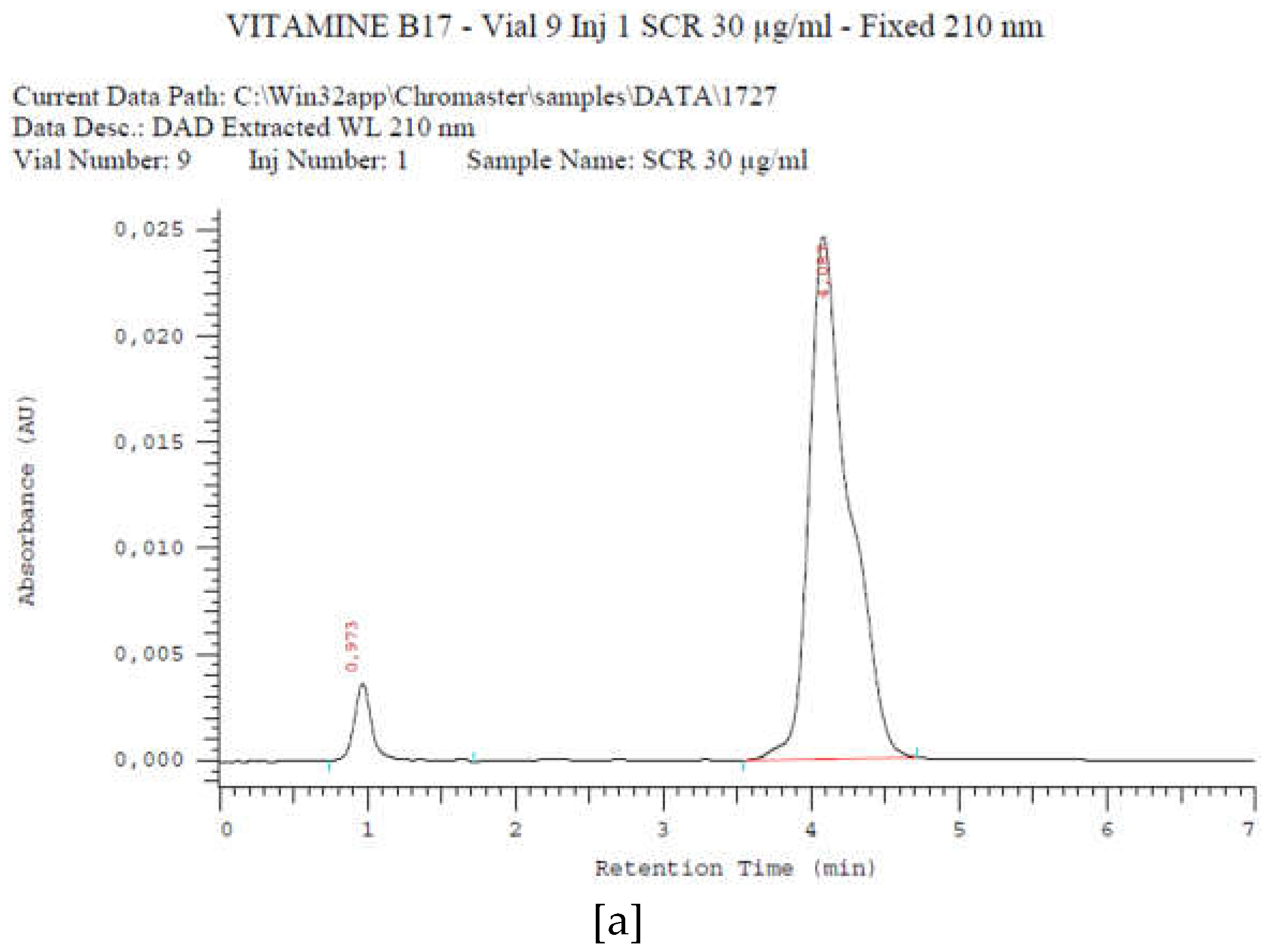
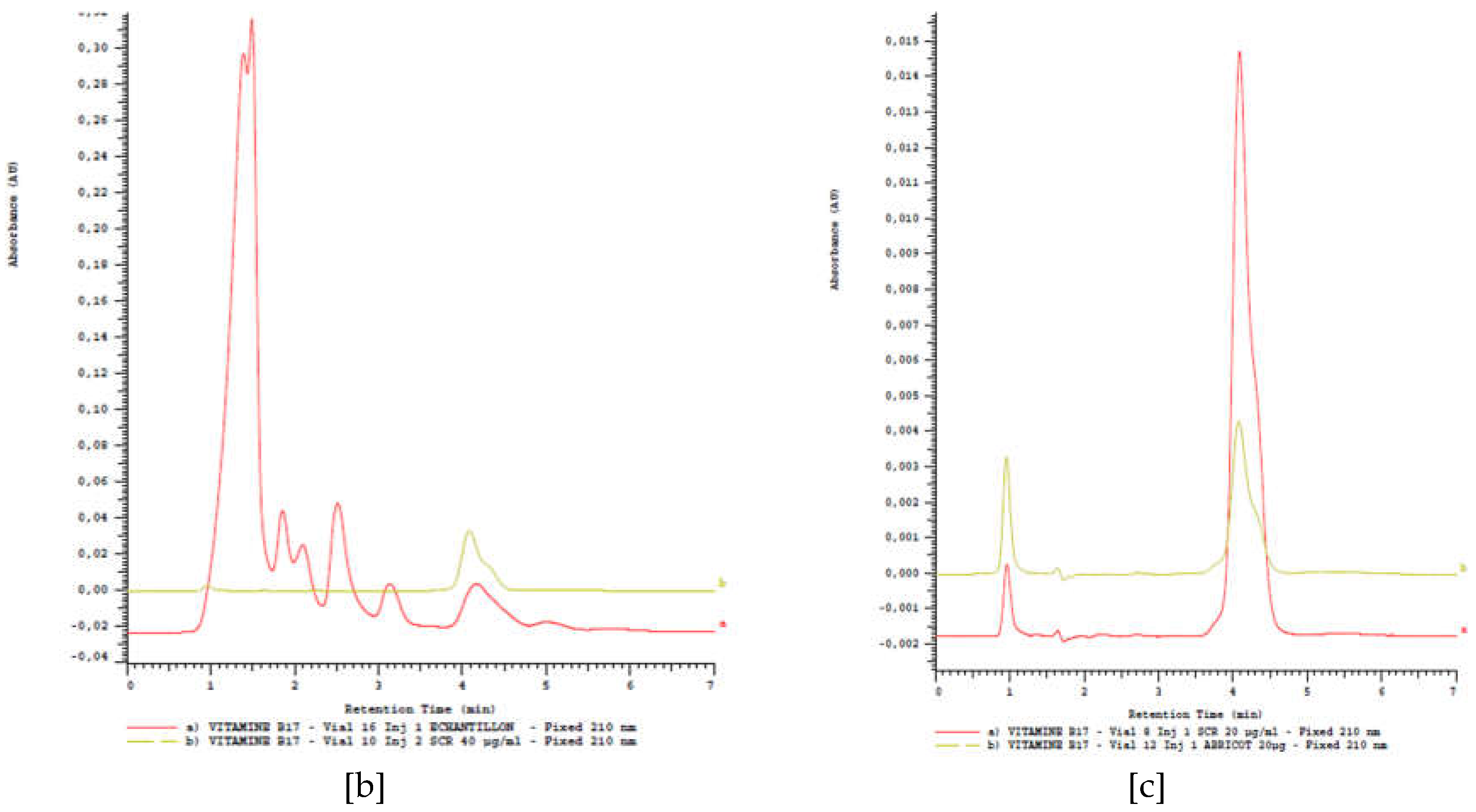
Amygdalin was identified by HPLC method in methanolic extracts by comparing the absorption chromatogram of the standard which is the standard of amygdalin (Sigma Aldrich - TCH - 10050-5G; purity ≥ 97%) at 30 µg.mL-1 and that of the samples. This chromatogram showed its peak at a retention time equal to 4.1 min.
Figure 1.
Chromatogram of amygdalin standard [a]; standard-sample [b] and standard-derived [c].
2.4.1. Amygdalin content in cassava stem organs
The amygdalin content of cassava stems according to cassava varieties is presented in Figure 2. Six (06) samples were submitted for amygdalin determination. Amygdalin is present in all samples submitted for analysis. Note that the young fresh stems of yellow cassava "MJT_F"
2.4.2. Amygdalin content in cassava leaves
The amygdalin content of cassava leaves according to variety is presented in Figure 3. Six (06) samples were submitted to the determination of the molecule. The presence of amygdalin is also noted in all the samples submitted for analysis. Here the contents found are function of the variety and of the drying because the contents of amygdalin in the fresh leaves are superior to those obtained in the leaves dried in the shade. Manioc Fresh Leaf Yellow “MJF_F” followed by BEN Fresh Leaf “BF_F” contained the highest levels of amygdalin (9251.74 µg and 7611.98 µg respectively). On the other hand, the variety RB Leaf dried in the shade “RBF_OM” contains the lowest amygdalin content (430.96 µg).
2.4.3. Amygdalin content in cassava derivatives
The amygdalin content of cassava derivatives is presented in Figure 4. 8 samples were subjected to amygdalin determination. Among these derivatives, Agbéli has the highest amygdalin content (401,56 µg/10g of sample). It should be remembered that Agbeli is fermented cassava starch. The boiled flesh was also found to have interesting amygdalin contents (from 30 to 138.38 µg/10g of sample). All the contents found are very variable from one derivative to another. This variation can be explained by the technology used to obtain each derivative. Furthermore, the Gari derivative has the lowest amygdalin content (28,09 µg/10g of sample), while the Tapioca derivative has no readable value. It should be noted that, of all the derivatives studied, the manufacture of these two derivatives is done by heating and at a very high temperature. The amygdalin content in Gari is low compared to those found in fresh roots.
2.5. Antioxidant activity
The variations of the antioxidant power by DPPH method of amygdalin extracted from different organs (flesh, 1st and 2nd skin) of three cassava varieties are presented in Table 4. In general, It is observed that the antiradical power varies from one extract to another and that the highest values are obtained with BEN 1st skin (PI: 95.38±0.07%; IC50 < 0. 19 µg.mL-1) followed by MJ flesh (PI: 95.11±0.18%; IC50 =0.5±0.22 µg.mL-1). The flesh of BEN and 1st skin MJ (respectively with PI: 94.71±0.07%; IC50 < 0.19 and PI: 93.77±0.54%; IC50 = 0.25±0.07 µg.mL-1) for sun-dried samples. There was no significant difference (p > 0.05) between these values. While the lowest values are obtained with MJ 2nd skin and RB flesh (PI: 85.12±0.67%; IC50 =4.6±1.97 µg.mL-1 and PI: 87.28±1.25%; IC50 =3 µg.mL-1). The percentages of inhibition are well above 50%.
For the shade-dried samples (Table 5), the highest average free radical scavenging power through DPPH is obtained with MJ 2nd skin (PI: 92.10±0.16%) with a IC50 equal to 0.5 µg.mL-1 followed by BEN 2nd skin (PI: 91.37±0.18%) with a IC50 = 2.75 µg.mL-1. The lowest value of antiradical power is obtained with BEN flesh (PI: 85.88±0.28; IC50 = 9.6±0.56 µg.mL-1) and RB flesh (PI: 88.93±0.12%; IC50 = 17.25±0.35 µg.mL-1). The reference molecule used as a standard (quercetin) has a lower free radical scavenging power (PI: 82.35±1.86%) than all samples and an IC50 of 5.75 µg.mL-1.
The variation in antioxidant power by DPPH method of leaf extracts of the three cassava varieties is presented in Table 6. It can be seen that the IC50 of BEN and MJ varieties are almost equal (3.79 and 3.02 µg.mL-1 respectively) and are lower than those of RB variety (8.11 µg.mL-1). Note that the IC50 of the reference molecule (ascorbic acid, IC50 = 1.11 µg.mL-1) is lower than the IC50 of amygdalin in all three varieties. There is no significant difference (p > 0.05) between the IC50 values obtained (µg.mL-1) with the reference molecule and the amygdalin of the leaves of the BEN and MJ varieties. The RB extract shows the highest reducing power.
The efficiency of the extracts in reducing Iron was determined. Table 6 shows that the highest reducing powers were recorded with the extracts from the leaves of varieties BEN and MJ (IC50 of 0.63 ± 0.04 µg.mL-1 and 0.69 ± 0.03 µg.mL-1 respectively) compared to the reducing power of the leaves of variety RB (IC50 of 0.52 ± 0.04 µg.mL-1). The comparative analysis between these values shows that there is a significant difference between MJ-BEN and RB (p < 0.05). The extracts of BEN and MJ varieties are more active in FRAP test compared to the extract of RB variety. Thus, the presence of the reductants in the extracts caused the reduction of Fe3+ ion (complexed) to Fe2+ ion.
2.6. Larval cytotoxicity of amygdalin extracted of cassava varieties
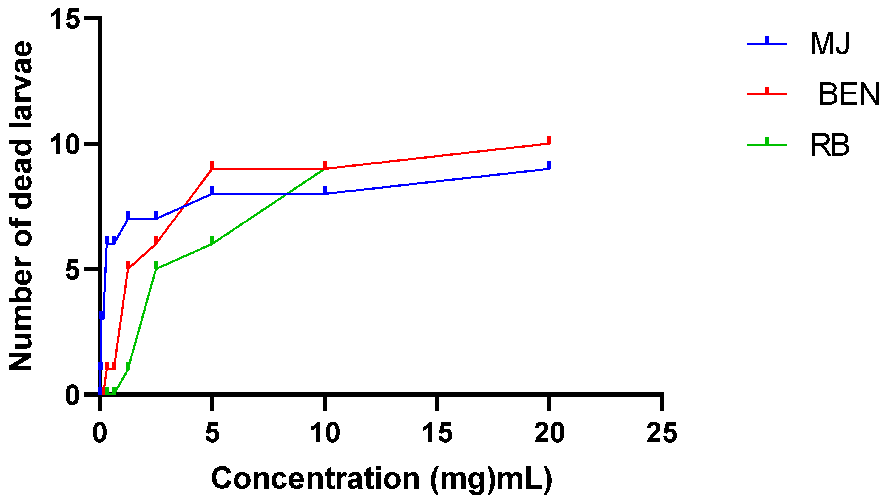
The cytotoxicity of the extracts was evaluated on Artemia salina larvae. The regression equation was used to determine the concentrations of ethanolic extracts of the leaves that caused the death of 50% of the larvae (LC50) previously introduced. The ethanolic extracts of the leaves of the three cassava varieties (BEN, MJ and RB) showed no toxicity or lethal dose in the tested larvae. Nevertheless, the CL50 of the RB and MJ extracts were almost the same (12.73 and 12.32 mg.mL-1). These values are higher than that of the BEN variety (Table 7). Of all the extracts tested, RB extract recorded more larval losses at high concentrations (10/16 dead larvae at 100 µg.mL-1).
Figure 5.
Larval cytotoxicity of extracts.
2.7. Anti-inflammatoiry activity of cassava leaf extracts
The 5% formalin-induced acute mouse hind paw edema model was used. The size of the edema was measured regularly every hour for 5 h and the percentages of increase and inhibition calculated. The results are shown in Figure 6 and Figure 7.
Administration of ethanolic extracts at 100 mg/kg and acetyl salicylic acid at 50 mg/kg prevents edema in treated mice compared with control mice (control) that received physiological water alone. The prevention is highly significant (p<0.0001) with BEN extract from the third hour to the fifth hour. An increase in the volume of the mice's paw was observed in the control group with a maximum of 55.16% at the first hour compared to a maximum of 48.88% in the lot that received aspirin and a maximum of 47.94% in the lot that received the BEN extract, always at the same hour. On the other hand, the batch of mice that received MJ extract showed a maximum of 43.61% at the second hour. If the percentage of increase in edema is more or less stable (between 53.69% and 55.16%) in control mice, in mice treated with BEN extract these percentages decrease considerably from the third hour to the fifth hour and mice treated with MJ extract the percentages of increase in edema decrease progressively from the second hour to the fifth hour. The batch treated with aspirin showed a first decrease between the first and second hour (48.88% to 41.16%) and a second decrease between the fourth and fifth hour (44.19% to 40.09%) (Figure 6).
Moreover, the percentages of edema inhibition increase with time and there is no significant difference (p>0.05) between the anti-inflammatory effect of the extracts at 100 mg/kg and that of Aspirin at 50 mg/kg from the first hour to the fifth hour. Better, like the reference molecule (Acetyl Salicylic Acid), our extracts have a more important anti-inflammatory activity in the second phase of the inflammatory process.
This while better inhibition of inflammatory paw edema was observed at the fifth hour in all treated mice with inhibition percentages ranging from 27.89% (BEN extract), 25.20% (Aspirin) and to 21.77% (MJ extract) (Figure 7).
2.8. Anticancer activity of amygdalin extracts from cassava variety BEN
2.8.1. Biochemical analysis
The variation in biochemical parameters is presented in Table 9. From this table it can be seen that the mean value of the creatinemia levels of the rats in group 2 is very high (19.57 mg.L-1) compared to the other batches. This high value indicates the onset of lesions and kidney dysfunction (renal failure) in the rats of batch 2. This finding is due to the treatment received by the rats of this batch, which consisted exclusively of DMH injections during the entire period. DMH therefore acted adversely on the liver and kidneys of rats in this batch.
Overall, the values of the biochemical parameters show a clear difference between the different batches of rats. Especially, there is a big difference between the parameter values of the rats of group 2 and the parameter values observed in all other remaining batches (R1, R3, R4 and R5). This diversity of values is observed in all parameters except those of monocytes. The variation of all the values of the biochemical parameters is highly significant (p<0.001), when comparing one batch to another, except for monocytes (p>0.005) as observed below (Table 9). In addition, after analysis of the comparison of the mean values of biochemical parameters made two by two between the batches of rats, it is found that the mean values of the rats of group 2 are largely higher than the mean values of the other batches. On the other hand, there is a clear difference between the values of the biochemical parameters of the rats of the control lot (R0), which did not receive any treatment, compared to the values of the other groups (R1 to R4), but these values of the lot show similarities with the values of lot 5 which received the treatment with the reference molecule (5-fluorouracil). The values of the biochemical parameters of the latter are similar to the values of the biochemical parameters of the rats (R1, R3, and R4) that received the treatment with amygdalin extracts (preventive and curative). This finding reflects the similarity in the mode of operation of 5-fluorouracil and amygdalin.
2.8.2. Histological analysis
2.8.1. Observation of the colon
The effects of DMH and treatments on the colon of rats are shown in Figure 8. In DMH treated group (b), the colonic mucosa shows altered Lieberkühn's glands (G) with areas of cellular infiltrates (CI), suggesting anarchic cellular proliferation. The caliciform cells seem to disappear from the glands. In batches 1, 4 and 5, the mucosa has its typical architecture as in normal rats (control batch) with Lieberkühn's glands (G) presenting their aspect of simple tubular glands with an abundance of caliciform cells (arrows). In batch 3, the glandular appearance is more or less normal with slightly fewer caliciform cells (arrows). We therefore deduce that DMH is at the root of the anarchic cell proliferation observed in the rat colon.
2.8.2. Observation on the liver
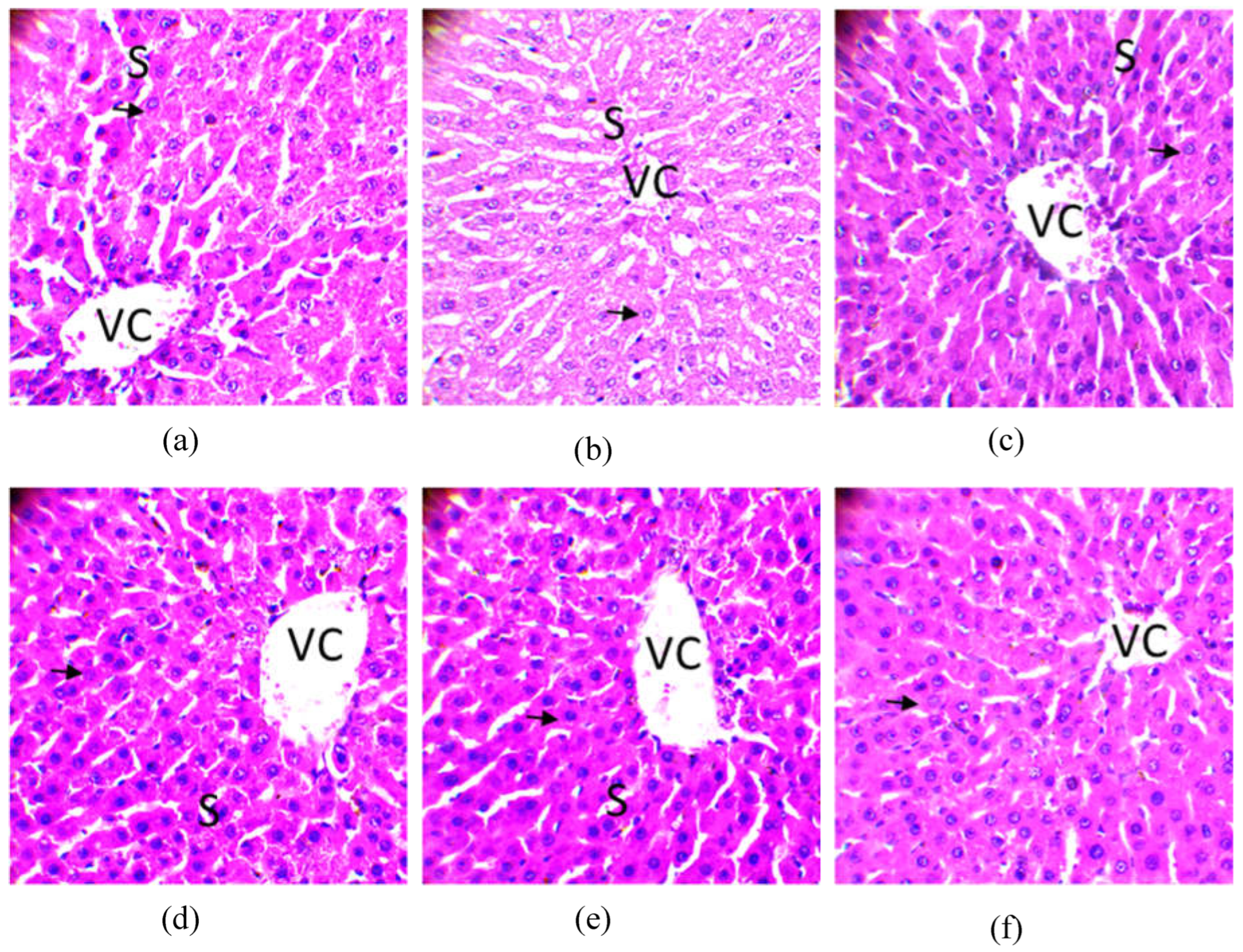
Apart from the observations on the colon, the possible effects of DMH on the liver were examined. The histology of the liver of experimental wistar rats has presented in figure 9. From this figure it can be seen that the liver parenchyma did not show any visibleatypia in the different batches. The hepatocytes (arrows) are well organized in cords around the centrilobular veins (VC). Venous sinusoids (S) are clearly visible between the hepatocyte cords. These observations show that DMH has not yet had any harmful effect on the liver of treated rats. This easily explains the mechanism of action of DMH, which is a molecule that acts primarily on the colon where its binding sites are present, allowing it to create damage in the colon. This damage in feedback can have harmful consequences on the liver later on.
Figure 9.
Histology of the liver of tested Wistar rats (original magnification × 400). Legend: Venous sinusoids (S); Centrilobular Veins (VC); (a) group 1: DMH and Amygdalin ; (b) group 2 : DMH only; (c) group 3 : DMH, then Amygdalin next week; (d) group 4 : Amygdalin, then DMH next week; (e) group 5 : DMH and 5-fluorouracil combined; (f) Control group: No treatment.
2.8.3. Observation of the kidney
The effects of DMH and treatments on the kidney of rats are shown in Figure 10. In the untreated positive control group (Batch 2), the renal parenchyma showed cellular debris (arrows) in some tubular lumens, an indicator of cellular injury. In the other groups, the appearance of the parenchyma is typical with characteristic glomeruli (G), renal tubules (RT). The tubular lumens are clearly visible. These observations show that only DMH acts negatively and moderately on the kidneys of treated rats. In the other groups, the appearance of the parenchyma is typical with characteristic glomeruli (G), renal tubules (RT). The tubular lumens are clearly visible. These observations show that only DMH acts negatively and moderately on the kidneys of treated rats.
3. Discussion
The phytochemical study characterized the presence of glycosides, flavonoids, saponosides, steroids, tannins, leuco-anthocyanins, coumarins and cyanogenic derivatives in ethanolic extracts of leaves of cassava varieties (BEN, RB and MJ). The content of these secondary metabolites varies from one variety to another. These same metabolites were found in the leaves of a cassava variety harvested in Côte d'Ivoire by [20]. Cyanogenetic compounds were detected in the sample powders. These are anti-nutritional factors, adding to the low protein and mineral content of cassava. However, this toxicity can be reduced, or even eliminated, in the finished ready-to-eat products obtained after various transformations.
The determination of total phenolic compounds, flavonoids and total tannins showed a variation in their contents from one variety to another. The ethanolic extracts from MJ and BEN varieties are much more concentrated in total flavonoids while these varieties show a low level of total phenols and total tannins compared to the extract from RB variety. Different studies have shown that external factors (geographical and climatic factors), genetic factors, but also the degree of maturation of the plant and storage time have a strong influence on the content of secondary metabolites [21].
Amygdalin was determined in 39 samples of cassava produced in southern Benin. Pure methanol (99.8% for HPLC. LABO CHEMIE PVT. LTD) was used for the HPLC assay, which allowed a good quantification of amygdalin in each sample. Because methanol is a good mobile phase for amygdalin separation by HPLC in less than 50mn [22]. The recovery rate found in this study was 98% (with a correlation coefficient R= 0.99). This result corroborates with that of [23] who separated amygdalin within 15mn in almond seeds and food products in the UK. The result is also similar to that of [24] who worked on artemisinin grown in Benin. These authors also found a recovery rate of 98%.
The content of amygdalin varied significantly (p<0,05) between different organs and derivatives of cassava. Among the samples analyzed, amygdalin was more abundant in young stems and fresh leaves of cassava with a content of 11142.99 µg.10g-1 and 9251.14 µg.10g-1 respectively. While, among cassava derivatives, the Agbeli derivative was more concentrated in amygdalin with a content of 401.56 µg/10g. These contents are lower than 141.000 µg.10g-1 and 155.000 µg.10g-1 obtained respectively with almond seeds and mongolian almond reported in the studies of [25] in China. High levels also obtained with green plum, apricot, black plum, peach, red cherry and black cherry were also reported by [23] (17.5 mg.g-1, 14.4 mg.g-1, 10 mg.g-1, 6.8 mg.g-1, 3.9 mg.g-1 and 2.7 mg.g-1 obtained respectively). The differences observed with these results can be explained by variation in the plant species studied, climatic and environmental factors, and the soil types exploited to grow these plants. Geographic environment and genomic differences could greatly influence amygdalin biosynthesis and accumulation [25].
Note that the antioxidant activity of plant extracts containing phenolic compounds is due to their ability to act as hydrogen or electron donors and scavenge free radicals. The DPPH test is one of the commonly used tests to prove the antioxidant capacity of fractions and isolated pure compounds to act as hydrogen atom donors [26]. The results obtained in our case study show that the sun-dried cassava organs proved to be the most potent scavengers of DPPH radicals than the shade-dried organs with IC50 values including one below 0.19 mg/mL and others of 0.25, 0.5, 0.75 and 2.35 mg.mL-1. These values are also more interesting than those of the leaves of the three varieties. They are also better than the IC50 of ethanolic extracts of cassava stems (0.518 and 0.616 mg.mL-1) reported by [27] in China and then those of methanolic extracts of peels and stems of a yellow-fleshed variety of cassava (425 and 234 μM TE g -1) reported in the studies conducted by [28] in Nigeria. While [29] reported that water yam and dasheen (Colocasia esculenta) had the same high percentage of DPPH inhibition activity with 95.83% and 93.41%, respectively. The high values of antioxidant activity can be attributed to high levels of phenols and flavonoids coupled with other compounds such as phenylpropanoids and anthocyanins [30]. Furthermore, ethanolic extracts of leaves showed significantly higher DPPH radical scavenging activity than those of cassava leaf stem extracted with acidified methanol, simple methanol and acetone reported by [31] in India. These observed differences may be related to the different phytochemical compositions of the plant parts used and also to the extraction solvents. Indeed, according to [32] the antioxidant capacities of the extracts have a strong relationship with the solvent used, mainly due to the different antioxidant potential of compounds of different polarities.
Also, the reducing power value of quercetin which is chosen as reference molecule is lower (82.35%) than the reducing power values of the majority of our samples. Therefore, the organs of all sun-dried varieties are more active in the DPPH test than the shade-dried varieties. This could be explained by the variation in the level of secondary metabolites contained in the different organs of each cassava variety and the temperature related to the drying methods of these organs.
Furthermore, extracts from BEN and MJ leaves are more active in the FRAP test compared to extracts from other organs. The reducing power of these extracts is certainly due to the presence of hydroxyl groups in the phenolic compounds which serve as electron donors. Thus, antioxidants are considered to be reducers and inactivators of oxidants [33].
This study also assessed possible risks to the population using the leaves of the three cassava varieties. The larval cytotoxicity curves showed that larval mortality increases with concentration and referring to the toxicity scale established by [34], all the LC50 values of our ethanolic extracts are higher than 0.1 mg/ml, value above which the extract is considered as not presenting toxicity. These results show that the ethanolic extracts of the leaves of the three cassava varieties (RB, BEN and MJ) are biologically active at a dose of 100mg/ml and are non-cytotoxic. Therefore, the medicinal and food use of these leaves does not present any risk of short or long term intoxication to the populations. It should be recalled that this study showed the presence of secondary metabolites and especially the presence of major chemical groups such as flavonoids and phenols. The detected components are known to have various therapeutic properties, such as astringent effects of tannins, anti-inflammatory and anti-allergic effects of flavonoids. Besides their antioxidant power, they are antiulcerous, antispasmodic, antisecretory and antidiarrheal [35]. They are also endowed with aphrodisiac virtues [36].
The results obtained from the anti-inflammatory tests show that ethanolic extracts of the leaves of the three cassava varieties at 100 mg/kg appreciably reduce the edema induced by formalin. Injection of 5% formalin into the paw of mice provoked an almost immediate inflammatory response manifested by the appearance of classical signs of acute local inflammation such as redness, pain, heat and oedema in all four experimental groups. This inflammation begins with a phase that lasts about 1h 30 min after the injection of 5% formalin and is triggered by the production of serotonin, histamine and bradykinin. Formalin causes local inflammation when injected into the fascia of the sole of the foot [37] as does carrageenan [38]. The second phase, which occurs after the second hour until the fifth hour, is due to the biosynthesis of prostaglandin [39] associated with leukocyte migration to the inflamed area [40]. The cause of this inflammatory response is tissue injury that induces the synthesis of histamine, prostaglandins, leukotrienes [41], PAF (p1aqueta activating factor), cytokines, NO (nitric oxide) and TNF (tumor necrosis factor) [42]. According to [43], these mediators promote vasodilation that causes redness and heat at the site of inflammation.
In addition, the 100 mg/kg ethanolic extract of BEN leaves more significantly (p<0.0001) reduced edema. On the other hand, there was no significant difference (p>0.05) between the anti-inflammatory effect of the two ethanolic extracts (BEN and MJ) and that of the 100 mg/kg standard. It can be deduced that the BEN and MJ extracts act in the same way as salicylic acid. Studies have shown that Salicylic Acid used as a standard, acts in the second phase of inflammation while inhibiting the synthesis of these different mediators [44]. By inhibiting the production of prostaglandins through the inhibition of Cyclooxygenase (COX2), it will limit the lowering of the pain threshold, hence its analgesic action, as well as inflammatory reactions, hence its antipyretic action [45-46]. This presages the same pharmacological responses with our extracts. These results suggest that amygdalin extracted from cassava leaves has an effect that opposes the action of endogenous pro-inflammatory mediators. This action would be exerted more on cyclooxygenase, the enzyme responsible for the synthesis of prostaglandins [47].
The anticancer activity test shows that subcutaneous injection of DMH to treated rats induces high levels of biochemical parameters and consequently observation of tumors in the colons of the rats that received it. 1,2-dimethylhydrazine is an effective carcinogen for induction of colon and rectal tumors in rats and mice by systemic subcutaneous or intraperitoneal injections [48]. In addition, researchers studied the efficacy of DMH in female mice and found that 83% of the mice developed visible tumors, and many had them primarily in the distal part of the colon [49]. This justifies the result observed in the colon of batch 2 rats (R2 treated with DMH). Recall that, the procarcinogen DMH, after a series of metabolic reactions, finally reaches the colon, where it produces the ultimate carcinogen and imbalance in the production of reactive oxygen species (ROS), which alkylate DNA and initiate the advent and development of colon carcinogenesis [50]. The preneolpetic lesions and histopathological observations of DMH-induced colon tumors can provide a typical understanding of the disease in rodents and humans. Therefore, the interpretation of histopathological observations revealed cellular abnormalities especially in the colon of these rats of batch 2. On the other hand, the absence of visible tumor in the colon, liver and kidney of rats of batches 1, 3, 4 and 5 (batches that received amygdalin from BEN leaves and 5-fluorouracil) attests that the ethanolic extracts (amygdalin) as well as the reference molecule had an effect on DMH-induced cell proliferation.
Note that, previous studies have reported that high levels of amygdalin ingested directly can be toxic to humans. Amygdalin is composed of two molecules of glucose, benzaldehyde and hydrogen cyanide and can exist as two epimers R and S [51]. The R -amygdalin is the natural amygdalin and the S -amygdalin is called neoamygdalin. Beta-glucosidase stored in plant cell compartments is also present in the human small intestine [52] and degrades amygdalin to prunasin, mandelonitrile, glucose, benzaldehyde and hydrogen cyanide. Hydrogen cyanide (HCN), benzaldehyde, prunasin, and mandelonitrile can be absorbed into the lymphatic and portal circulations [53]. The anticancer activity of amygdalin is thought to be related to the cytotoxic effects of enzymatically released HCN and unhydrolyzed cyanogenic glycosides [54]. But low and medium doses (50 and 100 mg.kg-1) of amygdalin administered orally do not induce any toxicity, while high dose amygdalin (200 mg.kg-1) is able to induce toxicity causing negative effects on the oxidative balance of liver tissues with an obvious effect on histopathology [55].
4. Materials and Methods
4.1. Chemicals
The extraction solvents, phosphate buffered saline (PBS), methanol (CH3OH) and ethanol (C2H5OH) were obtained from Sigma-Aldrich Chemical Company (St. Louis, USA). The 2,2-diphenyl-2-picrylhydrazyl (DPPH), potassium hexacyano ferrate [K3Fe(CN)6], trichloroacetic acid (C2HCl3O2), gallic acid (C7H6O5), ascorbic acid (C6H8O6), quercetin (C15H10O7), iron chloride (FeCl3), folin-ciocalteu reagent (FCR), anhydrous sodium carbonate (Na2CO3), aluminum chloride (AlCl3), potassium acetate (CH3CO2K) were of analytical grade. 1,2-Dimethylhydrazine (DMH) was purchased from Macklin (Shanghai Macklin Biochemical Co. , Ltd, China). Standard Amygdalin was purchased from Sigma-Aldrich (Sigma-Aldrich, Co, 3050 Spruce Street, St. Louis, MO, USA) and 5-Fluorouracil are provided by Sigma Aldrich, Germany (St Louis, MO, USA). Biochemical analysis kits were BIOLABO diagnostic kits.
4.2. Plant material and sample collection
The plant material used consists of the peelings (1st and 2nd skin) of the root, pulp (root), stem and leaves of three different varieties. These are the BEN variety; the RB variety and the Yellow Manioc (MJ) variety (Table 10). The samples were collected in the commune of Toffo located in the Atlantic Department. These samples were rinsed lightly with water to remove dust.
4.3. Methods of drying cassava organs
4.3.1. Shade drying in the laboratory
Organs such as leaves, pulp, stems of cassava were dried in the shade. This drying consisted of carefully cleaning the bench. On this bench, the different samples were dried under air conditioning at 16 o C for six (6) days.
4.3.2. Traditional sun drying
The organs such as the flesh and the second skin of the cassava root were dried in the sun. This drying consists in spreading an empty and clean PET bag on the ground. On this bag, the different samples are spread out in the sun for six (06) days.
The dried samples were ground to powder using a mill (IKA Bro-03-PATACE-018, Germany).
4.4. Animal material and acclimatization conditions
The animals used were albino mouses, and Wistar rats of EOPS (Exempt from specific pathogenic organism) health status, age approximately eight and wetghing between 150 g and 200g. The animals were housed in polypropylene cages integrated with water pots and under hygienic conditions with standard rat food and free access to water. After two weeks of acclimatization at a constant temperature of 22 ± 2oC under 12/12 light/dark cycle, the rat were divided into batches for the different tests. The body weight of the mouses and rats was recorded at the beginning and at the end of the experiment. This study was carried out within the framework of a doctoral thesis. The protocol received the favorable opinion of the scientific committee of the Doctoral School of Life and Earth Sciences (ED-SVT) of the University of Abomey-Calavi (UAC).
4.5. Phytochemical screening of cassava organs
The phytochemical screening carried out on the organ powder is based on differential precipitation and coloring reactions, using the method of [56]. This study allowed the identification of the main chemical groups (tannins, flavonoids, coumarins, terpenes, alkaloids, etc.) contained in the plant material.
4.5.1. Preparation of extracts
Dried powdered cassava organs were subjected to reflux conditions using the solvent ethanol at 78.5°C according to the method of [57]. 20 g of each ground organ was mixed with 200 mL of ethanol. Each mixture was boiled under reflux for 40 min. After refluxing, the mixture was subjected to ultrasonication for 15 min at 30°C and then completely filtered using Whatman No. 1 filter paper and the solvents were evaporated using a rotary evaporator. After drying the filtrate in the oven, 10 ml of diethyl ether was added to each filtrate and stirred for 1 min at room temperature (22°C) to obtain the precipitate. Diethyl ether was evaporated overnight in a fume hood. The crude extract was collected in petri dishes placed in the oven for drying and then scraped into sterile glass pillboxes for cool storage at 4 oC for biological testing. The extraction yield is defined as the ratio of the mass of dry extract obtained to the mass of plant material processed [58].
4.5.2. Determination of total phenols
Total phenolics are quantified in leaf extracts of each cassava variety (BEN, RB, MJ) using the Folin-Ciocalteu method described by [59] and adapted to an in-house method developed by [60]. A quantity of 10 µl of concentrated extract at 100 mg/ml (w/v) was introduced into the wells of 96-well plates with three replicates. A quantity of 25 µl of Folin-Ciocalteu reagent (50% v/v), was added and incubated 5min at room temperature, 25 µl of sodium carbonate (Na2CO3) at 20% (w/v) was added and then reduced to 200 µl with distilled water per well. Blanks were prepared by replacing the reagent with distilled water to correct for interfering compounds. Incubate for 30min, and absorbance is read at 760 nm on a multi-well plate reader. Gallic acid (0 - 500 µg/ml) is used as a standard and results are expressed as micrograms of gallic acid equivalent per 100 grams of extract.
4.5.3. Determination of total flavonoids
Total flavonoids in each sample were quantified by the aluminum trichloride method described by [61] and adapted to 96-well plates by [60]. It consists in adding and mixing 100 µl of methanolic AlCl3 (2%) to 100 µl of the appropriate dilution of the extract solution. Incubate for 15 min, then read the absorbance at 415 nm with the "Gen5" software using an Epoch Biotech multiplate spectrophotometer connected to a computer against a blank (mixture of 100 µl of methanolic extract solution and 100 µl of methanol) against a standard. Values were compared to a calibration curve for quercetin (0 - 500µg/ml) at R2 = 0.99. Flavonoid content is expressed as micrograms of quercetin equivalents per 100 g of extract.
4.5.4. Determination of hydrolysable tannins
The hydrolyzable tannins were determined according to the method of [62]. Indeed, 1 ml of each extract (5 mg/ml) is mixed with 3.5 ml of reagent (ferric chloride FeCl3 10-2 M in hydrochloric acid HCl 10-3 M). The absorbance of the mixture is measured (UV spectrophotometer, Shimadzu) at 660 nm after 15s of incubation. The content of hydrolyzable tannins T (%) is determined according to the formula below:
T (%) = (A × PM × V ×FD) / ε mole × P
A = absorbance, PM = weight of gallic acid (170.12 g/mol), V =volume of extract, DF = dilution factor, ε mole = 2169 (gallic acid equivalence constant), P = weight of extract.
4.6. Method for the determination of amygdalin by HPLC
4.6.1. Extraction of amygdalin from cassava samples and derivatives
Extraction of amygdalin from cassava organs and derivatives was performed according to the method described by [63] with some modifications. A quantity of 10 g of each sample was macerated in 150 mL of absolute methanol (Sigma Aldrich) for 15 min at 30 °C. Vacuum filtration was performed using a buchniii system and then concentrated in the rotavapor at 40 °C. The resulting filtrate was washed with 25 mL of diethyl ether and allowed to decant for 3 h. After decantation, the crude extract was obtained by removing the supernatant.
4.6.2. Analysis conditions
Amygdalin was separated and quantified with the system the HPLC-VWR-HITACHI system following the following analytical conditions: C18 column (LiChrospher 100 (5 μm), 100 × 4.6 mm Merck Chemicals, Darmstadt, Germany); Mobile Phase: Methanol/water (20/80, v / v); Flow rate: 0.7mL/min; Analysis time: 7 min; Detection wavelength: 280 nm.
A Hitachi VWR 5160 HPLC pump; a HITACHI VWR 5430 UV-DAD detector; a 100µl rheodyne loop injection valve (USA); a HITACHI VWR 5260 autosampler; a DELL Optiplex 7010 Couputer computer running Microsoft Windows 7 Enterprise, using CHROMASTER SYSTEM MANAGER software version 1. 1 for data acquisition; a HITACHI VWR 5310 column thermostat; a DELL 15" monitor; a DELL PIH keyboard; a Microsoft HID mouse.
4.6.3. Quantification procedure
To validate the method developed by the precision criteria, we performed an intra-day and inter-day replicate. Precision was determined for the different concentrations of amygdalin, three times on the same day for intra-day precision and three times on four different days for inter-day precision. Three injection tests were performed for each dilution and the means of the areas obtained under the curves were calculated. These means and their standard deviations were used to calculate the coefficients of variation (CV). The extraction yield was determined from 10 g of cassava organ or derivatives with the addition of 1 mg of the pure amygdalin. The recovery rate (RR) is given by the formula:
With A: amount of amygdalin extracted from cassava samples mixed with pure amygdalin; B: amount of amygdalin in cassava samples; C: amount of amygdalin added.
After calibration, the method was applied to the samples. The expression of the content (T) of amygdalin in µg in 100 g of sample is T = 10 q with the amount of amygdalin in 10 µL of injected extract.
4.6.4. Validation of the amygdalin identification method
Amygdalin was identified by HPLC method in methanolic extracts by comparing the absorption chromatogram of the standard which is the standard of amygdalin (Sigma Aldrich - TCH - 10050-5G; purity ≥ 97%) at 30 µg.mL-1 and that of the samples. This chromatogram showed its peak at a retention time equal to 4.1 min.
4.7. In vitro evaluation of the antioxidant activity of different extracts
4.7.1. Reduction of the DPPH radical
It was evaluated according to the method described by [64]. Thus, starting from 200 μg/ml of the extract solution, a series of 10 successive dilutions (to 1/2) of each extract was made with methanol so as to have 100 μl of volume. Then 100 μl of DPPH (100 μg/ml in methanol) was added to all dilutions in a cascade. At the same time, a negative control was prepared by mixing 100 μl of methanol with 100µl of the methanolic DPPH solution. The optical density reading was taken against a blank prepared for each concentration at 517 nm after 20 min of incubation in the dark at room temperature. The positive control is represented by a solution of a standard antioxidant; ascorbic acid whose optical density was also measured under the same conditions as the samples; and for each concentration, the test was repeated 3 times. The anti-free radical activity is estimated according to the following equation:
PI (DPPH) = Percentage of Inhibition of DPPH. OD = Optical Density
4.7.2. Evaluation of the reducing power of extracts
The ability of the extracts to reduce Fe3+ was assessed using the Ferric Reducing Antioxidant Power (FRAP) method described by (2017) [65] with a slight modification. 1 mL of the extracts was mixed with 2.5 mL of phosphate buffer (0.2 M, pH 6.6) and 2.5 mL of 1% potassium ferricyanide [K3]. Potassium ferricyanide [K3Fe(CN) 6]. After 20 minutes of incubation at 50°C, 2.5 mL of trichloroacetic acid (10%) was added to the mixture and mixed and centrifuged for 10 minutes (3000 r/t). 2.5 mL of the supernatant was mixed with 2.5 mL of distilled water and 0.5 mL of ferric chloride (0.1%). The absorbance was read at 700 nm. Ascorbic acid was used for the calibration curve and rutin (200 µg/mL) as the standard. Ferric iron (Fe3+) reducing activity was determined as follows ascorbic acid equivalents (mmol ascorbic acid/g extract). The antioxidant activity related to the reducing power of the extracts is expressed as Reducing Power (RP) using the following formula:
OD: Optical Density
The IC50 (Concentration that inhibits the 50% of DPPH or reduces the 50% of Fe3+) were determined by the probit method.
4.8. Evaluation of the cytotoxicity of extracts
The larval toxicity of the extracts was evaluated according to the method described by [66]. Larvae were obtained by hatching 10 mg of freeze-dried eggs of Artemia salina (ARTEMIO JBL GmbH D-67141 Neuhofem) under continuous agitation in one liter of sea water for 72 h. Thus, starting from a stock concentration of extracts of 100 mg/ml, a range of 10 concentrations is made with sterile distilled water following dilutions of ½ reason in test tubes numbered from T1 to T10. Then 1ml of seawater containing 16 live larvae was added to all tubes. The number of surviving larvae was counted under the microscope after 24 h of incubation. For each extract, the lethal concentration 50 (LC50) was determined from the regression line obtained from the curve representing the number of surviving larvae as a function of the extract concentration. The results were interpreted using the correlation grid associating the degree of toxicity with the LC50 proposed by [34].
4.9. In vivo evaluation of the anti-inflammatory activity of leaf extracts from three varieties of cassava
To highlight the anti-inflammatory activity of the studied cassava extracts, the experimental model of acute inflammation of the mouse paw induced by 5% formalin was used following the protocol used by [67]. The experiments were performed in male and female Swiss albino mice, weighing between 25 g and 43 g. They were fed with pellets containing 29% protein and received water and libitum. They were maintained at a temperature of 22 ± 2o C with a relative humidity of 55% ± 5.2. After fasting for 18 hours before the test, the mice were divided into 4 homogeneous batches of 3 mice according to weight. The diameter (Do) of the right hind leg of each mouse was measured one hour before the different treatments using an electronic caliper. The treatment was done orally (gavage) as follows:
Group 1: animals served as controls and received only physiological water at 0.2 mL per 25 g body weight;
Group 2: animals received amygdalin extract of the MJ variety at 1000 mg/kg body weight;
Group 3: animals received amygdalin extract of the BEN variety at 1000 mg/kg body weight;
Group 4: animals were used as a reference and received a reference anti-inflammatory drug, salicylic acetic acid at 50 mg/kg body weight.
One hour after gavage, 0.1 ml of a 5% formalin solution was injected into each mouse under the plantar pad of the right hind paw. The paw diameter at the arch was measured every hour until the fifth hour, using electronic display calipers. Edema increase (AOP) and inhibition (IOP) percentages were calculated according to the formula:
With Dt: Mean diameter of the right hind leg at time t; Do: Mean diameter of the right hind leg at time 0 (before treatment).
4.10. Evaluation of anticancer activity
It was performed in vivo with Wistar rats following the protocol adapted from [68]. All handling procedures also considered the guidelines for the study of animal models of cancer. The effect of amygdalin extracted from BEN variety (variety with the best antibacterial and anti-inflammatory activities) of cassava on anarchic cell proliferation of rat was tested by analysis of biochemical parameters and histopathological observations. 1,2-Dimethylhydrazine (DMH) was the molecule used to induce colon cancer in vivo. It was prepared in 1 Mm EDTA saline at pH 7.1 for subcutaneous injection of 45 mg/kg of body weight. The reference molecule used was 5-fluorouracil at 0.6 ml/kg/body weight. The extract was administered by gavage at 200 mg/mL in a volume of 100 mL/kg body weight.
4.10.1. Experimental design
Rats were randomly matched and body marked for indication. A number of 18 rats were divided into 6 groups of 3 rats each according to their body weight. All animals were provided with food, water and libitum throughout the experiment period.
Group 0: negative control, received plain water and food during the six weeks of experiment.
Group 1: inhibition effect of the extract. These rats received twice weekly subcutaneous injection of DMH plus daily gavage of the amygdalin extract.
Group 2: positive control, treated with DMH. Rats received a subcutaneous injection of DMH twice a week for the five weeks of the experiment.
Group 3: curative effect of extracts. Rats were injected subcutaneously with DMH twice a week for two weeks and at the third week, a daily dose of extract is applied until the end of the five weeks.
Group 4: preventive effect of extracts. Rats were given amygdalin extract daily for three weeks and then from the fourth week onwards, a subcutaneous injection of DMH twice a week for the next two weeks.
Group 5: effects of the reference molecule. Rats received a subcutaneous injection of DMH twice a week combined with an intravenous injection of 5-fluorouracil.
1,2 Dimethylhydrazine (DMH. Sigma Aldrich, Germany) was prepared fresh weekly in EDTA saline with a pH that was adjusted to 7.1 using NaOH solution immediately prior to subcutaneous injection at a dose of 45mg/kg body weight DMH. The follow-up time of the rats was six weeks for all batches.
4.10.2. Determination of biochemical parameters
In addition to the weight of the animals, biochemical parameters (hematocrit, hemoglobin, red blood cells, white blood cells, leukocytes and neutrophils) were measured before and at the end of the experiment from the animals' whole blood samples. In addition, plasma creatinine (REF 80107, LOT 012042A) and urea (REF 92032, LOT 041915A) concentrations, as well as as aspartate aminotransferase (AST) and alanine aminotransferase (ALT) (REF 51830, LOT 072105A7) were determined with BIOLABO (FRANCE) diagnostic kits on plasma collected after centrifugation of blood at 3000rpm. Blood samples were taken by puncturing the retroorbital sinus of the animals (under diethyl ether anesthesia).
4.10.3. Anatomical observations
After six weeks of follow-up, all animals were sacrificed under the same fasting conditions, in the evening (22 hours), under yellow light according to the protocol described by [68]. The liver, kidneys, and colon were removed and flushed with saline. The colon was opened longitudinally and examined microscopically to observe the presence of multiple identifiable lesions and the appearance of Aberrant Crypt Foci (ACF). Colon segments containing Multiple Plaque Lesions (MPL) were dissected, fixed immediately in 10% formalin over kerosene, sectioned, and stained with hematoxylin and eosin for histopathologic observations. The liver and kidneys were then placed in 10% formalin for histological sections.
4.10.4. Statistical analysis
GraphPad Prism 8 software was used for the analyses of variances. The Newman Skeuls test (SNK) was applied in a first step to compare the average amygdalin contents of the tested organs and derivatives. In a second step, the Student's t test was used to compare the average amygdalin content within each organ.
5. Conclusions
The present study evaluated the antioxydant, anti-inflammatory and anticancer activities of amygdalin extracted from organs and derivatives of the most produced cassava varieties in Benin. The study carried out on the powders obtained from the leaves of Manihot Esculenta allowed to identify some large families of its molecules among which we can quote alkaloids, flavonoids, tannins, glycosides, saponosides, steroids, leuco-anthocyanins, coumarins etc.
Cytotoxicity tests did not reveal any toxic effects of the extracts at the dose studied. So this work brings the reasons which underlie the results of our investigations stipulating the strong production and transformations of the three varieties of cassava (BEN, RB, MJ) in Benin.
This study highlights all the organs (leaves, stems, roots, peelings, second skin) of the three varieties of Manihot Esculenta, some of which were neglected by the population, thus causing environmental pollution. It also enhances the value of the by-products resulting from the processing of cassava produced in Benin. This can contribute to the fight against problems related to food insecurity and the improvement of living conditions of our populations on the one hand, and to the prevention and relief of chronic non-communicable diseases on the other hand.
The study evaluated the in vitro and in vivo biological activities of amygdalin extracts of the plant. The results revealed that cassava varieties grown in Benin possess remarkable antioxidant activities with anti-inflammatory similar to non-steroidal anti-inflammatory drugs (NSAIDs). In addition, amygdalin extracts of BEN cassava leaves had anticancer activities in cancer-prone Wistar rats. This molecule has been shown to be very effective in preventing and curing cancer by inhibiting the proliferation of cancer cells induced by DMH, which is confirmed by biochemical analysis of blood samples and histological examination.
Indeed, amygdalin, a molecule known for its numerous pharmacological properties, was quantified in cassava organs and derivatives. The results revealed the presence of this molecule in all samples but at variable levels. The leaves of the different cassava varieties and the Agbeli derivative showed a higher level of amygdalin. It could then be said that the anticancer power of the plant is due to the presence of a high level of amygdalin in its leaves.
Author Contributions
Conceptualization, L.B.M., H.L., R.B. and D.D.N.; methodology, H.L., A.N.A., A.P.T., S.M. and D.D.N.; software, H.L., A.N.A. and D.D.N.; validation, H.L., A.N.A., R.B., and D.D.N.; formal analysis, H.L., A.N.A., A.P.T., H.S. and D.D.N.; investigation, H.L., A.N.A., R.B., O.D., and D.D.N.; resources, H.L., A.N.A., and D.D.N.; data curation, S.H., S.M., A.A. and L.B.M.; writing—original draft preparation, H.L., A.N.A., R.B., O.D., H.S. and D.D.N.; writing—review and editing, S.H., S.M., A.A. ans L.B.M.; visualization, H.L., A.N.A., H.S., A.P.T. and D.D.N. and B.M.L.; supervision, S.H., A.A. and L.B.M.; project administration, A.A., B.M.L.; funding acquisition, L.B.M. All authors have read and agreed to the published version of the manuscript.
Funding
This research did not receive any external funding.
Conflicts of Interest
The authors declare that they have no conflicts of interest.
References
- Gnangnon F.; Egue M.; Akele-Akpo M.; Amidou S.; Brun L.; Mehinto D.; Houinato D.; Parkin D. Incidence des cancers à Cotonou entre 2014–2016 : les premiers résultats du premier registre des cancers en République du Bénin. Revue d'Épidémiologie et de Santé Publique. 2020; 68, 3 : pp. 0398-7620. [CrossRef]
- Ferlay J.; Ervik M.; Lam F.; Colombet M.; Mery L.; Piñeros M. Observatoire mondial du cancer : « Cancer Today ». Lyon : Centre international de recherche sur le cancer, 2020.
- WHO. Global Health Observatory. (2021). Available online at: https://www.who.int/data/gho/data/indicators/indicator-details/GHO/girls-aged-15-years-old-that-received-the-recommended-doses-of-hpv-vaccine (accessed June 15, 2022).
- WHO Globocan 2020. The Global Cancer Observatory/Mars 2021.
- Pezzatini M.; Marino G.; Conte S.; Catracchia V. Oncology: a forgotten territory in Africa. Annals of Oncology. 2007; 18, 12 : pp. 2046- 7. [CrossRef]
- Gombé C.M.; Godet J.; Gueye S.M. Cancers in Francophone Africa (Alliance des Ligues Africaines et Méditerranéennes contre le cancer). 2017. Accessed 17/ 04/ 2022.
- Egue M.; Gnangnon F.; Akele-Akpo M.T.; Parkin D. Incidence du cancer à Cotonou (Bénin), premiers résultats du registre des cancers de Cotonou. 2014-2016.
- Astier A.; Blanc-Leger F.; Burtin C.; Cazin J.L.; Chenailler C.; Daouphars M.; Anticancéreux : utilisation pratique (7ème édition). Dossier du CNHIM 2013: XXXIV.
- Yadav A.; Rene E.R.; Mandal M.K.; Dubey K.K. Threat and sustainable technological solution for antineoplastic drugs pollution: Review on a persisting global issue. Chemosphere. 2021; 263:128285. [CrossRef]
- NIOSH list of antineoplastic and other hazardous drugs in healthcare settings, 2016. Connor TH, MacKenzie BA, DeBord DG, Trout DB, O’Callaghan JP. Cincinnati. U.S. Department of Health and Human Services, Centers for Disease Control (CDC) and Prevention, National Institute for Occupational Safety and Health (NIOSH). Publication Number 2016-161. 2016;42. [CrossRef]
- Valanis BG.; Vollmer W.M.; Steele P. Occupational exposure to antineoplastic agents: selfreported miscarriages and stillbirths among nurses and pharmacists. J Occup Environ Med. 1999;41(8): pp. 632-8. [CrossRef]
- Dranitsaris G.; Johnston M.; Poirier S.; Schueller T.; Milliken D.; Green E. Are health care providers who work with cancer drugs at an increased risk for toxic events? A systematic review and meta-analysis of the literature. J Oncol Pharm Pract. 2005;11(2): pp. 69-78. [CrossRef]
- Halfane Lehmane, Rafiatou Ba, Durand Dah-Nouvlessounon, Haziz Sina, Orou Daouda Bello, Horace Degnonvi, Farid T. Bade, Farid Baba-Moussa, Adolphe Adjanohoun, Lamine Baba-Moussa. Cassava use in southern Benin: Importance and perception of actors involved in the value chain. African Journal of Agricultural Research. 2022. 18(11), pp. 919-932.
- Bahekar S.; Kale R. Phytopharmacological Aspects of Manihot Esculenta Crantz (Cassava) - A Review, Mintage Journal of Pharmaceutical & Medicinal Sciences. 2013. 2, 1: pp. 4-5.
- Andrianarison et al., (2015.
- Viorica-Mirela G.; Socaciu C.; Jianu I.; Florica R.; Florinela F. Identification et évaluation quantitative de l'amygdaline des huiles et noyaux d'abricot, de prune et de pêche. Bulletin USAMV-CN. 2006; 62: pp. 246-253. [CrossRef]
- Yang H.; Chang H.; Lee J.; Kim Y.; Kim H.; Lee M.H.; Shin M.S.; Ham D.H.; Park HK.; Lee H.; Kim CJ. L'amygdaline supprime les expressions induites par les lipopolysaccharides de la cyclooxygénase-2 et de l'oxyde nitrique synthase inductible dans les cellules microgliales BV2 de souris. Neurology Research. 2007; 29 (Suppl 1): pp. 59-64. [CrossRef]
- Zhou C.; Qian L.; Ma H.; Yu X.; Zhang Y. Amélioration de l'amygdaline activée avec la D-glucosidase sur la prolifération et l'apoptose des cellules HepG2. Carbohydr Polym. 2012; 90, 1: pp. 516-523. [CrossRef]
- Liczbinski. 2018.
- Brou K.G.; Mamyrbekova-Bekro J.A.; Dogbo D.O.; Gogbeu S.J.; Bekro Y.A. Sur la Composition Phytochimique Qualitative des Extraits bruts Hydrométhanoliques des Feuilles de 6 Cultivars de Manihot Esculenta Crantz de Côte d’Ivoire. European Journal of Scientific Research. 2010; 45, 2: pp. 200-211.
- El Hazzat N.; Iraqi R.; Bouseta A. Identification par GC-MS et GCFID-O des composés volatils des olives vertes de la variété « Picholine marocaine » : effet de l’origine géographique. International Journal of Biological and Chemical Sciences. 2015; 9(4): pp. 2219-2233. [CrossRef]
- Wei-Feng L.V.; Ding M.Y.; Zheng R. Isolation and quantitation of amygdalin in Apricot-kernel and Prunus Tomentosa Thunb by HPLC with solid-phase extraction. J Chromatogr Sci. 2005; 43(7): pp. 383-7. PMID: 16176653. [CrossRef]
- Bolarinwa I.F.; Orfila C.; Morgan M.R.A. Détermination de l'amygdaline dans les pépins de pomme, les pommes fraîches et les jus de pomme transformés. Food Chemistry. 2014. 170, pp. 437-442. [CrossRef]
- Ganse H.; Gbaguidi F.; Aminou T.; Zime H.; Moudachirou M.; Quentin-Leclercq J. Développement et validation d’une méthode quantitative de dosage par Chromatographie Liquide à Haute Performance UltraViolet (CLHP6UV) de l’artémimisine cultivé au Bénin. International Journal of Biological and Chemical Sciences. 2013. 5(1): pp. 142-149. [CrossRef]
- Wang W.; Xiao X.Z.; Xu X.Q.; Li Z.J.; Zhang J.M. Variation in Amygdalin Content in Kernels of Six Almond Species (Prunus spp. L.) Distributed in China. Frontier in Plant Science. 2022. 12: pp. 753-151.
- Stoilova L.; Krastanov A.; Stoyanova A.; Denev P.; Gargova S. 2007. Activité antioxydante d'un extrait de gingembre (Zingiber officinale). Food Chemistry. 2007.102: pp. 764–770.
- Yi, B.; Hu, L.; Mei, W.; Zhou, K.; Wang, H.; Luo, Y.; Wei, X.; Dai, H. Antioxidant Phenolic Compounds of Cassava (Manihot esculenta) from Hainan. Molecules. 2011; 16, pp. 10157-10167. [CrossRef]
- Ekeledo E.; Sajid L.; Adebayo A.; Joachim M. Potentiel antioxydant des extraits d'écorces et de tiges de variétés de manioc à chair jaune et blanche. Journal international des sciences et technologies alimentaires. 2020; 56, pp. 1333–1342.
- Dilworth L.; Brown R.; Wright M.; Oliver S.; Hall H.; Asemota H. Antioxydants, minéraux et composés bioactifs dans les aliments de base tropicaux. Journal Africain des Sciences et Technologies Alimentaires. 2012; 3, pp. 90 – 98.
- Spina M.; Cuccioloni M.; Sparapani L.; Acciarro S.; Eleuteri A.; Fioretti E. Évaluation comparative de la teneur en flavonoïdes dans l'appréciation de la qualité des légumes sauvages et cultivés destinés à la consommation humaine. Journal des sciences de l'alimentation et de l’agriculture, 2008 ; 88 : pp. 294 – 296.
- Nampoothiri S.V.; Prathapan A.; Cherian O.L.; Raghu K.G.; Venugopalan V.V.; Sundaresan A. 2011. In vitro antioxidant and inhibitory potential of Terminalia bellerica and Emblica officinalis fruits against LDL oxidation and key enzymes linked to type 2 diabetes. Food Chem Toxicol. 2011; 49(1): pp. 125-31. [CrossRef]
- Boeing J.S.; Barizão É.O.; Silva B.C.; Montanher P.F.; de Cinque A.V.; Visentainer J.V. Evaluation of solvent effect on the extraction of phenolic compounds and antioxidant capacities from the berries: application of principal component analysis. Chemistry Central Journal. 2014; 8, pp. 48. [CrossRef]
- Siddhuraju P.; Becker K. The antioxidant and free radical scavenging activities of processed cowpea (Vigna unguiculata Walp) seed extracts. Food Chemistry, 2007; 101, 1 : pp. 10-19. [CrossRef]
- Moshi J.M.; Cosam J.C.; Mbwambo Z.H.; Kapingu M.; Nkunya M.H.H. Testing Beyond Ethnomedical Claims : Brine Shrimp Lethality of some Tanzanian Plants. Journal of Pharmaceutical Biology. 2004; 42: pp. 547-551. [CrossRef]
- Di Carlo K.G.; Mascolo N.; Izzo A.A.; Capasso F. Flavonoids: Old and new aspects of a class of natural therapeutic drugs. Life Sciences. 1999; 65, 4 : pp. 337-353. [CrossRef]
- Boua B.B.; Békro Y.A.; Mamyrbékova-Békro J.A.; Wacothon K.C.; Ehilé E.E. Assessment of sexual stimulant potential of total flavonoïds extracted from leaves of Palisota hirsuta Thumb. 2008.
- Singh S.; Bani S.; Singh S.; Gupta B.D.; Banerjee S.K.; Singh B. Antinflammatory activity of lupeo1. Fitoterapia, 1997; 68, 1: pp. 9-16.
- Ossipov M.H.; Kovelowski C.J.; Porreca F.The increase in morphine antinoceptive potency produced by carrageenan-induced hindpaw inflammation is blocked by nalttrindole, a selective delta-opiod antagonist. Neuroscience Letter, 1995. 184: pp. 173-176. [CrossRef]
- Maity T.K.; Mandal S.C.; Mukherjee P.K. Studies on antiinflammatory effect of Cassia tora leaf extract (fam Leguminosae). Phytotherapy Research, 1998. 12: pp. 221–3.
- Ndiaye M.; Sy G.; Diéye A.M.; Touré M.T.; Faye B. Evaluation de l’activité des feuilles de Annona reticulata (Annonaceae) sur l’œdème aigue de la patte de rat induit par la carragenine. Pharmacology Medicinal Traditional in Africa. 2006. 14: pp. 186-179.
- Ammon H.P.T.; Safayhi H.; Mack T.; Sabieraj J. Mechanism of antiinflammtory actions of curcumne and bowellic acids. Ethnopharmacology, 1993; 38: pp. 113-119.
- Clarke J.M.; Sabrena M.B.; Edward C.; Jo R.W. Surfactant protein A protects growing cells and reduces TNF-alpha activity from LPS-stimulated macrophages. American Journal of Physiology, 1996; 271: pp. 310-319. [CrossRef]
- Huanga M.H.; Wang B.S.; Chiu C.S.; Amagaya S.; Hsieh W.T.; Huang S.S.; Shie P.H.; Huang G.J. Antioxidant, antinociceptive and anti-inflammatory activities of Xanthii Fructus extract. Journal of Ethnopharmacology, 2011; (135): pp. 545–552. [CrossRef]
- Gilles C.; Nicolas L. 2010. https://planet-vie.ens.fr/thematiques/sante/pharmacologie/l-aspirine#:~:text=L'action%20de%20l'aspirine,d'o%C3%B9%20son%20action%20antipyr%C3%A9tique.
- Alam K.; Pathak D.; Ansari S.H. Evaluation of Anti-inflammatory Activity of Ammomum subulatum fruit extract. International Journal of Pharmaceutical Sciences and Drug Research. 2011; 3: pp. 37-35.
- Talwar S.; Nandakumar K.; Nayak P.G.; Bansal P.; Mudgal J.; Mor V.; Rao C.M.; Lobo R. Anti-inflammatory activity of Terminalia paniculata bark extract against acute and chronic inflammation in rats. Journal of Ethnopharmacology. 2011; 134: pp. 328-323. [CrossRef]
- Perez-Guerrero C.; Herrera M.D.; Ortiz R. Alvarez de Sotomayor M., Fernandez MA: A pharmacological study of Cecropia obtusifolia Bertol aqueous extract. Journal of Ethnopharmacology, 2001; 76: pp. 279–84.
- Bandaru S.; Reddy. 2002. Encyclopédie du cancer (deuxième édition).
- Tôt B.; Malick L.; Shimizu H. Production de tumeurs intestinales et autres par le dichlorhydrate de 1,2-diméthylhydrazine chez la souris. I. Une étude au microscope électronique à lumière et à transmission des néoplasmes du côlon. Suis J Pathol; 1976; 84: pp. 69–86.
- Karthikkumar V.; Ramachandran V.; Mariadoss A.V.A.; Nurulfiza M.I.; Rajasekar P. Biochemical and molecular aspects of 1,2-dimethylhydrazine (DMH)-induced colon carcinogenesis: a review, Toxicology Research, 2020; Volume 9, pp. 2–18, . [CrossRef]
- Chang H.K.; Shin M.S.; Yang H.Y.; Lee J.W.; Kim Y.S.; Lee M.H.; Kim J.; Kim K.H.; Kim C.J. L'amygdaline induit l'apoptose par la régulation des expressions de Bax et Bcl-2 dans les cellules cancéreuses de la prostate humaine DU145 et LNCaP. Biol. Pharm. Taureau. 2006 ; 29 : pp. 1597–1602. [CrossRef]
- Shim S.M.; Kwon H. Métabolites de l'amygdaline dans des fluides digestifs humains simulés. Int. J. Food Sci. Nutr. 2010 ; 61 : pp. 770–779. [CrossRef]
- Chang J.; Zhang Y. Dégradation catalytique de l'amygdaline par des enzymes extracellulaires d'Aspergillus niger. Processus. Biochimie. 2012 ; 47 : pp. 195–200. [CrossRef]
- Nowak A.; Zielińska A. Aktywność przeciwnowotworowa amigdaliny Activité anticancéreuse de l'amygdaline. Postępy Fitoter. 2016 ; 17 : pp. 282–292.
- Albogami S.; Hassan A.; Ahmed N.; Alnefaie A.; Alattas A.; Alquthami L.; Alharbi A. Évaluation de la dose efficace d'Amygdalin pour l'amélioration de l'expression des gènes antioxydants et la suppression des dommages oxydatifs chez les souris. 2020; PeerJ, 8, pp. 9232. [CrossRef]
- Houghton P.J.; Raman A. Laboratory Handbook for fractionation of natural extracts. Pharmacognosy Research Laboratories, Departement of Pharmacy, king’s college, London, 1998; pp. 212.
- Sushma P.; Bindu J.; Et R.T.; Narendhirakannan. Évaluation des propriétés antioxydantes et de cytotoxicité de l'amygdaline extraite de prunus dulcis. Kongunadu Research Journal. 2019; 6, 2: pp. 8-12.
- Harborne J.B. 1998. Phytochemical methods: A guide to modern techniques of plant analysis. 3rd ed. chapman and hall international (Ed). NY, pp. 49–188.
- Singleton V.L.; Orthofer R.; Lamuela-Raventós R.M. Analysis of Total Phenols and Other Oxidation Substrates and Antioxidants by Means of Folin-Ciocalteu Reagent. Methods in Enzymology. 1999. 299 : pp. 152-178. [CrossRef]
- Durand D.N.; Hubert A.S.; Nafan D.; Haziz S.; Adolphe A.; Mariam I.; Donald A.; Joachim D.G.; Simeon O.; Kotchoni M.; Dicko H.; Lamine B.M. Phytochemical Analysis and Biological Activities of Cola nitida Bark. Biochemistry Research International, 2015: pp. 1-12, ID 493879.
- Yi Z.B.; Yu Y.; Liang Y.Z.; Zeng B. 2007. In Vitro Antioxidant and Antimicrobial Activities of the Extract of Pericarpium Citri Reticulatae of a New Citrus Cultivar and Its Main Flavonoids. LWT-Food Science and Technology, 2007; 4: pp. 597-603. [CrossRef]
- Mole S.; Waterman P.G. Une analyse critique des techniques de mesure des tanins dans les études écologiques. I. Techniques de définition chimique des tanins. Oecologia (Berlin), 1987; 72: pp. 137–147.
- Badr S.E.A.; Ola A.; Wahdan.; Mohamed S.A.F. Characterization and cytotoxic activity of amygdalin extracted from apricot kernels growing in Egypt. International Research Journal of Public and Environmental Health, 2020; Volume.7 (2), pp. 37-44. [CrossRef]
- Lamien-Médard A.; Lamien-Médard C.E.; Compaoré M.M.H.; Lamien-Medard N.T.R.; Kiendrebeogo M.; Zeba B.; Millogo J.F.; Nacoulma O.G. Polyphenol content and antioxidant activity of fourteen wild edible fruits from Burkina Faso. Molecules. 2008. 13: pp. 581-594. [CrossRef]
- Dieng S.I.M.; Fall A.D.; Diatta-Badji K.; Sarr A.; Sene M.; Sene M.; Mbaye A.; Diatta W.; Bassene E. Evaluation de l’activité antioxydante des extraits hydro-ethanoliques des feuilles et écorces de Piliostigma thonningii Schumach. International Journal of Biological and Chemical Sciences, 2017; volume 11, 2: pp. 768-776. [CrossRef]
- Kawsar S.M.A.; Huq E.; Nahar N. Cytotoxicity assessment of the aerial part of Macrotyloma uniflorum Lim. International Journal of Pharmaceutics, 2008 ; 4: pp. 297-300.
- Bassène E. Initiation à la Recherche sur les Substances Naturelles ; Extraction, Analyse, Essais Biologiques. Presses Universitaires de Dakar. 2012.
- Sharma P.; Kaur J.; Sanyal S.N. Effect of etoricoxib, a cyclooxygenase-2 selective inhibitor on aberrant crypt formation and apoptosis in 1, 2 dimethyl hydrazine induced colon carcinogenesis in rat model. Nutrición Hospitalaria, 2010; volume 25, 1: pp. 39-48.
Figure 2.
Amygdalin content (µg/10g of cassava stem powder). Legend: BJT_F=BEN Young Fresh Stem; RBJT_F=RB Young Fresh Stem; MJT_F=MJ Young Fresh Stem; BGT_F=BEN Large Fresh Stem; MJGT_F=MJ Large Fresh Stem; RBGT_F=RB Large Fresh Stem.
Figure 2.
Amygdalin content (µg/10g of cassava stem powder). Legend: BJT_F=BEN Young Fresh Stem; RBJT_F=RB Young Fresh Stem; MJT_F=MJ Young Fresh Stem; BGT_F=BEN Large Fresh Stem; MJGT_F=MJ Large Fresh Stem; RBGT_F=RB Large Fresh Stem.
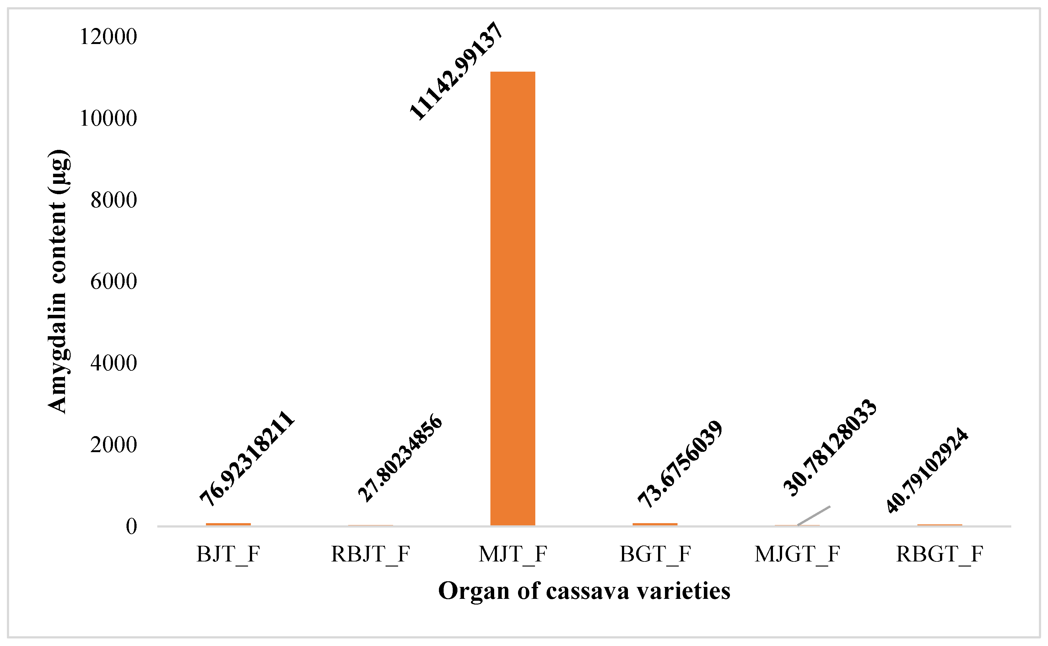
Figure 3.
Amygdalin content (µg/10g of cassava leaf powder). Legend: BF_F=BEN Fresh leaf; RBF_F=RB Fresh leaf; MJF_F=MJ Fresh leaf; BF_OM=BEN Shade dried leaf; RBF_OM=RB Shade dried leaf; MJF_OM=MJ Shade dried leaf.
Figure 3.
Amygdalin content (µg/10g of cassava leaf powder). Legend: BF_F=BEN Fresh leaf; RBF_F=RB Fresh leaf; MJF_F=MJ Fresh leaf; BF_OM=BEN Shade dried leaf; RBF_OM=RB Shade dried leaf; MJF_OM=MJ Shade dried leaf.

Figure 4.
Amygdalin content (µg/10g of cassava derivatives powder). Legend: RBCBO= RB Boiled Flesh; BCBO= BEN Boiled Flesh; MJCBO= MJ Boiled Flesh.
Figure 4.
Amygdalin content (µg/10g of cassava derivatives powder). Legend: RBCBO= RB Boiled Flesh; BCBO= BEN Boiled Flesh; MJCBO= MJ Boiled Flesh.
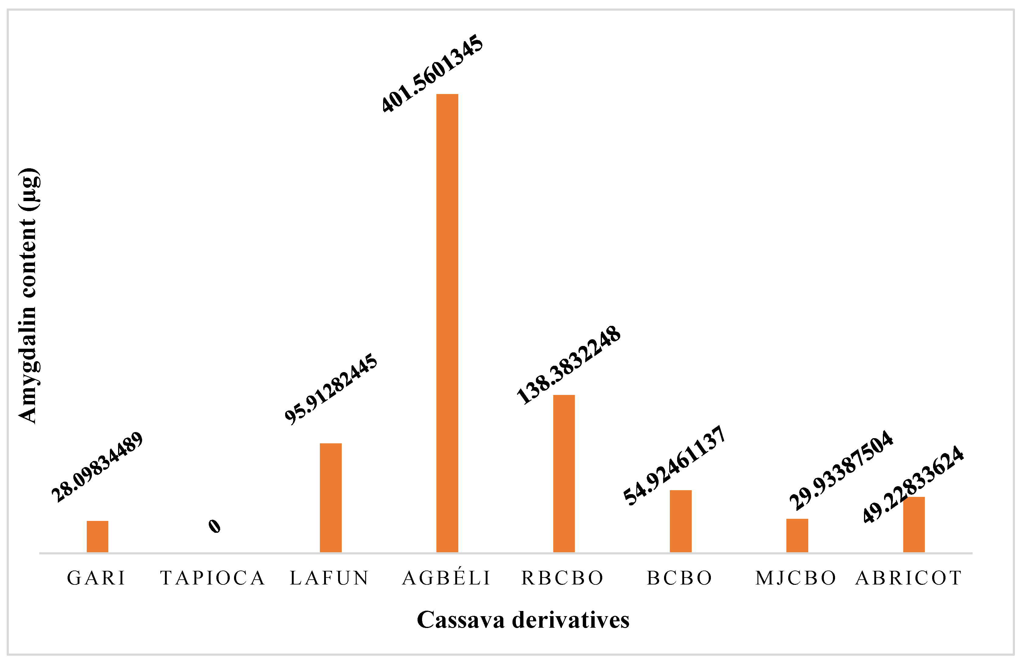
Figure 6.
Percentage increase in hind paw edema in mice as a function of time. Legend: E/MJ (amygdalin Extract of MJ variety); E/BEN (amygdalin Extract of BEN variety); AAS (Acetyl Salicylic Acid).
Figure 6.
Percentage increase in hind paw edema in mice as a function of time. Legend: E/MJ (amygdalin Extract of MJ variety); E/BEN (amygdalin Extract of BEN variety); AAS (Acetyl Salicylic Acid).
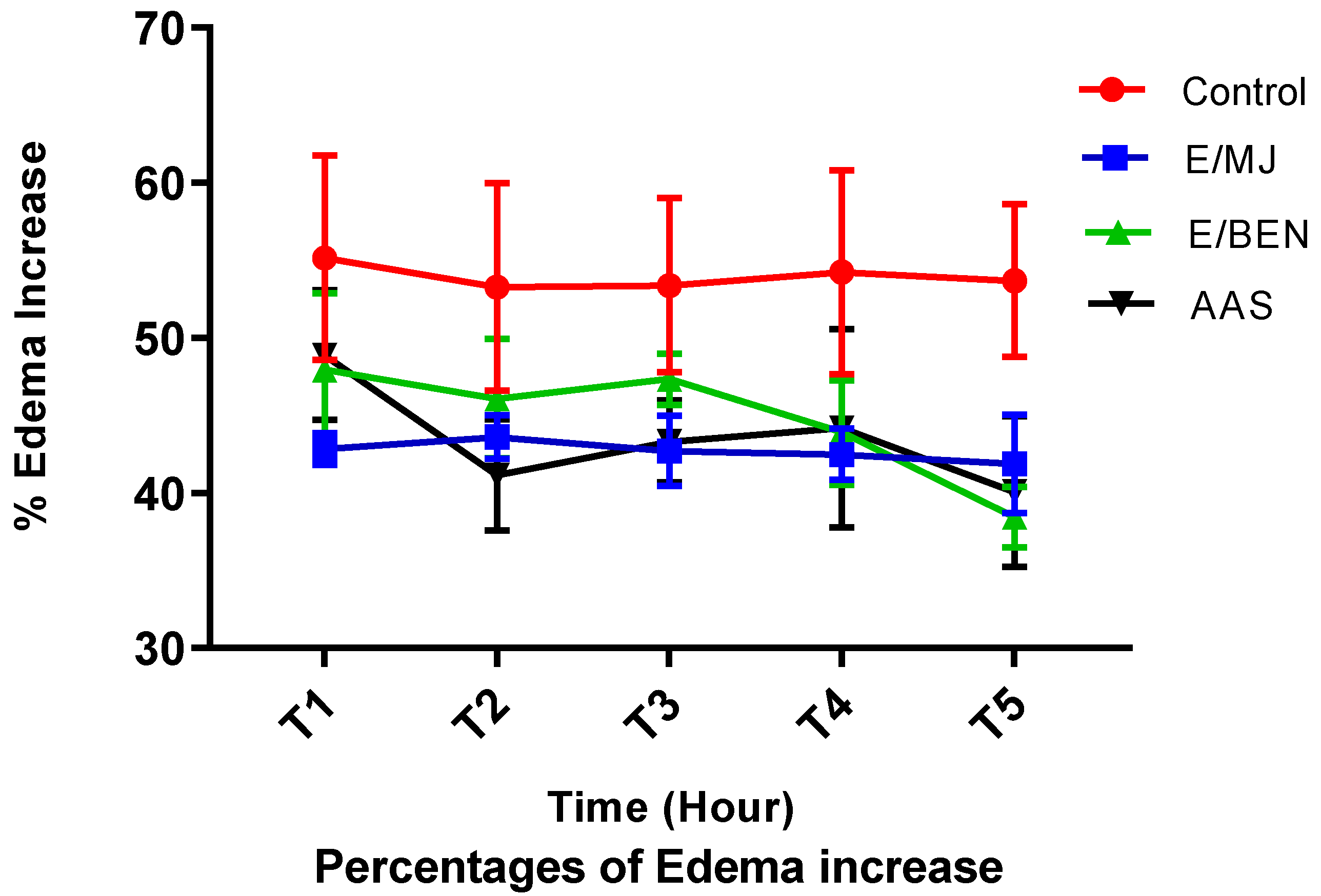
Figure 7.
Percentage inhibition of hind paw edema in mice as a function of time. Legend: E/MJ (amygdalin Extract of MJ variety); E/BEN (amygdalin Extract of BEN variety); AAS (Acetyl Salicylic Acid).
Figure 7.
Percentage inhibition of hind paw edema in mice as a function of time. Legend: E/MJ (amygdalin Extract of MJ variety); E/BEN (amygdalin Extract of BEN variety); AAS (Acetyl Salicylic Acid).
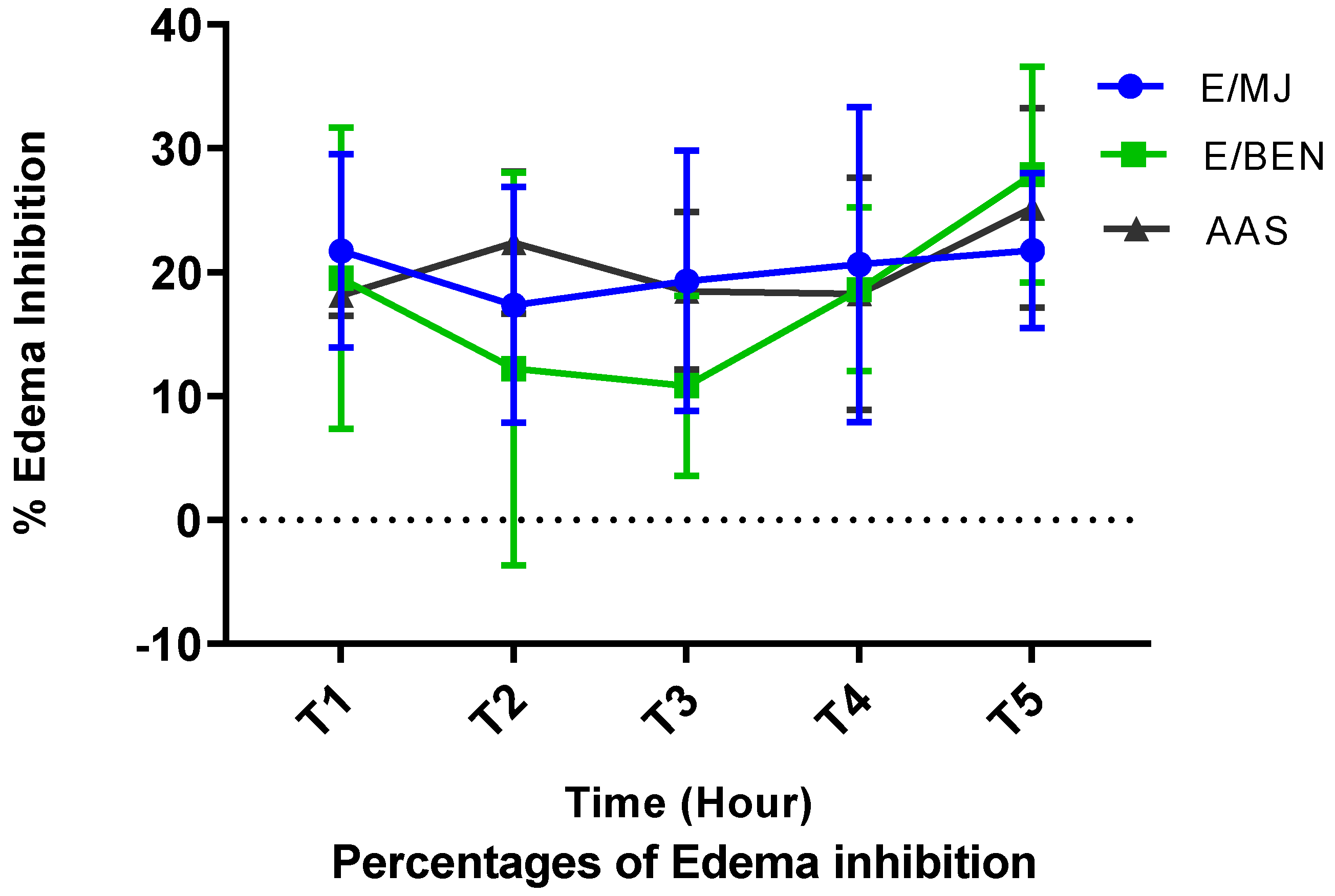
Figure 8.
Histology of experienced Wistar rat colon (original magnification × 400). Legend: Lieberkühn Gland (G); Cell Infiltrates (CI); Caliciform cells (arrows). Magnification: 4000X. (a) group 1: DMH and Amygdalin ; (b) group 2 : DMH only; (c) group 3 : DMH, then Amygdalin next week; (d) group 4 : Amygdalin, then DMH next week; (e) group 5 : DMH and 5-fluorouracil combined; (f) Control group: No treatment.
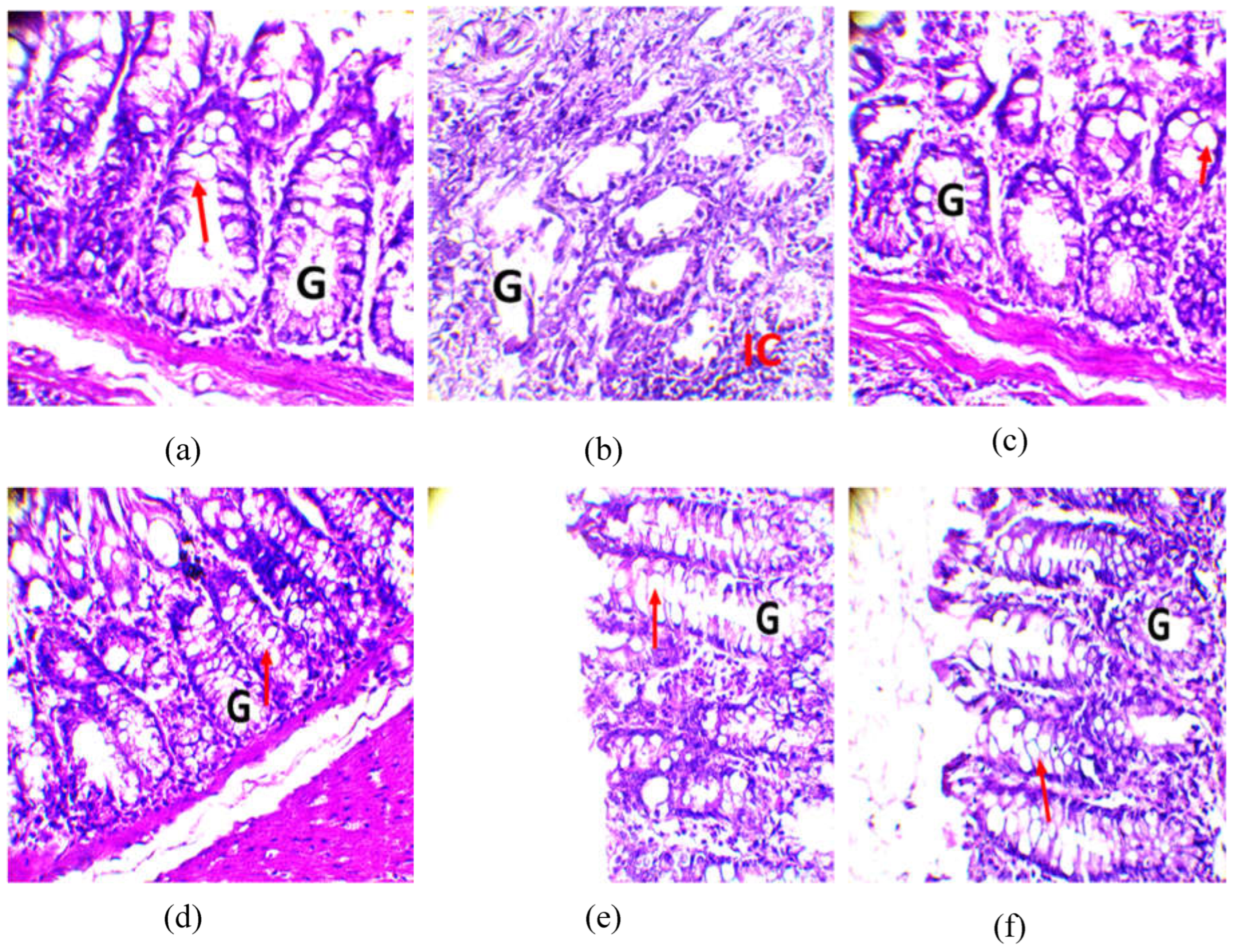
Figure 10.
Histology of the kidneys of tested Wistar rats (original magnification × 400). Legend: Glomeruli (G); Renal Tubules (RT); (a) group 1: DMH and Amygdalin ; (b) group 2 : DMH only; (c) group 3 : DMH, then Amygdalin next week; (d) group 4 : Amygdalin, then DMH next week; (e) group 5 : DMH and 5-fluorouracil combined; (f) Control group: No treatment.
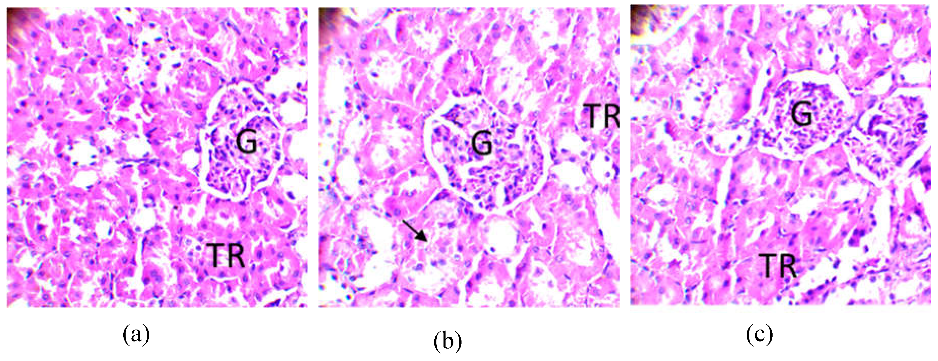
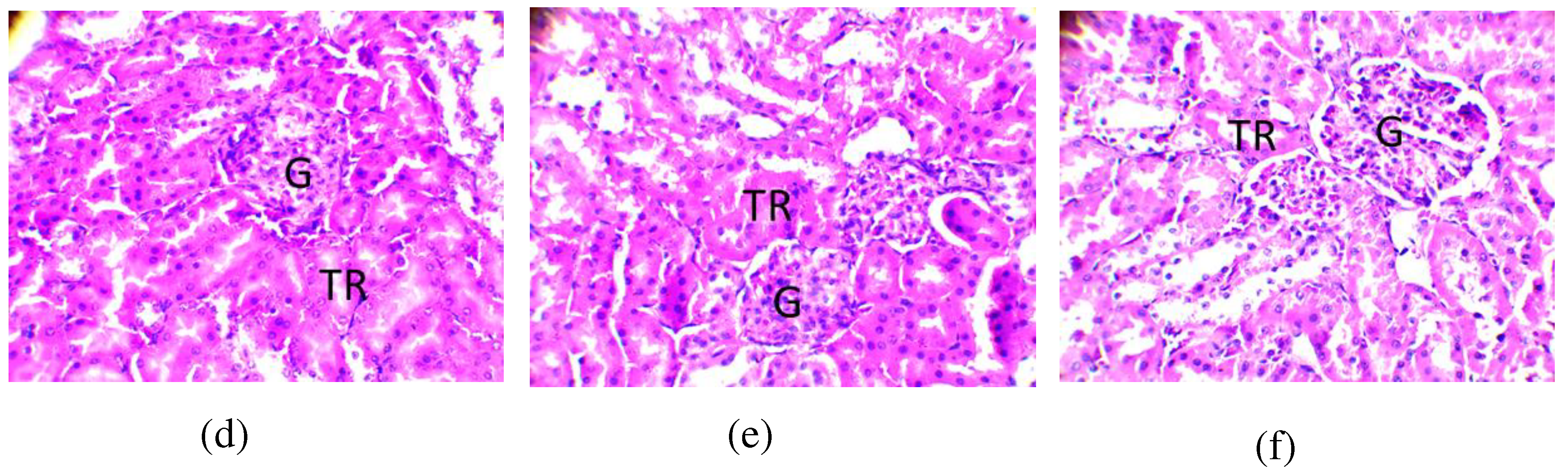
Table 1.
Result of the phytochemical screening test.
| Secondary metabolites | Cassava varieties leaves | ||
|---|---|---|---|
| BEN | MJ | RB | |
| Alkaloids | + | + | + |
| Tannins | + | + | + |
| Saponosides | + | + | + |
| Leuco-anthocyanins | + | + | + |
| Flavonoids | + | + | + |
| Steroids | + | + | + |
| Triterpenes | - | - | - |
| Coumarins | + | + | + |
| Glycosides | + | + | + |
| Cyanogenic derivatives | + | + | + |
Legend: (+): presence of metabolite; (-): absence of metabolite.
Table 2.
Yield of extracts from sun and shade dried samples.
| Samples | Organs | Yield (%) | |
|---|---|---|---|
| Drying in the sun | Drying in the shade | ||
|
MJ |
Chair | 11.40 | 9.23 |
| 1st skin | 1.36 | 1.18 | |
| 2nd skin | 14.20 | 14.78 | |
| Leaves | - | 1.50 | |
|
BEN |
Chair | 12.41 | 3.55 |
| 1st skin | 1.13 | 1.05 | |
| 2nd skin | 12.26 | 10.95 | |
| Leaves | - | 1.5 | |
|
RB |
Chair | 13.08 | 4.65 |
| 1st skin | 0.98 | 0.95 | |
| 2nd skin | 18.21 | 12.75 | |
| Leaves | - | 1.50 | |
Table 4.
In vitro antioxidant activity of amygdalin extracted from different organs of sun-dried cassava varieties by DPPH essay.
Table 4.
In vitro antioxidant activity of amygdalin extracted from different organs of sun-dried cassava varieties by DPPH essay.
| Substances | Extracts of sun-dried organs | PI (%) | IC50 (µg/mL-1) |
|---|---|---|---|
| RB | flesh | 87.28±1.25 | 3.00±0.00 |
| 1st skin | 91.72±0.00 | 0.75±0.07 | |
| 2nd skin | 91.15±0.14 | 2.35±1.20 | |
| MJ | flesh | 95.11±0.18 | 0.5±0.22 |
| 1st skin | 93.77±0.54 | 0.25±0.07 | |
| 2nd skin | 85.12±0.67 | 4.6±1.97 | |
| BEN | flesh | 94.71±0.07 | < 0.19 |
| 1st skin | 95.38±0.07 | < 0.19 | |
| 2nd skin | 89.38±0,23 | 7.25±0.35 |
Table 5.
In vitro antioxidant activity of amygdalin extracted from different organs of shade-dried cassava varieties.
Table 5.
In vitro antioxidant activity of amygdalin extracted from different organs of shade-dried cassava varieties.
| Substances | Extracts of organs dried in the shade | PI (%) | IC50 (µg/mL-1) |
|---|---|---|---|
| Molecule | Quercetine | 82.35±1.86 | 5.75±1.06 |
| RB | flesh | 88.93±0.12 | 17.25±0.35 |
| 2nd skin | 89.60±0.98 | 4.1±0.14 | |
| MJ | flesh | 90.14±0.32 | 21±14.14 |
| 2nd skin | 92.10±0.16 | 0.5±0 | |
| BEN | flesh | 85.88±0.28 | 9.6±0.56 |
| 2nd skin | 91.37±0.18 | 2.75±0.35 |
Table 6.
Antioxydant activity (IC50 µg.mL-1) of cassava varieties leaves by DPPH and FRAP essays.
| Standard/samples | IC50 (µg.mL-1) | |
|---|---|---|
| DPPH | FRAP | |
| Ascorbic acid | 1,11±0,09 | - |
| RB | 8,11±0,70 | 0.63±0.04 |
| MJ | 3,25±0,32 | 0.69±0.03 |
| BEN | 3,99±0,27 | 0.52±0.04 |
Table 7.
LC50 of ethanolic extracts from leaves of cassava varieties.
| Amygdalin extracts | RB | BEN | MJ |
|---|---|---|---|
| LC50 (mg/mL) | 12,73 | 11,60 | 12,32 |
| R2 | 0,80 | 0,64 | 0,41 |
Table 9.
Variations in biochemical parameters of rats in each batch.
| Variables | R0 | R1 | R2 | R3 | R4 | R5 | P-value | Significativity |
|---|---|---|---|---|---|---|---|---|
| Hb | 13.66b±0.49 | 16.43a±0.9 | 9.00c±1.0 | 15.6a±0.70 | 14.2b±0.30 | 13.6b±0.60 | 7.215e-07 | *** |
| Hte | 41.00b±2.00 | 9.00c±1.00 | 19.33d±2.00 | 30.33c±3.20 | 42.00b±2.00 | 39.33b±1.50 | 6934e-08 | *** |
| NR | 4.43b±0.26 | 5.46b±0.30 | 10.79a±2.50 | 6.16b±1.80 | 7.62b±2.40 | 4.26b±0.00 | 0.003153 | ** |
| NB | 7.77ab±1.15 | 4.18b±0.80 | 13.82a±6.10 | 7.91ab±2.40 | 8.86ab±0.40 | 7.12ab±1.00 | 0.02841 | * |
| Lym | 28.00c±7.21 | 39.33c±7.20 | 114.66a±7.30 | 84.00b±11.10 | 37.33c±11.90 | 21.66c±1.50 | 5.907e-08 | *** |
| Neut | 67.33b±11.84 | 61.66b±6.40 | 114.33a±8.50 | 82.00b±16.30 | 62.33b±11.50 | 72.00b±11.20 | 0.0008887 | *** |
| Eos | 0.33b±0.57 | 0.33b±0.50 | 1.66a±0.50 | 00b±00 | 0.33b±0.50 | 00b±00 | 0.009067 | ** |
| Mono | 0.33b±0.57 | 00.00a±00.00 | 00.00a±00.00 | 0.66a±1.10 | 00.00a±00.00 | 0.66a±0.50 | 0.4755 | NS |
| ASAT | 97.66b±9.07 | 96.66b±4.04 | 570.33a±39.80 | 147.33b±46.50 | 121.00b±8.80 | 102.33b±2.50 | 1.427e-10 | *** |
| ALAT | 86.33c±10.21 | 67.33c±10.10 | 567.66a±55.01 | 160.33b±26.50 | 122.66bc±8.10 | 91.00c±5.20 | 1.311e-10 | *** |
| CREAT | 6.92c±0.56 | 8.10c±0.40 | 19.57a±1.50 | 8.21c±0.80 | 13.10b±5.30 | 7.60c±1.40 | 0.0001756 | *** |
R1: amygdalin and DMH simultaneously (inhibition effect of the extract); R2: treated with DMH; R3: DMH then amygdalin (inhibition effect of the extract); R4: amygdalin, then DMH (preventive effect of the extract); R5: effects of 5-Fluoro uracil used as reference molecule. HB: Hemoglobin level; Hte: Hematocrit level; NR: Red cell; NB: White cell; Lym: Lymphocyte; Neut: Neutrophils; Eos: Eosinophil; Mono: Monocyte; ASAT: Aspartate Aminotransferase; ALAT: Alanine Aminotransferase; CREAT: Creatinine.
Table 10.
Characteristic of cassava varieties and their derived products.
| Name of the varieties | Characteristics | Quality and different processing options |
|---|---|---|
|
BEN 86052 |
soft variety |
Gari, Tapioca, Agbéli, lafoun |
| brown stem | ||
| Dark green leaf, edible. | ||
| Average yield in pods: 30%. | ||
|
RB 89509 |
soft variety |
Gari, Tapioca, Agbéli, lafoun |
| brown stem | ||
| green leaf, edible | ||
| low hydrocyanic acid content | ||
|
Yellow cassava (MJ) |
soft variety |
Gari, semolina |
| white stem | ||
| edible leaf | ||
| low hydrocyanic acid content |
Disclaimer/Publisher’s Note: The statements, opinions and data contained in all publications are solely those of the individual author(s) and contributor(s) and not of MDPI and/or the editor(s). MDPI and/or the editor(s) disclaim responsibility for any injury to people or property resulting from any ideas, methods, instructions or products referred to in the content. |
© 2023 by the authors. Licensee MDPI, Basel, Switzerland. This article is an open access article distributed under the terms and conditions of the Creative Commons Attribution (CC BY) license (http://creativecommons.org/licenses/by/4.0/).
Copyright: This open access article is published under a Creative Commons CC BY 4.0 license, which permit the free download, distribution, and reuse, provided that the author and preprint are cited in any reuse.


